
Back to Journals » Journal of Hepatocellular Carcinoma » Volume 13
Prognostic Value of the SEA Score in Double-Negative DCP/AFP Unresectable HCC with Triple Therapy
Authors Lin H  , Ran F, Wu J , Huang C, Wu J, Liu C , Wang X, Ya H, Jiang K , Gu J, Yan M , Li S
Received 7 March 2026
Accepted for publication 3 June 2026
Published 16 June 2026 Volume 2026:13 606282
DOI https://doi.org/10.2147/JHC.S606282
Checked for plagiarism Yes
Review by Single anonymous peer review
Peer reviewer comments 2
Editor who approved publication: Dr David Gerber
Hesheng Lin,1,* Fulin Ran,2,* Jiayi Wu,3,* Chaoyue Huang,2,* Junyi Wu,3,* Chengyuan Liu,1 Xueyao Wang,4 Houxiang Ya,1 Kaiwen Jiang,1 Jing Gu,1 Maolin Yan,3 Shuqun Li1
1Department of Hepatobiliary and Pancreatic Surgery, The First Affiliated Hospital of Guilin Medical University, Guilin, Guangxi, People’s Republic of China; 2Department of Gastrointestinal Surgery, The First Affiliated Hospital of Guilin Medical University, Guilin, Guangxi, People’s Republic of China; 3Department of Hepatobiliary and Pancreatic Surgery, Fujian Provincial Hospital, Fuzhou, Fujian, People’s Republic of China; 4Department of Hepatobiliary and Pancreatic Surgery, The Second Affiliated Hospital of Guilin Medical University, Guilin, Guangxi, People’s Republic of China
*These authors contributed equally to this study
Correspondence: Shuqun Li, Email [email protected] Maolin Yan, Email [email protected]
Background & Aims: The combination of transarterial chemoembolization (TACE), lenvatinib and PD-1 inhibitors (triple therapy) demonstrates encouraging efficacy in patients with unresectable hepatocellular carcinoma (uHCC). But its prognostic value remains unclear in patients with double negative des gamma carboxy prothrombin (DCP) and alpha fetoprotein (AFP) uHCC. To identify the best candidates for this treatment, this study aimed to evaluate the prognosis of this specific population and to establish a practical prognostic scoring model.
Methods: This multicenter retrospective study included 136 patients with double-negative DCP/AFP uHCC receiving triple therapy, stratified into training (n=91) and validation (n=45) cohorts. Independent predictors for overall survival (OS) were identified by Cox regression to build a scoring system. The model’s performance was validated using Kaplan-Meier curves and the area under the receiver operating characteristic curve (AUC).
Results: In the training cohort, median OS was not reached, and median progression-free survival was 18.9 months. Additionally, triple therapy produced a 79.1% objective response rate and a 95.6% disease control rate. Four variables (maximal tumor size ≥ 5 cm, absence of conversion surgery, presence of extrahepatic metastasis, and age < 55 years), were independent risk factors for prognosis and were utilized to develop the SEA score. The SEA score effectively stratified patients into low risk (0– 3 points) and high risk (4– 6 points) groups, with significantly different survival outcomes in both cohorts. This model showed robust discriminative performance, with AUCs of 0.80 in the training cohort and 0.68 in the validation cohort, and outperformed all individual prognostic factors.
Conclusion: Triple therapy demonstrates promising efficacy and an acceptable safety profile in patients with double-negative DCP/AFP uHCC. The SEA score effectively stratifies patient outcomes, facilitating the identification of optimal candidates for triple therapy, though prospective validation is warranted before widespread clinical use.
Keywords: unresectable hepatocellular carcinoma, triple therapy, overall survival, progression-free survival, SEA score
Introduction
Hepatocellular carcinoma (HCC) continues to be a serious global health concern, endangering human life and imposing a heavy burden on healthcare systems worldwide. It is the sixth most prevalent cancer and the third leading cause of cancer-related mortality globally.1 The majority of patients (about 70%) are discovered at an advanced stage due to the absence of obvious symptoms, which means they miss the opportunity for radical treatment, even though surgical resection, liver transplantation, and radiofrequency ablation may cure early stage HCC.2
Monotherapies, including transarterial chemoembolization (TACE), radiotherapy, molecular targeted drugs, and immunotherapy, have shown limited efficacy.3,4 The latest progress in HCC treatment has shifted from single treatment to multimodal treatment. In advanced HCC, combination therapy with tyrosine kinase inhibitors (TKIs, such as lenvatinib) and immune checkpoint inhibitors (ICIs, such as PD-1/PD-L1 inhibitors) is associated with a significantly improved prognosis. However, challenges such as tumor heterogeneity, drug resistance and increased toxicity still exist.
In this case, the integration of local treatments (such as TACE) with systemic therapies has emerged as a promising strategy. Previous studies have indicated that triple therapy, combining TACE, lenvatinib and PD-1 inhibitors improves overall survival (OS) and progression-free survival (PFS) in patients with unresectable HCC (uHCC) while maintaining a favorable safety profile.5,6 Notably, des-gamma-carboxy prothrombin (DCP) and alpha-fetoprotein (AFP) are two well-established serum biomarkers that play pivotal roles in the prognosis stratification and treatment response prediction of HCC.7–9 However, a subset of HCC patients presents with double negative serum levels of DCP and AFP, which poses substantial challenges to biomarker based monitoring, prognostic stratification, and individualized treatment selection. Notably, existing prognostic models (eg., TAE, CRAFITY, PPRD scores) were not developed for this specific double negative population, and the therapeutic effectiveness of the triple regimen in these patients remains to be fully clarified.10–12
In this multicenter retrospective study, we evaluated the efficacy and safety of triple therapy in patients with double negative DCP/AFP uHCC. We further constructed and externally validated a novel dedicated prognostic model (SEA score) to improve risk stratification, identify optimal candidates for triple therapy, and help guide personalized treatment strategies for this clinically unique population.
Materials and Methods
Patients
This multicenter retrospective study enrolled patients diagnosed with uHCC who underwent first-line triple therapy (TACE + lenvatinib + PD-1 inhibitors) across three hospitals from June 2018 to December 2023. The training cohort was from the First Affiliated Hospital of Guilin Medical University. The independent external cohort comprised patients from the Second Affiliated Hospital of Guilin Medical University and Fuzhou University Affiliated Provincial Hospital. We retrospectively analyzed demographic characteristics, clinical parameters (including hepatic function, tumor burden metrics, and other relevant indices), laboratory results, imaging studies, and treatment response data. Serum biomarker negativity was defined as follows: DCP <40 mAU/mL and AFP <400 ng/mL.13,14 This study adhered to the Declaration of Helsinki and obtained approval from the institutional review boards of all participating centers, and the requirement for written informed consent was waived due to the retrospective nature of the study. As a retrospective study, selection bias was minimized by consecutively enrolling all eligible patients according to uniform inclusion and exclusion criteria across three centers.
HCC was diagnosed in accordance with the Chinese Guidelines for Liver Cancer, relying on either typical imaging features or histopathological confirmation.15 All patients with HCC were categorized according to the Barcelona Clinic Liver Cancer (BCLC) staging system.16 Tumor unresectability was assessed by a multidisciplinary team (MDT) and was primarily defined as: (1) bilobar liver involvement precluding R0 resection, (2) inadequate surgical margins or insufficient future hepatic volume, and (3) extrahepatic metastasis (EHM) or extrahepatic lymph node metastasis.17
Inclusion criteria for this study were: (1) age ranging from 18 to 75 years, (2) an Eastern Cooperative Oncology Group performance status (ECOG-PS) score of 0 to 1, (3) no previous systemic or locoregional HCC therapy, and (4) double-negative DCP/AFP uHCC, with triple therapy administered as first-line treatment. The exclusion criteria were: (1) concurrent malignancies, (2) incomplete clinical data, (3) treatment discontinuation due to intolerability or non-compliance, and (4) BCLC stage A disease (patients eligible for curative therapies).
TACE Procedure
All enrolled patients underwent standardized conventional TACE via right femoral artery puncture under local anesthesia. TACE procedures adhered to established clinical standards.15 A 2.7F microcatheter was advanced superselectively into tumor-supplying segmental or subsegmental hepatic arteries under imaging guidance. The chemoembolization protocol utilized a precisely calibrated combination of pirarubicin (20–60 mg), oxaliplatin (200 mg), and lipiodol (5–20 mL), with individualized dosages adjusted according to comprehensive assessment of tumor burden and residual hepatic functional reserve. Subsequent embolization was performed administering absorbable gelatin sponge particles until near stasis of arterial flow was confirmed angiographically. The total emulsion volume was optimized based on three-dimensional tumor measurements from pretreatment imaging.
Post-procedure follow-up assessments were performed at 4–6 weeks following the initial TACE. Whether to repeat TACE was determined according to residual tumor activity and imaging results. Patients ineligible for additional TACE received supportive care. All TACE interventions were performed by board-certified interventional radiologists possessing at least 5 years of practice experience.
Lenvatinib and PD-1 Inhibitors
Systemic therapy was initiated within 2 weeks after the initial TACE session, with the lenvatinib dose adjusted based on body weight: 8 mg once daily for patients weighing <60 kg, or 12 mg once daily for those weighing ≥60 kg. Dose reductions were implemented for treatment-related toxicities, with permanent discontinuation required for persistent grade 3–4 adverse events until resolution. PD-1 inhibitors (camrelizumab 200 mg, sintilimab 200 mg, tislelizumab 200 mg, toripalimab 240 mg, or pembrolizumab 200 mg) were administered intravenously every 21 days, discontinued only for progressive disease or intolerable toxicity. For patients requiring repeat TACE sessions, both lenvatinib and PD-1 inhibitors were temporarily withheld 3 days before the procedure and reinstated 3 days after the procedure, unless significant TACE-related complications occurred. Different PD 1 inhibitors were administered according to clinical availability and routine institutional practice, which reflects real world clinical conditions. Owing to the relatively small sample size in each subgroup, formal comparative analyses of efficacy and safety among different PD 1 inhibitors were not performed.
Treatment Response and Safety Assessment
Tumor response was evaluated via contrast-enhanced computed tomography (CT) or magnetic resonance imaging (MRI) findings based on the modified Response Evaluation Criteria in Solid Tumors (mRECIST), and classified into complete response (CR), partial response (PR), stable disease (SD), or progressive disease (PD).18 The objective response rate (ORR) was defined as the percentage of patients with confirmed CR or PR lasting for at least 4 weeks, and the disease control rate (DCR) including CR, PR, and SD. Treatment safety was assessed by recording treatment-related adverse events (TRAEs) rated following the National Cancer Institute Common Terminology Criteria for Adverse Events version 5.0 (NCI-CTCAE v5.0).
Conversion Surgery
Following initiation of triple therapy, tumor resectability was assessed at 4- to 6-week intervals. When meeting criteria for resectability, conversion surgery would be contemplated following MDT discussion and acquisition of informed consent. The criteria for resectable HCC conversion included: (1) technical feasibility of achieving R0 resection while maintaining adequate liver remnant volume and function, (2) well-preserved liver function (Child-Pugh class A), (3) good performance status (ECOG-PS score of 0 to 1), (4) absence of extrahepatic metastatic lesions and (5) no medical contraindications to hepatectomy.15 Perioperative management involved withholding lenvatinib 1 week before and after surgery, while PD-1 inhibitors were discontinued 4 weeks. The extent and method of liver resection in each patient were tailored based on stage, location and size of tumors, quantitative liver function assessment, and patient’s physiological reserve.19
Follow-Up
All participants underwent regular follow-up assessments at intervals ranging from 4 to 8 weeks, during which they were closely monitored for indications of disease recurrence. Each follow-up appointment included laboratory tests, chest CT imaging, and contrast-enhanced abdominal CT/MRI scans. Postoperative adjuvant treatment, comprising lenvatinib plus PD-1 inhibitors, was given for 3–12 months, with the treatment duration customized based on pathological findings, radiological assessments, and patient therapy tolerance. For patients who failed conversion therapy, triple regimen was discontinued upon disease progression, occurrence of severe TRAEs, or patient withdrawal of consent. The subsequent treatment strategy was formulated through MDT consensus, incorporating shared decision-making with patients in accordance with personalized medicine principles.20
This study’s primary endpoint was OS, calculated from the commencement of triple therapy until mortality due to any reason. Secondary endpoints included PFS, ORR, DCR and TRAEs. PFS was defined as the duration between triple therapy initiation and disease progression or death from any cause. Data for patients who were progression-free or alive at final follow-up were right-censored during analysis. The data cutoff date fell on October 31, 2024.
Statistical Analysis
Patients with incomplete key data (age, tumor size, EHM, conversion surgery, survival status) were excluded. No imputation was performed for missing values. Frequencies and percentages were used to summarize categorical variables, and Fisher’s exact test or Pearson’s chi-square test, depending on the data distribution, were used for between-group comparisons. Variables with significant univariate associations with OS (P<0.05) were included in the multivariate Cox proportional hazards model. Backward stepwise elimination was employed to identify independent prognostic factors. Continuous variables were dichotomized using clinically relevant cutoff values. The prognostic scoring system was developed by weighting each independent predictor according to its β coefficient from the final multivariate model. The predictive performance of the model was quantified by calculating the area under the receiver operating characteristic curve (AUROC). Risk stratification was visualized through Kaplan-Meier survival curves, with between-strata comparisons conducted via Log rank testing (two-sided α=0.05). The prognostic scoring model was validated through an independent external validation cohort. All analyses were conducted using R statistical software (version 4.5.1), with two-tailed P-values <0.05 considered statistically significant.
Results
Patient Characteristics
This multicenter retrospective analysis ultimately included 136 eligible patients from an initial cohort of 246 patients with double-negative DCP/AFP uHCC who received first-line triple therapy across three medical centers between June 2018 and December 2023 (Figure 1). The training cohort comprised 91 eligible patients from a single center who met the inclusion criteria. Following a ratio of 2:1, we established an independent validation cohort consisting of 45 eligible patients from two additional centers. The training cohort comprised 84 males (92.3%) and 7 females (7.7%), with a predominance of patients aged ≥55 years (70.3%). The prevalence of infection with the hepatitis B virus (HBV) was observed in 77 patients (84.6%). Following reassessment of resectability criteria, 40 patients (44%) underwent successful conversion surgery after providing informed consent. Radiological assessment revealed the presence of macrovascular invasion in 43 patients, accounting for 47.3% of the training cohort. Tumor characteristics revealed 70 cases (76.9%) with maximal diameter ≥5 cm and 22 cases (24.2%) with solitary lesions. Table 1 displays all of the baseline characteristics.
 |
Table 1 Baseline Demographic and Clinical Characteristics of Included Patients |
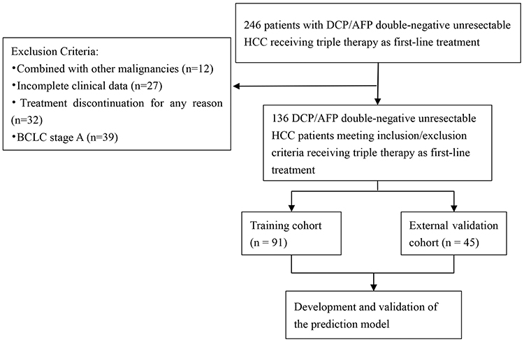 |
Figure 1 Flowchart of patient selection. |
Treatment Response and Safety Assessment
Per mRECIST criteria, the optimal tumor responses observed among the 91 patients included in the training cohort were as follows: CR in 18 (19.8%), PR in 54 (59.3%), SD in 15 (16.5%), and PD in 4 (4.4%). These outcomes corresponded to an ORR of 79.1% and a DCR of 95.6%, as illustrated in Table 2.
 |
Table 2 Tumor Responses per mRECIST in the Training Cohort |
Within the training cohort, TRAEs were reported in 71 of 91 patients (78.0%), with the majority classified as grade 1 or 2 events (Table 3). Grade 4 TRAEs were identified in 3 patients (3.3%), specifically manifesting as hand-foot syndrome, rash, and abnormal liver function. The most frequently observed TRAEs of any grade in this cohort included abnormal liver function (65.9%), hypertension (33.0%), fever (27.5%), fatigue (23.1%), decreased appetite (20.9%), thrombocytopenia (16.5%), nausea (15.4%), hand-foot syndrome (15.4%), diarrhea (12.1%), rash (12.1%), hypothyroidism (11.0%), vomiting (9.9%), abdominal pain (9.9%) and proteinuria (7.7%). TRAEs led to dosage reduction of lenvatinib for 7 patients (7.7%) and discontinuation of the PD-1 inhibitors for 2 patients (2.2%). During the follow-up period, no deaths attributable to the treatment were observed.
 |
Table 3 Treatment-Related Adverse Events in Training Cohort |
Survival Analysis
During the median follow-up period of 27.7 months (95% confidence interval [CI]: 22.0–30.1), 30 patients (32.9%) in the training cohort succumbed to death. Median OS was not attained. The 1-year, 2-year, and 3-year OS rates were 86.9%, 63.3%, and 54.4%, respectively (Figure 2A). Median PFS was 18.9 months (95% CI: 15.7–23.9), with 1-year, 2-year, and 3-year PFS rates of 69.6%, 36.9%, and 33.0%, respectively (Figure 2B).
 |
Figure 2 Kaplan-Meier curves for (A) overall survival and (B) progression-free survival for patients in the training cohort. Median overall survival was not reached. |
Independent Prognostic Factors
Univariate analysis identified several variables significantly associated with OS, including patient age (<55 years vs. ≥55years), surgery (no vs. yes), maximum tumor size (≥5 cm vs. <5 cm), EHM (yes vs. no) and hemoglobin (≥120 g/L vs. <120 g/L). In the multivariate Cox proportional hazards analysis, age, conversion surgery, maximum tumor size, and EHM, were identified as independent predictors significantly associated with OS (p < 0.05) (Table 4).
 |
Table 4 Univariate and Multivariate Cox Regression Analyses for OS in the Training Cohort |
Development and Validation of Prognostic Scoring Model
Scores for the four independent predictors were created by rounding their β coefficients to the nearest integer. The detailed scoring methodology is presented in Table 5. These four factors were integrated to establish the SEA score (range 0–6 points). Patients were stratified into low-risk (0–3 points) and high-risk (4–6 points) groups.
 |
Table 5 Calculation and Classification of the SEA Score |
The 2-year OS rates for the subgroups within the training cohort were 80.9% and 19.7%, respectively. Kaplan-Meier survival analysis showed significant differences in OS between these subgroups (p < 0.0001). Survival rates decreased markedly as the SEA score increased (Figure 3A). Specifically, the low-risk group showed favorable survival outcomes, while the high-risk group had a poor survival, with a median OS of just 14.4 months. PFS also differed significantly based on the SEA score (Figure 3B). The 2-year PFS rates for the high-risk group were 4.6% and the low-risk group was 49.9% (p < 0.0001).
 |
Figure 3 Kaplan–Meier curves according to SEA score. (A) overall survival and (B) progression-free survival according to the SEA score in the training cohort; (C) overall survival and (D) progression-free survival according to the SEA score in the validation cohort. |
An independent external cohort was used to further confirm the SEA score’s predictive performance. The baseline features of the training and validation cohorts did not differ significantly (Table 1). Within the validation cohort (n = 45), the majority of patients (73.3%) were aged ≥55 years. Conversion surgery was successfully performed in 16 cases (35.6%). Maximum tumor size ≥5 cm was present in 75.6% of patients and only 10 (22.2%) patients were identified with EHM. In alignment with the findings observed in the training cohort, the validation cohort demonstrated effective risk stratification based on the SEA score, with 2-year OS rates of 90.0% for the low-risk group and 20.0% for the high-risk group. Kaplan-Meier survival analysis revealed statistically significant differences in OS between these subgroups (p = 0.0013) (Figure 3C). The 2-year OS rates were 73.9% for the low-risk group and 26.7% for the high-risk group. In addition, Kaplan-Meier analysis revealed a 2-year PFS rates of 61.8% for low-risk patients versus 0% for high-risk patients (p<0.0001) (Figure 3D). In both the training and validation cohorts, the OS and PFS demonstrated significant stratification between their respective high-risk and low-risk subgroups.
Performance of SEA Score
The SEA scoring system demonstrated strong discriminative capacity across both cohorts, with AUC values of 0.800 (95% CI, 0.693–0.888) in the training cohort and 0.680 (95% CI, 0.548–0.811) in the validation cohort (Figure 4). Notably, the composite SEA score outperformed all single prognostic factors in predictive accuracy.
 |
Figure 4 Comparison of ROC curves for overall survival in the (A) training cohort and (B) validation cohort. ROC, receiver operating characteristic curve; AUC, area under the receiver operating characteristic curve. |
Discussion
Our study demonstrates promising efficacy of triple therapy in patients with double-negative DCP/AFP uHCC. The treatment demonstrated an ORR of 79.1% and a DCR of 95.6% by mRECIST criteria in the training cohort. The median OS was not reached and the median PFS of 18.9 months. These outcomes are generally consistent with those reported in previous studies of triple therapy for uHCC.10,21,22 The favorable survival outcomes observed in this study indicate that triple therapy may serve as an effective treatment for patients with double-negative DCP/AFP uHCC. Collectively, these results provide substantial support for the implementation of triple therapy as a potentially effective initial standard treatment option for this specific patient population.
The encouraging clinical results observed in triple therapy likely stem from synergistic mechanisms.22 TACE, as a locoregional treatment for uHCC, induces tumor necrosis and may promote antitumor immunity by releasing tumor-associated antigens, thereby potentiating the effects of PD-1 inhibitors.23,24 PD-1 inhibitors block the PD-1/PD-L1 checkpoint, reversing T-cell suppression and enabling them to recognize and attack cancer cells.25,26 Lenvatinib, a multitargeted antiangiogenic TKI, not only suppresses post-TACE neovascularization but also modulates the tumor immune microenvironment, further enhancing systemic immunotherapy.27–29 These collectively delay tumor progression and recurrence. The justification for this combination therapy is supported by clinical data showing triple therapy provides better survival advantages than dual therapy or monotherapy.22 The improved prognosis associated with low DCP levels may be attributed to enhanced T-cell infiltration and a less immunosuppressive tumor microenvironment, fostering stronger antitumor immune responses.14 Additionally, AFP-negative HCC exhibits better outcomes due to the absence of FOXM1-driven aggressive phenotypes and vascular invasion, which are hallmarks of AFP-positive tumors.30,31 Accordingly, all these indicate the therapeutic potential of combining locoregional, targeted, and immunotherapeutic strategies in patients with double-negative DCP/AFP uHCC.
The safety profile of triple therapy in our study was acceptable and consistent with previous reports of TACE combined with lenvatinib and PD-1 inhibitors.21,22,32 The overall incidence of any-grade TRAEs (78.0%) and grade≥3 TRAEs (17.6%) were comparable or even lower than those in similar real-world cohorts, in which the rates of any-grade TRAEs ranged from 82% to 91% and grade≥3 TRAEs from 22% to 31%.21,22,32,33 The most common adverse event was elevation of AST and ALT levels, reflecting the transient hepatotoxicity associated with TACE procedures.34 These biochemical alterations primarily reflect localized hepatic parenchymal injury resulting from tumor-directed ischemia and necrosis, rather than systemic drug-induced toxicity.35 Importantly, grade 4 TRAEs were rare in our study cohort, and no fatalities were attributed to therapy during the follow-up period. These findings collectively suggest that the triple therapy regimen demonstrates an acceptable safety profile for the treatment of DCP/AFP-negative uHCC.
Multivariate analysis demonstrated that tumor size ≥5 cm, conversion surgery, EHM, and age <55 years were independent prognostic factors, which were subsequently integrated into the SEA scoring model. Larger tumor size (≥5 cm) was significantly associated with more aggressive disease biology and poorer survival outcomes, consistent with previous reports demonstrating shorter median OS in this patient subgroup.36 Notably, patients who successfully underwent conversion surgery achieved substantially better prognosis compared to non-surgical candidates, highlighting the critical importance of surgery in advanced HCC management.37 EHM proved to be a robust negative prognostic factor, aligning with prior studies that have established metastatic disease as a key determinant of poor prognosis in HCC patients receiving triple therapy.38 Interestingly, younger age (<55 years) was associated with worse outcomes, potentially reflecting more aggressive tumor biology in this demographic, as supported by previous investigations reporting faster disease progression and reduced survival in younger HCC populations.39,40 However, the underlying biological mechanisms for this age-related prognostic difference remain to be fully elucidated, and further mechanistic research is warranted. These findings collectively provide a robust framework for prognostic stratification in advanced HCC and underscore the complex interplay between tumor characteristics, treatment modalities, and patient factors in determining clinical outcomes.
Numerous prognostic scoring models have been established for HCC patients treated with systemic or locoregional therapies. However, none of these tools are specifically designed for patients with double-negative DCP/AFP uHCC. Specifically, Zeng et al established the TAE score, Scheiner et al constructed the CRAFITY score, and Zhang et al proposed the PPRD score, all of which were developed to forecast survival outcomes in unselected HCC populations.10–12 The SEA score exhibits comparable or improved prognostic performance compared with these existing models, as it was specifically developed and externally validated for the double negative DCP/AFP uHCC population, thereby addressing a critical unmet clinical need. This study is the first to focus on double negative DCP/AFP uHCC patients treated with triple therapy and to establish a dedicated prognostic scoring system, which may provide a useful reference for clinical practice.
There are some limitations that warrant consideration in this study. First, this was a retrospective, single-arm, observational study with inherent selection bias, and no control group was included for comparison, which limits the causal inference of treatment efficacy. Future prospective, controlled, and multicenter studies are needed to provide higher-level evidence. Second, the sample size was relatively small, especially in the external validation cohort (n=45). Further investigations with larger sample sizes are required to improve statistical power and generalizability. Third, multiple types of PD-1 inhibitors were used interchangeably according to clinical practice, and the number of TACE sessions was individualized, both of which represent treatment heterogeneity and potential confounders. Meanwhile, the number of double negative DCP/AFP uHCC patients was insufficient to support robust subgroup analyses. Future studies should adopt standardized treatment protocols and perform well-powered subgroup analyses. Fourth, the study population was predominantly HBV-related HCC from Chinese centers, which may limit the extrapolation of results to other etiologies and ethnicities. Future research should enroll more diverse cohorts to enhance generalizability. Finally, the median follow-up period was relatively limited, and long-term survival data require further confirmation. Extended follow-up is needed to verify long-term outcomes.
Conclusion
The combination of TACE, lenvatinib and PD 1 inhibitors demonstrates favorable clinical efficacy and manageable safety profile in patients with double negative DCP/AFP uHCC. Our study established and externally validated the novel SEA prognostic scoring system. This practical model may help clinicians identify favorable candidates for triple therapy and facilitate individualized treatment planning. Nevertheless, given the retrospective design and relatively small sample size, large scale prospective studies are warranted to further verify the prognostic value and clinical utility of the SEA score before routine clinical application.
Generative AI Statement
The authors declare that no Generative AI was used in the creation of this manuscript.
Data Sharing Statement
The raw data supporting the conclusions of this article will be made available by the corresponding author, Professor Shuqun Li, upon reasonable request.
Ethics Approval
The research protocol received formal approval from the Ethics Committee of The First Affiliated Hospital of Guilin Medical University (2024KJTSC‐58), and study protocol was approved by the institutional review boards of other centres. All patient data used in this study were fully anonymized and de-identified before analysis. The study was conducted in accordance with the Declaration of Helsinki and local data protection regulations to ensure patient privacy and confidentiality.
Acknowledgments
We would like to express our gratitude to all participants and researchers for their contributions to this study.
Author Contributions
Hesheng Lin, Fulin Ran, Jiayi Wu, Chaoyue Huang, and Junyi Wu are co-first authors. All authors made a significant contribution to the work reported, whether that is in the conception, study design, execution, acquisition of data, analysis and interpretation, or in all these areas; took part in drafting, revising or critically reviewing the article; gave final approval of the version to be published; have agreed on the journal to which the article has been submitted; and agree to be accountable for all aspects of the work.
Funding
This study was funded by Guangxi Medical and Health Key discipline construction project, Guangxi medical and healthcare appropriate technology development and promotion and application projects (grant number:S2022132), Guangxi Natural Science Foundation (grant number: 2025GXNSFHA069049), Self-financed Science and Technology Project of Guilin City (grant number: 20250120z).
Disclosure
The authors declare that the research was conducted in the absence of any commercial or financial relationships that could be construed as a potential conflict of interest.
References
1. Bray F, Laversanne M, Sung H, et al. Global cancer statistics 2022: GLOBOCAN estimates of incidence and mortality worldwide for 36 cancers in 185 countries. CA Cancer J Clin. 2024;74(3):229–14. doi:10.3322/caac.21834
2. Devarbhavi H, Asrani SK, Arab JP, Nartey YA, Pose E, Kamath PS. Global burden of liver disease: 2023 update. J Hepatol. 2023;79(2):516–537. doi:10.1016/j.jhep.2023.03.017
3. Ciliberto D, Caridà G, Staropoli N, et al. First-line systemic treatment for hepatocellular carcinoma: a systematic review and network meta-analysis. Heliyon. 2023;9(8):e18696. doi:10.1016/j.heliyon.2023.e18696
4. Zhu H-D, Liu R, Jia -Z-Z, et al. Transarterial chemoembolization for hepatocellular carcinoma: treatment algorithm proposed by Chinese college of interventionalists (CCI). EngMedicine. 2024;1(3):100037. doi:10.1016/j.engmed.2024.100037
5. Wang L, Lin L, Zhou W. Efficacy and safety of transarterial chemoembolization combined with lenvatinib and PD-1 inhibitor in the treatment of advanced hepatocellular carcinoma: a meta-analysis. Pharmacol Ther. 2024;257:108634. doi:10.1016/j.pharmthera.2024.108634
6. Li S, Wu J, Wu J, et al. Prediction of early treatment response to the combination therapy of TACE plus lenvatinib and anti-PD-1 antibody immunotherapy for unresectable hepatocellular carcinoma: multicenter retrospective study. Front Immunol. 2023;14:1109771. doi:10.3389/fimmu.2023.1109771
7. Piñero F, Dirchwolf M, Pessôa MG. Biomarkers in hepatocellular carcinoma: diagnosis, prognosis and treatment response assessment. Cells. 2020;9(6):1370. doi:10.3390/cells9061370
8. Toyoda H, Kumada T, Tada T, Sone Y, Kaneoka Y, Maeda A. Tumor markers for hepatocellular carcinoma: simple and significant predictors of outcome in patients with HCC. Liver Cancer. 2015;4(2):126–136. doi:10.1159/000367735
9. Teng W, Wang H-W, Lin S-M; On behalf of Diagnosis Group and Systemic Therapy Group of TLCA. Management consensus guidelines for hepatocellular carcinoma: 2023 update on surveillance, diagnosis, systemic treatment, and posttreatment monitoring by the Taiwan liver cancer association and the gastroenterological society of taiwan. Liver Cancer. 2024;13(5):468–486. doi:10.1159/000537686
10. Zeng Z-X, Wu J-Y, Wu J-Y, et al. The TAE score predicts prognosis of unresectable HCC patients treated with TACE plus lenvatinib with PD-1 inhibitors. Hepatol Int. 2024;18(2):651–660. doi:10.1007/s12072-023-10613-x
11. Scheiner B, Pomej K, Kirstein MM, et al. Prognosis of patients with hepatocellular carcinoma treated with immunotherapy – development and validation of the CRAFITY score. J Hepatol. 2022;76(2):353–363. doi:10.1016/j.jhep.2021.09.035
12. Zhang Y, Miao H, Xie W, et al. The PPRD score stratifies patients with hepatocellular carcinoma and portal vein tumor thrombus treated with sorafenib plus transarterial chemoembolization. Eur Radiol. 2021;31(1):232–243. doi:10.1007/s00330-020-07078-z
13. Xie H, Li Y, Yang J, Tan Y, Xu J, Yang X. DCP as a biomarker for TACE efficacy in hepatocellular carcinoma. Front Oncol. 2025;15:1560210. doi:10.3389/fonc.2025.1560210
14. Wan Y, Geng X, Liu Q, et al. CTSE inhibits anti-tumor T cell response by promoting des-γ-carboxy prothrombin releasing in hepatocellular carcinoma. Cell Death Dis. 2025;16(1):434. doi:10.1038/s41419-025-07753-3
15. Zhou J, Sun H, Wang Z, et al. Guidelines for the diagnosis and treatment of primary liver cancer (2022 edition). Liver Cancer. 2023;12(5):405–444. doi:10.1159/000530495
16. Reig M, Forner A, Rimola J, et al. BCLC strategy for prognosis prediction and treatment recommendation: the 2022 update. J Hepatol. 2022;76(3):681–693. doi:10.1016/j.jhep.2021.11.018
17. Bi X, Zhao H, Zhao H, et al. Consensus of Chinese experts on neoadjuvant and conversion therapies for hepatocellular carcinoma: 2023 update. Liver Cancer. 2025;14(2):223–238. doi:10.1159/000541249
18. Llovet JM, Lencioni R. mRECIST for HCC: performance and novel refinements. J Hepatol. 2020;72(2):288–306. doi:10.1016/j.jhep.2019.09.026
19. Wu J-Y, Zhang Z-B, Zhou J-Y, et al. Outcomes of salvage surgery for initially unresectable hepatocellular carcinoma converted by transcatheter arterial chemoembolization combined with lenvatinib plus anti-PD-1 antibodies: a multicenter retrospective study. Liver Cancer. 2023;12(3):229–237. doi:10.1159/000528356
20. Goetz LH, Schork NJ. Personalized medicine: motivation, challenges, and progress. Fertil Steril. 2018;109(6):952–963. doi:10.1016/j.fertnstert.2018.05.006
21. Wang -Y-Y, Yang X, Wang Y-C, et al. Clinical outcomes of lenvatinib plus transarterial chemoembolization with or without programmed death receptor-1 inhibitors in unresectable hepatocellular carcinoma. World J Gastroenterol. 2023;29(10):1614–1626. doi:10.3748/wjg.v29.i10.1614
22. Xin Y, Zhang X, Liu N, et al. Efficacy and safety of lenvatinib plus PD-1 inhibitor with or without transarterial chemoembolization in unresectable hepatocellular carcinoma. Hepatol Int. 2023;17(3):753–764. doi:10.1007/s12072-023-10502-3
23. Jin Z-C, Zhong B-Y, Chen -J-J, et al. Real-world efficacy and safety of TACE plus camrelizumab and apatinib in patients with HCC (CHANCE2211): a propensity score matching study. Eur Radiol. 2023;33(12):8669. doi:10.1007/s00330-023-09754-2
24. Tan J, Fan W, Liu T, et al. TREM2+ macrophages suppress CD8+ T-cell infiltration after transarterial chemoembolisation in hepatocellular carcinoma. J Hepatol. 2023;79(1):126–140. doi:10.1016/j.jhep.2023.02.032
25. Ono A, Hayes CN, Miura R, Kawaoka T, Tsuge M, Oka S. Noninvasive prediction of the clinical benefit of immunotherapy in hepatocellular carcinoma. J Gastroenterol. 2025;60(9):1053–1069. doi:10.1007/s00535-025-02251-x
26. Pardoll DM. The blockade of immune checkpoints in cancer immunotherapy. Nat Rev Cancer. 2012;12(4):252–264. doi:10.1038/nrc3239
27. Cai M, Huang W, Huang J, et al. Transarterial chemoembolization combined with lenvatinib plus PD-1 inhibitor for advanced hepatocellular carcinoma: a retrospective cohort study. Front Immunol. 2022:13. doi:10.3389/fimmu.2022.848387
28. Kimura T, Kato Y, Ozawa Y, et al. Immunomodulatory activity of lenvatinib contributes to antitumor activity in the Hepa1-6 hepatocellular carcinoma model. Cancer Sci. 2018;109(12):3993–4002. doi:10.1111/cas.13806
29. Kishore SA, Bajwa R, Madoff DC. Embolotherapeutic strategies for hepatocellular carcinoma: 2020 update. Cancers. 2020;12(4):791. doi:10.3390/cancers12040791
30. Li R, Okada H, Yamashita T, et al. FOXM1 is a novel molecular target of AFP-positive hepatocellular carcinoma abrogated by proteasome inhibition. Int J Mol Sci. 2022;23(15):8305. doi:10.3390/ijms23158305
31. He H, Chen S, Fan Z, et al. Multi-dimensional single-cell characterization revealed suppressive immune microenvironment in AFP-positive hepatocellular carcinoma. Cell Discov. 2023;9(1):60. doi:10.1038/s41421-023-00563-x
32. Cai M, Huang W, Liang W, et al. Lenvatinib, sintilimab plus transarterial chemoembolization for advanced stage hepatocellular carcinoma: a Phase II study. Liver Int. 2024;44(4):920–930. doi:10.1111/liv.15831
33. Jin M, Jiang Z-Q, Qin J-H, et al. Efficacy and safety of lenvatinib plus programmed death-1 inhibitors with or without transarterial chemoembolization in the treatment of unresectable hepatocellular carcinoma. J Hepatocell Carcinoma. 2024;11:2309–2320. doi:10.2147/JHC.S485047
34. Brown KT, Do RK, Gonen M, et al. Randomized trial of hepatic artery embolization for hepatocellular carcinoma using doxorubicin-eluting microspheres compared with embolization with microspheres alone. J Clin Oncol. 2016;34(17):2046–2053. doi:10.1200/JCO.2015.64.0821
35. Forner A, Reig M, Bruix J. Hepatocellular carcinoma. Lancet Lond Engl. 2018;391(10127):1301–1314. doi:10.1016/S0140-6736(18)30010-2
36. Renne SL, Woo HY, Allegra S, et al. Vessels encapsulating tumor clusters (VETC) is a powerful predictor of aggressive hepatocellular carcinoma. Hepatology. 2020;71(1):183. doi:10.1002/hep.30814
37. Zhang W, Tong S, Hu B, et al. Lenvatinib plus anti-PD-1 antibodies as conversion therapy for patients with unresectable intermediate-advanced hepatocellular carcinoma: a single-arm, phase II trial. J Immunother Cancer. 2023;11(9):e007366. doi:10.1136/jitc-2023-007366
38. Sun J, Mao F, Liu C, et al. Combined FOLFOX4 with all-trans retinoic acid versus FOLFOX4 with placebo in treatment of advanced hepatocellular carcinoma with extrahepatic metastasis: a randomized, double-blind comparative study. Signal Transduct Target Ther. 2023;8(1):368. doi:10.1038/s41392-023-01604-3
39. Zhang Q, Liang Q, Xu C. Exploring the association between chemotherapy and prognosis among patients less than 50 years old with hepatocellular carcinoma: a retrospective cohort study based on the SEER database. Discov Oncol. 2025;16(1):682. doi:10.1007/s12672-025-02490-7
40. Gao T-M, Bai D-S, Qian -J-J, Zhang C, Jin S-J, Jiang G-Q. The growth rate of hepatocellular carcinoma is different with different TNM stages at diagnosis. Hepatobiliary Pancreat Dis Int. 2021;20(4):330–336. doi:10.1016/j.hbpd.2021.02.005
 © 2026 The Author(s). This work is published and licensed by Dove Medical Press Limited. The
full terms of this license are available at https://www.dovepress.com/terms
and incorporate the Creative Commons Attribution
- Non Commercial (unported, 4.0) License.
By accessing the work you hereby accept the Terms. Non-commercial uses of the work are permitted
without any further permission from Dove Medical Press Limited, provided the work is properly
attributed. For permission for commercial use of this work, please see paragraphs 4.2 and 5 of our Terms.
© 2026 The Author(s). This work is published and licensed by Dove Medical Press Limited. The
full terms of this license are available at https://www.dovepress.com/terms
and incorporate the Creative Commons Attribution
- Non Commercial (unported, 4.0) License.
By accessing the work you hereby accept the Terms. Non-commercial uses of the work are permitted
without any further permission from Dove Medical Press Limited, provided the work is properly
attributed. For permission for commercial use of this work, please see paragraphs 4.2 and 5 of our Terms.
Recommended articles

Long Non-Coding RNAs ASB16-AS1 and AFAP1-AS1: Diagnostic, Prognostic Impact and Survival Analysis in Colorectal Cancer
Elabd NS, Soliman SE, Elhamouly MS, Gohar SF, Elgamal A, Alabassy MM, Soliman HA, Gadallah AA, Elbahr OD, Soliman G, Saleh AA
The Application of Clinical Genetics 2022, 15:97-109
Published Date: 1 August 2022
Efficacy of Bevacizumab in High-Grade Meningiomas: A Retrospective Clinical Study
Bai X, Liu X, Wen J
Neuropsychiatric Disease and Treatment 2022, 18:1619-1627
Published Date: 6 August 2022
Real-World Outcomes and Prognostic Factors Among Patients with Advanced Non-Small Cell Lung Cancer and High PD-L1 Expression Treated with Immune Checkpoint Inhibitors as First-Line Therapy
Ge W, Wu N, Jalbert JJ, Quek RGW, Liu J, Rietschel P, Pouliot JF, Harnett J, Hsu ML, Feliciano JL
Cancer Management and Research 2022, 14:3191-3202
Published Date: 16 November 2022
The Systemic Inflammation Score is Associated with the Survival of Patients with Prostate Cancer
Xie J, Xiao X, Dong Z, Wang Q
Journal of Inflammation Research 2023, 16:963-975
Published Date: 7 March 2023
Unveiling the Landscape of PD-L1 Expression and Tumor-Infiltrating Lymphocyte Subtypes in Advanced Triple-Negative Breast Cancer in Brazil
Dos Santos ALS, Da Silva JL, De Albuquerque LZ, Neto ALA, Da Silva CF, Cerva LAM, Small IA, Rodrigues FR, De Macedo FC, Marcelino CP, Batista PDM, Rego MADC, Borba MACSM, De Melo AC
Breast Cancer: Targets and Therapy 2025, 17:349-358
Published Date: 15 April 2025