
Back to Journals » OncoTargets and Therapy » Volume 18
Patients with Advanced Non-Small Cell Lung Cancer and the EGFR Exon 19 Deletion p.L747 Benefit from Chemotherapy and First-Generation Tyrosine Kinase Inhibitors Compared with Patients with p.E746
Authors Guan M  , Shi Q, Ye W , Gu K
Received 20 April 2025
Accepted for publication 29 October 2025
Published 7 November 2025 Volume 2025:18 Pages 1227—1241
DOI https://doi.org/10.2147/OTT.S535531
Checked for plagiarism Yes
Review by Single anonymous peer review
Peer reviewer comments 4
Editor who approved publication: Dr John Maher
Maojing Guan,1,2 Qingming Shi,2 Wei Ye,3 Kangsheng Gu1
1Department of Oncology, the First Affiliated Hospital of Anhui Medical University, Hefei, 230022, People’s Republic of China; 2Department of Oncology, Anhui Chest Hospital, Hefei, 230022, People’s Republic of China; 3Department of Pathology, Anhui Chest Hospital, Hefei, 230022, People’s Republic of China
Correspondence: Kangsheng Gu, Department of Oncology, the First Affiliated Hospital of Anhui Medical University, 218 Jixi Road, Hefei, 230022, People’s Republic of China, Email [email protected]
Purpose: Sensitivity to epidermal growth factor receptor tyrosine kinase inhibitors (EGFR-TKIs) varies among individuals harboring exon 19 deletions (19del) at different amino acid positions and EGFR 19del or deletion–insertions (19delins), and the role of chemotherapy in this context remains unknown. Therefore, we investigated how chemotherapy and the EGFR 19del subtype affect the clinical outcomes of patients with advanced non-small cell lung cancer (NSCLC) treated with first-generation TKIs.
Patients and Methods: Eighty patients at one hospital who harbored an EGFR 19del mutation were retrospectively included. Survival analyses were performed by comparing first-line treatments, EGFR 19del variants, and the coding positions at which the deletions began.
Results: Among the 80 patients, 37 and 43 received first-generation TKIs and TKIs and chemotherapy, respectively. Progression-free survival (PFS) and overall survival (OS) were comparable between the two groups. The results were the same for patients with the EGFR p.E746 mutation (n = 56) and those with the p.L747 mutation (n = 23). However, in the subgroup of patients treated with TKIs, the results favored patients with EGFR p.E746 mutations over those with p.L747 mutations, as the median PFS differed by 4 months. In the EGFR p.L747 subgroup, PFS and OS were significantly longer in patients treated with chemotherapy and TKIs than in those treated with TKIs alone. Both EGFR p.L747 and treatment with TKIs were significant risk factors for poor PFS. Eastern Cooperative Oncology Group performance status was the only significant independent risk factor for poor OS. Compared with TKIs alone, combination therapy was associated with more grade III or IV toxicity effects.
Conclusion: Additional chemotherapy did not benefit patients with p.E746 mutations but did significantly improve the PFS and OS of those with p.L747 mutations. Thus, chemotherapy + first-generation TKI combination therapy for patients with advanced NSCLC should be carefully selected.
Keywords: progression-free survival, overall survival, first-line therapy, combination therapy
Introduction
Lung cancer is the leading cause of cancer-related deaths worldwide,1 and most primary lung cancers are NSCLCs. The EGFR gene was the first druggable gene identified in NSCLC. In-frame EGFR exon 19 deletion (19del) and the exon 21 L858R point mutation, which account for 85% of all EGFR mutations, were subsequently identified as the most prevalent mutations.2 These mutations are sensitive to EGFR-TKIs,3 as several clinical studies have demonstrated the efficacy of these agents over chemotherapy in patients with EGFR mutations.4–10 Consequently, first-generation TKIs have become the standard treatment for NSCLC.
On the basis of the deletion location, EGFR 19del can be divided into several subtypes. Deletions starting from E746 (delE746-A750) are the most frequent, followed by delL747-P753insS, delL747-A750insP, and delL747-T751.11 Additionally, deletions in exon 19 that occur at different amino acid positions and EGFR 19del or deletion–insertions (19delins) result in differential sensitivity to EGFR-TKIs; however, the conclusions have been inconsistent.12–19 Moreover, resistance to TKIs remains a major concern. Thus, several approaches have been proposed to improve the efficiency of and delay resistance to first-generation TKIs.
A combination of bevacizumab and first-line erlotinib improved PFS but did not improve OS in 154 patients with stage IIIB/IV or postoperative recurrent NSCLC.20,21 Moreover, the efficacy outcomes of FASTACT-2 improved with an intercalated regimen of chemotherapy and an EGFR inhibitor in patients with advanced NSCLC.22 The Phase II clinical study NEJ005 revealed that OS greatly improved (41.9 vs 30.7 months, P = 0.036) after first-line combination therapy with chemotherapy and gefitinib compared with that after gefitinib monotherapy.23 NEJ009 and another Phase III clinical study conducted in India revealed that compared with gefitinib alone, gefitinib combined with platinum-pemetrexed resulted in better PFS and OS.24,25 Furthermore, JMIT, a phase II clinical study conducted in East Asia, reached its primary endpoint and reported that the PFS was significantly longer with pemetrexed + gefitinib than with gefitinib alone.26 In contrast, while the OS was long, the length of time was insignificant.27
More treatments mean a higher incidence of toxicity and side effects. In previous studies,24,25 grade 3 or greater toxicity was more common in the combination therapy group than in the TKI alone group. A more precise way to balance the toxicity risks of combination therapy against potential benefits in specific subpopulations is needed.28
Nevertheless, none of these clinical studies considered the EGFR 19del subtype. Therefore, we performed a retrospective, single-center study to determine whether EGFR mutation subtypes influence treatment outcomes and to identify patients who would derive the greatest benefit from combination therapy with first-generation TKIs and chemotherapy. Our findings may lead to more precise treatments for these patients in the future.
Materials and Methods
Patients
In this retrospective study, we reviewed the next-generation sequencing (NGS) data of 1190 patients diagnosed with NSCLC at the Anhui Chest Hospital between October 2018 and February 2022. Among them, 259 patients had confirmed EGFR 19del. The inclusion criteria for this study were a) advanced-stage NSCLC, b) age > 18 years, and c) first-line treatment with first-generation EGFR-TKIs. The exclusion criteria were a) anti-vascular endothelial growth factor receptor (VEGFR) therapy as a first-line treatment and b) patients who had received thoracic radiotherapy. All patients included in the study had complete clinical records and tissue samples for histological evaluation. Clinical staging was performed according to the eighth edition of the Tumor Node Metastasis Classification for NSCLC.29 In all, 80 patients were included in the study after the inclusion and exclusion criteria were applied. This study was conducted in accordance with the Declaration of Helsinki and received approval from the Institutional Review Board of the Anhui Chest Hospital on April 18, 2022 (approval number: K2022-029). Written informed consent was waived due to the study’s retrospective nature. The study workflow is illustrated in Figure 1.
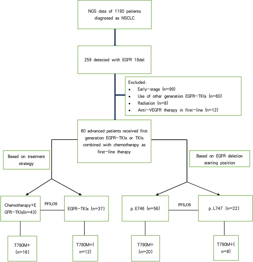 |
Figure 1 Experimental workflow. First, we retrospectively reviewed the NGS data of 1190 patients diagnosed with NSCLC in our hospital. Second, 259 patients with the EGFR 19del were identified. Finally, on the basis of the inclusion and exclusion criteria, we enrolled 80 patients in our study. |
NGS
All genomic profiling was performed at the Pathology Department of Anhui Chest Hospital. EGFR mutations were detected via NGS using the DA8600 platform, as previously described.30 DNA was extracted using a TIANamp FFPE DNA Kit (TIANGEN, Beijing, China). In all, 400 ng of DNA per sample was used for the preparation of a DNA library. The concentration of the DNA library was measured using a Qubit dsDNA HS Assay Kit (Thermo Fisher Scientific, Waltham, MA, USA). RNA was extracted using the RecoverAllTM Total Nucleic Acid Isolation Kit (Thermo Fisher Scientific). The quality of the RNA obtained was assessed using a Qubit RNA HS Assay Kit (Thermo Fisher Scientific). Total RNA (100 ng) was reverse transcribed using a random primer mixture. The synthesized cDNA was end-repaired and subsequently used to generate sequencing libraries according to the manufacturer’s instructions. The sequencing library was generated using a Novogene library construction kit (Tianjin Novogene Bioinformatics Technology Co., Ltd., Tianjin, China) according to the manufacturer’s instructions. This kit uses multiplex PCR capture and semiconductor sequencing technologies to purify DNA and RNA from formalin-fixed paraffin-embedded (FFPE) sections. The DNA and RNA were subsequently purified, the DNA and cDNA fragments of the target region were subsequently enriched using multiplex PCR, and the enriched libraries were quantified and subjected to quality control. The quantified libraries were then sequenced using a gene sequencer (DA8600; Zhongshan University Daan Gene Co., Ltd., Registration No.: 20,143,401,961) to obtain the DNA and RNA sequences of the target regions. In this study, we performed preliminary bioinformatics analyses involving data preprocessing, DNA comparison, and variant detection using Ion Reporter software (version 5.6) integrated with the DA8600 sequencer. The DNA variants and RNA fusions were subsequently transferred to the “Human EGFR, KRAS, BRAF, PIK3CA, ALK, and ROS1 Gene Mutation Detection Kit Analysis Software” by Tianjin Novozymes Bioinformatics Co., Ltd., for further advanced processing. Initial processing was performed to achieve base identification and data filtration, resulting in DNA/RNA sequences in the BAM format. DNA sequencing data were aligned against the reference genome (hg19) using TMAP software, which resulted in the generation of postcomparison BAM files. We employed TVC Variation Detection Software to identify single-nucleotide variants (SNVs) and insertions/deletions (indels), which are reported in VCF format. A mutation frequency threshold of 0.7% was applied for SNVs, while 1% was applied for Indels. The analysis progressed with data transferred to the gene mutation detection kit software, after which in-depth evaluations were performed. This included maintaining a minimum sequencing depth of 3000X for DNA mutation hotspots and at least 60,000 sequences for RNA data to meet quality control standards. Mutation results underwent annotation and filtering to retain hotspot annotations, while RNA sequences were assessed for fusion using TMAP, which validated sequences with overlap lengths of no fewer than 25 bp and error rates below 4%. Finally, mutations that met quality benchmarks were automatically classified by the software as positive or negative based on established thresholds: SNVs with mutation frequencies ≥0.7% and Indels ≥1% were considered positive, as were fusion sequences supported by at least 200 sequences. First-line NGS-detected samples included FFPE tumor tissue (n = 57), plasma (n = 22), and malignant pleural effusion (n = 1) samples.
Random Assignment
The enrolled patients were recruited not only from the Department of Oncology but also from the Department of Respiratory Medicine, the Department of Surgery, and other departments. Treatment plans were determined by treating physicians on the basis of age, ECOG PS, disease stage, patient willingness to receive certain treatments, and economic status, among other factors. However, EGFR subtypes were not considered. Therefore, the treatment decisions were considered randomized.
Treatment Protocol
Patients received initial doses of gefitinib, erlotinib, and icotinib at 250, 150, and 375 mg/day, respectively. Patients in the TKI + chemotherapy (T+C) cohort received intravenous pemetrexed (500 mg/m2) and cisplatin (75 mg/m2) or carboplatin (5 × area under the curve) on day 1 of a 21-day cycle for four or six cycles, followed by concurrent TKI and pemetrexed maintenance. Each treatment was administered until progressive disease or unbearable toxicity was observed.
Response and Toxicity Evaluation
Chest CT scans were performed every 1–2 months to evaluate treatment response and disease progression. Tumor response was evaluated according to the Response Evaluation Criteria in Solid Tumors version 1.1.31 Responses were classified as complete response (CR), partial response (PR), stable disease (SD), progressive disease (PD) or nonevaluable. PD was defined as a ≥20% increase in the sum of the longest diameters of target lesions, calculated relative to the smallest sum recorded during treatment, with an absolute increase of ≥5 mm, or progression of nontarget lesions or new lesions. The ORR was defined as the proportion of patients who achieved CR or PR. The DCR was defined as the proportion of participants who achieved CR, PR, or SD. PFS was calculated from treatment initiation to disease progression or death. OS was determined from the date of treatment initiation to the date of death or until the last follow-up on March 31, 2024. The National Cancer Institute Common Terminology Criteria for Adverse Events 5.0 was used to grade adverse events.
Statistical Analysis
The primary endpoint was PFS, and the secondary endpoints were OS and safety. PFS and OS were estimated using the Kaplan–Meier method. Independent risk factors were assessed using multivariate analysis with a Cox proportional hazards model. Baseline variables that were considered clinically relevant or that showed a univariate relationship with the outcome were included in the multivariate Cox proportional hazards regression model. Chi-square and Fisher’s exact tests were used to analyze the correlations among EGFR variants and clinicopathological variables. Statistical significance was set at P < 0.05. GraphPad Prism software (GraphPad Inc., La Jolla, CA, USA) was used to generate survival curves, and all the statistical analyses were performed using SPSS version 26.0 (IBM Corp., Armonk, NY, USA).
Results
Baseline Patient Characteristics
Among the 259 patients diagnosed with EGFR 19del, 69 had uncommon EGFR 19delins. The distribution of variants is shown in Figure 2a. Patients were classified into the EGFR p.E746 and EGFR p.L747 subgroups according to the starting position of the deletion. A greater percentage of EGFR 19delins was observed in the EGFR p.L747 subgroup than in the EGFR p.E746 subgroup (43/67 vs 25/190, P < 0.001; Figure 2b). Eighty patients were included in the study, and of these, 43 were in the T+C cohort and 37 were in the T cohort. The median PFS and OS of the 80 patients were 12 and 35 months, respectively. No significant differences were detected in basic clinical features, including age, sex, baseline brain metastasis status, Eastern Cooperative Oncology Group (ECOG) performance status (PS), and smoking history, between the subgroups (see Table 1 for the baseline characteristics of the patients). Among them, one patient harbored an S752_I759del.
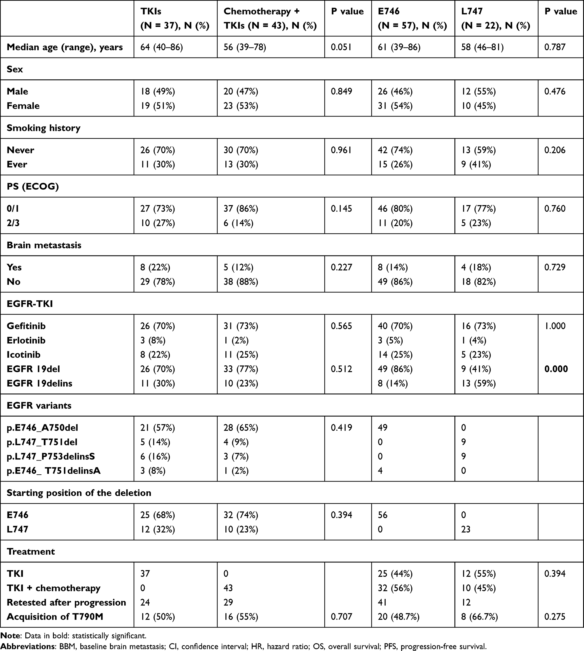 |
Table 1 Baseline Characteristics of the Included Patients |
 |
Figure 2 Mutation overview. (a) Pie chart illustrating the distribution of EGFR exon 19 deletion variants among 259 patients. (b) The percentage of EGFR exon 19 deletion–insertions was greater in patients with the EGFR p.L747 mutation than in those with the EGFR p.E746 mutation (43/67 vs 25/190, P < 0.001). EGFR, epidermal growth factor receptor; EGFR 19delins, exon 19 deletion-insertion; **, statistically significant. |
Survival Analysis
TKIs vs TKIs + Chemotherapy
The objective response rate (ORR) and disease control rate (DCR) in the TKI + chemotherapy cohort were 83.7% and 95.3%, respectively, whereas the 37 patients who received first-generation TKIs had an ORR of 78.4% and a DCR of 94.6%. No significant difference was observed in the ORR (P = 0.542) or DCR (P = 1) between the groups. Patients had a median PFS of 16 months (95% CI 12.377–19.623) and 11 months (95% CI 8.454–13.546) in the T+C and T cohorts, respectively, although the difference was not statistically significant (P = 0.062; see Figure 3A for PFS between T vs T+C); this was similar to the difference in median OS between the two groups (37 months [95% CI 26.289–47.711] vs 34 months [95% CI 25.396–44.604], P = 0.14) (see Figure 3B for OS between T vs T+C).
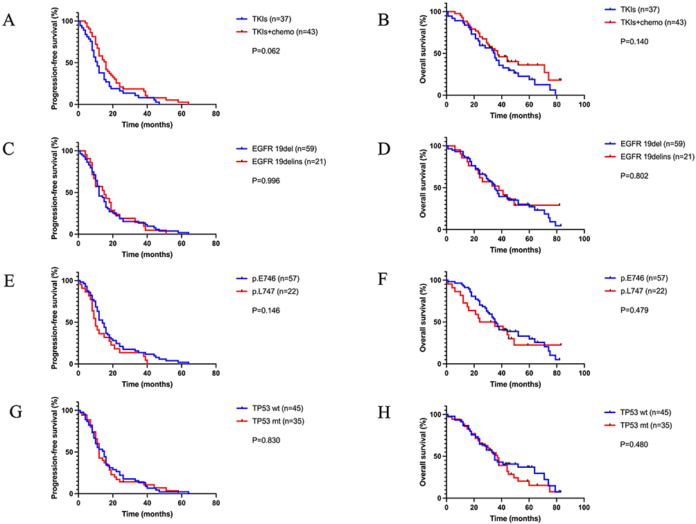 |
Figure 3 Kaplan–Meier curves for progression-free survival (PFS) and overall survival (OS) between groups. Comparison of PFS (A) and OS (B) between the TKI and TKI+chemotherapy groups. Comparison of PFS (C) and OS (D) between the EGFR exon 19 deletion and exon 19 deletion–insertion groups. Comparison of PFS (E) and OS (F) between the EGFR p.E746 and p.L747 groups. Comparison of PFS (G) and OS (H) between patients with wild-type and mutant TP53. Abbreviations: EGFR, epidermal growth factor receptor; TKIs, tyrosine kinase inhibitors; chemo, chemotherapy; EGFR 19delins, exon 19 deletion-insertion; TP53 wt, TP53 wild-type; TP53 mt, mutant TP53. |
EGFR 19del vs 19delins
We divided the patients into the EGFR 19del (n = 59) and EGFR 19delins (n = 21) subgroups according to the presence or absence of insertions. The baseline characteristics, including age, baseline brain metastasis status, sex, and smoking history, were similar between the two groups. The median PFS was 15 months (95% CI 6.028–23.972) in patients with EGFR 19del and 12 months (95% CI 9.187–14.813, P = 0.997) in those with EGFR 19delins (see Figure 3C for PFS between EGFR 19del and 19delins). The median OS was similar between patients with EGFR 19del and those with EGFR 19delins (38 months [95% CI 17.144–58.856] vs 35 months [95% CI 30.891–39.109]; P = 0.802) (see Figure 3D for OS between patients with EGFR 19del and those with EGFR 19delins).
p.E746del vs p.L747del Subtypes
The median PFS of patients harboring EGFR p.L747 mutations was shorter than that of patients harboring EGFR p.E746 mutations (10 months [95% CI 7.253–12.747] vs 14 months [95% CI 11.310–16.690, P = 0.146]) (see Figure 3E for PFS between the EGFR p.E746del subgroup and the p.L747 subgroup). In contrast, patients with EGFR p.L747 mutations had a shorter median OS (24 months, 95% CI 3.241–44.759) than did patients with EGFR p.E746 mutations (36 months, 95% CI 31.281–40.719 [P = 0.479]; see Figure 3F for OS between the EGFR p.E746del subgroup and the p.L747 subgroup), although these differences were not statistically significant.
Wild-Type vs Mutant TP53
The median PFS in patients harboring TP53 mutations was numerically shorter than that in patients with wild-type TP53 (12 months [95% CI 11.044–12.956] vs 15 months [95% CI 9.773–20.227, P = 0.830]) (see Figure 3G for PFS between patients with wild-type and those with mutant TP53). The median OS was similar between patients with TP53 mutations and those with wild-type TP53 (37 months [95% CI 30.527–43.473] vs 35 months [95% CI 30.059–39.941]; P = 0.480) (see Figure 3H for OS between patients with wild-type and mutant TP53).
Exploratory Subgroup Survival Analyses (TKIs vs TKIs + Chemotherapy Combined with p.E746del vs p.L747del)
Among the 37 patients who received only TKIs, 25 had EGFR p.E746 mutations, whereas 12 had EGFR p.L747 mutations. The median PFS of the EGFR p.E746 subgroup was longer than that of the EGFR p.L747 subgroup (12 months [7.104–16.896] vs 8 months [5.454–10.546], P = 0.011; Figure 4A). Similarly, the median OS of patients with p.E746 EGFR mutations was significantly longer than that of patients with p.L747 EGFR mutations (36 months [31.314–40.686] vs 21 months [9.118–32.882], P = 0.026; Figure 4B). However, the median PFS (P = 0.800) and the median OS (P = 0.439) were not significantly different between the two groups of patients who received TKIs plus chemotherapy (Figure 4C and D).
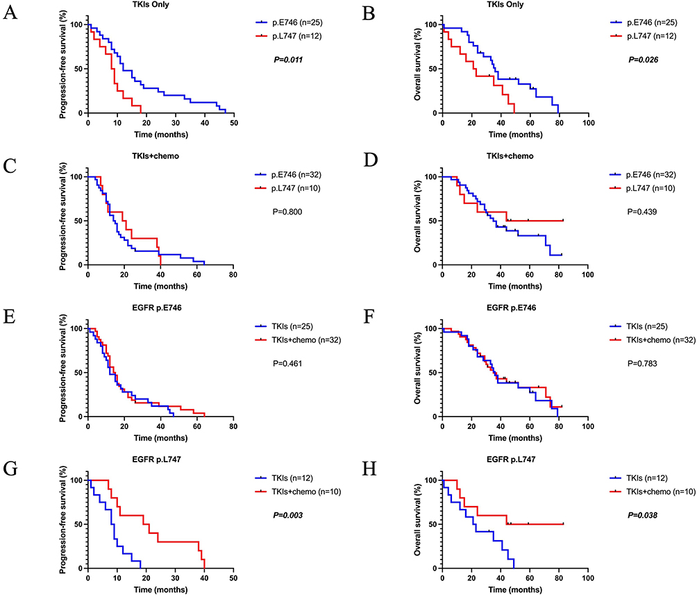 |
Figure 4 Kaplan–Meier curves for progression-free survival (PFS) and overall survival (OS) between subgroups. Comparison of PFS (A) and OS (B) between patients with the EGFR p.E746 mutation and those with the EGFR p.L747 mutation who were treated with TKIs. Comparison of PFS (C) and OS (D) between patients with the EGFR p.E746 mutation and those with the EGFR p.L747 mutation who were treated with TKIs + chemotherapy. Comparison of PFS (E) and OS (F) between patients with the EGFR p.E746 mutation who were treated with TKIs and those who were treated with TKIs + chemotherapy. Comparison of PFS (G) and OS (H) between patients with the EGFR p.L747 mutation who were treated with TKIs and those who were treated with TKIs + chemotherapy. EGFR, epidermal growth factor receptor; TKIs, tyrosine kinase inhibitors; chemo, chemotherapy. Data in bold and italics are statistically significant. |
Among patients harboring EGFR p.E746 mutations, 25 received first-generation TKIs as first-line treatment, while 32 received TKIs and chemotherapy. Age, baseline brain metastasis, sex, and smoking history were similar between the groups. The median PFS was 12 months (95% CI 7.104–16.896) for patients who received TKIs and 14 months (95% CI 10.304–17.696, P = 0.461) for those who received combination therapy (Figure 4E). Furthermore, the median OS was similar between patients who received TKIs alone and those who received combination therapy (36 months, 95% CI 31.314–40.686 vs 35 months, 95% CI 27.093–42.907; P = 0.783) (Figure 4F).
Among patients harboring an EGFR p.L747 mutation, 12 received TKIs as first-line therapy, while 10 received TKIs combined with chemotherapy. Clinical characteristics, including age, baseline brain metastasis status, sex, and smoking history, were not significantly different between the groups. The median PFS of patients who received combination therapy was significantly longer than that of patients who received only TKIs (19 months [95% CI 3.505–34.495] vs 8 months [95% CI 5.454–10.546] P = 0.003) (Figure 4G). Similarly, the median OS was significantly longer in patients treated with combination therapy than in those treated with TKIs alone (44 months [95% CI could not be attained] vs 21 months [95% CI 9.118–32.882]) (P = 0. 038) (Figure 4H).
Univariate and Multivariate Cox Proportional Hazards Models
Multivariate Cox proportional hazards models were used to identify the predictive effects of different variables on PFS and OS (see Table 2 for univariate and multivariate Cox proportional hazards models).
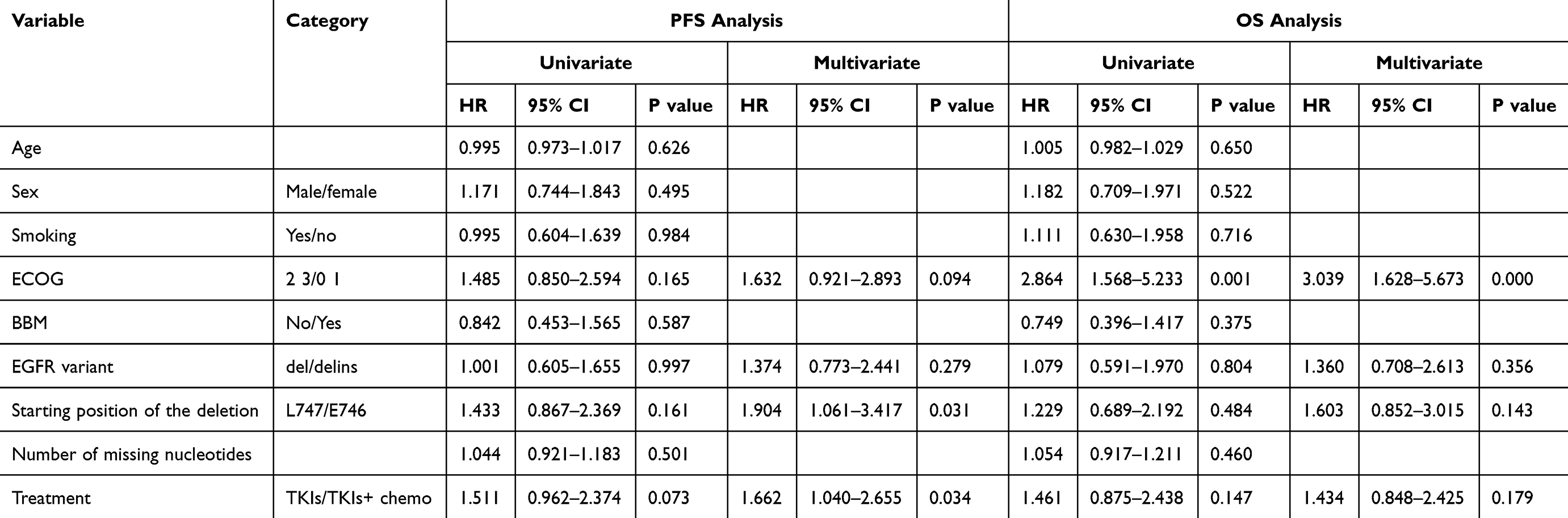 |
Table 2 Results of Univariate and Multivariate Survival Analyses of Patients with Lung Adenocarcinoma with EGFR Exon 19 Deletion |
Age, sex, smoking status, ECOG PS, baseline brain metastasis, EGFR variant type, starting position of the deletion, number of missing nucleotides, and first-line treatment were included in the univariate analysis, which revealed that none of the variables was significantly associated with PFS. ECOG PS (P = 0.165), EGFR variant type (P = 0.997), starting position of the deletion (P = 0.161), and treatment (P = 0.073) were subsequently included in the multivariate Cox proportional hazards model. Both the EGFR p.L747 mutation (HR 1.904 [95% CI 1.061–3.417]; P = 0.031) and treatment with TKIs alone (HR 1.662 [95% CI 1.040–2.655]; P = 0.034) were significant risk factors for poor PFS according to the multivariate Cox proportional hazards model.
The univariate analysis revealed that only the ECOG PS (P = 0.001) was significantly associated with OS. Therefore, we included the ECOG PS (P = 0.001), the EGFR variant type (P = 0.804), the starting position of the deletion (P = 0.484), and treatment (P = 0.147) in the multivariate Cox proportional hazards model. Notably, the ECOG PS was the only significant independent risk factor for OS according to the multivariate Cox proportional hazards model (P = 0.000).
Distribution of EGFR 19del Variants and the Mechanism of Resistance
The E746_A750del, which occurred in 49 patients (61.2%), was the most common EGFR 19del variant, whereas L747_P753delinsS and L747_T751del were the second most common variants, as they were each observed in nine patients (11.2%). Among the 57 patients harboring EGFR p.E746 mutations, 13 had TP53 mutations. The prevalence of TP53 mutations was significantly greater in patients with EGFR p.L747 mutations (10/22, P = 0.047).
A second genetic test was performed for patients who experienced disease progression (53/79, 67%) after first-line treatment. The EGFR T790M mutation was identified as the major acquired mechanism of resistance (28/53, 52.8%). The rate of T790M mutation in the TKI + chemotherapy subgroup (55%, 16/29) was slightly greater than that in the TKI group (50%, 12/24), although this difference was not statistically significant (P = 0.707). Additionally, the T790M mutation rate was similar between patients with EGFR p.E746 and those with EGFR p.L747 mutations (20/41 vs 8/12, P = 0.275), which is consistent with that observed between patients with EGFR 19del and 19delins (20/39 vs 8/14, P = 0.706).
Safety Analysis
The most common adverse events in the TKI + chemotherapy group were myelosuppression (46.5%, grades I–IV), liver function abnormalities (39.5%, grades I–III), and gastrointestinal toxicity (diarrhea, vomiting, 25.5%, grades I–III). Grade III–IV adverse events were noted in nine patients, and six patients discontinued chemotherapy due to toxicity. The most common adverse events observed in the TKI group were rashes (48.6%, grades I–II), diarrhea (32.4%, grades I–II), and liver function abnormalities (16.2%, grades I–II). One patient (2.7%) in the TKI group discontinued therapy due to grade III interstitial pneumonia. Clinically relevant grade III or higher toxicities occurred in 20.9% and 2.7% of patients in the TKI + chemotherapy and TKI groups, respectively (P = 0.02).
Discussion
To our knowledge, this is the first study to report the effects of chemotherapy and the EGFR 19del subtype on the clinical outcomes of patients with advanced NSCLC treated with first-generation TKIs. Compared with patients with EGFR p.E746 mutations, patients with EGFR p.L747 mutations can benefit more from chemotherapy plus first-generation TKIs than from TKI monotherapy, as shown by the higher PFS and OS observed in these patients.
In this real-world, retrospective study, we comprehensively evaluated the effects of chemotherapy and various EGFR exon 19 deletion subtypes on the efficacy of first-generation EGFR-TKIs and the survival outcomes after treatment. However, no significant differences were observed between the groups. In the exploratory subgroup analysis, we were surprised to find that compared with patients with EGFR p.E746 mutations, those with p.L747 mutations could benefit from chemotherapy combined with EGFR-TKIs. This finding was also verified through a multivariate analysis.
EGFR p.E746-A750 is the most common subtype of the EGFR 19del and accounts for 60% of all variants.19 Previous studies have shown that EGFR p.L747 differs from EGFR p.E746 not only in the molecular conformation of the resulting protein structure but also in the frequency of TP53 co-mutations and secondary T790M mutations. The frequency of EGFR 19delins in our study was higher in patients with EGFR p.L747 mutations than in those with p.E746 mutations, as previously reported.15 Deletions starting at L747 are often complex insertion deletions that result in the replacement of the deleted amino acids with a nonnative residue.2 As in L747_A750delinsP, the presence of a proline at this position displaces the adjacent β1/β2 glycine-rich loop and thus impairs drug binding.32 In our study, EGFR p.L747 comprised L747_P753delinsS (9, 40.9%), L747_T751del (9, 40.9%), L747_A750delinsP (2, 9.1%), and L747_T751delinsP (2, 9.1%). L747_A750delinsP has been reported to exhibit primary resistance to first-generation TKIs and sensitivity to second-generation TKIs both in vitro and in clinical case reports.32–35 Additionally, compared with the E746_A750del mutation, L747_A750delinsP is associated with inferior PFS in patients treated with first-line osimertinib.36 Accordingly, of the two patients in our study who harbored L747_A750delinsP, one experienced disease progression after 4 months of gefitinib treatment, while the other responded to chemotherapy and first-generation TKI therapy and had a PFS of 38 months.
Since EGFR p.L747 is more frequently accompanied by a TP53 mutation, which is related to TKI resistance,37,38 the addition of chemotherapy to the EGFR p.L747 subgroup may reverse the disadvantages of TKI monotherapy and provide survival benefits.
Unlike previous gene detection methods, NGS allows parallel sequencing of numerous small DNA fragments, which enables concurrent testing for a very wide array of mutational gene panels and provides in-depth insights into mutation subtypes. Tissue-based genetic testing is the gold standard, whereas liquid biopsy is an important supplemental method.39 NGS analysis of ctDNA using liquid biopsies can overcome the limitations of spatial tumor heterogeneity in tissue testing and is more feasible and noninvasive. However, ctDNA accounts for only a tiny fraction of the total cell-free DNA and can lead to false-positive or false-negative results.40 In contrast to what has been reported in previous studies,41,42 the rate of T790M mutation after disease progression despite first-line therapy, was higher in patients with p.L747 mutations than in those with p.E746 mutations (66.7% vs 48.7%), albeit the difference was not statistically significant. This was likely because of a lower but insignificant detection rate after progression in p.L747 patients (54.5% vs 74.5%, P=0.087). The small sample size, heterogeneity of the timing and method of resistance detection and the addition of chemotherapy resulted in bias.43 The inferior outcome of patients with p.L747 mutations may indicate more complicated resistance mechanisms, and thus further research is needed to explain these results.
Insertions in EGFR tyrosine kinase lead to domain amino acid substitution and disruption of the hydrophobic core, resulting in EGFR activation.44 Although this mutation is considered TKI-sensitive, we found that the PFS and OS of patients with EGFR 19delins did not differ from those of patients with EGFR 19del. Overall, these findings are consistent with those reported previously.45
Although combination therapy was associated with a high incidence of toxicity, this toxicity was manageable. However, grade III or higher toxicities were more common in the combination therapy group (20.9% vs 2.7%, P = 0.02). Therefore, caution should be exercised when chemotherapy is administered in addition to TKIs. Moreover, cytotoxic chemotherapy continues to play an important role in anticancer therapy even though TKIs are the standard first-line treatment for metastatic NSCLC. Although the FLAURA2 study (osimertinib with or without chemotherapy) recently revealed that PFS in the osimertinib-chemotherapy cohort was 9 months longer than that in the osimertinib monotherapy cohort among patients with EGFR-mutated advanced NSCLC, this prolongation occurred at the cost of high adverse events in the combined therapy group.46 The occurrence of adverse events was the most frequent reason for the discontinuation of carboplatin or cisplatin (47 patients [17%]) and pemetrexed (119 patients [43%]).46 The discontinuation rate in our study was 13.9% (6/43), which was lower than that in the FLAURA2 study. This is probably because if toxicity occurs, clinicians will decide to extend the treatment cycle or reduce the dosage, as well as a lower hematological toxicity of first-generation TKIs.
In the FLAURA2 study, differences in PFS and OS between patients with EGFR 19del and those with 21L858R were observed. Our findings suggest differences in PFS and OS between patients with different EGFR 19del variants. Thus, our clinical data underscore the importance of combination therapy for unique EGFR exon 19 subtypes. For the EGFR L747-specific subgroup, further research is needed to determine whether first-/third-generation TKIs combined with chemotherapy, second-generation TKIs, or other treatments should be selected.
Nonetheless, our study has several limitations. First, this was a retrospective single-institution study. Therefore, our results should be verified in future multi-institution prospective studies. Second, the sample size was relatively small, and differences between the subtypes of the EGFR p.L747 mutation were not specifically analyzed. The results of the subgroup analysis are exploratory rather than confirmatory. Our findings should therefore be interpreted with caution. Third, the patients included in this study were treated with first-generation EGFR-TKIs, while third-generation TKIs are now recommended as the preferred first-line treatment. Therefore, future prospective trials that integrate molecular subtyping and modern TKIs are essential to translate these findings into clinical practice.
Conclusion
Additional chemotherapy with EGFR-TKIs did not affect the clinical outcomes of NSCLC patients harboring the EGFR p.E746 mutation but greatly improved PFS and OS in those with the p.L747 mutation. Thus, a detailed analysis of the EGFR 19del should be performed to guide the selection of combination therapy for NSCLC patients, which may be tailored to their molecular profiles.
Data Sharing Statement
The data generated in this study are available upon request from the corresponding author.
Ethical Approval
This study was approved by the Institutional Review Board of the Anhui Chest Hospital (approval number: K2022-029). Given its retrospective nature, the committee waived the informed consent requirement for this study. We declare that patient information will be kept confidential and that we adhere to the principles of the Declaration of Helsinki.
Acknowledgments
This work was supported by the Anhui Provincial Clinical Key Specialty [grant number [2020]243]. The funder had no role in the study design, data collection and analysis, decision to publish, or preparation of the manuscript.
Author Contributions
All the authors put forth significant contributions to the work reported, whether in the conception, study design, execution, data acquisition, analysis and interpretation, or in all these areas; participated in drafting, revising or critically reviewing the article; provided final approval of the version to be published; agreed on the journal to which the article has been submitted; and agreed to be accountable for all aspects of the work.
Disclosure
The authors have no conflicts of interest to declare for this work.
References
1. Bray F, Laversanne M, Sung H, et al. Global cancer statistics 2022: GLOBOCAN estimates of incidence and mortality worldwide for 36 cancers in 185 countries. CA Cancer J Clin. 2024;74(3):229–263. doi:10.3322/caac.21834
2. Sharma SV, Bell DW, Settleman J, Haber DA. Epidermal growth factor receptor mutations in lung cancer. Nat Rev Cancer. 2007;7(3):169–181. doi:10.1038/nrc2088
3. Lynch TJ, Bell DW, Sordella R, et al. Activating mutations in the epidermal growth factor receptor underlying responsiveness of non-small-cell lung cancer to gefitinib. N Engl J Med. 2004;350(21):2129–2139. doi:10.1056/NEJMoa040938
4. Zhou C, Wu YL, Chen G, et al. Erlotinib versus chemotherapy as first-line treatment for patients with advanced EGFR mutation-positive non-small-cell lung cancer (OPTIMAL, CTONG-0802): a multicentre, open-label, randomised, Phase 3 study. Lancet Oncol. 2011;12(8):735–742. doi:10.1016/S1470-2045(11)70184-X
5. Rosell R, Carcereny E, Gervais R, et al. Erlotinib versus standard chemotherapy as first-line treatment for European patients with advanced EGFR mutation-positive non-small-cell lung cancer (EURTAC): a multicentre, open-label, randomised phase 3 trial. Lancet Oncol. 2012;13(3):239–246. doi:10.1016/S1470-2045(11)70393-X
6. Wu YL, Zhou C, Liam CK, et al. First-line erlotinib versus gemcitabine/cisplatin in patients with advanced EGFR mutation-positive non-small-cell lung cancer: analyses from the Phase III, randomized, open-label, ENSURE study. Ann Oncol. 2015;26(9):1883–1889. doi:10.1093/annonc/mdv270
7. Mitsudomi T, Morita S, Yatabe Y, et al. Gefitinib versus cisplatin plus docetaxel in patients with non-small-cell lung cancer harbouring mutations of the epidermal growth factor receptor (WJTOG3405): an open label, randomised phase 3 trial. Lancet Oncol. 2010;11(2):121–128. doi:10.1016/S1470-2045(09)70364-X
8. Sequist LV, Yang JC-H, Yamamoto N, et al. Phase III study of Afatinib or cisplatin plus pemetrexed in patients with metastatic lung adenocarcinoma with EGFR mutations. J Clin Oncol. 2013;31(27):3327–3334. doi:10.1200/JCO.2012.44.2806
9. Wu Y-L, Zhou C, Hu C-P, et al. Afatinib versus cisplatin plus gemcitabine for first-line treatment of Asian patients with advanced non-small-cell lung cancer harbouring EGFR mutations (LUX-Lung 6): an open-label, randomised phase 3 trial. Lancet Oncol. 2014;15(2):213–222. doi:10.1016/S1470-2045(13)70604-1
10. Papadimitrakopoulou VA, Mok TS, Han JY, et al. Osimertinib versus platinum‐pemetrexed for patients with EGFR T790M advanced NSCLC and progression on a prior EGFR‐tyrosine kinase inhibitor: AURA3 overall survival analysis. Ann Oncol. 2020;31(11):1536–1544. doi:10.1016/j.annonc.2020.08.2100
11. COSMIC database. 2019. Available from: https://cancer.sanger.ac.uk/cosmic/gene/analysis?ln=EGFR.
12. Chung KP, Wu SG, Wu JY, et al. Clinical outcomes in non-small cell lung cancers harboring different exon 19 deletions in EGFR. Clin Cancer Res. 2012;18(12):3470–3477. doi:10.1158/1078-0432.CCR-11-2353
13. Lee VHF, Tin VPC, Choy TS, et al. Association of exon 19 and 21 EGFR mutation patterns with treatment outcome after first-line tyrosine kinase inhibitor in metastatic non-small-cell lung cancer. J Thorac Oncol. 2013;8(9):1148–1155. doi:10.1097/JTO.0b013e31829f684a
14. Su J, Zhong W, Zhang X, et al. Molecular characteristics and clinical outcomes of EGFR exon 19 indel subtypes to EGFR TKIs in NSCLC patients. Oncotarget. 2017;8(67):111246–111257. doi:10.18632/oncotarget.22768
15. Kaneda T, Hata A, Tomioka H, et al. Possible differential EGFR-TKI efficacy among exon 19 deletional locations in EGFR-mutant non-small cell lung cancer. Lung Cancer. 2014;86(2):213–218. doi:10.1016/j.lungcan.2014.09.014
16. Sutiman N, Tan SW, Tan EH, et al. EGFR mutation subtypes influence survival outcomes following first-line gefitinib therapy in advanced Asian NSCLC patients. J Thorac Oncol. 2017;12(3):529–538. doi:10.1016/j.jtho.2016.11.2225
17. Rossi S, Toschi L, Finocchiaro G, et al. Impact of exon 19 deletion subtypes in EGFR-mutant metastatic non-small-cell lung cancer treated with first-line tyrosine kinase inhibitors. Clin Lung Cancer. 2019;20(2):82–87. doi:10.1016/j.cllc.2018.10.009
18. Peng X, Long X, Liu L, et al. Clinical impact of uncommon epidermal growth factor receptor exon 19 insertion-deletion variants on epidermal growth factor receptor-tyrosine kinase inhibitor efficacy in non-small-cell lung cancer. Eur J Cancer. 2020;141:199–208. doi:10.1016/j.ejca.2020.10.005
19. Zhao C, Jiang T, Li J, et al. The impact of EGFR exon 19 deletion subtypes on clinical outcomes in non-small cell lung cancer. Transl Lung Cancer Res. 2020;9(4):1149–1158. doi:10.21037/tlcr-19-359
20. Yamamoto N, Seto T, Nishio M, et al. Erlotinib plus bevacizumab vs erlotinib monotherapy as first-line treatment for advanced EGFR mutation-positive non-squamous non-small-cell Lung Cancer: survival follow-up results of the randomized JO25567 study. Lung Cancer. 2021;151:20–24. doi:10.1016/j.lungcan.2020.11.020
21. Saito H, Fukuhara T, Furuya N, et al. Erlotinib plus bevacizumab versus erlotinib alone in patients with EGFR-positive advanced non-squamous non-small-cell lung cancer (NEJ026): interim analysis of an open-label, randomised, multicentre, phase 3 trial. Lancet Oncol. 2019;20(5):625–635. doi:10.1016/S1470-2045(19)30035-X
22. Wu YL, Lee JS, Thongprasert S, et al. Intercalated combination of chemotherapy and erlotinib for patients with advanced stage non-small-cell lung cancer (FASTACT-2): a randomised, double-blind trial. Lancet Oncol. 2013;14(8):777–786. doi:10.1016/S1470-2045(13)70254-7
23. Oizumi S, Sugawara S, Minato K, et al. Updated survival outcomes of NEJ005/TCOG0902: a randomised phase II study of concurrent versus sequential alternating gefitinib and chemotherapy in previously untreated non-small cell lung cancer with sensitive EGFR mutations. ESMO Open. 2018;3(2):e000313. doi:10.1136/esmoopen-2017-000313
24. Hosomi Y, Morita S, Sugawara S, et al. Gefitinib alone versus gefitinib plus chemotherapy for non-small-cell lung cancer with mutated epidermal growth factor receptor: NEJ009 study. J Clin Oncol. 2020;38(2):115–123. doi:10.1200/JCO.19.01488
25. Noronha V, Patil VM, Joshi A, et al. Gefitinib versus gefitinib plus pemetrexed and carboplatin chemotherapy in EGFR-mutated lung cancer. J Clin Oncol. 2020;38(2):124–136. doi:10.1200/JCO.19.01154
26. Cheng Y, Murakami H, Yang PC, et al. Randomized Phase II trial of gefitinib with and without pemetrexed as first-line therapy in patients with advanced nonsquamous non-small-cell lung cancer with activating epidermal growth factor receptor mutations. J Clin Oncol. 2016;34(27):3258–3266. doi:10.1200/JCO.2016.66.9218
27. Yang JCH, Cheng Y, Murakami H, et al. A randomized Phase 2 study of gefitinib with or without pemetrexed as first-line treatment in nonsquamous NSCLC with EGFR mutation: final overall survival and biomarker analysis. J Thorac Oncol. 2020;15(1):91–100. doi:10.1016/j.jtho.2019.09.008
28. Tran T-O, Vo TH, Le NQK, et al. Omics-based deep learning approaches for lung cancer decision-making and therapeutics development. Briefings Functional Genomics Vol. 2024;23(3):181–192. doi:10.1093/bfgp/elad031
29. Detterbeck FC, Chansky K, Groome P, et al. The IASLC lung cancer staging project: methodology and validation used in the development of proposals for revision of the stage classification of NSCLC in the forthcoming (eighth) edition of the TNM classification of lung cancer. J Thorac Oncol. 2016;11(9):1433–1446. doi:10.1016/j.jtho.2016.06.028
30. Guan M, Xu J, Shi Q. Molecular determinants of clinical outcomes for anaplastic lymphoma kinase-positive non-small cell lung cancer in Chinese patients: a retrospective study. Cancer Genet. 2023;270–271:32–38. doi:10.1016/j.cancergen.2022.11.005
31. Eisenhauer EA, Therasse P, Bogaerts J, et al. New response evaluation criteria in solid tumours: revised RECIST guideline (version 1.1). Eur J Cancer. 2009;45(2):228–247. doi:10.1016/j.ejca.2008.10.026
32. Truini A, Starrett JH, Stewart T, et al. The EGFR exon 19 mutant L747-A750>P exhibits distinct sensitivity to tyrosine kinase inhibitors in lung adenocarcinoma. Clin Cancer Res. 2019;25(21):6382–6391. doi:10.1158/1078-0432.CCR-19-0780
33. Di Federico A, Filippini DM, Dall’Olio FG, et al. The EGFR exon 19 mutant L747-A750>P exhibits distinct sensitivity to tyrosine kinase inhibitors in lung adenocarcinoma-Letter. Clin Cancer Res. 2020;26(2):518–519. doi:10.1158/1078-0432.CCR-19-2441
34. Wei Q, Zhang J, Chen D, Li S, Liu Y. Primary resistance to gefitinib in a patient with lung adenocarcinoma harboring an EGFR exon 19 L747-A750>P mutation. Lung Cancer. 2020;148:175–176. doi:10.1016/j.lungcan.2020.08.013
35. Guida A, Tassi V, Facchinetti F, et al. Ad hoc Afatinib in an eldery lung cancer patient with EGFR exon 19 deletion L747-A750>. P Adv Respir Med. 2022;90(3):234–235. doi:10.5603/ARM.a2022.0034
36. Grant MJ, Aredo JV, Starrett JH, et al. Efficacy of osimertinib in patients with lung cancer positive for uncommon EGFR exon 19 deletion mutations. Clin Cancer Res. 2023;29(11):2123–2130. doi:10.1158/1078-0432.CCR-22-3497
37. Chen Y, Xu J, Zhang L, et al. A multicenter-retrospective study of non-small-cell lung carcinoma harboring uncommon epidermal growth factor receptor (EGFR) mutations: different subtypes of EGFR exon 19 deletion-insertions exhibit the clinical characteristics and prognosis of non-small cell lung carcinoma. Transl Lung Cancer Res. 2022;11(2):238–249. doi:10.21037/tlcr-22-48
38. Vanderlaan PA, Rangachari D, Mockus SM, et al. Mutations in TP53, PIK3CA, PTEN and other genes in EGFR mutated lung cancers: correlation with clinical outcomes. Lung Cancer. 2017;106:17–21. doi:10.1016/j.lungcan.2017.01.011
39. Sabour L, Sabour M, Ghorbian S. Clinical applications of next-generation sequencing in cancer diagnosis. Pathol Oncol Res. 2017;23(2):225–234. doi:10.1007/s12253-016-0124-z
40. Esagian SM, Grigoriadou GΙ, Nikas IP, et al. Comparison of liquid-based to tissue-based biopsy analysis by targeted next generation sequencing in advanced non-small cell lung cancer: a comprehensive systematic review. J Cancer Res Clin Oncol. 2020;146(8):2051–2066. doi:10.1007/s00432-020-03267-x
41. Huang LT, Zhang SL, Han CB, Ma JT. Impact of EGFR exon 19 deletion subtypes on clinical outcomes in EGFR-TKI-treated advanced non-small-cell lung cancer. Lung Cancer. 2022;166:9–16. doi:10.1016/j.lungcan.2022.01.014
42. Huang YH, Hsu KH, Tseng JS, et al. The association of acquired T790M mutation with clinical characteristics after resistance to first-line epidermal growth factor receptor tyrosine kinase inhibitor in lung adenocarcinoma. Cancer Res Treat. 2018;50(4):1294–1303. doi:10.4143/crt.2017.512
43. Wu SG, Gow CH, Chen YL, Liu YN, Tsai MF, Shih JY. Different treatment efficacies and T790M acquisition of EGFR-TKIs on NSCLC patients with variable Del-19 subtypes of EGFR. Int J Cancer. 2023;153(2):352–363. doi:10.1002/ijc.34507
44. He M, Capelletti M, Nafa K, et al. EGFR exon 19 insertions: a new family of sensitizing EGFR mutations in lung adenocarcinoma. Clin Cancer Res. 2012;18(6):1790–1797. doi:10.1158/1078-0432.CCR-11-2361
45. Wang Y, Zheng R, Hu P, Zhang Z, Shen S, Li X. Patients harboring uncommon EGFR exon 19 deletion-insertion mutations respond well to first-generation EGFR inhibitors and osimeritinib upon acquisition of T790M. BMC Cancer. 2021;21(1):1215. doi:10.1186/s12885-021-08942-x
46. Planchard D, Jänne PA, Cheng Y, et al. Osimertinib with or without chemotherapy in EGFR-mutated advanced NSCLC. N Engl J Med. 2023;389(21):1935–1948. doi:10.1056/NEJMoa2306434
 © 2025 The Author(s). This work is published and licensed by Dove Medical Press Limited. The
full terms of this license are available at https://www.dovepress.com/terms
and incorporate the Creative Commons Attribution
- Non Commercial (unported, 4.0) License.
By accessing the work you hereby accept the Terms. Non-commercial uses of the work are permitted
without any further permission from Dove Medical Press Limited, provided the work is properly
attributed. For permission for commercial use of this work, please see paragraphs 4.2 and 5 of our Terms.
© 2025 The Author(s). This work is published and licensed by Dove Medical Press Limited. The
full terms of this license are available at https://www.dovepress.com/terms
and incorporate the Creative Commons Attribution
- Non Commercial (unported, 4.0) License.
By accessing the work you hereby accept the Terms. Non-commercial uses of the work are permitted
without any further permission from Dove Medical Press Limited, provided the work is properly
attributed. For permission for commercial use of this work, please see paragraphs 4.2 and 5 of our Terms.
Recommended articles

Long Non-Coding RNAs ASB16-AS1 and AFAP1-AS1: Diagnostic, Prognostic Impact and Survival Analysis in Colorectal Cancer
Elabd NS, Soliman SE, Elhamouly MS, Gohar SF, Elgamal A, Alabassy MM, Soliman HA, Gadallah AA, Elbahr OD, Soliman G, Saleh AA
The Application of Clinical Genetics 2022, 15:97-109
Published Date: 1 August 2022
Efficacy of Bevacizumab in High-Grade Meningiomas: A Retrospective Clinical Study
Bai X, Liu X, Wen J
Neuropsychiatric Disease and Treatment 2022, 18:1619-1627
Published Date: 6 August 2022
Real-World Outcomes and Prognostic Factors Among Patients with Advanced Non-Small Cell Lung Cancer and High PD-L1 Expression Treated with Immune Checkpoint Inhibitors as First-Line Therapy
Ge W, Wu N, Jalbert JJ, Quek RGW, Liu J, Rietschel P, Pouliot JF, Harnett J, Hsu ML, Feliciano JL
Cancer Management and Research 2022, 14:3191-3202
Published Date: 16 November 2022
The Systemic Inflammation Score is Associated with the Survival of Patients with Prostate Cancer
Xie J, Xiao X, Dong Z, Wang Q
Journal of Inflammation Research 2023, 16:963-975
Published Date: 7 March 2023
Unveiling the Landscape of PD-L1 Expression and Tumor-Infiltrating Lymphocyte Subtypes in Advanced Triple-Negative Breast Cancer in Brazil
Dos Santos ALS, Da Silva JL, De Albuquerque LZ, Neto ALA, Da Silva CF, Cerva LAM, Small IA, Rodrigues FR, De Macedo FC, Marcelino CP, Batista PDM, Rego MADC, Borba MACSM, De Melo AC
Breast Cancer: Targets and Therapy 2025, 17:349-358
Published Date: 15 April 2025