Back to Journals » Journal of Inflammation Research » Volume 19
Lactate Dehydrogenase as a Potential Mediator Between Immature Granulocytes and Tumor Burden in Breast Cancer
Authors Liang H, Pan K, Liang X, Wang J, Xie X, Wan N, Lin J
Received 15 October 2025
Accepted for publication 7 January 2026
Published 17 January 2026 Volume 2026:19 573858
DOI https://doi.org/10.2147/JIR.S573858
Checked for plagiarism Yes
Review by Single anonymous peer review
Peer reviewer comments 3
Editor who approved publication: Dr Alberto Caminero
Huikai Liang,1,2,* Kelun Pan,1,* Xinlan Liang,3,* Jiayi Wang,1,2 Xinru Xie,1,2 Ningning Wan,1 Jianqing Lin1
1Department of Breast Surgery, The Second Affiliated Hospital of Fujian Medical University, Quanzhou, People’s Republic of China; 2The Second Clinical College of Fujian Medical University, Quanzhou, People’s Republic of China; 3The Fifth Clinical College, Zhuhai Campus of Zunyi Medical University, Zhuhai, 519041, People’s Republic of China
*These authors contributed equally to this work
Correspondence: Ningning Wan, Email [email protected] Jianqing Lin, Email woaizgfj@sina,com
Background: Chronic inflammation and metabolic dysregulation contribute to breast cancer initiation and progression. Immature granulocytes (IG) and lactate dehydrogenase (LDH) reflect systemic inflammation and metabolic activity, respectively, but their interplay in tumor growth remains unclear.
Objective: To investigate the associations among IG, LDH, and breast tumor size, and to evaluate whether LDH mediates the relationship between IG and tumor burden.
Methods: A total of 778 breast cancer patients undergoing primary surgery were included. Peripheral blood IG counts and LDH levels were measured within two weeks preoperatively, and tumor size was obtained from postoperative pathology reports. Associations were assessed using SHAP feature importance analysis, univariate and multivariate linear regression, weighted linear regression, and subgroup analyses. Mediation analysis evaluated the potential mediating role of LDH.
Results: In weighted and multivariable linear regression analyses, both IG count and LDH levels were significantly positively associated with tumor size. After full adjustment, IG remained an independent predictor of tumor size (β = 6.09, P = 0.01), and LDH showed a similar association (β = 0.01, P = 0.014). IG count was also strongly correlated with LDH levels (β = 241.52, 95% CI: 86.97– 396.06, P < 0.01). Mediation analysis indicated that LDH partially mediated the IG–tumor size association, accounting for 9.86% of the total effect. Subgroup analysis suggested that the relationship between IG and tumor size is modulated by hypertension.
Conclusion: These findings suggest a potential interplay between systemic inflammation and tumor metabolism in breast cancer progression. IG and LDH may serve as accessible biomarkers associated with tumor burden and could assist in risk stratification and clinical decision-making. Multicenter prospective studies are required to validate these associations and further elucidate the underlying biological mechanisms.
Keywords: breast cancer, inflammation, lactate dehydrogenase, immature granulocytes, tumor size
Introduction
Breast cancer (BC) is a malignant tumor arising from mammary epithelial tissue, characterized by uncontrolled cellular proliferation and malignant transformation.1 Despite extensive research, its precise etiology remains incompletely elucidated. According to the latest global cancer statistics, breast cancer accounts for 11.6% of all newly diagnosed cancer cases, ranking as the most prevalent malignancy and the leading cause of cancer-related death among women worldwide, thereby posing a substantial threat to women’s health.2
Emerging evidence suggests that the development and progression of breast cancer are determined not only by the intrinsic biological characteristics of the tumor but also by host-related factors such as systemic inflammation and metabolic state.3,4 Inflammation and tumors engage in a complex bidirectional interplay, and several inflammatory markers—such as the neutrophil-to-lymphocyte ratio (NLR), C-reactive protein (CRP), and immature granulocyte (IG) count—have been applied in the prognostic evaluation of multiple malignancies.5–8 Nevertheless, the specific association between systemic inflammation and breast cancer tumor burden, particularly tumor size, remains insufficiently explored.
In parallel, lactate dehydrogenase (LDH), a key metabolic enzyme in glycolysis, reflects both tumor metabolic activity and tissue injury.9 Elevated LDH levels have been consistently associated with unfavorable prognosis in various cancers.10,11 Importantly, tumor-associated inflammation and metabolic reprogramming are not independent processes but often act in concert to promote cancer progression. However, systematic investigations examining the interrelationship among inflammation, metabolism, and tum or burden in breast cancer remain limited.
Therefore, the present study aims to elucidate the associations between systemic inflammatory markers, LDH levels, and breast cancer tumor size. In addition, mediation analysis will be performed to determine whether LDH mediates the link between inflammation and tumor burden, thereby providing novel insights into the mechanistic interplay of inflammation and metabolism in breast cancer. This work seeks to contribute new evidence to refine risk stratification and advance precision management in breast cancer.
Materials and Methods
Study Population
A total of 778 patients with breast cancer who underwent surgery at the Department of Breast Surgery, The Second Affiliated Hospital of Fujian Medical University, between July 1, 2021, and June 30, 2025, were included in this study. Clinical data were extracted from the institutional electronic medical record system. All personal identifiers were removed to ensure compliance with patient privacy protection regulations.
Inclusion Criteria:
- Female, aged >20 years;
- Underwent primary breast cancer surgery;
- Pathologically confirmed as the first and only primary malignant breast tumor;
- No evidence of distant metastasis at diagnosis;
- No history of neoadjuvant therapy.
Exclusion Criteria:
- Missing or incomplete covariate data;
- Severe inflammatory disease or acute infection within one month prior to diagnosis;
- Significant dysfunction of major organs.
The detailed patient selection process is presented in Figure 1.
 |
Figure 1 Flowchart of participant selection. |
Tumor Size Measurement
To minimize the confounding effects of treatment on tumor growth, this study included only patients who had not received neoadjuvant therapy. Tumor size was defined as the maximum tumor diameter documented in the postoperative pathology report. For patients presenting with multiple lesions, the diameter of the largest lesion was recorded as the representative tumor size.
Assessment of Metabolic Levels and Systemic Inflammation
Peripheral blood samples were collected from each participant within two weeks prior to breast cancer surgery and analyzed in the hospital laboratory. To assess systemic inflammation, three hematological indicators were selected: immature granulocyte (IG) count, neutrophil-to-lymphocyte ratio (NLR), and platelet-to-lymphocyte ratio (PLR). These indices provide a comprehensive reflection of the host’s inflammatory status and immune balance, thereby enabling a more accurate evaluation of systemic inflammation. The calculation formulas were as follows:
NLR = neutrophil count/lymphocyte count;
PLR = platelet count/lymphocyte count.
In addition, lactate dehydrogenase (LDH) levels were measured concurrently. As a key metabolic enzyme, LDH serves as an important biomarker of tumor cell metabolic activity and the degree of tissue injury.
Covariates
Based on previous studies, the following variables were included as covariates in this analysis: age, body mass index (BMI), hypertension (yes/no), diabetes (yes/no), marital status (unmarried/married), history of procreation (yes/no), pausimenia status (yes/no), tumor laterality (left/right), tumor count (single/multiple), histological grade (I, II, III), presence of perineural or vascular invasion (yes/no), estrogen receptor (ER) status (positive/negative), progesterone receptor (PR) status (positive/negative), human epidermal growth factor receptor 2 (HER2) status (positive/negative), and axillary lymph node metastasis (ALNM) (yes/no). Because fewer than ten participants reported a history of smoking or alcohol consumption, these two variables were excluded from the final analysis.
Statistical Analysis
Participants were stratified into two groups according to tumor size (Group 1: ≤2 cm; Group 2: >2 cm), and baseline characteristics were compared between the groups. Continuous variables that satisfied normality assumptions, evaluated using the Kolmogorov–Smirnov test, were expressed as mean ± standard deviation and compared using independent samples t-tests. Categorical variables were summarized as frequencies and percentages, with between-group differences assessed by chi-square tests or Fisher’s exact tests, as appropriate.
To identify variables associated with tumor size, we first applied SHAP-based feature importance analysis in combination with univariate and multivariate linear regression. Based on these results, three weighted linear regression models were constructed to assess the associations among immature granulocyte (IG) count and tumor size, IG count and lactate dehydrogenase (LDH), and LDH and tumor size, respectively. Subgroup analyses were further conducted using fully adjusted models to evaluate the relationship between IG count and tumor size across different clinical subgroups.
Finally, mediation analysis was performed using the “mediation” package in R (v4.3.2) to test whether LDH acted as a mediator in the association between IG count and tumor size. A two-sided p-value < 0.05 was considered statistically significant.
Results
Baseline Characteristics of Study Participants
A total of 778 patients with breast cancer were included in this study, and their baseline characteristics are summarized in Table 1. When stratified by tumor size (≥2 cm), patients in the larger tumor group (G2) exhibited significantly higher mean values of several continuous variables, including age, BMI, Ki-67, LDH, IG count, and NLR, compared with those in the smaller tumor group (G1) (all P-value < 0.05).
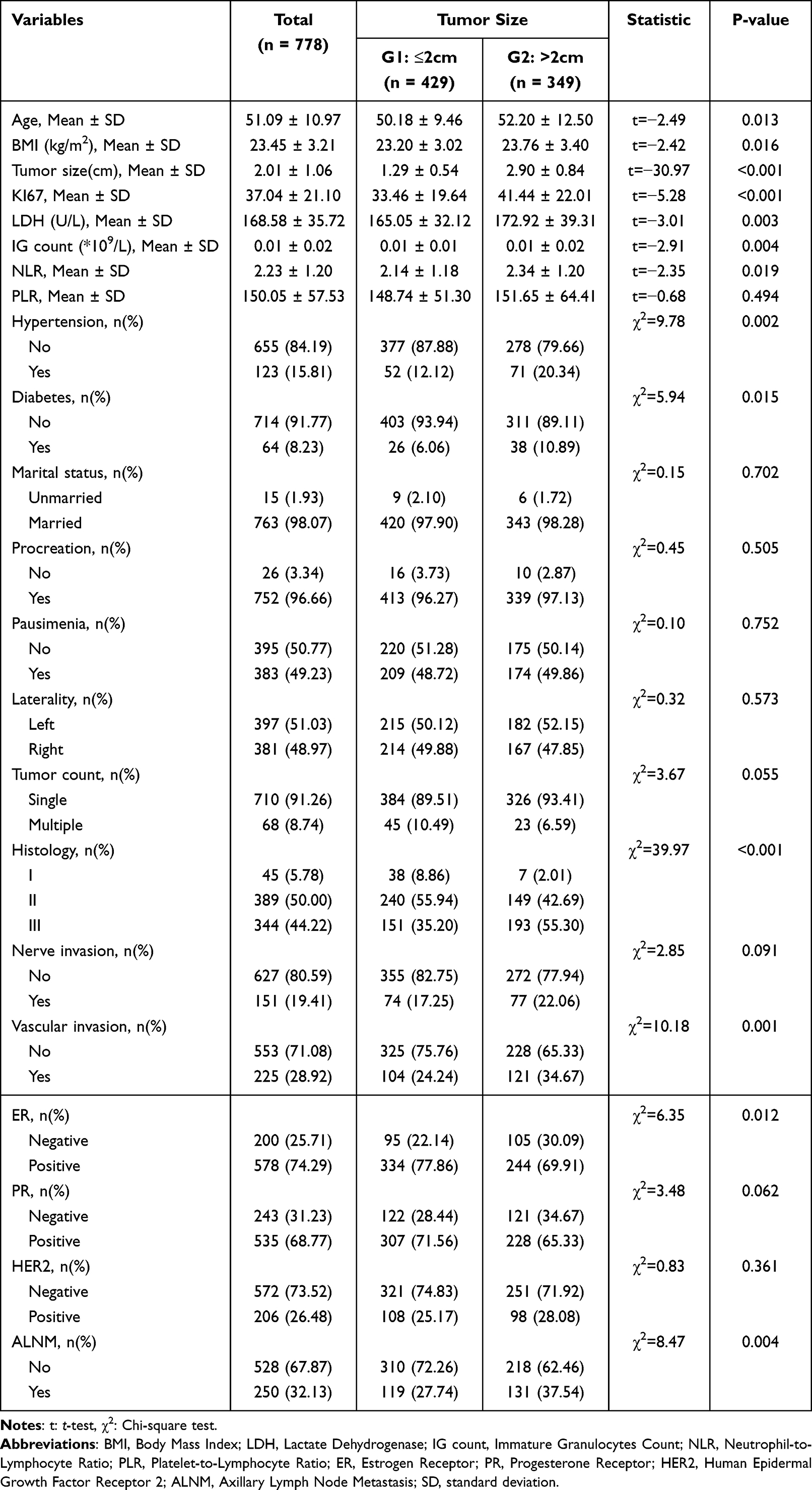 |
Table 1 The Baseline Characteristics of 778 Breast Cancer Participants |
With respect to categorical variables, the G2 group demonstrated significantly higher proportions of vascular invasion, ER-negative status, axillary lymph node metastasis (ALNM) positivity, histological grade III, hypertension, and diabetes (all P < 0.05). In contrast, no significant differences were observed between the two groups in terms of tumor laterality, nerve invasion, PR status, HER2 status, parity, menopausal status, or marital status.
SHAP Feature Importance
In the machine learning–based SHAP interpretability analysis (Figure 2), LDH, IG count, NLR, and PLR were identified as the top predictors of tumor size among all evaluated variables. Notably, LDH ranked second in feature importance, underscoring its pivotal role in predicting tumor size.
 |
Figure 2 SHAP-based interpretation of the machine learning model for tumor size prediction. (A) SHAP beeswarm plot showing the distribution and magnitude of SHAP values for LDH, IG count, NLR, PLR, and other clinical variables, reflecting their individual contributions to tumor size prediction. (B) Feature importance plot ranking variables according to the mean absolute SHAP values, indicating their overall importance in the model. |
Univariate and Multivariate Linear Regression Analysis
The results of univariate and multivariate linear regression analyses indicate that, while NLR and PLR were not significantly associated with tumor size, both LDH and IG count exhibited strong positive correlations with tumor size (Table 2). In the multivariate model, LDH (β = 0.01, 95% CI: 0.01–0.01, P-value = 0.019) and IG count (β = 7.31, 95% CI: 2.89–11.73, P-value = 0.001) remained independent predictors. These findings suggest that increased tumor volume is directly associated with elevated LDH levels and higher immature granulocyte counts. Accordingly, LDH and IG count may serve as potential biological markers, warranting further investigation into their roles in tumor progression.
 |
Table 2 Univariate and Multivariate Linear Regression Analysis for Tumor Size of 778 Breast Cancer Participants |
Weighted Linear Regression (WLR)
Using weighted linear regression (WLR), we examined the associations between tumor size, lactate dehydrogenase (LDH) levels, and immature granulocyte (IG) count in 778 patients with breast cancer (Table 3). IG count, analyzed as a continuous variable, was significantly positively correlated with tumor size, and this association remained robust after multivariable adjustment (β = 6.09, 95% CI: 1.46–10.72, P = 0.01). When the IG count was treated as a categorical variable, tumors in the IG > 0 group were significantly larger than those in the IG = 0 group (β = 0.21, 95% CI: 0.06–0.35, P = 0.005). Similarly, LDH levels were positively associated with tumor size (β = 0.01, P = 0.014), suggesting that higher tumor burden is linked to both inflammatory activation and increased metabolic activity.
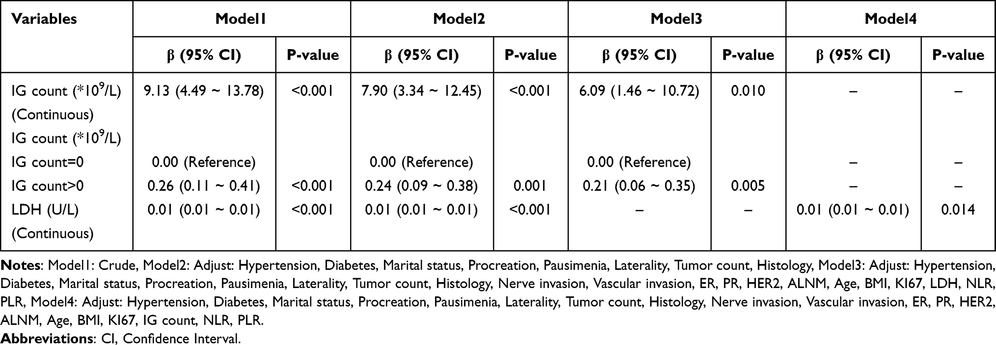 |
Table 3 Association of Tumor Size and IG Count/Lactate Dehydrogenase Among 778 Breast Cancer Participants |
Further analysis revealed a strong positive linear relationship between IG count and LDH levels (Table 4). In continuous variable analysis, LDH levels increased substantially with each unit increase in IG count (β = 241.52, 95% CI: 86.97–396.06, P-value < 0.002). In subgroup analysis, LDH levels were significantly higher in the IG > 0 group compared with the IG = 0 group (β = 6.27, 95% CI: 1.45–11.08, P-value = 0.011).
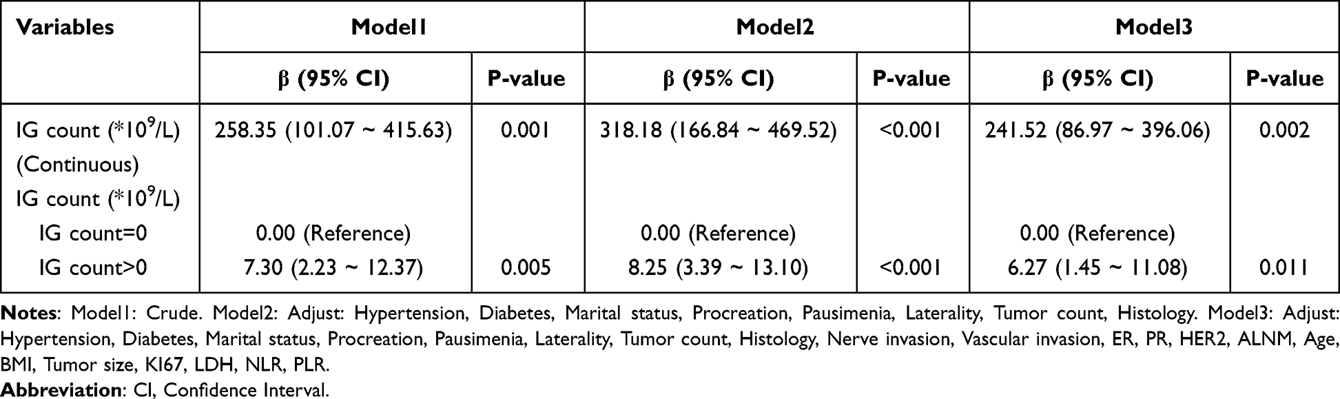 |
Table 4 Association of Immature Granulocytes Count and Lactate Dehydrogenase Among 778 Breast Cancer Participants |
Collectively, these findings suggest a potential synergistic interplay among systemic inflammation, metabolic activity, and tumor growth, as an elevated proportion of immature granulocytes in peripheral blood is associated with both increased tumor size and concomitant rises in LDH levels.
Subgroup Analysis
Subgroup analyses were performed to assess the consistency of the association between IG count and tumor size across different clinical characteristics, including age, hypertension, diabetes, marital status, history of procreation, pausimenia status, BMI, histological type, and ER, PR, and HER2 status. A positive association between IG count and tumor size was observed in most subgroups. However, the relationship was significantly modified by hypertension (P-value for interaction = 0.012) (Table 5).
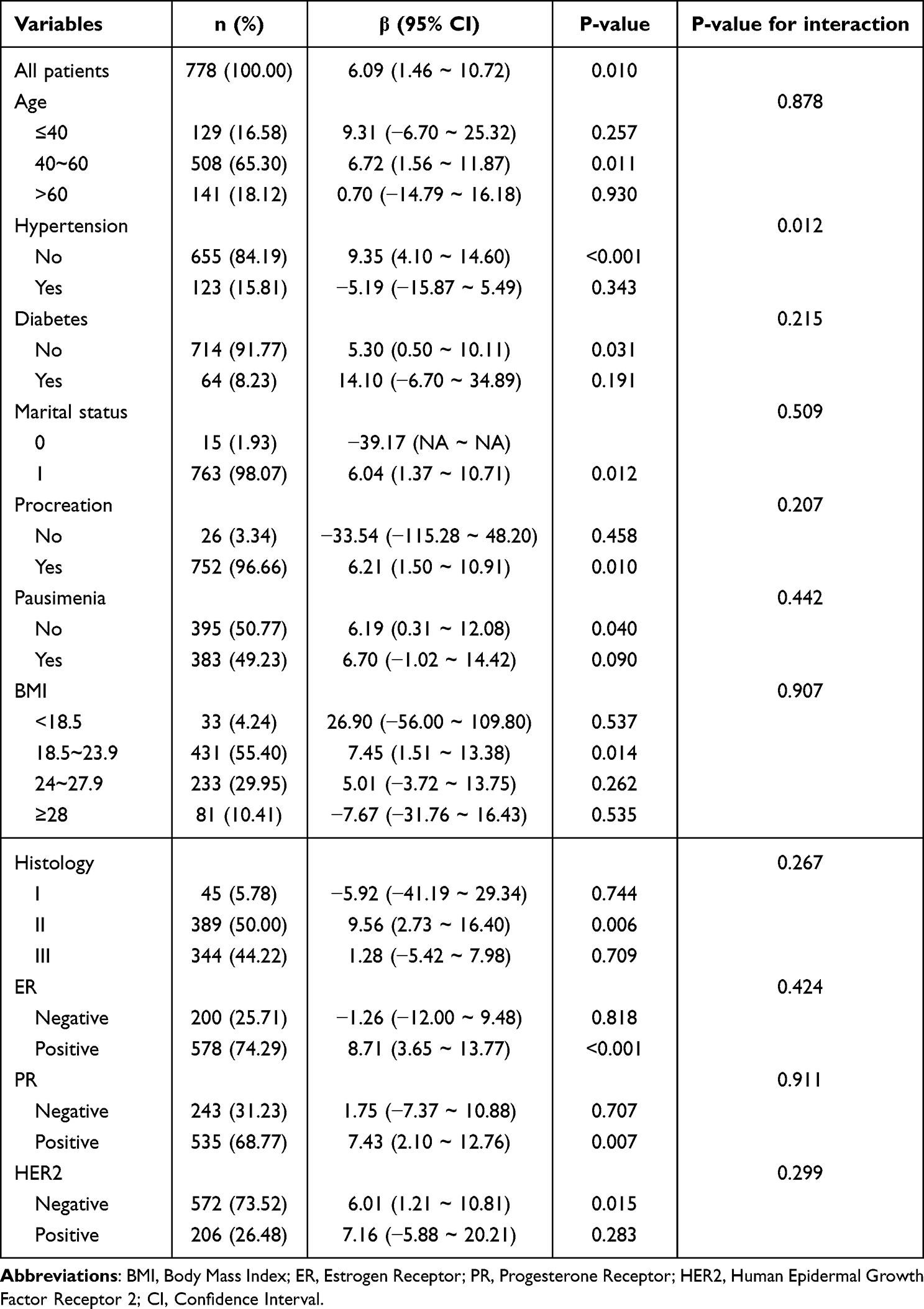 |
Table 5 Subgroup Analysis for the Association Between Immature Granulocytes Count and the Tumor Size in Breast Cancer Population |
Exploring Potential Mechanistic Links
To explore the potential underlying mechanisms linking IG count, LDH, and tumor growth, mediation analysis was performed. As shown in Figure 3, LDH partially mediated the association between IG count and tumor size, and this mediating effect was statistically significant. Specifically, LDH accounted for approximately 9.86% of the total effect, suggesting that it may serve as a biological mediator that partially explains the positive correlation between elevated IG levels and increased tumor size.
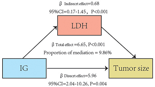 |
Figure 3 LDH as a mediator in the association between IG count and tumor size. |
Discussion
In the present study, we demonstrated that immature granulocyte (IG) count and lactate dehydrogenase (LDH) levels were both positively associated with tumor size in patients with breast cancer. Mediation analysis revealed that LDH partially mediated the relationship between IG and tumor size, highlighting a potential interplay between systemic inflammation and metabolic activity in promoting tumor growth. Furthermore, subgroup analyses suggested that this association may be influenced by the presence of hypertension, indicating that host comorbidities could modulate the impact of inflammatory responses on tumor burden.
Accumulating evidence indicates that chronic inflammation plays a critical role in the onset and progression of various diseases, including cancer and cardiovascular disorders.12–15 As early as the 19th century, researchers proposed a close association between chronic inflammation and carcinogenesis.16 While inflammation represents a natural protective response to tissue injury, its persistence can lead to a chronic state, which substantially increases the risk of cancer and other diseases.17 The inflammatory tumor microenvironment, shaped by interactions among tumor cells, stromal cells, and inflammatory immune cells, provides a permissive milieu that fosters tumor initiation and progression through multiple mechanisms.18,19
Clinically, severe stressors such as sepsis, trauma, or viral infections can induce “emergency granulopoiesis”, a hematopoietic response in which the bone marrow rapidly produces neutrophils to meet acute demands. This process, however, also results in the release of immature neutrophils into the peripheral blood.20–22 Immature granulocytes (IG), as a novel inflammatory biomarker, are easily detectable and have been widely applied in the prognostic evaluation of various diseases.23,24 Recent studies have shown that immature neutrophils are markedly enriched in bone metastasis tissues in various cancers, where they contribute to immunosuppressive processes.25 Similarly, myeloid-derived suppressor cells (MDSCs), a well-recognized immunosuppressive cell population, are increased in the peripheral blood and tumor tissues of cancer patients, correlating with impaired T-cell function, resistance to immunotherapy, and unfavorable prognosis.26 Consistent with these findings, Salma M Saed et al reported that breast cancer patients with tumors >2 cm exhibited significantly higher levels of g-MDSCs compared with those with tumors ≤2 cm, in line with the results of the present study.27
LDH plays a critical role in malignant tumor progression.28 Elevated LDH levels have been consistently associated with poor prognosis in cancer patients and are considered a classical hallmark of the “Warburg effect”, in which tumor cells preferentially utilize glycolysis even under normoxic conditions, producing excessive lactate.29–31 The accumulation of lactic acid acidifies the tumor microenvironment, thereby promoting invasion, metastasis, angiogenesis, and immune evasion.28 Moreover, LDH contributes to malignant progression by inducing epithelial–mesenchymal transition (EMT) and enhancing angiogenesis.32,33 Recently, LDH has attracted increasing attention as both a prognostic biomarker and a potential therapeutic target.34 Inhibiting LDH not only disrupts the Warburg effect and tumor energy metabolism but may also improve responsiveness to chemotherapy, immunotherapy, and radiotherapy, providing a promising avenue for combined cancer treatment strategies.35,36
Our findings indicate that LDH partially mediates the relationship between IG and breast tumor size. Consistently, a study on pancreatitis reported a positive correlation between immature granulocytes and LDH, supporting our observations.37 Inflammation is known to upregulate LDH through multiple mechanisms. For example, inflammatory microenvironments can activate signaling pathways such as HIF-1α, which induces LDHA expression.38,39 Additionally, doxorubicin-induced neutrophil extracellular traps (NETs) have been shown to regulate ferroptosis in cardiomyocytes via the HMGB1/TLR4/YAP axis, causing myocardial injury and subsequent elevation of peripheral LDH levels.40 Inflammatory responses can also damage tumor or surrounding stromal cells, releasing intracellular LDH into the extracellular space and circulation, thereby increasing serum LDH levels. The concurrent elevation of IG and LDH may further acidify the tumor microenvironment, accelerating tumor progression and malignancy.
Our study indicates that the relationship between IG and tumor size is modulated by hypertension. In normotensive patients, IG counts and tumor size show a consistent positive correlation, whereas this association is not statistically significant in hypertensive individuals. Hypertension is characterized by dysregulated reactive oxygen species (ROS) production, a key contributor to oxidative stress, which also plays a pivotal role in inflammation-driven tumorigenesis and progression, potentially altering systemic inflammatory responses.41,42 Moreover, hypertension-associated endothelial dysfunction, vascular injury, and cardiovascular remodeling may further impact tumor development.43 The use of antihypertensive medications may additionally modulate inflammatory status, influencing tumor progression. However, studies specifically evaluating the impact of hypertension on tumor burden in breast cancer patients remain scarce, highlighting an important gap in the current evidence. These observations highlight the need for further studies to elucidate the complex interplay between hypertension, inflammation, and tumor growth.
Our findings carry important biological and clinical implications, highlighting the central roles of systemic inflammation and metabolic alterations in breast cancer progression. Both immature granulocytes and LDH reflect aspects of the host inflammatory response and tumor metabolic activity, and their elevations were consistently associated with larger tumor size. These markers may therefore provide valuable insights into tumor burden and could assist in the early identification of patients with more aggressive disease. In addition, IG and LDH represent biologically meaningful pathways involving neutrophil-driven inflammation and glycolytic reprogramming, which have been increasingly recognized as contributors to tumor growth. These factors may serve not only as accessible biomarkers but also as potential entry points for therapeutic strategies that target inflammation–metabolism interactions in breast cancer.
Nevertheless, several limitations should be acknowledged. Due to the cross-sectional nature of this study, causal inference cannot be established, and the temporal relationship among IG, LDH, and tumor burden cannot be determined. The single-center design may also limit the generalizability of the findings. Furthermore, the distribution of IG counts restricted our ability to investigate more complex or nonlinear associations. Although multiple covariates were adjusted for, the possibility of residual confounding cannot be excluded. To address these limitations and further clarify the biological pathways linking inflammation, LDH elevation, and tumor growth, future research will require multicenter prospective studies as well as mechanistic investigations.
Conclusion
These findings suggest a potential interplay between systemic inflammation and tumor metabolism in breast cancer progression. IG and LDH may serve as accessible biomarkers associated with tumor burden and could assist in risk stratification and clinical decision-making. Multicenter prospective studies are required to validate these associations and further elucidate the underlying biological mechanisms.
Data Sharing Statement
The data are available from the corresponding author upon reasonable request.
Ethical Approval
This retrospective study was approved by the Clinical Research Ethics Committee, The Second Affiliated Hospital of Fujian Medical University (Approval No. 2025-105). All procedures were conducted in strict accordance with the principles of the Declaration of Helsinki. Due to the retrospective nature of the study and the complete removal of all patient-identifying information to ensure anonymity, the Ethics Committee granted a waiver of written informed consent. All data were analyzed in an anonymized manner. All operations in this study complied with relevant guidelines and regulatory requirements.
This study was conducted and reported in accordance with the RECORD (REporting of studies Conducted using Observational Routinely-collected health Data) guidelines.
Author Contributions
All authors have made substantial intellectual contributions to the work and approved it for publication. The specific contributions, following the CRediT taxonomy, are as follows:
Huikai Liang: Conceptualization, Investigation, Formal analysis, Writing – original draft.
Kelun Pan: Methodology, Investigation, Data curation, Writing – original draft.
Xinlan Liang: Formal analysis, Validation, Visualization, Writing – review & editing.
Jiayi Wang: Investigation, Resources, Data curation, Writing – review & editing.
Xinru Xie: Investigation, Resources, Writing – review & editing.
Ningning Wan: Conceptualization, Supervision, Project administration, Writing – review & editing.
Jianqing Lin: Supervision, Funding acquisition, Resources, Writing – review & editing.
All authors gave final approval of the version to be published; have agreed on the journal to which the article has been submitted; and agree to be accountable for all aspects of the work.
Huikai Liang, Kelun Pan, and Xinlan Liang contributed equally to this work and are designated as co-first authors.
Funding
This research received no external funding.
Disclosure
The authors declare no competing interests.
References
1. Amadou A, Torres-Mejia G, Hainaut P, Romieu I. Breast cancer in Latin America: global burden, patterns, and risk factors. Salud Publica Mex. 2014;56(5):547–13. doi:10.21149/spm.v56i5.7379
2. Bray F, Laversanne M, Sung H, et al. Global cancer statistics 2022: GLOBOCAN estimates of incidence and mortality worldwide for 36 cancers in 185 countries. CA Cancer J Clin. 2024;74(3):229–263. doi:10.3322/caac.21834
3. Nishida A, Andoh A. The role of inflammation in cancer: mechanisms of tumor initiation, progression, and metastasis. Cells. 2025;14(7). doi:10.3390/cells14070488
4. Turizo-Smith AD, Córdoba-Hernandez S, Mejía-Guarnizo LV, Monroy-Camacho PS, Rodríguez-García JA. Inflammation and cancer: friend or foe? Front Pharmacol. 2024;15:1385479. doi:10.3389/fphar.2024.1385479
5. Cupp MA, Cariolou M, Tzoulaki I, Aune D, Evangelou E, Berlanga-Taylor AJ. Neutrophil to lymphocyte ratio and cancer prognosis: an umbrella review of systematic reviews and meta-analyses of observational studies. BMC Med. 2020;18(1):360. doi:10.1186/s12916-020-01817-1
6. Rocco A, Sgamato C, Pelizzaro F, et al. Systemic inflammatory response markers improve the discrimination for prognostic model in hepatocellular carcinoma. Hepatol Int. 2025;19(4):915–928. doi:10.1007/s12072-025-10806-6
7. Zhao Z, Zhu X, Xu J, et al. CRP and HNF1A collaborate to regulate the progression of laryngeal cancer through the Wnt signaling pathway. Funct Integr Genomics. 2025;25(1):160. doi:10.1007/s10142-025-01670-6
8. Bozan MB, Yazar FM, Kale IT, Topuz S, Bozan AA, Boran OF. Immature granulocyte count and delta neutrophil index as new predictive factors for axillary metastasis of breast cancer. J Coll Physicians Surg Pak. 2022;32(2):220–225. doi:10.29271/jcpsp.2022.02.220
9. Ding J, Karp JE, Emadi A. Elevated lactate dehydrogenase (LDH) can be a marker of immune suppression in cancer: interplay between hematologic and solid neoplastic clones and their microenvironments. Cancer Biomark. 2017;19(4):353–363. doi:10.3233/cbm-160336
10. Chen J, Huang Z, Chen Y, et al. Lactate and lactylation in cancer. Signal Transduct Target Ther. 2025;10(1):38. doi:10.1038/s41392-024-02082-x
11. Tjokrowidjaja A, Lord SJ, John T, et al. Pre- and on-treatment lactate dehydrogenase as a prognostic and predictive biomarker in advanced non-small cell lung cancer. Cancer. 2022;128(8):1574–1583. doi:10.1002/cncr.34113
12. Mao H, Zhao X, Sun SC. NF-κB in inflammation and cancer. Cell Mol Immunol. 2025;22(8):811–839. doi:10.1038/s41423-025-01310-w
13. Huang Y, Zhou Y, Xu Y, et al. Inflammatory markers link triglyceride-glucose index and obesity indicators with adverse cardiovascular events in patients with hypertension: insights from three cohorts. Cardiovasc Diabetol. 2025;24(1):11. doi:10.1186/s12933-024-02571-x
14. Cifuentes M, Verdejo HE, Castro PF, et al. Low-grade chronic inflammation: a shared mechanism for chronic diseases. Physiology. 2025;40(1). doi:10.1152/physiol.00021.2024
15. Liang H, Pan K, Wang J, Lin J. Association between neutrophil percentage-to-albumin ratio and breast cancer in adult women in the US: findings from the NHANES. Front Nutr. 2025;12:1533636. doi:10.3389/fnut.2025.1533636
16. Balkwill F, Mantovani A. Inflammation and cancer: back to Virchow? Lancet. 2001;357(9255):539–545. doi:10.1016/s0140-6736(00)04046-0
17. Lin WW, Karin M. A cytokine-mediated link between innate immunity, inflammation, and cancer. J Clin Invest. 2007;117(5):1175–1183. doi:10.1172/jci31537
18. Li Q, He G, Yu Y, Li X, Peng X, Yang L. Exosome crosstalk between cancer stem cells and tumor microenvironment: cancer progression and therapeutic strategies. Stem Cell Res Ther. 2024;15(1):449. doi:10.1186/s13287-024-04061-z
19. Coussens LM, Werb Z. Inflammation and cancer. Nature. 2002;420(6917):860–867. doi:10.1038/nature01322
20. Hou L, Voit RA, Shibamura-Fujiogi M, et al. CD11c regulates neutrophil maturation. Blood Adv. 2023;7(7):1312–1325. doi:10.1182/bloodadvances.2022007719
21. Mare TA, Treacher DF, Shankar-Hari M, et al. The diagnostic and prognostic significance of monitoring blood levels of immature neutrophils in patients with systemic inflammation. Crit Care. 2015;19(1):57. doi:10.1186/s13054-015-0778-z
22. Matsuda K, Ide N, Xu Y, Iijima A, Shibuya A. Type 1 innate lymphoid cell-immature neutrophil axis suppresses acute tissue inflammation. Nat Commun. 2025;16(1):6574. doi:10.1038/s41467-025-61504-8
23. Hampson P, Dinsdale RJ, Wearn CM, et al. Neutrophil dysfunction, immature granulocytes, and cell-free dna are early biomarkers of sepsis in burn-injured patients: a prospective observational cohort study. Ann Surg. 2017;265(6):1241–1249. doi:10.1097/sla.0000000000001807
24. Sauneuf B, Bouffard C, Cornet E, et al. Immature/total granulocyte ratio: a promising tool to assess the severity and the outcome of post-cardiac arrest syndrome. Resuscitation. 2014;85(8):1115–1119. doi:10.1016/j.resuscitation.2014.04.017
25. Shi T, Liu W, Luo Y, et al. CHI3L3(+) immature neutrophils inhibit anti-tumor immunity and impede immune checkpoint blockade therapy in bone metastases. Cancer Cell. 2025. doi:10.1016/j.ccell.2025.07.007
26. Lasser SA, Ozbay Kurt FG, Arkhypov I, Utikal J, Umansky V. Myeloid-derived suppressor cells in cancer and cancer therapy. Nat Rev Clin Oncol. 2024;21(2):147–164. doi:10.1038/s41571-023-00846-y
27. Saed SM, Abbas S, El Ansary MS, et al. Phenotypic analysis of circulating myeloid derived suppressor cells and their subpopulations in egyptian females with breast cancer: a single-centre case-control study. Asian Pac J Cancer Prev. 2024;25(1):257–263. doi:10.31557/apjcp.2024.25.1.257
28. Claps G, Faouzi S, Quidville V, et al. The multiple roles of LDH in cancer. Nat Rev Clin Oncol. 2022;19(12):749–762. doi:10.1038/s41571-022-00686-2
29. Dong T, Liu Z, Xuan Q, Wang Z, Ma W, Zhang Q. Tumor LDH-A expression and serum LDH status are two metabolic predictors for triple negative breast cancer brain metastasis. Sci Rep. 2017;7(1):6069. doi:10.1038/s41598-017-06378-7
30. Chen H, Li Y, Li H, et al. NBS1 lactylation is required for efficient DNA repair and chemotherapy resistance. Nature. 2024;631(8021):663–669. doi:10.1038/s41586-024-07620-9
31. Wang Z, Jiang Q, Dong C. Metabolic reprogramming in triple-negative breast cancer. Cancer Biol Med. 2020;17(1):44–59. doi:10.20892/j.issn.2095-3941.2019.0210
32. Khajah MA, Khushaish S, Luqmani Y. Lactate is a major promotor of breast cancer cell aggressiveness. Cancers. 2025;17(11). doi:10.3390/cancers17111793
33. Guo X, Wan P, Shen W, et al. Fusobacterium periodonticum BCT protein targeting glucose metabolism to promote the epithelial-mesenchymal transition of esophageal cancer cells by lactic acid. J Transl Med. 2024;22(1):401. doi:10.1186/s12967-024-05157-z
34. Miholjcic TBS, Halse H, Bonvalet M, et al. Rationale for LDH-targeted cancer immunotherapy. Eur J Cancer. 2023;181:166–178. doi:10.1016/j.ejca.2022.11.032
35. Augoff K, Hryniewicz-Jankowska A, Tabola R. Lactate dehydrogenase 5: an old friend and a new hope in the war on cancer. Cancer Lett. 2015;358(1):1–7. doi:10.1016/j.canlet.2014.12.035
36. Verma S, Budhu S, Serganova I, et al. Pharmacologic LDH inhibition redirects intratumoral glucose uptake and improves antitumor immunity in solid tumor models. J Clin Invest. 2024;134(17). doi:10.1172/jci177606
37. Bedel C, Korkut M, Selvi F. New markers in predicting the severity of acute pancreatitis in the emergency department: immature granulocyte count and percentage. J Postgrad Med. 2021;67(1):7–11. doi:10.4103/jpgm.JPGM_784_20
38. Zhu B, Cheng L, Huang B, Liu R, Ren B. Central role of hypoxia-inducible factor-1α in metabolic reprogramming of cancer cells: a review. Medicine. 2024;103(44):e40273. doi:10.1097/md.0000000000040273
39. Corcoran SE, O’Neill LA. HIF1α and metabolic reprogramming in inflammation. J Clin Invest. 2016;126(10):3699–3707. doi:10.1172/jci84431
40. Zhao P, Li Y, Xu X, et al. Neutrophil extracellular traps mediate cardiomyocyte ferroptosis via the Hippo-Yap pathway to exacerbate doxorubicin-induced cardiotoxicity. Cell Mol Life Sci. 2024;81(1):122. doi:10.1007/s00018-024-05169-4
41. Camargo LL, Rios FJ, Montezano AC, Touyz RM. Reactive oxygen species in hypertension. Nat Rev Cardiol. 2025;22(1):20–37. doi:10.1038/s41569-024-01062-6
42. Chen A, Huang H, Fang S, Hang Q. ROS: a “booster” for chronic inflammation and tumor metastasis. Biochim Biophys Acta Rev Cancer. 2024;1879(6):189175. doi:10.1016/j.bbcan.2024.189175
43. Xu S, Touyz RM. Reactive oxygen species and vascular remodelling in hypertension: still alive. Can J Cardiol. 2006;22(11):947–951. doi:10.1016/s0828-282x(06)70314-2
 © 2026 The Author(s). This work is published and licensed by Dove Medical Press Limited. The
full terms of this license are available at https://www.dovepress.com/terms
and incorporate the Creative Commons Attribution
- Non Commercial (unported, 4.0) License.
By accessing the work you hereby accept the Terms. Non-commercial uses of the work are permitted
without any further permission from Dove Medical Press Limited, provided the work is properly
attributed. For permission for commercial use of this work, please see paragraphs 4.2 and 5 of our Terms.
© 2026 The Author(s). This work is published and licensed by Dove Medical Press Limited. The
full terms of this license are available at https://www.dovepress.com/terms
and incorporate the Creative Commons Attribution
- Non Commercial (unported, 4.0) License.
By accessing the work you hereby accept the Terms. Non-commercial uses of the work are permitted
without any further permission from Dove Medical Press Limited, provided the work is properly
attributed. For permission for commercial use of this work, please see paragraphs 4.2 and 5 of our Terms.
Recommended articles
Prognostic Significance of Preoperative Lactate Dehydrogenase to Albumin Ratio in Breast Cancer: A Retrospective Study
He J, Tong L, Wu P, Wu Y, Shi W, Chen L
International Journal of General Medicine 2023, 16:507-514
Published Date: 8 February 2023
Serum Lactate Dehydrogenase Is a Novel Predictor for the Severity in the Patients With MAFLD: A Cross-Sectional Study in Hefei, China
Yu L, Bao S, Zhu F, Xu Y, Zu F, Liu Y, Jiang R, Chen W, Chen S
Diabetes, Metabolic Syndrome and Obesity 2025, 18:345-361
Published Date: 5 February 2025
Inflammatory and Nutritional Biomarkers Predict Response to Neoadjuvant Dual Anti-HER2 Therapy in HER2-Positive Breast Cancer: A Retrospective Cohort Study
Şahinli H, Uyar GC, Yeşilbaş E
Cancer Management and Research 2026, 18:598948
Published Date: 16 April 2026
Prognostic Factors in Upper Gastrointestinal Neuroendocrine Carcinoma: A Retrospective Cohort Study of Clinicopathological Features and Survival Outcomes
Şeyran E, Gökmen A
Cancer Management and Research 2026, 18:618644
Published Date: 17 July 2026