
Back to Journals » Drug Design, Development and Therapy » Volume 19
Intranasal Dexmedetomidine with Propofol Provides Superior Sedation for Pediatric Contrast-Enhanced CT: A Randomized Controlled Trial
Authors Zhang SJ, Weng YJ, Lei Q, Lin MY, Chen HL, Fang TF, Lu GL 
Received 4 June 2025
Accepted for publication 29 September 2025
Published 24 October 2025 Volume 2025:19 Pages 9523—9532
DOI https://doi.org/10.2147/DDDT.S544737
Checked for plagiarism Yes
Review by Single anonymous peer review
Peer reviewer comments 2
Editor who approved publication: Professor Tamer Ibrahim
Su-Jing Zhang,1– 3,* Yan-Jun Weng,1,* Qian Lei,1 Min-Yi Lin,1 Hua-Lin Chen,1,4,* Tuan-Fang Fang,1,3,* Guo-Lin Lu1,3,*
1Fujian Children’s Hospital (Fujian Branch of Shanghai Children’s Medical Center), College of Clinical Medicine for Obstetrics & Gynecology and Pediatrics, Fujian Medical University, Fuzhou, People’s Republic of China; 2Department of Anesthesiology, Fujian Maternity and Child Health Hospital, College of Clinical Medicine for Obstetrics & Gynecology and Pediatrics, Fujian Medical University, Fuzhou, People’s Republic of China; 3Fujian Key Laboratory of Women and Children’s Critical Diseases Research, Fuzhou, People’s Republic of China; 4Department of Anesthesia, Shanghai Children’s Medical Center, School of Medicine, Shanghai Jiao Tong University, Shanghai, People’s Republic of China
*These authors contributed equally to this work
Correspondence: Guo-Lin Lu, Fujian Children’s Hospital (Fujian Branch of Shanghai Children’s Medical Center), College of Clinical Medicine for Obstetrics & Gynecology and Pediatrics, Fujian Medical University, Fuzhou, People’s Republic of China, Tel +86-18059007715, Email [email protected]
Background: Effective and safe sedation with rapid recovery remains a critical unmet need for pediatric patients undergoing contrast-enhanced computed tomography (contrast-enhanced CT). We compared the efficacy of intranasal dexmedetomidine (DEX) combined with intravenous propofol (D-P) versus DEX with buccal midazolam (D-M) for sedation during pediatric contrast-enhanced CT.
Methods: In this single-center, prospective, randomized controlled trial, 110 children (6 months– 6 years, ASA I/II) were allocated to D-M (2 μg/kg intranasal DEX + 0.2 mg/kg buccal midazolam) or D-P (2 μg/kg intranasal DEX + 1 mg/kg intravenous propofol). Primary outcome was one-time success rate (completed contrast-enhanced CT without additional sedation). Secondary outcomes included onset time, recovery metrics (Ramsay Sedation Scale [RSS] at 30 minutes, time to oral intake), and adverse events. Analyses followed full-analysis-set (FAS) and per-protocol-set (PPS) principles (ChiCTR2300067469).
Results: The D-P group demonstrated superior one-time success rates in both FAS (96.4% vs 74.5%; OR 9.05, 95% CI 1.95– 42.05, P =0.001) and PPS analyses (96.4% vs 77.1%; OR 7.88, 95% CI 1.65– 37.6, P =0.003). Sedation onset was faster with D-P (median 17 vs 20 minutes, P < 0.001), with 98.2% achieving sleep within 20 minutes versus 54.5% for D-M. Recovery was accelerated in D-P: 61.8% attained RSS ≤ 3 by 30 minutes (vs 30.9%, P < 0.001), and 77.3% resumed oral intake within 1 hour (vs 25.4%, P < 0.001). Bradycardia occurred more frequently with D-P (29.1% vs 5.4%, P =0.001), but no interventions were required.
Conclusion: Intranasal dexmedetomidine combined with propofol significantly improves sedation success, accelerates recovery, and reduces procedural delays in pediatric contrast-enhanced CT compared to midazolam, offering a clinically advantageous regimen for short-duration imaging.
Keywords: dexmedetomidine, propofol, midazolam, sedation, pediatrics, computed tomography
Introduction
Contrast-enhanced computed tomography (contrast-enhanced CT) stands as an indispensable imaging modality in the diagnostic arsenal for a wide spectrum of diseases, offering enhanced visualization and improved diagnostic accuracy.1 While the duration of a contrast-enhanced CT examination is brief, pediatric patients may experience injection-associated pain when the contrast medium is administered intravenously via an automated power injector.2 Deep sedation is frequently essential for children to remain still in a contrast-enhanced CT examination, ensuring the acquisition of high-quality diagnostic images. Despite the availability of multiple sedative drug regimens capable of achieving deep sedation in pediatric patients, both onset and recovery times remain prolonged. A fundamental problem is the lack of an optimal sedation technique for children undergoing contrast-enhanced CT, characterized by rapid onset and swift recovery times.
Dexmedetomidine is extensively used for sedation across various medical procedures due to its minimal impact on respiratory function and its relatively short elimination half-life.3 Dexmedetomidine is well tolerated by children and easy to administer. However, even at high doses, patients receiving dexmedetomidine as a single agent often require an additional bolus or supplementary medications to achieve the desired level of sedation.4 Increased or repeated doses of dexmedetomidine can induce deep sedation, but they are also associated with a higher incidence of bradycardia and hypotension. While the duration of a contrast-enhanced CT examination is brief (typically 1–5 minutes of actual scanning time), pediatric patients may experience injection-associated pain when the contrast medium is administered intravenously via an automated power injector. Previous studies have demonstrated that the combination of intranasal dexmedetomidine and midazolam (administered buccally or orally) is both effective and safe for sedation in pediatric patients.5,6 The success rate is notably higher than when sedation is administered with intranasal dexmedetomidine alone7 or buccal midazolam alone.8 However, it exhibits certain drawbacks, including a delayed onset of action (up to 55 minutes) and unpredictable efficacy, accompanied by an extended recovery period9 and the potential for buccal midazolam to induce a paradoxical reaction.10 Hence, it is imperative to investigate an efficacious and safe regimen combining intranasal dexmedetomidine for pediatric sedation during contrast-enhanced CT examinations.
Propofol is widely used for sedation in various procedures resulting from its rapid induction of deep sedation, high efficacy rate, and quick post-sedation recovery.11 The biggest problem of intravenous propofol is the potential to induce hypotension and dose-dependent respiratory inhibition.11 Dexmedetomidine has an effect on preserving the upper respiratory tract protective reflexes.12 Combination with intranasal dexmedetomidine decreases the risk of adverse airway events compared to intravenous propofol alone for pediatric sedation. The combination of intranasal dexmedetomidine and intravenous propofol exhibited a synergistic effect, enhancing safety and efficacy for sedation.13,14 A loading dose of intranasal dexmedetomidine incorporated into the intravenous propofol sedation for magnetic resonance imaging (MRI) significantly reduced the required propofol dosage, the incidence of adverse airway events, and necessary interventions.15 Little is known about a better choice of intranasal dexmedetomidine combined with propofol or midazolam for pediatric sedation in contrast-enhanced CT. In this study, we aimed to investigate the efficacy and safety of dexmedetomidine in combination with a single low dose of intravenous propofol, compared to the combination of intranasal dexmedetomidine and midazolam, for sedation in pediatric patients undergoing contrast-enhanced CT examinations. We hypothesized that the combination of intranasal dexmedetomidine and a single low-dose infusion of propofol is associated with higher one-time success rate than the combination of intranasal dexmedetomidine and buccal midazolam in children undergoing contrast-enhanced CT examinations.
Methods
Ethics and Registration
This study was approved by the Institutional Review Board of Fujian Children’s Hospital (IRB 2022ETKLR10055), and written informed consent was obtained from the legal guardians of all the subjects participating in the trial. The clinical trial reported in the manuscript was registered with the Chinese Clinical Trial Registry (ChiCTR), a WHO-approved public registry, prior to the enrollment of participants. The trial registration number is ChiCTR2300067469, and the full registration details can be accessed at https://www.chictr.org.cn/showproj.html?proj=184391. The trial was registered on January 9, 2023, in compliance with the guidelines set by the ICTRP. All legal guardians have the right to decline participation or request withdrawal from the study at any time, without the need to provide specific reasons.
Study Design
This researcher-initiated, single-center, prospective, randomized, controlled trial was carried out at Fujian Children’s Hospital from February 1, 2023, to August 26, 2023. The randomization sequence was generated by statistical software SPSS to allocate children to either the D-M group or the D-P group. The allocation ratio between two groups was 1:1. The nurse responsible for preparing and administering study drugs, as well as the legal guardians, were not blinded to treatment group allocation owing to the distinct characteristics and administration protocols of the study drugs. However, an independent research nurse involved in observation and data collection remained blinded to group allocation.
Participants
Children with ASA physical status I or II, aged between 6 months and 6 years, who were scheduled for a contrast-enhanced CT examination under sedation at our sedative center, were invited to participate in this study. Contrast-enhanced CT was performed for routine clinical indications such as evaluation of congenital anomalies, neoplastic conditions, trauma, and vascular abnormalities requiring high-resolution imaging under sedation. The exclusion criteria included multiple examinations under sedation, patients with upper respiratory tract infections (URTI), history of diagnosed obstructive sleep apnea, history of allergy or sensitivity to study drugs, history of propofol general anesthesia, history of propofol sedation in the ICU, and history of significant active cardiac, pulmonary, hepatic, or renal disease.
Sedation for Contrast-Enhanced CT Procedures
Fasting instructions were provided to parents prior to scheduled study, and a pre-sedation evaluation was conducted on the day of the study. All children arrived at the induction room with intravenous catheters inserted for the administration of contrast medium in the radiology department. Dexmedetomidine (Sichuan Meidakang Huakang Pharmaceutical Co. LTD, China) stock solution (concentration of 100 μg/mL) was drawn into a 1 mL tuberculin syringe and administered into both nostrils while the child assumed a recumbent position. Oral midazolam solution (Xiao Er’jing®; Yichang Renfu Pharmaceutical Co. LTD, Hubei, China) was dripped in both sides of the buccal mucosa using a tuberculin syringe without dilution (concentration at 20 mg/mL). Propofol (Beijing Fresenius Kabi Pharmaceutical Co., Ltd, China) was intravenously administered over 30 seconds at a concentration of 5 mg/mL diluted with 0.9% saline. In the D-M group, intranasal dexmedetomidine (2 μg/kg) was administered into both nostrils using a tuberculin syringe, while buccal midazolam (0.2 mg/kg) was dripped onto both sides of the buccal mucosa.5 Repeat intranasal dexmedetomidine boluses of 1μg/kg were administered when children did not achieve the target level of sedation 25 minutes later. If sedation failed again, the children were advised to receive 1 mg/kg of propofol. The D-P sedation protocol consisted of intranasal dexmedetomidine at 2 μg/kg followed by intravenous propofol at 1 mg/kg 15 minutes later. Repeat intravenous propofol boluses of 0.5 mg/kg were administered when children did not achieve the target level of sedation 3 minutes after the last dose of propofol.
The baseline heart rate (HR) and peripheral oxygen saturation (SpO2) were recorded before sedation in a waiting area. After induction, HR and SpO2 were continuously monitored using a pulse oximeter until discharge. Hypoxemia (defined as SpO2 < 90% for more than 10 seconds) was treated with supplemental oxygen at a flow rate of 2 L/min through a nasal cannula or face mask. Airway maneuvers, such as jaw thrust or chin lift, were performed if needed. Bradycardia was defined as a decrease of HR > 20% from baseline. Severe bradycardia (defined as a decrease of HR > 30% from baseline) was treated with atropine 0.1 mg/kg. Non-invasive blood pressure (NIBP) was not routinely recorded in this study, which will be included in future studies. We used Ramsay sedation scale (RSS)16,17 (Supplemental Table 1) to assess the depth of sedation. The optimal depth of sedation is unresponsive to verbal stimuli but responds purposefully to tactile or painful stimulation (ie, a Ramsay Sedation Scale score of 4–5). Failed sedation was defined as the inability to complete contrast-enhanced CT procedures due to body movement during the injection of the contrast agent. Upon the completion of contrast-enhanced CT, all participants were transferred to the sedation room for recovery for at least 30 minutes, in accordance with hospital regulations to facilitate early identification of contrast agent allergies. Children were assessed before discharge using the Aldrete score, which is a standardized tool used to assess recovery from anesthesia, based on five criteria: activity, respiration, circulation, consciousness, and oxygen saturation, each scored from 0 to 2; a total score of ≥9 indicates readiness for discharge from the post-anesthesia care unit.18
Primary and Secondary Outcomes
The primary outcome was defined as the one-time success rate, specifically the percentage of children who successfully completed the examination in a single session of sedation, without the need for additional sedative administration. The secondary outcomes included the onset time, examination time, RSS at 30 minutes after the completion of the examination, and the incidence of side effects (oxygen desaturation, bradycardia, and severe bradycardia). Post-discharge side effects in the following 24 hours include the timing to resume intake, the presence of vomiting, agitation, and altered bowel habits.
The onset time was defined as the period between administering premedication and reaching the target level of sedation. Examination time was defined as the period between entering and exiting the CT room. Agitation was defined as follows: (1) crying and not easily calmed, (2) agitated, (3) restless, and (4) excited, combative, or disoriented, or thrashing around.10
Sample Size Calculation
In our pilot study, we observed a one-time success rate of 70.0% in the D-M group and 96.7% in the D-P group (unpublished data). Based on these preliminary findings, we hypothesized that the one-time success rate would be 70% in the D-M group and 95% in the D-P group. Specifically, based on a two-sided alpha of 0.05, power of 90%, and an expected effect size of 92, the required sample size was calculated to be 46 patients per group. To account for an anticipated dropout rate of 20%, we planned to enroll 46×1.2 = 55 patients per group.
Statistical Analysis
Statistical analysis was performed using SPSS version 26.0 (IBM Corp., Armonk, NY). Continuous variables with normal distribution were expressed as mean ± standard deviation, while those with skewed distribution were summarized as median (interquartile range [IQR]). Categorical variables were described as frequencies and percentages. Between-group comparisons were conducted using the independent samples t-test for normally distributed continuous variables, the Mann–Whitney U-test for non-normally distributed continuous variables, and the chi-square test (or Fisher’s exact test, as appropriate) for categorical variables. A two-sided p-value <0.05 was considered statistically significant.
We adhered to the CONSORT guidelines for the reporting of randomized trials and pre-specified three analysis sets:
The intention-to-treat (ITT) population comprised all 110 randomly assigned participants, analyzed according to their originally assigned treatment group, irrespective of protocol deviations or subsequent treatment changes.
The full analysis set (FAS) included all participants who received at least one dose of the study medication and had at least one post-baseline assessment (n=110).
The per-protocol set (PPS) consisted of participants who completed the study without major protocol violations (n=107), excluding three children from the D-M group: two due to regurgitation of midazolam and one due to body movement during contrast injection requiring additional sedation.
For the primary outcome (one-time success rate), both FAS and PPS analyses were performed. Sensitivity analyses were conducted to assess the robustness of the results. Missing data (lost to follow-up, n=1; surgical intervention, n=1) were handled using multiple imputation by chained equations, with 10 imputed datasets created based on relevant baseline and outcome variables. Results from imputed datasets were pooled using Rubin’s rules. Bonferroni correction was applied to control the family-wise error rate. The corrected significance threshold for the four pre-specified secondary outcomes (onset time, examination time, RSS at 30 mins, time to resume intake) was set at α = 0.0125 (0.05/4). All significant secondary outcomes reported in the results remained statistically significant after this adjustment. All analyses were conducted using SPSS, and multiple imputation was performed with the R mice package (version 4.3.1) interfaced via SPSS.
Results
In total, 148 patients aged 6 months to 6 years were recruited between March 2023 and August 2023. Among them, 31 patients did not meet the inclusion criteria and were excluded, while 8 patients refused to participate. Accordingly, 110 patients were randomly divided into two groups. All children completed the examination except for one in the D-M group, who was excluded due to body movement during the injection of the contrast agent (Figure 1). In the D-M group, three children refused a second dose of dexmedetomidine after 25 minutes. Two children in this group did not reach the target sedation level after the second dose and required supplemental propofol at 1 mg/kg to complete the examination. Additionally, two children regurgitated some midazolam, and one of them needed extra sedatives to finish the examination. In the D-P group, one child was lost to follow-up, and another underwent surgical treatment on the same day. Demographic information of patients is presented in Table 1. There were no significant differences in gender, age, and weight between two groups.
 |
Table 1 Demographic Characteristics of Participants |
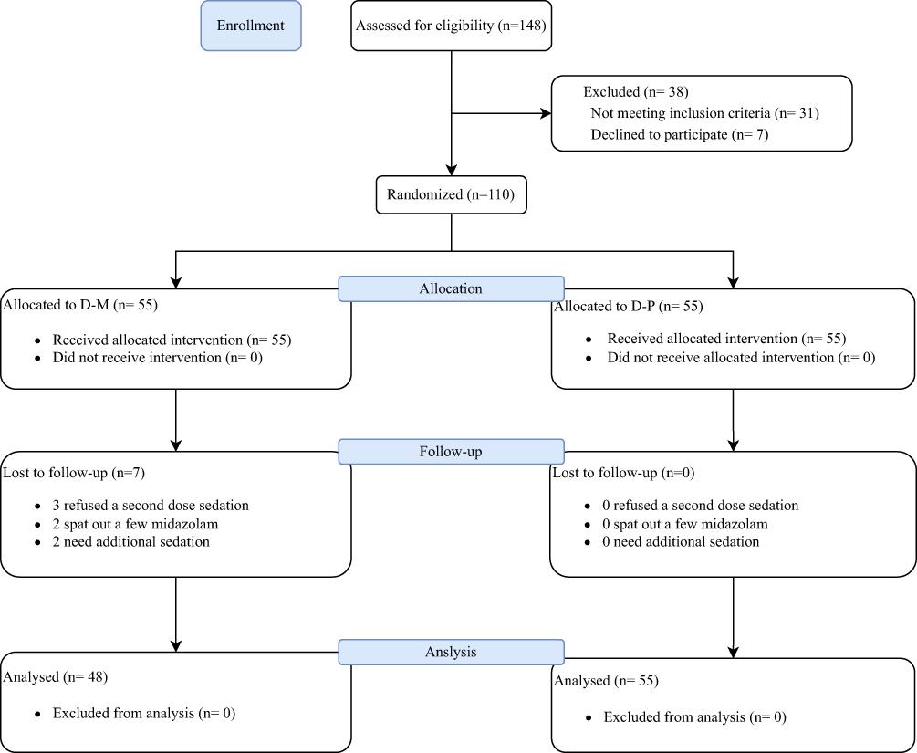 |
Figure 1 Consort flow diagram. |
The one-time success rate in the D-P group is significantly higher than in the D-M group in both the FAS [96.4% vs 74.5%, OR (95% CI): 9.05 (1.95‐42.05), P = 0.001] and PPS populations [96.4% vs 77.1%, OR (95% CI): 7.88 (1.65‐37.6), P = 0.003] (Table 2). The median (IQR) onset time of sedation was 17 (17–18) minutes in the D-P group and 20 (16–28) minutes in the D-M group. In the D-P group, 98.2% of children, compared to 54.5% in the D-M group, achieved sleep within 20 minutes (Figure 2). The OR (95% CI) for achieving sleep within 20 minutes was 45.0 (5.80‐348.86), P < 0.001. Regarding sedation effectiveness, 61.8% of children in the D-P group attained an RSS score ≤ 3 within 30 minutes post-examination, compared to 30.9% in the D-M group. The OR (95% CI) for RSS ≤ 3 was 5.27 (2.46‐11.29), P < 0.0001 (Table 3).
 |
Table 2 The Difference of One-Time Success Rate and Examination Time Between D-M and D-P Groups |
 |
Table 3 Ramsay Sedation Score at 30min After the Completion of Examination |
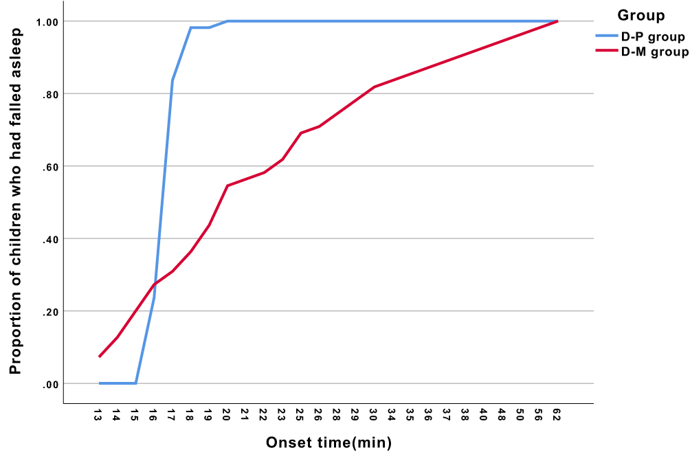 |
Figure 2 Onset time of sedation in D-P group and D-M group. Symbols and Abbreviations: D-P, dexmedetomidine–propofol group; D-M, dexmedetomidine–midazolam group. |
The examination duration was consistent between the two groups. In the D-M group, 3 out of 55 children (5.4%) experienced bradycardia, compared to 16 out of 55 children (29.1%) in the D-P group. The OR was 7.11 (95% CI: 1.94‐26.12), P = s0.001. No children required medical intervention for severe bradycardia, and none experienced respiratory depression or required oxygen therapy. Post-sedation surveys were completed by all patients in the D-M group and 53 out of 55 children in the D-P group. The IQR for time to resume intake was 48 minutes (37–61) in the D-P group and 85 minutes (65–116) in the D-M group. In the D-P group, 41 out of 53 children (77.3%) resumed intake within 1 hour, compared to only 14 out of 55 children (25.4%) in the D-M group. The OR was 10.06 (95% CI: 4.13–24.22), P < 0.001 (Figure 3). We performed a post hoc analysis to evaluate the correlation between total procedural time and the need for rescue medication. Using Spearman’s rank correlation test, we found no significant correlation in either group (P>0.05). After discharge, 8 out of 55 children (14.5%) in the D-M group showed signs of agitation, whereas no children in the D-P group exhibited agitation. Additionally, 2 children in the D-M group experienced gastrointestinal upset with loose stools, which resolved without medical intervention.
 |
Figure 3 Time resumes to intake in D-P group and D-M group. Symbols and Abbreviations: D-P, dexmedetomidine–propofol group; D-M, dexmedetomidine–midazolam group. |
Discussion
In this study, the combination of intranasal dexmedetomidine and a single low dose of propofol significantly increased the one-time success rate compared to the combination of intranasal dexmedetomidine and buccal midazolam. Moreover, the D-P regimen shortened both the onset time and the time to resume oral intake. It also resulted in lower RSS scores at 30 minutes post-examination, indicating more effective sedation. Adverse events were minimal, with eight patients experiencing agitation and two reporting gastrointestinal upset with loose stools. Although 16 children in the D-P group experienced bradycardia, none required pharmacological intervention, and no instances of respiratory depression were observed. These findings suggested that the D-P combination is a more efficient and safer sedative option for pediatric patients undergoing procedures.
The one-time successful sedation rate is 96.4% in the D-P group, which is significantly greater than that in the D-M group in both the FAS (74.5%) and PSS (77.1%). The rate in the D-M group is consistent with the 79.8% reported by our colleagues.5 Dexmedetomidine is absorbed mainly through the rich vascular plexus in the nasal cavity.19 It has a bioavailability of up to 93% with similar pharmacological effects to intravenous administration. The onset time of intranasal dexmedetomidine is 15–30 minutes. Propofol was administered 15 minutes after the dexmedetomidine administration, and the onset time less than 20 minutes is in 98.2% of children. However, the onset time in the D-M group is quite delayed and poorly predictable, with the longest onset time being up to 57 minutes in this study, which is consistent with the study reported by Cossvel et al.9 Previous studies have reported that midazolam can induce a paradoxical reaction in some instances10,20 and its taste is unpleasant, leading to children often being noncompliant with oral intake. Propofol exhibits to be an ideal sedation agent due to its rapid induction of deep sedation, high efficacy rate, and rapid post-sedation recovery.16,21 This study demonstrated that D-P has a rapid onset time and a shortened recovery period. No significant correlation was found between the duration of the CT procedure and the need for additional sedative doses in either the D-P or D-M group. Thus, D-P is superior to D-M for sedation during contrast-enhanced CT.
There is a suitable pharmacological rationale in our research. Dexmedetomidine has a longer half-life compared to propofol. However, its use in combination with a single low-dose propofol bolus was specifically chosen to leverage the synergistic sedative effect while minimizing propofol-related respiratory depression and injection pain. This combination has been shown in previous studies to provide rapid onset and predictable sedation with faster recovery than dexmedetomidine alone or with midazolam. Although CT is a short procedure, the need for contrast injection requires brief but deep sedation, which our regimen effectively provided without prolonged recovery (median recovery time ≤1 hour).
Propofol has a narrow therapeutic window in pediatric patients.22 Although increased or repeated doses of propofol may lead to deep sedation, they also increase the adverse effects of upper airway obstruction, oxygen desaturation, and hypotension. In our study, we did not find respiratory depression, which can be explained to be the low dose of propofol. The dose of propofol used in this study is lower than that of propofol sedation alone (2mg/kg or more),11 resulting from the concurrent use of dexmedetomidine. It was reported that adding dexmedetomidine to propofol can result in decreased propofol requirements and significantly fewer adverse airway events and interventions.11,14 Previous studies have shown that the combination of dexmedetomidine and propofol provided sufficient sedation for children undergoing MRI. In those studies, propofol was continuously infused following the loading dose during the procedural period.15,16 In this study, we found that a single dose of propofol combined with dexmedetomidine can provide an adequate level of sedation for contrast-enhanced CT examination, and also can avoid a prolonged time to recovery. D-P might reduce the workload of medical staff and enhance patient satisfaction.
While this study demonstrates the efficacy of the D-P regimen, several methodological limitations must be acknowledged. First, the inclusion criteria were restricted to children aged 6 months to 6 years, which limits the generalizability of our findings to older pediatric populations or infants younger than 6 months undergoing CECT. Second, the fixed dosing interval and single low dose of propofol, while effective in this protocol, may not represent the optimal dosing strategy; future pharmacodynamic studies could refine the timing and dosage to maximize efficacy and minimize any potential adverse effects. Third, the absence of non-invasive blood pressure (NIBP) monitoring represents a significant limitation, particularly given the observed incidence of bradycardia in the D-P group. Although no hemodynamically significant events required intervention, the lack of systematic BP data means we cannot fully exclude episodes of subclinical hypotension. Fourth, the 25-minute interval for rescue dexmedetomidine administration was based on pilot data, but a longer interval (eg, 30 minutes) might have improved the success rate in the D-M group and should be explored in future comparative trials. Finally, assessment of post-discharge outcomes relied on parental reporting, which introduces the potential for recall bias and variability in interpretation. These limitations underscore the need for broader validation in more diverse pediatric cohorts, with rigorous hemodynamic monitoring and standardized rescue protocols, before widespread clinical adoption can be recommended.
Conclusions
In this randomized controlled trial, the combination of intranasal dexmedetomidine and a single low-dose propofol bolus significantly improved one-time sedation success and accelerated recovery compared to dexmedetomidine–midazolam in young children undergoing CECT. However, these findings should be interpreted in light of several methodological limitations, including the pharmacological profile of dexmedetomidine in short-duration imaging, the use of a fixed, non-standard midazolam dose in the comparator group, and the potential for type I error in secondary outcomes due to multiple testing without formal adjustment. Although the regimen appears efficacious and safe within the study context, we recommend that these results be validated in larger, multicenter trials with broader age inclusion, extended hemodynamic monitoring, and optimized dosing protocols before widespread adoption into routine clinical practice.
Data Sharing Statement
The datasets used and/or analyzed during the current study are available from the corresponding author upon reasonable request.
Ethics Approval and Consent to Participate
This study was approved by the Institutional Review Board of Fujian Children’s Hospital (IRB 2022ETKLR10055), and written informed consent was obtained from the legal guardians of all the subjects participating in the trial. Written informed consent was acquired from the legal guardians of all the subjects. The study adhered to the Good Clinical Practice guidelines, followed the principles outlined in the Declaration of Helsinki, and conformed to the Strengthening the Reporting of the Consolidated Standards of Reporting Trials (CONSORT) guidelines.
Acknowledgment
We sincerely thank all the staff of the anesthesiology department of Fujian Children’s Hospital for helping to conduct this study. We appreciate the Cai-Wen Duan’s team for constructive suggestions.
Author Contributions
All authors made a significant contribution to the work reported, whether that is in the conception, study design, execution, acquisition of data, analysis and interpretation, or in all these areas; took part in drafting, revising or critically reviewing the article; gave final approval of the version to be published; have agreed on the journal to which the article has been submitted; and agree to be accountable for all aspects of the work.
Funding
This study was supported by the Excellence Program of Fujian Children’s Hospital (Grant No. YCXZ202204), the Natural Science Foundation of Fujian Province, China (Grant No. 2023J011321), and Joint Funds for the Innovation of Science and Technology, Fujian Province (Grant No. 2021Y9172).
Disclosure
The authors report no conflicts of interest in this work.
References
1. Ragel M, Nedumaran A, Makowska-Webb J. Prospective comparison of use of contrast-enhanced ultrasound and contrast-enhanced computed tomography in the Bosniak classification of complex renal cysts. Ultrasound. 2016;24(1):6–16. doi:10.1177/1742271X15626959
2. Masui T, Katayama M, Kobayashi S, Sakahara H. Intravenous injection of high and medium concentrations of computed tomography contrast media and related heat sensation, local pain, and adverse reactions. J Comput Assist Tomogr. 2005;29(5):704–708. doi:10.1097/01.rct.0000171238.10678.54
3. Kim JY, Kim KN, Kim DW, Lim HJ, Lee BS. Effects of dexmedetomidine sedation for magnetic resonance imaging in children: a systematic review and meta-analysis. J Anesth. 2021;35(4):525–535. doi:10.1007/s00540-021-02946-4
4. Siddappa R, Riggins J, Kariyanna S, Calkins P, Rotta AT. High‐dose dexmedetomidine sedation for pediatric MRI. Paediatr Anaesth. 2011;21(2):153–158. doi:10.1111/j.1460-9592.2010.03502.x
5. Gu HB, Miao LY, Bai J, Lu GL, Lei Q, Yang LJ, Wang DG. Combined use of intranasal Dexmedetomidine and an oral novel formulation of Midazolam for sedation of young children during brain MRI examination: a prospective, single-center, randomized controlled trial. BMC Anesthesiol. 2022;22(1):357. doi:10.1186/s12871-022-01897-x
6. Salvatore P, Giancarlo B, Vittorio C, AnnaGiulia, DA, Stanislao, P, Antonio, S, Renato, G. Combination of intranasal dexmedetomidine and midazolam for sedation in pediatric magnetic resonance imaging: a retrospective observational study. Pediatr Emerg Care. 2024;40(6):430–433. doi:10.1097/PEC.0000000000003086
7. Li BL, Yuen VM, Zhang N, Zhang HH, Huang JX, Yang SY, Jeffery W, Song XR. A comparison of intranasal dexmedetomidine and dexmedetomidine plus buccal midazolam for non-painful procedural sedation in children with autism. J Autism Dev Disord. 2019;49(9):3798–3806. doi:10.1007/s10803-019-04095-w
8. Nie J, Chen C, Xie J, Ding G. Oral midazolam vs. intranasal dexmedetomidine plus oral midazolam for sedation of pediatric outpatients: a double-blinded randomized controlled trial. BMC Anesthesiol. 2023;23(1):341. doi:10.1186/s12871-023-02289-5
9. Cossovel F, Trombetta A, Ramondo A, Guglielmo R, Luca R, Alessia S, Giorgio C, Egidio B. Intranasal dexmedetomidine and intranasal ketamine association allows shorter induction time for pediatric sedation compared to intranasal dexmedetomidine and oral midazolam. Ital J Pediatr. 2022;48(1):5. doi:10.1186/s13052-021-01196-0
10. Sun Y, Lu Y, Huang Y, Jiang H, Lonnqvist PA. Is dexmedetomidine superior to midazolam as a premedication in children? A meta‐analysis of randomized controlled trials. Paediatr Anaesth. 2014;24(8):863–874. doi:10.1111/pan.12391
11. Olivia NW, Denis S, Mario G, Françoise DP, Giulia C, Jean FF, Philippe VL. Prospective, randomized, double-blind, double-dummy, active-controlled, Phase 3 clinical trial comparing the safety and efficacy of intranasal dexmedetomidine to oral midazolam as premedication for propofol sedation in pediatric patients undergoing magnetic resonance imaging: the MIDEX MRI trial. Trials. 2023;24(1):518. doi:10.1186/s13063-023-07529-0
12. Jung SM. Drug selection for sedation and general anesthesia in children undergoing ambulatory magnetic resonance imaging. Yeungnam Univ J Med. 2020;37(3):159–168. doi:10.12701/yujm.2020.00171
13. Johnson EG, Weaver SG, Batt KL, Weaver RH, Schadler A, Hall SJ. Low‐dose adjuvant dexmedetomidine did not decrease propofol sedation requirements in children undergoing gastrointestinal endoscopy. Pharmacotherapy. 2022;42(10):792–797. doi:10.1002/phar.2729
14. Mason KP, Park RS, Sullivan CA, Lukovits, K, Halpin, EM, Imbrescia, ST, Cavanaugh, D, Prescilla, R, Fox, VL. The synergistic effect of dexmedetomidine on propofol for paediatric deep sedation. Eur J Anaesthesiol. 2021;38(5):541–547. doi:10.1097/eja.0000000000001350
15. Ahmed SS, Unland TL, Slaven JE, Nitu ME. Dexmedetomidine versus propofol: is one better than the other for MRI sedation in children? J Pediatr Intensive Care. 2016;06(02):117–122. doi:10.1055/s-0036-1584683
16. Xu SX, Shan XS, Gao JM, Liu HX, Chen WR, Gao SS, Ji FH, Peng K, Wang Q. Effect of esketamine vs dexmedetomidine adjunct to propofol sedation for pediatric 3Tesla magnetic resonance imaging: a randomized, double-blind, controlled trial. Eur J Med Res. 2022;27(1):258. doi:10.1186/s40001-022-00890-x
17. Malviya S, Voepel-Lewis T, Tait AR, Merkel S, Tremper K, Naughton N. Depth of sedation in children undergoing computed tomography: validity and reliability of the University of Michigan Sedation Scale (UMSS). Br J Anaesth. 2002;88(2):241–245. doi:10.1093/bja/88.2.241
18. Aldrete JA. The post-anesthesia recovery score revisited. J Clin Anesth. 1995;7(1):89–91. doi:10.1016/0952-8180(94)00001-k
19. Wu ZF, He L, Lai Y, Li SJ, Zhang YP. Observation of the sedative effect of dexmedetomidine combined with midazolam nasal drops before a pediatric craniocerebral MRI. J Craniofac Surg. 2020;31(6):1796–1799. doi:10.1097/scs.0000000000006657
20. Li BL, Luo H, Huang JX, Zhang HH, Paquin JR, Yuen VM, Song XR. Using intranasal dexmedetomidine with buccal midazolam for magnetic resonance imaging sedation in children: a single-arm prospective interventional study. Front Pediatr. 2022;10:889369. doi:10.3389/fped.2022.889369
21. Chidambaran V, Costandi A, D’Mello A. Propofol: a review of its role in pediatric anesthesia and sedation. CNS Drugs. 2015;29(7):543–563. doi:10.1007/s40263-015-0259-6
22. Machata AM, Willschke H, Kabon B, Kettner SC, Marhofer P. Propofol-based sedation regimen for infants and children undergoing ambulatory magnetic resonance imaging. Br J Anaesth. 2008;101(2):239–243. doi:10.1093/bja/aen153
 © 2025 The Author(s). This work is published and licensed by Dove Medical Press Limited. The
full terms of this license are available at https://www.dovepress.com/terms
and incorporate the Creative Commons Attribution
- Non Commercial (unported, 4.0) License.
By accessing the work you hereby accept the Terms. Non-commercial uses of the work are permitted
without any further permission from Dove Medical Press Limited, provided the work is properly
attributed. For permission for commercial use of this work, please see paragraphs 4.2 and 5 of our Terms.
© 2025 The Author(s). This work is published and licensed by Dove Medical Press Limited. The
full terms of this license are available at https://www.dovepress.com/terms
and incorporate the Creative Commons Attribution
- Non Commercial (unported, 4.0) License.
By accessing the work you hereby accept the Terms. Non-commercial uses of the work are permitted
without any further permission from Dove Medical Press Limited, provided the work is properly
attributed. For permission for commercial use of this work, please see paragraphs 4.2 and 5 of our Terms.
Recommended articles

Low-Dose Esketamine as an Adjuvant to Propofol Sedation for Same-Visit Bidirectional Endoscopy: Protocol for a Multicenter Randomized Controlled Trial
Song N, Shan XS, Yang Y, Zheng Z, Shi WC, Yang XY, Li Y, Tan AP, Liu H, Peng K, Ji FH
International Journal of General Medicine 2022, 15:4733-4740
Published Date: 6 May 2022
Efficacy and Safety of Ciprofol for Sedation/Anesthesia in Patients Undergoing Hysteroscopy: A Randomized, Parallel-Group, Controlled Trial
Lan H, Shan W, Wu Y, Xu Q, Dong X, Mei P, Duan G, You M, Jin L, Wu J
Drug Design, Development and Therapy 2023, 17:1707-1717
Published Date: 11 June 2023
Successful Use of Low-Dose Combination Propofol and Fentanyl in Cataract Surgery Phacoemulsification
Irawati D, Adli M, Yadi DF
Clinical Ophthalmology 2023, 17:1929-1937
Published Date: 5 July 2023
Investigation of the Protective Effects of Dexmedetomidine, Midazolam, Propofol, and Intralipid on Oxidative Stress and Inflammation in Rats with Lidocaine-Induced Toxicity
Kucun MK, Guler EM, Saracoglu A, Yildirim M, Demirtas C, Serdogan F, Beyaztas H, Aktas S, Kacan M, Gaszynski T, Ratajczyk P, Saracoglu KT
Journal of Inflammation Research 2025, 18:431-443
Published Date: 10 January 2025
Real-World Evidence for Combination Sedation: Propofol Plus Dexmedetomidine and Clinical Outcomes in Mechanically Ventilated ICU Patients
Qi J, Gao W, Ni Q, Liang Z, Wang W, Qiao T, Wang B
Therapeutics and Clinical Risk Management 2026, 22:597422
Published Date: 27 May 2026