
Back to Journals » Journal of Inflammation Research » Volume 18
Hemoglobin-Albumin-Lymphocyte-Platelet Score as an Integrative Biomarker for Prognosis and Sarcopenia in Colorectal Cancer
Authors Xie H, Wei L, Tang S, Gan J 
Received 6 May 2025
Accepted for publication 10 October 2025
Published 24 October 2025 Volume 2025:18 Pages 14709—14720
DOI https://doi.org/10.2147/JIR.S531116
Checked for plagiarism Yes
Review by Single anonymous peer review
Peer reviewer comments 2
Editor who approved publication: Dr Junhao Wang
Hailun Xie,1,2,* Lishuang Wei,3,* Shuangyi Tang,4 Jialiang Gan2,5
1Department of Gastrointestinal and Gland Surgery, the First Affiliated Hospital, Guangxi Medical University, Nanning, Guangxi, People’s Republic of China; 2Guangxi Key Laboratory of Enhanced Recovery After Surgery for Gastrointestinal Cancer, Nanning, Guangxi, People’s Republic of China; 3Department of Geriatric Respiratory Disease Ward, the First Affiliated Hospital, Guangxi Medical University, Nanning, Guangxi, People’s Republic of China; 4Department of Pharmacy, the First Affiliated Hospital, Guangxi Medical University, Nanning, Guangxi, People’s Republic of China; 5Department of Colorectal and Anal Surgery, the First Affiliated Hospital, Guangxi Medical University, Nanning, Guangxi, People’s Republic of China
*These authors contributed equally to this work
Correspondence: Shuangyi Tang, Department of Pharmacy, the First Affiliated Hospital of Guangxi Medical University, 6 Shuangyong Road, Nanning, Guangxi, 530021, People’s Republic of China, Email [email protected] Jialiang Gan, Department of Colorectal and Anal Surgery, the First Affiliated Hospital of Guangxi Medical University, 6 Shuangyong Road, Nanning, Guangxi, 530021, People’s Republic of China, Tel +86-13878155172, Email [email protected]
Objective: This study aimed to comprehensively investigate the dose-response relationship between the Hemoglobin-Albumin-Lymphocyte-Platelet (HALP) score, which reflects the combined inflammatory and nutritional status, and progression-free survival (PFS), overall survival (OS), as well as sarcopenia in patients with colorectal cancer (CRC).
Methods: A retrospective analysis was conducted on 1,441 CRC patients who underwent surgery. HALP score was calculated as Hemoglobin (g/L) × Albumin (g/L) × Lymphocyte count (109/L) / Platelet count (109/L). Survival analysis was carried out using Kaplan - Meier curves and Cox regression analysis. Logistic regression analysis was employed to analyze the relationship between the HALP and sarcopenia. HALP - based nomograms were developed to predict 1 - and 5 - year PFS and OS.
Results: Kaplan - Meier analysis showed that patients with low HALP scores had worse PFS (51.2% vs 63.3%, p < 0.001) and OS (54.1% vs 66.0%, p < 0.001). For every standard deviation increase in HALP scores, the HR for PFS decreased by 13.3% (HR = 0.867, 95% CI = 0.780– 0.963, p = 0.008), and for OS by 15.2% (HR = 0.848, 95% CI = 0.759– 0.948, p = 0.004). Multivariate Cox regression analysis confirmed HALP as an independent prognostic factor for PFS and OS. Logistic regression analysis indicated that low HALP scores were an independent predictor of sarcopenia (OR = 0.550, 95% CI = 0.420– 0.963, p < 0.001). The HALP-based nomograms demonstrated strong predictive performance for prognosis, with C-indexes of 0.726 (PFS) and 0.728 (OS) and 5-year AUCs exceeding 0.76.
Conclusion: Low HALP scores are independent risk factors for PFS, OS, and sarcopenia in CRC patients, highlighting HALP’s potential for clinical application in CRC management and may thus assist in personalized treatment decisions.
Keywords: colorectal cancer, HALP score, progression-free survival, overall survival, sarcopenia
Introduction
Colorectal cancer (CRC) ranks third among the most prevalent malignancies globally, with over 1.92 million new cases and 900,000 deaths reported annually.1 In China, it is the second most commonly diagnosed cancer and the fourth leading cause of cancer - related mortality. Both globally and nationally, CRC consistently ranks among the top five malignancies in terms of incidence and mortality, posing a significant threat to public health and imposing a substantial disease burden.2–4 The increasing incidence of CRC in recent years is largely attributed to lifestyle changes. Although advancements in treatment strategies, such as surgery combined with chemoradiotherapy, have improved outcomes for early - stage patients, the 5 - year survival rate for advanced CRC remains alarmingly low, less than 15%.5–7 The limitations of current clinical staging systems in predicting individualized CRC prognosis, especially in advanced cases, have become more evident. The lack of precise prognostic biomarkers has made treatment decision - making challenging, emphasizing the urgent need for novel, comprehensive biomarkers to refine risk stratification systems and improve patient outcomes.
Emerging evidence suggests that systemic inflammation plays a crucial role in cancer initiation and progression.8–10 Numerous studies have identified various hematological inflammatory markers as significant prognostic factors in CRC, including the neutrophil - to - lymphocyte ratio (NLR),11 platelet-to-lymphocyte ratio (PLR),12 and systemic inflammation response index (SIRI).13 Meanwhile, the nutritional status of cancer patients has been shown to significantly impact survival outcomes and quality of life. Malnutrition not only reduces the efficacy of therapeutic interventions but may also accelerate disease progression. Several nutrition - related indices, such as the prognostic nutritional index (PNI) and geriatric nutritional risk index (GNRI), have been validated as effective tools for predicting adverse outcomes in cancer patients.14–17
Recently, the HALP score has gained attention as a novel composite biomarker. It integrates four key parameters - hemoglobin, albumin, lymphocyte count, and platelet count - providing a comprehensive assessment of the interaction between the tumor and the host. The HALP score offers advantages like low-cost testing, ease of operation, and convenient data accessibility, reflecting both systemic inflammatory status and nutritional conditions. This fits the precision medicine-driven trend of prognostic models. While TNM staging combined with molecular markers (eg, MSI-H/dMMR) improves stratification, it suffers from high costs and complex techniques, limiting use in resource-constrained areas. As a routine hematological index, HALP avoids these issues, making it highly promotable there. Notably, HALP also holds distinct advantages over other clinically relevant biomarkers such as the Systemic Inflammation Response Index (SIRI) and Albumin-Bilirubin (ALBI) score. As previously documented, SIRI primarily serves as an inflammatory-specific marker, calculated based on neutrophil, monocyte, and lymphocyte counts to reflect systemic inflammatory activation, but it lacks integration of nutritional status—an oversight given that malnutrition frequently coexists with inflammation in CRC patients and independently impacts prognosis.18–20 In contrast, the ALBI score focuses narrowly on liver function, with its calculation relying solely on serum albumin and bilirubin levels, and thus fails to capture the inflammatory derangements (eg, lymphocyte depletion, platelet activation) that are central to tumor progression and muscle catabolism.21,22 By uniquely integrating both inflammatory indicators (lymphocytes, platelets) and nutritional markers (hemoglobin, albumin), HALP addresses the limitations of these single-domain biomarkers: it simultaneously quantifies the pro-inflammatory cascade that drives tumor invasion/metastasis and the nutritional reserve that determines treatment tolerance (eg, surgery, chemotherapy) and tissue repair capacity. This multi-dimensional assessment of the host-tumor interaction underscores HALP’s superior prognostic utility compared to SIRI (inflammation-only) and ALBI (liver function-only) in CRC management.
Initial studies have demonstrated its prognostic value in various solid tumors, such as gastric and lung cancers.23–25 However, its role in predicting survival outcomes in CRC patients undergoing surgery remains under - explored. Additionally, sarcopenia, characterized by the loss of skeletal muscle mass, has emerged as an important prognostic factor significantly affecting the survival and quality of life of CRC patients.26–28 Early identification and management of sarcopenia may enhance patient tolerance to surgery and anticancer therapies. Notably, sarcopenia in cancer patients is closely intertwined with systemic inflammatory responses and nutritional derangements—a pathological axis that the HALP score inherently reflects. Specifically, a low HALP score typically indicates concurrent chronic inflammatory status (evidenced by lymphopenia, which impairs immune regulation, and platelet activation, a driver of pro-inflammatory signaling) and protein-energy malnutrition (manifested by decreased hemoglobin and albumin, key markers of iron stores and systemic protein synthesis, respectively). These two processes synergistically promote muscle catabolism: chronic inflammation upregulates pro-inflammatory cytokines that activate the ubiquitin-proteasome system and autophagic pathways in skeletal muscle, while protein-energy malnutrition reduces muscle anabolism by limiting amino acid substrates and impairing insulin-like growth factor-1 (IGF-1) signaling.29,30 Despite this mechanistic plausibility, the direct relationship between the HALP score and sarcopenia in CRC patients has yet to be investigated.
Against this backdrop, our study aimed to systematically explore the dose - response relationship between the HALP score and PFS, OS, and sarcopenia in CRC patients. We also developed a dynamic nomogram incorporating the HALP score, TNM staging, and molecular markers to provide individualized prognostic predictions for CRC patients. This research aimed to fill critical gaps in CRC prognostication and contribute to the development of more precise and personalized treatment strategies.
Materials and Methods
Study Population
This retrospective study analyzed patients with CRC who underwent surgical treatment at the First Affiliated Hospital of Guangxi Medical University from 2015 to 2017. The inclusion criteria for patients were as follows: (1) histologically or cytologically confirmed CRC; (2) patients undergoing primary surgical resection; (3) availability of complete clinicopathological data; and (4) age ≥ 18 years at the time of diagnosis. Patients were excluded if they met any of the following criteria: (1) concurrent malignancies in other sites; (2) receipt of preoperative anticancer therapy (eg, chemotherapy or radiotherapy); (3) metastatic or recurrent CRC.
Data Collection
Preoperative clinical data were collected from electronic medical records, covering patient - related factors, tumor - related factors, treatment - related information, and laboratory parameters. Follow - up data were obtained through telephone interviews and electronic medical records, with the last follow - up conducted in January 2023.
Body mass index (BMI) was calculated as weight (kg) divided by the square of height (m²). Carcinoembryonic antigen (CEA) levels were categorized as normal (<5.00 ng/mL) or elevated (≥5.00 ng/mL). The HALP score was calculated using the formula: HALP = Hemoglobin (g/L) × Albumin (g/L) × Lymphocyte (109/L) / Platelet (109/L). The PNI was computed as: serum albumin (g/L) + 5 × total lymphocyte (109/L). The NLR was determined as neutrophil (109/L) / lymphocyte (109/L), and the PLR as platelet (109/L) / lymphocyte (109/L).
Outcomes
The primary outcomes of this study were PFS and OS. PFS was defined as the time from the date of surgery to the first occurrence of local or distant disease recurrence or death. OS was defined as the time from the date of surgery to the date of death from any cause or the last follow - up. The secondary outcome was sarcopenia, which was diagnosed based on the 2019 Asian Working Group for Sarcopenia (AWGS) consensus criteria: skeletal muscle mass index (SMI) < 6.92 kg/m² for males and < 5.13 kg/m² for females. According to previous literature,27 the SMI derived from the anthropometric equation was calculated as follows: [0.193 × weight (kg) + 0.107 × height (cm) - 4.157 × sex (1 = male, 2 = female) - 0.037 × age (year) - 2.631] / height squared (m²).
Statistical Analysis
Continuous variables were assessed for normality using the Kolmogorov–Smirnov test. Normally distributed variables were expressed as mean ± standard deviation (SD), while non - normally distributed variables were presented as median (interquartile range, IQR). Categorical variables were expressed as frequencies and percentages. The optimal cutoff value for the HALP score, derived from the Youden index, was determined using receiver operating characteristic (ROC) curve analysis. Time - dependent ROC curves were used to compare the predictive performance of different prognostic markers. Restricted cubic splines (RCS) were used to evaluate the non - linear dose - response relationship between the HALP score and outcomes in CRC patients. Survival curves were generated using the Kaplan - Meier method, and differences between groups were assessed using the log - rank test. Univariate and multivariate Cox proportional hazards regression analyses were performed to identify independent prognostic factors. Logistic regression analysis was used to explore the independent association between the HALP score and sarcopenia. To reduce baseline confounding between high- and low-HALP groups, propensity score matching (PSM) was performed. Variables with a p - value < 0.05 in univariate analysis were included in the multivariate analysis. Variables with a p - value ≤ 0.05 in multivariate analysis were incorporated into the construction of nomograms for predicting 1 - and 5 - year PFS and OS. The discriminative ability of the nomograms was evaluated using the concordance index (C - index), and goodness of fit was assessed using the coefficient of determination (R²). Calibration curves were plotted to compare predicted probabilities of PFS and OS with actual observed outcomes. Decision curve analysis (DCA) was used to compare the prognostic ability of nomograms with that of traditional TNM stages. Internal validation of the model was performed using 1000 bootstrap resamples. Additionally, the total cohort was randomly divided into training (70%) and validation (30%) subsets for internal validation of the nomogram. All statistical tests were two - sided, and a p - value < 0.05 was considered statistically significant. Statistical analyses were performed using R version 4.0.2 (http://www.R-project.org).
Results
Baseline Clinical Characteristics
A total of 1,441 CRC patients with stage I–IV who underwent surgical treatment were included in this study. Among them, 904 (62.7%) were male, and 537 (37.3%) were female. The mean age of the cohort was 58.12 ± 13.15 years, with a median follow - up duration of 61.46 months. During follow - up, 400 patients (27.8%) experienced recurrence, and 582 (40.4%) died from any cause. Among the study population, 762 patients (53.0%) were diagnosed with stage I–II disease, while 677 (47.0%) had stage III–IV disease. Sarcopenia was identified in 271 patients (18.8%) based on the AWGS criteria. The HALP scores ranged from 0.27 to 106.8, with a mean of 61.46 ± 25.97 and a median of 65.23. The optimal cutoff value for the HALP score was 32.4 (Figure S1). Based on this cutoff, 668 patients (46.4%) were classified into the high - HALP group, and 773 (53.6%) were categorized into the low - HALP group. Notably, low HALP scores were significantly associated with advanced age, lower BMI, advanced T stage, metastasis, larger tumor diameter, and elevated CEA levels. Compared to the high - HALP group, the low - HALP group had a higher overall mortality rate and recurrence risk. Additionally, patients in the low - HALP group had a 1 - day longer hospital stay and higher hospitalization costs (RMB 2,143.77) than those in the high - HALP group (Table S1). To further explore the distribution of HALP scores across clinicopathological characteristics, we analyzed the median HALP scores. Lower HALP scores were observed in female patients, those aged ≥60 years, individuals with low BMI, advanced - stage disease, sarcopenia, and those who died during follow - up (Figure S2).
Comparison of HALP with Other Prognostic Markers
For 3 - year PFS and OS, HALP did not show a significant advantage over other markers (Figure S3A and B). However, for 5 - year PFS, HALP outperformed NLR, PLR, and PNI (AUC: 0.556 vs 0.546 vs 0.541 vs 0.561) (Figure S3C). Similarly, for 5 - year OS, HALP also had a superior AUC compared to NLR, PLR, and PNI (0.561 vs 0.553 vs 0.546 vs 0.562) (Figure S3D).
Kaplan-Meier Analysis of HALP Scores
Kaplan - Meier survival analysis revealed significant associations between HALP scores and patient outcomes. Patients with low HALP scores had worse PFS compared to those with high HALP scores (51.2% vs 63.3%, p < 0.001; Figure 1A). Similarly, low HALP scores were associated with poorer OS (54.1% vs 66.0%, p < 0.001; Figure 1B). Subgroup analysis, stratified by TNM stage, showed that for both stage I–II and stage III–IV patients, low HALP scores were associated with worse PFS (Figure 2A–C), with a more pronounced difference in advanced - stage patients. High HALP scores conferred a significant survival advantage in both early and advanced stages for OS (Figure 2B–D). In the colon and rectal cancer subgroups, low HALP scores were consistently associated with inferior PFS and OS (Figure S4). Additionally, HALP scores effectively stratified PFS and OS in both normal and elevated CEA subgroups (Figure S5). To further precisely assess the association between HALP scores and survival outcomes, we performed PSM to eliminate intergroup confounding bias (Table S2). After adjusting for confounding factors via PSM, Kaplan-Meier survival curve analysis was re-conducted. The results revealed that patients with high HALP exhibited flatter survival curves and slower PFS progression (63.3% vs 51.9%, p<0.001; Figure S6A). Similarly, high HALP was associated with improved OS (66.0% vs 54.9%, p<0.001; Figure S6B).
 |
Figure 1 Kaplan-Meier curve of HALP scores in CRC patients. Notes: (A) PFS; (B) OS. |
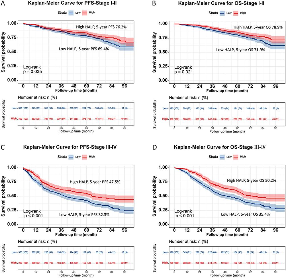 |
Figure 2 Stratified Kaplan-Meier curve of HALP scores based on TNM stage subgroup in CRC patients. Notes: (A) PFS (Stage I–II); (B) OS (Stage I–II); (C) PFS (Stage III–IV); (D) OS (Stage III–IV). |
Low Preoperative HALP Scores as an Independent Prognostic Factor
RCS analysis demonstrated that higher HALP scores were associated with a gradual decline in hazard ratios (HRs) for PFS, regardless of calibration models (Figure 3A). For every SD increase in HALP scores, the risk of PFS decreased by 13.3% (HR = 0.867, 95% CI = 0.780–0.963, p = 0.008). Patients in the low - HALP group had a 22.4% higher risk of adverse PFS compared to the high - HALP group (HR = 0.776, 95% CI = 0.652–0.924, p = 0.004). Univariate and multivariate Cox regression analyses identified preoperative HALP scores as an independent prognostic factor for PFS. When HALP scores were analyzed by quartiles, patients in Q2, Q3, and Q4 had progressively lower risks of adverse PFS compared to Q1 (Table 1). Similarly, RCS analysis showed that higher HALP scores were associated with a 15.2% reduction in adverse OS for every SD increase (HR = 0.848, 95% CI = 0.759–0.948, p = 0.004) (Figure 3B). Patients in the low - HALP group had a 22.4% increased risk of adverse OS compared to the high - HALP group (HR = 0.776, 95% CI: 0.648–0.929, p = 0.006). Progressive increases in HALP scores were associated with declining HRs for OS (Table 2). Subgroup analysis using forest plots confirmed that low HALP scores were an independent risk factor for both PFS and OS in most subgroups (Figure S6A and B).
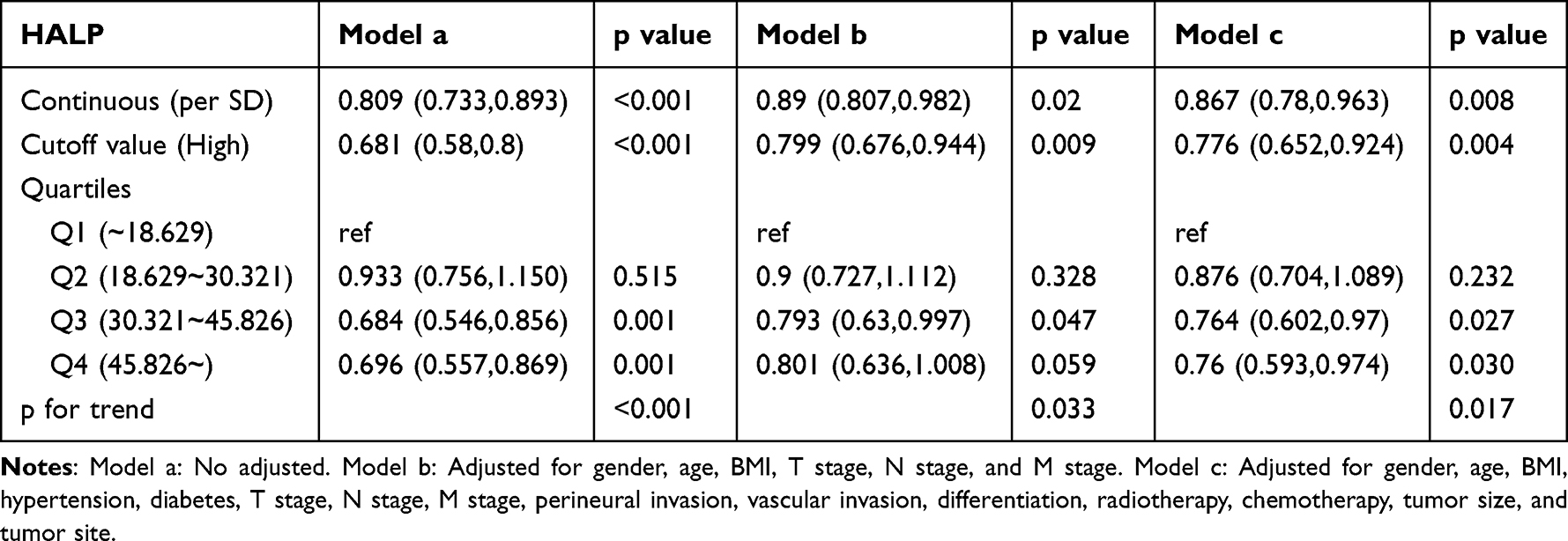 |
Table 1 Association between HALP and PFS of CRC patients |
 |
Table 2 Association between HALP and OS of CRC patients |
 |
Figure 3 The association between HALP scores and survival in CRC patients. Notes: (A) PFS; (B) OS. Model 1: No adjusted. Model 2: Adjusted for gender, age, and BMI. Model 3: Adjusted for gender, age, BMI, hypertension, diabetes, T stage, N stage, M stage, tumor size, perineural invasion, vascular invasion, macroscopic type, differentiation, radiotherapy, chemotherapy. |
Preoperative HALP Scores as an Independent Predictor of Sarcopenia
The incidence of sarcopenia was significantly higher in the low - HALP group than in the high - HALP group (23.8% vs 13.0%). Multivariate logistic regression analysis revealed that for every SD increase in HALP scores, the risk of sarcopenia decreased by 45% (OR = 0.550, 95% CI = 0.420–0.963, p < 0.001). Preoperative low HALP scores were identified as an independent predictor of sarcopenia (OR = 0.462, 95% CI = 0.305–0.699, p < 0.001). In quartile analysis, the risk of sarcopenia progressively decreased in Q2, Q3, and Q4 compared to Q1 (Table 3).
 |
Table 3 Association between HALP and sarcopenia of CRC patients |
Development of HALP-Based Prediction Models
Multivariate Cox regression identified six independent prognostic factors for PFS, including age, T stage, N stage, M stage, CEA level, and HALP scores (Table S3). These variables were used to construct a nomogram for predicting PFS (Figure S7). For OS, eight independent prognostic factors were identified: age, T stage, N stage, M stage, vascular invasion, CEA level, tumor differentiation, and HALP scores (Table S4). The corresponding nomogram for OS is shown in Figure S8. The 1 -, 3 -, and 5 - year AUC values for the PFS and OS nomograms were (0.803, 0.772, 0.761), and (0.766, 0.775, 0.765), respectively (Figure S9A and B). The C - indices for PFS and OS were 0.726 (95% CI = 0.705–0.747) and 0.728 (95% CI = 0.707–0.749), respectively. Calibration curves demonstrated excellent agreement between predicted and observed probabilities for 1-, 3-, and 5-year PFS and OS (Figure S10A and B). DCA revealed that the HALP - based nomograms provided superior clinical benefit compared to traditional tumor staging for both PFS and OS (Figure S11A and B). Patients stratified into high and low nomogram score groups exhibited significantly different PFS and OS outcomes (Figure S12A and B). To facilitate personalized prognostication for CRC patients, we deployed the PFS and OS nomogram models on a Shiny server, developing dedicated webpages for predicting PFS and OS in CRC patients. These webpages enable more convenient and individualized prognostic prediction for patients, with the URLs available at https://hailun.shinyapps.io/HALP_PFS/ and https://hailun.shinyapps.io/HALP-/respectively.
Validation of HALP-Based Prediction Models
Internal validation was performed by randomly splitting the cohort into validation cohort A (n = 721) and validation cohort B (n = 720). No significant differences in baseline characteristics were observed between the two cohorts (Table S5). In validation cohort A, HALP effectively stratified PFS and OS (Figure 4). Similarly, in validation cohort B, low HALP scores were associated with worse 5 - year PFS and OS (Figure 5). Both validation cohorts demonstrated excellent accuracy by ROC analysis (Figure S13), and calibration curves showed strong agreement between predicted and observed outcomes (Figure S14). DCA curves confirmed the superior clinical utility of HALP - based nomograms over traditional tumor staging in both validation cohorts (Figure S15). Patients with high nomogram scores consistently exhibited worse PFS and OS compared to those with low scores in both validation cohorts (Figure S16).
 |
Figure 4 The validation cohort A of Kaplan-Meier curve of HALP scores. Notes: (A) PFS; (B) OS. |
 |
Figure 5 The validation cohort B of Kaplan-Meier curve of HALP scores. Notes: (A) PFS; (B) OS. |
Discussion
This study systematically investigated the relationship between the HALP (hemoglobin, albumin, lymphocyte, and platelet) score and the prognosis of CRC patients undergoing surgical treatment. Moreover, we developed a HALP - based nomogram for individualized prognostic prediction. Our findings demonstrate that HALP is closely associated with PFS and OS in CRC patients. Specifically, low HALP scores were identified as an independent risk factor for adverse outcomes, a conclusion validated through multivariate and subgroup analyses. These results highlight the significant prognostic value of HALP in CRC management. Notably, HALP outperformed traditional inflammatory and nutritional markers, such as NLR, PLR, and PNI, in predicting 5 - year PFS and OS. This advantage likely stems from HALP’s integration of hemoglobin, albumin, lymphocytes, and platelets, which together offer a more comprehensive reflection of systemic inflammation and nutritional status than individual parameters. The biological variability of HALP scores may also be shaped by individual genetic differences, which could influence baseline immune and nutritional status independently of tumor characteristics.Tumor development and progression are intricately linked to inflammation and nutritional status. Inflammation promotes tumor cell proliferation, invasion, and metastasis, while malnutrition compromises immune function and facilitates tumor progression.31–34 By simultaneously addressing these two critical factors, HALP provides a more accurate prognostic assessment than single - parameter indicators.
HALP also showed robust prognostic stratification across various subgroups. Patients with low HALP scores had worse outcomes regardless of the TNM stage, and this difference was more pronounced in advanced - stage disease. This is particularly relevant for the clinical management of advanced CRC patients, where treatment decisions are often complex. HALP can assist clinicians in more accurately assessing prognosis, thereby guiding tailored therapeutic strategies. Similarly, HALP effectively stratified PFS and OS in subgroups based on tumor location (colon or rectum) and CEA levels, underscoring its broad applicability across diverse clinical profiles.
While our study establishes a robust independent association between low HALP scores and sarcopenia in CRC patients, the mechanistic link between HALP and sarcopenia, thus far framed around “inflammatory-nutritional interaction”, remains largely theoretical and lacks direct correlation data between HALP components and specific pro-inflammatory cytokines (eg, IL-6, TNF-α) in our cohort—representing a key limitation in mechanistic depth. Notably, existing literature provides plausible pathways to contextualize this association, which we can leverage to refine the mechanistic narrative. Lymphocyte count, a core component of HALP, is inversely correlated with circulating IL-6 and TNF-α levels in cancer patients; these pro-inflammatory cytokines suppress lymphocyte proliferation and induce apoptosis, leading to lymphopenia—a hallmark of low HALP.35,36 Concurrently, IL-6 and TNF-α directly activate skeletal muscle catabolism by upregulating atrophy-related E3 ligases via the NF-κB signaling pathway, while also impairing albumin synthesis in the liver and reducing iron availability for hemoglobin production—collectively driving the nutritional derangements reflected in low HALP.37,38 Platelet count, another HALP parameter, is elevated in response to TNF-α-mediated platelet activation, and activated platelets further amplify inflammatory cascades by releasing platelet-derived growth factor, creating a feedforward loop that exacerbates muscle breakdown.39 Thus, HALP may indirectly quantify the intensity of IL-6/TNF-α-driven inflammatory stress, which synergizes with nutritional deficits to promote sarcopenia.
The HALP - based nomogram developed in this study integrates multiple key prognostic factors and demonstrates excellent accuracy in predicting 1-, 3-, and 5 - year PFS and OS. The high C - index and calibration curves indicate strong agreement between predicted and observed outcomes, with clinical utility surpassing traditional tumor staging. This nomogram provides clinicians with an intuitive and quantifiable tool for precision prognostication, facilitating individualized treatment decisions. For example, patients with a predicted poor prognosis may benefit from intensified adjuvant therapy or exploration of novel treatments, while those with a favorable prognosis could avoid overtreatment, thereby reducing unnecessary morbidity and healthcare costs.
However, this study has several limitations. First, its retrospective design, despite controlling for confounders, may still be subject to selection bias and incomplete or inaccurate data, potentially limiting the generalizability of the findings. Second, the single-center nature of the study, combined with a relatively modest sample size, may not fully capture regional or institutional variations in CRC patient characteristics. Notably, while our internal validation efforts provide preliminary support for the model’s reliability—we randomly split the total cohort into two validation subsets (A and B), and both cohorts demonstrated excellent predictive accuracy by ROC analysis, with calibration curves showing strong agreement between predicted and observed PFS/OS outcomes and DCA confirming superior clinical utility over traditional TNM staging—such internal validation remains confined to a single institutional population. Thus, multicenter, large-scale prospective studies are still urgently needed to perform external validation of the prognostic value of HALP and the accuracy of the nomogram, ensuring its applicability across diverse clinical settings and patient cohorts. Additionally, HALP components (hemoglobin, albumin, lymphocytes, platelets) are susceptible to interference from non-cancer conditions (eg, chronic inflammation, anemia, liver dysfunction, infections)—representing a potential source of confounding. A critical additional limitation is the lack of data on key molecular therapeutic biomarkers in CRC, including KRAS, BRAF mutations, and MSI status. These markers are central to clinical treatment stratification. Although we included CEA in our nomogram, we were unable to explore how HALP correlates with these molecular markers or whether it provides additive prognostic value beyond them. This gap hinders our ability to clarify HALP’s role in precision treatment algorithms.
Conclusion
The HALP score is significantly associated with PFS, OS, and sarcopenia in CRC patients. Low HALP scores are an independent risk factor for adverse outcomes and sarcopenia, demonstrating superior prognostic performance compared to traditional markers. The HALP - based nomograms for PFS and OS exhibit high predictive accuracy, offer greater clinical utility than traditional tumor staging, and have been validated internally for reliability. These findings underscore the potential of HALP as a valuable tool in CRC prognosis and management.
Data Sharing Statement
The original contributions presented in the study are included in the article. Further inquiries can be directed to the corresponding author (Jialiang Gan).
Ethics Approval and Informed Consent
This study followed the Helsinki declaration. All participants signed an informed consent form and this study was approved by the Institutional Review Board of the First Affiliated Hospital, Guangxi Medical University (Registration number: NO.2022-KY-(043)).
Acknowledgments
The authors thank the members for their substantial work on data collection and patient follow-up.
Author Contributions
All authors made a significant contribution to the work reported, whether that is in the conception, study design, execution, acquisition of data, analysis and interpretation, or in all these areas; took part in drafting, revising or critically reviewing the article; gave final approval of the version to be published; have agreed on the journal to which the article has been submitted; and agree to be accountable for all aspects of the work.
Funding
This study was supported by the Young Elite Scientist Sponsorship Program by Cast (YESS20220687), Youth Science Foundation Project of Guangxi Medical University (GXMUYSF 202548), and 18th batch of Special Funding from the China Postdoctoral Science Foundation (2025T180639).
Disclosure
The authors declare that they have no competing interests in this work.
References
1. Filho AM, Laversanne M, Ferlay J, et al. The GLOBOCAN 2022 cancer estimates: data sources, methods, and a snapshot of the cancer burden worldwide. Int J Cancer. 2025;156(7):1336–1346. doi:10.1002/ijc.35278
2. Han B, Zheng R, Zeng H, et al. Cancer incidence and mortality in China, 2022. J Natl Cancer Cent. 2024;4(1):47–53. doi:10.1016/j.jncc.2024.01.006
3. Chen X, Dong X, Zheng Y, et al. Global trajectories of colorectal cancer burden from 1990 to 2021 and projection to 2040. Cancer Innov. 2025;4(4):e70020. doi:10.1002/cai2.70020
4. Wang S, Zheng R, Li J, et al. Global, regional, and national lifetime risks of developing and dying from gastrointestinal cancers in 185 countries: a population-based systematic analysis of GLOBOCAN. Lancet Gastroenterol Hepatol. 2024;9(3):229–237. doi:10.1016/s2468-1253(23)00366-7
5. Dekker E, Tanis PJ, Vleugels JLA, Kasi PM, Wallace MB. Colorectal cancer. Lancet. 2019;394(10207):1467–1480. doi:10.1016/s0140-6736(19)32319-0
6. Brenner H, Kloor M, Pox CP. Colorectal cancer. Lancet. 2014;383(9927):1490–1502. doi:10.1016/s0140-6736(13)61649-9
7. Ciardiello F, Ciardiello D, Martini G, Napolitano S, Tabernero J, Cervantes A. Clinical management of metastatic colorectal cancer in the era of precision medicine. CA Cancer J Clin. 2022;72(4):372–401. doi:10.3322/caac.21728
8. Diakos CI, Charles KA, McMillan DC, Clarke SJ. Cancer-related inflammation and treatment effectiveness. Lancet Oncol. 2014;15(11):e493–503. doi:10.1016/s1470-2045(14)70263-3
9. Coussens LM, Werb Z. Inflammation and cancer. Nature. 2002;420(6917):860–867. doi:10.1038/nature01322
10. Candido J, Hagemann T. Cancer-related inflammation. J Clin Immunol. 2013;33(Suppl 1):S79–84. doi:10.1007/s10875-012-9847-0
11. Li MX, Liu XM, Zhang XF, et al. Prognostic role of neutrophil-to-lymphocyte ratio in colorectal cancer: a systematic review and meta-analysis. Int J Cancer. 2014;134(10):2403–2413. doi:10.1002/ijc.28536
12. Peng HX, Lin K, He BS, et al. Platelet-to-lymphocyte ratio could be a promising prognostic biomarker for survival of colorectal cancer: a systematic review and meta-analysis. FEBS Open Bio. 2016;6(7):742–750. doi:10.1002/2211-5463.12083
13. Li KJ, Zhang ZY, Sulayman S, et al. Prognostic value of combined systemic inflammation response index and prognostic nutritional index in colorectal cancer patients. World J Gastrointest Surg. 2024;16(12):3794–3805. doi:10.4240/wjgs.v16.i12.3794
14. Li J, Zhu N, Wang C, et al. Preoperative albumin-to-globulin ratio and prognostic nutritional index predict the prognosis of colorectal cancer: a retrospective study. Sci Rep. 2023;13(1):17272. doi:10.1038/s41598-023-43391-5
15. Xie H, Wei L, Yuan G, Liu M, Tang S, Gan J. Prognostic value of prognostic nutritional index in patients with colorectal cancer undergoing surgical treatment. Front Nutr. 2022;9:794489. doi:10.3389/fnut.2022.794489
16. Xie H, Wei L, Yuan G, et al. Combination of geriatric nutritional risk index and carcinoembryonic antigen to predict the survival of patients with colorectal cancer. Front Nutr. 2022;9:902080. doi:10.3389/fnut.2022.902080
17. Hayama T, Hashiguchi Y, Ozawa T, et al. The preoperative geriatric nutritional risk index (GNRI) is an independent prognostic factor in elderly patients underwent curative resection for colorectal cancer. Sci Rep. 2022;12(1):3682. doi:10.1038/s41598-022-07540-6
18. Menyhart O, Fekete JT, Győrffy B. Inflammation and colorectal cancer: a meta-analysis of the prognostic significance of the systemic immune-inflammation index (SII) and the systemic inflammation response index (SIRI). Int J Mol Sci. 2024;25(15):8441. doi:10.3390/ijms25158441
19. Hayama T, Ochiai H, Ozawa T, et al. High systemic inflammation response index (SIRI) level as a prognostic factor for colorectal cancer patients after curative surgery: a single-center retrospective analysis. Sci Rep. 2025;15(1):1008. doi:10.1038/s41598-024-84991-z
20. Wei L, Xie H, Yan P. Prognostic value of the systemic inflammation response index in human malignancy: a meta-analysis. Medicine. 2020;99(50):e23486. doi:10.1097/md.0000000000023486
21. Kim D, Lee J-H, Cho E-S, et al. Clinical significance of combining preoperative and postoperative albumin-bilirubin score in colorectal cancer. Cancer Res Treat. 2023;55(4):1261–1269. doi:10.4143/crt.2022.1444
22. Shi X, Zhang S, Bao B, Cong H, Lu X, Shi A. Albumin-bilirubin score: a promising predictor of postoperative distant metastasis in patients with colorectal cancer. Biomarker Med. 2025;19(3):73–79. doi:10.1080/17520363.2025.2455928
23. Xu H, Zheng X, Ai J, Yang L. Hemoglobin, albumin, lymphocyte, and platelet (HALP) score and cancer prognosis: a systematic review and meta-analysis of 13,110 patients. Int Immunopharmacol. 2023;114:109496. doi:10.1016/j.intimp.2022.109496
24. Köşeci T, Seyyar M, Aydınalp Camadan Y, et al. HALP score in predicting response to treatment in patients with early-stage gastric cancer: a multi-centred retrospective cohort study. Medicina. 2024;60(12). doi:10.3390/medicina60122087
25. Zhao B, Guo H, Wu W, Duan G. Hemoglobin, albumin, lymphocyte and platelet (HALP) score can predict the prognosis of patients with non-small cell lung cancer (NSCLC). Asian J Surg. 2023;46(11):4891–4892. doi:10.1016/j.asjsur.2023.05.152
26. Li Z, Yan G, Liu M, et al. Association of perioperative skeletal muscle index change with outcome in colorectal cancer patients. J Cachexia Sarcopenia Muscle. 2024;15(6):2519–2535. doi:10.1002/jcsm.13594
27. Xie H, Wei L, Gao S, et al. Prognostic significance of sarcopenia diagnosed based on the anthropometric equation for progression-free survival and overall survival in patients with colorectal cancer. Front Nutr. 2023;10:1076589. doi:10.3389/fnut.2023.1076589
28. Xie H, Wei L, Liu M, Yuan G, Tang S, Gan J. Preoperative computed tomography-assessed sarcopenia as a predictor of complications and long-term prognosis in patients with colorectal cancer: a systematic review and meta-analysis. Langenbecks Arch Surg. 2021;406(6):1775–1788. doi:10.1007/s00423-021-02274-x
29. Späte U, Schulze PC. Proinflammatory cytokines and skeletal muscle. Curr Opin Clin Nutr Metab Care. 2004;7(3):265–269. doi:10.1097/00075197-200405000-00005
30. Lu Y, Bradley JS, McCoski SR, Gonzalez JM, Ealy AD, Johnson SE. Reduced skeletal muscle fiber size following caloric restriction is associated with calpain-mediated proteolysis and attenuation of IGF-1 signaling. Am J Physiol Regul Integr Comp Physiol. 2017;312(5):R806–r815. doi:10.1152/ajpregu.00400.2016
31. Arends J. Malnutrition in cancer patients: causes, consequences and treatment options. Eur J Surg Oncol. 2024;50(5):107074. doi:10.1016/j.ejso.2023.107074
32. Stumpf F, Keller B, Gressies C, Schuetz P. Inflammation and Nutrition: friend or Foe? Nutrients. 2023;15(5):1159. doi:10.3390/nu15051159
33. Xie H, Ruan G, Ge Y, et al. Inflammatory burden as a prognostic biomarker for cancer. Clin Nutr. 2022;41(6):1236–1243. doi:10.1016/j.clnu.2022.04.019
34. Arends J, Baracos V, Bertz H, et al. ESPEN expert group recommendations for action against cancer-related malnutrition. Clin Nutr. 2017;36(5):1187–1196. doi:10.1016/j.clnu.2017.06.017
35. Chen JL, Huang YS, Su YH, Kuo CY, Chang YC. Lymphopenia, IL6, and CCL2 after thoracic radiotherapy predict poor prognosis in patients with lung cancer. J Formos Med Assoc. 2025. doi:10.1016/j.jfma.2025.07.007
36. Maeda K, Baba Y, Nagai Y, et al. IL-6 blocks a discrete early step in lymphopoiesis. Blood. 2005;106(3):879–885. doi:10.1182/blood-2005-02-0456
37. Li YP, Schwartz RJ, Waddell ID, Holloway BR, Reid MB. Skeletal muscle myocytes undergo protein loss and reactive oxygen-mediated NF-kappaB activation in response to tumor necrosis factor alpha. FASEB J. 1998;12(10):871–880. doi:10.1096/fasebj.12.10.971
38. Radigan KA, Nicholson TT, Welch LC, et al. Influenza A virus infection induces muscle wasting via IL-6 regulation of the E3 ubiquitin ligase atrogin-1. J Immunol. 2019;202(2):484–493. doi:10.4049/jimmunol.1701433
39. Davizon-Castillo P, McMahon B, Aguila S, et al. TNF-α-driven inflammation and mitochondrial dysfunction define the platelet hyperreactivity of aging. Blood. 2019;134(9):727–740. doi:10.1182/blood.2019000200
 © 2025 The Author(s). This work is published and licensed by Dove Medical Press Limited. The
full terms of this license are available at https://www.dovepress.com/terms
and incorporate the Creative Commons Attribution
- Non Commercial (unported, 4.0) License.
By accessing the work you hereby accept the Terms. Non-commercial uses of the work are permitted
without any further permission from Dove Medical Press Limited, provided the work is properly
attributed. For permission for commercial use of this work, please see paragraphs 4.2 and 5 of our Terms.
© 2025 The Author(s). This work is published and licensed by Dove Medical Press Limited. The
full terms of this license are available at https://www.dovepress.com/terms
and incorporate the Creative Commons Attribution
- Non Commercial (unported, 4.0) License.
By accessing the work you hereby accept the Terms. Non-commercial uses of the work are permitted
without any further permission from Dove Medical Press Limited, provided the work is properly
attributed. For permission for commercial use of this work, please see paragraphs 4.2 and 5 of our Terms.
Recommended articles

Long Non-Coding RNAs ASB16-AS1 and AFAP1-AS1: Diagnostic, Prognostic Impact and Survival Analysis in Colorectal Cancer
Elabd NS, Soliman SE, Elhamouly MS, Gohar SF, Elgamal A, Alabassy MM, Soliman HA, Gadallah AA, Elbahr OD, Soliman G, Saleh AA
The Application of Clinical Genetics 2022, 15:97-109
Published Date: 1 August 2022
Efficacy of Bevacizumab in High-Grade Meningiomas: A Retrospective Clinical Study
Bai X, Liu X, Wen J
Neuropsychiatric Disease and Treatment 2022, 18:1619-1627
Published Date: 6 August 2022
Real-World Outcomes and Prognostic Factors Among Patients with Advanced Non-Small Cell Lung Cancer and High PD-L1 Expression Treated with Immune Checkpoint Inhibitors as First-Line Therapy
Ge W, Wu N, Jalbert JJ, Quek RGW, Liu J, Rietschel P, Pouliot JF, Harnett J, Hsu ML, Feliciano JL
Cancer Management and Research 2022, 14:3191-3202
Published Date: 16 November 2022
The Systemic Inflammation Score is Associated with the Survival of Patients with Prostate Cancer
Xie J, Xiao X, Dong Z, Wang Q
Journal of Inflammation Research 2023, 16:963-975
Published Date: 7 March 2023
Unveiling the Landscape of PD-L1 Expression and Tumor-Infiltrating Lymphocyte Subtypes in Advanced Triple-Negative Breast Cancer in Brazil
Dos Santos ALS, Da Silva JL, De Albuquerque LZ, Neto ALA, Da Silva CF, Cerva LAM, Small IA, Rodrigues FR, De Macedo FC, Marcelino CP, Batista PDM, Rego MADC, Borba MACSM, De Melo AC
Breast Cancer: Targets and Therapy 2025, 17:349-358
Published Date: 15 April 2025