
Back to Journals » Journal of Inflammation Research » Volume 19
A Machine Learning-Based Nomogram for Predicting Overall Survival in BCLC Stage 0–B Hepatocellular Carcinoma Patients with Low HALP Score
Authors Liu Y, Shi K, Wang X, Liu Y, Wang X  , Feng Y
Received 14 January 2026
Accepted for publication 4 June 2026
Published 22 July 2026 Volume 2026:19 592293
DOI https://doi.org/10.2147/JIR.S592293
Checked for plagiarism Yes
Review by Single anonymous peer review
Peer reviewer comments 4
Editor who approved publication: Dr Fatih Türker
Yao Liu, Ke Shi, Xiaojing Wang, Yao Liu, Xianbo Wang, Ying Feng
Center of Integrative Medicine, Beijing Ditan Hospital, Capital Medical University, Beijing, 100015, People’s Republic of China
*These authors contributed equally to this work
Correspondence: Ying Feng, Beijing Ditan Hospital, Capital Medical University, No. 8, Jingshundong Street, Chaoyang District, Beijing, Email [email protected] Xianbo Wang, Beijing Ditan Hospital, Capital Medical University, No. 8, Jingshundong Street, Chaoyang District, Beijing, Email [email protected]
Purpose: This study aimed to investigate the prognostic value of the hemoglobin, albumin, lymphocyte, and platelet (HALP) score in patients with Barcelona Clinic Liver Cancer (BCLC) stage 0–B hepatocellular carcinoma (HCC) and to develop a prognostic nomogram for the low-HALP subgroup.
Patients and Methods: We retrospectively analyzed 1,281 patients with BCLC stage 0–B HCC treated at Beijing Ditan Hospital between 2015 and 2021. Using a HALP cutoff of 47, patients were divided into a low‑HALP group (n = 457) and a high‑HALP group (n = 824). The low‑HALP cohort was randomly divided into a derivation set (n = 305) and an internal validation set (validation set 1, n = 152). An additional temporal validation set (validation set 2) comprised 251 patients treated between 2022 and 2025. Candidate predictors of 5‑year overall survival (OS) were screened using random survival forest, LASSO regression, and multivariate Cox analysis. A nomogram was then built and assessed by time‑dependent ROC curves, calibration plots, and decision curve analysis (DCA). Survival differences between risk groups were compared using the Kaplan–Meier method.
Results: A high HALP score was significantly associated with longer OS (p < 0.001). The final nomogram included gamma‑glutamyl transferase, creatinine, C‑reactive protein, and BCLC stage. The AUCs for 1‑, 3‑, and 5‑year OS were 0.80, 0.75, and 0.77 in the derivation set; 0.80, 0.83, and 0.77 in validation set 1; and 0.79, 0.73, and 0.76 in validation set 2. Compared with conventional staging systems, the nomogram showed better discrimination for 5‑year OS. Calibration and decision curve analyses confirmed good agreement and clinical usefulness. Using an optimal cutoff of 9.5, the nomogram stratified patients into high‑ and low‑risk groups with markedly different 5‑year mortality rates: 86.9% versus 46.4% in the derivation set, 90.0% versus 43.4% in validation set 1, and 86.1% versus 43.5% in validation set 2.
Conclusion: A low HALP score defines a subgroup of BCLC stage 0‑B HCC patients with poorer survival. To our knowledge, this is the first nomogram developed specifically for low‑HALP patients. It provides a tool for individualized prognostic assessment, although external validation is still needed.
Keywords: hepatocellular carcinoma, HALP, nomogram, survival, prognosis
Introduction
Hepatocellular carcinoma (HCC) is one of the most prevalent cancers worldwide and remains a leading cause of cancer-related deaths.1 China bears a disproportionately high burden, accounting for approximately half of all liver cancer cases worldwide.2 Curative treatments, including liver resection, transplantation, or ablation, are feasible for early-stage HCC and are associated with 5-year overall survival (OS) rates of approximately 60–70%.3 However, the Barcelona Clinic Liver Cancer (BCLC) stage 0–B encompasses a broad spectrum of disease severity and prognosis.4 Many patients within this group receive local or palliative therapies only, such as transarterial chemoembolization (TACE), resulting in 5-year OS rates below 30%.5 Thus, accurate risk stratification remains a crucial challenge in the management of BCLC 0–B HCC.
Systemic inflammation and immune dysfunction are well-recognized drivers of tumor progression in HCC. Chronic inflammation promotes hepatocarcinogenesis through sustained activation of nuclear factor‑κB and signal transducer and activator of transcription 3 pathways, which enhance tumor cell proliferation, survival, and angiogenesis.6,7 The hemoglobin, albumin, lymphocyte, and platelet (HALP) score serves as a composite indicator of systemic inflammation, nutritional status, and immune competence.8 Unlike single inflammatory markers such as neutrophil‑to‑lymphocyte ratio or platelet‑to‑lymphocyte ratio, the HALP score provides a more comprehensive reflection of the host’s immunonutritional state. A low HALP score indicates not only heightened systemic inflammation but also impaired nutritional reserve and immune surveillance, factors that synergistically promote tumor immune evasion and metastasis.9 Consequently, a low HALP score serves as a surrogate marker of an immunocompromised, nutritionally depleted state.
Patients with low HALP scores often present more pronounced immunosuppression and nutritional depletion, which substantially affect treatment tolerance, postoperative recovery, and long-term survival outcomes.10 However, most existing prognostic models treat early-stage HCC as a relatively homogeneous entity, overlooking distinct characteristics of the low-HALP patients. Although several studies have incorporated HALP into prognostic models for HCC, most have focused on patients receiving specific treatments such as surgical resection, TACE, or liver transplantation.11,12 None have specifically targeted BCLC stage 0–B patients with low HALP scores, who are vulnerable but are often grouped together with better‑prognosis patients. Importantly, the BCLC staging system does not capture the substantial heterogeneity in immunonutritional status among patients within the same stage; two patients with identical BCLC stage may have markedly different prognoses depending on their HALP score. Therefore, developing a dedicated prognostic tool for this high-risk subgroup is warranted to enable more individualized risk stratification.
Given this gap, more advanced analytical approaches may offer a solution. Unlike conventional statistical methods, machine learning (ML) techniques are capable of analyzing high‑dimensional clinical data and capturing complex non‑linear interactions among variables without relying on strict distributional assumptions. This inherent flexibility enables ML‑based models to achieve superior predictive accuracy and discrimination.13 This study aimed to develop and validate a ML–based nomogram for predicting OS among patients with low HALP scores and BCLC stage 0–B HCC, with the ultimate aim of optimizing clinical management for this vulnerable population.
Materials and Methods
Study Population
A total of 1,281 patients with BCLC stage 0–B HCC treated between 2015 and 2021 were included after applying the following exclusion criteria: (1) age < 18 or > 75 years; (2) BCLC stage C–D; (3) metastatic liver cancer or other malignancies; (4) severe cardiac or renal dysfunction; (5) HIV infection; and (6) loss to follow-up within 5 years without documented death. Using a HALP cutoff of 47 determined by time-dependent ROC analysis for 5-year OS with the maximum Youden index, the study population was categorized into low‑HALP (n = 457) and high‑HALP (n = 824) subgroups. In the low‑HALP subgroup from the 2015–2021 cohort, patients were randomly allocated in a 2:1 ratio to either the derivation cohort (n = 305) for model development or the internal validation cohort (n = 152) for model validation. To further evaluate the temporal stability of the nomogram, an additional temporal validation cohort comprising 251 consecutive patients with BCLC stage 0–B HCC and low HALP scores treated at the same institution between 2022 and 2025 was included (Figure 1). The research was conducted in compliance with the ethical principles outlined in the Declaration of Helsinki and received formal approval from the Ethics Committee of Beijing Ditan Hospital. This retrospective cohort study followed the Strengthening the Reporting of Observational Studies in Epidemiology (STROBE) reporting guideline. The completed STROBE checklist is provided as Supplementary Material S1.
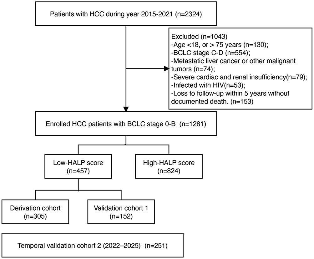 |
Figure 1 Flow chart of the study process. Abbreviations: HCC, hepatocellular Carcinoma; BCLC, Barcelona Clinic Liver Cancer; HIV, human immunodeficiency virus; HALP, Hemoglobin, Albumin, Lymphocyte and Platelet. |
Diagnosis and Staging
HCC was diagnosed according to the American Association for the Study of Liver Diseases guidelines based on histopathological examination or noninvasive criteria via dynamic imaging.14 The study population comprised patients with early- to intermediate-stage HCC (BCLC stages 0–B), as defined by the BCLC staging system,15 which integrates tumor burden, liver function, and performance status.
Data Collection and Follow-Up
Baseline clinical and laboratory data were collected, including patient demographics, comorbidities, and laboratory parameters such as complete blood count, comprehensive metabolic panels (assessing hepatic and renal function), and coagulation studies. All laboratory assessments were performed within 48 hours of admission. C‑reactive protein (CRP) was measured by immunoturbidimetry, and creatinine (Cr) and gamma‑glutamyl transferase (GGT) were measured by enzymatic methods using a Beckman AU5800 automated analyzer (Beckman Coulter, Brea, CA, USA). Reference ranges according to the manufacturer’s instructions were as follows: CRP 0–5 mg/L, Cr 57–111 µmol/L, and GGT 10–60 U/L. The laboratory participated in external quality assessment programs, and internal quality control was performed daily. Follow-up evaluations, including computed tomography/magnetic resonance imaging, ultrasonography, and serum alpha-fetoprotein testing, were conducted every three months. Treatment modalities were categorized as curative (surgical resection, liver transplantation, or local ablation) or noncurative (TACE, targeted therapy, immunotherapy, radiotherapy, chemotherapy, or best supportive care). OS was defined as the time from diagnosis to death from any cause or the last follow-up.
Statistical Analysis
Statistical analyses were performed using IBM SPSS 26.0 and R 4.4.0. Continuous variables were summarized as the mean ± standard deviation or median with interquartile range, as appropriate, whereas categorical variables were reported as counts and percentages. Comparisons among the three cohorts were made using one-way ANOVA or the Kruskal–Wallis H-test for continuous variables and the chi-square test for categorical variables. HALP was calculated as hemoglobin × albumin × lymphocyte/platelet.16 The optimal HALP cutoff for stratifying patients into low- and high-HALP groups was determined by time-dependent ROC analysis for 5-year OS using the maximum Youden index, yielding a cutoff of 47. Sensitivity analyses using cutoffs from the 20th to 80th percentiles confirmed the robustness of the findings (Supplementary Table S1).
To ensure robust feature selection, two complementary methods were applied: least absolute shrinkage and selection operator (LASSO) regression and random survival forest (RSF).17,18 For LASSO, 10‑fold cross‑validation was used to select the optimal penalty parameter λ (λ.min = 0.023), and variables with non‑zero coefficients at λ.min were retained. For RSF, the randomForestSRC package was used with 1,000 survival trees, sqrt(p) variables per node (where p is the number of predictor variables), a node size of 3, and log‑rank splitting. Variable importance was evaluated by permutation‑based variable importance measures (VIMP), with higher positive values indicating greater predictive relevance. Variables selected by both LASSO and RSF were then entered into a multivariate Cox regression model. To assess potential confounding by treatment strategy, two sensitivity analyses were performed. First, treatment type (curative vs. non‑curative) was forced as a covariate in the multivariate Cox model. Second, subgroup analyses stratified by treatment type were conducted.
Based on the selected predictors, a nomogram was developed to estimate 1‑, 3‑, and 5‑year OS probabilities. Model performance was assessed using time‑dependent receiver operating characteristic (ROC) analysis, calibration curves, and decision curve analysis (DCA). The discriminative performance of the nomogram for 5‑year OS was compared with that of the BCLC stage, the Model for End‑Stage Liver Disease (MELD) score, and the Albumin‑Bilirubin (ALBI) grade.19 Risk stratification was performed using the optimal cutoff identified by maximally selected rank statistics. Survival differences between risk groups were compared using Kaplan–Meier analysis with the log‑rank test. A two‑sided p‑value < 0.05 was considered statistically significant.
Results
HALP Cutoff and Survival Stratification
The optimal HALP cutoff of 47 was determined by time‑dependent ROC analysis for 5‑year OS using the maximum Youden index. At this cutoff, patients with low HALP (≤ 47, n = 457) had significantly higher 5‑year mortality than those with high HALP (>47, n = 824) (59.7% vs. 45.0%, p < 0.001; Figure 2). Significant survival differences were also observed using the median cutoff or the 25th/75th percentiles (Supplementary Table S1, all p < 0.05), indicating that the prognostic value of HALP was robust to the choice of cutoff threshold.
 |
Figure 2 Kaplan-Meier survival curves for overall survival based on HALP score stratification in patients with BCLC stage 0–B HCC. Abbreviations: HCC, hepatocellular carcinoma; HALP, Hemoglobin, Albumin, Lymphocyte and Platelet. |
Baseline Characteristics of Low-HALP Patients
The low‑HALP cohort from the 2015–2021 period was randomly divided into a derivation cohort (n = 305) and an internal validation cohort (validation cohort 1, n = 152). An additional temporal validation cohort (validation cohort 2, n = 251) comprising patients treated between 2022 and 2025 was included (Table 1). The overall cohort was predominantly male, with a high prevalence of cirrhosis and HBeAg positivity. The median age was 59 years (IQR 52–65) in the derivation cohort, and approximately half of the patients were classified as BCLC stage 0–A. Tumor size and multifocality distributions were similar across the three cohorts, and about 45% of patients received curative‑intent treatment. No statistically significant differences in baseline demographics or laboratory parameters were observed among the three cohorts.
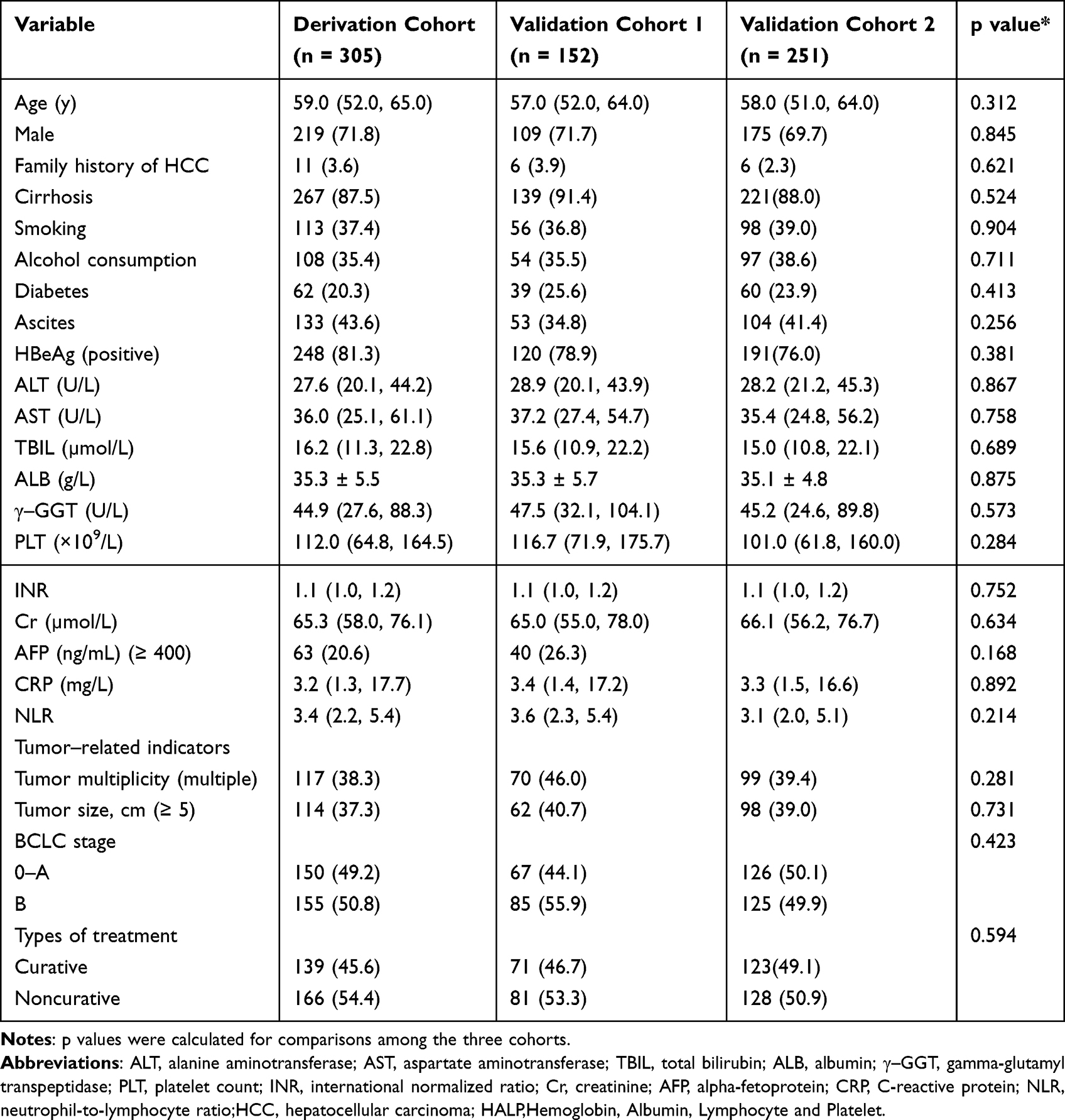 |
Table 1 Clinical Baseline Characteristics of Patients with BCLC Stage 0–B HCC and Low HALP Scores in the Derivation and Validation Sets |
Prognostic Factor Selection
In the derivation cohort, LASSO regression identified 11 variables with non‑zero coefficients at the optimal λ (λ.min = 0.023) as potential predictors of OS: sex, MELD score, cirrhosis, CRP, albumin, total bilirubin, GGT, INR, creatinine, tumor multiplicity, and BCLC stage (Figure 3A and B). Subsequently, RSF analysis was performed to assess variable importance for 5‑year OS prediction (Figure 3C). The model error rate declined as the number of trees increased and stabilized after 400 trees (Figure 3D). The key predictors identified by RSF included GGT, tumor multiplicity, creatinine, BCLC stage, and CRP (Figure 3E).
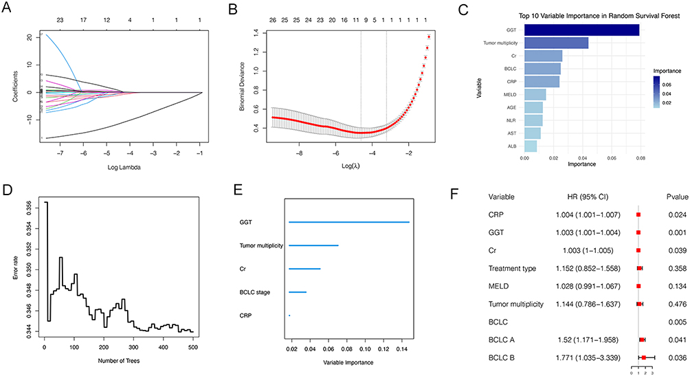 |
Figure 3 Feature selection and analysis for OS prediction in low-HALP HCC patients. Abbreviations: OS,overall survival; LASSO, Least Absolute Shrinkage and Selection Operator; RSF, Random Survival Forest; CRP, C-reactive protein; GGT, Gamma-glutamyl transferase; Cr, Creatinine; BCLC, Barcelona Clinic Liver Cancer; MELD, Model for End-Stage Liver Disease. Note: (A) LASSO coefficient profiles of the 11 candidate clinical variables; (B) Cross-validation curve for tuning parameter (λ) selection in the LASSO regression; (C) Top 10 variable importance ranking as determined by the RSF model for predicting 5-year OS; (D) Error rate of the RSF model as a function of the number of decision trees; (E) Predictors based on RSF analysis; (F) Multivariate Cox proportional hazards regression results for the final independent predictors of 5-year OS. |
Variables selected by both LASSO and RSF were included in a multivariate Cox regression model, which identified CRP (HR = 1.004, 95% CI: 1.001–1.007; p = 0.023), GGT (HR = 1.003, 95% CI: 1.001–1.004; p = 0.001), creatinine (HR = 1.003, 95% CI: 1.001–1.005; p = 0.038), and more advanced BCLC stage (stage A vs. 0: HR = 1.520, 95% CI: 1.171–1.958, p = 0.039; stage B vs. 0: HR = 1.771, 95% CI: 1.035–3.395, p = 0.036) as independent risk factors for 5‑year OS (Figure 3F). Adjustment for treatment type did not substantially change the HRs of GGT, CRP, creatinine, or BCLC stage, and treatment type itself was not an independent prognostic factor (HR = 1.152, p = 0.358; Supplementary Table S2). Based on these independent predictors, a prognostic nomogram was constructed incorporating these four variables (Figure 4).
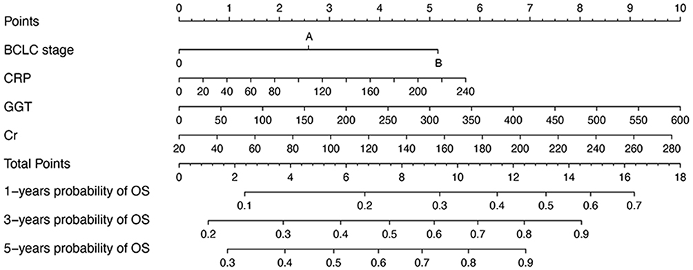 |
Figure 4 Prognostic nomogram for predicting 1-, 3-, and 5-year OS in low-HALP HCC patients. The nomogram integrates BCLC stage, CRP, GGT, and Cr to predict OS probabilities. Each variable is assigned a point value on the top “Points” scale (0–10). The sum of these points is plotted on the “Total Points” scale (0–18), from which the corresponding 1‑, 3‑, and 5‑year OS probabilities can be read. Clinical example: A patient with BCLC stage B (5 points), GGT 200 U/L (3.5 points), CRP 40 mg/L (1 point), and Cr 100 µmol/L (3 points) achieves a total score of 12.5 points, which corresponds to approximately 55%, 20%, and 10% probabilities of 1‑, 3‑, and 5‑year. Abbreviations: OS, respectively, HCC, hepatocellular carcinoma; OS, overall survival; BCLC, Barcelona Clinic Liver Cancer; CRP, C-reactive protein; GGT, Gamma-glutamyl transferase; Cr, Creatinine. |
Nomogram Performance and Validation
Time‑dependent ROC analysis for 1‑, 3‑, and 5‑year OS showed AUCs of 0.80, 0.75, and 0.77 in the derivation cohort; 0.80, 0.83, and 0.77 in the internal validation cohort (validation cohort 1); and 0.79, 0.73, and 0.76 in the temporal validation cohort (validation cohort 2) (Figure 5A–C). Compared with BCLC stage, MELD score, and ALBI grade, the nomogram demonstrated superior discriminatory ability for 5‑year OS across all three cohorts (Figure 5D–F and Table 2). Calibration plots showed good agreement between predicted and observed survival probabilities at all time points across the three cohorts (Figure 6A–C). DCA further demonstrated that the nomogram provided a higher net benefit than BCLC stage, MELD score, and ALBI grade across clinically meaningful risk thresholds (Figure 6D–F).
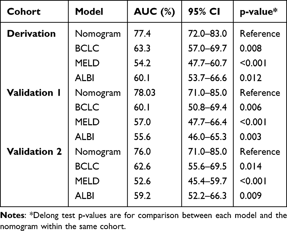 |
Table 2 Comparison of 5-Year AUC Between the Nomogram and Other Prognostic Models |
 |
Figure 5 Performance evaluation of the prognostic nomogram for low-HALP HCC patients. Time‑dependent ROC curves for 1‑, 3‑, and 5‑year OS in the derivation cohort (A), validation cohort 1 (B), and validation cohort 2 (C). ROC curves comparing the nomogram, BCLC stage, MELD score, and ALBI grade for 5‑year OS prediction in the derivation cohort (D), validation cohort 1 (E), and validation cohort 2 (F). Abbrevations: HALP, hemoglobin‑albumin‑lymphocyte‑platelet; HCC, hepatocellular carcinoma; ROC, receiver operating characteristic; OS, overall survival; BCLC, Barcelona Clinic Liver Cancer; MELD, Model for End‑Stage Liver Disease; ALBI, albumin‑bilirubin. |
 |
Figure 6 Calibration plots and DCA for the nomogram. Calibration plots for 1‑, 3‑, and 5‑year OS in the derivation cohort (A), validation cohort 1 (B), and validation cohort 2 (C). DCA curves showing net benefit of the nomogram in the derivation cohort (D), validation cohort 1 (E), and validation cohort 2 (F). Abbreviations: DCA, decision curve analysis; OS, overall survival. |
Risk Stratification and Survival Analysis
For risk stratification, an optimal cutoff score of 9.5 was established using maximally selected rank statistics, dividing patients into high‑risk (≥ 9.5) and low‑risk (< 9.5) categories (Figure 7A). Survival analysis confirmed significantly inferior OS in high‑risk patients across the derivation, internal validation, and temporal validation sets (all p < 0.001; Figure 7B–D). Using this cutoff, the nomogram successfully distinguished high‑risk from low‑risk patients, with markedly different 5‑year mortality rates: 86.9% versus 46.4% in the derivation set, 90.0% versus 43.4% in validation set 1, and 86.1% versus 43.5% in validation set 2. To further assess the robustness of the nomogram across different treatment strategies, subgroup survival analyses were performed stratified by treatment type. As shown in Supplementary Figure S1, the nomogram consistently distinguished high‑risk from low‑risk patients across all three cohorts, regardless of treatment type (curative vs. non‑curative) (all log‑rank p < 0.0001).
 |
Figure 7 Risk stratification and survival analysis based on the nomogram-derived risk score. Notes: (A) Determination of the optimal cutoff value (9.5 points) for risk stratification using maximally selected rank statistics. Kaplan-Meier survival curves comparing overall survival between the low-risk (< 9.5 points) and high-risk (≥ 9.5 points) groups in the derivation cohort (B), validation cohort 1 (C), and validation cohort 2 (D). |
Discussion
This study constructed and validated a ML-based nomogram to predict OS in patients with BCLC stage 0–B HCC presenting with low HALP scores. By incorporating four readily available clinical parameters (GGT, BCLC stage, CRP, and creatinine), the nomogram exhibited strong discriminatory performance and good calibration across both the derivation and validation sets, suggesting its potential utility for prognostic assessment in this population.
ML may offer potential advantages over conventional regression-based approaches by capturing complex, non‑linear relationships and interactions among variables, which may improve prognostic model performance.20 To our knowledge, no prior studies have employed ML to predict 5‑year OS in patients with low‑HALP, early‑ to intermediate‑stage HCC. Conventional models often overlook the distinct clinicopathological features of this subgroup, in which impaired immunonutritional status is closely linked to treatment response and survival. In our cohort, the 5‑year OS rate in the low‑HALP group was 40.3%, significantly lower than the 55.0% observed in the high‑HALP group. The HALP cutoff of 47 used in this study is comparable to previously reported thresholds: 45.6 in HCC patients undergoing curative resection and 47.89 in breast cancer patients.21,22 This consistency across different populations and study designs supports the robustness of our threshold. To address potential threshold‑selection bias, we conducted extensive sensitivity analyses, which confirmed that a low HALP score was significantly associated with poor prognosis regardless of the cutoff value applied, further supporting the robustness of the HALP prognostic value. The HALP score serves as a comprehensive reflection of the tumor immune microenvironment, integrating inflammatory, nutritional, and immune parameters.23 While HALP‑based models exist for HCC, our nomogram specifically addresses the BCLC stage 0–B low‑HALP subgroup, where prognostic heterogeneity is often overlooked.
By combining ML algorithms with traditional Cox regression, we identified four independent risk factors that are biologically plausible and clinically accessible: GGT, BCLC stage, CRP, and creatinine. These were incorporated into a nomogram for 5‑year OS prediction. The nomogram demonstrated superior discriminative ability for 5‑year OS across all three cohorts when compared with the BCLC stage, MELD score, and ALBI grade (all p < 0.05). Calibration curves further confirmed good concordance between model predictions and actual observed outcomes. Moreover, DCA substantiated the clinical value of the nomogram across a wide range of risk thresholds, indicating its potential for practical implementation. Based on routinely available clinical parameters, the nomogram offers an easily interpretable means of individualized outcome prediction with adequate performance. By applying the optimal cutoff, the nomogram effectively stratified patients into high‑ and low‑risk categories, and Kaplan–Meier analysis demonstrated a marked survival difference between the two groups. In the derivation set, the 5‑year mortality rates were 86.9% in the high‑risk group versus 46.4% in the low‑risk group; corresponding figures were 90.0% versus 43.4% in validation set 1, and 86.1% versus 43.5% in validation set 2. By enabling precise risk stratification, the nomogram provides a useful tool for individualized prognostic assessment in patients with low HALP scores and BCLC stage 0–B HCC.
Although the underlying mechanisms cannot be directly determined from our data, the following biological rationales may explain the observed associations. The BCLC clinical algorithm is the most widely adopted system for HCC staging and risk stratification, and plays a pivotal role in guiding clinical management decisions.24 In the context of low HALP levels, which signifies a state of impaired immunonutrition, the prognostic weight of systemic inflammation and organ dysfunction appears amplified. Systemic inflammation is a key driver of tumor progression, facilitating cancer growth, invasion, and metastasis.25 CRP, whose production is stimulated by cytokines such as interleukin (IL)-1 and IL-6, is released into the systemic circulation and serves as an inflammatory marker.26 Mechanistically, CRP can inhibit the activation of CD8+ and CD4+ T cells while promoting the expansion and immunosuppressive functions of myeloid-derived suppressor cells.27 Notably, in our low-HALP cohort, characterized by underlying immune and nutritional depletion, elevated CRP levels may more sensitively reflect a severe and poorly controlled systemic inflammatory state, thereby exhibiting a stronger association with mortality. Our results corroborate those of Hatanaka et al, who documented that the prognostic impact of inflammatory markers is particularly pronounced in patients with cancer-related cachexia or malnutrition.28
Renal function, as indicated by serum creatinine, also strongly correlated with survival. Elevated creatinine may indicate impaired kidney function, which can limit treatment tolerance and worsen the prognosis. In the setting of low HALP scores, which inherently suggest a poorer physiological reserve and higher vulnerability, even mild renal impairment may have disproportionately severe consequences on outcomes. Moreover, creatinine levels may be influenced by the systemic inflammatory states that are commonly present in patients with HCC. Inflammatory responses and renal dysfunction often interact synergistically, and elevated creatinine levels may reflect this intertwined pathophysiology. The interplay between renal dysfunction and the inflammatory response may create a vicious cycle that accelerates disease progression.29 Additionally, elevated GGT levels are associated with advanced liver dysfunction and poor prognosis in HCC.30 GGT contributes to carcinogenesis via prooxidant mechanisms and facilitates iron release from transferrin.31 In patients with low HALP levels whose livers may already be stressed by malnutrition and systemic inflammation, elevated GGT levels may indicate a greater cumulative burden of hepatic injury and oxidative stress, making it a more critical prognostic indicator in this subgroup.
This study had several limitations. First, the nomogram was derived from a single-center dataset. Although internal and temporal validation were performed, both validation cohorts originated from the same institution. External validation in independent multicenter cohorts is therefore needed to confirm generalizability. Second, although we identified several independent prognostic factors, we cannot exclude the possibility of unmeasured confounding. Despite subgroup analyses suggesting that treatment type did not independently affect OS, confounding by indication cannot be fully ruled out. Third, we focused solely on baseline HALP scores and did not analyze the dynamic changes during treatment. Finally, although the model demonstrated strong predictive performance for 5-year OS, long-term follow-up is needed to assess its validity for extended survival outcomes.
Conclusion
We developed and validated a prognostic nomogram for predicting the 5-year OS in patients with BCLC stage 0–B HCC and low HALP scores. By integrating ML and traditional statistical methods, the model identified high-risk individuals using routinely available clinical parameters. Given its favorable accuracy, convenience, and non‑invasiveness, this nomogram could assist clinicians in risk stratification and individualized prognostic assessment in this population.
Data Sharing Statement
The original contributions presented in the study are included in the article. Further inquiries can be directed to the corresponding author.
Ethics Approval and Informed Consent
This study followed the Helsinki declaration. All participants signed an informed consent form and this study was approved by Ethics Committee of Beijing Ditan Hospital. (No. DTEC-KY2024-017-01).
Acknowledgments
We gratefully recognize the patients who participated in this study.
Author Contributions
Ying Feng: Conceptualization, Funding Acquisition, Project Administration, Supervision, Writing - Review & Editing.
Xianbo Wang: Conceptualization, Funding Acquisition, Project Administration, Supervision, Writing - Review & Editing.
Yao Liu: Data Curation, Methodology, Validation, Visualization, Writing - Original Draft Preparation, Software, Writing - Review & Editing.
Ke Shi: Data Curation, Methodology, Software, Validation, Visualization, Writing - Original Draft Preparation, Funding Acquisition.
Xiaojing Wang: Writing - Review & Editing, Software, Data Curation.All authors gave final approval of the version to be published; have agreed on the journal to which the article has been submitted; and agree to be accountable for all aspects of the work.
Funding
This study was supported by the Beijing High-Level Innovation and Entrepreneurship Talent Support Programein – Young Backbone Talent Projects (Grant No. G202532309), High-Level Chinese Medicine Key Discipline Construction Project (Grant No. zyyzdxk-2023005), the Capital Health Development Research Project (Grant No. 2024-1-2173), the National Natural Science Foundation of China (Grant Nos. 82474419 and 82474426), and the Research-Oriented Ward Excellence Clinical Research Program Parallel Support Project (Grant No. BRWEP2024W102170107).
Disclosure
The author(s) report no conflicts of interest in this work.
References
1. Sung H, Ferlay J, Siegel RL, et al. Global cancer statistics 2020: GLOBOCAN estimates of incidence and mortality worldwide for 36 cancers in 185 countries. CA Cancer J Clin. 2021;71:209–13. doi:10.3322/caac.21660
2. Long J, Cui K, Wang D, et al. Burden of hepatocellular carcinoma and its underlying etiologies in China, 1990-2021: findings from the global burden of disease study 2021. Cancer Control. 2024;31:10732748241310573. doi:10.1177/10732748241310573
3. Altaf S, Saleem F, Sher AA, et al. Potential therapeutic strategies to combat HCC. Curr Mol Pharmacol. 2022;15:929–942. doi:10.2174/1874467215666220103111009
4. Yan H, Wang X, Liu X, et al. The survival strength of younger patients in BCLC stage 0-B of hepatocellular carcinoma: basing on competing risk model. BMC Cancer. 2022;22:185. doi:10.1186/s12885-022-09293-x
5. Raoul JL, Forner A, Bolondi L, et al. Updated use of TACE for hepatocellular carcinoma treatment: how and when to use it based on clinical evidence. Cancer Treat Rev. 2019;72:28–36. doi:10.1016/j.ctrv.2018.11.002
6. Zhang H, Song Y, Yang H, et al. Tumor cell-intrinsic Tim-3 promotes liver cancer via NF-κB/IL-6/STAT3 axis. Oncogene. 2018;37:2456–2468. doi:10.1038/s41388-018-0140-4
7. Kanda T, Sasaki-Tanaka R, Terai S. Inflammation of the liver, HCC development and HCC establishment. Hepatol Int. 2024;18:1090–1092. doi:10.1007/s12072-024-10707-0
8. Fang D, Yin X, Ding X, et al. Recurrence of hepatocellular carcinoma in patients with high HALP score in TACE combined with ablation. Front Oncol. 2025;15:1609260. doi:10.3389/fonc.2025.1609260
9. Farag CM, Antar R, Akosman S, et al. What is hemoglobin, albumin, lymphocyte, platelet (HALP) score? A comprehensive literature review of HALP’s prognostic ability in different cancer types. Oncotarget. 2023;14(1):153–172. doi:10.18632/oncotarget.28367
10. George ES, Sood S, Broughton A, et al. The association between diet and hepatocellular carcinoma: a systematic review. Nutrients. 2021;13:172. doi:10.3390/nu13010172
11. Bati IB, Tuysuz U, Eygi E. Prognostic significance of Hemoglobin, Albumin, Lymphocyte, and Platelet (HALP) score in liver transplantation for hepatocellular carcinoma. Curr Oncol. 2025;32:464. doi:10.3390/curroncol32080464
12. Zhou J, Yang D. Prognostic significance of Hemoglobin, Albumin, Lymphocyte and Platelet (HALP) score in hepatocellular carcinoma. J Hepatocell Carcinoma. 2023;10:821–831. doi:10.2147/JHC.S411521
13. Char DS, Burgart A. Machine-learning implementation in clinical Anesthesia: opportunities and challenges. Anesthesia and Analgesia. 2020;130:1709–1712. doi:10.1213/ane.0000000000004656
14. Singal AG, Llovet JM, Yarchoan M, et al. AASLD Practice guidance on prevention, diagnosis, and treatment of hepatocellular carcinoma. Hepatology. 2023;78:1922–1965. doi:10.1097/HEP.0000000000000466
15. Grieco A, Pompili M, Caminiti G, et al. Prognostic factors for survival in patients with early-intermediate hepatocellular carcinoma undergoing non-surgical therapy: comparison of Okuda, CLIP, and BCLC staging systems in a single Italian centre. Gut. 2005;54:411–418. doi:10.1136/gut.2004.048124
16. Li Q, Chen M, Zhao H, et al. The prognostic and clinicopathological value of HALP score in non-small cell lung cancer. Front Immunol. 2025;16:1576326. doi:10.3389/fimmu.2025.1576326
17. Wang Q, Qiao W, Zhang H, et al. Nomogram established on account of Lasso-Cox regression for predicting recurrence in patients with early-stage hepatocellular carcinoma. Front Immunol. 2022;13:1019638. doi:10.3389/fimmu.2022.1019638
18. Tian D, Yan HJ, Huang H, et al. Machine learning-based prognostic model for patients after lung transplantation. JAMA Netw Open. 2023;6:e2312022. doi:10.1001/jamanetworkopen.2023.12022
19. Demirtas CO, D’Alessio A, Rimassa L, et al. ALBI grade: Evidence for an improved model for liver functional estimation in patients with hepatocellular carcinoma. JHEP Rep. 2021;3:100347. doi:10.1016/j.jhepr.2021.100347
20. Campagnini S, Arienti C, Patrini M, et al. Machine learning methods for functional recovery prediction and prognosis in post-stroke rehabilitation: a systematic review. J Neuroeng Rehabil. 2022;19(1):54. doi:10.1186/s12984-022-01032-4
21. Toshida K, Itoh S, Kayashima H, et al. The hemoglobin, albumin, lymphocyte, and platelet score is a prognostic factor for Child-Pugh A patients undergoing curative hepatic resection for single and small hepatocellular carcinoma. Hepatol Res. 2023;53:522–530. doi:10.1111/hepr.13885
22. Jiang T, Sun H, Xue S, et al. Prognostic significance of hemoglobin, albumin, lymphocyte, and platelet (HALP) score in breast cancer: a propensity score-matching study. Cancer Cell Int. 2024;24:230. doi:10.1186/s12935-024-03419-w
23. Li H, Zhou Y, Zhang X, et al. The relationship between hemoglobin, albumin, lymphocyte, and platelet (HALP) score and 28-day mortality in patients with sepsis: a retrospective analysis of the MIMIC-IV database. BMC Infect Dis. 2025;25(1):333. doi:10.1186/s12879-025-10739-3
24. Reig M, Forner A, Rimola J, et al. BCLC strategy for prognosis prediction and treatment recommendation: the 2022 update. J Hepatol. 2022;76:681–693. doi:10.1016/j.jhep.2021.11.018
25. Yang YM, Kim SY, Seki E, et al. Inflammation and liver cancer: molecular mechanisms and therapeutic targets. Semin Liver Dis. 2019;39:26–42. doi:10.1055/s-0038-1676806
26. Okumura T, Kimura T, Iwadare T, et al. Prognostic significance of C-Reactive protein in lenvatinib-treated unresectable hepatocellular carcinoma: a multi-institutional study. Cancers. 2023;15:5343. doi:10.3390/cancers15225343
27. Yoshida T, Ichikawa J, Giuroiu I, et al. C reactive protein impairs adaptive immunity in immune cells of patients with melanoma. J Immunother Cancer. 2020;8:e000234. doi:10.1136/jitc-2019-000234
28. Hatanaka T, Yata Y, Hiraoka A, et al. Clinical impact of serum CRP levels on advanced HCC treated with durvalumab and tremelimumab: a multicentre study. Liver Int. 2025;45:e70192. doi:10.1111/liv.70192
29. Sohn W, Ham CB, Kim NH, et al. Effect of acute kidney injury on the patients with hepatocellular carcinoma undergoing transarterial chemoembolization. PLoS One. 2020;15:e0243780. doi:10.1371/journal.pone.0243780
30. Lu N, Sheng S, Xiong Y, et al. Prognostic model for predicting recurrence in hepatocellular carcinoma patients with high systemic immune-inflammation index based on machine learning in a multicenter study. Front Immunol. 2024;15:1459740. doi:10.3389/fimmu.2024.1459740
31. Liu Y, Zhang Q, Yang X, et al. Effects of various interventions on the occurrence of macrovascular invasion of hepatocellular carcinoma after the baseline serum γ-glutamyltransferase stratification. Onco Targets Ther. 2019;12:1671–1679. doi:10.2147/OTT.S184302
 © 2026 The Author(s). This work is published and licensed by Dove Medical Press Limited. The
full terms of this license are available at https://www.dovepress.com/terms
and incorporate the Creative Commons Attribution
- Non Commercial (unported, 4.0) License.
By accessing the work you hereby accept the Terms. Non-commercial uses of the work are permitted
without any further permission from Dove Medical Press Limited, provided the work is properly
attributed. For permission for commercial use of this work, please see paragraphs 4.2 and 5 of our Terms.
© 2026 The Author(s). This work is published and licensed by Dove Medical Press Limited. The
full terms of this license are available at https://www.dovepress.com/terms
and incorporate the Creative Commons Attribution
- Non Commercial (unported, 4.0) License.
By accessing the work you hereby accept the Terms. Non-commercial uses of the work are permitted
without any further permission from Dove Medical Press Limited, provided the work is properly
attributed. For permission for commercial use of this work, please see paragraphs 4.2 and 5 of our Terms.
Recommended articles

Histological Severity of Cirrhosis Influences Surgical Outcomes of Hepatocellular Carcinoma After Curative Hepatectomy
Liang BY, Gu J, Xiong M, Zhang EL, Zhang ZY, Lau WY, Wang SF, Guan Y, Chen XP, Huang ZY
Journal of Hepatocellular Carcinoma 2022, 9:633-647
Published Date: 23 July 2022
Nomogram for the Preoperative Prediction of the Macrotrabecular-Massive Subtype of Hepatocellular Carcinoma
Shan Y, Yu X, Yang Y, Sun J, Wu S, Mao S, Lu C
Journal of Hepatocellular Carcinoma 2022, 9:717-728
Published Date: 10 August 2022
Establishment and Validation of a Predictive Nomogram for Postoperative Survival of Stage I Non-Small Cell Lung Cancer
Wang ZH, Deng L
International Journal of General Medicine 2022, 15:7287-7298
Published Date: 14 September 2022
Development and Validation of a Novel Nomogram Integrated with Hypoxic and Lactate Metabolic Characteristics for Prognosis Prediction in Hepatocellular Carcinoma
Qiu X, Dong L, Wang K, Zhong X, Xu H, Xu S, Guo H, Wei X, Chen W, Xu X
Journal of Hepatocellular Carcinoma 2024, 11:241-255
Published Date: 2 February 2024
Development and Validation of a Prognostic Nomogram for Patients with AFP and DCP Double-Negative Hepatocellular Carcinoma After Local Ablation
Qiao W, Li J, Wang Q, Jin R, Zhang H
Journal of Hepatocellular Carcinoma 2024, 11:271-284
Published Date: 3 February 2024