Back to Journals » Journal of Pain Research » Volume 16
The Link Between Empty Sella Syndrome, Fibromyalgia, and Chronic Fatigue Syndrome: The Role of Increased Cerebrospinal Fluid Pressure
Authors Hulens M ![]() , Dankaerts W
, Dankaerts W ![]() , Rasschaert R
, Rasschaert R ![]() , Bruyninckx F
, Bruyninckx F ![]() , De Mulder P
, De Mulder P ![]() , Bervoets C
, Bervoets C ![]()
Received 21 October 2022
Accepted for publication 17 January 2023
Published 25 January 2023 Volume 2023:16 Pages 205—219
DOI https://doi.org/10.2147/JPR.S394321
Checked for plagiarism Yes
Review by Single anonymous peer review
Peer reviewer comments 2
Editor who approved publication: Dr Alaa Abd-Elsayed
Mieke Hulens,1 Wim Dankaerts,1 Ricky Rasschaert,2 Frans Bruyninckx,3 Peter De Mulder,4 Chris Bervoets5– 7
1Department of Rehabilitation Sciences, KU Leuven, Leuven, Belgium; 2Department of Neurosurgery, AZ Rivierenland, Bornem, Belgium; 3Department of Physical Medicine & Rehabilitation, University Hospitals of Leuven, Leuven, Belgium; 4Department of Anesthesiology and Pain Therapy, Imelda Hospital, Bonheiden, Belgium; 5Department of Neurosciences, KU Leuven, Leuven, Belgium; 6Department of Ophthalmology, University Hospitals of Leuven, Leuven, Belgium; 7Department Adult Psychiatry, University Psychiatric Center of KU Leuven, Leuven, Belgium
Correspondence: Mieke Hulens, Department of Rehabilitation Sciences, KU Leuven, Overwegstraat 14, 3051 Sint-Joris-Weert, Leuven, Belgium, Tel +32 477 338003, Fax +32 16 329197, Email [email protected]
Abstract: The etiopathogenesis of fibromyalgia (FM) and chronic fatigue syndrome (CFS) is not yet elucidated. Hypothalamo-pituitary-adrenal (HPA) axis dysfunction is reflected in the hormonal disturbances found in FM and CFS. Some study groups have introduced a novel hypothesis that moderate or intermittent intracranial hypertension may be involved in the etiopathogenesis of FM and CFS. In these conditions, hormonal disturbances may be caused by the mechanical effect of increased cerebrospinal fluid pressure, which hampers blood flow in the pituitary gland. Severe intracranial pressure may compress the pituitary gland, resulting in primary empty sella (ES), potentially leading to pituitary hormone deficiencies. The aim of this narrative review was to explore whether similar hormonal changes and symptoms exist between primary ES and FM or CFS and to link them to cerebrospinal fluid pressure dysregulation. A thorough search of the PubMed and Web of Science databases and the reference lists of the included studies revealed that several clinical characteristics were more prevalent in primary ES, FM or CFS patients than in controls, including increased cerebrospinal fluid pressure, obesity, female sex, headaches and migraine, fatigue, visual disturbances (visual acuity and eye motility abnormalities), vestibulocochlear disturbances (vertigo and neurosensorial hearing loss), and bodily pain (radicular pain and small-fiber neuropathy). Furthermore, challenge tests of the pituitary gland showed similar abnormalities in all three conditions: blunted adrenocorticotropic hormone, cortisol, growth hormone, luteinizing hormone, and thyroid stimulating hormone responses and an increased prolactin response. The findings of this narrative review provide further support for the hypothesis that moderately or intermittently increased cerebrospinal fluid pressure is involved in the pathogenesis of FM and CFS and should stimulate further research into the etiopathogenesis of these conditions.
Keywords: HPA axis, intracranial hypertension, prolactin, growth hormone, cortisol, thyroid hormones, hormone levels
Plain Language Summary
The cause of fibromyalgia (FM) and chronic fatigue syndrome (CFS) is not yet elucidated. Disturbances in the interactions between the hypothalamus, pituitary gland, and adrenal glands (the hypothalamo-pituitary-adrenal (HPA) axis) in FM and CFS may result in abnormal hormone production. However, a novel hypothesis proposes that moderate increases in cerebral and spinal fluid pressure may underlie both FM and CFS. Increased cerebral pressure may impede blood flow in the pituitary gland, resulting in hormonal disturbances. Indeed, it is well known that severely increased cerebral pressure can cause compression and flattening of the pituitary gland, potentially leading to pituitary hormone deficiency, termed empty sella syndrome. The sella (turcica) is a saddle-shaped notch in the bone at the base of the skull where the pituitary gland is located.
A search of the scientific literature revealed that increased cerebrospinal fluid pressure, obesity, female sex, headache and migraine, fatigue, visual disturbances, vertigo, hearing loss, and widespread pain were all more prevalent in empty sella, FM and CFS patients than in healthy individuals. Furthermore, it was demonstrated that the injection of substances to stimulate pituitary hormone production resulted in similar responses, including reduced cortisol, growth hormone, luteinizing hormone, and thyroid-stimulating hormone production and increased prolactin production, in all three conditions compared to healthy individuals. The findings of this review provide further support for the hypothesis that moderate or intermittent increases in cerebrospinal fluid pressure are involved in the pathogenesis of FM and CFS and should stimulate further research into the causes of these conditions.
Introduction
Fibromyalgia (FM) and chronic fatigue syndrome (CFS) are overlapping pain conditions whose etiopathogenesis is not yet elucidated. Hypothalamo-pituitary-adrenal (HPA) axis dysfunction is reflected in the hormonal disturbances found in FM and CFS.1–3 Several study groups have previously proposed that mild or intermittent forms of intracranial hypertension or cerebrospinal fluid pressure dysregulation may be involved in the etiopathogenesis of FM/CFS.4–8 Indeed, idiopathic intracranial hypertension and FM/CFS have several clinical characteristic symptoms in common, such as fatigue, widespread pain and paraesthesia, headaches, cognitive dysfunction, and brain fog.5
More severe cases of intracranial hypertension may cause significant compression of the pituitary gland, eventually leading to empty sella (ES) on brain magnetic resonance imaging (MRI). ES involves the herniation of the subarachnoid space within the sella turcica, associated with an elongated pituitary stalk and a flattening of the pituitary gland to less than 2 mm. Partial ES is diagnosed when < 50% of the sella is filled with cerebrospinal fluid, and total ES is diagnosed when >50% is filled with cerebrospinal fluid.
Primary ES is diagnosed in cases with unknown etiology when all secondary causes of ES are excluded, such as previous surgical, pharmacological or radiotherapy treatment of the pituitary gland.9
Primary ES is not just a neuroradiological finding without clinical implications. Indeed, one or more pituitary hormone deficiencies may be present in a significant proportion of patients with total and partial ES.10–14 We hypothesize that hormonal disturbances in FM/CFS can be the result of pituitary gland compression owing to intermittently or moderately increased cerebrospinal fluid pressure, which impedes the blood flow in the pituitary gland and stalk.
The aim of this narrative review was to explore the similarities in clinical characteristics between primary ES and FM/CFS.
Materials and Methods
PubMed and Web of Science (WOS) databases and the reference lists of the included studies were searched for English language articles published from 1985 until 2022 using the terms empty sella, FM and CFS in combination with the terms pituitary gland, increased cerebral pressure, growth hormone (GH), prolactin (PRL), cortisol, adrenocorticotropic hormone (ACTH), thyroid hormones, and gonadal hormones. Data regarding symptoms and hormonal levels in patients with primary ES, FM and CFS were compared and evaluated in reference to the hypothesis of a shared mechanism of underlying increased or dysregulated cerebrospinal fluid pressure.
A Discussion of the Similarities Between Primary ES and FM/CFS
The similarities between primary ES and FM/CFS are listed in Table 1.
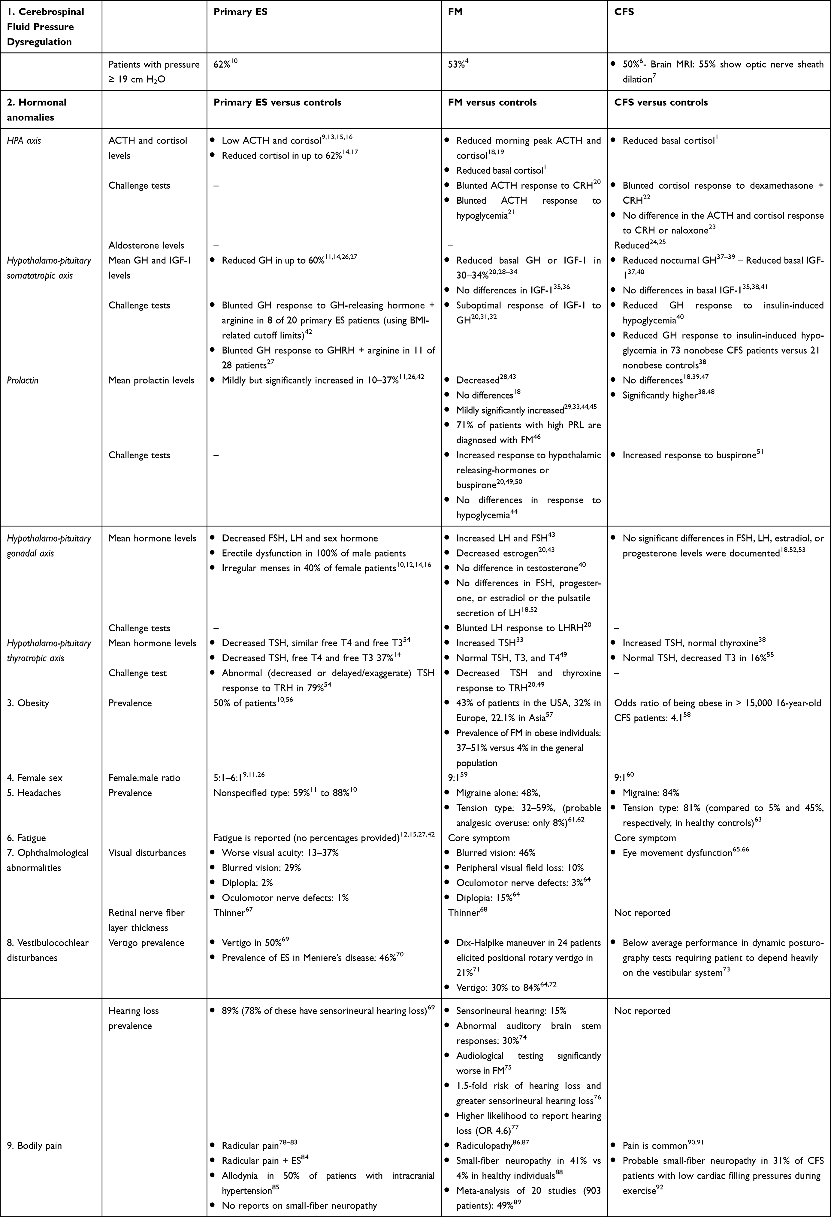 |
Table 1 Similar Characteristics of Primary ES, FM and CFS |
Cerebrospinal Fluid Pressure Dysregulation
In patients with intracranial hypertension, insufficient hormone secretion from the pituitary gland is caused by the compression of the pituitary gland against the sella turcica and the compression and eventual stretching of the pituitary stalk by the downward compression of the pituitary gland.9,11,26 Thus, similar to pituitary compression by an adenoma (secondary ES), increased cerebrospinal fluid pressure may hamper hormone production.13
Primary ES is almost always secondary to dysregulation of intracranial pressure even when incidentally found in asymptomatic cases. Idiopathic intracranial hypertension without papilledema is currently severely underestimated or misdiagnosed because the upper limit of the current normal range is set too high and because of a largely unrecognized high prevalence of asymptomatic cases. It has been suggested that the prevalence of idiopathic intracranial hypertension without papilledema might be up to three times higher than that of idiopathic intracranial hypertension.93 A possible mechanism that has been proposed is reduced intracranial compliance that precedes the increase in intracranial pressure and that may already be pathologic in cases lacking a detectable increase in intracranial pressure at lumbar puncture. Hence, opening pressure values greater than 25 cm H2O are not required (and potentially misleading) in diagnosis of idiopathic intracranial hypertension without papilledema.94
Intracranial hypertension involves a spectrum that begins with milder and intermittent forms that progress into ES and ends with more severe forms of idiopathic intracranial hypertension, presenting with papilledema, severe headache and visual field defects. Indeed, only 8 to 15% of patients with ES progress to develop these severe idiopathic intracranial hypertension symptoms, but in contrast, up to 94% of patients with severe idiopathic intracranial hypertension already show ES.9
Using a lumbar constant-rate infusion test, pressure anomalies were revealed in 109 (77%) of 142 primary ES patients: pathological resting intracranial pressure (range 19–35 cm H2O) in 88 patients (62%) and a reduction in the cerebrospinal fluid absorption rate despite normal basal intracranial pressure in 21 patients (15%).10
In patients with FM/CFS, moderately high cerebrospinal fluid pressures were found during lumbar puncture. In 30 patients with chronic widespread pain or FM, the mean cerebrospinal opening pressure was 19.7 cm H2O (range 11–32 cm H2O). Sixteen patients (53%) exhibited a pressure ≥19 cm H2O. Moreover, spinal fluid evacuation temporarily improved bodily pain in 21 of 30 patients (70%).4
Similar results were seen in patients with CFS. A lumbar puncture in 20 patients revealed a mean cerebrospinal fluid pressure of 19 cmH2O (range 12–41 cmH2O). Ten patients (50%) exhibited a pressure >19 cm H2O. Spinal fluid evacuation temporarily improved headaches in 17 CFS patients (85%).95 Additionally, brain MRI in 205 patients with CFS revealed an increased optic nerve sheath diameter as a sign of intracranial hypertension in 55%.7
Hormonal Anomalies in Primary ES and FM/CFS
The anterior lobe of the pituitary gland releases PRL, GH, ACTH, follicle-stimulating hormone (FSH), luteinizing hormone (LH) and thyroid-stimulating hormone (TSH) (Table 2). The posterior lobe of the pituitary gland stores and releases the hormones produced by the hypothalamus—antidiuretic hormone (ADH) and oxytocin. The pituitary stalk contains the vascular connections between the hypothalamus and the pituitary gland and the neuronal connections between the pituitary gland and the brain. A disruption of the blood flow in the pituitary gland or of the connections in the stalk due to compression from increased cerebrospinal fluid pressure may result in a disturbance of pituitary hormone secretion. Moreover, increased pressure on the pituitary tissue may also lead to functional insufficiency of the pituitary cells.42
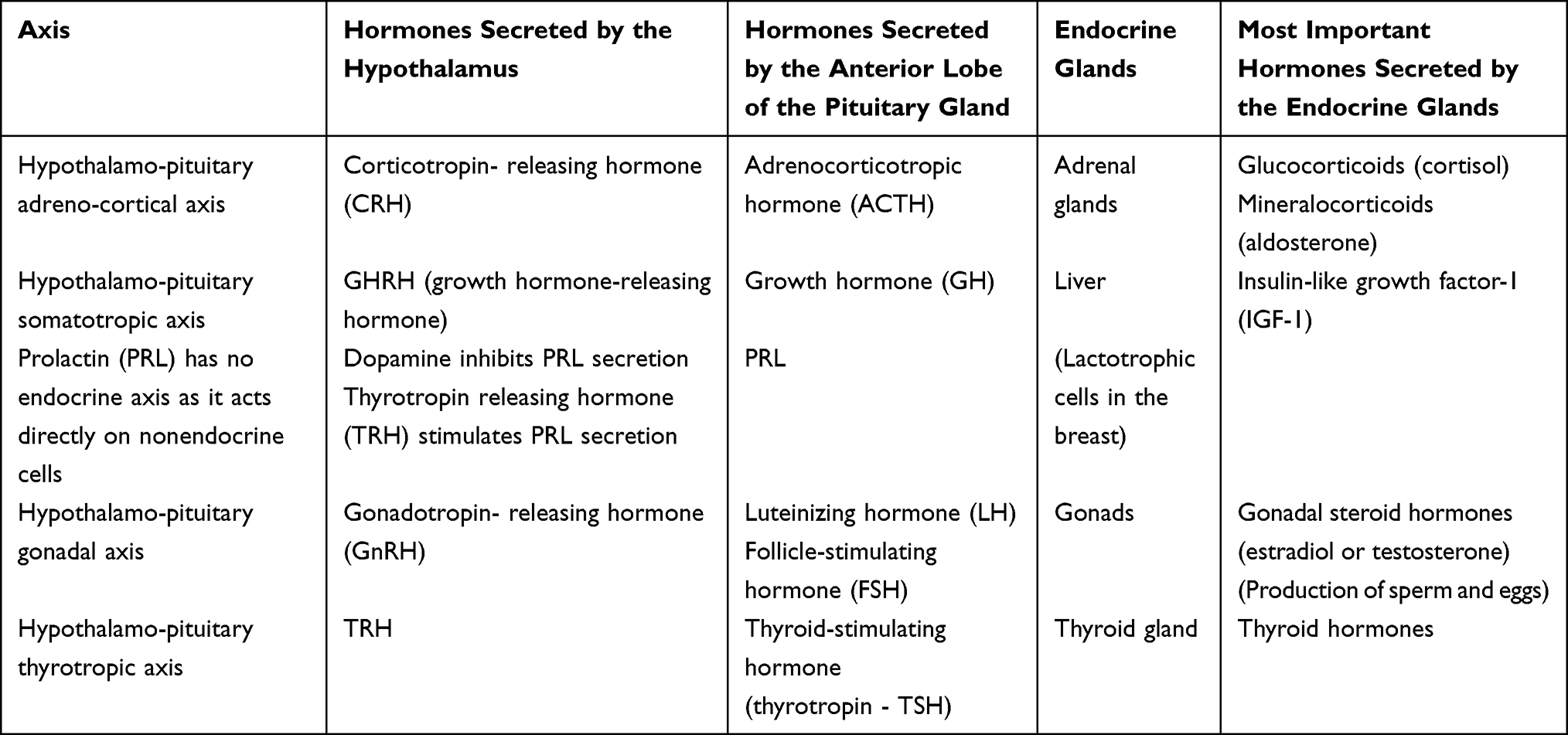 |
Table 2 Overview of the Hypothalamic and Pituitary Hormones |
An examination to evaluate pituitary function includes blood tests of hormone levels. However, stimulation tests are required for a more comprehensive examination of the adrenocortical axis and somatotropic axis.9,11,96 Some degree of hypopituitarism (one or more hormone deficiencies) is present in two-thirds of patients with total primary ES and is present in an important number of patients with partial primary ES.12,14
The Hypothalamo-Pituitary Adrenocortical Axis (HPA Axis)
Corticotropin releasing hormone (CRH) from the hypothalamus stimulates the secretion of ACTH, which in turn leads to the adrenal secretion of cortisol and aldosterone. ACTH deficiency in association with primary ES has been reported in several case studies. Low ACTH levels in primary ES were found to be associated with low cortisol levels.9,13,15,16 Low cortisol levels were found in up to 62% of patients with primary ES.14,17
In CFS/FM, reduced ACTH and cortisol levels are found during the morning physiological peak ACTH secretion.18,19 A meta-analysis of 85 studies found a significant reduction in basal cortisol levels in male and female patients with CFS and in female patients with FM compared to healthy controls.1
A pituitary challenge test in FM using a CRH injection demonstrated a blunted ACTH response.20 Additionally, a significant 30% reduction in the ACTH responses to hypoglycemia was observed in women with FM.21
Globally, a pituitary challenge test in CFS patients using low-dose dexamethasone and CRH combined reduced cortisol responses.22 Conversely, another study using a challenge test with CRH or naloxone detected no difference with respect to the ACTH and cortisol response in CFS patients and healthy individuals.23
Furthermore, two studies also found significantly lower aldosterone levels in patients with CFS than in controls.24,25
In a narrative review, Cleare41 found no changes in HPA axis function during the onset and early stages of CFS. According to our hypothesis, this late effect on the HPA axis may be due to the limited effect of the moderately increased cerebrospinal fluid pressure in the early stages, whereas longstanding increased cerebrospinal fluid pressure is more likely to affect pituitary function.
The Hypothalamo-Pituitary Somatotropic Axis
GH stimulates the liver to secrete insulin-like growth factor-1 (IGF-1). Therefore, IGF-1 levels are a surrogate marker for GH secretion. IGF-1 mediates the anabolic and growth-promoting effects of GH. It has been postulated that GH deficiency is an early event in patients with primary ES and that this deficiency could be related to the anatomical disposition of the cells within the anterior pituitary that produce GH, which could make these cells more vulnerable to increased pressure. However, some authors suggest that obesity might play a role because of its action in decreasing GH secretion.11
GH deficiency is one of the most common pituitary disorders associated with primary ES.9,96 When the GH levels of patients with primary ES were compared with those of healthy individuals, most studies reported decreased levels in up to 60% of patients.11,14,26,27
The stimulation of the pituitary gland using growth hormone-releasing hormone (GHRH) + arginine elicited a lower GH response in 40% of primary ES patients than in controls. In this study, BMI-related cutoff limits were used; thus, weight did not play a role.42 GHRH + arginine injection also revealed GH/IGF-I axis dysfunction in 39% of adults with primary ES.27 Serum IGF-1 levels in primary ES patients were positively correlated with pituitary volume.16
In FM, approximately 30–34% of patients present lower levels of basal GH or IGF-1 than healthy individuals.28–34 In a review of 26 studies, overall, low levels of IGF-1 and often a suboptimal response to GH stimulation tests were documented in patients with FM.20,31,32 Two studies did not find any differences in IGF-1 levels between patients with FM and healthy controls.35,36
In patients with CFS, lower nocturnal levels of GH37–39 or lower levels of IGF-1 than in controls were documented.37,40 Other studies found no differences in basal IGF-1 levels.35,38,41 However, challenge tests in CFS patients revealed a reduced peak value GH response to insulin-induced hypoglycemia versus controls40 and in nonobese CFS patients versus nonobese controls.38 Thus, in the latter study, obesity did not interfere with the results.
Prolactin (PRL)
Dopamine, produced in the hypothalamus, is an inhibitor of pituitary PRL secretion, whereas thyrotropin releasing hormone (TRH) is a stimulator. Hyperprolactinemia, usually mild (less than 50 ng/mL), is probably the most common endocrine disturbance in patients with primary ES, with an incidence from 10 to 37.5%. The proposed mechanism is pituitary stalk compression due to increased cerebrospinal fluid pressure, with a subsequent decrease in dopamine.11,26,42
In FM, the PRL serum levels vary across studies. Compared to controls, lower28,43 or similar18 or slightly higher PRL levels29,33,44,45 have been documented. On the other hand, aFM diagnosis was highly prevalent in patients with high PRL serum levels. In a study of women with hyperprolactinemia, 71% were diagnosed with FM versus only 4.5% in women with normal PRL levels.46
When challenging the pituitary gland in FM patients using an injection of a cocktail of hypothalamic releasing hormones or an injection of buspirone, a higher PRL response was elicited in FM patients than in controls.20,49,50 Similar to ES, the PRL response to buspirone in FM has been suggested to be mediated through the antagonistic effects of dopamine.50 During a hypoglycemia challenge test in FM patients, the PRL response showed wide interindividual variation and did not differ between patients and controls.44
In CFS patients, PRL serum levels also vary across studies. Compared to controls, similar39,44,47 or significantly higher levels have been reported.38,48 Similar to FM, stimulating the pituitary gland with buspirone significantly increased the PRL response in male CFS patients compared to controls.51
According to our hypothesis, moderately increased cerebrospinal fluid pressure in FM/CFS may be involved in the dysfunction of the pituitary gland and the pituitary stalk. The intracranial pressure in FM and CFS may not be high enough to significantly change basal PRL levels, explaining the variability across studies. However, challenge tests may expose minor pituitary dysfunction in FM/CFS, such as increased PRL secretion due to decreased dopamine availability.
The Hypothalamo-Pituitary Gonadal Axis
Gonadotropin-releasing hormone stimulates the pituitary gland to produce and secrete LH and FSH. These hormones stimulate the ovaries to produce estrogen and progesterone or the testicles to produce testosterone.
Blood samples from primary ES patients showed significantly decreased FSH, LH and sex hormone levels. Additionally, erectile dysfunction was reported in 53–100% of male patients, and irregular menses were reported in 40% of female patients.10,12,14,16 The latter symptoms may be indications of abnormal hormone levels.
The results of comparisons of the LH, FSH, and sex hormone levels in patients with FM versus healthy controls vary across studies, with higher serum levels of LH and FSH;43 significantly lower estrogen levels;20,43 no difference in testosterone levels;43 and no differences in FSH, progesterone, or estradiol levels or the pulsatile secretion of LH all being reported.18,52 However, there was a significant blunted LH response to challenge with LHRH in FM patients, while the increase in FSH did not differ compared to that in controls.20
In CFS, no significant differences in FSH, LH, estradiol, or progesterone levels were documented.18,52,53 The findings of challenge tests were not reported.
The Hypothalamo-Pituitary Thyrotropic Axis
In one study in primary ES patients versus controls, TSH serum levels were lower in ES patients, but the mean serum free T4 and free T3 values were similar.54 Another study found lower TSH and lower free T4 and free T3 levels in 37% of primary ES patients than in controls.14 Moreover, abnormal (impaired, or exaggerated/delayed) TSH responses to exogenous TRH injection were found in 79% of patients with primary ES when compared to healthy individuals.54 In 38 patients with asthenia, an MRI and endocrinological evaluation revealed 22 (58%) patients with ES (6 complete ES and 16 partial ES). In this study, TSH and T4 serum levels were lower in the ES group and partial ES group than in the asthenia patients with normal pituitary size. TSH serum levels were not correlated with pituitary size.97
In FM/CFS patients, TSH levels vary across studies. In one study in patients with FM, basal TSH serum levels were significantly higher than those in controls,33 whereas another study demonstrated normal TSH and normal basal T3 and T4 levels.49 Significantly higher serum TSH levels were found in CFS patients than controls, whereas the free thyroxine level of CFS patients was comparable to that of controls.38 In a case‒control study in CFS patients, free T3 was below the reference range in 16% of patients versus 7% of controls (odds ratio (OR) 2.56), whereas TSH was in the normal range.55
A challenge test in FM patients using intravenous injection of TRH elicited a lower TSH and thyroid hormone secretion response,20,49 which is a similar response to TRH as in primary ES patients. To our knowledge, no challenge tests have been reported for CFS patients.
Conclusion on Hormone Serum Levels and Challenge Test Responses in Patients with Primary ES, FM and CFS
Lower pituitary hormone levels have been documented in a significant proportion of patients with primary ES than in healthy controls, indicating pituitary insufficiency. On the other hand, in primary ES, PRL serum levels were elevated due to insufficient supply of the PRL inhibitor dopamine to the pituitary gland.
The results of comparisons of ACTH, cortisol, GH, FSH, sex hormone, TSH, and PRL levels in patients with FM/CFS versus healthy individuals vary across studies. These disparities among studies may be due to the limited number of studies performed, the limited number of patients in the studies, diurnal variation, and the methods used. However, according to our hypothesis, the milder form of intracranial hypertension in FM/CFS than in ES will likely have less impact on basal pituitary hormone secretion.
Nevertheless, when using challenge tests to evaluate pituitary function, the hormone level responses in patients with FM/CFS are generally the same as in patients with primary ES. In patients with FM/CFS, challenge tests may identify minor pituitary dysfunction, such as a blunted ACTH and cortisol response, a blunted GH response, an increased PRL response (due to a lack of dopamine), a blunted LH response, and a blunted TSH response to the stimulation of the pituitary gland.
Obesity
In obese patients, excess visceral fat raises intra-abdominal pressure, which leads to decreased thoracal distensibility and increased intrathoracic pressure. Consequently, pleural and cardiac filling pressures increase, which obstructs venous return from the brain, leading to increased intracranial pressure.9 Additionally, morbid obesity can induce hypoxia and hypercapnia, which may be associated with chronically elevated cerebrospinal fluid pressure.11,69 Hence, 50% of primary ES patients are obese.56
Obesity is also a prevalent comorbidity in patients with FM/CFS. A systematic review of studies found that the overall prevalence of obesity in FM patients ranged from 43% in the USA to 32% in Europe and 22.1% in Asia. On the other hand, the prevalence of FM in obese individuals was between 37 and 51% compared to 4% reported in the general population. The majority of studies also demonstrated that obesity is associated with worse FM symptoms, including severe pain, a higher number of tender points, stiffness, fatigue, poor physical functioning/disability, poor sleep, cognitive dysfunction, and lower quality of life.57,98
An observational study in a large cohort of 16-year-old CFS patients demonstrated that adolescents with CFS were four times more likely to be obese than healthy adolescents of the same age.58
Obesity has been identified as a risk factor for FM.57 Moreover, obesity has been identified as a risk factor for HPA axis dysfunction in FM patients.36,41 According to our hypothesis, as obesity increases intracranial pressure, it may increase the risk for developing FM and primary ES.
Female Sex
Primary ES, FM and CFS are significantly more prevalent in female patients. The female-to-male sex ratio of primary ES ranges from 5:126 to 6:1.9,11 The female-to-male sex ratio in both FM59 and CFS is estimated to be 9:1.60 The reason for this overrepresentation of women is not yet known.
Headaches
The predominant symptom in primary ES is headache, which is the main reason for imaging tests. Headache has been reported in between 59% and 88% of patients.10,11,26,69
According to De Simone et al,99 increased intracranial pressure is likely involved in the progression of migraine in most patients diagnosed with unresponsive chronic migraine. Indeed, in unresponsive migraine patients, lumbar puncture with cerebrospinal fluid withdrawal resulted in sustained remission of chronic migraine pain in 77%.99
FM/CFS patients also often suffer headaches and migraines. An observational study in FM patients reported a 29% prevalence of migraine and a 59% prevalence of tension-type headache.61 An epidemiological study reported a high prevalence of headaches in patients with FM. Chronic headache was reported by 76% of patients, with 84% patients reporting substantial or severe impact from their headaches. Migraine was diagnosed in 63% of FM patients who reported headaches, with probable analgesic overuse headaches in only 8%.62 In a cross-sectional study in patients with CFS, the prevalence of migraine headaches was 84%, and that of tension-type headaches was 81% compared to 5% and 45% in healthy controls, respectively.63
Fatigue
Fatigue is a common symptom in primary ES patients.12,15,27,42 Additionally, an MRI study in patients with asthenia as the main complaint revealed that 58% of patients had ES.97 Fatigue may be due to hormonal disturbances27 or to increased intracranial pressure.6
In FM/CFS patients, fatigue is one of the core symptoms.5,95
Ophthalmological Abnormalities
A comprehensive review on ES syndrome revealed that ophthalmological disturbances included visual acuity worsening (13–37%), blurred vision (29%), diplopia (2%), and defects in the oculomotor nerve (1%). The ophthalmological signs in primary ES syndrome were mainly due to the presence of increased intracranial pressure.9 Additionally, an analysis using optical coherence tomography revealed that the retinal nerve fiber layer thickness of asymptomatic ES patients was thinner than that of controls. The authors assumed that ischemic and mechanical mechanisms rather than a mass effect on the optical nerve may be the cause of these abnormalities.67
In FM, retinal nerve fiber thinning,68 decreased optic disc perfusion,100,101 blurred vision (in 46%),64 and visual field defects64 have been detected. According to our hypothesis, similar to primary ES, these abnormalities may be due to perfusion changes in the optic nerve due to the mechanic and ischemic effects of moderately or intermittently increased intracranial pressure.
Additionally, diplopia (eye motility dysfunction) has been reported in FM64,102 and CFS.65,66 According to our hypothesis, eye motility dysfunction in FM may be caused by the forced filling of the third, fourth or sixth cranial nerve with cerebrospinal fluid due to increased intracranial pressure.5
Vestibulocochlear Disturbances
In primary ES patients, dizziness, syncope, cranial nerve disorders, convulsions, and depression have been reported in approximately 40% of patients.26,69 In a case‒control study, the prevalence of partial or total ES in patients with Menière’s disease was 46%. There was also a high prevalence of radiographic signs of intracranial hypertension in patients with Menière’s disease.70 The neurological signs in patients with primary ES syndrome are predominantly due to the presence of increased intracranial pressure: values from 19 to 33 cm H2O were found to be insufficient to provoke a clear case of intracranial hypertension but sufficient to maintain the neurological symptoms of primary ES syndrome, such as headache and visual disturbances.9 Up to 89% of patients with primary ES are diagnosed with hearing loss, most frequently sensorineural hearing loss (78%).69
In FM patients, a Dix-Halpike maneuver elicited positional rotary vertigo in 21%.71 Additionally, 30% to 84% of FM patients report vertigo.64,72 FM patients have a higher likelihood of reporting hearing loss than controls (OR 4.6).77 In a nationwide population-based retrospective cohort study in patients with FM, the overall hearing loss risk was found to be 1.5-fold higher than that in a non-FM group after adjustment for sex, age, and comorbidities. Specifically, similar to primary ES, significantly greater sensorineural hearing loss was detected in patients with FM than in those without FM.76 Other studies in FM patients versus controls found worse audiological test results in FM patients versus controls.74,75
Vestibular function testing in CFS patients revealed that patients performed below average in dynamic posturography testing, especially in tests requiring individuals to depend heavily on the vestibular system.73 To our knowledge, there are no studies on hearing loss in patients with CFS.
In intracranial hypertension, hearing loss and vertigo may result from the forced filling of the middle and inner ear with cerebrospinal fluid via the cochlear aqueduct. When a lumbar puncture was performed in patients with idiopathic intracranial hypertension, vertigo, dizziness, fluctuating hearing loss, tinnitus, and aural fullness improved.103
Bodily Pain
Whereas bodily pain is a core symptom in FM/CFS, it is less well known that idiopathic intracranial hypertension is associated with bodily pain. Allodynia, defined as the perception of pain or discomfort induced by non noxious stimuli, has been reported in 50% of patients with intracranial hypertension.85 Additionally, neck pain, backpain, and radicular pain due to the forced filling of the nerve root sheaths with cerebrospinal fluid have been reported in patients with intracranial hypertension.78–84
Bodily pain in FM/CFS has been attributed to the mechanism of central nervous sensitization. However, in FM, electrodiagnostic assessments have detected radicular abnormalities in the majority of patients.86,87 Additionally, a meta-analysis of 20 biopsy studies including 903 FM patients revealed small-fiber neuropathy in up to 49% of FM patients.89 Small-fiber neuropathy is the consequence of damage to the fibers that are responsible for nociceptive processing and may produce debilitating pain.
According to our hypothesis, in the same way that the thinning of the optic nerve fiber layer in primary ES is due to ischemic and mechanic mechanisms,67 small or large nerve fibers or neurons may be damaged in the dorsal root ganglion due to ischemia and mechanical stress from increased cerebrospinal fluid pressure.
Pain is also a common complaint in patients with CFS.90 The 2011 International Consensus Criteria on CFS describe pain with neuropathic characteristics (sharp, shooting, burning) as a common symptom in CFS.91 Skin biopsy studies or electrodiagnostic tests have not been reported in CFS. One study in CFS patients with low cardiac filling pressures during exercise indirectly suggested small-fiber neuropathy in 31% of patients.92
Limitations
The hormone level variability among studies may be due to the paucity of available studies and the often limited sample sizes and different methodologies used in the studies. However, almost all of the studies demonstrate similar responses to pituitary challenge tests in primary ES and in FM/CFS.
Conclusions
In this narrative review, we found several disease characteristics that are similar in primary ES and FM/CFS. Hormone secretion responses to pituitary challenge tests were similar in all three conditions. Primary ES is mainly caused by increased intracranial pressure. According to our hypothesis, similar to primary ES, the hormonal disturbances in FM/CFS may be attributed to compressive effects of the cerebrospinal fluid on the pituitary gland, impeding the blood flow in the pituitary gland and the pituitary stalk.
Furthermore, similar neurological and ophthalmological signs, obesity, headaches, and fatigue are prevalent in primary ES and FM/CFS.
Therefore, these findings provide further support for the hypothesis that moderately or intermittently increased cerebrospinal fluid pressure is involved in the pathogenesis of FM/CFS.
Abbreviations
FM, fibromyalgia; CFS, chronic fatigue syndrome; HPA, hypothalamo-pituitary-adrenal; ES, empty sella; MRI, magnetic resonance imaging; GH, growth hormone; PRL, prolactin; ACTH, adrenocorticotropic hormone; FSH, follicle-stimulating hormone; LH, luteinizing hormone; TSH, thyroid-stimulating hormone; ADH, antidiuretic hormone; CRH, corticotropin releasing hormone; IGF-1, insulin-like growth factor-1; GHRH, growth hormone-releasing hormone; TRH, thyrotropin releasing hormone; OR, odds ratio.
Funding
This research received no specific grant from any funding agency in the public, commercial, or not-for-profit sectors.
Disclosure
The authors declare that they have no competing interests in this work.
References
1. Tak LM, Cleare AJ, Ormel J, et al. Meta-analysis and meta-regression of hypothalamic-pituitary-adrenal axis activity in functional somatic disorders. Biol Psychol. 2011;87(2):183–194. doi:10.1016/j.biopsycho.2011.02.002
2. Van Houdenhove B, Van Den Eede F, Luyten P. Does hypothalamic-pituitary-adrenal axis hypofunction in chronic fatigue syndrome reflect a ‘crash’ in the stress system? Med Hypotheses. 2009;72(6):701–705. doi:10.1016/j.mehy.2008.11.044
3. Martinez-Lavin M. Fibromyalgia in women: somatisation or stress-evoked, sex-dimorphic neuropathic pain? Clin Exp Rheumatol. 2021;39(2):422–425. doi:10.55563/clinexprheumatol/0c7d6v
4. Hulens M, Rasschaert R, Dankaerts W, Stalmans I, Vansant G, Bruyninckx F. Spinal fluid evacuation may provide temporary relief for patients with unexplained widespread pain and fibromyalgia. Med Hypotheses. 2018;118:55–58. doi:10.1016/j.mehy.2018.06.017
5. Hulens M, Rasschaert R, Vansant G, Stalmans I, Bruyninckx F, Dankaerts W. The link between idiopathic intracranial hypertension, fibromyalgia, and chronic fatigue syndrome: exploration of a shared pathophysiology. J Pain Res. 2018;11:3129–3140. doi:10.2147/JPR.S186878
6. Higgins JNP, Pickard JD, Lever AML. Chronic fatigue syndrome and idiopathic intracranial hypertension: different manifestations of the same disorder of intracranial pressure? Med Hypotheses. 2017;105:6–9. doi:10.1016/j.mehy.2017.06.014
7. Bragee B, Michos A, Drum B, Fahlgren M, Szulkin R, Bertilson BC. Signs of intracranial hypertension, hypermobility, and craniocervical obstructions in patients with myalgic encephalomyelitis/chronic fatigue syndrome. Front Neurol. 2020;11:828. doi:10.3389/fneur.2020.00828
8. Wostyn P, Van Dam D, Audenaert K, Paul De Deyn P. Fibromyalgia as a glymphatic overload syndrome. Med Hypotheses. 2018;115:17–18. doi:10.1016/j.mehy.2018.03.014
9. Chiloiro S, Giampietro A, Bianchi A, et al. Diagnosis of endocrine disease: primary empty sella: a comprehensive review. Eur J Endocrinol. 2017;177(6):R275–R285. doi:10.1530/EJE-17-0505
10. Maira G, Anile C, Mangiola A. Primary empty sella syndrome in a series of 142 patients. J Neurosurg. 2005;103(5):831–836. doi:10.3171/jns.2005.103.5.0831
11. Guitelman M, Basavilbaso NG, Vitale M, et al. Primary empty sella (PES): a review of 175 cases. Pituitary. 2013;16(2):270–274. doi:10.1007/s11102-012-0416-6
12. Zuhur SS, Kuzu I, Ozturk FY, Uysal E, Altuntas Y. Anterior pituitary hormone deficiency in subjects with total and partial primary empty sella: do all cases need endocrinological evaluation? Turk Neurosurg. 2014;24(3):374–379. doi:10.5137/1019-5149.JTN.8671-13.0
13. De Marinis L, Bonadonna S, Bianchi A, Maira G, Giustina A. Primary empty sella. J Clin Endocrinol Metab. 2005;90(9):5471–5477. doi:10.1210/jc.2005-0288
14. Colao A, Cotta OR, Ferone D, et al. Role of pituitary dysfunction on cardiovascular risk in primary empty sella patients. Clin Endocrinol (Oxf). 2013;79(2):211–216. doi:10.1111/cen.12122
15. Gulcan E, Gulcan A, Taser F, Korkmaz U, Erbilen E. May primary empty sella turcica be a cause of isolated ACTH deficiency? A case report and the review of related literature. Neuro Endocrinol Lett. 2007;28(6):745–748.
16. Akkus G, Sozutok S, Odabas F, et al. Pituitary volume in patients with primary empty sella and clinical relevance to pituitary hormone secretion: a retrospective single center study. Curr Med Imaging. 2021;17(8):1018–1024. doi:10.2174/1573405617666210525111218
17. Rani PR, Maheshwari R, Reddy TS, Prasad NR, Reddy PA. Study of prevalence of endocrine abnormalities in primary empty sella. Indian J Endocrinol Metab. 2013;17(Suppl 1):S125–S126. doi:10.4103/2230-8210.119527
18. Gur A, Cevik R, Nas K, Colpan L, Sarac S. Cortisol and hypothalamic-pituitary-gonadal axis hormones in follicular-phase women with fibromyalgia and chronic fatigue syndrome and effect of depressive symptoms on these hormones. Arthritis Res Ther. 2004;6(3):R232–R238. doi:10.1186/ar1163
19. Tomas C, Newton J, Watson S. A review of hypothalamic-pituitary-adrenal axis function in chronic fatigue syndrome. ISRN Neurosci. 2013;2013:784520. doi:10.1155/2013/784520
20. Riedel W, Layka H, Neeck G. Secretory pattern of GH, TSH, thyroid hormones, ACTH, cortisol, FSH, and LH in patients with fibromyalgia syndrome following systemic injection of the relevant hypothalamic-releasing hormones. Z Rheumatol. 1998;57(Suppl 2):81–87. doi:10.1007/s003930050242
21. Adler GK, Kinsley BT, Hurwitz S, Mossey CJ, Goldenberg DL. Reduced hypothalamic-pituitary and sympathoadrenal responses to hypoglycemia in women with fibromyalgia syndrome. Am J Med. 1999;106(5):534–543. doi:10.1016/S0002-9343(99)00074-1
22. Van Den Eede F, Moorkens G, Hulstijn W, et al. Combined dexamethasone/corticotropin-releasing factor test in chronic fatigue syndrome. Psychol Med. 2008;38(7):963–973. doi:10.1017/S0033291707001444
23. Inder WJ, Prickett TC, Mulder RT. Normal opioid tone and hypothalamic-pituitary-adrenal axis function in chronic fatigue syndrome despite marked functional impairment. Clin Endocrinol (Oxf). 2005;62(3):343–348. doi:10.1111/j.1365-2265.2005.02220.x
24. Miwa K. Down-regulation of renin-aldosterone and antidiuretic hormone systems in patients with myalgic encephalomyelitis/chronic fatigue syndrome. J Cardiol. 2017;69(4):684–688. doi:10.1016/j.jjcc.2016.06.003
25. Boneva RS, Decker MJ, Maloney EM, et al. Higher heart rate and reduced heart rate variability persist during sleep in chronic fatigue syndrome: a population-based study. Auton Neurosci. 2007;137(1–2):94–101. doi:10.1016/j.autneu.2007.08.002
26. Miljic D, Pekic S, Popovic V, et al. Empty sella. In: Feingold KR, Anawalt B, Boyce A, editors. Endotext. South Dartmouth (MA): MDText.com, Inc; 2000.
27. Poggi M, Monti S, Lauri C, Pascucci C, Bisogni V, Toscano V. Primary empty sella and GH deficiency: prevalence and clinical implications. Ann Ist Super Sanita. 2012;48(1):91–96. doi:10.4415/ANN_12_01_15
28. Landis CA, Lentz MJ, Rothermel J, et al. Decreased nocturnal levels of prolactin and growth hormone in women with fibromyalgia. J Clin Endocrinol Metab. 2001;86(4):1672–1678. doi:10.1210/jcem.86.4.7427
29. Gursel Y, Ergin S, Ulus Y, Erdogan MF, Yalcin P, Evcik D. Hormonal responses to exercise stress test in patients with fibromyalgia syndrome. Clin Rheumatol. 2001;20(6):401–405. doi:10.1007/s100670170003
30. Bennett RM. Adult growth hormone deficiency in patients with fibromyalgia. Curr Rheumatol Rep. 2002;4(4):306–312. doi:10.1007/s11926-002-0039-4
31. Jones KD, Deodhar P, Lorentzen A, Bennett RM, Deodhar AA. Growth hormone perturbations in fibromyalgia: a review. Semin Arthritis Rheum. 2007;36(6):357–379. doi:10.1016/j.semarthrit.2006.09.006
32. Cuatrecasas G, Gonzalez MJ, Alegre C, et al. High prevalence of growth hormone deficiency in severe fibromyalgia syndromes. J Clin Endocrinol Metab. 2010;95(9):4331–4337. doi:10.1210/jc.2010-0061
33. Al-Nimer MSM, Mohammad TAM, Maroof AMA. Dysfunction of anterior pituitary gland in women patients with recent fibromyalgia: a cross-sectional observational study. Electron J Gen Med. 2018;15(4):1–7. doi:10.29333/ejgm/90278
34. Gruber LM, Nanda S, Nippoldt T, Chang AY, Bancos I. Secondary adrenal insufficiency and growth hormone deficiency in patients with fibromyalgia. J Pain Res. 2021;14:1323–1329. doi:10.2147/JPR.S302291
35. Buchwald D, Umali J, Stene M. Insulin-like growth factor-I (somatomedin C) levels in chronic fatigue syndrome and fibromyalgia. J Rheumatol. 1996;23(4):739–742.
36. McCall-Hosenfeld JS, Goldenberg DL, Hurwitz S, Adler GK. Growth hormone and insulin-like growth factor-1 concentrations in women with fibromyalgia. J Rheumatol. 2003;30(4):809–814.
37. Berwaerts J, Moorkens G, Abs R. Secretion of growth hormone in patients with chronic fatigue syndrome. Growth Horm IGF Res. 1998;8(Suppl B):127–129. doi:10.1016/S1096-6374(98)80036-1
38. Moorkens G, Berwaerts J, Wynants H, Abs R. Characterization of pituitary function with emphasis on GH secretion in the chronic fatigue syndrome. Clin Endocrinol (Oxf). 2000;53(1):99–106. doi:10.1046/j.1365-2265.2000.01049.x
39. Di Giorgio A, Hudson M, Jerjes W, Cleare AJ. 24-hour pituitary and adrenal hormone profiles in chronic fatigue syndrome. Psychosom Med. 2005;67(3):433–440. doi:10.1097/01.psy.0000161206.55324.8a
40. Allain TJ, Bearn JA, Coskeran P, et al. Changes in growth hormone, insulin, insulin like growth factors (IGFs), and IGF-binding protein-1 in chronic fatigue syndrome. Biol Psychiatry. 1997;41(5):567–573. doi:10.1016/S0006-3223(96)00074-1
41. Cleare AJ. The HPA axis and the genesis of chronic fatigue syndrome. Trends Endocrinol Metab. 2004;15(2):55–59. doi:10.1016/j.tem.2003.12.002
42. DEl Monte P, Foppiani L, Cafferata C, Marugo A, Bernasconi D. Primary “empty sella” in adults: endocrine findings. Endocr J. 2006;53(6):803–809. doi:10.1507/endocrj.K06-024
43. El Maghraoui A, Tellal S, Achemlal L, et al. Bone turnover and hormonal perturbations in patients with fibromyalgia. Clin Exp Rheumatol. 2006;24(4):428–431.
44. Griep EN, Boersma JW, de Kloet ER. Pituitary release of growth hormone and prolactin in the primary fibromyalgia syndrome. J Rheumatol. 1994;21(11):2125–2130.
45. Samborski W, Stratz T, Schochat T, Mennet P, Muller W. Biochemical changes in fibromyalgia. Z Rheumatol. 1996;55(3):168–173.
46. Buskila D, Fefer P, Harman-Boehm I, et al. Assessment of nonarticular tenderness and prevalence of fibromyalgia in hyperprolactinemic women. J Rheumatol. 1993;20(12):2112–2115.
47. Ottenweller JE, Sisto SA, McCarty RC, Natelson BH. Hormonal responses to exercise in chronic fatigue syndrome. Neuropsychobiology. 2001;43(1):34–41. doi:10.1159/000054863
48. Racciatti D, Guagnano MT, Vecchiet J, et al. Chronic fatigue syndrome: circadian rhythm and hypothalamic-pituitary-adrenal (HPA) axis impairment. Int J Immunopathol Pharmacol. 2001;14(1):11–15. doi:10.1177/039463200101400103
49. Neeck G, Riedel W. Thyroid function in patients with fibromyalgia syndrome. J Rheumatol. 1992;19(7):1120–1122.
50. Malt EA, Olafsson S, Aakvaag A, Lund A, Ursin H. Altered dopamine D2 receptor function in fibromyalgia patients: a neuroendocrine study with buspirone in women with fibromyalgia compared to female population based controls. J Affect Disord. 2003;75(1):77–82. doi:10.1016/S0165-0327(02)00025-3
51. Sharpe M, Clements A, Hawton K, Young AH, Sargent P, Cowen PJ. Increased prolactin response to buspirone in chronic fatigue syndrome. J Affect Disord. 1996;41(1):71–76. doi:10.1016/0165-0327(96)00075-4
52. Korszun A, Young EA, Engleberg NC, et al. Follicular phase hypothalamic-pituitary-gonadal axis function in women with fibromyalgia and chronic fatigue syndrome. J Rheumatol. 2000;27(6):1526–1530.
53. Cevik R, Gur A, Acar S, Nas K, Sarac AJ. Hypothalamic-pituitary-gonadal axis hormones and cortisol in both menstrual phases of women with chronic fatigue syndrome and effect of depressive mood on these hormones. BMC Musculoskelet Disord. 2004;5:47. doi:10.1186/1471-2474-5-47
54. Cannavo S, Curto L, Venturino M, et al. Abnormalities of hypothalamic-pituitary-thyroid axis in patients with primary empty sella. J Endocrinol Invest. 2002;25(3):236–239. doi:10.1007/BF03343996
55. Ruiz-Nunez B, Tarasse R, Vogelaar EF, Dijck-Brouwer DAJ, Muskiet FAJ. Higher prevalence of “low T3 syndrome” in patients with chronic fatigue syndrome: a case-control study. Front Endocrinol. 2018;9:97. doi:10.3389/fendo.2018.00097
56. Degnan AJ, Levy LM. Pseudotumor cerebri: brief review of clinical syndrome and imaging findings. AJNR Am J Neuroradiol. 2011;32(11):1986–1993. doi:10.3174/ajnr.A2404
57. D’Onghia M, Ciaffi J, Lisi L, et al. Fibromyalgia and obesity: a comprehensive systematic review and meta-analysis. Semin Arthritis Rheum. 2021;51(2):409–424. doi:10.1016/j.semarthrit.2021.02.007
58. Norris T, Hawton K, Hamilton-Shield J, Crawley E. Obesity in adolescents with chronic fatigue syndrome: an observational study. Arch Dis Child. 2017;102(1):35–39. doi:10.1136/archdischild-2016-311293
59. Clauw DJ. Fibromyalgia: a clinical review. JAMA. 2014;311(15):1547–1555. doi:10.1001/jama.2014.3266
60. Faro M, Saez-Francas N, Castro-Marrero J, Aliste L, de Sevilla TF, Alegre J. Gender differences in chronic fatigue syndrome. Reumatol Clin. 2016;12(2):72–77. doi:10.1016/j.reuma.2015.05.007
61. de Tommaso M, Federici A, Serpino C, et al. Clinical features of headache patients with fibromyalgia comorbidity. J Headache Pain. 2011;12(6):629–638. doi:10.1007/s10194-011-0377-6
62. Marcus DA, Bernstein C, Rudy TE. Fibromyalgia and headache: an epidemiological study supporting migraine as part of the fibromyalgia syndrome. Clin Rheumatol. 2005;24(6):595–601. doi:10.1007/s10067-005-1121-x
63. Ravindran MK, Zheng Y, Timbol C, Merck SJ, Baraniuk JN. Migraine headaches in chronic fatigue syndrome (CFS): comparison of two prospective cross-sectional studies. BMC Neurol. 2011;11:30. doi:10.1186/1471-2377-11-30
64. Watson NF, Buchwald D, Goldberg J, Noonan C, Ellenbogen RG. Neurologic signs and symptoms in fibromyalgia. Arthritis Rheum. 2009;60(9):2839–2844. doi:10.1002/art.24772
65. Badham SP, Hutchinson CV. Characterising eye movement dysfunction in myalgic encephalomyelitis/chronic fatigue syndrome. Graefes Arch Clin Exp Ophthalmol. 2013;251(12):2769–2776. doi:10.1007/s00417-013-2431-3
66. Godts D, Moorkens G, Mathysen DG. Binocular vision in chronic fatigue syndrome. Am Orthopt J. 2016;66(1):92–97. doi:10.3368/aoj.66.1.92
67. Yilmaz A, Gok M, Altas H, Yildirim T, Kaygisiz S, Isik HS. Retinal nerve fibre and ganglion cell inner plexiform layer analysis by optical coherence tomography in asymptomatic empty sella patients. Int J Neurosci. 2020;130(1):45–51. doi:10.1080/00207454.2019.1660328
68. Garcia-Martin E, Garcia-Campayo J, Puebla-Guedea M, et al. Fibromyalgia is correlated with retinal nerve fiber layer thinning. PLoS One. 2016;11(9):e0161574. doi:10.1371/journal.pone.0161574
69. Delgado-Hernandez A, Verduzco-Mendoza A, Luna-Reyes FA, Marquez-Palacios S, Arch-Tirado E. Analysis of the joint and a posteriori probability between primary empty sella, its comorbidities and audiovestibular pathology. Cir Cir. 2015;83(6):459–466. doi:10.1016/j.circir.2015.04.031
70. Tawfik KO, Stevens SM, Mihal D, et al. Radiographic evidence of occult intracranial hypertension in patients with meniere’s disease. Otolaryngol Head Neck Surg. 2017;157(2):260–268. doi:10.1177/0194599817699401
71. Bayazit YA, Gursoy S, Ozer E, Karakurum G, Madenci E. Neurotologic manifestations of the fibromyalgia syndrome. J Neurol Sci. 2002;196(1–2):77–80. doi:10.1016/S0022-510X(02)00032-1
72. Koca TT, Seyithanoglu M, Sagiroglu S, Berk E, Dagli H. Frequency of audiological complaints in patients with fibromyalgia syndrome and its relationship with oxidative stress. Niger J Clin Pract. 2018;21(10):1271–1277. doi:10.4103/njcp.njcp_95_18
73. Ash-Bernal R, Wall C, Komaroff AL, et al. Vestibular function test anomalies in patients with chronic fatigue syndrome. Acta Otolaryngol. 1995;115(1):9–17. doi:10.3109/00016489509133339
74. Rosenhall U, Johansson G, Orndahl G. Otoneurologic and audiologic findings in fibromyalgia. Scand J Rehabil Med. 1996;28(4):225–232.
75. Gencer ZK, Balbaloglu O, Ozkiris M, Saydam L. Does fibromyalgia have an effect on hearing loss in women? Turk J Med Sci. 2017;47(6):1699–1702. doi:10.3906/sag-1511-25
76. Le TP, Tzeng YL, Muo CH, et al. Risk of hearing loss in patients with fibromyalgia: a nationwide population-based retrospective cohort study. PLoS One. 2020;15(9):e0238502. doi:10.1371/journal.pone.0238502
77. Stranden M, Solvin H, Fors EA, Getz L, Helvik AS. Are persons with fibromyalgia or other musculoskeletal pain more likely to report hearing loss? A HUNT study. BMC Musculoskelet Disord. 2016;17(1):477. doi:10.1186/s12891-016-1331-1
78. Groves MD, McCutcheon IE, Ginsberg LE, Kyritsis AP. Radicular pain can be a symptom of elevated intracranial pressure. Neurology. 1999;52(5):1093–1095. doi:10.1212/WNL.52.5.1093
79. Kincaid O, Rowin J. Intracranial hypertension causing polyradiculopathy and late or absent F-waves. J Neurol Neurosurg Psychiatry. 2006;77(12):1384–1386. doi:10.1136/jnnp.2006.092387
80. Moosa A, Joy MA, Kumar A. Extensive radiculopathy: another false localising sign in intracranial hypertension. J Neurol Neurosurg Psychiatry. 2004;75(7):1080–1081.
81. Obeid T, Awada A, Mousali Y, Nusair M, Muhayawi S, Memish S. Extensive radiculopathy: a manifestation of intracranial hypertension. Eur J Neurol. 2000;7(5):549–553. doi:10.1046/j.1468-1331.2000.t01-1-00099.x
82. Round R, Keane JR. The minor symptoms of increased intracranial pressure: 101 patients with benign intracranial hypertension. Neurology. 1988;38(9):1461–1464. doi:10.1212/WNL.38.9.1461
83. Wall M, Kupersmith MJ, Kieburtz KD, et al. The idiopathic intracranial hypertension treatment trial: clinical profile at baseline. JAMA Neurol. 2014;71(6):693–701. doi:10.1001/jamaneurol.2014.133
84. Santinelli R, Tolone C, Toraldo R, Canino G, De Simone A, D’Avanzo M. Familial idiopathic intracranial hypertension with spinal and radicular pain. Arch Neurol. 1998;55(6):854–856. doi:10.1001/archneur.55.6.854
85. Ekizoglu E, Baykan B, Orhan EK, Ertas M. The analysis of allodynia in patients with idiopathic intracranial hypertension. Cephalalgia. 2012;32(14):1049–1058. doi:10.1177/0333102412457091
86. Hulens M, Bruyninckx F, Rasschaert R, et al. Electrodiagnostic abnormalities associated with fibromyalgia. J Pain Res. 2020;13:737–744. doi:10.2147/JPR.S234475
87. Caro XJ, Galbraith RG, Winter EF. Evidence of peripheral large nerve involvement in fibromyalgia: a retrospective review of EMG and nerve conduction findings in 55 FM subjects. Eur J Rheumatol. 2018;5(2):104–110. doi:10.5152/eurjrheum.2018.17109
88. Oaklander AL, Herzog ZD, Downs HM, Klein MM. Objective evidence that small-fiber polyneuropathy underlies some illnesses currently labeled as fibromyalgia. Pain. 2013;154(11):2310–2316. doi:10.1016/j.pain.2013.06.001
89. Galosi E, Truini A, Di Stefano G. A systematic review and meta-analysis of the prevalence of small fibre impairment in patients with fibromyalgia. Diagnostics. 2022;12(5):1135. doi:10.3390/diagnostics12051135
90. Wyller VBB. Pain is common in chronic fatigue syndrome - current knowledge and future perspectives. Scand J Pain. 2019;19(1):5–8. doi:10.1515/sjpain-2018-2007
91. Carruthers BM, van de Sande MI, De Meirleir KL, et al. Myalgic encephalomyelitis: international consensus criteria. J Intern Med. 2011;270(4):327–338. doi:10.1111/j.1365-2796.2011.02428.x
92. Joseph P, Arevalo C, Oliveira RKF, et al. Insights from invasive cardiopulmonary exercise testing of patients with myalgic encephalomyelitis/chronic fatigue syndrome. Chest. 2021;160(2):642–651. doi:10.1016/j.chest.2021.01.082
93. De Simone R, Ranieri A, Sansone M, et al. Dural sinus collapsibility, idiopathic intracranial hypertension, and the pathogenesis of chronic migraine. Neurol Sci. 2019;40(Suppl 1):59–70. doi:10.1007/s10072-019-03775-w
94. Sansone M, De Angelis M, Bilo L, Bonavita V, De Simone R. Idiopathic intracranial hypertension without intracranial hypertension. Neurol Clin Pract. 2021;11(3):e350–e352. doi:10.1212/CPJ.0000000000001022
95. Higgins N, Pickard J, Lever A. Lumbar puncture, chronic fatigue syndrome and idiopathic intracranial hypertension: a cross-sectional study. JRSM Short Rep. 2013;4(12):2042533313507920. doi:10.1177/2042533313507920
96. Auer MK, Stieg MR, Crispin A, Sievers C, Stalla GK, Kopczak A. Primary empty sella syndrome and the prevalence of hormonal dysregulation. Dtsch Arztebl Int. 2018;115(7):99–105. doi:10.3238/arztebl.2018.0099
97. Yamada T, Nojiri K, Sasazawa H, et al. Correlation between the pituitary size and function in patients with asthenia. Endocr J. 2005;52(4):441–444. doi:10.1507/endocrj.52.441
98. Atzeni F, Alciati A, Salaffi F, et al. The association between body mass index and fibromyalgia severity: data from a cross-sectional survey of 2339 patients. Rheumatol Adv Pract. 2021;5(1):rkab015. doi:10.1093/rap/rkab015
99. De Simone R, Ranieri A, Montella S, et al. Intracranial pressure in unresponsive chronic migraine. J Neurol. 2014;261(7):1365–1373. doi:10.1007/s00415-014-7355-2
100. Bambo MP, Garcia-Martin E, Gutierrez-Ruiz F, et al. Study of perfusion changes in the optic disc of patients with fibromyalgia syndrome using new colorimetric analysis software. J Fr Ophtalmol. 2015;38(7):580–587. doi:10.1016/j.jfo.2015.01.010
101. Zdebik N, Zdebik A, Boguslawska J, Przezdziecka-Dolyk J, Turno-Krecicka A. Fibromyalgia syndrome and the eye-a review. Surv Ophthalmol. 2021;66(1):132–137. doi:10.1016/j.survophthal.2020.05.006
102. Rosenhall U, Johansson G, Orndahl G. Eye motility dysfunction in chronic primary fibromyalgia with dysesthesia. Scand J Rehabil Med. 1987;19(4):139–145.
103. Ranieri A, Cavaliere M, Sicignano S, Falco P, Cautiero F, De Simone R. Endolymphatic hydrops in idiopathic intracranial hypertension: prevalence and clinical outcome after lumbar puncture. Preliminary data. Neurol Sci. 2017;38(Suppl 1):193–196. doi:10.1007/s10072-017-2895-8
 © 2023 The Author(s). This work is published and licensed by Dove Medical Press Limited. The
full terms of this license are available at https://www.dovepress.com/terms
and incorporate the Creative Commons Attribution
- Non Commercial (unported, 3.0) License.
By accessing the work you hereby accept the Terms. Non-commercial uses of the work are permitted
without any further permission from Dove Medical Press Limited, provided the work is properly
attributed. For permission for commercial use of this work, please see paragraphs 4.2 and 5 of our Terms.
© 2023 The Author(s). This work is published and licensed by Dove Medical Press Limited. The
full terms of this license are available at https://www.dovepress.com/terms
and incorporate the Creative Commons Attribution
- Non Commercial (unported, 3.0) License.
By accessing the work you hereby accept the Terms. Non-commercial uses of the work are permitted
without any further permission from Dove Medical Press Limited, provided the work is properly
attributed. For permission for commercial use of this work, please see paragraphs 4.2 and 5 of our Terms.
Recommended articles
Effects of Anterior Pituitary Adenomas’ Hormones on Glucose Metabolism and Its Clinical Implications
Li M, Zhang J, Yang G, Zhang J, Han M, Zhang Y, Liu Y
Diabetes, Metabolic Syndrome and Obesity 2023, 16:409-424
Published Date: 13 February 2023
Targeting the Arginine Vasopressin V1b Receptor System and Stress Response in Depression and Other Neuropsychiatric Disorders
Kanes SJ, Dennie L, Perera P
Neuropsychiatric Disease and Treatment 2023, 19:811-828
Published Date: 12 April 2023