
Back to Journals » ImmunoTargets and Therapy » Volume 14
Neutrophil Extracellular Traps in Sepsis and Sepsis-Related Organ Dysfunction
Authors Yan W, Xu X, Li X, Ma Y, Guo L, Yang J, Jin Z, Zhang J, Li T 
Received 24 September 2025
Accepted for publication 15 November 2025
Published 27 November 2025 Volume 2025:14 Pages 1373—1393
DOI https://doi.org/10.2147/ITT.S569962
Checked for plagiarism Yes
Review by Single anonymous peer review
Peer reviewer comments 3
Editor who approved publication: Prof. Dr. Michael Shurin
Wenbo Yan,1,2,* Xiyuan Xu,2,* Xiaojuan Li,1 Yushu Ma,1 Lining Guo,3,4 Jingping Yang,2 Zhipeng Jin,1 Jie Zhang,3– 5 Tiewei Li1
1Department of Clinical Laboratory, Zhengzhou Key Laboratory of Children’s Infection and Immunity, Henan Children’s Hospital, Children’s Hospital Affiliated to Zhengzhou University, Zhengzhou, Henan, People’s Republic of China; 2Department of Respiratory and Critical Care Medicine, Inner Mongolia Baogang Hospital, Baotou, Inner Mongolia, People’s Republic of China; 3Department of Otorhinolaryngology Head and Neck Surgery, Beijing Children’s Hospital, Capital Medical University, National Center for Children’s Health, Beijing, People’s Republic of China; 4Beijing Key Laboratory for Pediatric Diseases of Otolaryngology Head and Neck Surgery, Beijing, People’s Republic of China; 5Department of Otorhinolaryngology Head and Neck Surgery, Henan Children’s Hospital, Children’s Hospital Affiliated to Zhengzhou University, Zhengzhou, Henan, People’s Republic of China
*These authors contributed equally to this work
Correspondence: Jie Zhang, Email [email protected] Tiewei Li, Email [email protected]
Abstract: Sepsis is a systemic inflammatory response triggered by infection, which can result in multiple organ dysfunctions, including disseminated intravascular coagulation (DIC) and acute lung injury (ALI), ultimately leading to patient mortality. The pathophysiology of sepsis is intricate, involving excessive immune activation, cytokine storms, endothelial damage, and microcirculatory dysfunction. Dysregulated host responses frequently give rise to severe complications, markedly elevating mortality rates. Neutrophil extracellular traps (NETs) are web-like structures consisting of DNA, histones, and granular proteins, released by neutrophils upon activation. Ongoing research into NETs has uncovered their significant pathophysiological roles in clinical conditions, including sepsis. This review outlines the mechanisms of NET formation, release, classification, detection methods, and relevant biomarkers. Additionally, it delves into the signaling pathways involved in NET generation, their pathophysiological implications in sepsis and its complications, and evaluates their potential utility in clinical laboratory diagnostics.
Keywords: sepsis, neutrophil extracellular trap, mechanism, biomarker, detection techniques
Introduction
Sepsis is a severe systemic inflammatory response syndrome typically triggered by infection. When infectious agents such as bacteria, viruses, or fungi enter the bloodstream and release toxins, the body mounts an excessive inflammatory response,1 resulting in widespread tissue damage and organ dysfunction, including the lungs, heart, kidneys, and brain.2 This systemic inflammation induces microthrombosis and can lead to complications such as disseminated intravascular coagulation (DIC), septic shock, and acute lung injury (ALI).3 Recent data from 2025 indicate an overall mortality rate of 35% in the general population, with a significantly higher rate of 66% in high-risk groups.4–6 Survivors often face long-term sequelae, including muscle atrophy, immune dysregulation, and the need for extended care.7,8
Neutrophils, as key players in the body’s early defense against infection, play a critical role in sepsis. In addition to their functions of phagocytosis9 and degranulation,10 neutrophils were first recognized by Brinkmann et al in 2004 for their involvement in a novel immune mechanism, the formation of neutrophil extracellular traps (NETs).11,12 NETs are a distinct form of cell death and have since become an established component of the innate immune response.13 However, beyond their established role in immune defense, NETs may also contribute to organ damage and exacerbate disease through a spectrum of mechanisms, including cytotoxicity,14 amplification of inflammatory responses,15 and physical induction of thrombosis.16 NETs contribute to various clinical conditions, including infectious diseases, autoimmune disorders, thrombotic conditions, and cancer.2,17 During early infection, NETs trap pathogens through the release of histones18 and DNA, which prevents pathogen spread, disrupts bacterial membranes, enhances antimicrobial defense, and supports immune and tissue homeostasis.19–21 However, excessive NET release can exacerbate inflammatory responses. In sepsis, NETs interact with platelets to promote aggregation and thrombosis, which further contribute to DIC, septic shock,22 and other complications.11 Additionally, NETs compromise the integrity of alveolar epithelial cell junctions, leading to pulmonary edema and ALI.23 There are several classification schemes for NETs, with two commonly accepted ones. One divides NETs into NOX-dependent and NOX-independent types, based on whether their formation relies on nicotinamide adenine dinucleotide phosphate (NADPH) oxidase (NOX) activity in the upstream signaling cascade.24 Another classification, based on neutrophil morphological changes during NET formation, distinguishes suicidal NETs, vital NETs, and mitochondrial NETs.25,26 Various detection methods for NETs are available, including immunofluorescence microscopy for DNA and ELISA for NET-associated proteases.27
This review summarizes the formation, release, and classification of NETs, along with the regulatory mechanisms and interactions among related signaling pathways.28 It also evaluates laboratory detection methods and biomarkers for NETs, highlights their pathophysiological roles in thrombosis and multi-organ injury during sepsis, and discusses their potential clinical implications.
NETosis and Related Signaling Pathways
Neutrophils in the body are activated by various agents, such as bacteria, viruses, lipopolysaccharide (LPS), and granulocyte-macrophage colony-stimulating factor (GM-CSF), at specific time points, each triggering distinct pathways for NET production and release.29 The time required for NET formation and release varies with the stimulus, and the NETs produced exhibit differing abilities to capture pathogens.25 The process by which neutrophils generate and release NETs is referred to as NETosis.30 NET formation pathways are broadly categorized into two types: NOX-dependent NETosis, which relies on NOX, and NOX-independent NETosis.31,32 NETs primarily consist of fibrous networks made up of DNA, histones (H1, H2A, H2B, H3, and H4), and granzymes, such as myeloperoxidase (MPO) and neutrophil elastase (NE)33,34 The NETosis process involves several signaling pathways, with the proteases and DNA components differing based on the specific pathway, as illustrated in Figure 1.
 |
Figure 1 NETosis and associated signaling pathways. The formation mechanisms of NOX-dependent and NOX-independent NETs differ, although they share certain common pathways. The NOX-dependent pathway relies on NOX-mediated ROS production, Ca2+ influx, and the activation of Akt, PKC-MEK-ERK, and other signaling pathways. In contrast, NOX-independent NETosis involves Ca2+ influx that activates small conductance potassium 3 (SK3) and Akt signaling, leading to mitochondrial ROS production. This mitochondrial ROS generation triggers histone citrullination and NET formation. The activation and interaction of these signaling pathways are essential steps for neutrophils to respond to inflammatory signals and form NETs. Abbreviations: Akt, Ak strain transforming; NETs, neutrophil extracellular traps; NOX, NADPH oxidase; ROS, active oxygen; SK3, small conductance potassium. |
NOX-Dependent NETosis
NOX-dependent NETosis refers to pathways where reactive oxygen species (ROS) are generated through NOX activation (specifically NOX2) during NET formation, with a relatively extended formation period of approximately 2 to 4 hours.35 Following neutrophil stimulation, such as by phorbol myristate acetate (PMA), the enzyme peptidylarginine deiminase 4 (PAD4) induces histone citrullination. This process is also accompanied by nuclear membrane rupture and DNA release, triggered by downstream signaling, including extracellular signal-regulated kinases (ERK), Ak strain transforming (Akt) kinases, and endogenous Ca2+ that activate NOX enzymes, catalyzing ROS production, including superoxide.36 Ras-related C3 botulinum toxin substrate 2 (Rac2), a small GTPase, regulates NOX in NOX-dependent NETosis by activating NOX to promote ROS generation.37 Subsequently, the generated ROS further promote the formation of NETs through a cascade of events, including activation of enzymes such as MPO and NE, disruption of membrane integrity, and stimulation of PAD4-mediated citH3.38 Additionally, p47phox and p67phox, regulatory subunits of the NOX complex, are critical for its activation and function.36,39 For instance, Pieterse et al22 demonstrated that NOX-dependent NETosis leads to the cleavage of the N-terminal tails of core histones by NE, while Yoko et al40 further confirmed that singlet oxygen, a type of ROS, plays a key role in NOX-dependent NETosis formation. In patients with chronic granulomatous disease (a congenital ROS deficiency), NET formation is impaired.41 This NOX-dependent NETosis pathway is commonly observed in pathogen defense responses and is also implicated in autoimmune diseases.42
NOX-Independent NETosis
Unlike NOX-dependent NETosis, NOX-independent NETosis does not rely on ROS generated by NOX enzymes but is triggered through alternative mechanisms. This process results in the rapid synthesis and release of NETs, typically occurring within 15 to 60 minutes.35 Due to its swift response, NOX-independent NETosis is often observed during the early stages of acute inflammation.43 It involves the production of mitochondrial ROS,44 calcium influx,31 specific metabolic pathways such as lactate production via glycolysis,45 and direct chemical stimuli, such as uric acid.22,46 For instance, Yutaka et al discovered that mitochondrial ROS plays a pivotal role in NOX-independent NETosis in cells with mitochondrial DNA deletion.31 This finding is closely linked to autophagy studies, suggesting that autophagy, not only a regulatory mechanism for intracellular metabolism, may also be involved in NOX-independent NETosis.47 Autophagosomes produced during autophagy can regulate intracellular ROS levels, thereby influencing NET formation.48 However, Nina et al49 found that NET formation does not depend on autophagy in human cells. Further research by Yasuyuki et al46 revealed that high concentrations of uric acid can directly induce NETosis without activating NOX. Deepika et al45 reported that lactate produced during glycolysis promotes NOX-independent NETosis. David et al50 emphasized that calcium ion influx drives NET formation by activating mitochondrial ROS through the SK3 channel, independent of the ERK pathway. Moreover, Cristiane et al44 demonstrated that shifts in local pH within inflammatory foci augment calcium influx and provoke the release of mitochondrial ROS. This mitochondrial ROS, in turn, serves as a key activator of PAD4, leading to chromatin citrullination and decondensation, thereby triggering NOX-independent NETosis.51 This pathway is commonly observed in inflammatory diseases, such as dengue fever, where platelet activation and non-structural protein 1 (NS1) mediate NETosis.52
Currently, some researchers have isolated neutrophils from the peripheral blood of healthy donors and induced NET formation via high-dose ultraviolet (UV) light.53 During this process, caspase-3 is activated in neutrophils exposed to UV, leading to mitochondrial ROS production and p38 phosphorylation.54 Interestingly, UV-induced NET formation does not rely on histone citrullination55 but occurs primarily through chromatin decondensation regulated at the transcriptional level. NETs induced by UV light may represent a distinct form of NOX-independent NETosis, but the detailed mechanism of their formation warrants further experimental investigation.
Cross-Sharing of the Two Generation Approaches
As depicted in Figure 1, the NETosis process involves multiple signaling pathways and substances, including DNA and proteases.56 Between the two distinct NET formation pathways, several signaling elements are not independent but are instead mutually regulated and shared in various forms. These include: (1) ROS: ROS act not only as direct inducers of NET formation but also as critical components of the signaling cascade.50 ROS generation initiates intracellular signal transduction and activates downstream transcription factors, thereby regulating NET formation.57,58 For instance, ROS accumulation promotes histone citrullination, a key step in NET formation.59 Moreover, ROS can facilitate NET release by impacting the integrity and dynamics of the cell membrane.39 (2) Akt pathway: Inhibiting Akt activity with the MK-2206 inhibitor, David et al demonstrated that the synthesis of both types of NETosis was blocked. This further clarified that, despite differing activation levels of the Akt pathway during the synthesis of the two NETosis types, its activation is essential for both forms of NET generation.50 (3) Glycolytic metabolism: Akt2 or calcium influx triggers lactate production, which either directly or indirectly activates pyruvate kinase muscle isozyme M2-lactate dehydrogenase (PKM2-LDH), driving NET formation through a series of cascade reactions.45 (4) Histone citrullination: In NOX-dependent NETosis, ROS generated by NOX activation activate PAD4, catalyzing the conversion of arginine to citrulline residues on histone H3. In NOX-independent NETosis, calcium ion influx in an alkaline environment or ROS produced by mitochondria activate PAD4 to induce histone citrullination.22,44 Since both types of NETosis involve the conversion of arginine to citrulline on histone H3, histone citrullination is regarded as a marker of NET formation.34
The Release of NETs
Building on the research regarding NET generation and signaling pathways (NOX-dependent and NOX-independent NETs), it is evident that the mechanisms of NET synthesis vary depending on the type of inducer involved.60–62 Current evidence suggests that the phenotypic diversity of NETs stems from the activation of different upstream signaling cascades in neutrophils, triggered by various microenvironmental stimuli.63 Notably, the downstream release process of NETs is not uniform,63 and the remaining neutrophils exhibit distinct morphological changes post-release.60 Some neutrophils die in a state between necrosis and apoptosis,64 while others retain functional capabilities such as phagocytosis.65 Based on these distinctions, NETs have been further classified into three categories: suicidal NETs, vital (also termed rapid or early) NETs, and mitochondrial NETs,23 as illustrated in Figure 2.
 |
Figure 2 NET release and the three types of NETosis. The release mechanisms of suicidal, vital, and mitochondrial NETosis differ significantly. In suicidal NETosis, neutrophils are stimulated, leading to the disintegration of the nuclear membrane. Chromatin depolymerizes and combines with granular proteins, followed by cell membrane lysis, releasing NETs into the extracellular space. This process, which depends on NOX and ROS production, results in cell death and takes a relatively long time. In vital NETosis, neutrophils release chromatin and granular proteins via nuclear budding or vesicle-mediated blistering, without compromising cell membrane integrity. Despite losing their nucleus, neutrophils remain active. Mitochondrial NETosis likely involves mitochondrial DNA and mitochondria-derived ROS in the release mechanism. Abbreviations: DNA, deoxyribonucleic acid; NETs, neutrophil extracellular traps; NOX, NADPH oxidase; ROS, active oxygen. |
Suicidal NETs
Suicidal NETosis is a NOX-dependent cell death process distinct from apoptosis and necrosis.66 NOX-derived ROS activate NE and MPO, which translocate to the nucleus.67 There, they collaborate with PAD4-mediated histone citrullination to drive chromatin decondensation.68 Subsequent nuclear and plasma membrane rupture releases decondensed chromatin complexed with antimicrobial proteins over 2–4 hours, resulting in neutrophil lysis and functional loss.69,70
Vital NETs
Vital NETosis is TLR-mediated and involves PAD4 and calpain synergizing for nuclear envelope dissolution.51,71 Calcium signaling can induce chromatin depolymerization independently of NOX.51 NETs are expelled via nuclear budding or vesicles within 30 minutes to 2 hours, preserving plasma membrane integrity.72,73 The resulting anucleated cytoplasts retain chemotactic and phagocytic capacities, enabling rapid pathogen containment during early infection.72–75
Mitochondrial NETs
Mitochondrial DNA-containing NETs (mtNETs), first described by Yousefi et al, consist of mitochondrial DNA released after GM-CSF priming and LPS/C5a stimulation.29,76 Observed in ATC and post-trauma neutrophils, their ROS-dependent formation involves calcium-induced SK3 signaling, leading to mitochondrial permeability increase, swelling, and mtDNA release.77–81 mtDNA-granule protein complexes are exported via vesicles or autophagy, with neutrophils remaining functional and release completing within 20 minutes.82–84
Biomarkers and Detection Techniques Related to NETs
Laboratory biomarkers for detecting NETs can currently be categorized into two main types. The first type focuses on compositional analysis, measuring specific components such as nucleosomes, citH3, and MPO to indirectly assess NET formation. The second approach involves the direct visualization of NET structures through histological sectioning and fluorescence staining techniques combined with electron microscopy.85 The second approach involves the direct visualization of NET structures through histological sectioning and fluorescence staining techniques combined with electron microscopy.86
Key Biomarker Characteristics, as Follows
- Nucleosomes/cfDNA/dsDNA: Nucleosomes, composed of histone octamers wrapped with 147 bp DNA, form the core scaffold of NETs.87 During NETosis, chromatin depolymerization generates dsDNA fragments, which are the main source of cell-free DNA (cfDNA).88,89 Elevated cfDNA levels thus closely reflect NET formation.
- CitH3: Generated by PAD4-mediated citrullination during NETosis, CitH3 promotes DNA binding and serves as a key chromatin component.90 Its quantity in NETs correlates with sepsis severity, indicating prognostic value.91
- MPO and NE: MPO binds NET DNA to form MPO-DNA complexes,92 while NE acts as a critical serine protease in neutrophil immunity.93,94
- Other Biomarkers: Glutathione peroxidase 3 (GPX3) synergizes with granular enzymes such as MPO and NE;95 its overexpression can suppress NETosis and alleviate renal injury.96 In transplant rejection, β2-microglobulin (B2M), CDK1, and MAP3K5 have emerged as novel NET biomarkers.96
Various detection techniques for NETs have been developed, with enzyme-linked immunosorbent assay (ELISA), flow cytometry, and fluorescent immunostaining being the most commonly employed laboratory methods. In tissues or cells, key biomarkers associated with NETs include double-stranded DNA (dsDNA), citH3, MPO, NE, NET-specific structures, and glutathione peroxidase 3 (GPX3). These biomarkers are primarily detected using advanced analytical techniques such as immunofluorescence microscopy97 and Western blot analysis.98 Imaging-based approaches utilize fluorescent DNA dyes (eg, Sytox Green) combined with MPO/NE immunofluorescence staining, allowing for direct visualization of NET structures via electron microscopy.99,100 Imaging-based approaches utilize fluorescent DNA dyes (eg, Sytox Green) combined with MPO/NE immunofluorescence staining, allowing for direct visualization of NET structures via electron microscopy.99–101 In peripheral blood, bronchoalveolar lavage fluid, or cerebrospinal fluid, circulating NET biomarkers include cfDNA, dsDNA, nucleosomes, MPO-DNA complexes, citH3, NE, and GPX3. These biomarkers are quantitatively measured using ELISA,102 flow cytometry,63 and quantitative real-time polymerase chain reaction (qPCR).103 Notably, the assessment of MPO and NE level through ELISA provides indirect evidence of NET formation.104 Despite significant advancements in NET detection technologies, several critical limitations remain. The primary challenge is the lack of standardized protocols, resulting in substantial variation in operational procedures and interpretation criteria across different studies.100 Current methodologies are limited by their inability to provide real-time in vivo monitoring of NET dynamics, necessitating the reliance on static tissue sections or ex vivo sample analyses.100 Additionally, the specificity of serum and plasma biomarkers, such as cfDNA, is questioned due to potential confounding factors from apoptotic or necrotic cellular processes. No single biomarker currently serves as a definitive indicator of NET formation. Emerging evidence indicates that multiplex biomarker detection significantly improves the specificity for NET identification. In this context, the colocalization of citH3+ and MPO+ has become the gold standard for NET verification.105 However, while promising markers such as citH3 have shown utility in animal models and in vitro systems, their clinical validation remains incomplete, with additional constraints related to technical challenges and cost considerations. While current technologies have enhanced our ability to quantify enzyme activity and characterize molecular markers, significant challenges persist in methodological standardization, in vivo application, and clinical translation.
NETs are Involved in the Pathological Mechanism of Sepsis
The Formation of NETs in Sepsis
NETs often exhibit dual roles in the pathophysiology of diseases. On one hand, their unique fibrous network structure enables NETs to physically trap and eliminate pathogenic microorganisms.20 Components such as citH3, MPO, and NE within NETs also contribute to their bactericidal effects.21 On the other hand, excessive NET release exacerbates inflammation-induced tissue damage.106 Anurag et al91 found that NOX-independent NETs exert a stronger pro-inflammatory effect compared to NOX-dependent NETs.
Recent studies examining NET-related markers in pediatric sepsis provide compelling evidence for NETs’ involvement in the disease’s pathophysiology. Quantitative analysis of plasma biomarkers (cfDNA, nucleosomes, NE, and citH3) alongside C-reactive protein (CRP) in children with early-onset and late-onset sepsis showed significantly elevated cfDNA levels in both patient groups.104 These findings support cfDNA as a reliable marker for NETs formation and degradation during sepsis.104 Serological analyses further demonstrated significantly higher concentrations of MPO-DNA complexes in patients with sepsis compared to healthy controls.107 Although these results confirm NETs formation during early sepsis, they do not elucidate their functional role in disease progression. A more comprehensive study by Shuofei et al108 quantified NETs biomarkers in three cohorts: 52 ICU patients with sepsis, 10 ICU patients without sepsis, and 40 healthy controls. Their analysis revealed markedly elevated levels of NET-specific markers (cfDNA, MPO-DNA, and NE) in patients with sepsis compared to both control groups.108 Notably, plasma and platelet fractions from patients with sepsis were shown to stimulate enhanced NETs release from healthy neutrophils ex vivo.108 These findings strongly implicate NETs as active contributors to the pathophysiological cascade of sepsis. Traditionally, the scientific community has emphasized NOX-dependent mechanisms in NETs generation. However, groundbreaking research by Elmar et al22 has challenged this view. Their antibody-based detection of N-terminal histone tails in patients with sepsis conclusively demonstrated that NET formation in sepsis predominantly occurs via NOX-independent pathways.
Sofie et al109 conducted a study in which 22 healthy volunteers were randomly injected with either LPS or normal saline to simulate the early inflammatory response of sepsis. Plasma samples were subsequently collected and analyzed using ELISA and flow cytometry.109 Flow cytometry results revealed that citH3 could bind to the surface of microvesicles (MVs).109 These MVs primarily expressed neutrophil markers (CD66b and MPO), with some also expressing platelet markers (CD42a).109 The interaction between citH3 and MVs suggests a connection between platelets, neutrophils, and NETs.109 Existing research indicates that platelet binding to neutrophils occurs as follows: platelets release chemokines and other factors to recruit neutrophils to the inflammatory site.110,111 Platelets selectively adhere to neutrophil surfaces, forming platelet–neutrophil complexes (PNCs).112 These complexes enhance the interaction of adhesion molecules (eg, integrin αIIbβ3 and CD11b/CD18), thereby accelerating neutrophil migration to the infected site and enhancing their pathogen-killing ability.112–114
Additionally, pathogen-associated molecular patterns (PAMPs) and damage-associated molecular patterns (DAMPs) bind to pattern recognition receptors (PRRs), triggering the release of inflammatory factors such as C-X-C motif chemokine receptor 2 (CXCR2).115,116 Activated inflammatory factors bind to G protein-coupled receptors on neutrophil surfaces, leading to neutrophil recruitment.117 PAMPs also interact with PRRs, such as TLRs on neutrophil surfaces, which stimulate the release of inflammatory mediators and NETs. The released NETs, along with PAMPs and other inflammatory mediators, further stimulate neutrophils, creating a cycle of excessive NET formation. This process exacerbates the inflammatory response, resulting in tissue damage50 and regulatory imbalance, which amplifies the inflammatory response.118 The pathological mechanism through which platelets and DAMPs promote NET formation in sepsis and contribute to related clinical complications is illustrated in Figure 3.
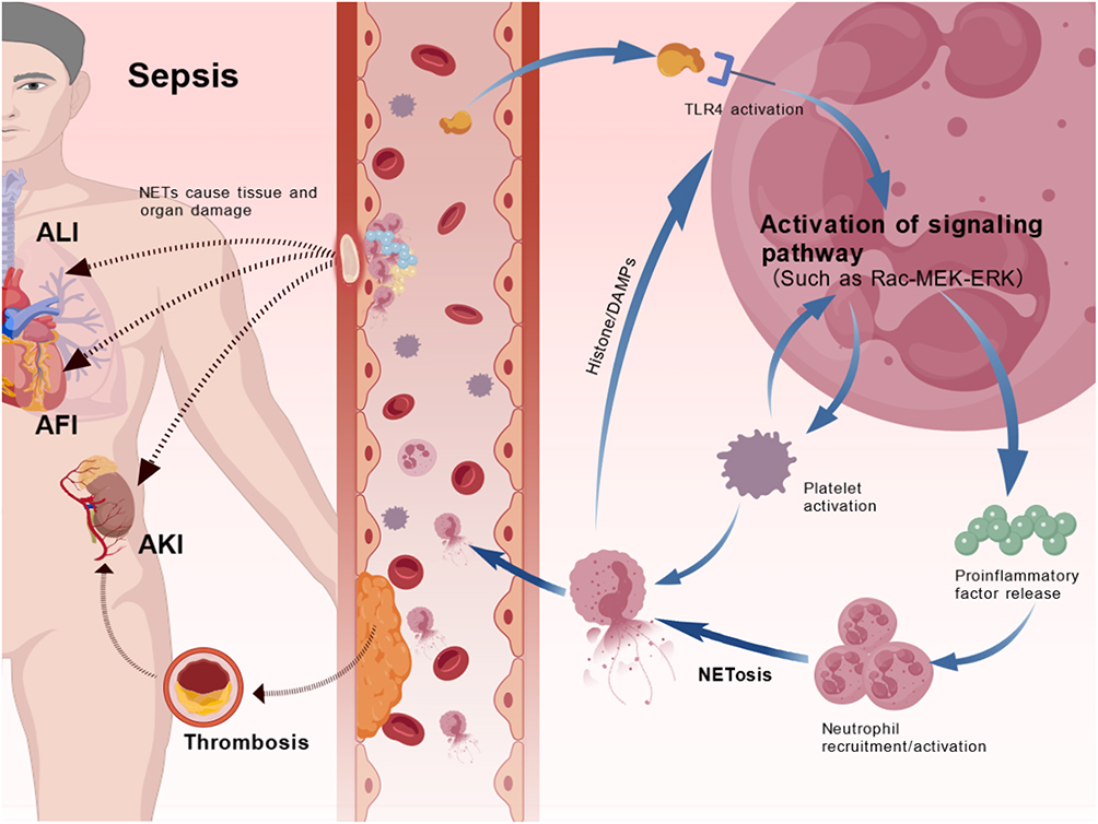 |
Figure 3 Pathological mechanism involving NETs. Activated TLR4 receptors on neutrophils mediate the release of inflammatory factors through the Rac-MEK-ERK signaling pathway, which also promotes neutrophil aggregation. Subsequently, neutrophils produce and release NETs. Additionally, activated platelets can further stimulate NET formation. Once formed, NETs activate neutrophils and amplify the inflammatory response. NETs can accumulate in microvessels, tissues, and organs. CitH3, a component of NETs, exerts cytotoxic effects on endothelial cells, and other enzymes and components of NETs contribute to these effects. These factors may lead to multiple organ damage, including thrombosis, ALI, AKI, and AFI. Abbreviations: ALI, acute lung injury; AKI, acute kidney injury; AMI, acute myocardial injury; citH3: NETs, neutrophil extracellular traps; TLR4, toll-like receptor4. |
The pathways through which PNCs release NETs are as follows:
- P-selectin on platelet surfaces binds to PSGL-1 on neutrophils, stimulating the release of ROS, MPO, NE, and other factors from neutrophils.110 Platelets also release high mobility group box 1 protein (HMGB1) and C3a, further enhancing neutrophil activity and promoting NET synthesis and release.119
- TLR4 on platelets detects TLR4 ligands in the blood, inducing platelet binding to adherent neutrophils and mediating increased NET release by polymorphonuclear neutrophils (PMNs).24,120
- Platelet-derived transforming growth factor-beta (TGF-β)121 enhances neutrophil retention in organs such as the lungs and liver by regulating NET formation. However, this pathway amplifies the inflammatory response122 and is often considered an aberrant activation mechanism for neutrophils.112
- Stimulation by LPS or DAMPs activates TLR4, triggering NET release via downstream signaling pathways, including the Raf-MEK-ERK pathway.123
NETs are Involved in Sepsis-Induced Thrombosis Formation
The synthesis and release of NETs lead to their binding with platelets, triggering the release of procoagulant factors. NETs also activate the extrinsic coagulation pathway by exposing tissue factor (TF) and inhibit the anticoagulation system, such as downregulating thrommoregulatory protein (TM), resulting in a systemic hypercoagulable state.110,124 Platelet–NET complexes accumulate in microvessels, further damaging endothelial cells through obstruction and local inflammation, which includes complement activation and ROS release. This exposure of subendothelial collagen recruits additional platelets and neutrophils, perpetuating a vicious cycle.121,125 Additionally, citH3 in NETs directly activates platelets and coagulation factor XII, thereby initiating the intrinsic coagulation pathway.126 Histones also exert cytotoxic effects, exacerbating endothelial cell damage. PNCs further promote the synthesis and release of NETs, which act as scaffolds for thrombosis by recruiting red blood cells, von Willebrand factor (VWF), and fibrin, thus enhancing the positive feedback loop in coagulation and thrombosis formation.111 P-selectin expressed on activated platelets binds to histones or DNA in NETs, while integrin αIIbβ3 adheres to the fibrous structure of NETs by binding to fibrinogen or VWF, stabilizing the platelet–NET complex.114,127 In summary, NETs and platelets activate both extrinsic and intrinsic coagulation pathways and promote vascular endothelial cell injury through multiple mechanisms. These processes culminate in microthrombi formation across various organs, depletion of coagulation factors, and the clinical manifestation of DIC, characterized by widespread hemorrhage and organ failure.
NETs are Involved in Sepsis-Associated ALI
Mengdi et al128 found that levels of MPO-DNA and cf-DNA were significantly elevated in serological tests of adult patients with sepsis-associated ALI. Following induction with PMA, neutrophils from these patients exhibited an enhanced ability to generate NETs, providing evidence that NETs contribute to ALI induced by sepsis.128 Sepsis pathogens activate the Raf-MEK-ERK pathway through TLR4 stimulation, promoting the release of inflammatory factors, which further recruit and activate neutrophils. These activated neutrophils release NETs within pulmonary microvessels. The DNA backbone and toxic components, such as citH3 and MPO, directly damage alveolar epithelial and endothelial cells, increasing vascular permeability, promoting microthrombi formation, and leading to pulmonary edema,129 which exacerbates ALI. Additionally, TLR4 on platelets binds to TLR4 ligands, inducing PMNs to release more NETs.24,120 The retention of PNCs in pulmonary capillaries facilitates fibrin deposition, triggering microthrombi formation, causing local ischemia, and worsening lung injury.121,127 Histones and proteases in NETs damage the alveolar-capillary barrier, aggravating pulmonary edema and oxygenation deficits.76 NETs further impair pulmonary microcirculation by activating platelets to form microthrombi.130 Mitochondrial DNA (mtDNA) in NETs activates macrophages via the TLR9/STING pathway, resulting in the release of pro-inflammatory factors, such as interleukin-1β (IL-1β) and tumor necrosis factor-α (TNF-α), which amplify inflammation and immune imbalance, thereby worsening lung injury.131,132 In the mouse cecal ligation and puncture (CLP) sepsis model, deletion of programmed death-ligand 1 (PD-L1) significantly reduced neutrophil infiltration into the lungs and lung injury.133 It also lowered plasma levels of pro-inflammatory factors like TNF-α and IL-6, while increasing the anti-inflammatory cytokine IL-10, leading to improved pathology.133 Meanwhile, inhibiting PD-L1 function (eg, by blocking the PI3K/Akt pathway with anti-PD-L1 antibodies) accelerated neutrophil apoptosis, reduced lung tissue damage, and alleviated the inflammatory burden.134 NETs contribute to ALI development in sepsis through immune thrombosis, inflammatory factor activation, disruption of signaling pathways, and endothelial injury.135–137 Clinical data show that increased NETs and platelet aggregation markers correlate with the severity of ALI. Inhibiting NET formation or employing antiplatelet therapies (eg, P2Y12 inhibitors) alleviates lung injury. Moreover, clinical trials have demonstrated that antiplatelet drugs, such as bellaprost, reduce the incidence of sepsis-ALI and ICU admissions.138,139
NETs are Involved in Sepsis-Associated Acute Myocardial Injury (SA-AMI)
Emerging evidence highlights the pivotal role of NETs in the pathogenesis of SA-AMI. Clinical studies consistently reveal elevated plasma NET levels in both pediatric and adult patients with sepsis,140–142 with a significant correlation between NET concentrations and the severity of myocardial injury.141,143 The harmful effects of NETs on myocardial tissue are mediated through several mechanisms:
- Prothrombotic Effects: NETs promote microvascular thrombosis by releasing histones and DNA, which activate coagulation factor XII, inducing platelet aggregation and fibrin deposition.108 This exacerbates myocardial ischemic injury in sepsis.144,145
- Direct Cardiomyocyte Injury: CitH3 directly damages endothelial cells, increases myocardial permeability, and triggers cardiomyocyte apoptosis via TLR4/9-mediated pathways, leading to the release of cardiac enzymes.141,143
- Structural and Functional Impairment: In LPS-induced septic rat models, myocardial NET deposition correlates with significant structural damage and functional deterioration of cardiomyocytes.143
- Mitochondrial Dysfunction: NETs stimulate neutrophil-derived ROS production, reducing mitochondrial membrane potential and impairing ATP synthesis in cardiomyocytes.146 Notably, ROS levels correlate positively with established markers of myocardial injury.145
The pathophysiological effects of NETs on myocardial conduction heterogeneity in sepsis are mediated by two key mechanisms: (1) Myocardial Fibrosis: NETs promote interstitial fibrosis and the formation of fibrous foci in myocardial tissue.143 PAD4-mediated histone citrullination induces atrial fibrosis and conduction heterogeneity.145 (2) Ion Channel Dysfunction: Histones inhibit potassium channel activity, leading to prolonged action potential duration.143 These alterations manifest clinically as a significantly increased risk of new-onset atrial fibrillation (NOAF) in patients with sepsis.143 Cumulatively, the evidence highlights NETs as critical mediators of sepsis-induced cardiac dysfunction, suggesting potential therapeutic targets for mitigating SA-AMI.
NETs are Involved in Sepsis-Associated Acute Kidney Injury (SA-AKI)
The pathogenesis of SA-AKI involves a key mechanism centered around the lactate-HMGB1-NETs axis. Lactate induces the lactylation of HMGB1, which subsequently triggers NETosis, leading to renal tubular damage and oxidative stress.147,148 Mechanistically, macrophages release lactylated HMGB1 via exosomes, activating neutrophils to release mitochondrial DNA and promoting NET formation.148 Clinical studies of peripheral blood from patients with SA-AKI showed that NET formation and oxidative stress responses were linked to the downregulation of epithelial cell transforming 2 (ECT2) and chordin-like 1 (CHRDL1) gene expression, as well as the upregulation of platelet-activating factor receptor (PTAFR), colony stimulating factor 3 (CSF3), and FBJ murine osteosarcoma viral oncogene homolog (FOS) gene expression.149 A 28-day follow-up study by Jian et al150 involving 136 patients with SA-AKI revealed that non-survivors exhibited significantly higher plasma NET markers compared to survivors, with these markers correlating positively with inflammatory indicators. Moreover, Jonas et al demonstrated in a prospective study of 601 patients with sepsis that elevated plasma levels of heparin-binding protein (HBP) were significantly associated with an increased risk of AKI development.151 Interestingly, a study of severely burned patients during the shock phase found that increased plasma HBP concentrations were associated with decreased NET release from neutrophils.152 However, whether HBP, as a neutrophil chemotactic factor, functionally contributes to NET generation and release during sepsis remains unclear and warrants further investigation.
NETs are Involved in Sepsis-Associated Other Organ Damage
Laboratory analysis of patients with sepsis revealed markedly elevated plasma levels of cfDNA and histones, which showed significant positive correlations with hepatic injury markers such as alanine aminotransferase (ALT) and aspartate aminotransferase (AST). These findings suggest that NETs play an active role in sepsis-induced liver injury.108,141 Mechanistic studies demonstrate that NETs contribute to hepatocyte damage through multiple pathways: (1) Released histones (H3 and H4) compromise plasma membrane integrity, induce oxidative stress, and disrupt mitochondrial function, ultimately triggering apoptotic or necrotic cell death;141,145 (2) NETs activate platelets and facilitate the binding of coagulation factors, accelerating thrombin generation. Concurrently, histones inhibit protein C anticoagulant pathways, exacerbating coagulopathy and promoting microthrombus formation, which induces localized ischemia and hypoxia, further aggravating parenchymal injury;108,153 (3) NET-associated proteases, particularly NE and MPO, activate Kupffer cells within the hepatic sinusoids, promoting the release of pro-inflammatory cytokines such as IL-1β and TNF-α, amplifying the inflammatory cascade and exacerbating tissue damage.141,145
In sepsis-associated brain injury, NETs contribute to neural dysfunction through dual mechanisms of inflammatory activation and coagulation cascade induction. Specifically, NETs exacerbate endothelial injury and disrupt blood-brain barrier integrity, promoting cerebral microvascular thrombosis.145 Clinical evidence indicates that NETs enhance the hypercoagulable state in sepsis, with elevated plasma NET levels significantly correlating with thromboembolic events in patients with sepsis.108 At the molecular level, NET formation upregulates pro-thrombotic and pro-inflammatory gene expression profiles. The subsequent release of inflammatory mediators traverses the compromised blood-brain barrier, activating microglia and leading to neuronal damage.143,145
Targeting NETosis in Sepsis: Therapeutic Strategies and Challenges
Recent studies have highlighted the critical role of NETs in exacerbating organ damage during sepsis. Concurrently, emerging evidence suggests that inhibiting NETosis to reduce NETs has emerged as a novel therapeutic strategy for sepsis, with the potential to mitigate multiple organ injury. The principal advantage of this approach lies in its direct intervention in NETosis, effectively lowering NETs levels and thereby attenuating inflammatory responses, thrombus formation, and organ dysfunction. For instance, in a neonatal sepsis model, treatment with recombinant human DNase (rhDNase) reduced plasma NET concentrations and ameliorated organ injury.141 In a neonatal mouse model of infectious peritonitis, DNase I directly degraded the DNA backbone of NETs, diminishing NET levels and inflammatory response.154 Moreover, DNase I interfered with NETosis, thereby improving the hypercoagulable state and reducing thrombosis, which contributed to the alleviation of sepsis-associated atrial fibrillation.108,143 Denorme et al154 demonstrated that the neonatal NET-Inhibitory Factor (nNIF), an endogenous inhibitor of NET formation, directly blocked NET generation, decreased inflammatory cytokine levels, and improved survival in a translational model of neonatal infectious peritonitis. Beyond directly targeting NETosis, another strategy involves inhibiting the activity of PAD4, which suppresses histone citrullination and indirectly blocks NET synthesis and release. For example, in both infant and neonatal sepsis models, administration of PAD4 inhibitors significantly reduced NET formation and enhanced survival rates.141,154 Additionally, Shirakawa et al155 reported that hydrogen gas (H2) therapy directly inhibited PAD4, leading to decreased NET levels. Independently of PAD4 activity, Fang et al135 showed that blocking macrophage-1 antigen (Mac-1) in an LPS-induced sepsis model suppressed direct contact between neutrophils and vascular endothelial cells, indirectly reducing NET formation.
However, targeting NETs presents several challenges. Although numerous studies suggest that excessive NET release exacerbates systemic inflammation and organ injury, NETs also serve as a crucial mechanism of neutrophil-mediated immune defense, particularly in early sepsis. Excessive inhibition of NETosis may therefore compromise antimicrobial immunity and increase infection risk.144,145 Future investigations into NETs-targeting therapies should address this dual role. Furthermore, given the complex pathophysiology of sepsis and the indirect relationship between NETs and disease progression, monotherapy targeting NETs alone may be insufficient to fully resolve sepsis-induced damage.108 A comprehensive therapeutic regimen, potentially combining NETs-targeting agents with antibiotics, may be necessary to form an integrated diagnostic and treatment framework.156
Potential Clinical Applications of Biomarkers of Nets in Sepsis
When sepsis occurs, tissue cells throughout the body are damaged, leading to the activation and death of immune cells, which results in the release of significant amounts of DNA into the bloodstream, forming cfDNA.119 CfDNA levels notably increase at the early onset of sepsis in patients.157 However, cfDNA can originate from various sources, including apoptotic cells, mitochondrial DNA, bacterial DNA, and others. It remains unclear whether cfDNA detected in the blood of patients with sepsis primarily originates from neutrophils or is associated with NETs. To investigate this, Nicholas et al158 found in a study of 49 patients with sepsis that the plasma concentration of cfDNA was significantly positively correlated with MPO levels. Additionally, citH3 levels were significantly higher compared to the control group.158 These findings suggest that the cfDNA detected in the serum of patients with sepsis primarily originates from NETs formed during NETosis.158 Supporting this, Joshua’s research on 14 patients with sepsis showed that cfDNA in 13 patients mainly came from neutrophils, although some patients had cfDNA derived from liver cells.159 Further methylation profiling of cfDNA revealed that it not only serves as a diagnostic marker for sepsis but also reflects organ damage caused by the condition.
Currently, studies on the use of citH3 for the early diagnosis of sepsis are limited. However, existing research highlights its diagnostic potential in other infectious diseases. CitH3 may serve as a biomarker for clinical diagnosis when combined with other detection indicators. Among patients with sepsis, MPO-DNA levels showed significant correlations with inflammatory markers like PCT and CRP, as well as coagulation function indicators such as prothrombin time-international normalized ratio (PT-INR), further reflecting the dual role of NETs in systemic inflammation and coagulopathy.160 The specific NET marker MPO-DNA was significantly elevated in the early stages (days 1, 3, and 7) of septic shock and can be detected by ELISA, providing evidence that MPO-DNA may serve as a diagnostic biomarker for sepsis.161 MPO-DNA levels were significantly correlated with mortality rates on days 3, 7, and 28. They were also strongly associated with organ function impairment: MPO-DNA levels were negatively correlated with mean arterial pressure and PaO2/FiO2 ratio, and positively correlated with the SOFA score.138 Moreover, neutrophil-derived exosomes, such as those containing MMP9, promote excessive NET formation, and their plasma levels are independently linked to the severity of sepsis and poor prognosis.162
A single biomarker is insufficient for the diagnosis of sepsis, as exclusive reliance on one marker increases the risk of false-positive results. Conditions such as trauma, surgery, ischemia-reperfusion injury, autoimmune diseases like rheumatoid arthritis (RA) and systemic lupus erythematosus (SLE), atherosclerosis, and others can also trigger the release of NETs.162–164 Therefore, detecting a single NET marker cannot reliably distinguish sepsis from these other conditions. Additionally, due to the multifaceted pathophysiology of sepsis, which involves inflammation, immune response, coagulation, and microcirculatory disturbances, measuring NETs alone does not fully reflect the complexity of the condition. NETs may represent only one aspect of sepsis’ pathophysiological cascade. A combined measurement of multiple biomarkers provides more accurate clinical diagnostic support. For example, the simultaneous detection of inflammatory markers like MPO-DNA complexes and CRP offers a more comprehensive view of the abnormal inflammatory responses and coagulation dysfunctions that extend beyond sepsis. Moreover, cfDNA combined with CRP can serve as valuable biomarkers for assessing the severity of sepsis and is particularly useful in differentiating complex bloodstream infections from milder forms.165 Additionally, citH3 and MPO co-localization staining has been employed to identify coronary thrombosis distribution in patients with ST-segment elevation myocardial infarction (STEMI).105
As discussed in Biomarkers and Detection Techniques Related to NETs, although a variety of biomarkers are available for NET detection, cfDNA, citH3, and MPO-DNA complexes are among the most frequently utilized diagnostic biomarkers in clinical settings. However, current clinical research predominantly compares patients with sepsis to healthy controls, without adequately addressing the distinction between septic and non-septic individuals within the same complication context. For example, in studies of ALI, most experimental designs have focused on comparing sepsis-induced ALI with healthy individuals, with a notable scarcity of studies comparing sepsis-induced ALI with non-sepsis-induced ALI. Thus, the reliability of these biomarkers in differentiating between these two distinct ALI etiologies remains uncertain and warrants further rigorous investigation. As shown in Table 1, NETs, as emerging biomarkers, hold substantial clinical potential in research on sepsis and its complications.
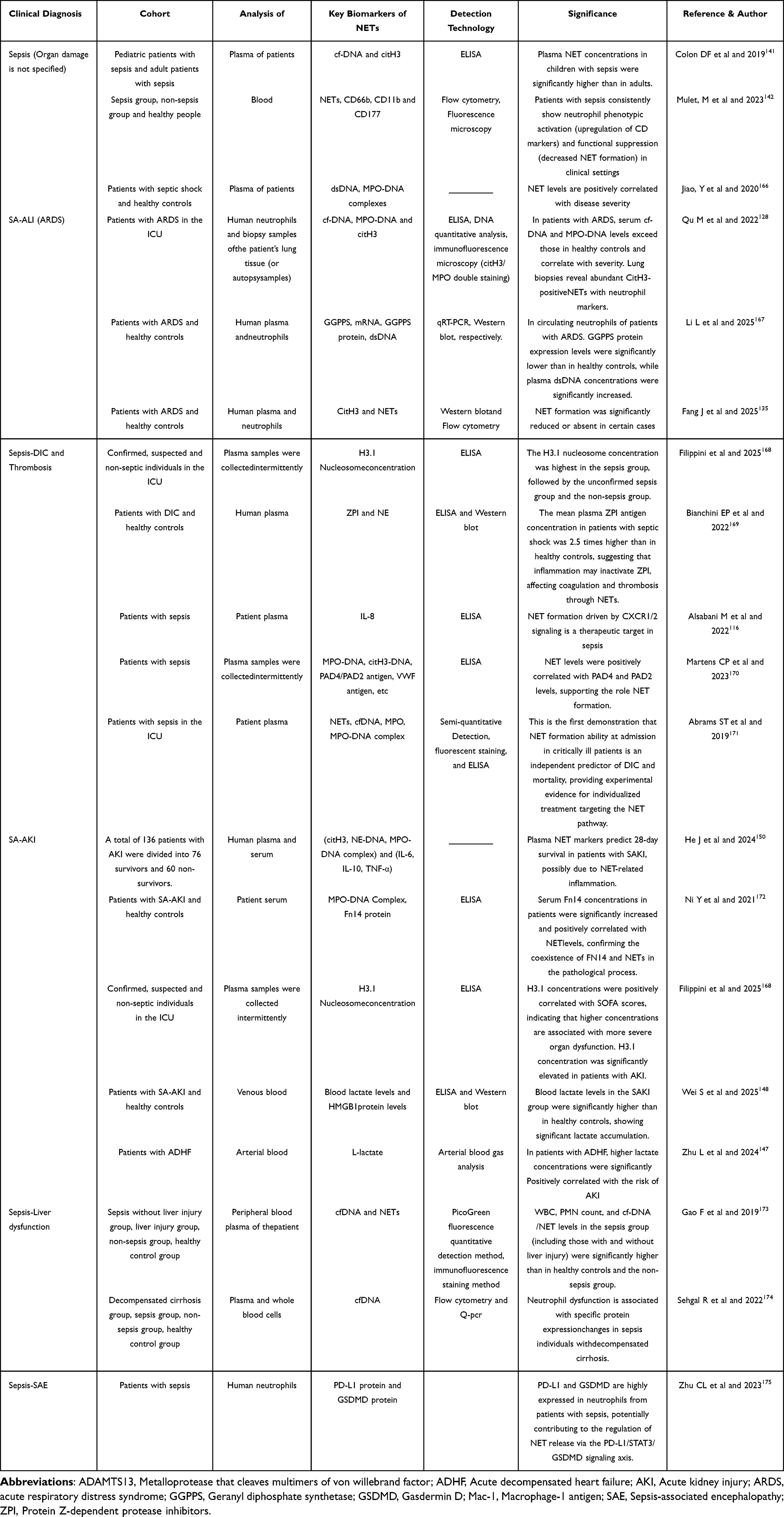 |
Table 1 Clinical Research of NET-Mediated Pathogenesis in Sepsis Complications (2019–2025) |
Conclusion
Sepsis is a complex clinical syndrome, and research on NETs has highlighted their pivotal role in host immune responses and pathophysiological processes. NETs are fibrous DNA structures released by activated neutrophils, capable of capturing and eliminating pathogens. However, excessive NET release can lead to host tissue damage. In the context of sepsis, the relationship between NET formation and clinical complications is intricate. While NETs help contain infections, their overproduction can contribute to intravascular coagulation, thrombosis, DIC, ARDS (ALI), and multiple organ dysfunction. The excessive formation and release of NETs correlate closely with disease severity and poor prognosis. Recent advances in detection technology have enhanced our ability to assess NETs’ presence and activity, offering new insights into both basic research and clinical diagnostics. For instance, NET formation can be indirectly evaluated by measuring plasma cfDNA levels and specific NET-associated proteins such as MPO, citH3, and NE. Clinical investigations have shown that the serum NET levels in pediatric patients with sepsis are significantly higher than in adult patients and healthy controls, suggesting an increased NET production in children with sepsis. Moreover, the level of NETs was found to correlate positively with the severity of sepsis in pediatric patients.141
Future research must further elucidate the mechanisms of NETosis in sepsis and develop therapies that balance the antimicrobial benefits of NET formation with the prevention of tissue damage. Establishing age-specific diagnostic criteria, particularly for pediatric populations, and optimizing rapid detection methods are critical for timely intervention. Standardizing NET quantification would enhance diagnostic accuracy. These advancements collectively offer: (1) A critical temporal window for life-saving interventions and (2) A foundation for personalized therapeutic approaches. The implementation of standardized NET assessment protocols could transform diagnostic practices, providing both prognostic value and therapeutic guidance in sepsis management.
Abbreviations
AST, aspartate aminotransferase; Akt, Ak strain transforming; AKI, acute kidney injury; ALT, alanine aminotransferase; ALI, acute lung injury; ATC, anaplastic thyroid carcinoma; AMI, acute myocardial injury; B2M, beta-2-Microglobulin; cfDNA, free deoxyribonucleic acid; citH3, histone H3 subunit citrullinated; CR3, complement receptor3; CDK1, cyclin-dependent kinase1; CRP, C-reactive protein; CXCR, C-X-C motif chemokine receptor; CLP, cecal ligation and puncture; CHRDL1, chordin-like 1 (CHRDL1); CSF3, colony stimulating factor 3; DIC, disseminated intravascular coagulation; dsDNA, double-stranded DNA; ECT2, epithelial cell transforming 2; ELISA, enzyme-linked immunosorbent assays; ERK, extracellular regulated protein kinases; FOS, FBJ murine osteosarcoma viral oncogene homolog; GTPases, guanosine triphosphatases; GPX3, glutathione peroxidase 3; GM-CSF, macrophage colony-stimulating factor; GSDMD, gasdermin D; HBP, heparin-binding protein; HMGB1, high mobility group box 1 protein; IL-1β, interleukin-1 β; LPS, lipopolysaccharide; mtDNA, mitochondrial DNA; MVs, microvesicles; MAP3K5, mitogen-activated protein kinase5; MPO, myeloperoxidase; MPO-DNA, myeloperoxidase DNA complex; NETs, neutrophil extracellular traps; NS1, non-structural protein 1; NADPH, nicotinamide adenine dinucleotide phosphate; NOX, NADPH oxidase; NE, neutrophil elastase; NOAF, new-onset atrial fibrillation; PKM2-LDH, pyruvate kinase muscle isozyme m2-lactate dehydrogenase; PAD4, peptidylarginine deiminase 4; PMA, phorbol myristate acetate; PD-L1, programmed death-ligand1; PNCs, platelet-neutrophil complexes; PAMPs, pathogen-associated molecular patterns; PRR, pattern recognition receptors; PMN, polymorphic neutrophils; PT-INR, prothrombin time-international normalized ratio; PTAFR, platelet-activating factor receptor; Rac2, ras-related C3 botulinum toxin substrate 2; ROS, active oxygen; SK3, small conductance potassium; STEMI, ST-segment elevation myocardial infarction; SAE, sepsis-associated encephalopathy; SA-AMI, sepsis associated atrial fibrillation; SA-AKI, sepsis-associated acute kidney injury; TLR, toll-like receptor; TNF-α, tumor necrosis factor-α; TGF-β, transforming growth factor-beta; TF, tissue factor; TM, thrombomodulin; UA, high-dose ultraviolet light; VWF, von willebrand factor.
Data Sharing Statement
Data sharing is not applicable to this article as no new data were created or analyzed in this study.
Ethical Approval
This review did not require ethical approval as it was based on previously published studies.
Acknowledgments
We thank Bullet Edits Limited for the language editing and proofreading of the manuscript.
Funding
This work was supported by the National Natural Science Foundation of China (82200097), the Key Research, Development, and Promotion Projects of Henan Province (252102310054 and 232102310122), the Inner Mongolia Natural Science Fund project (2024LHMS08032, 2023QN08063 and 2025QN08087), and the Aerospace Medical Research Project (2025YK03).
Disclosure
No potential conflict of interest was reported by the author(s).
References
1. Cox D. Sepsis - it is all about the platelets. Front Immunol. 2023;14:1210219. doi:10.3389/fimmu.2023.1210219
2. Fousert E, Toes R, Desai J, Behrends J, Hölscher C. Neutrophil extracellular traps (NETs) take the central stage in driving autoimmune responses. Cells. 2020;10:9. doi:10.3390/cells10010009
3. Chen W, Pan JY. Anatomical and pathological observation and analysis of SARS and COVID-19: microthrombosis is the main cause of death. Biol Proced Online. 2021;23:4. doi:10.1186/s12575-021-00142-y
4. Hong G, Ju H, Oh DK, et al. Clinical characteristics and prognostic factors of sepsis in patients with malignancy. Sci Rep. 2025;15:7078. doi:10.1038/s41598-025-87457-y
5. Abe T, Saito K, Nagano T, Yamada Y, Ochiai H. Complement system activation through the alternative pathway associates with disseminated intravascular coagulation to increase mortality in sepsis. Thromb Res. 2025;247:109281. doi:10.1016/j.thromres.2025.109281
6. Thang NVV, Luyen LT, Vi NTT, Hai PD. Neutrophil-to-lymphocyte-to-albumin ratio as a prognostic marker for mortality in sepsis and septic shock in Vietnam. Acute Crit Care. 2025;40:244–251. doi:10.4266/acc.003576
7. Cheng H, Shao L, Wu H, Mi B, Li Q, Zhang J. Older adult sepsis survivors discharged to skilled nursing facilities: age-adjusted charlson comorbidity index as a predictor of 6-month mortality. Nurs Crit Care. 2025;30:e70078. doi:10.1111/nicc.70078
8. Xu Y, Zhang H, Li J, Wang N, Yuan H. Risk factors analysis of 90-day mortality in patients with sepsis in intensive care unit. PLoS One. 2025;20:e0325813. doi:10.1371/journal.pone.0325813
9. Gierlikowska B, Stachura A, Gierlikowski W, Demkow U. Phagocytosis, degranulation and extracellular traps release by neutrophils-the current knowledge, pharmacological modulation and future prospects. Front Pharmacol. 2021;12:666732. doi:10.3389/fphar.2021.666732
10. Lee M, Lee SY, Bae YS. Emerging roles of neutrophils in immune homeostasis. BMB Rep. 2022;55:473–480. doi:10.5483/BMBRep.2022.55.10.115
11. Brinkmann V, Reichard U, Goosmann C, et al. Neutrophil extracellular traps kill bacteria. Science. 2004;303:1532–1535. doi:10.1126/science.1092385
12. Pignataro G, Gemma S, Petrucci M, et al. Unraveling NETs in sepsis: from cellular mechanisms to clinical relevance. Int J Mol Sci. 2025;26: 7464.
13. Lee KH, Kronbichler A, Park DD, et al. Neutrophil extracellular traps (NETs) in autoimmune diseases: a comprehensive review. Autoimmun Rev. 2017;16:1160–1173. doi:10.1016/j.autrev.2017.09.012
14. Wang H, Kim SJ, Lei Y, et al. Neutrophil extracellular traps in homeostasis and disease. Signal Transduct Target Ther. 2024;9:235. doi:10.1038/s41392-024-01933-x
15. Drury B, Hardisty G, Gray RD, Ho GT. Neutrophil extracellular traps in inflammatory bowel disease: pathogenic mechanisms and clinical translation. Cell Mol Gastroenterol Hepatol. 2021;12:321–333. doi:10.1016/j.jcmgh.2021.03.002
16. Zlatar L, Knopf J, Singh J, et al. Neutrophil extracellular traps characterize caseating granulomas. Cell Death Dis. 2024;15:548. doi:10.1038/s41419-024-06892-3
17. Mutua V, Gershwin LJ. A review of neutrophil extracellular traps (NETs) in disease: potential Anti-NETs therapeutics. Clin Rev Allergy Immunol. 2021;61:194–211. doi:10.1007/s12016-020-08804-7
18. Fei Y, Huang X, Ning F, et al. NETs induce ferroptosis of endothelial cells in LPS-ALI through SDC-1/HS and downstream pathways. Biomed Pharmacother. 2024;175:116621. doi:10.1016/j.biopha.2024.116621
19. Meng W, Paunel-Gorgulu A, Flohe S, et al. Depletion of neutrophil extracellular traps in vivo results in hypersusceptibility to polymicrobial sepsis in mice. Crit Care. 2012;16:R137. doi:10.1186/cc11442
20. King PT, Dousha L, Pasalic L. Neutrophil extracellular traps and respiratory disease. J Clin Med. 2024;14:13. doi:10.3390/jcm14010013
21. Arazna M, Pruchniak MP, Zycinska K, Demkow U. Neutrophil extracellular trap in human diseases. Adv Exp Med Biol. 2013;756:1–8.
22. Pieterse E, Rother N, Yanginlar C, et al. Cleaved N-terminal histone tails distinguish between NADPH oxidase (NOX)-dependent and NOX-independent pathways of neutrophil extracellular trap formation. Ann Rheum Dis. 2018;77:1790–1798. doi:10.1136/annrheumdis-2018-213223
23. Nomura K, Miyashita T, Yamamoto Y, et al. Citrullinated histone H3: early biomarker of neutrophil extracellular traps in septic liver damage. J Surg Res. 2019;234:132–138. doi:10.1016/j.jss.2018.08.014
24. Ravindran M, Khan MA, Palaniyar N. Neutrophil extracellular trap formation: physiology, pathology, and pharmacology. Biomolecules. 2019;10:9. doi:10.3390/biom10010009
25. Tan C, Aziz M, Wang P. The vitals of NETs. J Leukoc Biol. 2021;110:797–808. doi:10.1002/JLB.3RU0620-375R
26. Wang Y, Du C, Zhang Y, Zhu L. Composition and function of neutrophil extracellular traps. Biomolecules. 2024;15:14. doi:10.3390/biom15010014
27. Berkes E, Oehmke F, Tinneberg HR, Preissner KT, Saffarzadeh M. Association of neutrophil extracellular traps with endometriosis-related chronic inflammation. Eur J Obstet Gynecol Reprod Biol. 2014;183:193–200. doi:10.1016/j.ejogrb.2014.10.040
28. Hornigold K, Chu JY, Chetwynd SA, et al. Age-related decline in the resistance of mice to bacterial infection and in LPS/TLR4 pathway-dependent neutrophil responses. Front Immunol. 2022;13:888415. doi:10.3389/fimmu.2022.888415
29. Yousefi S, Mihalache C, Kozlowski E, Schmid I, Simon HU. Viable neutrophils release mitochondrial DNA to form neutrophil extracellular traps. Cell Death Differ. 2009;16:1438–1444. doi:10.1038/cdd.2009.96
30. Zhu Y, Xia X, He Q, et al. Diabetes-associated neutrophil NETosis: pathogenesis and interventional target of diabetic complications. Front Endocrinol. 2023;14:1202463. doi:10.3389/fendo.2023.1202463
31. Takishita Y, Yasuda H, Shimizu M, et al. Formation of neutrophil extracellular traps in mitochondrial DNA-deficient cells. J Clin Biochem Nutr. 2020;66:15–23. doi:10.3164/jcbn.19-77
32. Garishah FM, Rother N, Riswari SF, et al. Neutrophil Extracellular Traps in Dengue Are Mainly Generated NOX-Independently. Front Immunol. 2021;12:629167. doi:10.3389/fimmu.2021.629167
33. Gardiner EE, Andrews RK. Neutrophil extracellular traps (NETs) and infection-related vascular dysfunction. Blood Rev. 2012;26:255–259. doi:10.1016/j.blre.2012.09.001
34. Sil P, Yoo DG, Floyd M, Gingerich A, Rada B. High throughput measurement of extracellular DNA release and quantitative NET formation in human neutrophils in vitro. J Vis Exp. 2016. doi:10.3791/52779
35. Li T, Zhang Z, Li X, et al. Neutrophil extracellular traps: signaling properties and disease relevance. Mediators Inflamm. 2020;2020:9254087. doi:10.1155/2020/9254087
36. Paclet MH, Laurans S, Dupre-Crochet S. Regulation of neutrophil NADPH oxidase, NOX2: a crucial effector in neutrophil phenotype and function. Front Cell Dev Biol. 2022;10:945749. doi:10.3389/fcell.2022.945749
37. Ansari J, Vital SA, Yadav S, Gavins FNE. Regulating neutrophil PAD4/NOX-dependent cerebrovasular thromboinflammation. Int J Biol Sci. 2023;19:852–864. doi:10.7150/ijbs.77434
38. Korba-Mikolajczyk A, Sluzalska KD, Kasperkiewicz P. Exploring the involvement of serine proteases in neutrophil extracellular traps: a review of mechanisms and implications. Cell Death Dis. 2025;16(1):535. doi:10.1038/s41419-025-07857-w
39. Azzouz D, Palaniyar N, Li N. How do ROS induce NETosis? Oxidative DNA damage, DNA repair, and chromatin decondensation. Biomolecules. 2024;15:14.
40. Nishinaka Y, Arai T, Adachi S, Takaori-Kondo A, Yamashita K. Singlet oxygen is essential for neutrophil extracellular trap formation. Biochem Biophys Res Commun. 2011;413:75–79. doi:10.1016/j.bbrc.2011.08.052
41. Yu HH, Yang YH, Chiang BL. Chronic granulomatous disease: a comprehensive review. Clin Rev Allergy Immunol. 2021;61:101–113. doi:10.1007/s12016-020-08800-x
42. Chen Y, Hu H, Tan S, et al. The role of neutrophil extracellular traps in cancer progression, metastasis and therapy. Exp Hematol Oncol. 2022;11:99. doi:10.1186/s40164-022-00345-3
43. Hurtado Gutierrez MJ, Allard FL, Mosha HT, Dubois CM, McDonald PP. Human neutrophils generate extracellular vesicles that modulate their functional responses. Cells. 2022;12:12. doi:10.3390/cells12010012
44. Naffah de Souza C, Breda LCD, Khan MA, et al. Alkaline pH promotes NADPH oxidase-independent neutrophil extracellular trap formation: a matter of mitochondrial reactive oxygen species generation and citrullination and cleavage of histone. Front Immunol. 2017;8:1849. doi:10.3389/fimmu.2017.01849
45. Awasthi D, Nagarkoti S, Sadaf S, Chandra T, Kumar S, Dikshit M. Glycolysis dependent lactate formation in neutrophils: a metabolic link between NOX-dependent and independent NETosis. Biochim Biophys Acta Mol Basis Dis. 2019;1865:165542. doi:10.1016/j.bbadis.2019.165542
46. Nishinaka Y, Arai Y, Arai T, et al. Uric acid induces NADPH oxidase-independent neutrophil extracellular trap formation. Biochem Biophys Res Commun. 2014;443:556–561. doi:10.1016/j.bbrc.2013.12.007
47. Raza S, Rajak S, Singh R, Zhou J, Sinha RA, Goel A. Cell-type specific role of autophagy in the liver and its implications in non-alcoholic fatty liver disease. World J Hepatol. 2023;15:1272–1283. doi:10.4254/wjh.v15.i12.1272
48. Sheahan BJ, Schubert AG, Schubert W, Sheats MK, Schnabel LV, Gilbertie JM. Equine neutrophils selectively release neutrophil extracellular traps in response to chemical and bacterial agonists. Front Vet Sci. 2025;12:1512343. doi:10.3389/fvets.2025.1512343
49. Germic N, Stojkov D, Oberson K, Yousefi S, Simon HU. Neither eosinophils nor neutrophils require ATG5-dependent autophagy for extracellular DNA trap formation. Immunology. 2017;152:517–525. doi:10.1111/imm.12790
50. Douda DN, Khan MA, Grasemann H, Palaniyar N. SK3 channel and mitochondrial ROS mediate NADPH oxidase-independent NETosis induced by calcium influx. Proc Natl Acad Sci U S A. 2015;112:2817–2822. doi:10.1073/pnas.1414055112
51. Gosswein S, Lindemann A, Mahajan A, et al. Citrullination licenses calpain to decondense nuclei in neutrophil extracellular trap formation. Front Immunol. 2019;10:2481. doi:10.3389/fimmu.2019.02481
52. Papayannopoulos V. Neutrophil extracellular traps in immunity and disease. Nat Rev Immunol. 2018;18:134–147. doi:10.1038/nri.2017.105
53. Azzouz D, Khan MA, Sweezey N, Palaniyar N. Two-in-one: UV radiation simultaneously induces apoptosis and NETosis. Cell Death Discov. 2018;4:51. doi:10.1038/s41420-018-0048-3
54. Azzouz D, Palaniyar N. ApoNETosis: discovery of a novel form of neutrophil death with concomitant apoptosis and NETosis. Cell Death Dis. 2018;9:839. doi:10.1038/s41419-018-0846-9
55. Strozyk E, Kulms D. The role of AKT/mTOR pathway in stress response to UV-irradiation: implication in skin carcinogenesis by regulation of apoptosis, autophagy and senescence. Int J Mol Sci. 2013;14:15260–15285. doi:10.3390/ijms140815260
56. Liu S, Feng M, Guan W. Mitochondrial DNA sensing by STING signaling participates in inflammation, cancer and beyond. Int J Cancer. 2016;139:736–741. doi:10.1002/ijc.30074
57. Fisher AB. Reactive oxygen species and cell signaling with lung ischemia. Undersea Hyperb Med. 2004;31:97–103.
58. Averill-Bates D. Reactive oxygen species and cell signaling. Biochim Biophys Acta Mol Cell Res. 2024;1871:119573. doi:10.1016/j.bbamcr.2023.119573
59. Shukrun R, Fidel V, Baron S, et al. Neutrophil extracellular traps in pediatric inflammatory bowel disease: a potential role in ulcerative colitis. Int J Mol Sci. 2024;26:25. doi:10.3390/ijms26010025
60. Liu Y, Ma YH, Yang JW, et al. Rethinking neutrophil extracellular traps. Int Immunopharmacol. 2023;124:110834. doi:10.1016/j.intimp.2023.110834
61. de Bont CM, Koopman WJH, Boelens WC, Pruijn GJM. Stimulus-dependent chromatin dynamics, citrullination, calcium signalling and ROS production during NET formation. Biochim Biophys Acta Mol Cell Res. 2018;1865:1621–1629. doi:10.1016/j.bbamcr.2018.08.014
62. Petretto A, Bruschi M, Pratesi F, et al. Neutrophil extracellular traps (NET) induced by different stimuli: a comparative proteomic analysis. PLoS One. 2019;14:e0218946. doi:10.1371/journal.pone.0218946
63. Carestia A, Kaufman T, Rivadeneyra L, et al. Mediators and molecular pathways involved in the regulation of neutrophil extracellular trap formation mediated by activated platelets. J Leukoc Biol. 2016;99:153–162. doi:10.1189/jlb.3A0415-161R
64. Caruso RA, Fedele F, Rigoli L, et al. Apoptotic-like tumor cells and apoptotic neutrophils in mitochondrion-rich gastric adenocarcinomas: a comparative study with light and electron microscopy between these two forms of cell death. Rare Tumors. 2013;5:68–71. doi:10.4081/rt.2013.e18
65. Stewart KS, Abdusselamoglu MD, Tierney MT, et al. Stem cells tightly regulate dead cell clearance to maintain tissue fitness. Nature. 2024;633:407–416. doi:10.1038/s41586-024-07855-6
66. Jiang X, Li Q, Huang R, et al. Giardia duodenalis triggered neutrophil extracellular traps in goats. Immunobiology. 2025;230:152894. doi:10.1016/j.imbio.2025.152894
67. Zhang S, Lu Y, Zhao Y, et al. Dectin-1 participates in beta-glucan- or PMA-induced neutrophil extracellular trap formation during antifungal defense. Am J Transl Res. 2025;17:3898–3907. doi:10.62347/YQCM4496
68. Zhang K, He W, Guo X, Zhang Y, Huang C. Progress in mechanism of formation of neutrophil extracellular traps: review. Xi Bao Yu Fen Zi Mian Yi Xue Za Zhi. 2020;36:561–564.
69. Porto BN, Stein RT. Neutrophil extracellular traps in pulmonary diseases: too much of a good thing? Front Immunol. 2016;7:311. doi:10.3389/fimmu.2016.00311
70. Domon H, Oda M, Maekawa T, Nagai K, Takeda W, Terao Y. Streptococcus pneumoniae disrupts pulmonary immune defence via elastase release following pneumolysin-dependent neutrophil lysis. Sci Rep. 2016;6:38013. doi:10.1038/srep38013
71. Chow JC, Young DW, Golenbock DT, Christ WJ, Gusovsky F. Toll-like receptor-4 mediates lipopolysaccharide-induced signal transduction. J Biol Chem. 1999;274:10689–10692. doi:10.1074/jbc.274.16.10689
72. Akk A, Springer LE, Pham CT. Neutrophil extracellular traps enhance early inflammatory response in sendai virus-induced asthma phenotype. Front Immunol. 2016;7:325. doi:10.3389/fimmu.2016.00325
73. Munoz Caro T, Hermosilla C, Silva LM, Cortes H, Taubert A. Neutrophil extracellular traps as innate immune reaction against the emerging apicomplexan parasite Besnoitia besnoiti. PLoS One. 2014;9:e91415. doi:10.1371/journal.pone.0091415
74. Pilsczek FH, Salina D, Poon KK, et al. A novel mechanism of rapid nuclear neutrophil extracellular trap formation in response to Staphylococcus aureus. J Immunol. 2010;185:7413–7425. doi:10.4049/jimmunol.1000675
75. Lv G, Wang H, Wang J, Lian S, Wu R. Effect of BLV infection on the immune function of polymorphonuclear neutrophil in dairy cows. Front Vet Sci. 2021;8:737608. doi:10.3389/fvets.2021.737608
76. Denning NL, Aziz M, Gurien SD, Wang P. DAMPs and NETs in sepsis. Front Immunol. 2019;10:2536. doi:10.3389/fimmu.2019.02536
77. Cristinziano L, Modestino L, Loffredo S, et al. Anaplastic thyroid cancer cells induce the release of mitochondrial extracellular DNA traps by viable neutrophils. J Immunol. 2020;204:1362–1372. doi:10.4049/jimmunol.1900543
78. Xia L, Yan X, Zhang H. Mitochondrial DNA-activated cGAS-STING pathway in cancer: mechanisms and therapeutic implications. Biochim Biophys Acta Rev Cancer. 2025;1880:189249. doi:10.1016/j.bbcan.2024.189249
79. Tumburu L, Ghosh-Choudhary S, Seifuddin FT, et al. Circulating mitochondrial DNA is a proinflammatory DAMP in sickle cell disease. Blood. 2021;137:3116–3126. doi:10.1182/blood.2020009063
80. Liu L, Mao Y, Xu B, et al. Induction of neutrophil extracellular traps during tissue injury: involvement of STING and Toll-like receptor 9 pathways. Cell Prolif. 2020;53:e12775. doi:10.1111/cpr.12775
81. Zheng Y, Zhu Y, Liu X, et al. The screening of albumin as a key serum component in preventing release of neutrophil extracellular traps by selectively inhibiting mitochondrial ROS generation. Can J Physiol Pharmacol. 2021;99:427–438. doi:10.1139/cjpp-2019-0670
82. Zeng FL, Zhang Y, Wang ZH, et al. Neutrophil extracellular traps promote Acetaminophen-induced acute liver injury in mice via AIM2. Acta Pharmacol Sin. 2024;45:1660–1672. doi:10.1038/s41401-024-01239-2
83. Delgado-Rizo V, Martinez-Guzman MA, Iniguez-Gutierrez L, Garcia-Orozco A, Alvarado-Navarro A, Fafutis-Morris M. Neutrophil extracellular traps and its implications in inflammation: an overview. Front Immunol. 2017;8:81. doi:10.3389/fimmu.2017.00081
84. Berthelot JM, Le Goff B, Neel A, Maugars Y, Hamidou M. NETosis: at the crossroads of rheumatoid arthritis, lupus, and vasculitis. Joint Bone Spine. 2017;84:255–262. doi:10.1016/j.jbspin.2016.05.013
85. Salvianti F, Gelmini S, Mancini I, et al. Circulating tumour cells and cell-free DNA as a prognostic factor in metastatic colorectal cancer: the OMITERC prospective study. Br J Cancer. 2021;125:94–100. doi:10.1038/s41416-021-01399-6
86. Sabbatini M, Bona E, Novello G, Migliario M, Reno F. Aging hampers neutrophil extracellular traps (NETs) efficacy. Aging Clin Exp Res. 2022;34:2345–2353. doi:10.1007/s40520-022-02201-0
87. Eickbush TH, Moudrianakis EN. The histone core complex: an octamer assembled by two sets of protein-protein interactions. Biochemistry. 1978;17:4955–4964. doi:10.1021/bi00616a016
88. Li P, Liang S, Wang L, Guan X, Wang J, Gong P. Predictive value of neutrophil extracellular trap components for 28-day all-cause mortality in patients with cardiac arrest: a pilot observational study. Shock. 2023;60:664–670. doi:10.1097/SHK.0000000000002225
89. Yang S, Xiao Y, Du Y, et al. Diagnostic and prognostic value of neutrophil extracellular trap levels in patients with acute aortic dissection. Front Cardiovasc Med. 2021;8:683445. doi:10.3389/fcvm.2021.683445
90. Sanders NL, Martin IMC, Sharma A, et al. Neutrophil extracellular traps as an exacerbating factor in bacterial pneumonia. Infect Immun. 2022;90:e0049121. doi:10.1128/iai.00491-21
91. Gupta AK, Joshi MB, Philippova M, et al. Activated endothelial cells induce neutrophil extracellular traps and are susceptible to NETosis-mediated cell death. FEBS Lett. 2010;584:3193–3197. doi:10.1016/j.febslet.2010.06.006
92. Yoo DG, Floyd M, Winn M, Moskowitz SM, Rada B. NET formation induced by Pseudomonas aeruginosa cystic fibrosis isolates measured as release of myeloperoxidase-DNA and neutrophil elastase-DNA complexes. Immunol Lett. 2014;160:186–194. doi:10.1016/j.imlet.2014.03.003
93. Huang H, Zhang H, Onuma AE, Tsung A. Neutrophil elastase and neutrophil extracellular traps in the tumor microenvironment. Adv Exp Med Biol. 2020;1263:13–23.
94. Liu H, Han J, Ni W, et al. The diagnostic value of plasma NETs levels and iCEB in silent myocardial ischemia in maintenance hemodialysis patients. J Inflamm Res. 2025;18:847–862. doi:10.2147/JIR.S488836
95. Yang Z, Wang S, Yin K, Zhang Q, Li S. MiR-1696/GPx3 axis is involved in oxidative stress mediated neutrophil extracellular traps inhibition in chicken neutrophils. J Cell Physiol. 2021;236:3688–3699. doi:10.1002/jcp.30105
96. Pei J, Weng H, Peng J, et al. Identification of potential targets regulating neutrophil extracellular traps in acute rejection of kidney transplantation based on transcriptomics and animal experiments. Int Immunopharmacol. 2025;147:114008. doi:10.1016/j.intimp.2024.114008
97. Rada B. Neutrophil extracellular traps. Methods Mol Biol. 2019;1982:517–528.
98. Nakazawa D, Kumar SV, Marschner J, et al. Histones and neutrophil extracellular traps enhance tubular necrosis and remote organ injury in ischemic AKI. J Am Soc Nephrol. 2017;28:1753–1768. doi:10.1681/ASN.2016080925
99. Ouyang J, Tian Y, Jiang C, Yang Q, Wang H, Li Q. Laboratory assays on the effects of a novel acaricide, SYP-9625 on Tetranychus cinnabarinus (Boisduval) and its natural enemy, Neoseiulus californicus (McGregor). PLoS One. 2018;13:e0199269. doi:10.1371/journal.pone.0199269
100. Liou JS, Derito CM, Madsen EL. Field-based and laboratory stable isotope probing surveys of the identities of both aerobic and anaerobic benzene-metabolizing microorganisms in freshwater sediment. Environ Microbiol. 2008;10:1964–1977. doi:10.1111/j.1462-2920.2008.01612.x
101. Islam MM, Salma U, Irahara T, Watanabe E, Takeyama N. Quantifying myeloperoxidase-DNA and neutrophil elastase-DNA complexes from neutrophil extracellular traps by using a modified sandwich ELISA. J Vis Exp. 2023. doi:10.3791/64644
102. Pastorek M, Konecna B, Janko J, et al. Mitochondria-induced formation of neutrophil extracellular traps is enhanced in the elderly via Toll-like receptor 9. J Leukoc Biol. 2023;114:651–665. doi:10.1093/jleuko/qiad101
103. Park JH, Kim JE, Gu JY, et al. Evaluation of circulating markers of neutrophil extracellular trap (NET) formation as risk factors for diabetic retinopathy in a case-control association study. Exp Clin Endocrinol Diabetes. 2016;124:557–561. doi:10.1055/s-0042-101792
104. Lenz M, Maiberger T, Armbrust L, et al. cfDNA and DNases: new biomarkers of sepsis in preterm neonates-A pilot study. Cells. 2022;12:11. doi:10.3390/cells12010011
105. Santos A, Martin P, Blasco A, et al. NETs detection and quantification in paraffin embedded samples using confocal microscopy. Micron. 2018;114:1–7. doi:10.1016/j.micron.2018.07.002
106. Huckriede J, Keulen GM, van de Poll MCG, Wichapong K, Reutelingsperger CPM, Nicolaes GAF. Proteolytic neutralization of extracellular histones by neutrophil elastase is enhanced by heparin. J Thromb Haemost. 2025;23:2483–2493. doi:10.1016/j.jtha.2025.03.039
107. Zhang H, Wu D, Wang Y, et al. METTL3-mediated N6-methyladenosine exacerbates ferroptosis via m6A-IGF2BP2-dependent mitochondrial metabolic reprogramming in sepsis-induced acute lung injury. Clin Transl Med. 2023;13:e1389. doi:10.1002/ctm2.1389
108. Yang S, Qi H, Kan K, et al. Neutrophil extracellular traps promote hypercoagulability in patients with sepsis. Shock. 2017;47:132–139. doi:10.1097/SHK.0000000000000741
109. Paues Goranson S, Thalin C, Lundstrom A, et al. Circulating H3Cit is elevated in a human model of endotoxemia and can be detected bound to microvesicles. Sci Rep. 2018;8:12641. doi:10.1038/s41598-018-31013-4
110. Zhou H, Deng M, Liu Y, et al. Platelet HMGB1 is required for efficient bacterial clearance in intra-abdominal bacterial sepsis in mice. Blood Adv. 2018;2:638–648. doi:10.1182/bloodadvances.2017011817
111. Chen Z, Zhang H, Qu M, et al. Review: the emerging role of neutrophil extracellular traps in sepsis and sepsis-associated thrombosis. Front Cell Infect Microbiol. 2021;11:653228. doi:10.3389/fcimb.2021.653228
112. Andonegui G, Kerfoot SM, McNagny K, Ebbert KV, Patel KD, Kubes P. Platelets express functional Toll-like receptor-4. Blood. 2005;106:2417–2423. doi:10.1182/blood-2005-03-0916
113. Wang X, Qin W, Sun B. New strategy for sepsis: targeting a key role of platelet-neutrophil interaction. Burns Trauma. 2014;2:114–120. doi:10.4103/2321-3868.135487
114. Hurley SM, Lutay N, Holmqvist B, Shannon O. The dynamics of platelet activation during the progression of streptococcal sepsis. PLoS One. 2016;11:e0163531. doi:10.1371/journal.pone.0163531
115. Cicchinelli S, Pignataro G, Gemma S, et al. PAMPs and DAMPs in sepsis: a review of their molecular features and potential clinical implications. Int J Mol Sci. 2024;26:25.
116. Alsabani M, Abrams ST, Cheng Z, et al. Reduction of NETosis by targeting CXCR1/2 reduces thrombosis, lung injury, and mortality in experimental human and murine sepsis. Br J Anaesth. 2022;128:283–293. doi:10.1016/j.bja.2021.10.039
117. Castanheira FVS, Kubes P. Neutrophils and NETs in modulating acute and chronic inflammation. Blood. 2019;133:2178–2185. doi:10.1182/blood-2018-11-844530
118. Martinod K, Fuchs TA, Zitomersky NL, et al. PAD4-deficiency does not affect bacteremia in polymicrobial sepsis and ameliorates endotoxemic shock. Blood. 2015;125:1948–1956. doi:10.1182/blood-2014-07-587709
119. Mandel J, Casari M, Stepanyan M, Martyanov A, Deppermann C. Beyond hemostasis: platelet innate immune interactions and thromboinflammation. Int J Mol Sci. 2022;23. doi:10.3390/ijms23073868
120. Ma AC, Kubes P. Platelets, neutrophils, and neutrophil extracellular traps (NETs) in sepsis. J Thromb Haemost. 2008;6:415–420. doi:10.1111/j.1538-7836.2007.02865.x
121. Chen S, Wu A, Shen X, Kong J, Huang Y. Disrupting the dangerous alliance: dual anti-inflammatory and anticoagulant strategy targets platelet-neutrophil crosstalk in sepsis. J Control Release. 2025;379:814–831. doi:10.1016/j.jconrel.2025.01.053
122. Li Y, Jiang H, Li X, et al. Platelet-specific deletion of TGF-beta1 impairs septic thrombosis in mice-brief report. Arterioscler Thromb Vasc Biol. 2025;45:136–143. doi:10.1161/ATVBAHA.124.322029
123. Fonseca Z, Diaz-Godinez C, Mora N, et al. Entamoeba histolytica induce signaling via Raf/MEK/ERK for neutrophil extracellular trap (NET) formation. Front Cell Infect Microbiol. 2018;8:226. doi:10.3389/fcimb.2018.00226
124. Granja T, Korner A, Gluck C, et al. Targeting CD39 toward activated platelets reduces systemic inflammation and improves survival in sepsis: a preclinical pilot study. Crit Care Med. 2019;47:e420–e7. doi:10.1097/CCM.0000000000003682
125. Cichon I, Ortmann W, Santocki M, Opydo-Chanek M, Kolaczkowska E. Scrutinizing mechanisms of the ‘Obesity Paradox in Sepsis’: obesity is accompanied by diminished formation of Neutrophil Extracellular Traps (NETs) due to restricted neutrophil-platelet interactions. Cells. 2021;11:10. doi:10.3390/cells11010010
126. Berends ET, Kuipers A, Ravesloot MM, Urbanus RT, Rooijakkers SH. Bacteria under stress by complement and coagulation. FEMS Microbiol Rev. 2014;38:1146–1171. doi:10.1111/1574-6976.12080
127. Sun X, Zhou M, Pu J, Wang T. Stachydrine exhibits a novel antiplatelet property and ameliorates platelet-mediated thrombo-inflammation. Biomed Pharmacother. 2022;152:113184. doi:10.1016/j.biopha.2022.113184
128. Qu M, Chen Z, Qiu Z, et al. Neutrophil extracellular traps-triggered impaired autophagic flux via METTL3 underlies sepsis-associated acute lung injury. Cell Death Discov. 2022;8:375. doi:10.1038/s41420-022-01166-3
129. Granja T, Kohler D, Tang L, et al. Semaphorin 7A coordinates neutrophil response during pulmonary inflammation and sepsis. Blood Adv. 2024;8:2660–2674. doi:10.1182/bloodadvances.2023011778
130. Semeraro F, Ammollo CT, Morrissey JH, et al. Extracellular histones promote thrombin generation through platelet-dependent mechanisms: involvement of platelet TLR2 and TLR4. Blood. 2011;118:1952–1961. doi:10.1182/blood-2011-03-343061
131. Zhou X, Jin J, Lv T, Song Y. A narrative review: the role of NETs in acute respiratory distress syndrome/acute lung injury. Int J Mol Sci. 2024;26:25.
132. Scozzi D, Liao F, Krupnick AS, Kreisel D, Gelman AE. The role of neutrophil extracellular traps in acute lung injury. Front Immunol. 2022;13:953195. doi:10.3389/fimmu.2022.953195
133. Wang JF, Wang YP, Xie J, et al. Upregulated PD-L1 delays human neutrophil apoptosis and promotes lung injury in an experimental mouse model of sepsis. Blood. 2021;138:806–810. doi:10.1182/blood.2020009417
134. Sun L, Fang K, Yang Z. Combination therapy with probiotics and anti-PD-L1 antibody synergistically ameliorates sepsis in mouse model. Heliyon. 2024;10:e31747. doi:10.1016/j.heliyon.2024.e31747
135. Fang J, Ding H, Huang J, et al. Mac-1 blockade impedes adhesion-dependent neutrophil extracellular trap formation and ameliorates lung injury in LPS-induced sepsis. Front Immunol. 2025;16:1548913. doi:10.3389/fimmu.2025.1548913
136. Gu J, Ran X, Deng J, et al. Glycyrrhizin alleviates sepsis-induced acute respiratory distress syndrome via suppressing of HMGB1/TLR9 pathways and neutrophils extracellular traps formation. Int Immunopharmacol. 2022;108:108730. doi:10.1016/j.intimp.2022.108730
137. Liu C, Zhou Y, Tu Q, Yao L, Li J, Yang Z. Alpha-linolenic acid pretreatment alleviates NETs-induced alveolar macrophage pyroptosis by inhibiting pyrin inflammasome activation in a mouse model of sepsis-induced ALI/ARDS. Front Immunol. 2023;14:1146612. doi:10.3389/fimmu.2023.1146612
138. Akinosoglou K, Alexopoulos D. Use of antiplatelet agents in sepsis: a glimpse into the future. Thromb Res. 2014;133:131–138. doi:10.1016/j.thromres.2013.07.002
139. Zhang H, Zhou Y, Qu M, et al. Tissue factor-enriched neutrophil extracellular traps promote immunothrombosis and disease progression in sepsis-induced lung injury. Front Cell Infect Microbiol. 2021;11:677902. doi:10.3389/fcimb.2021.677902
140. Hashiba M, Huq A, Hirakawa A, et al. Neutrophil extracellular traps in patients with sepsis. J Surg Res. 2015;194:248–254. doi:10.1016/j.jss.2014.09.033
141. Colon DF, Wanderley CW, Franchin M, et al. Neutrophil extracellular traps (NETs) exacerbate severity of infant sepsis. Crit Care. 2019;23:113. doi:10.1186/s13054-019-2407-8
142. Mulet M, Osuna-Gomez R, Zamora C, et al. Dysregulated neutrophil extracellular traps formation in sepsis. Immunology. 2023;170:374–387. doi:10.1111/imm.13676
143. Xiang J, Cao J, Wang X, et al. Neutrophil extracellular traps and neutrophil extracellular traps-related genes are involved in new-onset atrial fibrillation in LPS-induced sepsis. Int Immunopharmacol. 2024;138:112550. doi:10.1016/j.intimp.2024.112550
144. Li RHL, Tablin F. A comparative review of neutrophil extracellular traps in sepsis. Front Vet Sci. 2018;5:291. doi:10.3389/fvets.2018.00291
145. Kumar S, Payal N, Srivastava VK, Kaushik S, Saxena J, Jyoti A. Neutrophil extracellular traps and organ dysfunction in sepsis. Clin Chim Acta. 2021;523:152–162. doi:10.1016/j.cca.2021.09.012
146. Park SY, Shrestha S, Youn YJ, et al. Autophagy primes neutrophils for neutrophil extracellular trap formation during sepsis. Am J Respir Crit Care Med. 2017;196:577–589. doi:10.1164/rccm.201603-0596OC
147. Zhu L, Zheng Q, Liu X, et al. HMGB1 lactylation drives neutrophil extracellular trap formation in lactate-induced acute kidney injury. Front Immunol. 2024;15:1475543. doi:10.3389/fimmu.2024.1475543
148. Wei S, Dai Z, Wu L, et al. Lactate-induced macrophage HMGB1 lactylation promotes neutrophil extracellular trap formation in sepsis-associated acute kidney injury. Cell Biol Toxicol. 2025;41:78. doi:10.1007/s10565-025-10026-6
149. Shaoqun T, Xi Y, Wei W, et al. Neutrophil extracellular traps-related genes contribute to sepsis-associated acute kidney injury. BMC Nephrol. 2025;26:235. doi:10.1186/s12882-025-04126-y
150. He J, Zheng F, Qiu L, et al. Plasma neutrophil extracellular traps in patients with sepsis-induced acute kidney injury serve as a new biomarker to predict 28-day survival outcomes of disease. Front Med. 2024;11:1496966. doi:10.3389/fmed.2024.1496966
151. Tverring J, Vaara ST, Fisher J, et al. Heparin-binding protein (HBP) improves prediction of sepsis-related acute kidney injury. Ann Intensive Care. 2017;7:105. doi:10.1186/s13613-017-0330-1
152. Yang Y, Liu L, Guo Z, et al. Investigation and assessment of neutrophil dysfunction early after severe burn injury. Burns. 2021;47:1851–1862. doi:10.1016/j.burns.2021.02.004
153. Zhang F, Zhang Z, Ma X. Neutrophil extracellular traps and coagulation dysfunction in sepsis. Zhonghua Wei Zhong Bing Ji Jiu Yi Xue. 2017;29:752–755. doi:10.3760/cma.j.issn.2095-4352.2017.08.017
154. Denorme F, Rustad JL, Portier I, et al. Neutrophil extracellular trap inhibition improves survival in neonatal mouse infectious peritonitis. Pediatr Res. 2023;93:862–869. doi:10.1038/s41390-022-02219-0
155. Shirakawa K, Kobayashi E, Ichihara G, et al. H(2) inhibits the formation of neutrophil extracellular traps. JACC Basic Transl Sci. 2022;7:146–161. doi:10.1016/j.jacbts.2021.11.005
156. Zhang J, Miao C, Zhang H. Targeting neutrophil extracellular traps in cancer progression and metastasis. Theranostics. 2025;15:5846–5869. doi:10.7150/thno.111096
157. Li Z, Yuan T. Neutrophil extracellular traps in adult diseases and neonatal bacterial infectious diseases: a review. Heliyon. 2024;10:e23559. doi:10.1016/j.heliyon.2023.e23559
158. Jackson Chornenki NL, Coke R, Kwong AC, et al. Comparison of the source and prognostic utility of cfDNA in trauma and sepsis. Intensive Care Med Exp. 2019;7:29. doi:10.1186/s40635-019-0251-4
159. Moss J, Magenheim J, Neiman D, et al. Comprehensive human cell-type methylation atlas reveals origins of circulating cell-free DNA in health and disease. Nat Commun. 2018;9:5068. doi:10.1038/s41467-018-07466-6
160. Zhang D, Guo J, Shi C, et al. MPO-DNA complexes and cf-DNA in patients with sepsis and their clinical value. Biomedicines. 2024;13:12. doi:10.3390/biomedicines13010012
161. Maruchi Y, Tsuda M, Mori H, et al. Plasma myeloperoxidase-conjugated DNA level predicts outcomes and organ dysfunction in patients with septic shock. Crit Care. 2018;22:176. doi:10.1186/s13054-018-2109-7
162. Xie X, Pi M, Zhang H, et al. Neutrophil-derived exosomes promote sepsis-related multiple organ dysfunction through the induction of neutrophil extracellular trap formation. Int Immunopharmacol. 2025;159:114892. doi:10.1016/j.intimp.2025.114892
163. Giaglis S, Hahn S, Hasler P. The NET outcome”: Are neutrophil extracellular traps of any relevance to the pathophysiology of autoimmune disorders in childhood? Front Pediatr. 2016;4:97. doi:10.3389/fped.2016.00097
164. Barnado A, Crofford LJ, Oates JC. At the bedside: Neutrophil extracellular traps (NETs) as targets for biomarkers and therapies in autoimmune diseases. J Leukoc Biol. 2016;99:265–278. doi:10.1189/jlb.5BT0615-234R
165. Urosevic N, Merritt AJ, Inglis TJJ. Plasma cfDNA predictors of established bacteraemic infection. Access Microbiol. 2022;4:acmi000373. doi:10.1099/acmi.0.000373
166. Jiao Y, Li W, Wang W, et al. Platelet-derived exosomes promote neutrophil extracellular trap formation during septic shock. Crit Care. 2020;24:380. doi:10.1186/s13054-020-03082-3
167. Li L, Ma L, Qian H, et al. GGPPS negatively regulates the formation of neutrophil extracellular traps in lipopolysaccharide-induced acute lung injury. Inflammation. 2025;48:1143–1158. doi:10.1007/s10753-024-02104-4
168. Filippini DFL, Jiang M, Kramer L, et al. Plasma H3.1 nucleosomes as biomarkers of infection, inflammation and organ failure. Crit Care. 2025;29:198. doi:10.1186/s13054-025-05415-6
169. Bianchini EP, Razanakolona M, Helms J, et al. The proteolytic inactivation of protein Z-dependent protease inhibitor by neutrophil elastase might promote the procoagulant activity of neutrophil extracellular traps in sepsis. Thromb Haemost. 2022;122:506–516. doi:10.1055/a-1530-3980
170. Martens CP, Peetermans M, Vanassche T, Verhamme P, Jacquemin M, Martinod K. Peptidylarginine deiminase 4 and ADAMTS13 activity in Staphylococcus aureus bacteraemia. Philos Trans R Soc Lond B Biol Sci. 2023;378:20230042. doi:10.1098/rstb.2023.0042
171. Abrams ST, Morton B, Alhamdi Y, et al. A novel assay for neutrophil extracellular trap formation independently predicts disseminated intravascular coagulation and mortality in critically ill patients. Am J Respir Crit Care Med. 2019;200:869–880. doi:10.1164/rccm.201811-2111OC
172. Ni Y, Hu BC, Wu GH, et al. Interruption of neutrophil extracellular traps formation dictates host defense and tubular HOXA5 stability to augment efficacy of anti-Fn14 therapy against septic AKI. Theranostics. 2021;11:9431–9451. doi:10.7150/thno.61902
173. Gao F, Hui J, Yang L, Zhang J, Yu X, Lu S. Changes in neutrophil function in septic liver injury and its effect on prognosis: a prospective observational study. Zhonghua Wei Zhong Bing Ji Jiu Yi Xue. 2019;31:1324–1329. doi:10.3760/cma.j.issn.2095-4352.2019.11.003
174. Sehgal R, Kaur N, Maiwall R, Ramakrishna G, Maras JS, Trehanpati N. Plasma proteomic analysis identified proteins associated with faulty neutrophils functionality in decompensated cirrhosis patients with sepsis. Cells. 2022;12:11.
175. Zhu CL, Xie J, Liu Q, et al. PD-L1 promotes GSDMD-mediated NET release by maintaining the transcriptional activity of Stat3 in sepsis-associated encephalopathy. Int J Biol Sci. 2023;19:1413–1429. doi:10.7150/ijbs.79913
 © 2025 The Author(s). This work is published and licensed by Dove Medical Press Limited. The
full terms of this license are available at https://www.dovepress.com/terms
and incorporate the Creative Commons Attribution
- Non Commercial (unported, 4.0) License.
By accessing the work you hereby accept the Terms. Non-commercial uses of the work are permitted
without any further permission from Dove Medical Press Limited, provided the work is properly
attributed. For permission for commercial use of this work, please see paragraphs 4.2 and 5 of our Terms.
© 2025 The Author(s). This work is published and licensed by Dove Medical Press Limited. The
full terms of this license are available at https://www.dovepress.com/terms
and incorporate the Creative Commons Attribution
- Non Commercial (unported, 4.0) License.
By accessing the work you hereby accept the Terms. Non-commercial uses of the work are permitted
without any further permission from Dove Medical Press Limited, provided the work is properly
attributed. For permission for commercial use of this work, please see paragraphs 4.2 and 5 of our Terms.
Recommended articles

Identifying Potential Effective Diagnostic and Prognostic Biomarkers in Sepsis by Bioinformatics Analysis and Validation
Huang X, Tan J, Chen X, Zhao L
International Journal of General Medicine 2022, 15:6055-6071
Published Date: 6 July 2022
Impaired Circulating Antibody-Secreting Cells Generation Predicts the Dismal Outcome in the Elderly Septic Shock Patients
Xu H, Li T, Zhang X, Li H, Lv D, Wang Y, Huo F, Bai J, Wang C
Journal of Inflammation Research 2022, 15:5293-5308
Published Date: 13 September 2022
SPINK1 is a Potential Diagnostic and Prognostic Biomarker for Sepsis
Chen D, Shi Z, Gao X, Yang Y, Lei X, Hu Y
Infection and Drug Resistance 2024, 17:875-884
Published Date: 8 March 2024
Integrated Analysis Identified TGFBI as a Biomarker of Disease Severity and Prognosis Correlated with Immune Infiltrates in Patients with Sepsis
Shi M, Wei Y, Guo R, Luo F
Journal of Inflammation Research 2024, 17:2285-2298
Published Date: 15 April 2024
PCT/ALB Ratio in Initial Three days for the Prediction of Secondary Infection in Septic Patients
Zhang C, Huang G, Li H, Zhang T, Jian M, Huang C, Zhang Y, Wang Z, Hou X, Xia Y, Jiang W
Infection and Drug Resistance 2025, 18:625-633
Published Date: 1 February 2025