Back to Journals » International Journal of General Medicine » Volume 18
Exploring Ferroptosis-Related Genes in Depression: Insights Into Molecular Mechanisms and Potential Therapies
Authors Li P ![]() , Gao Y, Li M, Du S, Zhao X, Zhou Y
, Gao Y, Li M, Du S, Zhao X, Zhou Y
Received 2 May 2025
Accepted for publication 7 October 2025
Published 31 October 2025 Volume 2025:18 Pages 6581—6593
DOI https://doi.org/10.2147/IJGM.S538103
Checked for plagiarism Yes
Review by Single anonymous peer review
Peer reviewer comments 2
Editor who approved publication: Dr Woon-Man Kung
Pengpeng Li,1,2 Yangyang Gao,3 Miaobo Li,4 Shiqing Du,2 Xudong Zhao,2 Yiting Zhou2
1Department of Neurosugery, Xi’an Aerospace Hospital of Northwest University, Xi’an, Shaanxi, 710000, People’s Republic of China; 2Department of Interventional Radiology, Affiliated Hospital of Jiangnan University, Wuxi, Jiangsu, 214125, People’s Republic of China; 3Department of Critical Care Medicine, Ningxia Medical University, Yinchuan, Ningxia, 750004, People’s Republic of China; 4Department of Critical Care Medicine, the Second Hospital of Lanzhou University, Lanzhou, Gansu, 730030, People’s Republic of China
Correspondence: Xudong Zhao, Department of Interventional Radiology, Affiliated Hospital of Jiangnan University, No. 1000, Hefeng Road, Wuxi, 214125, People’s Republic of China, Email [email protected] Yiting Zhou, Email [email protected]
Objective: This study aimed to investigate the role of ferroptosis-related genes in depression pathogenesis and identify potential therapeutic strategies through integrated bioinformatics and traditional Chinese medicine (TCM) approaches.
Methods: We analyzed the GSE38206 dataset comprising peripheral blood mononuclear cell samples from 9 MDD patients and 9 healthy controls. Bioinformatics analyses including differential expression screening, functional enrichment, and protein-protein interaction network construction were performed to identify ferroptosis-associated hub genes. Potential therapeutic agents were screened through connectivity mapping and TCM network pharmacology analysis.
Results: Our analysis identified 60 ferroptosis-related differentially expressed genes and revealed 17 core genes that form a central regulatory network in depression. These genes are primarily involved in inflammatory response, iron metabolism, and oxidative stress pathways. We identified five potential small-molecule compounds (VX-702, gossypol, atomoxetine, TWS-119, and ibudilast) that may target these ferroptosis-related pathways. Additionally, network pharmacology analysis revealed several TCM compounds (including ginseng, saffron, and Salvia miltiorrhiza) that demonstrate multi-target regulatory capabilities consistent with TCM treatment principles.
Conclusion: This study provides evidence supporting the involvement of ferroptosis-related mechanisms in depression and proposes a integrative treatment strategy combining targeted molecular interventions with TCM approaches. The findings offer new perspectives for developing therapeutic interventions, particularly for treatment-resistant depression.
Keywords: depression, ferroptosis, bioinformatics, traditional chinese medicine, oxidative stress
Introduction
Depression has emerged as a significant global public health challenge, with its disability rate ranking first among mental disorders. Recent epidemiological investigations reveal that this disease exhibits chronic recurrent characteristics, with a global prevalence rate reaching 4.4%.1 Notably, a marked upward trend has been observed in the 10–20 age group, with females demonstrating higher susceptibility than males.2 Current therapeutic approaches primarily rely on selective serotonin reuptake inhibitors (SSRIs), which enhance synaptic neurotransmitter concentration by inhibiting 5-HT transporter function.3 However, clinical data indicate that approximately 30–50% of patients with major depressive disorder (MDD) exhibit treatment resistance or inadequate response to existing medications, accompanied by a delayed onset of action exceeding four weeks and adverse effects such as nausea and sexual dysfunction.4 These limitations underscore the urgent need for novel therapeutic strategies.
Emerging research has revealed a potential link between ferroptosis, a distinct form of programmed cell death, and the pathological mechanisms underlying depression.5,6 This process is characterized by the accumulation of lipid peroxidation and is regulated in an iron-dependent manner.7 Unlike apoptosis and other forms of cell death, the molecular mechanisms of ferroptosis involve disruptions in iron metabolism, imbalances in redox homeostasis, and a cascade of lipid peroxidation reactions.8 Specifically, the transferrin-TFRC (Transferrin receptor) complex complex mediates the endocytosis of ferric iron, which is subsequently reduced to Fe²+. In cases of iron dysregulation, this Fe²+ can catalyze excessive generation of reactive oxygen species through the Fenton reaction, triggering pathological peroxidation of polyunsaturated fatty acids and ultimately leading to membrane structural damage.9 This iron-dependent oxidative stress pathway provides a novel theoretical framework for understanding the molecular mechanisms of neurodegenerative diseases, while also suggesting that key regulatory factors such as GPX4 (glutathione peroxidase 4, a key antioxidant enzyme) and ACSL4 (acyl-CoA synthetase long-chain family member 4, which promotes lipid peroxidation) may serve as potential targets for antidepressant therapy.10
Despite these insights, systematic research on ferroptosis-related genes in the context of depression remains limited. This study employs bioinformatics analysis to address this gap, focusing on two key areas: (1) the functional modules and interaction networks of critical regulatory genes, and (2) the identification of drug targets with potential translational value. By exploring these aspects, the study aims to provide new perspectives for the precision diagnosis and treatment of depression, offering innovative approaches to address this complex and multifaceted condition.
Materials and Methods
Microarray Data
Clinical data from patients with depression were obtained from the Gene Expression Omnibus (GEO) database, specifically the GSE38206 dataset. The GSE38206 dataset comprises gene expression profiles from peripheral blood mononuclear cells (PBMCs) of 9 MDD patients and 9 healthy controls. PBMCs were selected as they represent the most clinically accessible tissue for depression studies while maintaining relevance to inflammatory pathways implicated in depression pathophysiology. As the study utilized publicly available anonymized data without accessing or processing personal information, neither patient consent nor ethical committee approval was required.
FerrDb Database
The FerrDb database (http://www.zhounan.org/ferrdb/) was employed as a comprehensive resource for ferroptosis-related genes.11 This database catalogs experimentally validated ferroptosis driver genes, suppressor genes, and marker genes, providing up-to-date resources for investigating ferroptosis mechanisms and biomarkers.
Differential Expression Analysis
Differentially expressed mRNAs were analyzed using the limma package (version 2.10) in R (version 3.18.0) on the GEO dataset. The criteria for identifying differentially expressed mRNAs were an adjusted P-value < 0.05 and an absolute log2 fold change > 1.3. To further elucidate the functional roles of these potential targets, gene ontology (GO) enrichment analysis and Kyoto Encyclopedia of Genes and Genomes (KEGG;https://www.genome.jp/kegg/) pathway analysis were performed. These analyses aimed to uncover the molecular functions, biological pathways, and cellular localizations of the genes.12–14 The Cluster Profiler package in R was used for detailed GO and KEGG pathway analysis, and the pheatmap package was utilized to generate heatmaps for visualization.
Ferroptosis-Related Differentially Expressed Genes in Depression
Data from the FerrDb database (version V2) were integrated with the GSE38206 dataset to identify ferroptosis-related differentially expressed genes (DEGs).11 The Venny 2.1 online tool was used to generate Venn diagrams illustrating the intersections between datasets. The ferroptosis-related DEGs were then uploaded to the Metascape platform (version v3.5.20240901) for gene functional annotation analysis, with a focus on the intersection between the GSE38206 dataset and ferroptosis-related genes.
Protein-Protein Interaction Network Analysis
The Metascape online platform was employed to perform functional annotation and biological process analysis on ferroptosis-related DEGs, aiming to explore the molecular mechanisms of ferroptosis in depth.10 Based on the GSE24265 dataset, a set of ferroptosis-related genes was identified, and the STRING database (v11.0) and Cytoscape software (v3.10.2) were used to construct a protein-protein interaction (PPI) network. To analyze the network structure further, the Molecular Complex Detection (MCODE) algorithm (v2.0.0) was applied to cluster the network and identify key functional modules within the PPI network.15 The MCODE algorithm detects and extracts significant subnetworks within the PPI network, revealing modules with specific biological functions. Module scores were used to identify core genes within the network, with a significance threshold of P < 0.05 to select statistically significant modules. Hub genes were identified using four topological algorithms in Cytoscape: (1) Maximal Clique Centrality (MCC) to detect densely connected subnetworks; (2) Degree (>15 connections) to select highly interactive nodes; (3) Maximum Neighborhood Component (MNC) and Closeness centrality (>0.5) to pinpoint key regulatory genes, with a significance threshold of p < 0.05 for MCODE modules (cluster score > 5).
Screening for Potential Therapeutic Compounds
The core target genes were imported into the Connectivity Map (CMAP) database for comparative analysis. CMAP integrates gene expression profiles from cells treated with a wide range of small-molecule compounds. By calculating the correlation between gene expression patterns, CMAP identifies compounds that exhibit negative correlations with disease-related gene expression profiles. These compounds may have the potential to reverse disease-associated gene expression patterns and are thus considered candidate therapeutic agents. This systems biology and computational pharmacology approach offers a novel strategy for drug repositioning and discovery.
Results
Data Normalization
The GSE38206 dataset underwent standardized preprocessing, including log2 transformation and Benjamini-Hochberg correction. All findings should be interpreted in the context of PBMC-derived data, which while sharing key inflammatory pathways with CNS tissue, may not fully capture neuron-specific molecular changes. Following quantile normalization, the median expression levels across samples demonstrated strong consistency, as illustrated in the boxplot. This confirmed the dataset’s suitability for subsequent analyses. See Figure 1 for details.
 |
Figure 1 Boxplot of Normalized Gene Expression Levels in the GSE38206 Dataset. The boxplot illustrates the distribution of gene expression levels across all samples after quantile normalization. The median expression levels (represented by the horizontal line within each box) show consistent alignment, indicating successful normalization and data standardization. This confirms the dataset’s reliability for downstream analyses. |
Identification of Differentially Expressed Genes
Differential expression analysis was performed using the limma package, with the filtering criteria set as an adjusted P-value < 0.05 and |log2(FC)| > 1.3.16 Statistical analysis identified a total of 840 significantly differentially expressed genes. To visually represent the distribution patterns of these genes, a volcano plot (Figure 2A) and a heatmap (Figure 2B) were generated. The volcano plot clearly illustrates the distribution of differentially expressed genes across two dimensions: fold change and statistical significance. Meanwhile, the heatmap systematically displays the expression patterns of these genes across different samples, providing a robust foundation for subsequent analyses.
 |
Figure 2 Visualization of Differentially Expressed Genes in the GSE38206 Dataset. (A) Volcano Plot: The plot displays the distribution of differentially expressed genes based on fold change (log2(FC)) and statistical significance (-log10(P-value)). Genes with significant differential expression (adjusted P-value < 0.05 and |log2(FC)| > 1.3). (B) Heatmap: The heatmap illustrates the expression patterns of the 840 significantly differentially expressed genes across all samples. Rows represent genes, and columns represent samples, with color intensity indicating expression levels (red for upregulation and blue for downregulation). The clustering of samples and genes provides insights into the overall expression trends and relationships. |
Functional Enrichment Analysis of Differentially Expressed Genes
To systematically investigate the biological functions of the differentially expressed genes, Gene Ontology (GO) and Kyoto Encyclopedia of Genes and Genomes (KEGG) pathway enrichment analyses were performed. GO analysis indicated that upregulated genes were significantly enriched in pathways associated with neutrophil degranulation (Figure 3A), implying their potential involvement in inflammatory and immune responses. Conversely, downregulated genes were mainly linked to processes related to cell growth and proliferation (Figure 3B), suggesting that disruptions in cellular development may contribute to the pathophysiology of depression.17
 |
Figure 3 Functional Enrichment Analysis of Differentially Expressed Genes. (A) GO Enrichment Analysis of Upregulated Genes: The bar plot highlights the top enriched biological processes, with a focus on pathways related to the regulation of cell size. (B) GO Enrichment Analysis of Downregulated Genes: The bar plot illustrates the top enriched biological processes, particularly those associated with neutrophil activation and immune response. (C) KEGG Pathway Analysis of Upregulated Genes: The bar plot shows significant enrichment in pathways involving PD-L1 expression and the PD-1 checkpoint, suggesting a potential role of immune regulation in depression. (D) KEGG Pathway Analysis of Downregulated Genes: The bar plot demonstrates significant enrichment in pathways related to leishmaniasis, indicating a possible link between infection-related mechanisms and depression. |
KEGG analysis further revealed that upregulated genes were notably enriched in pathways such as Lipid and atherosclerosis and Ferroptosis (Figure 3C), highlighting potential mechanisms related to lipid metabolism and iron-dependent cell death in depression. In contrast, downregulated genes were significantly associated with Hypertrophic cardiomyopathy and Melanogenesis pathways (Figure 3D), indicating possible connections between cardiac pathophysiology, pigment biosynthesis, and depressive disorders.
Identification and Functional Analysis of Ferroptosis-Related Differentially Expressed Genes
To investigate the potential role of ferroptosis in depression, ferroptosis-related genes were extracted from the FerrDb database and intersected with the GSE38206 dataset, identifying a set of ferroptosis-related differentially expressed genes associated with depression (Figure 4A). Subsequently, pathway enrichment analysis was performed on these genes using the Metascape platform. The results revealed significant enrichment in key pathways, including ferroptosis, nuclear receptor meta-pathway, and glucocorticoid response (Figure 4B).
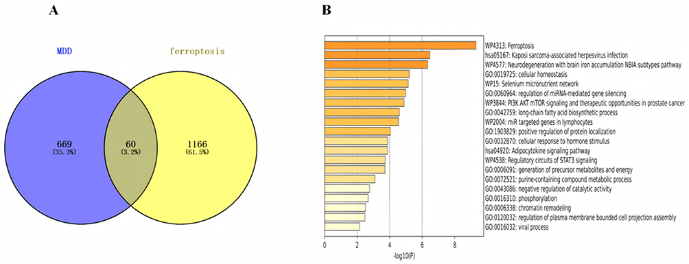 |
Figure 4 Identification and Functional Analysis of Ferroptosis-Related Differentially Expressed Genes (A) Venn Diagram of Ferroptosis-Related Genes in Depression: The diagram illustrates the intersection between ferroptosis-related genes from the FerrDb database and differentially expressed genes from the GSE38206 dataset, identifying genes potentially involved in both ferroptosis and depression. (B) Pathway Enrichment Analysis of Ferroptosis-Related Genes: The bar plot highlights the top enriched pathways, including ferroptosis, nuclear receptor meta-pathway, and glucocorticoid response, providing insights into the molecular mechanisms linking ferroptosis to depression. |
The enrichment of the ferroptosis pathway suggests that iron-dependent cell death may play a critical role in the pathological mechanisms of depression. The enrichment of pathways related to neurodegeneration with brain iron accumulation (NBIA) further supports the involvement of iron dysregulation in depressive disorders. Additionally, the observed enrichment of the PI3K-AKT-mTOR signaling pathway—a key regulator of cellular metabolism, growth, and survival—aligns with the well-documented dysfunction of the hypothalamic-pituitary-adrenal (HPA) axis in depression, reflecting potential disruptions in neuroendocrine and stress-response systems. These findings provide novel insights into the molecular mechanisms linking ferroptosis and iron homeostasis to depression, offering new avenues for mechanistic research and therapeutic exploration.
Construction of Protein-Protein Interaction Network and Core Module Analysis
To systematically explore the interaction relationships among ferroptosis-related genes in depression, a protein-protein interaction (PPI) network was constructed using the STRING database and visualized using Cytoscape software (Figure 5A). By applying the MCODE algorithm for modular analysis, three functional modules with significant biological relevance were successfully identified (Figure 5B–D).
 |
Figure 5 Protein-Protein Interaction Network and Core Module Analysis of Ferroptosis-Related Genes in Depression. (A) PPI Network Visualization: The network illustrates the interactions among ferroptosis-related genes, with nodes representing genes and edges representing interactions. The network was constructed using the STRING database and visualized in Cytoscape. (B–D) Functional Modules Identified by MCODE Analysis: The three panels highlight the top functional modules extracted from the PPI network using the MCODE algorithm. Each module represents a cluster of highly interconnected genes, revealing potential biological pathways and mechanisms associated with ferroptosis in depression. |
Functional Analysis of Key Genes Identified by MCODE Algorithm
The 17 genes belong to module 1 which has the highest MCODE score. The 17 key genes identified through the MCODE algorithm were further investigated for their potential regulatory roles in ferroptosis associated with depression. Functional enrichment analysis of Cluster 1 using the Metascape platform revealed that these genes are primarily involved in biological processes such as Ferroptosis, pcarboxylic acid metabolic process, and Selenium micronutrient network (Figure 6). The significant enrichment of ferroptosis-related pathways confirms the central role of this programmed cell death mechanism in depression pathophysiology. The identification of carboxylic acid metabolic processes suggests alterations in cellular energy metabolism that may contribute to neuronal dysfunction.18 Most notably, the selenium micronutrient network enrichment highlights the crucial role of this essential trace element in antioxidant defense systems, particularly through its incorporation into glutathione peroxidase (GPX4), the key enzyme preventing ferroptosis execution.15 These findings not only elucidate the multifunctional characteristics of ferroptosis-related genes in depression but also provide a molecular foundation for developing trace element-based therapeutic strategies.
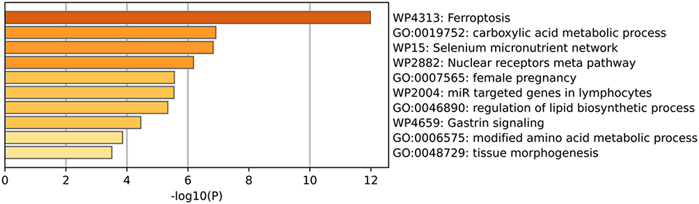 |
Figure 6 Functional Enrichment Analysis of Key Genes in Cluster 1. The bar plot illustrates the top enriched biological processes associated with the 17 key genes identified by the MCODE algorithm. These genes are primarily involved in cell activation, positive regulation of response to external stimuli, and osteoclast differentiation, highlighting their potential roles in neuroinflammation, stress response, and bone metabolism in the context of depression. |
Screening of Potential Small-Molecule Drugs Based on Hub Genes
Among these candidates, VX-702, a p38 MAPK inhibitor, may attenuate neuroinflammatory responses and oxidative stress, both implicated in ferroptosis-related depressive pathology.19 Gossypol, originally identified as a BH3 mimetic, has been shown to modulate glutathione metabolism and lipid peroxidation, potentially influencing ferroptotic pathways.20 Atomoxetine, a selective norepinephrine reuptake inhibitor already in clinical use for ADHD, may confer neuroprotective benefits beyond its primary mechanism, possibly through anti-apoptotic and antioxidant effects.21 TWS-119, a GSK-3β inhibitor, could promote neuronal survival and synaptic plasticity, thereby counteracting ferroptosis-associated neuronal loss.22 Lastly, ibudilast, a phosphodiesterase and macrophage migration inhibitory factor inhibitor, exhibits broad anti-inflammatory and neuroprotective properties relevant to ferroptosis-mediated depression.23
The identification of these compounds provides a rational basis for developing novel therapeutic strategies that target ferroptosis-related mechanisms in depression. These findings also establish a foundation for subsequent validation through in vitro and in vivo experiments (Table 1).
 |
Table 1 Potential Small-Molecule Compounds Targeting Ferroptosis-Related Hub Genes in Depression |
Potential Traditional Chinese Medicine Screening Based on Hub Genes
In this study, we identified nine hub genes (STAT3, PTGS2, PKM, PCBP1, KRAS, IDH1, HSPA8, FTH1, ELAVL1) through the intersection of four topological algorithms (MCC, MNC, Degree, and Closeness) in Cytoscape (Figure 7). These genes were subsequently mapped using the Coremine Medical database to screen associated traditional Chinese medicines (TCMs). The screening yielded the following results:
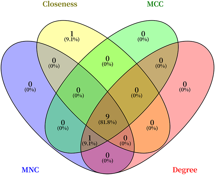 |
Figure 7 Schematic Diagram of TCM Screening Based on Hub Genes. The gene network was analyzed using Cytoscape software with four algorithms (MCC, MNC, Degree, and Closeness). |
STAT3 was associated with Salvia miltiorrhiza Bunge, Crocus sativus L. (saffron), Cannabis sativa L. seed, Curcuma longa L. (turmeric tuber), and Curcuma wenyujin Y. H. Chen et C. Ling (turmeric). These TCMs are known to promote blood circulation, resolve stasis, soothe the liver, and alleviate depression, corresponding to the “qi stagnation and blood stasis” syndrome.
PTGS2 was linked to Crocus sativus L., Astragalus membranaceus (Fisch). Bunge, Poria cocos (Schw). Wolf, Cannabis sativa L. seed, and Polygonum multiflorum Thunb.
PKM was connected to Clematis chinensis Osbeck, Celastrus orbiculatus Thunb. fruit, Celastrus orbiculatus Thunb., Carpesium abrotanoides L. fruit, and Allium chinense G. Don.
PCBP1 was associated with Rabdosia rubescens (Hemsl). Hara herb.
KRAS was related to Taxillus chinensis (DC). Danser herb, Ardisia crispa (Thunb). A. DC. root, and Hedyotis diffusa Willd.
IDH1 was matched with Ganoderma sinense Zhao, Xu et Zhang, Chloriti Lapis (chlorite schist), and Eucommiae Cortex Colla (eucommia bark gelatin).
HSPA8 was aligned with Paederia scandens (Lour). Merr. herb, Dichroa febrifuga Lour. twig and leaf, and Dichroa febrifuga Lour. root.
FTH1 was correlated with Edgeworthia chrysantha Lindl. root, Euphorbia humifusa Willd. herb, and Curcuma kwangsiensis S. G. Lee et C. F. Liang rhizome.
ELAVL1 was tied to Glycyrrhiza uralensis Fisch. root and rhizome, and Salvia miltiorrhiza Bunge.
These TCMs possess qi-tonifying (modulating mitochondrial bioenergetics and redox homeostasis), blood-nourishing, mind-calming, and spirit-stabilizing (neuroprotective and anxiolytic activities) effects, suitable for depression with “heart and spleen deficiency” syndrome.
The screening results not only suggest potential TCM candidates for depression treatment but also highlight the theoretical strengths of TCM in “syndrome differentiation and treatment” and “holistic regulation.” This study provides novel insights and evidence for integrating traditional Chinese and Western medicine in managing depression.
Discussion
Association Between Ferroptosis and Pathogenesis of Depression
Ferroptosis, a newly identified form of programmed cell death, demonstrates potential links to the dysregulation of iron metabolism and lipid peroxidation, which are implicated in the pathogenesis of depression.23 Our bioinformatic analysis identified 60 ferroptosis-related differentially expressed genes (DEGs) from peripheral blood mononuclear cells (PBMCs) of MDD patients, which are primarily involved in iron ion metabolism, reactive oxygen species response, and glutathione metabolic processes. These findings align with growing evidence that iron dyshomeostasis and oxidative stress play crucial roles in depressive disorders.24 Notably, our PPI network analysis revealed 17 core genes central to ferroptosis regulation in depression, we found that genes such as ACSL4, FTL formed key regulatory nodes, providing molecular insights into lipid peroxidation and antioxidant defense mechanisms relevant to depression pathology.25,26
Depression remains a leading cause of global disability, with rising incidence rates worldwide.23 The use of PBMC data offers both advantages and limitations. While PBMCs cannot directly reflect neuronal molecular events, they provide a clinically feasible platform for translational research and have been successfully used to identify depression biomarkers in multiple studies.27 Our findings should therefore be viewed as identifying peripheral correlates of potentially CNS-relevant ferroptosis pathways.
Analysis of Core Gene Regulatory Network
The protein-protein interaction network analysis identified 17 core genes that constitute a functionally interconnected module central to ferroptosis regulation in depression. Among these, STAT3, PTGS2, HSPA8, FTH1, and ELAVL1 emerged as hub genes with the highest connectivity, suggesting their pivotal roles in coordinating ferroptosis-related pathways.
Notably, our network pharmacology analysis revealed several TCM compounds with multi-target regulatory capabilities against these core genes. Salvia miltiorrhiza (Danshen) and Crocus sativus (saffron) demonstrated particularly strong interactions with multiple hub genes, including STAT3, PTGS2, and FTH1. These botanicals are known to possess potent antioxidant and anti-inflammatory properties, with documented effects on iron metabolism and glutathione synthesis.28,29
The regulatory patterns observed suggest that these core genes form a coordinated network that maintains iron and redox homeostasis. Disruption of this network, may lead to increased susceptibility to ferroptosis, ultimately contributing to neuronal dysfunction and depressive symptomatology.30 The identification of this core gene module provides a mechanistic framework for understanding how ferroptosis contributes to depression pathogenesis and offers specific molecular targets for therapeutic intervention.
Characteristics of Herbal Properties in Gene Regulation
Network analysis revealed that the identified traditional Chinese medicines target key pathways in ferroptosis-related depression. Most herbs regulate oxidative stress and neuroinflammation, while others modulate immune response and iron homeostasis.
Herbal properties showed distinct regulatory patterns: bitter-cold herbs primarily target pro-inflammatory genes,31 whereas sweet-warm herbs focus on immune regulation, demonstrating complementary therapeutic effects.32
Mechanistic studies indicate these herbs contain bioactive compounds that modulate oxidative stress and inflammatory responses, providing a molecular basis for their traditional use in depression treatment. These findings support the integration of TCM approaches targeting ferroptosis pathways for depression management.
Innovation in Treatment Principles Based on Ferroptosis Regulation
The five candidates small-molecule compounds (VX-702, gossypol, atomoxetine, TWS-119, and ibudilast) identified in this study demonstrate synergistic potential with active components from traditional Chinese medicines in regulating ferroptosis pathways. These compounds target key mechanisms including neuroinflammation, oxidative stress, and lipid peroxidation—core pathological processes in ferroptosis-related depression.
Notably, these small molecules complement the action mechanisms of the identified TCMs. For instance, both ibudilast and Salvia miltiorrhiza components target inflammatory pathways,33 while atomoxetine and saffron active compounds share neuroprotective properties.34 This convergence of mechanisms suggests potential for combined therapeutic strategies that simultaneously address multiple facets of ferroptosis dysregulation.
The predicted compounds offer novel targeting approaches for depression treatment, particularly through their effects on glutathione metabolism, iron homeostasis, and antioxidant defense systems. Their mechanisms align with emerging evidence that ferroptosis inhibition may represent a promising strategy for treatment-resistant depression. These findings provide a foundation for developing integrated treatment approaches that combine targeted molecular interventions with multi-component TCM formulations, potentially offering enhanced efficacy through complementary mechanisms of action.
Limitations and Future Directions for Integrated Research
This study has several limitations that should be addressed in future research. First, the use of peripheral blood mononuclear cell (PBMC) data, while clinically accessible, may not fully represent molecular changes in the central nervous system. Future studies should incorporate postmortem brain tissues or neuroimaging approaches to validate these findings in relevant CNS contexts.
Second, the bioinformatics predictions require experimental validation. We recommend employing chronic unpredictable mild stress (CUMS) animal models to investigate the relationship between key gene expression and therapeutic outcomes. Specifically, studies should measure ferroptosis markers (eg, ACSL4, GPX4) following pharmacological interventions targeting the identified pathways.
For clinical translation, we propose developing integrated formulations that combine the predicted small-molecule compounds with screened traditional Chinese medicines. Systematic evaluation of these combinations should focus on their effects on ferroptosis signaling pathways and their potential for treating refractory depression.
Finally, while ferroptosis appears to be an important mechanism in depression, it represents only one aspect of this complex disorder. Future research should explore the interactions between ferroptosis and other pathological processes, including genetic predisposition, environmental stressors, and neuroendocrine adaptations, to develop more comprehensive treatment strategies.
Conclusion
This study demonstrates the significant involvement of ferroptosis-related mechanisms in the pathogenesis of depression through integrated bioinformatics and network pharmacology approaches. We identified 60 ferroptosis-associated differentially expressed genes and 17 core genes that form a central regulatory network in depression. These findings provide novel insights into the molecular mechanisms linking iron dysregulation, lipid peroxidation, and oxidative stress to depressive disorders.
Our analysis revealed five promising small-molecule compounds (VX-702, gossypol, atomoxetine, TWS-119, and ibudilast) that target key ferroptosis pathways, along with several traditional Chinese medicines with multi-target regulatory capabilities. The convergence of mechanisms between these Western and traditional approaches suggests strong potential for developing integrated treatment strategies.
While this study provides valuable insights, future research should focus on experimental validation using animal models and clinical samples to confirm these findings. The development of combined therapeutic approaches that simultaneously target multiple facets of ferroptosis dysregulation may offer new avenues for treating depression, particularly for medication-resistant cases.
This work establishes a foundation for advancing integrative medicine approaches that combine targeted molecular interventions with traditional medicine wisdom, potentially leading to more effective and comprehensive treatment strategies for depression.
Data Sharing Statement
The datasets analyzed during this study are publicly available in the Gene Expression Omnibus (GEO) repository under accession number GSE38206 (https://www.ncbi.nlm.nih.gov/geo/query/acc.cgi?acc=GSE38206). All other data generated or analyzed during this study are included in this published article.
Research Ethics
This study has been approved by the Ethics Committee of Wuxi Second Hospital (Approval No.: 2024Y-226).
Author Contributions
All authors made a significant contribution to the work reported, whether that is in the conception, study design, execution, acquisition of data, analysis and interpretation, or in all these areas; took part in drafting, revising or critically reviewing the article; gave final approval of the version to be published; have agreed on the journal to which the article has been submitted; and agree to be accountable for all aspects of the work Use of LLM, AI None declared.
Funding
This study was supported by the National Natural Science Foundation of China (grant no. 82071381 awarded to Xudong Zhao).
Disclosure
The authors state no conflicts of interest in this work.
References
1. Marwaha S, Palmer E, Suppes T, et al. Novel and emerging treatments for major depression. Lancet. 2023;401(10371):141–153. doi:10.1016/S0140-6736(22)02080-3
2. Thapar A, Eyre O, Patel V, et al. Depression in young people. Lancet. 2022;400(10352):617–631. doi:10.1016/S0140-6736(22)01012-1
3. Kraus C, Castrén E, Kasper S, et al. Serotonin and neuroplasticity - Links between molecular, functional and structural pathophysiology in depression. Neurosci Biobehav Rev. 2017;77:317–326. doi:10.1016/j.neubiorev.2017.03.007
4. Cipriani A, Furukawa TA, Salanti G, et al. Comparative efficacy and acceptability of 21 antidepressant drugs for the acute treatment of adults with major depressive disorder: a systematic review and network meta-analysis. Lancet. 2018;391(10128):1357–1366. doi:10.1016/S0140-6736(17)32802-7
5. Liu J, Zhang G, Chen L, et al. Natural products targeting ferroptosis in depression: research progress and therapeutic prospects. Phytomedicine. 2025:142.
6. Mao L, You J, Xie M, et al. Arginine methylation of β-catenin induced by PRMT2 aggravates LPS-induced cognitive dysfunction and depression-like behaviors by promoting ferroptosis. Molecular Neurobiol. 2024;61(10):7796–7813. doi:10.1007/s12035-024-04019-5
7. Dixon SJ, Lemberg K, Lamprecht M, et al. Ferroptosis: an iron-dependent form of nonapoptotic cell death. Cell. 2012;149(5):1060–1072. doi:10.1016/j.cell.2012.03.042
8. Stockwell BR. Ferroptosis turns 10: emerging mechanisms, physiological functions, and therapeutic applications. Cell. 2022;185(14):2401–2421. doi:10.1016/j.cell.2022.06.003
9. Shah R, Shchepinov MS, Pratt DA. Resolving the role of lipoxygenases in the initiation and execution of ferroptosis. Acs Central Sci. 2018;4(3):387–396. doi:10.1021/acscentsci.7b00589
10. Liu G, Deng B, Huo L, et al. Tetramethylpyrazine alleviates ferroptosis and promotes functional recovery in spinal cord injury by regulating GPX4/ACSL4. Euro J Pharmacol. 2024;977:176710. doi:10.1016/j.ejphar.2024.176710
11. Zhou N, Yuan X, Du Q, et al. FerrDb V2: update of the manually curated database of ferroptosis regulators and ferroptosis-disease associations. Nucleic Acids Res. 2023;51(D1):D571–D582. doi:10.1093/nar/gkac935
12. Kanehisa M, Furumichi M, Sato Y, et al. KEGG: biological systems database as a model of the real world. Nucleic Acids Res. 2024;53(D1):D672–D677. doi:10.1093/nar/gkae909
13. Kanehisa M. Toward understanding the origin and evolution of cellular organisms. Protein Sci. 2019;28(11):1947–1951. doi:10.1002/pro.3715
14. Kanehisa M. KEGG: kyoto encyclopedia of genes and genomes. Nucleic Acids Res. 2000;28(1):27–30. doi:10.1093/nar/28.1.27
15. Angeli JPF, Conrad M. Selenium and GPX4, a vital symbiosis. Free Radic Biolo Med. 2018;127:153–159. doi:10.1016/j.freeradbiomed.2018.03.001
16. Belzeaux R, Bergon A, Jeanjean V, et al. Responder and nonresponder patients exhibit different peripheral transcriptional signatures during major depressive episode. Transl Psychiatry. 2012;2.
17. Chen X, Kang R, Kroemer G, et al. Ferroptosis in infection, inflammation, and immunity. J Exp Med. 2021;218(6). doi:10.1084/jem.20210518
18. Wang T, Shah YM, Matsubara T, et al. Control of steroid 21-oic acid synthesis by peroxisome proliferator-activated receptor α and role of the hypothalamic-pituitary-adrenal axis. J Biologic Chem. 2010;285(10):7670–7685. doi:10.1074/jbc.M109.090175
19. Han Y, Wang J, Zhang J, et al. VX-702 ameliorates the severity of sepsis-associated acute kidney injury by downregulating inflammatory factors in macrophages. J Inflamm Res. 2024;17:4037–4054. doi:10.2147/JIR.S464018
20. Shang JJ, Xiong C, Jiang W, et al. Gossypol acetic acid alleviates the ferroptosis of chondrocytes in osteoarthritis by inhibiting GPX4 methylation. Curr Med Chem. 2025;32(12):2422–2439. doi:10.2174/0109298673280730231211092905
21. Kratochvil CJ, Newcorn JH, Arnold LE, et al. Atomoxetine alone or combined with fluoxetine for treating ADHD with comorbid depressive or anxiety symptoms. J Ame Acad Child Adolescent Psychiatry. 2005;44(9):915–924. doi:10.1097/01.chi.0000169012.81536.38
22. Guo A, et al. Bioinformatic identification of hub genes Myd88 and Ccl3 and TWS-119 as a potential agent for the treatment of massive cerebral infarction. Front Neurosci. 2023;17.
23. Wang LY, Xu R, Huang C, et al. Targeting the ferroptosis crosstalk: novel alternative strategies for the treatment of major depressive disorder. General Psychiatry. 2023;36(5):e101072. doi:10.1136/gpsych-2023-101072
24. El-Abasy HM, Elsaid ME, Abdelkader EM, et al. Metformin’s cardioprotective role in isoprenaline-induced myocardial infarction: unveiling insights into the AMPK, NF-κB, JAK2/STAT3 pathways, and cholinergic regulation. Life Sci. 2024;357.
25. Ciobanu LG, Sachdev PS, Trollor JN, et al. Downregulated transferrin receptor in the blood predicts recurrent MDD in the elderly cohort: a fuzzy forests approach. J Affect Disord. 2020;267:42–48. doi:10.1016/j.jad.2020.02.001
26. Geng RJ, Dai M-S, Wang Y, et al. Evaluation the therapeutic effect of adipose-derived mesenchymal stem cells on chronic mild stress by activating PEBP1-GPX4 axis in ferroptosis using qRT-PCR, fluorescence microscope and iron determination analysis. J Biomed Nanotechnol. 2022;18(12):2828–2838. doi:10.1166/jbn.2022.3475
27. Chen J, Zhu X, Yang F, et al. Exploring male-specific synaptic plasticity in major depressive disorder: a single-nucleus transcriptomic analysis using bioinformatics methods. Inter J Mole Sci. 2025;26(7):3135.
28. Boskabady M, Farkhondeh T. Antiinflammatory, antioxidant, and immunomodulatory effects of crocus sativus L. and its main constituents. Phytotherapy Res. 2016;30(7):1072–1094. doi:10.1002/ptr.5622
29. Jiang C, Yan Y, Long T, et al. Ferroptosis: a potential therapeutic target in cardio-cerebrovascular diseases. Mole Cellular Biochem. 2025;480(7):4379–4399. doi:10.1007/s11010-025-05262-7
30. Jin Y, et al. Dimethyl malonate preserves brain and neurobehavioral phenotype following neonatal hypoxia-ischemia by inhibiting FTH1-mediated ferritinophagy. Redox Biology. 2025;86.
31. Zhou W, Dai Y, Meng J, et al. Network pharmacology integrated with molecular docking reveals the common experiment-validated antipyretic mechanism of bitter-cold herbs. J Ethnopharmacol. 2021;274:114042. doi:10.1016/j.jep.2021.114042
32. Pan SY, Ou SC, Chang TT. Integrating traditional Chinese medicine in managing neutropenic fever during chemotherapy for pediatric lymphoma: a case report. Explore. 2025;21(3):103166. doi:10.1016/j.explore.2025.103166
33. Ko G, Kim J, Jeon YJ, et al. Salvia miltiorrhiza alleviates memory deficit induced by ischemic brain injury in a transient MCAO mouse model by inhibiting ferroptosis. Antioxidants. 2023;12(4):785.
34. Levey AI, Qiu D, Zhao L, et al. A Phase II study repurposing atomoxetine for neuroprotection in mild cognitive impairment. Brain. 2022;145(6):1924–1938. doi:10.1093/brain/awab452
 © 2025 The Author(s). This work is published and licensed by Dove Medical Press Limited. The
full terms of this license are available at https://www.dovepress.com/terms
and incorporate the Creative Commons Attribution
- Non Commercial (unported, 4.0) License.
By accessing the work you hereby accept the Terms. Non-commercial uses of the work are permitted
without any further permission from Dove Medical Press Limited, provided the work is properly
attributed. For permission for commercial use of this work, please see paragraphs 4.2 and 5 of our Terms.
© 2025 The Author(s). This work is published and licensed by Dove Medical Press Limited. The
full terms of this license are available at https://www.dovepress.com/terms
and incorporate the Creative Commons Attribution
- Non Commercial (unported, 4.0) License.
By accessing the work you hereby accept the Terms. Non-commercial uses of the work are permitted
without any further permission from Dove Medical Press Limited, provided the work is properly
attributed. For permission for commercial use of this work, please see paragraphs 4.2 and 5 of our Terms.
Recommended articles
Transcriptome Analysis Identifies Biomarkers for the Diagnosis and Management of Psoriasis Complicated with Depression
Xia X, Yu H, Li Y, Liang Y, Li G, Huang F
Clinical, Cosmetic and Investigational Dermatology 2023, 16:1287-1301
Published Date: 18 May 2023
Exploration and Validation of Potential Biomarkers and Therapeutic Targets in Ferroptosis of Asthma
Xing Y, Feng L, Dong Y, Li Y, Zhang L, Wu Q, Huo R, Dong Y, Tian X, Tian X
Journal of Asthma and Allergy 2023, 16:689-710
Published Date: 12 July 2023
Identification of Ferroptosis-Related Genes in Heart Failure Induced by Transverse Aortic Constriction
Gu JJ, Du TJ, Zhang LN, Zhou J, Gu X, Zhu Y
Journal of Inflammation Research 2023, 16:4899-4912
Published Date: 31 October 2023
Integrated Analysis of Ferroptosis and Immunity-Related Genes Associated with Diabetic Kidney Disease
Wang J, Wang L, Pang Z, Ge Q, Wu Y, Qi X
Diabetes, Metabolic Syndrome and Obesity 2023, 16:3773-3793
Published Date: 23 November 2023
Bioinformatics and Integrative Experimental Method to Identifying and Validating Co-Expressed Ferroptosis-Related Genes in OA Articular Cartilage and Synovium
Ma J, Yu P, Ma S, Li J, Wang Z, Hu K, Su X, Zhang B, Cheng S, Wang S
Journal of Inflammation Research 2024, 17:957-980
Published Date: 12 February 2024