
Back to Journals » ImmunoTargets and Therapy » Volume 15

The Immune Architecture of Eosinophilic Esophagitis: Mechanisms, Therapeutic Targets, and Precision Management
Authors Bertin L  , Caldart F, Barchi A, Seregni N, Pasta A, Calabrese F , Marabotto E , Mari A , Farah A, Sirinic E, Sorge A, Ghisa M, Chahuán J, Savarino V , Savarino EV
Received 31 March 2026
Accepted for publication 17 May 2026
Published 27 May 2026 Volume 2026:15 510865
DOI https://doi.org/10.2147/ITT.S510865
Checked for plagiarism Yes
Review by Single anonymous peer review
Peer reviewer comments 2
Editor who approved publication: Dr Sarah Wheeler
Luisa Bertin,1,2 Federico Caldart,3 Alberto Barchi,4 Niccolò Seregni,4 Andrea Pasta,5,6 Francesco Calabrese,5,6 Elisa Marabotto,5,6 Amir Mari,7,8 Amir Farah,9 Emma Sirinic,1,2 Andrea Sorge,1,2 Matteo Ghisa,2 Javier Chahuán,1,10 Vincenzo Savarino,5 Edoardo Vincenzo Savarino1,2
1Department of Surgery, Oncology and Gastroenterology, University of Padua, Padua, Italy; 2Gastroenterology Unit, Azienda Ospedale-Università Padova, Padua, Italy; 3Department of Medicine, University of Verona, Verona, Italy; 4Digestive Physiopathology Unit, IRCCS San Raffaele, Milan, Italy; 5Department of Internal Medicine, University of Genoa, Genoa, Italy; 6IRCCS-Ospedale Policlinico San Martino, Genoa, Italy; 7Gastroenterology Institute, Nazareth EMMS Hospital, Nazareth, Israel; 8Azrieli Faculty of Medicine, Bar-Ilan University, Safed, Israel; 9Surgery, Medical College of
Wisconsin, Milwaukee, Wisconsin, USA; 10Department of Gastroenterology, Pontificia Universidad Católica de Chile, Santiago, Chile
Correspondence: Edoardo Vincenzo Savarino, Department of Surgery, Oncology and Gastroenterology (DiSCOG), University of Padua – Azienda Ospedaliera di Padova, Via Giustiniani, 2, Padova, 35128, Italy, Tel +39-049-8217749, Email [email protected]
Background: Eosinophilic esophagitis (EoE) is a chronic, allergen-driven, type 2 immune-mediated disease of the esophagus defined by symptoms of esophageal dysfunction and eosinophil-predominant mucosal inflammation (≥ 15 eosinophils per high-power field), with a rising global prevalence now exceeding 1 in 1,000 individuals in Western countries. Its pathogenesis extends far beyond eosinophils, encompassing epithelial alarmin signaling (TSLP, IL-33, IL-18), ILC2 and Th2 immune activation, and a convergent effector network of eosinophils, mast cells, and fibroblasts that drives barrier disruption and progressive fibrostenotic remodeling.
Purpose: This narrative review synthesizes current evidence on the multi-layered immunopathogenesis of EoE, the evolving conceptualization of disease remission, established and emerging therapeutic options, and precision medicine strategies incorporating phenotypic and molecular endotype classification.
Key Findings: EoE shares a pathophysiologic continuum with other atopic diseases, underpinned by convergent type 2 immune mechanisms and epithelial barrier gene defects. A recently characterized ILC2–amphiregulin–EGFR axis directly drives structural esophageal remodeling independent of eosinophil recruitment, providing a mechanistic explanation for the histology-symptom dissociation that limits several biologic therapies. First-line therapies—proton pump inhibitors (PPIs), swallowed topical corticosteroids (STCs), and dietary elimination—achieve histologic remission in 45– 90% of patients. Dupilumab, the first approved biologic, achieves histologic remission in approximately 59– 60% of patients versus 5– 6% with placebo at week 24. An expanding pipeline targets IL-13, IL-5R, TSLP, mast cells, and JAK-STAT pathways alongside optimized steroid formulations and oral small molecules.
Conclusion: EoE is entering an era of precision therapeutics. Integrating molecular endotyping, multi-effector immunopathology, and multidimensional disease activity assessment will be essential to optimize long-term outcomes, prevent fibrostenotic progression, and individualize the expanding armamentarium of targeted therapies.
Plain Language Summary: Eosinophilic esophagitis (EoE) is a chronic inflammatory disease of the food pipe (esophagus) that causes difficulty swallowing. It is linked to food allergens and has become much more common over the past thirty years, now affecting roughly 1 in every 1,000 people in Western countries.
For a long time, doctors believed EoE was caused mainly by one type of immune cell called the eosinophil. We now know the picture is more complex. This review explains that EoE involves many parts of the immune system working together: the lining of the esophagus releases alarm signals that trigger a chain reaction; a group of immune cells called ILC2s amplify this reaction and directly cause thickening of the esophageal wall; and other cells, including mast cells and fibroblasts (which form scar tissue), each play distinct roles in causing symptoms and structural damage.
This complexity explains a puzzling observation: several newer treatments that successfully clear the inflammatory cells from the esophagus do not reliably improve swallowing. Our review explains why — because structural damage and nerve sensitization that cause symptoms can persist even after the inflammatory cells are gone.We review three established treatments (acid-suppressing medicines, swallowed steroid preparations, and elimination diets), the first approved targeted therapy (dupilumab, which blocks two key immune signals at once), and a pipeline of newer treatments. We also describe how people with EoE can be grouped into molecular subtypes that may predict which treatment is most likely to help them, moving toward personalized care. The infographic titled ’Immunotargets and Therapy for Eosinophilic Esophagitis’ explores the hidden immune landscape of eosinophilic esophagitis. It features an iceberg metaphor, with eosinophils at the top, associated with IL-5 and IL-5Rα. Below the surface, various immune components are listed: Mast cells linked to Siglec-8 and KIT, ILC2s associated with TSLP, IL-33/ST2 and Areg-EGFR, Fibroblasts connected to TGF-β, LIGHT and TSPAN12 and Sensory neurons related to TRPV1 and the mast cell-neuroimmune axis. This visual representation emphasizes the complexity of immune interactions in eosinophilic esophagitis.Infographic on eosinophilic esophagitis therapy, focusing on immune components and targets.
Keywords: eosinophilic esophagitis, type 2 inflammation, ILC2-amphiregulin-EGFR, IL-33/ST2 axis, GPR15, IFN signature, mast cell, neuroimmune, TGF-β, fibrostenosis, dupilumab, precision medicine
Introduction
Eosinophilic esophagitis (EoE) is a chronic, antigen-driven, type 2 immune-mediated disease defined by the triad of esophageal dysfunction symptoms, eosinophil-predominant inflammation (≥15 eosinophils/hpf on biopsy), and exclusion of secondary causes of esophageal eosinophilia.1–4 First characterized as a distinct clinicopathologic entity in the early 1990s, EoE has since transformed from a curiosity of case reports into one of the most actively studied gastrointestinal inflammatory diseases of the modern era, with a global prevalence now exceeding 1 in 1,000 persons in industrialized countries and an incidence that outpaces the rate of increase in endoscopy and biopsy, implying genuine environmental drivers beyond diagnostic ascertainment.5
EoE occupies a unique position at the crossroads of allergology and gastroenterology.6,7 It shares a pathophysiologic continuum with asthma, atopic dermatitis, allergic rhinitis, and chronic rhinosinusitis with nasal polyps—a cluster of diseases increasingly recognized as related expressions of type 2 mucosal immunopathology—yet it is distinguished by its organ-specific manifestation, its propensity for fibrostenotic remodeling if inadequately controlled, and the critical role of food antigens as principal environmental triggers.8 Untreated or undertreated EoE carries the risk of progressive esophageal remodeling, luminal narrowing, and a clinical phenotype dominated by dysphagia and food bolus impaction that profoundly impairs quality of life.9 Diagnostic delays of more than a decade remain common in adult patients.10
The therapeutic landscape has been reshaped over the past decade. The approval of dupilumab in 2022 as the first biologic for EoE validated the IL-4/IL-13 axis as a clinically actionable target and inaugurated a new era of mechanism-based treatment.11 Concurrently, the concept of therapeutic success has evolved from a single histologic threshold into a multidimensional construct encompassing endoscopic, symptomatic, and patient-reported outcomes, increasingly framed within a Treat-to-Target (T2T) paradigm.12 Critically, recent mechanistic research has revealed that disease pathogenesis extends well beyond eosinophilic inflammation: an ILC2-driven amphiregulin–EGFR axis directly induces esophageal epithelial hyperplasia; the IL-33/ST2 signaling axis amplifies the entire innate–adaptive cascade; mast cells persist in histologic remission and contribute independently to smooth muscle dysfunction and fibrosis; and distinctive sphingolipid dysregulation in esophageal epithelium impairs barrier integrity through cytokine-regulated ceramide biosynthesis. This expanding mechanistic landscape reframes EoE as a multi-effector immune disorder requiring multi-target therapeutic strategies.
This review provides a comprehensive synthesis of EoE immunopathogenesis—with particular focus on immune dysfunction at its multiple levels—alongside the evidence base for established and emerging therapies, evolving definitions of disease control, and the emerging framework of precision medicine incorporating molecular endotyping and phenotypic stratification. Our goal is to offer the clinician-scientist a coherent roadmap of where the field stands and where it is headed, illuminating both the therapeutic opportunities and the scientific gaps that remain.
Methods
This is a narrative review. A structured literature search was conducted in PubMed and EMBASE from January 2015 through March 2026, supplemented by manual review of reference lists of key publications and selected congress abstracts from major gastroenterology and allergy congresses. Search terms included “eosinophilic esophagitis”, combined with “immunopathogenesis”, “epithelial barrier”, “ILC2”, “IL-33”, “amphiregulin”, “mast cell”, “biologics”, “dupilumab”, “treat-to-target”, “endotype”, “fibrostenosis”, and “precision medicine.” Original research articles, randomized controlled trials, systematic reviews and meta-analyses, and consensus guidelines were prioritized. Studies published in English were included. Given the rapidly evolving nature of the field, particular attention was paid to publications from 2022 onward. The selection of studies for inclusion was based on relevance to the review’s thematic structure, quality of evidence, and clinical or mechanistic significance, as assessed by all authors by consensus.
Epidemiology
The global burden of EoE has risen substantially and continuously over the past three decades.13,14 A recent meta-analysis, published in 2023 and drawing on 40 population-based studies from 15 countries spanning five continents, reported a global pooled incidence of 5.3 per 100,000 person-years (95% CI 4.0–6.6) and prevalence of 40.0 per 100,000 persons (95% CI 31.1–49.0).15 Critically, prevalence increased by more than 800% from the earliest studies (1976–2001) to the most recent cohorts (2017–2022)—a rate of change that outstrips the growth in endoscopy and biopsy practices. Comparable findings from the United States Veterans Health Administration, which documented a greater than 300% increase in annual incident cases between 2009 and 2018, and from European population-based registries converge on this conclusion: the epidemiological rise of EoE is real, not artifactual.16,17
Current estimates in Western countries position the prevalence at greater than 1 in 1,000 individuals, comparable to inflammatory bowel disease, making EoE the most common cause of dysphagia and food bolus impaction in children and young adults, and the second most frequent cause of chronic esophagitis after gastresophageal reflux disease (GERD).18 The disease predominantly affects white males, with a consistent male-to-female ratio of approximately 3:1. Incidence rises during adolescence and peaks in early adulthood, although EoE is increasingly recognized across all age groups and both sexes.
Multiple risk and protective factors have been identified.19,20 Early-life exposures associated with increased EoE risk include preterm birth, neonatal intensive care unit admission, Cesarean delivery, early antibiotic or acid-suppressive use, and maternal smoking, all of which may perturb the intestinal microbiome during a critical immunological window. Conversely, Helicobacter pylori infection and early exposure to furred pets appear protective, consistent with the hygiene hypothesis and its proposition that reduced early microbial stimulation promotes Th2 immune polarization.21
Clinical Presentation and Atopic Comorbidities
The clinical presentation of EoE is highly age-dependent, reflecting the dynamic interplay between developing anatomy, evolving immune phenotypes, and the progressive structural consequences of untreated inflammation.1,22,23 In adolescents and adults, solid-food dysphagia is the cardinal symptom, frequently accompanied by food bolus impaction—an event that precipitates emergency endoscopy in 33–54% of adult patients at some point during the disease course.24 In infants and young children, EoE manifests as vomiting, regurgitation, food refusal, failure to thrive, and failure to progress to age-appropriate food textures. An important clinical caveat applies across all age groups: patients with long-standing EoE frequently adopt maladaptive eating strategies that mask the severity of dysphagia at clinical interview, and these compensatory behaviors may progress to avoidant/restrictive food intake disorder.25–29
Atopic comorbidities are extraordinarily prevalent in EoE. At least 60–80% of patients carry at least one concomitant allergic diagnosis, and the probability of an EoE diagnosis rises steeply with atopic burden: a pediatric birth-cohort analysis demonstrated hazard ratios of 2.5 (95% CI 1.6–3.9), 5.6 (95% CI 3.5–8.9), and 9.1 (95% CI 5.2–16.0) for any one, two, and three atopic diagnoses, respectively.30,31 These data strongly support the conceptualization of EoE as a late esophagus-specific manifestation of the allergic march, yet a substantial minority of patients lack objective evidence of systemic atopy, indicating that non-IgE-mediated mechanisms operate in parallel.
Psychiatric and neurodevelopmental comorbidities deserve equal clinical attention. Approximately 31% of adults with EoE carry at least one psychiatric diagnosis, most commonly anxiety and depression.32–35 Symptom-specific anxiety and esophageal hypervigilance are independent predictors of dysphagia severity after accounting for endoscopic and histologic disease activity, highlighting the relevance of central sensitization pathways. The diagnosis requires fulfillment of three criteria: symptoms of esophageal dysfunction; a peak eosinophil density of at least 15 eos/hpf confirmed by biopsies from at least two esophageal segments; and exclusion of alternative causes of esophageal eosinophilia.36 The contemporary diagnostic framework no longer requires a mandatory PPI trial before establishing the diagnosis.37
Immunopathogenesis: A Multi-Effector Model of Immune Dysfunction
The immunopathogenesis of EoE can no longer be reduced to a simple eosinophil-centric or even Th2-centric narrative. An emerging multi-effector model positions the eosinophil as one—albeit prominent—player within a hierarchical immune cascade that is initiated at the level of the esophageal epithelium, amplified through both innate and adaptive lymphoid circuits including ILC2s, basophils, iNKTs, GPR15⁺ clonally expanded Th2 cells, and CD8⁺ tissue-resident memory cells, and ultimately converges on a spectrum of effectors—eosinophils, mast cells, fibroblasts, and sensory neurons—each contributing distinct pathobiological activities.38 TGF-β, LIGHT (TNFSF14), and endothelial TSPAN12 further link inflammatory signaling to the fibrostenotic remodeling compartment through mechanisms largely independent of eosinophil density. A concurrently active IFN-γ/type I IFN signature, the AHR–SPINK7–OVOL1 barrier regulation axis, and the esophageal microbiome add environmental and non-type 2 dimensions to this pathobiological landscape. The clinical consequence of this multi-effector architecture is that suppressing a single node, however effectively, may not resolve all disease manifestations, a mechanistic principle that now directly shapes therapeutic strategy. Figure 1 summarizes the main mechanisms involved in EoE pathophysiology.
 |
Figure 1 Main mechanisms involved in Eosinophilic esophagitis’ pathophysiology. |
The Esophageal Epithelium as an Active Immune Organ
The esophageal epithelium is not a passive barrier that simply fails under immune attack; it is an active immunological organ whose intrinsic molecular architecture simultaneously determines susceptibility to antigen penetration and orchestrates the upstream activation of the entire type 2 cascade.39–41 Three converging lines of evidence establish this dual role: the alarmin signaling machinery that translates mechanical and proteolytic damage into immune activation; the genetic and lipid-metabolic vulnerability that impairs structural barrier integrity; and a hypoxia-driven adenosine axis that links inflammatory oxygen demand to epithelial repair failure.
The type 2 cascade42 is initiated by epithelial-derived alarmins, of which TSLP, IL-33, and IL-18 are the dominant orchestrators.43
TSLP, encoded by a gene harboring EoE-associated variants together with its receptor gene CRLF2, is constitutively expressed at low levels in the esophageal epithelium and is markedly upregulated in active EoE biopsies.43 TSLP attracts, activates, and potentiates survival and degranulation of eosinophils; enhances IL-1-induced cytokine secretion by mast cells; and potently activates dendritic cells and ILC2s.44 A pivotal mechanistic discovery linked TSLP production to the serine protease inhibitor SPINK7: loss of SPINK7 expression amplifies kallikrein-5 proteolytic activity, impairs epithelial differentiation, and drives TSLP production, directly connecting epithelial protease dysregulation to upstream type 2 immune priming.45,46 IL-33, a member of the IL-1 cytokine superfamily, signals through its cognate receptor ST2 and activates NF-κB to drive production of IL-1β, IL-3, IL-4, IL-5, IL-6, IL-13, and TNF-α across a broad range of target cells.47,48
ST2 is expressed on eosinophils, mast cells, basophils, ILC2s, regulatory T cells, macrophages, dendritic cells, and vascular endothelial cells, underlining IL-33’s capacity to simultaneously coordinate the entire innate–adaptive immune spectrum.49 The causal significance of IL-33 was established by a murine transgenic model (EoE33 mice) in which epithelial overexpression of a secreted active form of IL-33 recapitulated the full EoE phenotype—eosinophilic infiltration, basal-zone hyperplasia, dilated intercellular spaces, mast cell accumulation, increased CD4⁺ T cells, and elevated IL-4, IL-9, and IL-13—through an ST2-dependent and IL-13-dependent mechanism.50 Critically, structural features including dilated intercellular spaces, basal-zone hyperplasia, and lamina propria fibrosis persisted in eosinophil-deficient EoE33 mice, reinforcing that IL-33/ST2 drives pathology through effectors beyond the eosinophil.51 IL-33 can additionally be triggered by proteolytic allergens through the ripoptosome—an intracellular platform activating RIPK1-caspase (8/3/7)-IL-33 signaling upon antigen exposure7—providing a mechanism by which food antigens initiate eosinophilic inflammation independent of classical adaptive recognition.52 IL-18 similarly supports eosinophil maturation and survival, acts in concert with TSLP to induce IL-13 secretion from ILC2s,53 and upregulates endothelial adhesion molecules enabling peripheral immune cell recruitment.54 Additionally, due to decreased expression of natural IL-1R antagonists, esophageal epithelial cells in EoE may be hyperresponsive to any IL-1 secreted into the local microenvironment, creating a state of amplified alarmin sensitivity.
The structural basis of barrier vulnerability in EoE is multidimensional.55–59 Dilation of intercellular spaces (DIS) is a histologic hallmark reflecting downregulation of barrier proteins—zonulin-3, filaggrin, claudins, and desmoglein-1—under Th2 cytokine influence. Genome-wide association studies have identified CAPN14, encoding calpain-14, as the most strongly EoE-associated gene; its IL-13-driven upregulation directly destabilizes desmosomal junctions via DSG1 and desmoplakin degradation.60,61 Loss of SPINK7 amplifies kallikrein-5–mediated protease activity and TSLP production, creating a barrier-to-alarmin feed-forward loop.61,62 Non-responders to PPI–topical steroid therapy exhibit significantly downregulated SPINK5, SPINK7, SPINK8, and DSG1 expression compared with responders, implicating barrier gene dysregulation as a molecular substrate of treatment refractoriness.63 A newly characterized lipidomic dimension of barrier failure concerns epithelial sphingolipid composition: esophageal brushings from patients with active EoE show selectively elevated non-hydroxy fatty acid sphingosine ceramide/phytoceramide (NS-CER/NP-CER) ratios, positively correlated with IL-5, IL-13, and CCL26 mRNA expression.64 IL-4/IL-13 directly dysregulate the ceramide desaturase enzymes DEGS1 and DEGS2 in primary esophageal epithelial cells, establishing cytokine-regulated lipid metabolism as a mechanism of barrier failure and potentially generating aberrant ceramide-based lipid antigens recognized by esophageal iNKTs through CD1d, linking barrier dysfunction to innate immune activation.
A further, recently characterized layer of barrier pathology involves the HIF-1α–CD73–adenosine signaling axis. Eosinophilic inflammation substantially increases esophageal epithelial oxygen demand, creating inflammatory hypoxia that paradoxically attenuates HIF-1α during extended insult. CD73 (NT5E), a direct HIF-1α transcriptional target that converts extracellular AMP to adenosine, is significantly diminished in active EoE biopsies and in the L2-IL5NˣA murine model, with accompanying reduction in tight junction occludin.65 Pharmacologic CD73 blockade impairs wound healing and barrier function in vitro, while activation of the downstream ADORA2B receptor—the most abundantly expressed adenosine receptor in active esophageal epithelium—restores fibronectin and occludin expression and improves barrier integrity both in vitro and in vivo, establishing ADORA2B agonism as a novel therapeutic strategy for epithelial repair that operates independently of cytokine suppression.65
This barrier dysfunction is not fully reversible upon histologic remission. Prospective transcriptomic-proteomic profiling of pediatric patients across active disease, inactive disease (<15 eos/hpf), and deep remission (0 eos/hpf) revealed persistent DSG1 downregulation in deep remission, indicating structurally impaired desmosome recovery independent of eosinophil density.66,67 Cadherin 26 (CDH26)—mediating leukocyte binding via α4 and αE integrins and regulating IL-4 receptor-mediated epithelial signaling—was persistently upregulated at both gene and protein level in both pediatric and adult cohorts at 0 eos/hpf. Periostin (POSTN) was the only protein persistently upregulated in deep-remission EoE at the proteomic level and correlated with the EREFS rings subscore (Spearman r=0.53, p=0.004), directly linking molecular residual barrier-remodeling activity to structural fibrostenotic endpoints.66,67 These data suggest that EoE may exhibit an “inflammatory memory”—stable epigenetic or structural changes that persist despite suppression of active inflammation—and provide a molecular rationale for disease relapse after treatment withdrawal.68
Two additional dimensions of epithelial immune function in EoE deserve emphasis. First, the aryl hydrocarbon receptor (AHR) serves as a global environmental sensor in esophageal epithelial cells, detecting microbiota-derived molecules, ingested tryptophan, and xenobiotics to maintain immune homeostasis and epithelial barrier integrity.69 In the human esophagus, AHR directly regulates SPINK7 through OVOL1, a transcription factor enriched in esophageal epithelium that supports epithelial differentiation and protease inhibitor expression—thereby creating a mechanistic link between diet, the esophageal microbiome, and the barrier gene program already identified as central to EoE susceptibility. AHR activation thus represents an underexplored therapeutic strategy for restoring barrier function through a microenvironmentally responsive pathway distinct from cytokine blockade. Second, esophageal epithelial cells function as non-professional antigen-presenting cells through IFN-γ-induced, CIITA-pIV-dependent MHCII upregulation during active EoE.70 In vitro, esophageal epithelial cells can proteolytically cleave food antigens and present them to CD4⁺ T cells via MHCII; genetic ablation of CIITA-pIV reduces esophageal MHCII expression and eosinophilia in murine EoE. Esophageal epithelial cells also express the co-stimulatory molecule CD80 at baseline, with decreased expression in active EoE. This epithelial antigen-presenting capacity positions the esophagus as a site of primary allergen–T cell interaction, not merely a downstream target of immune activation, and may explain how local esophageal sensitization drives disease independently of systemic atopy.
The esophageal microbiome constitutes a further dimension of barrier regulation and immune priming. EoE is associated with characteristic microbial dysbiosis: enrichment of Proteobacteria including Neisseria and Corynebacterium, elevated total bacterial load, and increased Haemophilus in active disease.71 Murine models with absent lactobacilli upregulate POSTN, KLK5, and HIF1, directly linking microbial composition to barrier-gene dysregulation central to EoE pathogenesis.72 Esophageal dysbiosis may amplify inflammation by disrupting proteolytic balance and activating epithelial pattern recognition receptors; microbiome composition is further modified by treatment modality, with steroid therapy, PPIs, and dietary modification each producing distinct microbial signatures that may contribute to differences in therapeutic response.
The homeostatic human esophagus hosts a complex immune landscape revealed by a 2024 single-cell atlas of 60 distinct cell types.39 Contrary to the predominant Th2 narrative, IFN-γ⁺CD8⁺ tissue-resident memory T cells—not CD4⁺ T cells—are the dominant lymphocyte in the homeostatic esophagus, with at least one CD8⁺ T cell subtype expressing MHCII genes suggestive of non-conventional antigen-presenting capacity. This CD8⁺-IFN-dominant homeostatic baseline is consistent with a conserved IFN-γ/type I IFN response gene signature detectable in active EoE epithelium across pediatric and adult cohorts by bulk RNA-seq, concurrent with the canonical Th2 signature.73 Because IFN-γ reduces epithelial tight junction expression, promotes epithelial apoptosis, and upregulates esophageal epithelial MHCII, this non-type 2 inflammatory layer may contribute to barrier disruption independently of Th2 cytokines and partially explain residual disease in patients achieving Th2 suppression.
Barrier dysfunction in EoE is not confined to the epithelial layer: a recent study demonstrated disruption of the esophageal vascular barrier (EVB) in active EoE using probe-based confocal laser endomicroscopy, with marked fluorescein leakage and upregulation of the vascular permeability marker PV-1 at both protein and transcript level compared to reflux esophagitis and healthy controls.59 Notably, EVB integrity was restored after dupilumab treatment, and PV-1 expression correlated significantly with mucosal eosinophil density, suggesting that vascular barrier disruption is inflammation-driven and potentially reversible, with implications for allergen translocation and disease perpetuation.
Innate Immune Amplification: ILC2s, Basophils, iNKTs, and Failed Immune Regulation
Epithelial alarmin signals are received and amplified by a network of innate immune cells that collectively prime the downstream adaptive type 2 response. Dendritic cells are activated by TSLP and IL-33 to drive T cell differentiation, a process continuously supplied by basophil-derived IL-4 as an obligate co-signal. Basophils thereby occupy a pivotal upstream node whose IL-4 production sustains both ILC2 activation and naive T cell Th2 polarization—a role insufficiently appreciated given the field’s predominant focus on eosinophils and ILC2s, and one that positions basophils as a potential upstream therapeutic target capable of simultaneously damping both ILC2-driven and adaptive Th2-driven circuits.
Type 2 innate lymphoid cells (ILC2s) serve as the principal cellular bridge between epithelial alarmin release and downstream adaptive Th2 polarization.74 In the homeostatic esophagus, ILC2s constitute the dominant lymphocyte population, comprising approximately 90% of esophageal ILCs, and express characteristically high levels of KLRG1—a marker of activated, cytokine-high ILC2s—relative to ILC2s in the lung.39 ILC2 frequency is markedly elevated in active EoE compared with healthy controls and histologic remission.74 Upon activation by alarmins, esophageal ILC2s secrete IL-5, IL-13, and amphiregulin (Areg), an EGF-family growth factor. Among these, Areg has emerged as a mechanistically critical but previously underappreciated driver of structural esophageal injury independent of eosinophilia. Single-cell RNA-sequencing revealed that ILC2s are the dominant source of esophageal Areg—with significantly higher AREG expression than Tregs or Th2 cells—and that Areg is the sole EGFR-binding ligand upregulated in the EoE esophagus.75 Areg binds to EGFR on esophageal basal cells, inducing prolonged phosphorylation of EGFR and downstream ERK1/2 and AKT activation that drives basal-cell hyperproliferation and pathological epithelial thickening.76 ILC-deficient mice fail to develop basal-cell hyperplasia or epithelial thickening even after IL-33 challenge, and recombinant Areg alone recapitulates these structural changes in naïve mice. EGFR inhibition with erlotinib and Areg-neutralizing antibodies significantly reduce both epithelial hyperplasia and eosinophilia in murine EoE models, establishing the ILC2–Areg–EGFR axis as a therapeutically actionable target for the structural dimension of EoE that operates upstream of and independently from eosinophil density.
Invariant natural killer T cells (iNKTs) bridge innate and adaptive immunity through TCR-mediated recognition of lipid antigens presented on CD1d.77 iNKTs are increased in EoE biopsy specimens and produce IL-5 and IL-13 upon activation, contributing directly to type 2 cytokine amplification.53 IL-18 activates iNKTs through a non-TCR-dependent pathway to produce IL-5 and IL-13, providing a direct mechanistic link between the IL-18 alarmin axis and iNKT-mediated cytokine amplification. iNKTs from children with food allergy exhibit differential responsiveness to milk-derived sphingomyelin presented via CD1d, potentially connecting the ceramide dysregulation to a CD1d-dependent innate recognition pathway through which altered epithelial lipid metabolism may generate aberrant lipid antigen signals that perpetuate esophageal inflammation through a mechanism entirely independent of classical adaptive allergen recognition.78,79
The immune dysregulation in EoE is not solely a failure of effector cell excess but also reflects impaired regulatory mechanisms. FoxP3⁺CD25⁺CD4⁺ Tregs are reduced in the esophageal tissue of adults with EoE independent of corticosteroid therapy, while paradoxically appearing increased in pediatric tissue but with reduced functional suppressive capacity.80 Under inflammatory conditions, Tregs lose FoxP3 expression and convert into pro-inflammatory Th1 or Th17 subsets, potentially amplifying rather than restraining esophageal inflammation. Eosinophils contribute to Treg homeostasis through production of TGF-β and all-trans retinoic acid (ATRA), promoting intestinal iTreg differentiation—a feedback loop that may be disrupted when eosinophils adopt a degranulating phenotype, removing a critical restraint on Th2 amplification and impairing oral tolerance mechanisms that would otherwise limit allergen-specific esophageal inflammation.81
The Adaptive Type 2 Response and Eosinophilic Effector Cascade
The adaptive arm of the type 2 response is driven by highly specialized CD4⁺ CRTH2⁺ central-memory effector Th2 cells82 together with activated ILC2s, which collectively produce large quantities of IL-4, IL-13, and IL-5.83 A crucial refinement of this Th2 biology was provided by single-cell immunology: Morgan et al identified by single-cell RNA-seq a population of clonally expanded, GPR15-expressing pathogenic effector Th2 cells as the dominant disease-associated adaptive immune population in EoE tissue84 GPR15 mediates tissue homing to mucosal sites including the esophagus, providing a mechanism for allergen-specific T cell tissue localization distinct from peripheral blood Th2 cells. These clonally expanded, esophagus-resident pathogenic Th2 cells represent a precision target whose esophageal-specific homing—mediated by GPR15—and clonal expansion distinguish them from circulating type 2 effectors and may underlie the local persistence of adaptive inflammation. IL-13 occupies the most central position in EoE pathobiology.85,86 It drives eotaxin-3 (CCL26) expression in esophageal epithelial cells—CCL26 is upregulated 53-fold in the EoE transcriptome87,88—recruits eosinophils from the circulation, and directly impairs epithelial barrier integrity by suppressing filaggrin, claudins, and desmoglein-1 and upregulating CAPN14.86,89 IL-4, acting through the shared IL-4Rα/JAK-STAT6 axis, amplifies eosinophil chemotaxis90 and upregulates endothelial adhesion molecules91 in order to facilitate transepithelial migration.92 IL-5 sustains eosinophil survival and maturation in esophageal tissue, preventing apoptosis.93 This cytokine network is tightly integrated:94,95 IL-13 enhances CCL26 expression, eotaxin-3 recruits eosinophils, and recruited eosinophils release further cytokines amplifying ILC2 and mast cell activation, creating self-sustaining inflammatory circuits.96 IL-9, produced by Th9 cells and ILC2s,97 further potentiates mast cell and ILC2 activation.98
The eosinophil itself is both an effector and an amplifier. The upper esophagus is physiologically devoid of eosinophils, rendering even modest esophageal eosinophilia pathologically significant.99 Within inflamed tissue, eosinophils degranulate, releasing MBP-1/2, ECP, EPO, and EDN, which damage epithelial cells, generate reactive oxygen species, trigger mast cell and basophil degranulation, and activate smooth muscle and fibroblasts.100 Eosinophils expressing MHCII and CD80 may present antigen to T cells,101 linking eosinophilia to adaptive immune perpetuation.102 They also produce IL-9, eicosanoids including PGD2 that signals through CRTH2 to support ILC2 accumulation, and importantly TGF-β and ATRA that feed back to promote Treg differentiation—providing both pathologic effector outputs and a regulatory counter-circuit that may be disrupted in active disease.103
B cells participate in EoE pathogenesis beyond the conventional IgE paradigm. Effector Th2 polarization and IL-4 stimulate B cells to increase IgE synthesis.104 The role of antigen-specific immune responses in EoE is more nuanced than a simple non-IgE model suggests. In pediatric and young populations, IgE-mediated sensitization to food proteins — particularly cow’s milk, wheat, egg, soy, and nuts — has been detected in a significant proportion of patients, and historically formed the empirical basis for multi-food elimination diets that achieve clinical and histopathologic resolution in a meaningful percentage of cases.105 Pollen-food cross-reactivity phenomena and sensitization to panallergens further complicate antigen identification, particularly in adults who may develop EoE in the context of prior aeroallergen sensitization.106 Over the disease course, the dominant immune mechanism may evolve: early or pediatric EoE can exhibit features of IgE-mediated hypersensitivity with demonstrable food-specific IgE, while established adult EoE is more consistently characterized by non-IgE-mediated, chronic eosinophilic, mast cell-, basophil-, and fibroblast-driven inflammation with prominent tissue IgG4 responses.107 This temporal and phenotypic immune evolution — from an IgE-dominant profile in some children toward a chronic, IgG4-associated, non-IgE-mediated inflammatory architecture in adults — may explain why IgE-directed diagnostic approaches perform inconsistently across the age spectrum and why elimination diets based on IgE testing alone may miss relevant triggers in adult patients. It also suggests that the pathogenic roles of food antigen recognition, antigen-specific IgG4 blocking activity, and humoral tolerance failure may change meaningfully over the natural history of the disease.
Elevated tissue IgG4 correlates with eosinophil counts, histologic grade, and stage, as well as with tissue IL-4, IL-10, and IL-13 expression.95 This IgG4 signature may reflect chronic local antigen stimulation analogous to IgG4-related disease in other organs, and its contribution to tolerance failure and food-specific humoral responses warrants further investigation.
Mast Cells, Fibroblasts, and the Structural Consequences of Chronic Inflammation
Beyond eosinophils, accumulating evidence implicates mast cells and fibroblasts as critical co-conspirators in EoE pathogenesis that contribute to disease dimensions such as progressive structural remodeling not captured by eosinophil counts alone: mast cells, drive smooth muscle and neural dysfunction; and fibroblasts, are reprogrammed into a self-perpetuating fibrostenotic state by TGF-β and LIGHT.
Single-cell RNA sequencing has revealed remarkable mast cell heterogeneity in EoE, with a proliferating, locally activated subpopulation that persists in histologic remission.108 Mast cell degranulation correlates with disease severity and contributes to epithelial barrier dysfunction, smooth muscle hypertrophy, esophageal dysmotility, and subepithelial fibrosis.109 MBP released by eosinophils independently triggers mast cell degranulation, creating an amplification loop; IL-9 from Th9 cells and ILC2s potentiates mast cell activation by upregulating FcεRI, CD117 (c-KIT), and Siglec family receptors.110,111 Serum proteomics using the Olink Explore 384 Inflammation Panel confirmed a dual-pathway architecture: CCL26 was the most significantly upregulated biomarker in both EoE (adjusted p=0.0013) and basal-cell hyperplasia (BCH; adjusted p=0.0049), with its BCH association persisting after controlling for eosinophil count; ITGA11 and TNFRSF11A (RANK) were nominally associated with BCH independently of eosinophil density; and Oncostatin M and TGF-α were nominally associated with food impaction, mechanistically linking structural complications to remodeling- rather than eosinophil-driven pathways.112
TGF-β is the dominant profibrotic cytokine in EoE, produced by eosinophils, mast cells, epithelial cells, and fibroblasts, and signals through SMAD2/3 to drive fibroblast-to-myofibroblast transdifferentiation, upregulate collagen type I (COL1A1) and fibronectin, and promote smooth muscle hypertrophy.113 Smad3-deficient mice are fully protected against esophageal fibrosis and angiogenesis, establishing SMAD3 as an obligate effector of fibrotic remodeling.114 A TGF-β1 promoter C-509 SNP associated with fibrostenotic EoE drives elevated TGF-β1 expression; fibroblasts from these patients display higher collagen1α1, periostin, and MMP2 levels and increased α-smooth muscle actin.115 Periostin, downstream of IL-13 and TGF-β in fibroblasts, promotes eosinophil adhesion via αM integrin binding and persists at the proteomic level in deep remission, where it correlates with EREFS rings subscore (r=0.53, p=0.004), linking residual fibrotic activity to endoscopic structural endpoints.116–118
Single-cell RNA sequencing of esophageal biopsies has revealed that EoE fibroblasts acquire an inflammatory, mechanically rigid, stress-responsive state with reduced CD73 activity and impaired regenerative capacity. Healthy fibroblasts cultured on EoE-derived extracellular matrix (ECM) are driven toward this dysregulated phenotype through a thrombospondin-1 (TSP-1) feed-forward loop: TSP-1, elevated in EoE ECM, drives collagen I overproduction and activates latent TGF-β, perpetuating fibrostenosis.119 Critically, fibroblasts differentiate into active myofibroblasts in a stiff ECM even without ongoing inflammatory stimuli, mechanistically explaining why fibrostenotic progression continues in histologic remission.120 LIGHT (TNFSF14/CD258), produced by hematopoietic cells in the esophageal inflammatory infiltrate, further activates fibroblasts through HVEM and LTβR signaling, and LIGHT deficiency in a murine EoE model markedly reduces fibroblast proliferation, IL-13 production, CD4⁺ T cell accumulation, and esophageal remodeling.121 Endothelial TSPAN12 is the most highly differentially expressed EoE Diagnostic Panel gene between fibrostenotic and non-fibrostenotic EoE; its expression correlates with BCH severity, lamina propria fibrosis, and esophageal narrowing independent of eosinophil burden, and is restored by anti-IL-13 but not fluticasone.122
Autophagy adds a cytoprotective counterweight to this structural injury program.123 ATG7 distinguishes active EoE from remission and GERD as a tissue biomarker, and inhibition of autophagic flux exacerbates oxidative stress in esophageal epithelial cells and organoids, suggesting that autophagy serves a protective rather than pathogenic role in the inflamed esophageal mucosa.124
The esophageal neuroimmune axis provides an additional eosinophil-independent mechanism for chronic functional impairment.41 Mast cell tryptase and histamine directly activate TRPV1⁺ nociceptors in the esophageal mucosa; TRPV1 expression correlates positively with mast cell-specific gene expression in EoE tissue.125 TRPV1⁺ neurons in turn release substance P in response to eosinophils, proteases, and allergens, stimulating mast cell degranulation and dendritic cell migration in a bidirectional neuroimmune loop.126 Because activated mast cells persist in histologic remission, their continued degranulation can maintain neural sensitization long after eosinophil depletion, directly explaining the incomplete symptom resolution observed in patients achieving full histologic remission—and positioning the mast cell–sensory neuron axis as an unexplored therapeutic target for the functional dimension of EoE.
Importantly, the multi-effector architecture described in does not operate uniformly across patients: treatment-refractory pediatric patients with EoE exhibit a metatranscriptomic signature of 6,318 differentially modulated genes—including amplified cytokine signaling, extracellular matrix remodeling, and severely impaired SPINK-family barrier gene expression—that distinguishes them from treatment-responsive patients and provides molecular evidence for distinct mechanistic subgroups.63 The clinical translation of this heterogeneity into a precision medicine framework is therefore needed.
These mechanistic insights underscore the potential clinical utility of peripheral biomarkers for noninvasive EoE monitoring. Serum eosinophil cationic protein (ECP) and eosinophil-derived neurotoxin (EDN) reflect eosinophil degranulation activity and correlate with histologic disease activity in both pediatric and adult cohorts. Serum periostin reflects fibroblast activation under IL-13 and TGF-β influence and tracks structural remodeling independently of eosinophil density, making it a candidate surrogate for the fibrostenotic disease compartment.68 Serum eotaxin-3 (CCL26) provides a direct readout of epithelial IL-13 signaling and has been proposed as a noninvasive correlate of mucosal type 2 activity. Together, these markers represent a candidate panel for monitoring disease activity and treatment response without repeated endoscopy, though prospective validation as surrogates for biopsy-based endpoints remains an essential research priority.
Established Therapies
Three therapeutic modalities—PPIs, swallowed topical corticosteroids (STCs), and dietary elimination—constitute the established first-line armamentarium for EoE, while dupilumab has been approved as the first disease-specific biologic.127,128 Endoscopic dilation plays a complementary role in patients with fibrostenotic disease. The selection of initial therapy should be individualized according to disease phenotype, severity, patient preference, access to care, and atopic comorbidity burden.
Proton Pump Inhibitors
PPIs have been established as effective first-line therapy for EoE through a mechanism that is not confined to acid suppression but encompasses direct anti-inflammatory activity, including downregulation of the Th2-driven STAT6-eotaxin-3 axis in esophageal epithelial cells, explaining why clinical response to PPIs cannot be reliably predicted by GERD-related variables.129–131 A large systematic review and meta-analysis of 73 studies and over 7,300 patients reported pooled clinical response rates of approximately 65% (95% CI 57.2–72.4) and histologic remission in approximately 45.4% (95% CI 41.6–49.3%) across adult and pediatric populations.132 Standard induction consists of an 8-week course of high-dose PPI—twice the standard reflux dose in adults, or 1–2 mg/kg/day in children.133,134 Predictors of PPI nonresponse include younger age, lower BMI, peripheral eosinophilia, diffuse or proximal esophageal eosinophilia, CYP2C19 rapid metabolizer status, and rhinoconjunctivitis.135
Swallowed Topical Corticosteroids
STCs represent the principal anti-inflammatory pharmacotherapy for EoE.136 Pooled analyses across randomized trials demonstrate histologic remission rates markedly superior to placebo (RR 11.94, 95% CI 6.56–21.75).137 Budesonide oral suspension (BOS), approved in the United States for induction up to 12 weeks, achieved histologic remission in 53.1% of patients versus 1% with placebo in its pivotal Phase 3 trial.138 Budesonide orally dispersible tablet (BOT), approved in Europe, Canada, and Australia, achieved 58% complete remission and 93% histologic remission in the EOS-1 induction trial, with 75% of patients maintaining remission at 48 weeks in EOS-2.139–141 Maintenance therapy is required in most patients given high relapse rates after treatment withdrawal.142 Long-term management should be individualized according to disease severity, relapse pattern, tolerability, and patient preference. An important practical consideration is the risk of corticosteroid dependence: a substantial proportion of patients relapse within weeks of discontinuing STCs, and indefinite maintenance therapy is required in those with persistent or frequently relapsing disease. Esophageal candidiasis is the most clinically relevant adverse event, occurring in approximately 5–10% of patients on long-term budesonide, and should be monitored for during follow-up endoscopies; oral hygiene measures, including rinsing with water after STC administration, may reduce fungal colonization risk.143
Dupilumab
Dupilumab, a fully human monoclonal antibody targeting the IL-4 receptor alpha subunit (IL-4Rα) and thereby concurrently blocking both IL-4 and IL-13 signaling, is the first biologic approved for EoE in adults and adolescents (≥12 years, ≥40 kg: 300 mg subcutaneously once weekly), with pediatric indication extended to ≥1 year (>15 kg), approved in the United States and European Union.144–149 In the pivotal phase 3 LIBERTY EoE TREET study, weekly dupilumab achieved histologic remission (<15 eos/hpf) in approximately 59–60% of patients versus 5–6% with placebo, with between-group differences in DSQ score of −12.32 (95% CI −19.11 to −5.54) and −9.92 (95% CI −14.81 to −5.02) at week 24 (both P<0.001). Long-term extension data for the open-label extension per-protocol subset at 52 weeks showed that 100% of continuously treated patients achieved histologic remission, with sustained DSQ improvements (mean −30.3 points) and eosinophil count reductions of approximately −96% from baseline. Notably, a proportion of initial non-responders achieved remission during the open-label extension period, suggesting that a longer induction window may be warranted. The most clinically relevant adverse events are injection site reactions, conjunctivitis, and nasopharyngitis, consistent with the known dupilumab safety profile in other atopic indications. Dupilumab mechanisms of action are represented in Figure 2.
 |
Figure 2 Dupilumab mechanism of action. |
Dietary Interventions
Diet therapy represents a distinct precision medicine dimension: the long-term goal of identifying individual food triggers through structured reintroduction would be transformative, as current skin prick testing and serum allergy testing have limited accuracy for identifying food triggers and should not guide therapeutic decisions.150 Biomarker-driven food trigger identification therefore remains a major unmet precision medicine need. It should be noted that while IgE-mediated testing alone is insufficient to guide dietary decisions in EoE, it should not be dismissed entirely: combined strategies integrating both IgE-mediated and non-IgE-mediated assessment, alongside emerging molecular allergology tools that characterize component-resolved sensitization profiles, represent an active area of investigation that may improve trigger identification — particularly in patients with complex polysensitization patterns or pollen-food cross-reactivity.
Dietary elimination can achieve histologic remission rates ranging from approximately 44–54% with single-food (milk) elimination to 90–94% with elemental formula.151–153 Randomized trial data in both children and adults support a step-up approach initiating with single-food elimination and adding further eliminations only if remission is not achieved.153 A central limitation of all dietary strategies is the requirement for serial endoscopies to guide structured food reintroduction.154
Endoscopic Dilation
Endoscopic dilation addresses the mechanical consequences of fibrostenotic EoE without modifying the underlying inflammatory process.155,156 It is indicated for patients with symptomatic strictures refractory to anti-inflammatory therapy. It is recommended to target a luminal diameter of 15–18 mm using a stepwise technique (≤3 mm per session).157 Clinically significant perforation occurs in approximately 0.03% of procedures and significant bleeding in 0.03%. Because dilation treats structure without addressing inflammation, it should be conceptualized as complementary rather than alternative strategy, and anti-inflammatory maintenance therapy is recommended in all patients with fibrostenotic disease.158 Table 1 reports a summary of established therapeutic options for EoE.
 |
Table 1 Summary of Established Therapeutic Options for Eosinophilic Esophagitis |
Emerging and Approved Biologic and Pharmacologic Therapies
The approval of dupilumab has validated cytokine-targeted immunotherapy in EoE and catalyzed a broad pipeline targeting complementary inflammatory nodes.159 However, clinical experience has illuminated a critical mechanistic gap: histologic eosinophil depletion does not reliably translate into symptomatic recovery.36 In the MESSINA trial of benralizumab, 87.4% of patients achieved histologic remission yet the co-primary DSQ endpoint was not met; in the KRYPTOS trial of lirentelimab, 88–92% achieved histologic remission with the same negative symptomatic result. These convergent failures carry both regulatory and methodological implications. From a regulatory standpoint, neither benralizumab nor lirentelimab has received approval for EoE, and their development programs have been effectively halted by the inability to demonstrate symptomatic benefit despite profound eosinophil depletion. From a trial design standpoint, these results establish that histologic remission alone is an insufficient primary endpoint for drugs targeting upstream or non-eosinophilic pathways; co-primary or composite endpoints that capture functional and structural disease dimensions are essential. Future trials should incorporate validated patient-reported outcome instruments with pre-specified minimal clinically important differences (MCIDs)—for the DSQ, a reduction of approximately 10 points or 30% from baseline has been proposed as a clinically meaningful threshold—and should consider endoscopic and histologic remodeling measures alongside eosinophil counts.
IL-4/IL-13 Axis: Dupilumab and Selective IL-13 Antagonists
Beyond dupilumab, selective neutralization of IL-13 alone has been pursued as a potentially more targeted strategy. Cendakimab (CC-93538) is a high-affinity monoclonal antibody that selectively neutralizes IL-13 by blocking its interaction with both IL-13Rα1 and IL-13Rα2, thereby inhibiting downstream signaling relevant to eosinophil recruitment, epithelial barrier dysfunction, and fibrotic remodeling. In its recently completed phase 3 trial, cendakimab achieved histologic remission in approximately 28.6% of patients compared with 2.2% with placebo—a rate substantially lower than observed with dupilumab, perhaps reflecting the residual pro-inflammatory contribution of IL-4 signaling, which is preserved when only IL-13 is blocked.160 Cendakimab currently awaits regulatory review. Dectrekumab (QAX576), another selective IL-13 antibody, achieved a 60% reduction in esophageal eosinophil counts but failed to meet its histologic remission primary endpoint, and development has been discontinued.160
IL-5 and IL-5 Receptor Blockade
Mepolizumab (anti-IL-5) and reslizumab (anti-IL-5), despite substantially reducing blood and tissue eosinophil counts, failed to achieve clinically meaningful histologic remission or symptom improvement in randomized trials.94,161 These discouraging results prompted the hypothesis that partial eosinophil depletion was insufficient—an hypothesis addressed by benralizumab, an afucosylated IgG1 anti-IL-5Rα antibody that depletes eosinophils with greater potency via antibody-dependent cell-mediated cytotoxicity. The phase 3 MESSINA trial of benralizumab, an afucosylated IgG1 anti-IL-5Rα antibody that depletes eosinophils via antibody-dependent cytotoxicity, reported 87.4% histologic remission versus 6.5% with placebo, yet the co-primary DSQ endpoint failed to demonstrate symptomatic benefit and endoscopic measures were similarly unimproved.162 None of the IL-5-directed agents is currently approved for this indication.
Upstream Alarmin Blockade: TSLP and the IL-33/ST2 Axis
Targeting upstream alarmins that initiate the type 2 cascade offers the theoretical advantage of broader pathway suppression. Tezepelumab, an anti-TSLP monoclonal antibody approved for severe asthma, is under evaluation in the randomized, double-blind phase 3 CROSSING trial, which enrolls adolescents and adults with active EoE to receive tezepelumab or placebo every four weeks for up to 52 weeks; co-primary endpoints include histologic remission and change in DSQ at week 16 (NCT05583227), with results expected in 2027. Solrikitug, a second-generation anti-TSLP antibody with higher binding affinity, is being investigated in the ALAMERE Phase 2 trial (NCT06598462). Anti-IL-33 and anti-ST2 approaches, validated by the striking EoE phenotype of IL-33-transgenic mice and the central role of the IL-33/ST2 axis in coordinating the innate–adaptive cascadE, represent a mechanistically compelling alternative to anti-TSLP strategies, particularly given that IL-33 simultaneously activates eosinophils, mast cells, ILC2s, and Tregs through ST2. Clinical evaluation of IL-33/ST2 targeting in EoE is an area warranting active investigation.
IL-15 Inhibition: CALY-002
IL-15, released by stressed esophageal epithelial cells, orchestrates a self-amplifying inflammatory niche by activating dendritic cells, cytotoxic T cells, innate lymphoid cells, and eosinophils. CALY-002, a humanized anti-IL-15 antibody, is under investigation in an ongoing Phase 1/2 study; preliminary data in steroid-refractory EoE demonstrate reductions in esophageal eosinophil density and improvements in dysphagia without serious adverse events (NCT04593251).163
Mast Cell Targeting: Lirentelimab and Barzolvolimab
Lirentelimab (AK002) is a humanized IgG1 antibody that engages Siglec-8, an inhibitory receptor expressed on mast cells and eosinophils, inducing selective apoptosis of both cell types. In the phase 2/3 KRYPTOS trial (n=277), histologic remission was achieved in 88–92% of patients receiving either dose of lirentelimab versus 11% with placebo, yet the co-primary DSQ endpoint was not met (NCT04856891).164 Barzolvolimab (CDX-0159), an anti-KIT monoclonal antibody that depletes mast cells by targeting their essential survival receptor, is being evaluated in the phase 2 EVOLVE trial. Unlike lirentelimab, early data from EVOLVE suggest that barzolvolimab-mediated mast cell depletion is accompanied by improvements in DSQ scores alongside reductions in mast cell and eosinophil density (NCT05774184), raising the hypothesis that mast cell depletion may produce a qualitatively different functional effect compared with Siglec-8-mediated co-inhibition, potentially because KIT blockade also suppresses mast cell mediators that contribute to smooth muscle hypertrophy and neural sensitization.165 Table 2 reports biologic therapies in development or approved for EoE.
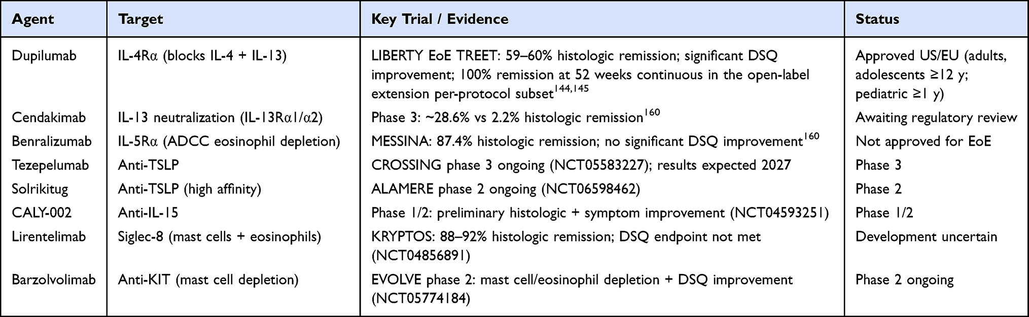 |
Table 2 Biologic Therapies in Development or Approved for Eosinophilic Esophagitis |
Optimized Topical Steroid Formulations
While biologic agents target upstream cytokine pathways, small-molecule drugs and refined topical steroid formulations aim to optimize mucosal drug delivery, improve adherence, reduce systemic exposure, and provide accessible alternatives to injectable biologics.166
The development of purpose-designed esophageal formulations has substantially improved mucosal drug residence. Besides BOT and BOS, gluticasone orally dispersible tablet (APT-1011) demonstrated 80–86% histologic remission with high-dose regimens in a phase 2b trial, with phase 3 trials ongoing.167 Extended-release injectable fluticasone crystals (EP-104GI), designed for prolonged esophageal residence via endoscopic deposition, show early data of 62% histologic remission at high dose without systemic adverse events.11 Mometasone mucoadhesive film (ESO-101), formulated for overnight esophageal delivery, achieved 48% histologic remission with significant endoscopic improvement in phase 2; phase 3 is ongoing. The progressive refinement of topical corticosteroid delivery underscores that formulation pharmacokinetics, not steroid identity, is the dominant determinant of efficacy in this disease.
Potassium-Competitive Acid Blockers
Vonoprazan, a potassium-competitive acid blocker providing more potent and predictable acid suppression than conventional PPIs, may augment the anti-inflammatory component of acid-suppressive therapy in EoE. A randomized phase 2 trial (pHalcon-EoE-201) comparing vonoprazan 20 mg daily with placebo for 12 weeks was initiated in 2025, with results expected in 2027.168,169
S1P Receptor Modulators
Etrasimod, an oral sphingosine-1-phosphate (S1P) receptor modulator targeting receptors 1, 4, and 5, limits lymphocyte egress from secondary lymphoid organs and thereby reduces esophageal mucosal infiltration. In the VOYAGE phase 2 trial, etrasimod 2 mg daily for 24 weeks reduced peak eosinophil counts by 52% compared with a 61% increase on placebo, and achieved histologic remission in approximately one-third of patients, with significant improvements in patient-reported dysphagia outcomes.170 Despite these promising results, the program was discontinued in 2025 for reasons unrelated to efficacy or safety, providing important proof-of-concept that immune-cell trafficking is a viable therapeutic target.
Janus Kinase Inhibitors
JAK-STAT signaling is the intracellular effector pathway downstream of IL-4, IL-13, IL-5, and other Th2 cytokines, positioning JAK inhibitors as pan-pathway immunomodulators with potential for broad anti-inflammatory activity in EoE. Case reports have described clinical and histologic remission with tofacitinib171 and with baricitinib and upadacitinib in eosinophilic gastrointestinal disease.172 These are uncontrolled observations. Selective JAK1 inhibition offers a potentially improved therapeutic index. AQ280 (Aqilion), a water-dissolvable selective JAK1 inhibitor tablet designed for patients with dysphagia, demonstrated dose-dependent JAK1 biomarker inhibition without off-target JAK2 effects in the phase 1 ARIA-1 trial and confirmed comparable pharmacokinetics between the dissolvable tablet and capsule in ARIA-2. Phase 2 trials are planned across North America and Europe.
Other Emerging Targets
Several additional pathways are under active preclinical or early clinical investigation. Alpha-1 antitrypsin, which restores epithelial barrier function by inhibiting kallikrein-5, has reversed EoE-like inflammation and barrier dysfunction in murine models.173 Losartan, an angiotensin II receptor blocker with antifibrotic properties, reduced esophageal eosinophilia in a small series.174 Vedolizumab, an anti-α4β7 integrin antibody, has shown preliminary efficacy signals in EoE175 although it has also been associated with eosinophilic pneumonia.176 CRTH2/DP2 antagonism, blocking PGD2-mediated chemotaxis of Th2 cells and eosinophils, produced eosinophil count reductions and symptom improvement with OC000459 in an early randomized study.177
Toward Personalized Management: Disease Control, Endotyping, and Treat-to-Target Strategies
The multi-effector pathobiology and the variable clinical responses converge on a single imperative: EoE cannot be managed with a single remission target or a single therapeutic sequence applied uniformly to all patients. The heterogeneity of disease—in inflammatory activity, fibrostenotic burden, molecular endotype, and treatment response—demands a precision medicine framework that aligns monitoring goals, therapeutic selection, and escalation criteria with the dominant pathobiological driver in each individual.
Evolving Concepts of Disease Control and Remission
Historically, histologic remission—defined as fewer than 15 eos/hpf—served as the sole therapeutic endpoint.178–180 This benchmark provided standardization across clinical trials but its limitations are now apparent: a substantial proportion of patients with marked histologic improvement experience persistent dysphagia due to structural remodeling not captured by eosinophil counts, while some patients with apparent histologic activity report minimal symptoms because of behavioral adaptation.68,181,182 The mast cell–neuroimmune crosstalk provides a cellular mechanism for this dissociation: mast cells persist in histologic remission in an activated state, maintaining neural sensitization independently of eosinophil levels.180 Histologic remission, even when defined stringently as 0 eos/hpf, does not equate to molecular remission. Multi-omics analyses of pediatric EoE reveal persistently dysregulated genes in deep remission—CDH26, DSG1, MMP12, POSTN, UPK1B, TRIM2, and MUC4—while mast cell DEG signatures including CPA3 and TPSAB1 persist in PPI-unresponsive patients after full induction.67 These molecular footprints argue for dual primary endpoints in future trials, separating inflammatory remission from remodeling remission.
The concept of deep remission therefore integrates three dimensions: histologic response (peak eosinophil count), endoscopic response quantified by the EREFS (≤2, grading edema, rings, exudates, furrows, and strictures), and patient-reported symptom improvement as captured by validated instruments such as the Dysphagia Symptom Questionnaire (DSQ).67,183 Fibrostenotic remodeling warrants particular attention: untreated EoE carries the risk of irreversible stricture formation, and sustained histologic remission in at least two consecutive endoscopies is associated with significantly lower risk of later stricture development.184,185 Figure 3 represents core outcomes in EoE management.
 |
Figure 3 Core outcome domains in eosinophilic esophagitis. Abbreviations: DIS, dilated intercellular spaces; EREFS, EoE Endoscopic Reference Score; eos/hpf, eosinophils per high-power field; EndoFLIP, Endoluminal Functional Lumen Imaging Probe. |
Clinical Phenotypes
The most clinically actionable distinction is between the inflammatory phenotype—characterized by mucosal edema, exudates, and furrows—and the fibrostenotic phenotype, characterized by rings, strictures, and esophageal narrowing. Patients with an inflammatory phenotype are generally younger, less likely to have required prior dilation, and significantly more likely to achieve clinico-histologic remission with PPIs. Esophageal distensibility measured by functional lumen imaging probe (FLIP) panometry further refines phenotypic characterization: a distensibility index below 4.5 mm2/mmHg identifies patients at high risk of food impaction and fibrostenotic progression. Recent pediatric data extend the utility of FLIP to younger patients: in a cohort of 112 patients under 21 years old undergoing EGD with concurrent EndoFLIP, the distensibility index at 30 mL inflation was significantly lower in EoE (median 2.1 mm2/mmHg) than in reflux (3.3) or normal/reactive (2.9) diagnoses (p=0.03), and DI inversely correlated with EoEHSS overall grade score as well as with subscores for eosinophil abscesses, surface layering, dilated intercellular spaces, and basal zone hyperplasia (all p<0.05).186 Because lamina propria fibrosis is challenging to sample adequately on superficial mucosal biopsy, EndoFLIP may serve as a complementary adjunct to the EoEHSS in characterizing EoE-associated remodeling in children, a patient group where established adult reference values for distensibility do not yet apply.
The atopic comorbidity burden defines a third phenotype of particular therapeutic relevance: patients with multiple coexisting type 2 inflammatory conditions are optimal candidates for dupilumab, which addresses all atopic manifestations through shared IL-4/IL-13 axis blockade. Careful characterization of the atopic versus non-atopic status also informs trigger identification: patients with high atopic burden, polysensitization, or active aeroallergen sensitization may represent a distinct endotypic subgroup in whom both food and environmental triggers contribute to esophageal inflammation. Distinguishing atopic from non-atopic EoE is therefore not merely a phenotypic label but a clinically actionable classification that guides both biological therapy selection and the scope of allergen investigation.
Molecular Endotypes and Their Therapeutic Implications
Machine-learning analysis of the 96-transcript EoE Diagnostic Panel (EDP) defined three endotypes whose differential responses to available therapies are now evident from trial data.187 EoEe1 (markedly low ALOX15, pauci-inflammatory, normal endoscopy; RR for steroid-sensitivity 3.27, 95% CI 1.04–10.27; p=0.0443) is the most treatment-responsive subgroup and is likely manageable with first-line PPIs or STCs; early biologic escalation is not warranted. EoEe2 (high IL-4, TSLP, and type 2 immunity gene expression, steroid-refractory; RR 2.77, 95% CI 1.11–6.95; p=0.0376) maps directly to the patient population in whom dupilumab performs best; its high TSLP and IL-33/ST2 burden also makes it the most compelling candidate for upcoming anti-TSLP (tezepelumab, solrikitug) or anti-IL-33/ST2 strategies, and early biologic intervention—rather than sequential first-line failure—is mechanistically rational. EoEe3 (adult-onset, low epithelial differentiation gene expression, narrow-caliber esophagus; RR for narrow-caliber 7.98, 95% CI 1.84–34.64; p=0.0013) is driven by TGF-β–fibroblast fibrostenotic pathways; combined anti-inflammatory and endoscopic dilation strategies, with attention to preventing further fibrotic progression, are required.88 Peak eosinophil counts did not differentiate the three endotypes, reinforcing that eosinophil density is an insufficient surrogate for molecular disease activity. Five molecular subgroups within these three endotypes have been described by Dunn et al, providing further granularity.188 The clinical translation of endotyping currently faces a practical obstacle: the EDP requires fresh biopsy tissue, specialized RNA processing, and bioinformatic infrastructure unavailable outside research centers. Future research must determine whether endotype classification can be approximated by serum biomarkers—eotaxin-3, IL-13, TSLP, sphingolipid ceramide ratios—and whether endotype-directed therapy improves outcomes over standard phenotypic approaches.
Treat-to-Target Framework
The concept of precision medicine in EoE ultimately requires not only patient classification but also a structured approach to defining when and how treatment goals are achieved and when escalation is warranted. The Treat-to-Target (T2T) strategy, modeled on the STRIDE and STRIDE-II initiatives that transformed inflammatory bowel disease management,189,190 applies this logic to EoE by defining distinct short-, intermediate-, and long-term therapeutic targets within explicit timeframes and linking failure to achieve these targets to predetermined escalation decisions.189 While EoE and IBD share epithelial barrier dysfunction and partially overlapping type 2 inflammatory pathways, important differences distinguish them: EoE has not been associated with colorectal cancer risk, histologic remission occupies a more central position than in IBD, and the symptom-histology dissociation is uniquely complex in EoE given behavioral dietary adaptations that may mask dysphagia severity while symptom-endoscopy dissociation is more prominent in IBD. This underscores the need for validated, disease-specific PRO instruments as well as non-invasive biomarkers rather than clinical interview alone. Interestingly, anxiety and depression have been associated with both diseases and quality of life restoration remains a treatment goal. T2T framework is illustrated in Figure 4.
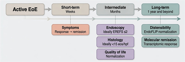 |
Figure 4 Treat-to-target framework for eosinophilic esophagitis. Adapted from Greuter & Straumann, Gastroenterol Hepatol 2024.12 Abbreviations: EREFS, EoE Endoscopic Reference Score; eos/hpf, eosinophils per high-power field; EndoFLIP, Endoluminal Functional Lumen Imaging Probe. |
Regarding specific outcomes, the COREOS collaborators have defined a core outcome set encompassing histopathology, endoscopy, patient-reported symptoms, and EoE-specific quality of life.167 Among these, patient-reported symptoms are essential for diagnosis but require careful interpretation: patients with longstanding dysphagia frequently adopt maladaptive eating behaviors that mask symptom severity at clinical interview. In randomized trials, validated instruments—the Dysphagia Symptom Questionnaire (DSQ) in adults or the EoE Activity Index (EEsAI)—should be used; in routine practice, simpler visual analogue or Likert scale tools may be more feasible. Italian EoE experts have proposed that controlled disease in adults requires a 30–90% reduction in symptom scores.167 Symptom improvement may be observed as early as week 2 with budesonide and week 4 with dupilumab; elemental diet can induce rapid complete remission within 2 weeks but practical limitations restrict widespread use.
Endoscopic activity, assessed with the EREFS, covers both inflammatory (edema, exudates, furrows) and fibrostenotic features (rings, strictures); endoscopic remission has been proposed as EREFS ≤2. Greuter and Straumann propose endoscopic remission as a short- to intermediate-term target achievable between 6 and 24 weeks, though EREFS alone does not substitute for histologic assessment.12
Histologic remission, defined as fewer than 15 eos/hpf, remains the central therapeutic target in EoE and is clinically important because patients achieving histologic remission in at least two consecutive endoscopies have a significantly lower risk of subsequent stricture development.191 After conventional induction therapy, histologic reassessment should occur approximately 8–12 weeks after treatment initiation; after biologic therapy, approximately 6 months.192 It should be noted that the Italian EoExpert consensus has proposed a two-tier internal nomenclature using the term “controlled disease” for fewer than 15 eos/hpf per 0.3 mm2 with EoEHSS improvement, and “histologic remission” for deeper suppression to fewer than 1 eos/hpf per 0.3 mm2 with marked histologic scoring system decrease.193 A stricter cutoff may be better reserved for clinical trials, as it does not appear to confer additional clinical benefit in routine practice. In randomized controlled trials, the EoEHSS—which evaluates eight biopsy features beyond eosinophil density—should be assessed; however, its complexity makes it challenging in real-world observational settings.194
Moving beyond EREFS and eosinophil counts, newer histologic scoring instruments such as the Index of Severity for Eosinophilic Esophagitis (I-SEE) evaluate not only mucosal eosinophilic infiltration but also lamina propria fibrosis, basal cell hyperplasia, dilated intercellular spaces, and surface epithelial alterations — features that more comprehensively capture the full histopathologic burden of EoE and may better correlate with structural complications and quality of life than eosinophil density alone.195 Adoption of multi-component histologic instruments alongside EREFS as composite assessment tools represents a priority for future clinical trial design and real-world practice standardization.
Quality of life is among the outcomes that matter most to patients with EoE and must be considered an integral component of disease control rather than a secondary endpoint. Improvement in at least one validated quality-of-life measure should form part of the definition of controlled disease, particularly because some highly effective therapies may control disease activity while imposing significant quality-of-life burden through route of administration, dietary restriction, or adverse effects.196
This multidimensional concept is conceptualized in Figure 5.
 |
Figure 5 Multidimensional framework for remission in eosinophilic esophagitis. Remission is conceptualized across four intersecting domains: histologic (peak eosinophil count <15 eos/hpf with EoEHSS improvement), endoscopic (EREFS ≤2), symptomatic and patient-reported quality of life (DSQ and QoL improvement), and functional (EndoFLIP-based distensibility normalization). Pairwise intersections define clinically meaningful partial response states. Deep remission requires concurrent improvement across all four domains. |
Integrating these dimensions, the T2T algorithm for EoE defines: short-term targets (6–12 weeks) of meaningful symptom improvement (≥30% DSQ reduction) and EREFS improvement; intermediate targets (8–12 weeks for conventional therapy, 6 months for biologics) of histologic remission and quality-of-life normalization; and long-term targets of fibrostenotic progression prevention, sustained molecular remission, and freedom from escalation. Failure to achieve short-term targets should prompt assessment of adherence, adequacy of delivery, and possible endotype re-evaluation before escalation. Failure to achieve intermediate histologic targets despite adequate short-term response should raise the question of endotype-directed escalation—particularly in patients whose molecular profile suggests EoEe2 biology, for whom early biologic introduction may be warranted rather than sequential first-line failure. T2T implementation is currently constrained by the endoscopic burden of repeated biopsy assessment, the absence of validated non-invasive surrogate biomarkers, and limited real-world penetration of standardized scoring systems outside academic centers. Addressing these implementation gaps is as urgent a research priority as the discovery of new therapeutic targets.
Conclusions
EoE is no longer a disease of eosinophils alone but a multi-effector immune disorder whose full complexity — from epithelial alarmin sensing and ILC2–Areg remodeling to mast cell–neuroimmune crosstalk and fibroblast-driven fibrostenosis — demands therapies, endpoints, and monitoring strategies that match the right treatment to the right patient across all dimensions of disease activity. Effective management requires multidimensional assessment integrating symptoms, histology, endoscopy, and quality of life rather than any single metric in isolation. Distinguishing the inflammatory from the fibrostenotic phenotype at diagnosis and during follow-up is clinically actionable, identifying patients who require early escalation to prevent irreversible structural progression from those for whom endoscopic intervention is a necessary complement to pharmacotherapy. Molecular endotyping holds promise for aligning patients with the therapy most matched to their dominant immune program, though translation to routine practice requires noninvasive, accessible biomarker platforms. Finally, long-term data on disease modification, fibrosis reversibility, and the durability of biologic-induced remission represent essential evidence gaps that the field must urgently address.
Data Sharing Statement
No new data were created or analyzed in this study. Data sharing is not applicable to this article.
Ethics Statement
This article is a narrative review of previously published literature. No ethics approval was required as no original human or animal studies were conducted.
Author Contributions
All authors made a significant contribution to the work reported, whether that is in the conception, study design, execution, acquisition of data, analysis and interpretation, or in all these areas; took part in drafting, revising or critically reviewing the article; gave final approval of the version to be published; have agreed on the journal to which the article has been submitted; and agree to be accountable for all aspects of the work.
AI Use Disclosure
The authors acknowledge use of Claude (Anthropic) for assistance in language refinement. All scientific content was specified and verified by the authors, who take full responsibility for the figure.
Funding
This research received no specific grant from any funding agency.
Disclosure
Luisa Bertin reports Honoraria from Giuliani, Edra SPA, Takeda; Meeting/travel support from Pfizer, Lilly, Falk, outside the submitted work. Federico Caldart has nothing to disclose.Dr Alberto Barchi reports Honoraria from Sanofi-Regeneron, Malesci, outside the submitted work. Andrea Pasta reports Grants or contracts from Dr Falk, Medtronic; Honoraria from Fenix Pharma, Dompè, Alfasigma, Teverpharma, Pfizer; Meeting/travel support from Dompé, Pfizer, Abbvie; Data safety/advisory board participation from Sanofi Opella, outside the submitted work. Vincenzo Savarino reports Honoraria from Honoraria for lectures, outside the submitted work. Edoardo Vincenzo Savarino reports Support for the manuscript from Bonollo, Difass, Pfizer, Reckitt Benckiser, Sanofi/Regeneron, SILA, Unifarco, Zeta Farmaceutici, during the conduct of the study; Consulting fees from Abbvie, Alfasigma, Apogee, AstraZeneca, Biogen, Bristol-Myers Squibb, Celltrion, Dr. Falk, Eli Lilly, Fenix Pharma, Ferring, Giuliani, Grunenthal, Johnson&Johnson, JB Pharmaceuticals, Merck & Co, Nestlè, Pfizer, PRO.MED.CS Praha a.s., Reckitt Benckiser, Recordati, Sanofi/Regeneron, SILA, Takeda, Unifarco; Honoraria from Abbvie, Aboca, Abivax, Agave, AGPharma, Alfasigma, Apoteca, Biosline, CaDiGroup, Celltrion, Dr Falk, EG Stada Group, Eli Lilly, Fenix Pharma, Galapagos, Giuliani, Johnson&Johnson, JB Pharmaceuticals, Innovamedica/Adacyte, Lionhealth, Malesci, Mayoly Biohealth, Montefarco, Novartis, Omega Pharma, Pfizer, Rafa, Reckitt Benckiser, Recordati, Sandoz, Sanofi/Regeneron, SILA, Takeda, Tillots, Unifarco, outside the submitted work. The authors report no other conflicts of interest in this work.
References
1. de BN, Visaggi P, Penagini R, et al. The 1st EoETALY consensus on the diagnosis and management of eosinophilic esophagitis – definition, clinical presentation and diagnosis. Digestive and Liver Disease. 2024;56(6):951–27. doi:10.1016/j.dld.2024.02.005
2. Savarino EV, Barbara G, Bilò MB, et al. Eosinophilic esophagitis in adults and adolescents: epidemiology, diagnostic challenges, and management strategies for a type 2 inflammatory disease. Therap Adv Gastroenterol. 2024;17:17562848241249570. doi:10.1177/17562848241249570
3. de Bortoli N, Visaggi P, Penagini R, et al. The 1st EoETALY Consensus on the Diagnosis and Management of Eosinophilic Esophagitis-Current Treatment and Monitoring. Dig Liver Dis. 2024;56(7):1173–1184. doi:10.1016/j.dld.2024.02.020
4. Mari A, Calabrese F, Pasta A, et al. Esophageal and oropharyngeal dysphagia: clinical recommendations from the united European gastroenterology and European society for neurogastroenterology and motility. United European Gastroenterology Journal. 2025;13(6):855–901. doi:10.1002/ueg2.70062
5. Bertin L, Pasta A, Ghisa M, et al. Research gaps in eosinophilic esophagitis: unanswered questions and future directions. Dig Liver Dis. 2025;2025:S1590. doi:10.1016/j.dld.2025.10.010.
6. Abonia JP, Blanchard C, Butz BB, et al. Involvement of mast cells in eosinophilic esophagitis. J Allergy Clin Immunol. 2010;126(1):140–149. doi:10.1016/j.jaci.2010.04.009
7. Aceves SS, Chen D, Newbury RO, Dohil R, Bastian JF, Broide DH. Mast cells infiltrate the esophageal smooth muscle in patients with eosinophilic esophagitis, express TGF-β1, and increase esophageal smooth muscle contraction. J Allergy Clin Immunol. 2010;126(6):1198–1204.e4. doi:10.1016/j.jaci.2010.08.050
8. González-Cervera J, Arias Á, Redondo-González O, Cano-Mollinedo MM, Terreehorst I, Lucendo AJ. Association between atopic manifestations and eosinophilic esophagitis: a systematic review and meta-analysis. Annals of Allergy, Asthma & Immunology. 2017;118(5):582–590.e2. doi:10.1016/j.anai.2017.02.006
9. Aceves SS. Remodeling and fibrosis in chronic eosinophil inflammation. Dig Dis. 2014;32(1–2):15–21. doi:10.1159/000357004
10. Lenti MV, Savarino E, Mauro A, et al. Diagnostic delay and misdiagnosis in eosinophilic oesophagitis. Dig Liver Dis. 2021;53(12):1632–1639. doi:10.1016/j.dld.2021.05.017
11. Pasta A, Bertin L, Mari A, et al. The therapeutic pipeline for eosinophilic esophagitis: current landscape and future directions. Pharmaceuticals. 2025;18(12). doi:10.3390/ph18121882
12. Greuter T, Straumann A. Rationale for an eosinophilic esophagitis treat-to-target concept. Gastroenterol Hepatol (N Y). 2024;20(10):583–590.
13. Mona R, Hruz P. Epidemiology of eosinophilic esophagitis: really a novel and evolving disease? Inflamm Intest Dis. 2025;10(1):34–40. doi:10.1159/000543022
14. Dlugosz A, Berglund A, Uhde M. The epidemiology of eosinophilic esophagitis in Sweden - a nationwide population-based study. Scandinavian Journal of Gastroenterology. 2025;60(1):1–9. doi:10.1080/00365521.2024.2440787
15. Hahn JW, Lee K, Shin JI, et al. Global incidence and prevalence of eosinophilic esophagitis, 1976–2022: a systematic review and meta-analysis. Clin Gastroenterol Hepatol. 2023;21(13):3270–3284.e77. doi:10.1016/j.cgh.2023.06.005
16. Visaggi P, Dellon ES. Epidemiology, Natural history, and treatment of eosinophilic gastrointestinal diseases. Gastroenterology. 2025;S0016–5085(25)06042–1. doi:10.1053/j.gastro.2025.09.030
17. Nudelman NT, Trovato A, Manem N, et al. Markedly increased prevalence of eosinophilic esophagitis in patients with atopic diseases in a US veteran population. Int Arch Allergy Immunol. 2025;2025:1–8. doi:10.1159/000547753
18. Thel HL, Anderson C, Xue AZ, Jensen ET, Dellon ES. Prevalence and costs of eosinophilic esophagitis in the United States. Clinical Gastroenterology and Hepatology. 2025;23(2):272–280.e8. doi:10.1016/j.cgh.2024.09.031
19. Chang JW, Jensen ET. Epidemiologic and clinical clues to the etiology of eosinophilic esophagitis. Immunology and Allergy Clinics of North America. 2024;44(2):145–155. doi:10.1016/j.iac.2023.12.003
20. Dellon ES, Shaheen O, Koutlas NT, et al. Early life factors are associated with risk for eosinophilic esophagitis diagnosed in adulthood. Dis Esophagus. 2021;34(2):doaa074. doi:10.1093/dote/doaa074
21. Bonazzi E, De Barba C, Lorenzon G, et al. Recent developments in managing luminal microbial ecology in patients with inflammatory bowel disease: from evidence to microbiome-based diagnostic and personalized therapy. Expert Review of Gastroenterology & Hepatology. 2025;19(5):563–576. doi:10.1080/17474124.2025.2495087
22. Miehlke S. Clinical features of Eosinophilic esophagitis in children and adults. Best Practice & Research Clinical Gastroenterology. 2015;29(5):739–748. doi:10.1016/j.bpg.2015.09.005
23. Bystrom J, O’Shea NR. Eosinophilic oesophagitis: clinical presentation and pathogenesis. Postgrad Med J. 2014;90(1063):282–289. doi:10.1136/postgradmedj-2012-131403
24. Alhmoud T, Ghazaleh S, Ghanim M, Redfern RE. the risk of esophageal food impaction in eosinophilic esophagitis patients: the role of Clinical and socioeconomic factors. Clin Exp Gastroenterol. 2022;15:153–161. doi:10.2147/CEG.S364994
25. Savarino E, Bredenoord AJ, Fox M, Pandolfino JE, Roman S, Gyawali CP. Advances in the physiological assessment and diagnosis of GERD. Nat Rev Gastroenterol Hepatol. 2017;14(11):665–676. doi:10.1038/nrgastro.2017.130
26. Savarino V, Visaggi P, Marabotto E, et al. Topical protection of esophageal mucosa as a new treatment of GERD. J Clin Gastroenterol. 2025;59(3):197–205. doi:10.1097/MCG.0000000000002128
27. Bertin L, Savarino V, Marabotto E, Ghisa M, de Bortoli N, Savarino EV. Pathophysiology of GERD. Digestion. 2025;2025:1. doi:10.1159/000547023
28. Gyawali CP, Yadlapati R, Fass R, et al. Updates to the modern diagnosis of GERD: lyon consensus 2.0. Gut. 2024;73(2):361–371. doi:10.1136/gutjnl-2023-330616
29. Visaggi P, Del Corso G, Solinas I, et al. Adaptive behaviors, esophageal anxiety and hypervigilance modify the association between dysphagia perception and histological disease activity in eosinophilic esophagitis. Am J Gastroenterol. 2025;120(8):1750–1759. doi:10.14309/ajg.0000000000003272
30. Hill DA, Grundmeier RW, Ramos M, Spergel JM. Eosinophilic esophagitis is a late manifestation of the allergic march. The Journal of Allergy and Clinical Immunology: In Practice. 2018;6(5):1528–1533. doi:10.1016/j.jaip.2018.05.010
31. Pesce M, Ricchiuti M, Cantone E, et al. The overlooked burden of atopic comorbidities in eosinophilic esophagitis: insights from a real-life comprehensive multidisciplinary evaluation. Journal of Clinical Medicine. 2025;14(20). doi:10.3390/jcm14207322
32. Taft TH, Guadagnoli L, Edlynn E. Anxiety and depression in eosinophilic esophagitis: a scoping review and recommendations for future research. J Asthma Allergy. 2019;12:389–399. doi:10.2147/JAA.S193045
33. Taft TH, Triggs JR, Carlson DA, et al. Validation of the oesophageal hypervigilance and anxiety scale for chronic oesophageal disease. Aliment Pharmacol Ther. 2018;47(9):1270–1277. doi:10.1111/apt.14605
34. Taft TH, Ballou S, Bedell A, Lincenberg D. Psychological Considerations and Interventions in Inflammatory Bowel Disease Patient Care. Gastroenterol Clin North Am. 2017;46(4):847–858. doi:10.1016/j.gtc.2017.08.007
35. Taft TH, Kern E, Kwiatek MA, Hirano I, Gonsalves N, Keefer L. The adult eosinophilic oesophagitis quality of life questionnaire: a new measure of health-related quality of life. Aliment Pharmacol Ther. 2011;34(7):790–798. doi:10.1111/j.1365-2036.2011.04791.x
36. Bertin L, Savarino EV. Editorial: targeting the Future of eosinophilic oesophagitis management. Aliment Pharmacol Ther. 2025;61(3):587–588. doi:10.1111/apt.18449
37. Dellon ES, Liacouras CA, Molina-Infante J, et al. Updated International Consensus Diagnostic Criteria for Eosinophilic Esophagitis. Proceedings of the AGREE Conference. Gastroenterology. 2018;155(4):1022–1033.e10. doi:10.1053/j.gastro.2018.07.009
38. Bertin L, Pasta A, Wechsler JB, et al. Beyond eosinophils: redefining the spectrum of esophageal inflammatory diseases through an immune-centric paradigm. Clin Gastroenterol Hepatol. 2026:S1542. doi:10.1016/j.cgh.2026.01.028.
39. Rochman M, Kellerman K, Jankowski MP, Rothenberg ME. The oesophagus as an immune organ. Nat Rev Gastroenterol Hepatol. 2025;22(9):657–667. doi:10.1038/s41575-025-01086-4
40. Leech T, Kelsell D, Blaydon D, Woodland P, Peiris M. A role for high mobility group box 1 (HMGB1) Release in the pathogenesis of gastroesophageal reflux disease. Neurogastroenterol Motil. 2025;e70083. doi:10.1111/nmo.70083
41. Leech T, Peiris M. Mucosal neuroimmune mechanisms in gastro-oesophageal reflux disease (GORD) pathogenesis. J Gastroenterol. 2024;59(3):165–178. doi:10.1007/s00535-023-02065-9
42. Sylvester M, Son A, Schwartz DM. The interactions between autoinflammation and type 2 immunity: from mechanistic studies to epidemiologic associations. Front Immunol. 2022;13:818039. doi:10.3389/fimmu.2022.818039
43. Gautam R, Ruffner MA. Eosinophilic esophagitis: mechanisms of disease and approach to treatment. Curr Allergy Asthma Rep. 2026;26(1):21. doi:10.1007/s11882-026-01253-w
44. Rothenberg ME, Spergel JM, Sherrill JD, et al. Common variants at 5q22 associate with pediatric eosinophilic esophagitis. Nat Genet. 2010;42(4):289–291. doi:10.1038/ng.547
45. Azouz NP, Ynga-Durand MA, Caldwell JM, et al. The antiprotease SPINK7 serves as an inhibitory checkpoint for esophageal epithelial inflammatory responses. Sci Transl Med. 2018;10(444):eaap9736. doi:10.1126/scitranslmed.aap9736
46. Azouz NP, Klingler AM, Pathre P, et al. Functional role of kallikrein 5 and proteinase-activated receptor 2 in eosinophilic esophagitis. Sci Transl Med. 2020;12(545):eaaz7773. doi:10.1126/scitranslmed.aaz7773
47. Schmitz J, Owyang A, Oldham E, et al. IL-33, an interleukin-1-like cytokine that signals via the IL-1 receptor-related protein ST2 and induces T helper type 2-associated cytokines. Immunity. 2005;23(5):479–490. doi:10.1016/j.immuni.2005.09.015
48. Venturelli N, Lexmond WS, Ohsaki A, et al. Allergic skin sensitization promotes eosinophilic esophagitis through the IL-33-basophil axis in mice. J Allergy Clin Immunol. 2016;138(5):1367–1380.e5. doi:10.1016/j.jaci.2016.02.034
49. Uchida AM, Lenehan PJ, Vimalathas P, et al. Tissue eosinophils express the IL-33 receptor ST2 and type 2 cytokines in patients with eosinophilic esophagitis. Allergy. 2022;77(2):656–660. doi:10.1111/all.15127
50. Travers J, Rochman M, Caldwell JM, Besse JA, Miracle CE, Rothenberg ME. IL-33 is induced in undifferentiated, non-dividing esophageal epithelial cells in eosinophilic esophagitis. Sci Rep. 2017;7(1):17563. doi:10.1038/s41598-017-17541-5
51. Masuda MY, Pyon GC, Luo H, et al. Epithelial overexpression of IL-33 induces eosinophilic esophagitis dependent on IL-13. J Allergy Clin Immunol. 2024;153(5):1355–1368. doi:10.1016/j.jaci.2024.01.017
52. Bouffi C, Rochman M, Zust CB, et al. IL-33 markedly activates murine eosinophils by an NF-κB-dependent mechanism differentially dependent upon an IL-4-driven autoinflammatory loop. J Immunol. 2013;191(8):4317–4325. doi:10.4049/jimmunol.1301465
53. Laky K, Frischmeyer-Guerrerio PA. Development and dysfunction of structural cells in eosinophilic esophagitis. J Allergy Clin Immunol. 2024;153(6):1485–1499. doi:10.1016/j.jaci.2024.04.006
54. Venkateshaiah SU, Mishra A, Manohar M, et al. A critical role for IL-18 in transformation and maturation of naive eosinophils to pathogenic eosinophils. J Allergy Clin Immunol. 2018;142(1):301–305. doi:10.1016/j.jaci.2018.02.011
55. Salzano S, Caltabiano R, Zizzo M, et al. Eosinophilic Esophagitis: a Review of Histopathological and Diagnostic Aspects. Diagnostics (Basel). 2025;15(23):3011. doi:10.3390/diagnostics15233011
56. Collins MH. Histopathology of eosinophilic esophagitis. Dig Dis. 2014;32(1–2):68–73. doi:10.1159/000357012
57. Heil A, Kuehlewindt T, Godat A, et al. Histological Phenotyping in Eosinophilic Esophagitis: localized Proximal Disease Is Infrequent but Associated with Less Severe Disease and Better Disease Outcome. Int Arch Allergy Immunol. 2024;185(1):63–72. doi:10.1159/000533815
58. Ma C, Jairath V, Feagan BG, et al. Responsiveness of a Histologic Scoring System Compared With Peak Eosinophil Count in Eosinophilic Esophagitis. Am J Gastroenterol. 2022;117(2):264–271. doi:10.14309/ajg.0000000000001571
59. Lenti MV, Rossi CM, Cannizzaro R, et al. Disrupted mucosal vascular barrier in eosinophilic esophagitis. Sci Rep. 2025;15(1):25600. doi:10.1038/s41598-025-09680-x
60. Kottyan LC, Davis BP, Sherrill JD, et al. Genome-wide association analysis of eosinophilic esophagitis provides insight into the tissue specificity of this allergic disease. Nat Genet. 2014;46(8):895–900. doi:10.1038/ng.3033
61. Davis BP, Stucke EM, Khorki ME, et al. Eosinophilic esophagitis-linked calpain 14 is an IL-13-induced protease that mediates esophageal epithelial barrier impairment. JCI Insight. 2016;1(4):e86355. doi:10.1172/jci.insight.86355
62. Meyer-Hoffert U, Wu Z, Schröder JM. Identification of Lympho-Epithelial Kazal-Type Inhibitor 2 in Human Skin as a Kallikrein-Related Peptidase 5-Specific Protease Inhibitor. PLoS One. 2009;4(2):e4372. doi:10.1371/journal.pone.0004372
63. Hiremath G, Choksi Y, Correa H, et al. Children with eosinophilic esophagitis non-responsive to combination therapy have distinct esophageal transcriptomic and microbiome profile. Allergy. 2024;79(10):2798–2811. doi:10.1111/all.16208
64. Braskett M, Goleva E, Bronova I, et al. Lipidomic Analysis of Esophageal Epithelia Reveals a Distinctive Sphingolipid Profile in Eosinophilic Esophagitis. Allergy. 2025;80(10):2849–2860. doi:10.1111/all.16660
65. Kellett SK, Crue T, Almeida Cruz SN, et al. Resolution of epithelial dysfunction in eosinophilic esophagitis is mediated by an HIF-1α-CD73-adenosine signaling axis. J Allergy Clin Immunol. 2026;157(3):693–710.e1. doi:10.1016/j.jaci.2025.09.006
66. Hoelz H, Faro T, Frank ML, et al. Persistent desmoglein-1 downregulation and periostin accumulation in histologic remission of eosinophilic esophagitis. J Allergy Clin Immunol. 2025;155(2):505–519. doi:10.1016/j.jaci.2024.09.016
67. Ruffner MA, Shoda T, Lal M, et al. Persistent esophageal changes after histologic remission in eosinophilic esophagitis. J Allergy Clin Immunol. 2024;153(4):1063–1072. doi:10.1016/j.jaci.2023.12.012
68. Russin M, Dellon ES. Emerging Tissue and Blood Biomarkers for Eosinophilic Esophagitis. Gastroenterol Clin North Am. 2026;55(1):39–54. doi:10.1016/j.gtc.2025.08.006
69. Dong F, Perdew GH. The aryl hydrocarbon receptor as a mediator of host-microbiota interplay. Gut Microbes. 2020;12(1):1859812. doi:10.1080/19490976.2020.1859812
70. Rodríguez-López EM, Clement RL, Lal M, et al. Oesophageal Epithelial Cell-Intrinsic MHCII Regulates Food Antigen-Dependent Eosinophilic Esophagitis in an IFNγ-Dependent Manner. Clin Exp Allergy. 2026;56(2):124–136. doi:10.1111/cea.70205
71. Facchin S, Bonazzi E, Tomasulo A, et al. Could modulating the esophageal microbiome be the answer for eosinophilic esophagitis treatment? Expert Rev Gastroenterol Hepatol. 2025;2025:1–9. doi:10.1080/17474124.2025.2530606
72. Zhernov YV, Vysochanskaya SO, Sukhov VA, et al. Molecular Mechanisms of Eosinophilic Esophagitis. Int J Mol Sci. 2021;22(24):13183. doi:10.3390/ijms222413183
73. Ruffner MA, Hu A, Dilollo J, et al. Conserved IFN Signature between Adult and Pediatric Eosinophilic Esophagitis. J Immunol. 2021;206(6):1361–1371. doi:10.4049/jimmunol.2000973
74. Lim M, Kim T, Kim H, Jang BG, Myung JK, Kim HY. Esophageal ILC2s mediate abnormal epithelial remodeling in eosinophilic esophagitis via Areg-EGFR signaling. Cell Mol Immunol. 2025;22(1):97–110. doi:10.1038/s41423-024-01242-x
75. Wen T, Aronow BJ, Rochman Y, et al. Single-cell RNA sequencing identifies inflammatory tissue T cells in eosinophilic esophagitis. J Clin Invest. 2025;129(5):2014–2028. doi:10.1172/JCI125917
76. Kim TR, Son B, Lee CG, Park HO. Amphiregulin in Fibrotic Diseases and Cancer. International Journal of Molecular Sciences. 2025;26(14). doi:10.3390/ijms26146945
77. Van KL, Parekh VV, Wu L. Invariant natural killer T cells: bridging innate and adaptive immunity. Cell Tissue Res. 2011;343(1):43–55. doi:10.1007/s00441-010-1023-3
78. Melum E, Jiang X, Baker KD, et al. Control of CD1d-restricted antigen presentation and inflammation by sphingomyelin. Nat Immunol. 2019;20(12):1644–1655. doi:10.1038/s41590-019-0504-0
79. Lee-Sarwar K, Kelly RS, Lasky-Su J, et al. Intestinal microbial-derived sphingolipids are inversely associated with childhood food allergy. Journal of Allergy and Clinical Immunology. 2018;142(1):335–338.e9. doi:10.1016/j.jaci.2018.04.016
80. Kheshtchin N, Kanannejad Z, Ghahramani Z, Esmaeilzadeh H, Sepahi N. Balancing immune responses: regulatory cells in eosinophilic gastrointestinal disorders. Front Immunol. 2024;15:1372009. doi:10.3389/fimmu.2024.1372009
81. Chen HH, Sun AH, Ojcius DM, et al. Eosinophils from Murine Lamina Propria Induce Differentiation of Naïve T Cells into Regulatory T Cells via TGF-β1 and Retinoic Acid. PLoS One. 2015;10(11):e0142881. doi:10.1371/journal.pone.0142881
82. Racca F, Pellegatta G, Cataldo G, et al. Type 2 Inflammation in Eosinophilic Esophagitis: from Pathophysiology to Therapeutic Targets. Front Physiol. 2021;12:815842. doi:10.3389/fphys.2021.815842
83. Gurram RK, Zhu J. Orchestration between ILC2s and Th2 cells in shaping type 2 immune responses. Cell Mol Immunol. 2019;16(3):225–235. doi:10.1038/s41423-019-0210-8
84. Morgan DM, Ruiter B, Smith NP, et al. Clonally expanded, GPR15-expressing pathogenic effector TH2 cells are associated with eosinophilic esophagitis. Sci Immunol. 2021;6(62):eabi5586. doi:10.1126/sciimmunol.abi5586
85. Blanchard C, Mingler MK, Vicario M, et al. IL-13 involvement in eosinophilic esophagitis: transcriptome analysis and reversibility with glucocorticoids. J Allergy Clin Immunol. 2007;120(6):1292–1300. doi:10.1016/j.jaci.2007.10.024
86. Blanchard C, Stucke EM, Rodriguez-Jimenez B, et al. A striking local esophageal cytokine expression profile in eosinophilic esophagitis. J Allergy Clin Immunol. 2011;127(1):208–217,217.e1–7. doi:10.1016/j.jaci.2010.10.039
87. Ryu S, Lee KH, Tizaoui K, et al. Pathogenesis of Eosinophilic Esophagitis: a Comprehensive Review of the Genetic and Molecular Aspects. Int J Mol Sci. 2020;21(19):7253. doi:10.3390/ijms21197253
88. Gueguen E, Morsy Y, Mamie C, et al. Novel transcriptomic panel identifies histologically active eosinophilic oesophagitis. Gut. 2024;73(7):1076–1086. doi:10.1136/gutjnl-2023-331743
89. Sherrill JD, Kc K, Wu D, et al. Desmoglein-1 regulates esophageal epithelial barrier function and immune responses in eosinophilic esophagitis. Mucosal Immunol. 2014;7(3):718–729. doi:10.1038/mi.2013.90
90. Blanchard C, Wang N, Stringer KF, et al. Eotaxin-3 and a uniquely conserved gene-expression profile in eosinophilic esophagitis. J Clin Invest. 2006;116(2):536–547. doi:10.1172/JCI26679
91. Nakagome K, Nagata M. The Possible Roles of IL-4/IL-13 in the Development of Eosinophil-Predominant Severe Asthma. Biomolecules. 2024;14(5). doi:10.3390/biom14050546
92. Krempski J, Yamani A, Thota LNR, et al. IL-4-STAT6 axis amplifies histamine-induced vascular endothelial dysfunction and hypovolemic shock. J Allergy Clin Immunol. 2024;154(3):719–734. doi:10.1016/j.jaci.2024.05.009
93. Yamaguchi Y, Suda T, Ohta S, Tominaga K, Miura Y, Kasahara T. Analysis of the survival of mature human eosinophils: interleukin-5 prevents apoptosis in mature human eosinophils. Blood. 1991;78(10):2542–2547.
94. Straumann A, Conus S, Grzonka P, et al. Anti-interleukin-5 antibody treatment (mepolizumab) in active eosinophilic oesophagitis: a randomised, placebo-controlled, double-blind trial. Gut. 2010;59(1):21–30. doi:10.1136/gut.2009.178558
95. Assa’ad AH, Gupta SK, Collins MH, et al. An antibody against IL-5 reduces numbers of esophageal intraepithelial eosinophils in children with eosinophilic esophagitis. Gastroenterology. 2011;141(5):1593–1604. doi:10.1053/j.gastro.2011.07.044
96. Vaccaro J, Canziani KE, Guzmán L, et al. Type-2 Cytokines Promote the Secretion of the Eosinophil–Attractant CCL26 by Intestinal Epithelial Cells in Food-Sensitized Patients. Front Immunol. 2022;13:909896. doi:10.3389/fimmu.2022.909896
97. Blanchard C, Durual S, Estienne M, Emami S, Vasseur S, Cuber JC. Eotaxin-3/CCL26 gene expression in intestinal epithelial cells is up-regulated by interleukin-4 and interleukin-13 via the signal transducer and activator of transcription 6. Int J Biochem Cell Biol. 2005;37(12):2559–2573. doi:10.1016/j.biocel.2005.06.010
98. Bick F, Blanchetot C, Lambrecht BN, Schuijs MJ. A reappraisal of IL-9 in inflammation and cancer. Mucosal Immunology. 2025;18(1):1–15. doi:10.1016/j.mucimm.2024.10.003
99. Ramirez GA, Yacoub M-R, Ripa M, et al. Eosinophils from Physiology to Disease: a Comprehensive Review. Biomed Res Int. 2018;2018:9095275. doi:10.1155/2018/9095275
100. Sastre B, Cañas JA, Rodrigo-Muñoz JM, Del Pozo V. Novel Modulators of Asthma and Allergy: exosomes and MicroRNAs. Front Immunol. 2017;8:826. doi:10.3389/fimmu.2017.00826
101. Le-carlson M, Seki S, Abarbanel D, Quiros A, Cox K, Nadeau KC. Markers of Antigen Presentation and Activation on Eosinophils and T Cells in the Esophageal Tissue of Patients With Eosinophilic Esophagitis. J Pediatr Gastroenterol Nutr. 2013;56(3):257–262. doi:10.1097/MPG.0b013e3182758d49
102. Wechsler ME, Munitz A, Ackerman SJ, et al. Eosinophils in Health and Disease: a State-of-the-Art Review. Mayo Clin Proc. 2021;96(10):2694–2707. doi:10.1016/j.mayocp.2021.04.025
103. Zhu C, Weng Q, Gao S, et al. TGF-β signaling promotes eosinophil activation in inflammatory responses. Cell Death Dis. 2024;15(8):637. doi:10.1038/s41419-024-07029-2
104. Davis BP, Rothenberg ME. Mechanisms of Disease of Eosinophilic Esophagitis. Annu Rev Pathol. 2016;11:365–393. doi:10.1146/annurev-pathol-012615-044241
105. Godat A, Greuter T. Food-related reactions in eosinophilic esophagitis: pathophysiology and treatment. Best Practice & Research Clinical Gastroenterology. 2025;79:102073. doi:10.1016/j.bpg.2025.102073
106. van Rhijn BD, van Ree R, Versteeg SA, et al. Birch pollen sensitization with cross-reactivity to food allergens predominates in adults with eosinophilic esophagitis. Allergy. 2013;68(11):1475–1481. doi:10.1111/all.12257
107. Nuyttens L, Dominicus T, Keppens C, et al. IgE-Mediated Food Sensitization, Management Strategies, and Quality of Life in Pediatric Eosinophilic Esophagitis: a Prospective Observational Study. Nutrients. 2025;17(12). doi:10.3390/nu17121980
108. N B-BM, Rochman M, Kotliar M, et al. Single-cell RNA-sequencing of human eosinophils in allergic inflammation in the esophagus. J Allergy Clin Immunol. 2024;154(4):974–987. doi:10.1016/j.jaci.2024.05.029
109. Kleuskens MTA, Bek MK, Al Halabi Y, et al. Mast cells disrupt the function of the esophageal epithelial barrier. Mucosal Immunol. 2023;16(5):567–577. doi:10.1016/j.mucimm.2023.06.001
110. Pajulas A, Fu Y, Cheung CCL, et al. Interleukin-9 promotes mast cell progenitor proliferation and CCR2-dependent mast cell migration in allergic airway inflammation. Mucosal Immunol. 2023;16(4):432–445. doi:10.1016/j.mucimm.2023.05.002
111. O’Donnell MC, Ackerman SJ, Gleich GJ, Thomas LL. Activation of basophil and mast cell histamine release by eosinophil granule major basic protein. J Exp Med. 1983;157(6):1981–1991. doi:10.1084/jem.157.6.1981
112. Eindor-Abarbanel A, Moss-Ophir N, Agajany N, et al. Beyond eosinophils: a proteomic dissection of remodeling and inflammation in suspected eosinophilic esophagitis. J Pediatr Gastroenterol Nutr. 2026;2026:1 doi:10.1002/jpn3.70347
113. Muir AB, Dods K, Henry SJ, et al. Eosinophilic esophagitis associated chemical and mechanical microenvironment shapes esophageal fibroblast behavior. J Pediatr Gastroenterol Nutr. 2016;63(2):200–209. doi:10.1097/MPG.0000000000001100
114. Cho JY, Doshi A, Rosenthal P, et al. Smad3-deficient mice have reduced esophageal fibrosis and angiogenesis in a model of egg-induced eosinophilic esophagitis. J Pediatr Gastroenterol Nutr. 2014;59(1):10–16. doi:10.1097/MPG.0000000000000343
115. Rawson R, Anilkumar A, Newbury RO, et al. The TGFβ1 Promoter SNP C-509T and Food Sensitization Promote Esophageal Remodeling in Pediatric Eosinophilic Esophagitis. PLoS One. 2015;10(12):e0144651. doi:10.1371/journal.pone.0144651
116. Maeda D, Kubo T, Kiya K, et al. Periostin is induced by IL-4/IL-13 in dermal fibroblasts and promotes RhoA/ROCK pathway-mediated TGF-β1 secretion in abnormal scar formation. J Plast Surg Hand Surg. 2019;53(5):288–294. doi:10.1080/2000656X.2019.1612752
117. Dellon ES, Higgins LL, Beitia R, et al. Prospective assessment of serum periostin as a biomarker for diagnosis and monitoring of eosinophilic esophagitis. Aliment Pharmacol Ther. 2016;44(2):189–197. doi:10.1111/apt.13672
118. Blanchard C, Mingler MK, McBride M, et al. Periostin facilitates eosinophil tissue infiltration in allergic lung and esophageal responses. Mucosal Immunol. 2008;1(4):289–296. doi:10.1038/mi.2008.15
119. Jumabay M, Abud EM, Okamoto K, et al. Eosinophilic esophagitis drives tissue fibroblast regenerative programs toward pathologic dysfunction. J Allergy Clin Immunol. 2025;155(4):1333–1345. doi:10.1016/j.jaci.2024.11.028
120. D’Urso M, Kurniawan NA. Mechanical and Physical Regulation of Fibroblast–Myofibroblast Transition: from Cellular Mechanoresponse to Tissue Pathology. Front Bioeng Biotechnol. 2020;8:609653. doi:10.3389/fbioe.2020.609653
121. Manresa MC, Miki H, Miller J, et al. A Deficiency in the Cytokine TNFSF14/LIGHT Limits Inflammation and Remodeling in Murine Eosinophilic Esophagitis. J Immunol. 2022;209(12):2341–2351. doi:10.4049/jimmunol.2200326
122. Shoda T, Wen T, Caldwell JM, et al. Loss of Endothelial TSPAN12 Promotes Fibrostenotic Eosinophilic Esophagitis via Endothelial Cell–Fibroblast Crosstalk. Gastroenterology. 2022;162(2):439–453. doi:10.1053/j.gastro.2021.10.016
123. Merves JF, Whelan KA, Benitez AJ, et al. ATG7 gene expression as a novel tissue biomarker in Eosinophilic Esophagitis. Am J Gastroenterol. 2016;111(1):151–153. doi:10.1038/ajg.2015.404
124. Whelan KA, Merves JF, Giroux V, et al. Autophagy mediates epithelial cytoprotection in Eosinophilic Oesophagitis. Gut. 2017;66(7):1197–1207. doi:10.1136/gutjnl-2015-310341
125. Zhang S, Shoda T, Aceves SS, et al. The mast cell pain connection in eosinophilic esophagitis. Allergy. 2022;77(6):1895–1899. doi:10.1111/all.15260
126. Perner C, Flayer CH, Zhu X, et al. Substance P release by sensory neurons triggers dendritic cell migration and initiates the Type-2 immune response to allergens. Immunity. 2020;53(5):1063–1077.e7. doi:10.1016/j.immuni.2020.10.001
127. Barchi A, Massimino L, Mandarino FV, et al. Clinical, Histologic, and Safety Outcomes With Long-term Maintenance Therapies for Eosinophilic Esophagitis: a Systematic Review and Meta-analysis. Clin Gastroenterol Hepatol. 2025;23(11):1890–1904.e7. doi:10.1016/j.cgh.2024.11.007
128. Visaggi P, Barberio B, Corso GD, et al. Comparison of drugs for active eosinophilic oesophagitis: systematic review and network meta-analysis. Gut. 2023;72(11):2019–2030. doi:10.1136/gutjnl-2023-329873
129. Rabinowitz SS, Sharma S, Pittman M. Coexistence of gastroesophageal reflux and eosinophilic esophagitis in a single patient: case report. Transl Pediatr. 2025;14(4):740–747. doi:10.21037/tp-2024-620
130. Laserna-Mendieta EJ, Casabona S, Guagnozzi D, et al. Efficacy of proton pump inhibitor therapy for eosinophilic oesophagitis in 630 patients: results from the EoE connect registry. Aliment Pharmacol Ther. 2020;52(5):798–807. doi:10.1111/apt.15957
131. Laserna-Mendieta EJ, Casabona S, Savarino E, et al. Efficacy of Therapy for Eosinophilic Esophagitis in Real-World Practice. Clin Gastroenterol Hepatol. 2020;18(13):2903–2911.e4. doi:10.1016/j.cgh.2020.01.024
132. Lucendo AJ, Gutiérrez-Ramírez L, Tejera-Muñoz A, Molina-Infante J, Arias Á. EUREOS Guidelines Committee. Proton Pump Inhibitors for Inducing and Maintaining Remission in Eosinophilic Esophagitis: an Updated Systematic Review and Meta-Analysis. Clin Gastroenterol Hepatol. 2025;23(12):2115–2127.e21. doi:10.1016/j.cgh.2025.01.016
133. Muftah M, Goldin AH, Barshop K, et al. Twice-Daily Proton Pump Inhibitor Induces Higher Remission Rate in Eosinophilic Esophagitis Than Once-Daily Regimen Regardless of Total Daily Dose. Am J Gastroenterol. 2024;119(5):991–995. doi:10.14309/ajg.0000000000002712
134. Dellon ES, Muir AB, Katzka DA, et al. ACG Clinical Guideline: diagnosis and Management of Eosinophilic Esophagitis. Am J Gastroenterol. 2025;120(1):31–59. doi:10.14309/ajg.0000000000003194
135. Alexander R, Alexander JA, Akambase J, et al. Proton Pump Inhibitor Therapy in Eosinophilic Esophagitis: predictors of Nonresponse. Dig Dis Sci. 2021;66(9):3096–3104. doi:10.1007/s10620-020-06633-4
136. Barchi A, Girelli M, Ventimiglia A, et al. Orally Dispersible Swallowed Topical Corticosteroids in Eosinophilic Esophagitis: a Paradigm Shift in the Management of Esophageal Inflammation. Pharmaceutics. 2025;17(10):1325. doi:10.3390/pharmaceutics17101325
137. Franciosi JP, Gordon M, Sinopoulou V, et al. Medical treatment of eosinophilic esophagitis. Cochrane Database Syst Rev. 2023;7(7):CD004065. doi:10.1002/14651858.CD004065.pub4
138. Hirano I, Collins MH, Katzka DA, et al. Budesonide Oral Suspension Improves Outcomes in Patients With Eosinophilic Esophagitis: results from a Phase 3 Trial. Clin Gastroenterol Hepatol. 2022;20(3):525–534.e10. doi:10.1016/j.cgh.2021.04.022
139. Miehlke S, Schlag C, Lucendo AJ, et al. Budesonide orodispersible tablets for induction of remission in patients with active eosinophilic oesophagitis: a 6-week open-label trial of the EOS-2 Programme. United European Gastroenterol J. 2022;10(3):330–343. doi:10.1002/ueg2.12220
140. Biedermann L, Schlag C, Straumann A, et al. Efficacy and Safety of Budesonide Orodispersible Tablets for Eosinophilic Esophagitis up to 3 Years: an Open-Label Extension Study. Clinical Gastroenterology and Hepatology. 2025;23(12):2144–2154.e6. doi:10.1016/j.cgh.2024.10.034
141. Maniero D, Ghisa M, Bruschi A, et al. Effectiveness and Safety of Orodispersible Budesonide for Eosinophilic Esophagitis: a Multicenter Real-World Study. Clinical Gastroenterology and Hepatology. 2026;24(4):986–994. doi:10.1016/j.cgh.2025.08.033
142. Straumann A, Lucendo AJ, Miehlke S, et al. Budesonide Orodispersible Tablets Maintain Remission in a Randomized, Placebo-Controlled Trial of Patients With Eosinophilic Esophagitis. Gastroenterology. 2020;159(5):1672–1685.e5. doi:10.1053/j.gastro.2020.07.039
143. Laserna-Mendieta EJ, Navarro P, Casabona-Francés S, et al. Swallowed topical corticosteroids for eosinophilic esophagitis: utilization and real-world efficacy from the EoE CONNECT registry. United European Gastroenterol J. 2024;12(5):585–595. doi:10.1002/ueg2.12533
144. Dellon ES, Rothenberg ME, Collins MH, et al. Dupilumab in Adults and Adolescents with Eosinophilic Esophagitis. N Engl J Med. 2022;387(25):2317–2330. doi:10.1056/NEJMoa2205982
145. Rothenberg ME, Dellon ES, Collins MH, et al. Efficacy and safety of dupilumab up to 52 weeks in adults and adolescents with eosinophilic oesophagitis (LIBERTY EoE TREET study): a multicentre, double-blind, randomised, placebo-controlled, phase 3 trial. Lancet Gastroenterol Hepatol. 2023;8(11):990–1004. doi:10.1016/S2468-1253(23)00204-2
146. Aceves SS, Dellon ES, Greenhawt M, Hirano I, Liacouras CA, Spergel JM. Clinical guidance for the use of dupilumab in eosinophilic esophagitis: a yardstick. Ann Allergy Asthma Immunol. 2023;130(3):371–378. doi:10.1016/j.anai.2022.12.014
147. Collins MH, Rothenberg ME, Dellon ES, et al. Dupilumab Improves Histopathologic Features in Patients With Eosinophilic Esophagitis: LIBERTY EoE TREET Study Results. Gastro Hep Advances. 2025;4(6). doi:10.1016/j.gastha.2025.100646
148. Chehade M, Dellon ES, Spergel JM, et al. Dupilumab for Eosinophilic Esophagitis in Patients 1 to 11 Years of Age. New England Journal of Medicine. 2024;390(24):2239–2251. doi:10.1056/NEJMoa2312282
149. Dellon ES, Kosloski MP, Shabbir A, Glotfelty L, Xu C, Kamal MA. Symptomatic Improvement in Adults and Adolescents with Eosinophilic Esophagitis Requires Higher Systemic Dupilumab Exposure than Histologic Response. Clin Transl Gastroenterol. 2025;16:e00793. doi:10.14309/ctg.0000000000000793
150. Chang JW, Kliewer K, Haller E, et al. Development of a Practical Guide to Implement and Monitor Diet Therapy for Eosinophilic Esophagitis. Clinical Gastroenterology and Hepatology. 2023;21(7):1690–1698. doi:10.1016/j.cgh.2023.03.006
151. Kliewer KL, Abonia JP, Aceves SS, et al. One-food versus 4-food elimination diet for pediatric eosinophilic esophagitis: a multisite randomized trial. J Allergy Clin Immunol. 2025;155(2):520–532. doi:10.1016/j.jaci.2024.08.023
152. Kliewer KL, Gonsalves N, Dellon ES, et al. One-food versus six-food elimination diet therapy for the treatment of eosinophilic oesophagitis: a multicentre, randomised, open-label trial. Lancet Gastroenterol Hepatol. 2023;8(5):408–421. doi:10.1016/S2468-1253(23)00012-2
153. Molina-Infante J, Arias Á, Alcedo J, et al. Step-up empiric elimination diet for pediatric and adult eosinophilic esophagitis: the 2-4-6 study. J Allergy Clin Immunol. 2018;141(4):1365–1372. doi:10.1016/j.jaci.2017.08.038
154. Visaggi P, Mariani L, Pardi V, et al. Dietary Management of Eosinophilic Esophagitis: tailoring the Approach. Nutrients. 2021;13(5):1630. doi:10.3390/nu13051630
155. Aceves SS, Alexander JA, Baron TH, et al. Endoscopic approach to eosinophilic esophagitis: American Society for Gastrointestinal Endoscopy Consensus Conference. Gastrointest Endosc. 2022;96(4):576–592.e1. doi:10.1016/j.gie.2022.05.013
156. Jung KW, Gundersen N, Kopacova J, et al. Occurrence of and risk factors for complications after endoscopic dilation in eosinophilic esophagitis. Gastrointest Endosc. 2011;73(1):15–21. doi:10.1016/j.gie.2010.09.036
157. Schoepfer AM, Gonsalves N, Bussmann C, et al. Esophageal dilation in eosinophilic esophagitis: effectiveness, safety, and impact on the underlying inflammation. Am J Gastroenterol. 2010;105(5):1062–1070. doi:10.1038/ajg.2009.657
158. Dougherty M, Runge TM, Eluri S, Dellon ES. Esophageal dilation with either bougie or balloon technique as a treatment for eosinophilic esophagitis: a systematic review and meta-analysis. Gastrointest Endosc. 2017;86(4):581–591.e3. doi:10.1016/j.gie.2017.04.028
159. Bertin L, Sirinic E, Ghisa M, et al. Eosinophilic esophagitis: from pathophysiology to diagnosis, monitoring and treatment. European Journal of Clinical Investigation. 2026;56(4):e70192. doi:10.1111/eci.70192
160. Dellon ES, Charriez CM, Zhang S, et al. Cendakimab in Adults and Adolescents with Eosinophilic Esophagitis. NEJM Evid. 2025;4(10):EVIDoa2500095. doi:10.1056/EVIDoa2500095
161. Spergel JM, Rothenberg ME, Collins MH, et al. Reslizumab in children and adolescents with eosinophilic esophagitis: results of a double-blind, randomized, placebo-controlled trial. J Allergy Clin Immunol. 2012;129(2):456–463,463.e1–3. doi:10.1016/j.jaci.2011.11.044
162. Rothenberg ME, Dellon ES, Collins MH, et al. Eosinophil Depletion with Benralizumab for Eosinophilic Esophagitis. N Engl J Med. 2024;390(24):2252–2263. doi:10.1056/NEJMoa2313318
163. Vicari AP, Schoepfer AM, Meresse B, et al. Discovery and characterization of a novel humanized anti-IL-15 antibody and its relevance for the treatment of refractory celiac disease and eosinophilic esophagitis. MAbs. 2017;9(6):927–944. doi:10.1080/19420862.2017.1332553
164. Youngblood BA, Brock EC, Leung J, et al. AK002, a Humanized Sialic Acid-Binding Immunoglobulin-Like Lectin-8 Antibody that Induces Antibody-Dependent Cell-Mediated Cytotoxicity against Human Eosinophils and Inhibits Mast Cell-Mediated Anaphylaxis in Mice. Int Arch Allergy Immunol. 2019;180(2):91–102. doi:10.1159/000501637
165. Alvarado D, Maurer M, Gedrich R, et al. Anti-KIT monoclonal antibody CDX-0159 induces profound and durable mast cell suppression in a healthy volunteer study. Allergy. 2022;77(8):2393–2403. doi:10.1111/all.15262
166. Rider P, Carmi Y, Cohen I. Biologics for Targeting Inflammatory Cytokines, Clinical Uses, and Limitations. Int J Cell Biol. 2016;2016:9259646. doi:10.1155/2016/9259646
167. Dellon ES, Lucendo AJ, Schlag C, et al. Fluticasone Propionate Orally Disintegrating Tablet (APT-1011) for Eosinophilic Esophagitis: randomized Controlled Trial. Clin Gastroenterol Hepatol. 2022;20(11):2485–2494.e15. doi:10.1016/j.cgh.2022.02.013
168. Kuzumoto T, Tanaka F, Sawada A, et al. Vonoprazan shows efficacy similar to that of proton pump inhibitors with respect to symptomatic, endoscopic, and histological responses in patients with eosinophilic esophagitis. Esophagus. 2021;18(2):372–379. doi:10.1007/s10388-020-00783-0
169. Marabotto E, Calabrese F, Pasta A, et al. Evaluating Vonoprazan for the treatment of erosive GERD and heartburn associated with GERD in adults. Expert Opin Pharmacother. 2024;25(17):2319–2325. doi:10.1080/14656566.2024.2427335
170. Dellon ES, Collins MH, Bredenoord AJ, et al. Etrasimod as a treatment for eosinophilic oesophagitis (VOYAGE): a double-blind, placebo-controlled, randomised, phase 2 trial. Lancet Gastroenterol Hepatol. 2025;10(7):622–633. doi:10.1016/S2468-1253(25)00062-7
171. Cheng E, Zhang X, Wilson KS, et al. JAK-STAT6 Pathway Inhibitors Block Eotaxin-3 Secretion by Epithelial Cells and Fibroblasts from Esophageal Eosinophilia Patients: promising Agents to Improve Inflammation and Prevent Fibrosis in EoE. PLoS One. 2016;11(6):e0157376. doi:10.1371/journal.pone.0157376
172. Luschnig P, Kienzl M, Roula D, et al. The JAK1/2 inhibitor baricitinib suppresses eosinophil effector function and restricts allergen-induced airway eosinophilia. Biochem Pharmacol. 2021;192:114690. doi:10.1016/j.bcp.2021.114690
173. Wang T, Shuai P, Wang Q, et al. α‑1 Antitrypsin is a potential target of inflammation and immunomodulation (Review). Mol Med Rep. 2025;31(4):107. doi:10.3892/mmr.2025.13472
174. Abonia JP, Rudman Spergel AK, Hirano I, et al. Losartan Treatment Reduces Esophageal Eosinophilic Inflammation in a Subset of Eosinophilic Esophagitis. J Allergy Clin Immunol Pract. 2024;12(9):2427–2438.e3. doi:10.1016/j.jaip.2024.07.011
175. Taft TH, Mutlu EA. The Potential Role of Vedolizumab in Concomitant Eosinophilic Esophagitis and Crohn’s Disease. Clinical Gastroenterology and Hepatology. 2018;16(11):1840–1841. doi:10.1016/j.cgh.2018.06.022
176. Zhu W, Zhao T, Wei J, et al. Case report: eosinophilic pneumonia associated with vedolizumab therapy in a patient with ulcerative colitis. Front Med (Lausanne). 2022;9. 942237. doi:10.3389/fmed.2022.942237
177. Straumann A, Hoesli S, Bussmann C, et al. Anti-eosinophil activity and clinical efficacy of the CRTH2 antagonist OC000459 in eosinophilic esophagitis. Allergy. 2013;68(3):375–385. doi:10.1111/all.12096
178. Straumann A, Katzka DA. Diagnosis and Treatment of Eosinophilic Esophagitis. Gastroenterology. 2018;154(2):346–359. doi:10.1053/j.gastro.2017.05.066
179. Muir A, Falk GW. Eosinophilic Esophagitis: a Review. JAMA. 2021;326(13):1310–1318. doi:10.1001/jama.2021.14920
180. Eke R, Li T, White A, Tariq T, Markowitz J, Lenov A. Systematic review of histological remission criteria in eosinophilic esophagitis. JGH Open. 2018;2(4):158–165. doi:10.1002/jgh3.12059
181. Al Abdulqader AK. Advances in diagnostic techniques for eosinophilic esophagitis: a systematic review of emerging noninvasive methods. European Journal of Medical Research. 2025;30(1):924. doi:10.1186/s40001-025-02989-3
182. Cotton CC, Moist SE, McGee SJ, Furuta GT, Aceves SS, Dellon ES. A Newly Proposed Severity Index for Eosinophilic Esophagitis is Associated With Baseline Clinical Features and Successful Treatment Response. Clinical Gastroenterology and Hepatology. 2023;21(10):2534–2542.e1. doi:10.1016/j.cgh.2023.03.047
183. Katzka DA, Falck-Ytter Y. AGA Commentary on Eosinophilic Esophagitis Guidelines. Gastroenterology. 2020;159(3):813–815. doi:10.1053/j.gastro.2020.05.070
184. Xu X, Kwiatek J, Siddall J, Genofre E, Stirnadel-Farrant H, Katial R. Patient-reported symptoms and burden of eosinophilic esophagitis: evidence from real-world clinical practice. BMC gastroenterology. 2024;24(1):246. doi:10.1186/s12876-024-03334-4
185. Collins MH, Pan Z, Yang GY, et al. Patient-Reported Outcome Scores Are Comparable in Multiregional Versus Uniregional Eosinophilic Esophagitis. Gastro Hep Advances 2025;4(9):100753. doi:10.1016/j.gastha.2025.100753
186. Almazan E, Liang TZ, Hohl B, Hoskins BJ, Birkness-Gartman JE, Ng K. EndoFLIP distensibility index correlates with histologic findings in children with eosinophilic esophagitis. J Pediatr Gastroenterol Nutr. 2025;80(5):824–831. doi:10.1002/jpn3.70013
187. Shoda T, Wen T, Aceves SS, et al. Eosinophilic oesophagitis endotype classification by molecular, clinical, and histopathological analyses: a cross-sectional study. Lancet Gastroenterol Hepatol. 2018;3(7):477–488. doi:10.1016/S2468-1253(18)30096-7
188. Dunn JLM, Shoda T, Caldwell JM, et al. Esophageal type 2 cytokine expression heterogeneity in eosinophilic esophagitis in a multi-site cohort. J Allergy Clin Immunol. 2020;145(6):1629–1640.e4. doi:10.1016/j.jaci.2020.01.051
189. Turner D, Ricciuto A, Lewis A, et al. STRIDE-II: an Update on the Selecting Therapeutic Targets in Inflammatory Bowel Disease (STRIDE) Initiative of the International Organization for the Study of IBD (IOIBD): determining Therapeutic Goals for Treat-to-Target strategies in IBD. Gastroenterology. 2021;160(5):1570–1583. doi:10.1053/j.gastro.2020.12.031
190. Bertin L, Crepaldi M, Zanconato M, et al. Refractory Crohn’s Disease: perspectives, Unmet Needs and Innovations. Clin Exp Gastroenterol. 2024;17:261–315. doi:10.2147/CEG.S434014
191. Maslenkina K, Mikhaleva L, Mikhalev A, et al. Assessment of the Severity and the remission criteria in eosinophilic esophagitis. Biomedicines. 2023;11(12). doi:10.3390/biomedicines11123204
192. von AU, Biedermann L, Aceves SS, et al. Monitoring patients with eosinophilic esophagitis in routine clinical practice - International expert recommendations. Clinical Gastroenterology and Hepatology. 2023;21(10):2526–2533. doi:10.1016/j.cgh.2022.12.018
193. Vincenzo Savarino E, Fassan M, de Bortoli N, et al. Italian EoExpert panel recommendation for disease control, switching criteria, and follow-up in eosinophilic esophagitis from pediatric to adult age. Therap Adv Gastroenterol. 2025;18:17562848251337515. doi:10.1177/17562848251337515
194. Amil-Dias J, Oliva S, Papadopoulou A, et al. Diagnosis and management of eosinophilic esophagitis in children: an update from the European Society for Paediatric Gastroenterology, Hepatology and Nutrition (ESPGHAN). J Pediatr Gastroenterol Nutr. 2024;79(2):394–437. doi:10.1002/jpn3.12188
195. Dellon ES, Khoury P, Muir AB, et al. A Clinical Severity Index for Eosinophilic Esophagitis: development, Consensus, and Future Directions. Gastroenterology. 2022;163(1):59–76. doi:10.1053/j.gastro.2022.03.025
196. McGarrigle V, Swaminathan A, Inns SJ. Quality of life factors in adults with eosinophilic oesophagitis in New Zealand. Nutrients. 2024;16(20):3437. doi:10.3390/nu16203437
 © 2026 The Author(s). This work is published and licensed by Dove Medical Press Limited. The
full terms of this license are available at https://www.dovepress.com/terms
and incorporate the Creative Commons Attribution
- Non Commercial (unported, 4.0) License.
By accessing the work you hereby accept the Terms. Non-commercial uses of the work are permitted
without any further permission from Dove Medical Press Limited, provided the work is properly
attributed. For permission for commercial use of this work, please see paragraphs 4.2 and 5 of our Terms.
© 2026 The Author(s). This work is published and licensed by Dove Medical Press Limited. The
full terms of this license are available at https://www.dovepress.com/terms
and incorporate the Creative Commons Attribution
- Non Commercial (unported, 4.0) License.
By accessing the work you hereby accept the Terms. Non-commercial uses of the work are permitted
without any further permission from Dove Medical Press Limited, provided the work is properly
attributed. For permission for commercial use of this work, please see paragraphs 4.2 and 5 of our Terms.
Recommended articles

Awareness and Predictors of the Use of Bioinformatics in Genome Research in Saudi Arabia
Alomair L, Abolfotouh MA
International Journal of General Medicine 2023, 16:3413-3425
Published Date: 11 August 2023
The Neuroimmune Axis in Atopic Dermatitis: From Pathogenic Mechanisms to Targeted Neuroimmunotherapy
Zhang Z, Chang C, Xiao L, Su H, Lyu Y, Zhao J, Chen J, Gou K, Zhou J, Wang C, Zhu X, Fu S, Lu M, Shi L
Journal of Inflammation Research 2025, 18:18079-18113
Published Date: 25 December 2025
Effectiveness of Mepolizumab in Severe Uncontrolled Asthma Associated or Not with EGPA Based on the Exacto Scale and Separ-Remas Criteria
Leon Lloreda A, Muñoz-Sánchez B, Echavarria Kashmiri D, Polonio Gonzalez ML, Maestre Sanchez MV, Ferrrer Galvan M, Romero Falcon A, Medina Gallardo JF, Alvarez-Gutierrez FJ
Journal of Asthma and Allergy 2026, 19:565740
Published Date: 11 February 2026
Efficacy and Safety of Dupilumab in Chronic Obstructive Pulmonary Disease with Type 2 Inflammation: A Single-Center Real-World Study
Chen Y, Shen X
International Journal of Chronic Obstructive Pulmonary Disease 2026, 21:614883
Published Date: 10 July 2026