Back to Journals » International Journal of General Medicine » Volume 19
Targeting Ferroptosis for Cerebral Neuroprotection in Ischemic Stroke: Pathophysiological Insights
Authors Song C ![]() , Tang S, Huang Y, Xie G, Tang J
, Tang S, Huang Y, Xie G, Tang J
Received 27 February 2026
Accepted for publication 16 May 2026
Published 26 May 2026 Volume 2026:19 605531
DOI https://doi.org/10.2147/IJGM.S605531
Checked for plagiarism Yes
Review by Single anonymous peer review
Peer reviewer comments 2
Editor who approved publication: Dr Woon-Man Kung
Chong Song,1,* Songlin Tang,2,* Yongpan Huang,1,* Guangdi Xie,3 Jiayu Tang4
1School of Medicine, Changsha Social Work College, Changsha, Hunan, People’s Republic of China; 2Department of Neurology, The First Affiliated Hospital of Shaoyang College, Shaoyang, Hunan, People’s Republic of China; 3Department of Neurology, Huitong People’s Hospital, Huitong, Hunan, People’s Republic of China; 4Department of Neurology, Brain Hospital of Hunan Province, Changsha, Hunan, People’s Republic of China
*These authors contributed equally to this work
Correspondence: Jiayu Tang, Department of Neurology, Brain Hospital of Hunan Province, Changsha, Hunan, People’s Republic of China, Email [email protected] Yongpan Huang, School of Medicine, Changsha Social Work College, Changsha, Hunan, People’s Republic of China, Email [email protected]
Abstract: Acute ischemic stroke, a leading cause of neurological disability, stemed from cerebral hypoperfusion-induced ischemia/reperfusion (I/R) injury. Ferroptosis, an iron-dependent, lipid peroxidation-driven cell death, has emerged as a key pathological driver. Unlike apoptosis, ferroptosis involves glutathione peroxidase 4 (GPX4) inactivation, iron dysregulation, and lethal lipid peroxides. Its preclinical inhibition reduced neuronal loss, demonstrating therapeutic promise. Ischemic injury activated accidental/regulated cell death pathways, with ferroptosis, apoptosis, and pyroptosis dynamically regulated by ischemia duration/severity. Convergent mechanisms included hypoxia-induced mitochondrial dysfunction, iron/lipid peroxidation disrupting blood-brain barrier integrity, glutamate-ferroptosis oxidative crosstalk, and Ca2+ overload via reversed Na+/Ca2+ exchange and NMDA hyperactivity. Clinically, cerebrospinal ferritin elevation and parenchymal iron deposition predicted poor outcomes, prioritizing iron homeostasis modulation. GPX4 activation, ACSL4/LOX inhibition, and ACSL3-mediated MUFA integration have showed efficacy in preclinical models. Translational barriers included poor blood-brain barrier permeability of inhibitors, unvalidated human pathways, and lack of relevant comorbid models. Advancing therapies required biomarker discovery, human tissue validation, and integrated models to bridge mechanisms and clinical translation. Ferroptosis inhibition emerged as a neuroprotective strategy with transformative therapeutic potential for acute ischemic stroke, offering a novel avenue to mitigate neuronal injury and improve clinical outcomes.
Keywords: ferroptosis, acute ischemic stroke, ischemia/reperfusion injury, lipid peroxidation, neuroprotection
Introduction
Acute ischemic stroke, a major contributor to global disability, primarily stemed from arterial occlusion-induced cerebral hypoperfusion.1 Current treatments prioritize revascularization (thrombolysis/endovascular intervention) to rescue metabolically vulnerable penumbral tissue, though reperfusion frequently exacerbates injury via oxidative/inflammatory cascades.2,3
Ferroptosis, an iron-dependent form of regulated cell death driven by GPX4 inactivation, dysregulated iron metabolism, and lipid peroxide accumulation, constitutes a mechanistically integral component of stroke pathology. This process, distinguished from classical apoptosis, is characterized by mitochondrial shrinkage accompanied by membrane rupture and cristae loss.4,5 The underlying pathophysiology involved synergistic iron overload, compromised antioxidant defenses, and enzymatic peroxidation of polyunsaturated fatty acids (PUFAs) phospholipids. Critically, preclinical studies have consistently demonstrated reduced cerebral infarct volumes following pharmacological inhibition of ferroptosis.6,7 Emerging research has further expanded the understanding of ferroptosis-targeted neuroprotection, particularly in the context of subarachnoid hemorrhage (SAH), a severe stroke subtype with high mortality and limited therapeutic options. Oroxin A (OA) demonstrates neuroprotection in SAH by activating the Nrf2/GPX4 pathway and upregulating FSP1 to inhibit ferroptosis and reduce early brain injury. Subsequently, idebenone (IDB) is shown to mitigate SAH-induced cognitive impairment and neuronal damage by enhancing FSP1 stability through N-myristoylation, thereby suppressing ferroptosis and neuroinflammation.8,9
Therapeutic strategies targeting iron chelation, GPX4 activation, and lipid antioxidant pathways have shown preclinical promise but face barriers to clinical translation, including the lack of validated biomarkers, insufficient understanding of pathway crosstalk, and challenges in blood-brain barrier (BBB) penetration. Convergent cellular signaling pathways, particularly those governing survival and stress responses, also regulate ferroptosis sensitivity. The PI3K/Akt/mTOR signaling cascade, a central hub for regulating cell growth, metabolism, and survival, plays a complex role during cerebral ischemia. While transient activation of Akt can be neuroprotective, sustained or aberrant modulation of its activity has been linked to various forms of cell death, potentially intersecting with ferroptosis by modulating oxidative stress responses and metabolic adaptation.10 Advancing neuroprotection required multi-omics-driven biomarker discovery, computationally optimized BBB-permeable agents, and preclinical models that incorporate comorbidities.
Multifaceted Regulation of Ferroptosis in Ischemic Stroke
Ischemic stroke results from cerebrovascular occlusion attributable to atherosclerosis, hypertension, diabetes, and/or dyslipidemia, which precipitates acute cerebral hypoperfusion.11 This critical deprivation of oxygen and glucose rapidly depletes ATP reserves, inducing lactic acidosis and activating the ischemic/hypoxic cascade. Subsequently, this cascade triggers neuronal death mediated through excitotoxicity, mitochondrial dysfunction, and necrotic core formation,12 manifesting clinically as hemiplegia, sensory deficits, and aphasia.13
Ischemic brain tissue differentiates into two distinct compartments based on perfusion thresholds. The ischemic core undergoes irreversible necrosis due to catastrophic ATP depletion resulting from collateral circulation failure (perfusion <20% of baseline).14 Adjacent to the core, the penumbra maintains partial perfusion (20–40% of baseline), sustaining metabolically compromised but potentially salvageable neurons within a 4.5- to 6-hour therapeutic window.15
Reperfusion paradoxically amplifies ischemic injury through oxidative stress exacerbation, neuroinflammatory cytokine storms, and glutamatergic excitotoxicity mediated by intracellular Calcium overload.16 These cascades intensify following BBB disruption, mitochondrial permeability transition pore (mPTP) activation, and ferroptosis, an iron-dependent, lipid peroxidation-driven cell death pathway initiated by GPX4 inactivation.17 Ferroptosis mechanistically intersects with excitotoxicity and inflammation via Fe2+-dependent Fenton chemistry, establishing self-amplifying cascades that propagate ischemic damage. Therapeutic targeting of ferroptosis regulators (ACSL4, SLC7A11) represents a promising strategy to stabilize the penumbra and improve neurological recovery (Figure 1).
 |
Figure 1 Ferroptosis mechanism diagram. |
Abnormal Glucose Metabolism and Ferroptosis in Ischemic Stroke
The brain’s disproportionate metabolic demand (consuming 20% of systemic resources despite comprising only 2% of body mass) renders it exceptionally vulnerable to ischemic cascades.18 Cerebral hypoxia induces anaerobic glycolysis in neural cells, generating graded acidosis through lactate accumulation. Moderate acidosis (pH 6.5–6.8) may attenuate excitotoxicity via NMDA receptor suppression,19 whereas severe acidemia (pH <6.2) triggers mitochondrial permeability transition and cytotoxic edema. Paradoxically, reperfusion amplifies ischemic injury, as demonstrated in MCAO models where transient pH recovery fails to reverse sustained core acidosis.20 Astrocyte-derived lactate demonstrates context-dependent duality: sustaining neuronal metabolism via the astrocyte, neuron lactate shuttle under normoxic conditions, yet mitigating ferroptosis through redox regulation during ischemia.21 HIF-1α-mediated metabolic reprogramming upregulates glucose transporters and glycolytic enzymes, prioritizing ATP production over metabolic efficiency; this intersects with ferroptosis pathways via iron retention—marked by enhanced transferrin receptors and mitochondrial reactive oxygen species (ROS)-induced lipid peroxidation.22,23 Further deepening the link between glucose metabolism and ferroptosis, molecular evidence highlights the pivotal roles of GABARAPL1(a core autophagy-related protein) and HIF-1α.24 Under ischemia, stabilized HIF-1α orchestrates an adaptive metabolic shift by upregulating genes for glucose transporters (GLUT1/3) and glycolytic enzymes, diverting flux towards lactate production but simultaneously promoting an “iron-sink” phenotype by increasing transferrin receptor (TfR) expression and suppressing ferritin heavy chain (FTH), thereby elevating the labile iron pool (LIP). This iron overload, coupled with HIF-1α-induced lactate production, accelerates lipid ROS generation within an environment primed for ferroptosis.25 Conversely, GABARAPL1 regulates glucose supply by mediating glycophagy (autophagy of glycogen) within astrocytes. During reperfusion or metabolic stress, GABARAPL1 downregulation leads to impaired glycogen degradation, glycogen accumulation, and disrupted glucose-to-lactate cycling. Crucially, this impairs the generation of NADPH via the pentose phosphate pathway (PPP), a metabolic axis critical for regenerating reduced glutathione (GSH) and detoxifying lipid peroxides, consequently sensitizing neurons to ferroptosis. Thus, while HIF-1α promotes glycolysis and co-opts iron metabolism to potentiate ferroptosis, GABARAPL1 mitigates it by ensuring efficient glycogen-to-glucose conversion and NADPH availability. The dysregulation of either axis, HIF-1α’s iron-retentive glycolysis or GABARAPL1’s support of the PPP, compromises metabolic defenses against ferroptosis.26 Emerging evidence establishes glycophagy as critical in cerebral I/R injury: GABARAPL1 downregulation impairs astrocytic glycogen clearance, exacerbating oxidative stress, while its overexpression preserves neuronal viability during reperfusion.27 Cross-pathological analyses reveal conserved metabolic adaptations against ferroptosis: tumor cells leverage pentose phosphate pathway (PPP) activation to sustain NADPH-dependent antioxidant defenses,28 while neuronal SK channel modulation shifts energy production toward glycolysis to suppress iron-mediated cell death.29 Notably, insulin enhances hippocampal glucose utilization and PPP flux, counteracting lipid peroxidation in lipopolysaccharide (LPS)-impaired cognition.30 Therapeutic strategies targeting nodal regulators including GABARAPL1-mediated glycophagy restoration, Hsp27-driven glycolytic enhancement, and insulin-induced PPP activation synergistically preserve metabolic homeostasis and neuronal survival in cerebral ischemia. This integrated approach concurrently addresses ischemia’s mechanistic triad: pH dysregulation, bioenergetic failure, and iron overload.
Integrated Mechanisms of BBB Disruption and Ferroptosis in Ischemic Stroke
I/R induces dynamic BBB breakdown through endothelial tight junction disassembly, pericyte dysfunction, and astrocytic end-foot detachment. This structural compromise promotes vasogenic edema, hemorrhagic transformation, and leukocyte-platelet aggregation-mechanisms perpetuating the post-recanalization “no-reflow” phenomenon.31,32 Ischemic injury initiates a biphasic neuroinflammatory cascade wherein M1-polarized microglia and infiltrating peripheral immune cells coordinate with reactive astrocytes to release cytokines, chemokines, and matrix metalloproteinases (MMPs), collectively amplifying vascular permeability and extracellular matrix degradation. Subsequent to neuronal necrosis, damaged cells release damage-associated molecular patterns (DAMPs) in temporally segregated phases: HMGB1 (peaking within 6h post-injury) followed by peroxiredoxins (12–72h post-injury). These DAMPs activate the TLR4/NF-κB signaling pathway across diverse cell types, establishing a self-sustaining necroinflammatory loop via persistent cross-cellular activation.33,34
Ferroptosis bridges metabolic collapse and neuroinflammation during cerebral ischemia. Iron overload fuels Fenton reactions that generate hydroxyl radicals, which subsequently oxidize PUFAs into lipid peroxides. Concurrently, inflammatory stress depletes GPX4, disabling antioxidant defenses and accelerating ferroptosis progression. Critically, ferroptotic cells release damage-associated molecular patterns (DAMPs) that amplify TLR4/NF-κB-mediated cytokine production, establishing a self-reinforcing cycle connecting iron dysregulation, oxidative injury, and neuroinflammation.35,36
MMP9 exacerbates ischemic injury through dual pathological mechanisms: degradation of BBB tight junction proteins and transcriptional suppression of GPX4/FSP-1 via SP1/NRF2/ATF4 regulators, thereby directly promoting iron retention and lipid peroxidation.26,37 Soluble epoxide hydrolase (sEH) compromises BBB integrity by catabolizing protective 14,15-EET; pharmacological inhibition of sEH attenuates pro-inflammatory cytokine release and enhances functional recovery.38,39 Post-reperfusion endothelial lysophospholipase activity facilitates parenchymal iron influx, which activates the ACSL4-LPCAT3-LOX enzymatic axis to amplify ferroptotic cascades.40,41 Collectively, MMP9-mediated barrier disruption, sEH-driven neuroinflammation, and lysophospholipase-induced iron dysregulation synergistically propagate oxidative injury and ferroptotic cell death.
Oxidative Stress Injury and Ferroptosis in Ischemic Stroke
Oxidative stress critically drives neuroinflammation and neuronal death in ischemic stroke through redox imbalance, damaging lipids, proteins, and nucleic acids while disrupting blood-brain barrier integrity.42 During I/R, lactic acidosis amplifies lipid peroxidation and free radical generation including ROS and RNS overwhelming endogenous antioxidant defenses.43,44 Mitochondrial electron transport chain leakage and enzymatic reactions (notably NADPH oxidases and xanthine oxidase) constitute primary ROS sources, though their relative contributions remain contended.45 This oxidative surge synergizes with ferroptosis via coordinated mechanisms: depleting GPX4, impairing lipid peroxide detoxification, and accelerating iron-dependent Fenton reactions, thereby perpetuating neuronal injury.46
Mitochondrial Dysfunction and Ferroptosis in Ischemic Stroke
Severe hypoxia disrupts mitochondrial homeostasis by impairing ATP synthesis, reducing membrane potential, and elevating mitochondrial reactive oxygen species (mtROS), which oxidize mitochondrial DNA (mtDNA) and drive neurodegeneration.47 Structural perturbations including cristae fragmentation and fission/fusion imbalance, further exacerbated energy metabolism collapse.48 While compensatory mechanisms such as mitochondrial fusion (facilitating genetic mixing), fission (segregating damaged DNA), and Parkin/PKM2-mediated mitophagy sustain homeostasis during mild ischemia, prolonged hypoxia/reperfusion pathologically overactivates mitophagy, accelerating neuronal death.49 Hypoxia-induced dysregulation of mitochondrial dynamics manifests through downregulation of fusion proteins MFN1/2, upregulation of fission factors (MFF and phosphorylated DRP1), and severe ATP depletion. Notably, adenylate kinase 4 overexpression restores cellular viability by rescuing mitophagic flux.50 Chronic ischemia ultimately collapses mitochondrial quality control, amplifying ferroptosis via mtROS-driven lipid peroxidation and iron release.
Mitochondrial autophagy mitigates ferroptosis through CHK2-dependent clearance of ROS-generating organelles.51 Conversely, mPTP opening induced by ROS and Ca2+ overload triggers bioenergetic failure, thereby bridging apoptosis, necrosis, and ferroptosis.52 Ischemic injury disrupts oxidative phosphorylation, amplifying ROS production at electron transport chain complexes. Complex I significantly contributes to ROS via NADH-driven electron leakage and reverse electron transfer (RET) from complex II, with RET-derived ROS dominating reperfusion pathology.53 The α-ketoglutarate dehydrogenase complex (composed of DLST/DLD/E3) regulates TCA cycle flux and ROS dynamics: DLST haploinsufficiency reduces basal ROS, whereas DLD deficiency suppresses RET-induced ROS accumulation.54 Necroptosis, a regulated form of necrosis triggered by TNFα and other cytokines, can share upstream signals with ferroptosis. Compound NecroX-7, a necroptosis and mPTP opening inhibitor, has demonstrated neuroprotective effects in preclinical cerebral I/R models by preserving mitochondrial membrane potential and reducing oxidative damage.22 While its primary target is RIPK1/RIPK3/MLKL-mediated necroptosis, its potent ROS scavenging ability also mitigates lipid peroxidation, suggesting a potential role in alleviating ferroptosis-associated injury. During I/R, TIGAR-mediated inhibition of succinate dehydrogenase (SDH) attenuates RET, mitochondrial ROS (mitoROS), and subsequent lipid peroxidation.55 Targeting regulators of RET may represent a therapeutic strategy to mitigate ferroptosis, though mechanistic validation remains imperative.
Enzyme-Driven ROS/RNS Generation in Ischemic Stroke
NADPH oxidase (NOX) and xanthine oxidase (XO) critically mediated neuronal injury and ferroptosis through ROS overproduction. Among seven NOX isoforms (NOX1-5, DUOX1-2), NOX1 knockdown reduced infarct volume and oxidative DNA damage in rodents. Microglial NOX2 activation exacerbates post-reperfusion neuroinflammation, establishing it as a therapeutic target in middle cerebral artery occlusion/reperfusion (MCAO/R) models.56,57 Clinical evidence indicates that NOX4 upregulation correlates with stroke severity and ischemia-induced ROS generation, highlighting its translational potential.58 Human-specific NOX5 aggravates BBB dysfunction via endothelial oxidative injury in humanized mouse models,59 while hypoxia-inducible DUOX1/2 activation promotes ferroptosis through HIF-2α-dependent ROS overproduction.60 Concurrently, I/R injury upregulates XO, which catalyzes xanthine oxidation to generate superoxide, hydrogen peroxide (H2O2), and peroxynitrite. Salivary XO activity clinically distinguishes ischemic from hemorrhagic stroke, while elevated serum XO levels predict unfavorable neurological outcomes.61,62
Arachidonic acid (AA) metabolites generated through cyclooxygenase (COX) and cytochrome P450 (CYP) pathways amplify ferroptosis by driving ROS-dependent lipid peroxidation. In MCAO/R models, pathological COX2 upregulation exacerbates oxidative neuronal injury, whereas its pharmacological inhibition attenuates damage.63 During reperfusion, peroxynitrite (ONOO−) formation via nitric oxide-superoxide interaction accumulates chronically through inducible nitric oxide synthase (iNOS) overactivation, overwhelming endogenous antioxidant defenses and promoting macromolecular oxidation.64 Therapeutic strategies targeting neuronal nitric oxide synthase (nNOS) or scavenging ONOO− demonstrate significant neuroprotective efficacy in preclinical studies.65 Preclinical evidence supports targeting NOX1/2/4 and xanthine oxidase (XO) to mitigate oxidative injury, though mechanistic roles of NOX3 and dual oxidase (DUOX) isoforms require further elucidation. While isoform-selective NOX inhibitors show therapeutic promise, interspecies variations, notably the absence of NOX5 in rodents, demand caution in clinical translation. Combinatorial antioxidant approaches simultaneously inhibiting NOX, XO, and COX/CYP pathways may synergistically suppress lipid peroxidation and ferroptosis. Nevertheless, optimal therapeutic windows and dosing regimens require refinement to maximize efficacy while minimizing off-target effects.
Antioxidant System Dysregulation and Therapeutic Implications in Ischemic Stroke
Ischemic stroke disrupts enzymatic antioxidant defenses, thereby exacerbating oxidative injury. Activity reduction of SOD in stroke patients positively correlates with impaired ROS detoxification pathways.66 GPX4 deficiency accelerates ferroptosis, whereas activation of the Nrf2/HO-1/GPX4 axis mitigates neuronal death in I/R models.67 The Nrf2 transcription factor coordinates antioxidant responses through transcriptional upregulation of SOD, catalase, and GPX via binding to antioxidant response elements (AREs). Ischemic conditions activate Nrf2 through Keap1 dissociation and PI3K/AKT/NF-κB signaling, enhancing ROS neutralization, mitochondrial integrity, and BBB stabilization.68,69 Notably, natural Nrf2 activators derived from botanical sources have garnered attention for their neuroprotective potential. For instance, eriodictyol-7-O-glucoside, a flavonoid glycoside, has been shown in preclinical models to activate the Nrf2/HO-1 signaling axis, thereby suppressing oxidative stress and inflammatory cascades. By enhancing the cellular antioxidant defense system, compounds like eriodictyol-7-O-glucoside may indirectly support GPX4 activity and inhibit ferroptosis, offering a complementary therapeutic approach.70 Despite adaptive antioxidant responses, persistent oxidative stress ultimately overwhelms cellular defense mechanisms. Nrf2-targeted therapeutics and exogenous antioxidants demonstrate potential in restoring redox homeostasis and suppressing ferroptosis. However, precise therapeutic timing and dosage optimization are critical to prevent disruption of physiological reactive oxygen species signaling cascades.71 These findings underscore the dual regulatory role of antioxidant modulation in both stroke pathogenesis and neuroprotective intervention strategies.
Calcium Overload and Ferroptosis in Ischemic Stroke
Ischemic stroke-induced ATP depletion impairs Na+/K+-ATPase and Na+/Ca2+-ATPase function, triggering pathological Ca2+ overload through three distinct mechanisms including (1) Reverse-mode operation of the Na+/Ca2+ exchanger during reperfusion; (2) Glutamate-mediated N-methyl-D-aspartate receptor (NMDAR) activation inducing mPTP opening and subsequent bioenergetic collapse; (3) Impaired sarco/endoplasmic reticulum Ca2+-ATPase (SERCA) activity, exacerbated by glucose-regulated protein 75 (GRP75)-dependent mitochondrial Ca2+ overload at endoplasmic reticulum-mitochondria contact sites, a process attenuated by GRP75 inhibition.72–74
Mitochondrial calpain activation destabilized mPTP, inducing cytochrome c release and apoptosis, while NOX2-driven ROS amplifies ER Ca2+ leakage, perpetuating Ca2+-ROS cycles.75,76 Targeting NCX modulation, SERCA activation, or mitochondria-ER crosstalk (eg, GRP75 inhibitors) may restore Ca2+ homeostasis, whereas ROS scavengers disrupt feedback loops.
Mitochondrial calpain activation promotes mPTP destabilization, inducing cytochrome c release and apoptotic pathways. Concurrently, NADPH oxidase 2 (NOX2)-derived reactive oxygen species amplify endoplasmic reticulum Ca2+ leakage, establishing self-amplifying Ca2+-ROS feedforward cycles.77 Therapeutic targeting of NCX modulation, SERCA activation, or mitochondria-endoplasmic reticulum crosstalk represents a strategic approach to restore calcium homeostasis. Conversely, ROS scavengers disrupt these pathological feedback loops.
Calcium dysregulation mechanistically intersects with ferroptosis through iron-calcium crosstalk. Influx of Ca2+ through voltage-gated calcium channels and N-methyl-D-aspartate receptors (NMDARs) potentiates intracellular iron accumulation, whereas endoplasmic reticulum Ca2+ release mediated by ryanodine receptors (RyRs) aggravates GPX4 inhibition-induced ferroptotic cell death.77,78 Intriguingly, ferroptosis inhibitors such as ferrostatin-1 demonstrate capacity to attenuate cytoplasmic Ca2+ surges, indicative of a bidirectional regulatory relationship.79 In fluoride-induced neuroinflammation models, L-type calcium channel (LTCC)-dependent iron entry initiates ferroptosis, a process pharmacologically reversible by nifedipine administration.80 The precise role of Ca2+ remains controversial, as redox-sensitive ion channels may potentiate iron-mediated toxicity, whereas early calcium dyshomeostasis might directly induce oxidative collapse independent of iron pathways.81,82 This functional duality positions Ca2+ as both a pathogenic mediator and regulatory modulator of ferroptosis, necessitating condition-specific mechanistic investigations.
Glutamate Homeostasis Disruption and Ferroptosis in Ischemic Stroke
Ischemic stroke disrupts glutamate homeostasis through multiple pathological mechanisms. Astrocytic excitatory amino acid transporter (EAAT) dysfunction secondary to energy failure elevates extracellular glutamate concentrations.83 Activation of volume-regulated anion channels (SWELL1) in neurons and astrocytes amplifies cytotoxic edema and pathological glutamate release; pharmacological inhibition of SWELL1 reduces neuronal death and improves functional outcomes in MCAO models.84 Pathological glutamate accumulation suppresses mitochondrial oxidative phosphorylation via AMP-activated protein kinase (AMPK)-mediated pathways, driving lactate accumulation and consequent tissue acidosis.59 Excessive activation of GluN2B-containing NMDARs exacerbates calcium overload and apoptotic pathways, while selective modulation of GluN2A-containing NMDARs demonstrates neuroprotective effects.85 Glutamate-iron interplay further drives ferroptosis. HIF-1α-dependent system xc− upregulation sustains glutamate release, while sorafenib-mediated inhibition attenuates neuronal injury.86 Paradoxically, GCPII elevation in MCAO rats promotes ferroptosis by hydrolyzing N-acetylglucosylglutamate, suppressing system xc−, depleting GSH, and impairing GPX4.87 Glutamate-activated acid sphingomyelinase in oligodendrocytes disrupted mitochondrial integrity via permeability transition pore opening, whereas folate supplementation mitigates injury by modulating GCPII.88 Therapeutic strategies included SWELL1 inhibition to attenuate edema, GluN2A-selective NMDAR activation combined with GluN2B blockade, and timed system xc− inhibition. Concurrent iron chelation or Wnt pathway activation synergistically combated ferroptosis by restoring redox balance and organelle function. These insights highlighted glutamate’s dual role as excitotoxic mediator and ferroptosis amplifier, necessitating multi-target therapeutic strategies.
Iron Homeostasis Dysregulation and Ferroptosis in Ischemic Stroke
Under physiological conditions, systemic iron homeostasis is maintained through tightly regulated mechanisms. Duodenal absorption initiates via reduction of dietary iron by duodenal cytochrome B, followed by divalent metal transporter 1 (DMT1)-mediated Fe2+ uptake. Absorbed iron is either stored as ferritin or circulated bound to transferrin (Tf). Hepatic ferroportin (FPN) serves as the primary regulator of systemic iron export, preventing pathological overload.89,90 Within the central nervous system, Tf-bound iron traverses the BBB through TfR1/DMT1 complexes expressed on brain microvascular endothelial cells (BMVECs), a process modulated by astrocytic ferro-modulin. Subsequent parenchymal iron release requires FPN stabilization by the cuproenzymes ceruloplasmin (Cp) and hephaestin.91,92
Ischemic stroke disrupts iron regulatory pathways, elevating non-transferrin-bound iron (NTBI) levels and promoting parenchymal deposition. Cerebrovascular endothelial dysfunction exacerbates iron extravasation, overwhelming astrocytic buffering capacity. Concurrent upregulation of neuronal DMT1 and impaired FPN/Cp/hephaestin (Heph) activity establish a cytotoxic intracellular iron pool that promotes ferroptosis. STEAP3-mediated ferric iron reduction and NCOA4-dependent ferritinophagy drive labile iron accumulation, while mitochondrial ferritin (FtMt) deficiency amplifies ferroptotic cascades.93,94 Dysregulation of iron transporters (SLC39A14), heme catabolism via heme oxygenase-1 (HO-1), and mitochondrial regulators (CISD1) further potentiate oxidative injury.95,96 Therapeutic interventions targeting ferroportin stabilization, iron regulatory protein 2 (IRP2)-mediated pathway modulation, or mitochondrial-iron crosstalk may restore cerebral iron homeostasis and mitigate ischemia-induced ferroptosis.97,98
Cerebral Iron Accumulation in Ischemic Stroke: Clinical and Mechanistic Insights
Clinical neuroimaging confirms pathological iron accumulation in ischemic territories. Quantitative susceptibility mapping (QSM) demonstrates significantly elevated iron deposition in acute infarcts compared to healthy controls, indicating regional regulatory failure.99 Complementary MRI signatures, notably T1 hypointensity and T2 hyperintensity within infarct cores, further corroborate iron overload. Cortical superficial siderosis independently serves as a biomarker for recurrent stroke risk, identified in 2.2% of stroke/transient ischemic attack (TIA) patients.100,101 Serum ferritin elevation correlates with ischemic stroke incidence in type 2 diabetes and associates causally with adverse post-stroke outcomes.102,103 Preclinical models parallel these observations, with hypoxic-ischemia inducing dynamic iron deposition that peaks at day 3 and persists for 28 days in neonatal rats.104 Ischemic injury disrupts iron homeostasis through three interconnected mechanisms: BBB compromise facilitating dysregulated iron influx, oxidative damage impairing ceruloplasmin Cp/hephaestin (Heph)-dependent ferroportin export complexes, and hypoxia-induced upregulation of neuronal TfR1DMT1 under bioenergetic stress. This pathogenic iron accumulation drives lethal lipid peroxidation and oxidative damage, establishing a self-perpetuating cytotoxic cascade. Pharmacological strategies targeting iron chelation, ferroportin stabilization, or key iron-trafficking proteins may disrupt this vicious cycle.
Iron Dysregulation and Ferroptosis in Ischemic Stroke
Ischemic stroke compromises BBB integrity, facilitating parenchymal iron influx through hemoglobin degradation pathways and NTBI transport. Hypoxia upregulates neuronal TfR1 and DMT1 expression via HIF-1α-mediated pathways, amplifying cellular iron uptake.105,106 Concurrent ceruloplasmin deficiency impairs FPN1 stability while promoting DMT1 activity, thereby exacerbating intracellular iron retention.107 Post-ischemic attenuation of ferroportin-modulatory mechanisms elevates neuronal FPN1 levels, mitigating iron overload and ferroptotic cell death.108 Astrocytic knockdown of ferroportin-regulatory components increases neuronal FPN1 expression, revealing intercellular iron-regulatory networks.109
NCOA4-dependent ferritinophagy liberates stored iron, which synergizes with lipoxygenase (LOX)-driven lipid peroxidation to amplify ferroptotic cell death.110,111 Ischemic acidosis potentiates iron toxicity through pH-sensitive upregulation of DMT1, augmenting cellular iron influx.112 This pathological triad comprising impaired iron export (FPN1 downregulation), enhanced import (TfR1/DMT1 upregulation), and dysregulated iron release (ferritinophagy) drives lethal accumulation of lipid ROS. Therapeutic strategies targeting ferroportin-modulatory interactions, NCOA4 inhibition, or LOX suppression may disrupt this vicious cycle, providing protection against ferroptosis.
Crosstalk Between Regulated Cell Death and Ferroptosis in Ischemic Stroke
Ischemic stroke triggers both accidental cell death (ACD) and regulated cell death (RCD), the latter encompassing multiple mechanistically distinct subtypes including apoptosis, ferroptosis, pyroptosis, cuproptosis, and immunogenic cell death (ICD). These cell death modalities exhibit dynamic spatiotemporal heterogeneity during cerebral ischemic progression, with their activation patterns tightly modulated by the duration and severity of ischemic insults.113
Core infarct neurons undergo necrotic cell death, whereas the penumbral region predominantly displays apoptotic features.114 Moderate I/R injury induces autophagic cell death, while severe I/R triggers mixed apoptosis-necrosis phenotypes.115 Dysregulation of autophagic flux exacerbates penumbral damage through impaired autophagosome clearance.116 Apoptosis manifests characteristic chromatin condensation and membrane blebbing, though therapeutic intervention remains viable only during early phases.117 Pyroptosis and necroptosis involve lytic plasma membrane rupture, releasing damage-associated molecular patterns (DAMPs) such as HMGB1 that amplify neuroinflammatory cascades.118 ICD facilitates recruitment of peripheral immune cells via BBB disruption, with nine identified genetic markers (eg, CASP1, MYD88) demonstrating diagnostic potential.119,120
Emerging cell death pathways include cuproptosis, which is mediated through mitochondrial enzyme disruption by Cu+ and suppressed through copper chelation strategies.121 Notably, retinal I/R models demonstrate sequential activation of necroptosis during early phases, followed by parthanatos, apoptosis, and ferroptosis.122 Furthermore, cerebral I/R injury exhibits time-dependent escalation of ferroptosis and necroptosis, with ACSL4 upregulation initiating within 2 hours post-reperfusion. While GPX4 and SLC7A11 activity show partial recovery, persistent ACSL4 dysregulation underscores its particular vulnerability to ischemic conditions.123,124
Mechanisms of Ferroptosis
Ferroptosis in ischemic stroke develops through a tripartite pathogenesis comprising iron overload, glutathione depletion, and lipid peroxidation. Iron dysregulation arises from imbalanced TfR1/DMT1-mediated uptake, FPN/ceruloplasmin Cp/hephaestin (Heph)-dependent export failure, and nuclear receptor coactivator 4 (NCOA4)-driven ferritinophagy, with mitochondrial iron-buffering deficits via FtMt/CDGSH iron sulfur domain 1 (CISD1) dysfunction exacerbating these perturbations.125,126 Excess Fe2+ catalyzes Fenton reactions that generate hydroxyl radicals, oxidizing phospholipids containing PUFAs while concurrently disabling lipid repair mechanisms.127
Glutathione system failure involves dual mechanisms encompassing GPX4-dependent and GPX4-independent pathways. Cysteine deprivation resulting from impaired SLC7A11 disrupts glutathione synthesis, with this impairment being exacerbated by p53/NRF2-mediated transcriptional imbalance.128 Concurrent GPX4 inactivation, whether through selenium deficiency or pharmacological inhibition combined with FSP1-CoQ10axis disruption impairs lipid hydroperoxide detoxification.129,130
Lipid peroxidation amplification occurs through ACSL4-LPCAT3-mediated incorporation of PUFAs coupled with POR-driven peroxide generation, concurrent with suppression of protective ACSL3-MUFA pathways.131,132 HIF-1α-dependent upregulation of ALOX12/15 further shifts this metabolic equilibrium toward peroxidation dominance.133 Combined therapeutic strategies concurrently targeting iron export (via ferroportin stabilization), GPX4 activation, and ALOX inhibition may restore redox homeostasis, providing multi-pathway neuroprotection.
Dual Regulatory Failures in Glutathione Metabolism and Redox Homeostasis Drive Ferroptosis in Ischemic Stroke
Ferroptosis in ischemic stroke pathogenesis stems from dual regulatory failures in glutathione metabolism and redox homeostasis. Dysfunction of the cystine/glutamate antiporter SLC7A11 disrupts GSH biosynthesis, with its expression subject to p53-mediated repression (reversed through ubiquitination) and NRF2 transcriptional activation.134,135 Post-transcriptional suppression by miRNA-27a and PUM2 promotes pathological iron accumulation, which can be pharmacologically countered by trifluoperazine via AMPK/FoxO3a/HIF-1α signaling.136 Concurrently, ATF3/ATF4-mediated transcriptional divergence and glutamate-NMDAR-induced GPX4/GSH depletion establish context-dependent ferroptotic cascades in neuronal cells.137,138
GPX4 serves as the central ferroptosis inhibitor, utilizing GSH to catalyze the reduction of lipid peroxides into non-toxic lipid alcohols and thereby neutralizing oxidative damage. Pharmacological agents such as Ferrostatin-1 amplify GPX4 enzymatic activity while concurrently scavenging ROS,139 with selenium supplementation sustaining GPX4 expression under ischemic stress conditions.140 Transcriptional regulators including RXRγ and GRSF1, along with post-translational modifications mediated by TRIM26 ubiquitination and creatine kinase B (CKB) phosphorylation, stabilize GPX4 during reperfusion phases.141 Complementary mitochondrial resilience mechanisms further fortify this system: FtMt overexpression diminishes cytosolic iron toxicity,142 SLC25A39 supports redox-dependent oxidative phosphorylation,143 and DHODH activation mitigates iron-driven ferroptosis.144 Coenzyme Q10 analogs act synergistically to suppress lipid peroxidation and counteract mitochondrial dysfunction.145
Moreover, alterations in lipid metabolism directly influence ferroptosis susceptibility, with emerging roles for lipid chaperones like Fatty Acid-Binding Protein 5 (FABP5). FABP5 facilitates the intracellular shuttling of PUFAs to sites of peroxidation and regulates ACSL4 activity. In models of ischemic brain injury, neuronal FABP5 expression is upregulated, where it co-localizes with the ferroptosis marker 4-HNE and interacts with mitochondrial membranes, promoting lipid peroxidation and exacerbating ferroptotic neuronal death. The co-upregulation of FABP5 with related isoform FABP3 in neurons post-ischemia further underscores the critical involvement of specific lipid-binding proteins in stroke-associated ferroptosis. This positions FABP5 as a promising and specific biomarker for ferroptosis in ischemic stroke, whose detection in cerebrospinal fluid or via targeted imaging could improve disease stratification and therapeutic monitoring.146
Therapeutic Targeting of Iron Metabolism in Stroke
Clinical evidence confirms that elevated serum and cerebrospinal fluid ferritin correlates with post-stroke neurological decline, establishing iron overload as a validated therapeutic target.147,148 Pharmacologically, VK-28 mitigates cerebral hemorrhage-induced white matter injury by reducing non-transferrin-bound iron, whereas deferiprone chelates labile iron in ischemia/reperfusion (I/R) models.149,150 Natural compounds inhibit hypoxia-induced TfR1 upregulation by suppressing HIF-1α, thereby limiting neuronal iron influx.151,152 Post-transcriptionally, iron dynamics are modulated through Roquin-mediated TfR1 mRNA stabilization, whereas FLCN deficiency impairs Rab11A-dependent TfR1 recycling via mTORC1 activation.153,154
Ferroptosis regulation in cytosolic iron dynamics involves STEAP3-mediated amplification, which suppresses the p53/SLC7A11 axis and is counteracted by STEAP3 inhibition during hypoxic injury.155,156 DMT1-driven Fe2+ influx exacerbates ferroptotic neuronal death, as demonstrated by ebselen’s neuroprotective effects through DMT1 blockade.157 NCOA4-mediated ferritinophagy releases labile iron pools, aggravating cerebral I/R injury; conversely, NCOA4 silencing or ginkgolide B-induced disruption of NCOA4-ferritin binding reduces infarct volume.158,159 Paradoxically, cGAS-STING pathway activation exacerbates early-phase injury via NCOA4 upregulation.160
FPN exhibits dual regulatory functions in ischemic stroke: its downregulation correlates with pathological iron accumulation in neurodegenerative contexts, while I/R-induced FPN upregulation alleviates neuronal Fe2+ overload.161,162 Although hepatocyte growth factor (HGF) enhances FPN expression to alleviate post-ischemic iron toxicity, temporal analyses reveal conflicting therapeutic outcomes: acute FPN inhibition reduces iron burden but impairs long-term functional recovery, highlighting the need for stage-specific interventions.163,164 Accumulating preclinical evidence implicates SLC39A14 in regulating Fe2+ transport dynamics.165
Targeting Lipid Metabolism to Counteract Stroke-Associated Ferroptosis
As the principal suppressor of ferroptosis, GPX4 requires GSH as an essential cofactor to catalyze the reduction of lipid peroxides. Pharmacological agents such as ferrostatin-1 demonstrate dual mechanisms of action by both enhancing GPX4 activity and directly scavenging lipid ROS.129 Selenium serves as a critical component for GPX4 functionality, with its supplementation shown to preserve mitochondrial integrity and upregulate GPX4 expression under hyperglycemic ischemic conditions.166 Transcriptional regulation through RXRγ and GRSF1 enhances GPX4 expression, thereby ameliorating cerebral I/R injury.167 Post-translational modifications, including TRIM26-mediated ubiquitination and CKB)-dependent phosphorylation, confer additional stabilization of GPX4 in stroke models.168,169
Mitochondrial pathways exhibit functional synergy with GPX4 through coordinated iron homeostasis and redox regulation. Overexpression of FtMt significantly reduces pathological cytosolic iron accumulation following ischemic insult.168 The mitochondrial GSH transporter SLC25A39 maintains redox capacity to support oxidative phosphorylation,161 while coenzyme Q10 (CoQ10) analogs effectively counteract both lipid peroxidation and mitochondrial dysfunction.147 Dihydroorotate dehydrogenase (DHODH) activation demonstrates protective effects against iron-mediated toxicity, in stark contrast to the ferroptosis exacerbation observed during its deficiency.160
Therapeutic innovation requires the integration of GPX4-centric molecular interventions with mitochondrial-directed approaches targeting ferritin upregulation and DHODH agonism. Combining CoQ10 antioxidants with GPX4 stabilizers represents a synergistic strategy to inhibit ferroptosis execution through concomitant neutralization of lipid peroxides and restoration of iron homeostasis.
Emerging agent L-F001 shows strong brain penetration and sustained ferroptosis inhibition in preclinical mouse models. It activates/stabilizes GPX4, downregulates ACSL4 to block lipid peroxidation initiation, and exhibits delayed/reversed neuronal death potential when administered within 24 hours post I/R, indicating a broad therapeutic window.169,170 Dimethyl fumarate, an Nrf2 activator, upregulates antioxidant genes (including GPX4) via the Nrf2/ARE pathway in CNS injury models, suppressing ferroptosis, reducing blood-brain barrier disruption and inflammation, with enhanced efficacy under continuous low-dose administration.171 Both can be integrated into comprehensive therapies targeting lipid metabolism and oxidative stress.
Future research in the ferroptosis field is poised to deepen our understanding of this distinct form of cell death and drive the development of innovative therapeutic strategies. Significant progress has been made, yet the intricate molecular mechanisms, particularly the interactions among the iron, lipid, glutamate, calcium, and mitochondrial pathways, warrant further investigation to gain a holistic view. Key areas of focus include uncovering the roles of specific genes and proteins involved, such as ACSL4 and GPX4, to identify novel therapeutic targets (Table 1).
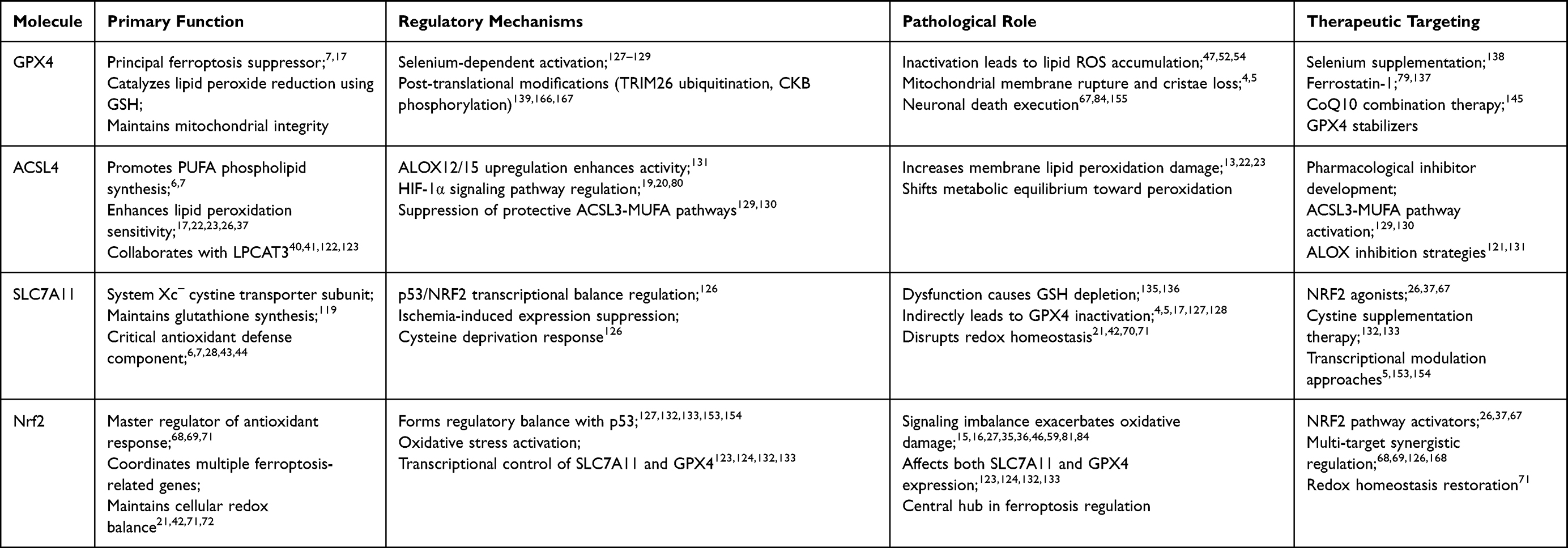 |
Table 1 Comparative Analysis of Key Ferroptosis Regulators in Ischemic Stroke. Summary Table of Core Signaling Molecules |
Discovering reliable biomarkers for ferroptosis is essential for early disease diagnosis and monitoring, necessitating research to validate these markers across diverse biological samples. Therapeutic development is another promising avenue, involving the design of targeted treatments like iron chelators and antioxidant supplements, as well as exploring combination therapies to enhance efficacy.
Tailoring therapeutic strategies to specific diseases, including neurodegenerative disorders, cancer, and cardiovascular diseases, is crucial due to the varied roles ferroptosis plays in each context. Leveraging advanced technologies such as single-cell sequencing and CRISPR-Cas9 gene editing will further elucidate ferroptosis at the molecular and cellular levels.
However, several challenges persist. The complexity of the pathways involved complicates the dissection of individual contributions and interactions, potentially hindering the identification of specific targets. Validating biomarkers across different patient populations and disease stages is a significant hurdle, requiring markers to be specific, sensitive, and reproducible. Developing therapies that specifically target ferroptosis without affecting other cellular processes is another major challenge, alongside ensuring their safety in preclinical and clinical studies.
The heterogeneity of diseases associated with ferroptosis, such as varying tumor types and stages in cancer, complicates the development of universal therapeutic strategies. Additionally, technological limitations in resolution, sensitivity, and throughput need to be addressed through continuous innovation. Overall, while future research in ferroptosis holds great promise, overcoming these challenges is essential for translating research findings into clinical applications.
Nanotherapeutic Strategies: ROS Scavenging by Recombinant Human Heavy Chain Ferritin Nanoparticles
Emerging nanotherapeutic approaches are revolutionizing targeted neuroprotection in ischemic stroke by addressing the dual challenges of blood-brain barrier penetration and off-target effects. Among these, recombinant human heavy chain ferritin nanoparticles (rHFn) have demonstrated remarkable potential, particularly leveraging the role of Ferritin heavy chain (FTH1) in modulating the labile iron pool and mitigating oxidative stress.172 FTH1 is primarily responsible for intracellular iron sequestration, thereby limiting the catalytic action of iron in Fenton reactions, a primary source of hydroxyl radicals that drive lethal lipid peroxidation characteristic of ferroptosis. Recent studies show that recombinant ferritin heavy chain nanoparticles offer a neuroprotective effect by integrating three synergistic mechanisms: (1) Inherent ROS Scavenging: rHFn directly scavenges deleterious reactive oxygen species, reducing oxidative damage to neuronal membranes and preventing the initiation of the peroxidation cascade; (2) Labile Iron Sequestration: It mimics and enhances the function of endogenous ferritin, chelating redox-active Fe2+ and preventing its participation in Fe2+-mediated oxidation reactions; and (3) Biomimetic Nanocarrier Properties: Exhibiting BBB permeability via transferrin receptor-1 (TFR1)-mediated transcytosis, enabling it to accumulate preferentially within ischemic penumbra tissues. Preclinical findings in models of middle cerebral artery occlusion (MCAO) revealed that systemic administration of rHFn significantly attenuated infarct volume, improved motor outcomes, and curtailed neuronal apoptosis.173,174 Moreover, the nanoparticle was found to diminish inflammatory cytokine expressions (TNF-α, IL-1β) and restored GPX4 activity, thereby protecting cells from lipid peroxidation, mediated cell death.175,176 Unlike small-molecule ROS scavengers (eg, Edaravone), rHFn displays minimal diffusion clearance and longer circulation times, prolonging its therapeutic window after reperfusion injury. This nanoenzyme platform underscores the potential of integrating bio-inspired antioxidant and iron-chelating properties into a single multifunctional entity, establishing new paradigms for ferroptosis-specific pharmacotherapy. Future translational efforts should focus on optimizing dosing regimens, elucidating rHFn’s long-term immunogenicity profile, and testing its efficacy in comorbid preclinical stroke models that mimic the clinical complexity of stroke patients. These advances highlight rHFn as a promising candidate for advancing ischemic stroke therapeutics into clinical trials.
Core Pathogenic Mechanisms
Ferroptosis in ischemic stroke develops through three synergistic pathways: iron dysregulation (TfR1/DMT1-mediated uptake, FPN/Cp/Heph export failure, NCOA4-driven ferritinophagy), glutathione depletion (system Xc− dysfunction, GPX4/FSP1-CoQ10axis disruption), and lipid peroxidation (ACSL4-LPCAT3-POR axis with suppressed ACSL3-MUFA pathway).
Therapeutic Strategies
Multitarget interventions include iron chelators, GPX4 activation (selenium/ferrostatin-1), RXRγ/GRSF1-mediated transcription, TRIM26/CKB-regulated stabilization, ACSL4/LOX inhibition, ACSL3 activation, and mitochondrial support (SLC25A39, CoQ10, DHODH). Combining GPX4 stabilizers with CoQ10 antioxidants provides synergistic protection by neutralizing peroxides and restoring iron homeostasis.
Future Directions
Key priorities involve: multi-omics to resolve cell death crosstalk; BBB-optimized inhibitor design; biomarker development; comorbidity-integrated animal models; multi-target cocktails and nanocarriers; integration with thrombolytic therapies.
Conclusion
The core contribution of this review lies in the systematic and multi-dimensional integration of the complex pathophysiological mechanisms of ferroptosis in acute ischemic stroke, establishing a clear framework that spans from fundamental mechanisms to clinical translation, and accordingly proposing a future research roadmap with a clear direction and a focus on clinical translation.
Author Contributions
All authors made a significant contribution to the work reported, whether that is in the conception, study design, execution, acquisition of data, analysis and interpretation, or in all these areas; took part in drafting, revising or critically reviewing the article; gave final approval of the version to be published; have agreed on the journal to which the article has been submitted; and agree to be accountable for all aspects of the work.
Funding
This work was supported by the Hunan Provincial Natural Science Foundation (no. 2025JJ70217, 2026JJ80933), Scientific Research Program of the Health Commission of Hunan Province (D202303078252) and Hunan University of Chinese Medicine (HUCMS) University-Hospital Joint Fund (2024XYLH209). The funders supported study design, data collection and analysis, decision to publish, and preparation of the manuscript.
Disclosure
The authors declare that the research was conducted in the absence of any commercial or financial relationships that could be construed as a potential conflict of interest.
References
1. Fu R, Zhao L, Guo Y, et al. Aim2 inflammasome: a potential therapeutic target in ischemic stroke. Clin Immunol. 2024;259:109881. doi:10.1016/j.clim.2023.109881
2. Morse PT, Wan J, Bell J, et al. Sometimes less is more: inhibitory infrared light during early reperfusion calms hyperactive mitochondria and suppresses reperfusion injury. Biochem Soc Trans. 2022;50:1377–18. doi:10.1042/BST20220446
3. Xie J, Zhang Z. Recent advances and therapeutic implications of 2-oxoglutarate-dependent dioxygenases in ischemic stroke. Mol Neurobiol. 2024;61:3949–3975. doi:10.1007/s12035-023-03790-1
4. Li L, Wang M, Ma YM, et al. Selenium inhibits ferroptosis in hyperglycemic cerebral ischemia/reperfusion injury by stimulating the hippo pathway. PLoS One. 2023;18:e291192.
5. Wang B, Kong W, Lv L, Wang Z. Plumbagin induces ferroptosis in colon cancer cells by regulating p53-related slc7a11 expression. Heliyon. 2024;10:e28364. doi:10.1016/j.heliyon.2024.e28364
6. Su Y, Lv M, Huang Z, et al. Defect engineering to tailor structure-activity relationship in biodegradable nanozymes for tumor therapy by dual-channel death strategies. J Control Release. 2024;367:557–571. doi:10.1016/j.jconrel.2024.01.066
7. Forcina GC, Dixon SJ. Gpx4 at the crossroads of lipid homeostasis and ferroptosis. Proteomics. 2019;19:e1800311. doi:10.1002/pmic.201800311
8. Chen J, Shi Z, Zhang C, Xiong K, Zhao W, Wang Y. Oroxin A alleviates early brain injury after subarachnoid hemorrhage by regulating ferroptosis and neuroinflammation. J Neuroinflammation. 2024;21(1):116. doi:10.1186/s12974-024-03099-3
9. Chen J, Shi Z, Chen Y, Xiong K, Wang Y, Zhang H. A CoQ10 analog ameliorates cognitive impairment and early brain injury after subarachnoid hemorrhage by regulating ferroptosis and neuroinflammation. Redox Biol. 2025;84:103684. doi:10.1016/j.redox.2025.103684
10. Gao N, Huang Z, Xie J, et al. Cryptotanshinone alleviates cerebral ischemia reperfusion injury by regulating ferroptosis through the PI3K/AKT/Nrf2 and SLC7A11/GPX4 signaling pathway. J Ethnopharmacol. 2025;348:119800. doi:10.1016/j.jep.2025.119800
11. Luo M, Luan X, Yang C, et al. Revisiting the potential of regulated cell death in glioma treatment: a focus on autophagy-dependent cell death, anoikis, ferroptosis, cuproptosis, pyroptosis, immunogenic cell death, and the crosstalk between them. Front Oncol. 2024;14:1397863. doi:10.3389/fonc.2024.1397863
12. Zhang C, Zhao Y, Yu M, Qin J, Ye B, Wang Q. Mitochondrial dysfunction and chronic liver disease. Curr Issues Mol Biol. 2022;44:3156–3165. doi:10.3390/cimb44070218
13. Shi P, Wu J, Li M, et al. Upregulation of hsp27 via further inhibition of histone h2a ubiquitination confers protection against myocardial ischemia/reperfusion injury by promoting glycolysis and enhancing mitochondrial function. Cell Death Discov. 2023;9:466. doi:10.1038/s41420-023-01762-x
14. Shi GS, Qin QL, Huang C, et al. The pathological mechanism of neuronal autophagy-lysosome dysfunction after ischemic stroke. Cell Mol Neurobiol. 2023;43:3251–3263. doi:10.1007/s10571-023-01382-0
15. Qian C, Liu DF, Wang CX, et al. Targeting early apoptosis in acute ischemic stroke with a small-molecule probe. Acs Biomater Sci Eng. 2018;4:1862–1870. doi:10.1021/acsbiomaterials.8b00213
16. Yawoot N, Chumboatong W, Sengking J, Tocharus C, Tocharus J. Chronic high-fat diet consumption exacerbates pyroptosis- and necroptosis-mediated hmgb1 signaling in the brain after ischemia and reperfusion injury. J Physiol Biochem. 2022;78:833–844. doi:10.1007/s13105-022-00906-4
17. Cai J, Ye Z, Hu Y, et al. Identification of immunogenic cell death-related gene classification patterns and immune infiltration characterization in ischemic stroke based on machine learning. Front Cell Neurosci. 2022;16:1094500. doi:10.3389/fncel.2022.1094500
18. Liu R, Song P, Gu X, et al. Comprehensive landscape of immune infiltration and aberrant pathway activation in ischemic stroke. Front Immunol. 2021;12:766724. doi:10.3389/fimmu.2021.766724
19. Guo Q, Ma M, Yu H, Han Y, Zhang D. Dexmedetomidine enables copper homeostasis in cerebral ischemia/reperfusion via ferredoxin 1. Ann Med. 2023;55:2209735. doi:10.1080/07853890.2023.2209735
20. Dvoriantchikova G, Lypka KR, Adis EV, Ivanov D. Multiple types of programmed necrosis such as necroptosis, pyroptosis, oxytosis/ferroptosis, and parthanatos contribute simultaneously to retinal damage after ischemia-reperfusion. Sci Rep. 2022;12:17152. doi:10.1038/s41598-022-22140-0
21. Du B, Deng Z, Chen K, et al. Iron promotes both ferroptosis and necroptosis in the early stage of reperfusion in ischemic stroke. Genes Dis. 2024;11:101262. doi:10.1016/j.gendis.2024.101262
22. Zhou Y, She R, Mei Z, Liu D, Ge J. Crosstalk between ferroptosis and necroptosis in cerebral ischemia/reperfusion injury and naotaifang formula exerts neuroprotective effect via hsp90-gcn2-atf4 pathway. Phytomedicine. 2024;130:155399. doi:10.1016/j.phymed.2024.155399
23. Kuriakose D, Xiao Z. Pathophysiology and treatment of stroke: present status and future perspectives. Int J Mol Sci. 2020;21:7609. doi:10.3390/ijms21207609
24. Kim JK, Silwal P, Kim YJ, et al. Gamma-aminobutyric acid type A receptor alpha 4 coordinates autophagy, inflammation, and immunometabolism to promote innate immune activation. Autophagy Rep. 2023;2(1):2181915. doi:10.1080/27694127.2023.2181915
25. Wang L, Li J, Wang Y, et al. Dan-Deng-Tong-Nao softgel capsule promotes angiogenesis of cerebral microvasculature to protect cerebral ischemia reperfusion injury via activating HIF-1alpha-VEGFA-Notch1 signaling pathway. Phytomedicine. 2023;118:154966. doi:10.1016/j.phymed.2023.154966
26. Guo H, Li Y, Wang S, et al. Dysfunction of astrocytic glycophagy exacerbates reperfusion injury in ischemic stroke. Redox Biol. 2024;74:103234. doi:10.1016/j.redox.2024.103234
27. Qin C, Yang S, Chu YH, et al. Correction to: signaling pathways involved in ischemic stroke: molecular mechanisms and therapeutic interventions. Signal Transduct Target Ther. 2022;7:278. doi:10.1038/s41392-022-01129-1
28. Radak D, Katsiki N, Resanovic I, et al. Apoptosis and acute brain ischemia in ischemic stroke. Curr Vasc Pharmacol. 2017;15:115–122. doi:10.2174/1570161115666161104095522
29. Sifat AE, Nozohouri S, Archie SR, Chowdhury EA, Abbruscato TJ. Brain energy metabolism in ischemic stroke: effects of smoking and diabetes. Int J Mol Sci. 2022;23:8512. doi:10.3390/ijms23158512
30. Denecke KM, Mcbain CA, Hermes BG, et al. Microfluidic model to evaluate astrocyte activation in penumbral region following ischemic stroke. Cells. 2022;11:2356. doi:10.3390/cells11152356
31. Zhao A, Liu N, Yao M, et al. A review of neuroprotective effects and mechanisms of ginsenosides from panax ginseng in treating ischemic stroke. Front Pharmacol. 2022;13:946752. doi:10.3389/fphar.2022.946752
32. Takahashi S. Metabolic contribution and cerebral blood flow regulation by astrocytes in the neurovascular unit. Cells. 2022;11:813. doi:10.3390/cells11050813
33. Ehresman J, Cottrill E, Caplan JM, Mcdougall CG, Theodore N, Nyquist PA. Neuroprotective role of acidosis in ischemia: review of the preclinical evidence. Mol Neurobiol. 2021;58:6684–6696. doi:10.1007/s12035-021-02578-5
34. Kelmanson IV, Shokhina AG, Kotova DA, et al. In vivo dynamics of acidosis and oxidative stress in the acute phase of an ischemic stroke in a rodent model. Redox Biol. 2021;48:102178. doi:10.1016/j.redox.2021.102178
35. Cerina M, Levers M, Keller JM, Frega M. Neuroprotective role of lactate in a human in vitro model of the ischemic penumbra. Sci Rep. 2024;14:7973. doi:10.1038/s41598-024-58669-5
36. Madai S, Kilic P, Schmidt RM, et al. Activation of the hypoxia-inducible factor pathway protects against acute ischemic stroke by reprogramming central carbon metabolism. Theranostics. 2024;14:2856–2880.
37. Pu J, Han J, Yang J, Yu L, Wan H. Anaerobic glycolysis and ischemic stroke: from mechanisms and signaling pathways to natural product therapy. Acs Chem Neurosci. 2024;15:3090–3105. doi:10.1021/acschemneuro.4c00371
38. Yao X, Li W, Fang D, et al. Emerging roles of energy metabolism in ferroptosis regulation of tumor cells. Adv Sci. 2021;8:e2100997. doi:10.1002/advs.202100997
39. Krabbendam IE, Honrath B, Dilberger B, et al. Sk channel-mediated metabolic escape to glycolysis inhibits ferroptosis and supports stress resistance in c. Elegans. Cell Death Dis. 2020;11:263.
40. Sun M, Liu M, Li Q, et al. Insulin attenuates lps-induced cognitive impairment and ferroptosis through regulation of glucose metabolism in hippocampus. Cns Neurosci Ther. 2024;30:e14887. doi:10.1111/cns.14887
41. Jia M, Jin F, Li S, et al. No-reflow after stroke reperfusion therapy: an emerging phenomenon to be explored. Cns Neurosci Ther. 2024;30:e14631. doi:10.1111/cns.14631
42. Zheng X, Ren B, Gao Y. Tight junction proteins related to blood-brain barrier and their regulatory signaling pathways in ischemic stroke. Biomed Pharmacother. 2023;165:115272. doi:10.1016/j.biopha.2023.115272
43. Gao HM, Chen H, Cui GY, Hu JX. Damage mechanism and therapy progress of the blood-brain barrier after ischemic stroke. Cell Biosci. 2023;13:196.
44. Zhao LX, Du JR, Zhou HJ, Liu DL, Gu MX, Long FY. Differences in proinflammatory property of six subtypes of peroxiredoxins and anti-inflammatory effect of ligustilide in macrophages. PLoS One. 2016;11:e164586.
45. Chen W, Zhou X, Meng M, Pan X, Huang L, Chen C. Hyperbaric oxygen improves cerebral ischemia-reperfusion injury in rats via inhibition of ferroptosis. J Stroke Cerebrovasc Dis. 2023;32:107395. doi:10.1016/j.jstrokecerebrovasdis.2023.107395
46. Zhao B, Yin Q, Fei Y, et al. Research progress of mechanisms for tight junction damage on blood-brain barrier inflammation. Arch Physiol Biochem. 2022;128:1579–1590. doi:10.1080/13813455.2020.1784952
47. Gawargi FI, Mishra PK. Mmp9 drives ferroptosis by regulating gpx4 and iron signaling. iScience. 2024;27(9):110622. doi:10.1016/j.isci.2024.110622
48. Montaner J, Ramiro L, Simats A, et al. Matrix metalloproteinases and adams in stroke. Cell Mol Life Sci. 2019;76:3117–3140. doi:10.1007/s00018-019-03175-5
49. Tu R, Armstrong J, Lee K, Hammock BD, Sapirstein A, Koehler RC. Soluble epoxide hydrolase inhibition decreases reperfusion injury after focal cerebral ischemia. Sci Rep. 2018;8:5279. doi:10.1038/s41598-018-23504-1
50. Wu J, Zhao Y, Fan Z, et al. Soluble epoxide hydrolase inhibitor protects against blood-brain barrier dysfunction in a mouse model of type 2 diabetes via the ampk/ho-1 pathway. Biochem Biophys Res Commun. 2020;524:354–359. doi:10.1016/j.bbrc.2020.01.085
51. Jia CL, Gou Y, Gao Y, et al. Rosmarinic acid liposomes suppress ferroptosis in ischemic brain via inhibition of tfr1 in bmecs. Phytomedicine. 2024;132:155835. doi:10.1016/j.phymed.2024.155835
52. Bhattarai S, Subedi U, Manikandan S, et al. Endothelial specific deletion of autotaxin improves stroke outcomes. Cells. 2023;12:511. doi:10.3390/cells12030511
53. Zhang LM, Liang XL, Xiong GF, et al. Analysis and identification of oxidative stress-ferroptosis related biomarkers in ischemic stroke. Sci Rep. 2024;14:3803. doi:10.1038/s41598-024-54555-2
54. Barrier L, Barrier J, Arnaud M, Piriou A, Tallineau C. Alterations in the ganglioside composition of rat cortical brain slices during experimental lactic acidosis: implications of an enzymatic process independent of the oxidative stress. Biochim Biophys Acta. 1997;1336:15–22. doi:10.1016/S0304-4165(97)00004-4
55. Lushchak VI, Lushchak O. Interplay between reactive oxygen and nitrogen species in living organisms. Chem Biol Interact. 2021;349:109680. doi:10.1016/j.cbi.2021.109680
56. Vinokurov AY, Stelmashuk OA, Ukolova PA, Zherebtsov EA, Abramov AY. Brain region specificity in reactive oxygen species production and maintenance of redox balance. Free Radic Biol Med. 2021;174:195–201. doi:10.1016/j.freeradbiomed.2021.08.014
57. Orellana-Urzua S, Claps G, Rodrigo R. Improvement of a novel proposal for antioxidant treatment against brain damage occurring in ischemic stroke patients. Cns Neurol Disord Drug Targets. 2021;20:3–21. doi:10.2174/1871527319666200910153431
58. Huang Z, Chen Y, Zhang Y. Mitochondrial reactive oxygen species cause major oxidative mitochondrial DNA damages and repair pathways. J Biosci. 2020;45:84. doi:10.1007/s12038-020-00055-0
59. Wen B, Xu K, Huang R, et al. Preserving mitochondrial function by inhibiting grp75 ameliorates neuron injury under ischemic stroke. Mol Med Rep. 2022;25(5):165. doi:10.3892/mmr.2022.12681
60. Lei L, Yang S, Lu X, Zhang Y, Li T. Research progress on the mechanism of mitochondrial autophagy in cerebral stroke. Front Aging Neurosci. 2021;13:698601. doi:10.3389/fnagi.2021.698601
61. Zhong Y, Jia B, Xie C, et al. Adenylate kinase 4 promotes neuronal energy metabolism and mitophagy in early cerebral ischemia via parkin/pkm2 pathway. Exp Neurol. 2024;377:114798. doi:10.1016/j.expneurol.2024.114798
62. Guo WZ, Fang HB, Cao SL, et al. Six-transmembrane epithelial antigen of the prostate 3 deficiency in hepatocytes protects the liver against ischemia-reperfusion injury by suppressing transforming growth factor-beta-activated kinase 1. Hepatology. 2020;71:1037–1054. doi:10.1002/hep.30882
63. Yang J, Wang Z, Liu X, Lu P. Modulation of vascular integrity and neuroinflammation by peroxiredoxin 4 following cerebral ischemia-reperfusion injury. Microvasc Res. 2021;135:104144. doi:10.1016/j.mvr.2021.104144
64. Napolitano G, Fasciolo G, Venditti P. Mitochondrial management of reactive oxygen species. Antioxidants. 2021;10(11):1824. doi:10.3390/antiox10111824
65. Horvath G, Svab G, Komlodi T, et al. Reverse and forward electron flow-induced h(2)o(2) formation is decreased in alpha-ketoglutarate dehydrogenase (alpha-kgdh) subunit (e2 or e3) heterozygote knock out animals. Antioxidants. 2022;11:1487. doi:10.3390/antiox11081487
66. Wang P, Huang Y, Sun B, et al. Folic acid blocks ferroptosis induced by cerebral ischemia and reperfusion through regulating folate hydrolase transcriptional adaptive program. J Nutr Biochem. 2024;124:109528. doi:10.1016/j.jnutbio.2023.109528
67. Choi DH, Kim JH, Lee KH, et al. Role of neuronal nadph oxidase 1 in the peri-infarct regions after stroke. PLoS One. 2015;10:e116814.
68. Yang Y, Hao T, Yao X, et al. Crebanine ameliorates ischemia-reperfusion brain damage by inhibiting oxidative stress and neuroinflammation mediated by nadph oxidase 2 in microglia. Phytomedicine. 2023;120:155044. doi:10.1016/j.phymed.2023.155044
69. Li G, Ye C, Zhu Y, et al. Oxidative injury in ischemic stroke: a focus on nadph oxidase 4. Oxid Med Cell Longev. 2022;2022:1148874. doi:10.1155/2022/1148874
70. Lu Y, Shen Z, Xu Y, et al. Discovery of new phenyltetrazolium derivatives as ferroptosis inhibitors for treating ischemic stroke: an example development from free radical scavengers. J Med Chem. 2024;67(14):11712–11731. doi:10.1021/acs.jmedchem.4c00211
71. Yang D, Xia X, Xi S. Salvianolic acid a attenuates arsenic-induced ferroptosis and kidney injury via hif-2alpha/duox1/gpx4 and iron homeostasis. Sci Total Environ. 2024;907:168073. doi:10.1016/j.scitotenv.2023.168073
72. Maciejczyk M, Nesterowicz M, Zalewska A, et al. Salivary xanthine oxidase as a potential biomarker in stroke diagnostics. Front Immunol. 2022;13:897413. doi:10.3389/fimmu.2022.897413
73. Yu H, Chen X, Guo X, et al. The clinical value of serum xanthine oxidase levels in patients with acute ischemic stroke. Redox Biol. 2023;60:102623. doi:10.1016/j.redox.2023.102623
74. Xiang P, Hu J, Wang H, et al. Mir-204-5p is sponged by tug1 to aggravate neuron damage induced by focal cerebral ischemia and reperfusion injury through upregulating cox2. Cell Death Discov. 2022;8:89. doi:10.1038/s41420-022-00885-x
75. Lu H, Li S, Dai D, et al. Enhanced treatment of cerebral ischemia-reperfusion injury by intelligent nanocarriers through the regulation of neurovascular units. Acta Biomater. 2022;147:314–326. doi:10.1016/j.actbio.2022.05.021
76. Xu M, Chen X, Gu Y, et al. Baicalin can scavenge peroxynitrite and ameliorate endogenous peroxynitrite-mediated neurotoxicity in cerebral ischemia-reperfusion injury. J Ethnopharmacol. 2013;150:116–124. doi:10.1016/j.jep.2013.08.020
77. Asmah RH, Sackey P, Adjei P, et al. Haematological indices and antioxidant enzyme activity in ghanaian stroke patients. Biomed Res Int. 2022;2022:1203120. doi:10.1155/2022/1203120
78. Xiao P, Huang H, Zhao H, et al. Edaravone dexborneol protects against cerebral ischemia/reperfusion-induced blood-brain barrier damage by inhibiting ferroptosis via activation of nrf-2/ho-1/gpx4 signaling. Free Radic Biol Med. 2024;217:116–125. doi:10.1016/j.freeradbiomed.2024.03.019
79. Wang P, Ren Q, Shi M, Liu Y, Bai H, Chang YZ. Overexpression of mitochondrial ferritin enhances blood-brain barrier integrity following ischemic stroke in mice by maintaining iron homeostasis in endothelial cells. Antioxidants. 2022;11:1257. doi:10.3390/antiox11071257
80. Miao Q, Wang R, Sun X, Du S, Liu L. Combination of puerarin and tanshinone iia alleviates ischaemic stroke injury in rats via activating the nrf2/are signalling pathway. Pharm Biol. 2022;60:1022–1031. doi:10.1080/13880209.2022.2070221
81. Yang M, He Y, Deng S, et al. Mitochondrial quality control: a pathophysiological mechanism and therapeutic target for stroke. Front Mol Neurosci. 2021;14:786099. doi:10.3389/fnmol.2021.786099
82. Orellana-Urzua S, Briones-Valdivieso C, Chichiarelli S, Saso L, Rodrigo R. Potential role of natural antioxidants in countering reperfusion injury in acute myocardial infarction and ischemic stroke. Antioxidants. 2023;12:1760. doi:10.3390/antiox12091760
83. Behera R, Sharma V, Grewal AK, et al. Mechanistic correlation between mitochondrial permeability transition pores and mitochondrial atp dependent potassium channels in ischemia reperfusion. Biomed Pharmacother. 2023;162:114599. doi:10.1016/j.biopha.2023.114599
84. Ludhiadch A, Sharma R, Muriki A, Munshi A. Role of calcium homeostasis in ischemic stroke: a review. Cns Neurol Disord Drug Targets. 2022;21:52–61. doi:10.2174/1871527320666210212141232
85. Han Y, Li X, Yang L, et al. Ginsenoside rg1 attenuates cerebral ischemia-reperfusion injury due to inhibition of nox2-mediated calcium homeostasis dysregulation in mice. J Ginseng Res. 2022;46:515–525. doi:10.1016/j.jgr.2021.08.001
86. Robichaux DJ, Harata M, Murphy E, Karch J. Mitochondrial permeability transition pore-dependent necrosis. J Mol Cell Cardiol. 2023;174:47–55. doi:10.1016/j.yjmcc.2022.11.003
87. Gleitze S, Ramirez OA, Vega-Vasquez I, et al. Ryanodine receptor mediated calcium release contributes to ferroptosis induced in primary hippocampal neurons by gpx4 inhibition. Antioxidants. 2023;12:705. doi:10.3390/antiox12030705
88. Pelizzoni I, Macco R, Morini MF, Zacchetti D, Grohovaz F, Codazzi F. Iron handling in hippocampal neurons: activity-dependent iron entry and mitochondria-mediated neurotoxicity. Aging Cell. 2011;10:172–183. doi:10.1111/j.1474-9726.2010.00652.x
89. Pedrera L, Espiritu RA, Ros U, et al. Ferroptotic pores induce ca(2+) fluxes and escrt-iii activation to modulate cell death kinetics. Cell Death Differ. 2021;28:1644–1657. doi:10.1038/s41418-020-00691-x
90. Wang D, Yin K, Zhang Y, et al. Fluoride induces neutrophil extracellular traps and aggravates brain inflammation by disrupting neutrophil calcium homeostasis and causing ferroptosis. Environ Pollut. 2023;331:121847. doi:10.1016/j.envpol.2023.121847
91. Gleitze S, Paula-Lima A, Nunez MT, Hidalgo C. The calcium-iron connection in ferroptosis-mediated neuronal death. Free Radic Biol Med. 2021;175:28–41. doi:10.1016/j.freeradbiomed.2021.08.231
92. Pedrera L, Ros U, Garcia-Saez AJ. Calcium as a master regulator of ferroptosis and other types of regulated necrosis. Cell Calcium. 2023;114:102778. doi:10.1016/j.ceca.2023.102778
93. Degregorio-Rocasolano N, Marti-Sistac O, Gasull T. Deciphering the iron side of stroke: neurodegeneration at the crossroads between iron dyshomeostasis, excitotoxicity, and ferroptosis. Front Neurosci. 2019;13:85. doi:10.3389/fnins.2019.00085
94. Chen J, Yang J, Chu J, et al. The swell1 channel promotes ischemic brain damage by mediating neuronal swelling and glutamate toxicity. Adv Sci. 2024;11:e2401085. doi:10.1002/advs.202401085
95. Xue D, Wei C, Zhou Y, et al. Triol inhibits rapid intracellular acidification and cerebral ischemic injury: the role of glutamate in neuronal metabolic reprogramming. Acs Chem Neurosci. 2022;13:2110–2121. doi:10.1021/acschemneuro.2c00119
96. Zhu H, Chen X, Zhang L, et al. Discovery of novel positive allosteric modulators targeting glun1/2a nmdars as anti-stroke therapeutic agents. Rsc Med Chem. 2024;15:1307–1319. doi:10.1039/D3MD00455D
97. Hsieh CH, Lin YJ, Chen WL, et al. Hif-1alpha triggers long-lasting glutamate excitotoxicity via system x(c)(-) in cerebral ischaemia-reperfusion. J Pathol. 2017;241:337–349. doi:10.1002/path.4838
98. Wang X, Li M, Wang F, et al. Tigar reduces neuronal ferroptosis by inhibiting succinate dehydrogenase activity in cerebral ischemia. Free Radic Biol Med. 2024;216:89–105. doi:10.1016/j.freeradbiomed.2024.03.011
99. Novgorodov SA, Voltin JR, Gooz MA, Li L, Lemasters JJ, Gudz TI. Acid sphingomyelinase promotes mitochondrial dysfunction due to glutamate-induced regulated necrosis. J Lipid Res. 2018;59:312–329. doi:10.1194/jlr.M080374
100. Fisher AL, Srole DN, Palaskas NJ, et al. Iron loading induces cholesterol synthesis and sensitizes endothelial cells to tnfalpha-mediated apoptosis. J Biol Chem. 2021;297:101156. doi:10.1016/j.jbc.2021.101156
101. Vogt AS, Arsiwala T, Mohsen M, Vogel M, Manolova V, Bachmann MF. On iron metabolism and its regulation. Int J Mol Sci. 2021;22:4591. doi:10.3390/ijms22094591
102. Xu Y, Zhang Y, Zhang JH, et al. Astrocyte hepcidin ameliorates neuronal loss through attenuating brain iron deposition and oxidative stress in app/ps1 mice. Free Radic Biol Med. 2020;158:84–95. doi:10.1016/j.freeradbiomed.2020.07.012
103. You L, Yu PP, Dong T, et al. Astrocyte-derived hepcidin controls iron traffic at the blood-brain-barrier via regulating ferroportin 1 of microvascular endothelial cells. Cell Death Dis. 2022;13:667. doi:10.1038/s41419-022-05043-w
104. Wang Z, Li Y, Ye Y, et al. Nlrp3 inflammasome deficiency attenuates cerebral ischemia-reperfusion injury by inhibiting ferroptosis. Brain Res Bull. 2023;193:37–46. doi:10.1016/j.brainresbull.2022.11.016
105. Song Y, Gao M, Wei B, et al. Mitochondrial ferritin alleviates ferroptosis in a kainic acid-induced mouse epilepsy model by regulating iron homeostasis: involvement of nuclear factor erythroid 2-related factor 2. Cns Neurosci Ther. 2024;30:e14663. doi:10.1111/cns.14663
106. Liu R, Zhang X, Nie L, Sun S, Liu J, Chen H. Heme oxygenase 1 in erythropoiesis: an important regulator beyond catalyzing heme catabolism. Ann Hematol. 2023;102:1323–1332. doi:10.1007/s00277-023-05193-7
107. Zhang X, Peng T, Li C, et al. Inhibition of cisd1 alleviates mitochondrial dysfunction and ferroptosis in mice with acute lung injury. Int Immunopharmacol. 2024;130:111685. doi:10.1016/j.intimp.2024.111685
108. Tsatsanis A, Wong BX, Gunn AP, et al. Amyloidogenic processing of Alzheimer’s disease beta-amyloid precursor protein induces cellular iron retention. Mol Psychiatry. 2020;25:1958–1966. doi:10.1038/s41380-020-0762-0
109. Qin X, Zhang J, Wang B, et al. Ferritinophagy is involved in the zinc oxide nanoparticles-induced ferroptosis of vascular endothelial cells. Autophagy. 2021;17:4266–4285. doi:10.1080/15548627.2021.1911016
110. Yang J, Lv M, Han L, et al. Evaluation of brain iron deposition in different cerebral arteries of acute ischaemic stroke patients using quantitative susceptibility mapping. Clin Radiol. 2024;79:e592–e598. doi:10.1016/j.crad.2024.01.007
111. Yilmazer-Hanke D, Mayer T, Muller HP, et al. Histological correlates of postmortem ultra-high-resolution single-section mri in cortical cerebral microinfarcts. Acta Neuropathol Commun. 2020;8:33. doi:10.1186/s40478-020-00900-1
112. Marti-Fabregas J, Camps-Renom P, Best JG, et al. Stroke risk and antithrombotic treatment during follow-up of patients with ischemic stroke and cortical superficial siderosis. Neurology. 2023;100:e1267–e1281. doi:10.1212/WNL.0000000000201723
113. Zhang Y, Wang H, Jia R, Chen D, Li Z. Serum ferritin is associated with the presence of ischemic stroke among individuals with type 2 diabetes. Heliyon. 2024;10:e27898. doi:10.1016/j.heliyon.2024.e27898
114. He Q, Wang W, Xu D, et al. Causal association of iron status with functional outcome after ischemic stroke. Stroke. 2024;55:423–431. doi:10.1161/STROKEAHA.123.044930
115. Hu DW, Zhang G, Lin L, Yu XJ, Wang F, Lin Q. Dynamic changes in brain iron metabolism in neonatal rats after hypoxia-ischemia. J Stroke Cerebrovasc Dis. 2022;31:106352. doi:10.1016/j.jstrokecerebrovasdis.2022.106352
116. Yang L, Wang D, Wang XT, Lu YP, Zhu L. The roles of hypoxia-inducible factor-1 and iron regulatory protein 1 in iron uptake induced by acute hypoxia. Biochem Biophys Res Commun. 2018;507:128–135. doi:10.1016/j.bbrc.2018.10.185
117. Li Y, Shen Q, Huang L, et al. Anti-aging factor grsf1 attenuates cerebral ischemia-reperfusion injury in mice by inhibiting gpx4-mediated ferroptosis. Mol Neurobiol. 2024;61:2151–2164. doi:10.1007/s12035-023-03685-1
118. Ryan F, Zarruk JG, Losslein L, David S. Ceruloplasmin plays a neuroprotective role in cerebral ischemia. Front Neurosci. 2018;12:988. doi:10.3389/fnins.2018.00988
119. Luo Q, Zheng J, Fan B, Liu J, Liao W, Zhang X. Enriched environment attenuates ferroptosis after cerebral ischemia/reperfusion injury by regulating iron metabolism. Brain Res Bull. 2023;203:110778. doi:10.1016/j.brainresbull.2023.110778
120. Gao SQ, Wang X, Li T, et al. Astrocyte-derived hepcidin aggravates neuronal iron accumulation after subarachnoid hemorrhage by decreasing neuronal ferroportin1. Free Radic Biol Med. 2024;210:318–332. doi:10.1016/j.freeradbiomed.2023.11.036
121. Zhu R, Kang Y, Li Q, et al. Alpha-tocopherol inhibits ferroptosis and promotes neural function recovery in rats with spinal cord injury via downregulating alox15. Biomed Pharmacother. 2024;175:116734. doi:10.1016/j.biopha.2024.116734
122. Xie LH, Fefelova N, Pamarthi SH, Gwathmey JK. Molecular mechanisms of ferroptosis and relevance to cardiovascular disease. Cells. 2022;11:2726. doi:10.3390/cells11172726
123. Tang X, Fang M, Cheng R, et al. Iron-deficiency and estrogen are associated with ischemic stroke by up-regulating transferrin to induce hypercoagulability. Circ Res. 2020;127:651–663. doi:10.1161/CIRCRESAHA.119.316453
124. Hu W, Zhang Y, Wang D, et al. Iron overload-induced ferroptosis impairs porcine oocyte maturation and subsequent embryonic developmental competence in vitro. Front Cell Dev Biol. 2021;9:673291. doi:10.3389/fcell.2021.673291
125. Yang Y, Zhu T, Wang X, et al. Acsl3 and acsl4, distinct roles in ferroptosis and cancers. Cancers. 2022;14:5896. doi:10.3390/cancers14235896
126. Dong H, Xia Y, Jin S, et al. Nrf2 attenuates ferroptosis-mediated iir-ali by modulating tert and slc7a11. Cell Death Dis. 2021;12:1027. doi:10.1038/s41419-021-04307-1
127. Zheng X, Toyama T, Siu S, et al. Selenoprotein p expression in glioblastoma as a regulator of ferroptosis sensitivity: preservation of gpx4 via the cycling-selenium storage. Sci Rep. 2024;14:682. doi:10.1038/s41598-024-51259-5
128. Kraft V, Bezjian CT, Pfeiffer S, et al. Gtp cyclohydrolase 1/tetrahydrobiopterin counteract ferroptosis through lipid remodeling. Acs Cent Sci. 2020;6:41–53. doi:10.1021/acscentsci.9b01063
129. Liu J, Kang R, Tang D. Signaling pathways and defense mechanisms of ferroptosis. FEBS J. 2022;289(22):7038–7050. doi:10.1111/febs.16059
130. Lei G, Zhang Y, Koppula P, et al. The role of ferroptosis in ionizing radiation-induced cell death and tumor suppression. Cell Res. 2020;30:146–162. doi:10.1038/s41422-019-0263-3
131. Merkel M, Goebel B, Boll M, et al. Mitochondrial reactive oxygen species formation determines acsl4/lpcat2-mediated ferroptosis. Antioxidants. 2023;12(8):1590. doi:10.3390/antiox12081590
132. Yang Z, Jiang Y, Xiao Y, et al. Di-huang-yin-zi regulates p53/slc7a11 signaling pathway to improve the mechanism of post-stroke depression. J Ethnopharmacol. 2024;319:117226. doi:10.1016/j.jep.2023.117226
133. Liu H, An N, Wang L, et al. Protective effect of xingnaojing injection on ferroptosis after cerebral ischemia injury in mcao rats and sh-sy5y cells. J Ethnopharmacol. 2023;301:115836. doi:10.1016/j.jep.2022.115836
134. Zhong S, Chen W, Wang B, et al. Energy stress modulation of ampk/foxo3 signaling inhibits mitochondria-associated ferroptosis. Redox Biol. 2023;63:102760. doi:10.1016/j.redox.2023.102760
135. Jiang H, Wang C, Zhang A, et al. Atf4 protects against sorafenib-induced cardiotoxicity by suppressing ferroptosis. Biomed Pharmacother. 2022;153:113280. doi:10.1016/j.biopha.2022.113280
136. Han Y, Fu L, Kong Y, Jiang C, Huang L, Zhang H. Steap3 affects ovarian cancer progression by regulating ferroptosis through the p53/slc7a11 pathway. Mediators Inflamm. 2024;2024:4048527. doi:10.1155/2024/4048527
137. Liu Y, Baumann B, Song Y, et al. Minimal effect of conditional ferroportin ko in the neural retina implicates ferrous iron in retinal iron overload and degeneration. Exp Eye Res. 2022;218:108988. doi:10.1016/j.exer.2022.108988
138. Li C, Zhu X, Chen J, et al. Multifaceted role of ferroptosis in cardiovascular disease. Acta Biochim Biophys Sin. 2023;55:183–193. doi:10.3724/abbs.2023019
139. Yang L, Du B, Zhang S, Wang M. Rxrgamma attenuates cerebral ischemia-reperfusion induced ferroptosis in neurons in mice through transcriptionally promoting the expression of gpx4. Metab Brain Dis. 2022;37:1351–1363. doi:10.1007/s11011-022-00988-5
140. Wang P, Cui Y, Liu Y, et al. Mitochondrial ferritin alleviates apoptosis by enhancing mitochondrial bioenergetics and stimulating glucose metabolism in cerebral ischemia reperfusion. Redox Biol. 2022;57:102475. doi:10.1016/j.redox.2022.102475
141. Shi X, Reinstadler B, Shah H, et al. Combinatorial gxgxe crispr screen identifies slc25a39 in mitochondrial glutathione transport linking iron homeostasis to oxphos. Nat Commun. 2022;13:2483. doi:10.1038/s41467-022-30126-9
142. Tian Y, Xie Y, Guo Z, Feng P, You Y, Yu Q. 17beta-oestradiol inhibits ferroptosis in the hippocampus by upregulating dhodh and further improves memory decline after ovariectomy. Redox Biol. 2023;62:102708. doi:10.1016/j.redox.2023.102708
143. Li D, Zhang G, Wang Z, et al. Idebenone attenuates ferroptosis by inhibiting excessive autophagy via the ros-ampk-mtor pathway to preserve cardiac function after myocardial infarction. Eur J Pharmacol. 2023;943:175569. doi:10.1016/j.ejphar.2023.175569
144. Gamez A, Alva N, Carbonell T, Rama R. Extracellular ferritin contributes to neuronal injury in an in vitro model of ischemic stroke. J Physiol Biochem. 2021;77:539–545. doi:10.1007/s13105-021-00810-3
145. Sultana J, Sharma DJ. Evaluation of serum ferritin as a prognostic marker in acute ischemic stroke patients-a prospective cohort study. J Assoc Physicians India. 2022;70:11–12.
146. Guo Q, Kawahata I, Cheng A, et al. Fatty acid-binding proteins 3 and 5 are involved in the initiation of mitochondrial damage in ischemic neurons. Redox Biol. 2023;59:102547. doi:10.1016/j.redox.2022.102547
147. Li Q, Wan J, Lan X, Han X, Wang Z, Wang J. Neuroprotection of brain-permeable iron chelator vk-28 against intracerebral hemorrhage in mice. J Cereb Blood Flow Metab. 2017;37:3110–3123. doi:10.1177/0271678X17709186
148. Behrouzi B, Weyers JJ, Qi X, et al. Action of iron chelator on intramyocardial hemorrhage and cardiac remodeling following acute myocardial infarction. Basic Res Cardiol. 2020;115:24. doi:10.1007/s00395-020-0782-6
149. Chen J, Ma D, Bao J, Zhang Y, Deng G. Roots of astragalus propinquus schischkin regulate transmembrane iron transport and ferroptosis to improve cerebral ischemia-reperfusion injury. Evid Based Complement Alternat Med. 2022;2022:7410865. doi:10.1155/2022/7410865
150. Wang GL, Xu SY, Lv HQ, Zhang C, Peng YJ. Electroacupuncture inhibits ferroptosis induced by cerebral ischemiareperfusion. Curr Neurovasc Res. 2023;20:346–353. doi:10.2174/1567202620666230623153728
151. Corral VM, Schultz ER, Eisenstein RS, Connell GJ. Roquin is a major mediator of iron-regulated changes to transferrin receptor-1 mrna stability. Iscience. 2021;24:102360. doi:10.1016/j.isci.2021.102360
152. Wang X, Wu H, Zhao L, et al. Flcn regulates transferrin receptor 1 transport and iron homeostasis. J Biol Chem. 2021;296:100426. doi:10.1016/j.jbc.2021.100426
153. Han WM, Hong YX, Xiao GS, Wang RY, Li G. Nmdars activation regulates endothelial ferroptosis via the pp2a-ampk-hmgb1 axis. Cell Death Discov. 2024;10:34. doi:10.1038/s41420-023-01794-3
154. Cui J, Wang Q, Li M. Xinnaotongluo liquid protects h9c2 cells from h/r-induced damage by regulating mdm2/steap3. PLoS One. 2024;19:e302407. doi:10.1371/journal.pone.0302407
155. Zhang H, Ostrowski R, Jiang D, et al. Hepcidin promoted ferroptosis through iron metabolism which is associated with dmt1 signaling activation in early brain injury following subarachnoid hemorrhage. Oxid Med Cell Longev. 2021;2021:9800794. doi:10.1155/2021/9800794
156. Li C, Sun G, Chen B, et al. Nuclear receptor coactivator 4-mediated ferritinophagy contributes to cerebral ischemia-induced ferroptosis in ischemic stroke. Pharmacol Res. 2021;174:105933. doi:10.1016/j.phrs.2021.105933
157. Yang Y, Wu Q, Shan X, et al. Ginkgolide b attenuates cerebral ischemia-reperfusion injury via inhibition of ferroptosis through disrupting ncoa4-fth1 interaction. J Ethnopharmacol. 2024;318:116982. doi:10.1016/j.jep.2023.116982
158. Li B, Wang W, Li Y, et al. Cgas-sting pathway aggravates early cerebral ischemia-reperfusion injury in mice by activating ncoa4-mediated ferritinophagy. Exp Neurol. 2023;359:114269. doi:10.1016/j.expneurol.2022.114269
159. Qian ZM, Li W, Guo Q. Ferroportin1 in the brain. Ageing Res Rev. 2023;88:101961. doi:10.1016/j.arr.2023.101961
160. Liu W, Wang L, Liu C, Dai Z, Li T, Tang B. Edaravone ameliorates cerebral ischemia-reperfusion injury by downregulating ferroptosis via the nrf2/fpn pathway in rats. Biol Pharm Bull. 2022;45:1269–1275. doi:10.1248/bpb.b22-00186
161. Li S, Qian ZM, Xu G, Zheng J, Wu Y. Hepatocyte growth factor protects pc12 cells against ogd/r-induced injury by reducing iron. Biosci Rep. 2020;40:BSR20200287.
162. Zheng H, Guo X, Kang S, et al. Cdh5-mediated fpn1 deletion exerts neuroprotective effects during the acute phase and inhibitory effects during the recovery phase of ischemic stroke. Cell Death Dis. 2023;14:161. doi:10.1038/s41419-023-05688-1
163. Zhang T, Yang M, Ma C, Wei X, Zhang Z. Bach1 encourages ferroptosis by activating kdm4c-mediated cox2 demethylation after cerebral ischemia-reperfusion injury. Eur J Neurosci. 2023;58:2194–2214. doi:10.1111/ejn.16035
164. Li W, Liang L, Liu S, Yi H, Zhou Y. Fsp1: a key regulator of ferroptosis. Trends Mol Med. 2023;29:753–764. doi:10.1016/j.molmed.2023.05.013
165. Li K, Wang A, Diao Y, Fan S. Oxidative medicine and cellular longevity the role and mechanism of ncoa4 in ferroptosis induced by intestinal ischemia reperfusion. Int Immunopharmacol. 2024;133:112155. doi:10.1016/j.intimp.2024.112155
166. Wu K, Yan M, Liu T, et al. Creatine kinase b suppresses ferroptosis by phosphorylating gpx4 through a moonlighting function. Nat Cell Biol. 2023;25:714–725. doi:10.1038/s41556-023-01133-9
167. Wang Z, Xia Y, Wang Y, et al. The e3 ligase trim26 suppresses ferroptosis through catalyzing k63-linked ubiquitination of gpx4 in glioma. Cell Death Dis. 2023;14:695. doi:10.1038/s41419-023-06222-z
168. Wang L, Zhang X, Xiong X, et al. Nrf2 regulates oxidative stress and its role in cerebral ischemic stroke. Antioxidants. 2022;11(12):2377. doi:10.3390/antiox11122377
169. Zhou R, Wu L, Jin N, Sha S, Ouyang Y. L-F001, a multifunctional fasudil-lipoic acid dimer, antagonizes hypoxic-ischemic brain damage by inhibiting the TLR4/MyD88 signaling pathway. Brain Behav. 2023;13(12):e3280. doi:10.1002/brb3.3280
170. Peng W, Ouyang Y, Wang S, et al. L-F001, a multifunctional fasudil-lipoic acid dimer prevents RSL3-induced ferroptosis via maintaining iron homeostasis and inhibiting JNK in HT22 cells. Front Cell Neurosci. 2022;16:774297. doi:10.3389/fncel.2022.774297
171. Mirmotahari SA, Aliomrani M, Hassanzadeh F, Sirous H, Rostami M. Hybrid derivatives containing dimethyl fumarate and benzothiazole scaffolds for the potential treatment of multiple sclerosis; in silico & in vivo study. Daru. 2024;32(2):599–615. doi:10.1007/s40199-024-00529-8
172. Qi M, Cheng Y, Liu K, et al. Recombinant human heavy chain ferritin nanoparticles serve as ROS scavengers for the treatment of ischemic stroke. Int J Nanomed. 2024;19:2285–2299. doi:10.2147/IJN.S449606
173. Zhang S, Li R, Zheng Y, Zhou Y, Fan X. Erythrocyte membrane-enveloped Salvianolic Acid B nanoparticles attenuate cerebral ischemia-reperfusion injury. Int J Nanomed. 2022;17:3561–3577. doi:10.2147/IJN.S375908
174. Kang H, Meng F, Liu F, et al. Nanomedicines targeting ferroptosis to treat stress-related diseases. Int J Nanomed. 2024;19:8189–8210. doi:10.2147/IJN.S476948
175. Wang K, Yu J, Luo Q, Bai Y, Gao Q, Yu R. siHIF-1alpha-loaded micellar nanoparticles inhibit M1 macrophage activation to ameliorate chronic rhinosinusitis. Free Radic Biol Med. 2025;237:403–418. doi:10.1016/j.freeradbiomed.2025.05.433
176. Niu T, Shi X, Liu X, Wang H, Liu K, Xu Y. Porous Se@SiO2 nanospheres alleviate diabetic retinopathy by inhibiting excess lipid peroxidation and inflammation. Mol Med. 2024;30(1):24. doi:10.1186/s10020-024-00785-z
 © 2026 The Author(s). This work is published and licensed by Dove Medical Press Limited. The
full terms of this license are available at https://www.dovepress.com/terms
and incorporate the Creative Commons Attribution
- Non Commercial (unported, 4.0) License.
By accessing the work you hereby accept the Terms. Non-commercial uses of the work are permitted
without any further permission from Dove Medical Press Limited, provided the work is properly
attributed. For permission for commercial use of this work, please see paragraphs 4.2 and 5 of our Terms.
© 2026 The Author(s). This work is published and licensed by Dove Medical Press Limited. The
full terms of this license are available at https://www.dovepress.com/terms
and incorporate the Creative Commons Attribution
- Non Commercial (unported, 4.0) License.
By accessing the work you hereby accept the Terms. Non-commercial uses of the work are permitted
without any further permission from Dove Medical Press Limited, provided the work is properly
attributed. For permission for commercial use of this work, please see paragraphs 4.2 and 5 of our Terms.
Recommended articles
Shikonin Could Be Used to Treat Tubal Pregnancy via Enhancing Ferroptosis Sensitivity
Lai Y, Zeng F, Chen Z, Feng M, Huang Y, Qiu P, Zeng L, Ke Y, Deng G, Gao J
Drug Design, Development and Therapy 2022, 16:2083-2099
Published Date: 1 July 2022
The Emerging Role of Ferroptosis in Sepsis, Opportunity or Challenge?
Huang Q, Ding Y, Fang C, Wang H, Kong L
Infection and Drug Resistance 2023, 16:5551-5562
Published Date: 23 August 2023
The Chains of Ferroptosis Interact in the Whole Progression of Atherosclerosis
Wan X, Zhang H, Tian J, Hao P, Liu L, Zhou Y, Zhang J, Song X, Ge C
Journal of Inflammation Research 2023, 16:4575-4592
Published Date: 16 October 2023
Targeting Ferroptosis in Bone-Related Diseases: Facts and Perspectives
Chen H, Han Z, Wang Y, Su J, Lin Y, Cheng X, Liu W, He J, Fan Y, Chen L, Zuo H
Journal of Inflammation Research 2023, 16:4661-4677
Published Date: 18 October 2023
Inhibition of Hippocampal Neuronal Ferroptosis by Liproxstatin-1 Improves Learning and Memory Function in Aged Mice with Perioperative Neurocognitive Dysfunction
Lin L, Ling X, Chen T, Zhou Q, Huang J, Huang L, Lin X, Lin L
Journal of Inflammation Research 2025, 18:2991-3007
Published Date: 27 February 2025