
Back to Journals » Journal of Inflammation Research » Volume 19
Neutrophils in Kidney Disease: Linking Neutrophil Function and Microenvironment to Therapeutic Targets
Authors Ku J, Flora KK, Steiger S, Anders HJ
Received 29 May 2026
Accepted for publication 7 July 2026
Published 24 July 2026 Volume 2026:19 605349
DOI https://doi.org/10.2147/JIR.S605349
Checked for plagiarism Yes
Review by Single anonymous peer review
Peer reviewer comments 2
Editor who approved publication: Dr Wenjian Li
John Ku, Kailey K Flora, Stefanie Steiger, Hans-Joachim Anders
Department of Medicine IV, Hospital of Ludwig-Maximilians-Universität München, LMU Medicine, Munich, Germany
Correspondence: Hans-Joachim Anders, Division of Nephrology, Department of Medicine IV, Hospital of Ludwig-Maximilians-Universität München, Ziemssenstr. 5, 80336 München, Munich, Germany, Tel +49 089440032623, Email [email protected]
Abstract: Neutrophils are central mediators of kidney immunity and injury, exerting both protective and pathogenic functions on tissue compartment, inflammatory response, and disease state. During urinary tract infection and pyelonephritis, neutrophils protect the host through phagocytosis, granule release, reactive oxygen species (ROS) production, and neutrophil extracellular trap (NET) formation. In contrast, dysregulated neutrophil activation contributes to sterile and autoimmune kidney disease, including cholesterol crystal embolism, anti-neutrophil cytoplasmic antibody -associated vasculitis (AAV), ischemic acute kidney injury, and progression to chronic kidney disease. Emerging evidence indicates that neutrophil function in the kidney is highly compartment-specific and shaped by local metabolic and inflammatory cues. Persistent neutrophil activation promotes thromboinflammation, endothelial injury, autoimmunity, and fibrogenic remodeling through release of cytokines, S100A8/A9, proteases, ROS, and NET-associated mediators. In chronic kidney disease, uremic toxins and metabolites, such as uric acid, further alter neutrophil function by impairing antimicrobial responses while sustaining inflammatory activation. This review summarizes current insights into neutrophil biology across infection, sterile, and autoimmune kidney diseases, with focus on complement-specific neutrophil functions and effector mechanisms. In addition, we discuss emerging therapeutic strategies targeting neutrophil activation pathways and highlight recent advances in spatial transcriptomics, single-cell technologies, and intravital imaging that are reshaping our understanding of neutrophil-mediated kidney injury.
Keywords: neutrophils, kidney injury, ANCA-associated vasculitis, complement, neutrophil extracellular traps, AKI-to-CKD transition
Introduction
The kidneys receive approximately 20% of cardiac output and contain a highly specialized glomerular and peritubular microvascular network that is continuously exposed to circulating leukocytes, immune complexes, pathogens, and inflammatory mediators.1 Hypertension, diabetes, and aging progressively impair this microvascular architecture through arteriolar hyalinosis, capillary rarefaction, glomerulosclerosis, podocyte loss, and nephron depletion; thereby increasing susceptibility to acute kidney injury (AKI) and chronic kidney disease (CKD).2,3 AKI is a frequent complication in hospitalized patients, particularly during sepsis and after major surgery, and often accelerates progression to CKD, which affects more than 850 million individuals worldwide and markedly increases cardiovascular morbidity and mortality.1
Despite their abundance and rapid recruitment during kidney injury, neutrophils have historically received less attention in nephrology than adaptive immune cells. Classification systems for glomerulonephritis largely focus on immunoglobulin and complement deposition, while therapeutic development has predominantly targeted lymphocytes and complement pathways.4 However, neutrophils are among the first leukocytes recruited to the injured and inflamed kidney, where they contribute to host defense but also mediate tissue injury through release of ROS, proteases, cytokines, and NETs. Persistent or dysregulated neutrophil activation has increasingly been implicated in thromboinflammation, endothelial injury, autoimmunity, and maladaptive repair during AKI-to-CKD transition.
Recent clinical success of complement-targeting therapies further highlights the importance of neutrophil-driven pathways in kidney disease. Iptacopan, an oral factor B inhibitor, reduces proteinuria in IgA nephropathy and C3 glomerulopathy; by suppressing C5a generation, it may also attenuate complement-mediated neutrophil activation..5,6
Importantly, neutrophil function in the kidney is highly context- and compartment-dependent.7,8 The same neutrophil can mediate antimicrobial defense during pyelonephritis yet promote immunothrombosis in cholesterol crystal embolism, and necroinflammation in crystal nephropathy, ischemic AKI, or autoimmune disease. Local metabolic and inflammatory microenvironments, including hypoxia, hyperosmolarity, uremic toxins and metabolites, and cytokine exposure, shape neutrophil activation states and effector functions. Understanding how neutrophil phenotypes are regulated across kidney compartments and disease states may therefore identify therapeutic strategies that limit tissue injury while preserving antimicrobial host defense.
Neutrophils have traditionally been viewed as short-lived innate effector cells primarily involved in antimicrobial defense to infection. This review summarizes recent advances in immunology and nephrology that reveal remarkable functional plasticity of neutrophils across distinct kidney microenvironments and disease states. Their interactions with tubular epithelial cells, endothelial cells, complement pathways, platelets, macrophages, and adaptive immune cells position neutrophils at the center of kidney inflammation and repair. At the same time, emerging technologies such as single-cell sequencing, spatial transcriptomics, and intravital imaging are redefining our understanding of neutrophil heterogeneity and tissue-specific behavior in the kidney. These insights are beginning to reshape therapeutic concepts beyond broad immunosuppression toward selective modulation of pathogenic neutrophil programs.
Neutrophil Biology and Diversity
Neutrophils are produced in the bone marrow at a rate of approximately 109 cells per day in healthy adults.10 Granulopoiesis takes about five days and is primarily regulated by granulocyte colony-stimulating factor (G-CSF). Mature neutrophils are subsequently released into the circulation, where they patrol the vasculature for inflammatory signals and have a circulating half-life of 6 to 12 hours.8–10 In the absence of tissue recruitment, aged neutrophils undergo apoptosis and are cleared by macrophages eg in the bone marrow. During infection or sterile inflammation, homeostatic granulopoiesis is replaced by emergency granulopoiesis, which accelerates neutrophil production and mobilizes bone marrow reserves.11 As a result, immature neutrophil populations may enter the circulation, often displaying reduced phagocytic capacity, altered chemokine receptor expression, and enhanced propensity for NET formation (Figure 1A).9,12
 |
Figure 1 Renal compartments shape neutrophil state in kidney injury. (A) Neutrophils occupy a continuum of functional states. A circulating neutrophil can be biased toward any of these programs by the signals it encounters. (B) The kidney provides anatomically distinct microenvironments that imprint the states defined in A, each compartment shown with its representative diseases and dominant neutrophil states alongside the cortex to medulla gradient. Arrows connect each compartment to its label and indicate microenvironmental imprinting of neutrophil state. Figure 1 was generated using an artificial intelligence tool, OpenAI ChatGPT (version 5.5), image generation feature, and was then edited and labeled by the authors. |
Neutrophils are essential for immediate host defense through phagocytosis, degranulation, ROS production, and NET formation.13 However, these effector functions are not deployed uniformly. A neutrophil that phagocytoses an opsonized bacterium does not necessarily form NETs, whereas neutrophils exposed to crystals or immune complexes within the glomerulus or interstitium may undergo NETosis.7,9,12 Which effector function predominates depends on the inflammatory context, tissue compartment, and activation state in eg AKI, CKD, and AAV.8
A central concept in neutrophil biology is the distinction between priming and activation. Resting neutrophils exposed to low-grade inflammatory stimuli such as tumor necrosis factor-α (TNF-α), granulocyte-macrophage colony-stimulating factor (GM-CSF), type I interferons, or subactivating concentrations of C5a become primed for exaggerated responses to subsequent stimulus.9,14,15 Priming increases surface expression of adhesion and Fc receptors, mobilizes granule proteins such as myeloperoxidase (MPO) and proteinase 3 (PR3) to the plasma membrane, promotes assembly of NADPH oxidase complexes and lowers the threshold for NET formation. Many neutrophils in autoimmune and complement-mediated kidney diseases are thought to exist in a primed rather than fully activated state. Clinically, disease activity therefore correlates less with absolute neutrophil counts than with markers of activation and priming, including MPO, soluble CD62L, and surface CD11b.16,17 This concept also provides a therapeutic rationale for targeting upstream activation pathways rather than broadly suppressing neutrophils.
Neutrophil granules provide the molecular machinery for their effector functions. Azurophilic granules contain MPO, neutrophil elastase, cathepsin G, defensins, and PR3, whereas specific and gelatinase granules contain lactoferrin, lysozyme, and matrix metalloproteinases. During priming, MPO and PR3 translocate to the cell surface, where they become accessible to circulating autoantibodies in AAV.14,18 NETs, consisting of decondensed chromatin decorated with granule proteins, were first described as antimicrobial structures,11 but are now recognized as important mediators of sterile inflammation and autoimmunity.12,19–21 NET-associated histones generated by peptidylarginine deiminase 4 (PAD4), MPO, and PR3 are directly tissue-damaging and immunogenic,22 while extracellular DNA and chromatin activate nucleic acid sensing pathways, including toll-like receptor 9 (TLR9) and cGAS-STING. In lupus nephritis, these pathways reinforce type I interferon responses and perpetuate autoimmunity.
Recent studies have further highlighted substantial neutrophil heterogeneity across inflammatory disease. Low-density granulocytes (LDGs), which co-separate with peripheral blood mononuclear cells during density-gradient centrifugation, are enriched in systemic lupus erythematosus (SLE) and can also be detected in AAV.23,24 LDGs exhibit increased spontaneous NET formation and enhanced type I interferon signaling,23,24 and their NETs are enriched in oxidized mitochondrial DNA capable of amplifying interferon responses in lupus nephritis.25 In parallel, aged neutrophils characterized by increased C-X-C motif chemokine receptor 4 (CXCR4) and reduced CXCR2 expression display altered trafficking and effector behavior during chronic inflammation.26–28
Importantly, these descriptors define overlapping functional states rather than fixed neutrophil lineages.8,29 A neutrophil may simultaneously exhibit features of aging, priming, NET susceptibility, or emergency granulopoiesis depending on the inflammatory milieu. Within the kidney, local factors including hypoxia, hyperosmolarity, complement activation, immune complexes, endothelial injury, and uremic metabolites further shape neutrophil phenotype and function. This context-dependent plasticity is increasingly recognized as a central determinant of neutrophil-mediated kidney injury.
The Kidney as a Regulator of Neutrophil Behavior
Neutrophils rapidly accumulate in the kidney during AKI, both in experimental models and in human biopsies, particularly following ischemia–reperfusion injury (IRI).30 Within minutes after reperfusion, neutrophils adhere to activated endothelial cells and platelets, contributing to capillary plugging, vascular congestion, and impaired microvascular perfusion.30 In the kidney interstitium, neutrophils amplify tissue injury and inflammation through degranulation, cytokine release, NETosis, and ROS production.30 Consistently, blocking neutrophil recruitment using anti-ICAM antibodies or ICAM deficiency attenuates kidney injury in murine IRI models.31,32
Sterile tissue injury itself further promotes neutrophil activation. Damage-associated molecular patterns (DAMPs) released during IRI, including histones and mitochondrial components, can directly trigger NET formation.33 NETs subsequently amplify oxidative stress and histone release, thereby promoting tubular epithelial cell death and necroinflammation.33 Recent experimental studies suggest that targeting NET formation may represent a therapeutic strategy in AKI. For example, pharmacological inhibition of PAD4, a key regulator of NETosis, reduces tubular apoptosis and pyroptosis in contrast-induced AKI.34,35 Importantly, the kidney is not merely a passive target of neutrophil-mediated injury but actively shapes neutrophil behavior through its unique anatomical and metabolic microenvironment. Distinct kidney compartments expose infiltrating neutrophils to different combinations of shear stress, osmolarity, hypoxia, endothelial activation, immune complexes, complement activation, and tissue-derived metabolites; thereby biasing neutrophil effector programs toward antimicrobial defense, thromboinflammation, NET formation, or maladaptive tissue remodelling. Figure 1B summarizes kidney compartments that imprint compartment-specific neutrophil states and highlights representative disease contexts associated with each microenvironment.
Renal Arterioles
Renal arterioles represent a vascular niche in which circulating neutrophils encounter endothelial injury, platelet activation, and high shear stress.36 These conditions favor leukocyte adhesion, β2-integrin activation, and platelet-neutrophil interactions that promote immunothrombosis. In cholesterol crystal embolism, cholesterol crystals released from atherosclerotic plaques lodge within small renal arteries and arterioles, where they induce endothelial injury, thrombotic microangiopathy, inflammasome activation, and necroinflammation.37–40 In this setting, infiltrating neutrophils acquire a highly proinflammatory and prothrombotic phenotype characterized by β2-integrin activation, NET formation, oxidative burst, and GSDMD-dependent pyroptosis.40,41
Glomerular Capillaries and Bowman’s Space
The glomerulus functions as a specialized filtration network in which its fenestrated capillaries create conditions that favor the trapping of neutrophils during inflammation.42 Unlike post-capillary venules, where leukocyte recruitment depends on P-selectin rolling followed by integrin-dependent adhesion, the glomerular circulation supports a distinct mechanism in which neutrophil arrest occurs largely independent of selectins and relies instead on integrin interactions, particularly MAC-1,43 often facilitated by platelet-mediated bridging.43 Once retained within capillary beds, neutrophils are exposed to circulating autoantibodies, immune complexes, and complement fragments, which drive strong activation through Fcγ and complement receptors.14 This environment promotes robust effector responses, including degranulation and NET formation,14 making neutrophils key contributors to glomerular injury in diseases such as AAV, lupus nephritis, immunoglobulin A (IgA) nephropathy, C3 glomerulopathy, and anti-glomerular basement membrane (GBM) disease.
In more severe forms of glomerular injury, particularly rapidly progressive or crescentic glomerulonephritis, damage to the glomerular basement membrane allows inflammatory mediators, coagulation factors, and plasma proteins to enter Bowman’s space. This transforms the normally protective urinary space into a procoagulant and proinflammatory niche.44,45 Consequently, parietal epithelial cells lining Bowman’s capsule become activated and together with recruited monocytes/macrophages form cellular crescents, which compress the glomerular tuft and disrupt filtration.45,46 Neutrophils play their most important role early in this process, amplifying endothelial injury and promoting capillary wall rupture through degranulation, NET release, and inflammatory signals.14,47 In this way, they act as upstream effectors of necrotizing glomerular injury in diseases that overlap with the glomerular compartment, including AAV, anti-GBM disease, severe lupus nephritis, and crescentic forms of IgA nephropathy.
Peritubular Capillaries
Peritubular capillaries constitute the post-glomerular microvascular network that surrounds the tubules and supplies oxygen and nutrients to highly metabolically active tubular epithelial cells. Unlike upstream arterioles, where flow limitation can be driven by discrete thromboinflammatory occlusion, this downstream exchange bed is particularly sensitive to subtle or heterogeneous reductions in perfusion. Even modest disturbances in capillary flow can therefore translate into rapid impairment of oxygen delivery to adjacent tubular segments. Consistent with this vulnerability, intravital imaging has shown that peritubular capillary dysfunction may arise before detectable changes in conventional biomarkers of kidney injury, underscoring the importance of microvascular perfusion as an early determinant of AKI.48
During kidney injury, peritubular capillary flow can be compromised without a single focal obstruction. Multiple overlapping mechanisms contribute, including endothelial swelling, leukocyte adhesion, platelet aggregation, erythrocyte congestion, pericyte-mediated capillary constriction, and NET-associated endothelial injury.34,49–51 In this context, neutrophils contribute less through classical high-shear vascular recruitment and more through their role in promoting capillary congestion, endothelial damage, and local perfusion failure.34,36,48–51 NET formation has been observed within both glomerular and peritubular capillary compartments in experimental models of AKI, and strategies that inhibit or degrade NETs have been associated with reduced endothelial injury and improved kidney functional outcomes.34,52 Accordingly, peritubular capillary dysfunction represents a convergent pathway rather than a disease-specific lesion, occurring in settings such as IRI, vascular forms of AKI, thrombotic microangiopathies, and cholesterol crystal embolism.34,49,53–56
The Outer Medulla
The outer medulla is characterized by inherently low oxygen availability due to countercurrent oxygen exchange within the vasa recta combined with high metabolic demands of local tubular segments, especially the proximal straight tubule and thick ascending limb.1,49 This baseline hypoxic state renders the region particularly vulnerable to ischemic and toxic injury. Within this environment, hypoxia-driven stabilization of HIF-1α may influence infiltrating neutrophils by promoting glycolytic metabolism and prolonging neutrophil survival, potentially contributing to sustained inflammatory activity, although direct in vivo confirmation remains limited.57,58 As a result, this compartment is especially relevant to forms of AKI involving secondary neutrophil recruitment and NET formation, such as IRI, toxin-induced AKI, and sepsis-associated AKI.53,54
The Interstitium
The interstitium represents a compartment where a failure to resolve inflammation becomes central to persistent or chronic inflammation. Following acute injury or repeated insults, maladaptive tubular adaptation, sustained transforming growth factor beta (TGF-β) signaling, persistent macrophage accumulation, and fibroblast activation converge to drive extracellular matrix deposition and interstitial fibrosis.59–61 Neutrophils can exacerbate this process when their clearance is inefficient and cellular recruitment persists,62,63 as uncleared apoptotic neutrophils may undergo secondary necrosis, releasing DAMPs, proteases, histones, and NET components that perpetuate macrophage activation and ongoing tissue injury.52,63,64 Over time, this creates a self-sustaining inflammatory-fibrotic loop underlying conditions such as AKI-to-CKD transition, chronic pyelonephritis, chronic interstitial nephritis, and chronic allograft injury with interstitial fibrosis and tubular atrophy.59,65
Tubule Lumen
The tubular lumen is characterized by direct exposure to urinary contents rather than blood or tissue fluid, making it uniquely vulnerable to injury from precipitated or accumulated intraluminal material. In diseases such as myeloma cast nephropathy, nephrocalcinosis, and chronic uric acid nephropathy, light chains, crystals, or urate deposits accumulate within the tubular lumen, leading to tubular injury and obstruction of tubular flow.40,66,67 In these conditions, the primary insult originates from luminal crystalline material itself, while neutrophils are recruited secondary in response to epithelial stress and danger signals, where they may further amplify injury in the surrounding interstitium.
In infectious settings such as pyelonephritis and sepsis, the role of the tubular lumen is distinct. Ascending bacteria enter the urinary tract,68 and tubular epithelial cells respond by producing chemokines that drive neutrophil migration across the epithelium into the lumen, resulting in pyuria.69 Here, neutrophil infiltration serves a protective function, contributing to bacterial clearance rather than primarily reflecting sterile crystal- or cast-mediated injury.
Osmotic and Metabolic Milieu
Beyond its anatomic compartments, the kidney exposes neutrophils to a highly dynamic biochemical environment defined by gradients in oxygen tension, osmolarity, metabolites, and retained solutes. The osmotic gradient is particularly important because the cortex remains relatively close to normal extracellular osmolarity, whereas the medulla becomes progressively hyperosmolar due to NaCl and urea handling during urinary concentration.1,70 Consequently, neutrophils entering the medulla encounter simultaneous hypoxic and osmotic stress. Cellular adaptation to hypertonicity is largely regulated by the transcription factor NFAT5/TonEBP, which is essential for medullary homeostasis71 and also participates in immune regulation in macrophages and T cells. However, whether NFAT5 directly programs neutrophil responses within the medulla remains unclear.72
The effects of hyperosmolarity on neutrophils are complex and appear to depend on both exposure time and the surrounding inflammatory environment. Short-term exposure to hypertonic NaCl can suppress NOX2-dependent NET formation and ROS production, whereas prolonged exposure to high salt may instead promote delayed neutrophil activation.73,74 Thus, the medullary osmotic gradient is best understood as a modulatory microenvironment able to shape neutrophil survival, chemokine responses, ROS production, and NET release rather than acting as a single inflammatory signal.
Metabolic factors provide an additional layer of regulation. Activated neutrophils rely strongly on glycolysis, while the pentose phosphate pathway supplies reducing equivalents necessary for NADPH oxidase activity. Glucose, and in some contexts glutamine, also contribute to NET formation.75 Within the kidney, these metabolic programs are particularly relevant in the outer medulla, where hypoxia and HIF-1α stabilization support glycolytic adaptation and prolong neutrophil survival.57 During ischemic stress, succinate can accumulate and function as a potential priming signal through SUCNR1/GPR91 signaling, although definitive in vivo evidence in kidney neutrophils remains limited.76,77
In CKD, the metabolic environment becomes further altered by the retention of uremic toxins/solutes and immunomodulatory mediators that contribute to chronic inflammation and immune dysfunction. Patients with advanced kidney disease, especially those on dialysis, exhibit increased susceptibility to infection and impaired vaccine responses due to the secondary immunodeficiency related to kidney disease (SIDKD).78 Uremic solutes/metabolites and immunoregulatory proteins, including indoxyl sulfate, p-cresyl sulfate, leptin, and fibroblast growth factor 23, have been implicated in dysregulated neutrophil function.78–81 Neutrophil dysfunctions include impaired phagocytosis, reduced respiratory burst, altered migration,82,83 and diminished NET release and NETosis.84 Mechanistically, these solutes act on distinct neutrophil effector arms rather than as nonspecific toxins. Indoxyl sulfate and p-cresyl sulfate, which are bound to protein and poorly cleared by conventional dialysis, raise basal ROS production and accelerate apoptosis while blunting the stimulated respiratory burst, chemotaxis, and bacterial killing.80,85 FGF23 acts directly on neutrophils to suppress the activation of the β2 integrin CD11b/CD18 by chemokines and its downstream signaling, and thereby reduces firm adhesion, transendothelial migration, and recruitment to infected tissue.81 In addition, soluble uric acid, which commonly accumulates in CKD-associated asymptomatic hyperuricemia, has been shown to impair β2 integrin activation and internalization/recycling, thereby altering neutrophil migration during sterile inflammation.86 At the same time, soluble uric acid drives hyperinflammation, characterized by increased cytokine levels, neutrophil activation and recruitment, while simultaneously impairs host defense in sepsis by suppressing neutrophil effector functions including phagocytosis, bacterial killing, and ROS generation.87 This suggests that soluble uric acid may compromise immune competence during infection. The net result is a maladaptive state in which diminished pathogen clearance coexists with persistent low-grade activation, linking uremic retention to both the infection susceptibility and the persistent inflammation that characterize SIDKD.78 Most evidence derives from in vitro exposure and murine models, and the relative contribution of individual solutes to intrarenal neutrophil function in patients remains to be defined.
Together, these observations emphasize that neutrophils entering different kidney compartments are exposed to distinct factors of oxygen availability, osmotic stress, metabolic substrates, retained solutes, and inflammatory mediators. As a result, neutrophil survival, activation state, migration, and effector function are continuously shaped by the unique microenvironment encountered across the kidney.
Therapeutic Settings
The disease contexts below can be organized into four therapeutic settings according to the dominant neutrophil state and the most appropriate therapeutic targets: (1) conditions in which neutrophil antimicrobial function must be preserved; (2) conditions in which neutrophils drive microvascular immunothrombosis; (3) conditions in which upstream immune signals pathologically activate neutrophils; and (4) conditions characterized by impaired clearance and failed resolution of neutrophilic inflammation. This framework emphasizes that neutrophils do not play a uniform role across kidney diseases and that effective intervention depends on identifying the dominant pathogenic mechanisms within a given disease.
Setting 1. Conditions in Which Neutrophil Function Must Be Preserved
In acute and chronic infections, neutrophils are important components of host defense, making preservation of their antimicrobial function a therapeutic priority. In acute pyelonephritis, ascending urinary tract infection, and urosepsis, intact neutrophil recruitment, migration, and activation are required for bacterial clearance. The therapeutic challenge is therefore not suppression of neutrophils themselves, but limiting of the collateral tissue injury caused by excessive ROS production, degranulation, and NET formation, while maintaining effective pathogen killing.53,54
A major limitation of neutrophil-mediated bacterial clearance is that pathogens are not always freely accessible within the urinary tract. Uropathogenic E. coli can invade epithelial cells, form intracellular bacterial communities, adopt filamentous morphologies, or persist in biofilm-like niches; thereby evading extracellular immune defenses. Consequently, even robust neutrophil recruitment may be insufficient when bacteria reside within protected epithelial or catheter-associated compartments.88,89
This balance becomes particularly apparent in sepsis-associated AKI, where neutrophils remain essential for antimicrobial defense but NET formation can simultaneously worsen peritubular capillary occlusion and amplify tubular necrosis.53,54,90 Therapeutic strategies therefore aim to selectively reduce NET-mediated injury while preserving phagocytic capacity. Approaches targeting downstream NET components, such as GSDMD or PAD4, are conceptually attractive because they interfere with NET formation without broadly suppressing upstream neutrophil activation. DNase I-mediated degradation of extracellular NETs has shown benefit in experimental sepsis models,91 although translation into clinical practice remains unclear.
Chronic infectious conditions, including reflux-associated pyelonephritis, obstructive nephropathy, schistosomiasis-associated kidney disease, and infection-associated immune complex glomerulonephritis, represent a distinct challenge characterized by persistent but incomplete bacterial clearance. In other cases, incomplete clearance stems not from an anatomical obstacle or the organism itself but from a primary immunodeficiency, whether in the neutrophils or in other components of host defense. In either setting, sustained neutrophil recruitment drives interstitial injury and progressive fibrosis; when the neutrophils themselves function normally, the damage reflects the persistence of this response rather than any failure of microbial killing. Accordingly, antimicrobial therapy with antibiotics and restoration of urinary drainage remain the primary interventions, whereas broad anti-inflammatory suppression risks worsening uncontrolled infection.
Setting 2. Conditions in Which Neutrophils Drive Microvascular Injury
In IRI, the microvascular component of sepsis-associated AKI, cholesterol crystal embolism, and thrombotic microangiopathies, neutrophils contribute primarily through immunothrombosis, the pathological convergence of innate immune activation and coagulation within the microvascular lumen.58 DAMPs released by necrotic tubular cells activate complement pathways and Toll-like receptors in recruited neutrophils, promoting degranulation and NET formation that propagate vascular occlusion and extend ischemic injury.
IRI represents one of the most common causes of intrinsic AKI in hospitalized patients and is central to delayed graft function after kidney transplantation.49 Ischemic injury induces ATP depletion, mitochondrial permeability transition cell death, and tubular necrosis, while reperfusion-associated succinate accumulation generates bursts of mitochondrial ROS that promote neutrophil activation.76 NET-derived histones further amplify tissue injury and may contribute to organ failure.52 Experimental studies have demonstrated protective effects from blocking complement C5a or its receptor (C5aR1/CD88), GSDMD, and PAD4 leading to reduced neutrophil-mediated IRI, although no neutrophil-targeted therapy has yet completed a successful Phase III trial for IRI-associated AKI.
Cholesterol crystal embolism provides a clinically relevant AKI model sterile immunothrombosis.38 Cholesterol crystals formed in small renal arteries trigger complement activation, coagulation and recruit neutrophils that release NETs and GSDMD-dependent inflammatory signals.39–41 Pharmacological inhibition of C5aR1 and GSDMD attenuates crystal-induced immunothrombosis in mouse models, and circadian variation in neutrophil aging appears to influence injury severity.37,41,56 Despite strong mechanistic data, anti-complement approaches have not been validated in randomized clinical trials.
Atypical hemolytic uremic syndrome (aHUS) occupies the intersection of complement-driven thrombotic microangiopathy (TMA) and sterile necroinflammation. Dysregulated activation of the alternative complement pathway, whether caused by mutations in complement regulatory proteins or autoantibodies against factor H, leads to persistent endothelial deposition of C3b and C5a. These signals recruit and activate neutrophils, amplifying platelet-rich microvascular thrombosis.92 In this setting, complement blockade with the anti-C5 monoclonal antibodies eculizumab and ravulizumab has become standard therapy by interrupting the upstream complement signal responsible for neutrophil activation and endothelial injury.93
Shiga toxin-producing E. coli-associated hemolytic uremic syndrome (STEC-HUS) is an infection-initiated TMA in which Shiga toxin induces endothelial injury, complement activation, and platelet aggregation. Neutrophils amplify vascular thrombosis through NET release and MPO-mediated oxidative stress.55 Although eculizumab is used off-label in severe STEC-HUS, the specific contribution of complement blockade to neutrophil-driven pathology remains incompletely defined. Although NETs contribute to microvascular injury across these disorders, the disease differ in their triggers. In IRI, NETs intensify hypoxia-associated no-reflow injury; in cholesterol crystal embolism, they propagate crystal-driven immunothrombosis; in aHUS, they amplify complement-saturated endothelial injury; and in STEC-HUS, they follow an infection-triggered toxin injury. These differences suggest that effective therapy will likely require disease-specific therapeutic approaches rather than a single generalized anti-NET therapy.
Setting 3. Conditions in Which Upstream Signals Activate Neutrophils
In AAV, anti-GBM disease, lupus nephritis, IgA nephropathy, and C3 glomerulopathy, neutrophils become pathogenic primarily because they are exposed to antibodies or complement factors. In these diseases, therapeutic strategies preferentially target the initiating immune pathways rather than globally suppressing neutrophil function.
AAV provides the clearest example of this paradigm. Circulating neutrophils are not intrinsically pathogenic during disease remission but become activated after priming by cytokines such as TNF-α and IL-17, which translocate MPO and PR3 to the neutrophil surface where they become targets for ANCA binding.14,15 Locally generated C5a further amplifies neutrophil priming through C5aR1 signaling.94
Anti-GBM disease similarly involves antibody-mediated injury amplified by recruited neutrophils.95 Autoantibodies directed against the α3 chain of type IV collagen bind the glomerular basement membrane, activate complement, and recruit neutrophils that release elastase and MPO, thereby worsening basement membrane disruption and crescent formation. However, because the initiating pathology remains antibody driven, plasma exchange and immunosuppression remain the therapeutic priorities rather than direct neutrophil-targeted interventions.
In lupus nephritis, immune complex deposition and complement activation generate C5a and inflammatory signals that recruit and prime neutrophils.96 LDGs in SLE are prone to spontaneous NET formation, and their NETs contain oxidized mitochondrial DNA that activates plasmacytoid dendritic cells to produce type I interferons.97 Therapeutic agents such as voclosporin, a calcineurin inhibitor, and belimumab, which neutralizes BAFF, target mechanisms upstream to immune complex formation rather than neutrophils directly.98,99
Post-infectious glomerulonephritis, including post-streptococcal GN, is characterized by complement-dependent glomerular neutrophil infiltration and endocapillary proliferation during the acute phase. In most pediatric patients, the disease is transient and followed by spontaneous resolution, whereas adults are more likely to experience incomplete recovery and progression toward CKD.
In IgA nephropathy, galactose-deficient IgA1 (Gd-IgA1) immune complexes deposit in the mesangium and activate the alternative complement pathway. Although neutrophils are not the dominant inflammatory infiltrate in most cases, complement-driven C5a recruits and primes circulating neutrophils during active disease flares, contributing to proteinuria and hematuria. This is therapeutically supported by factor B inhibitor with iptacopan, which reduces alternative complement pathway amplification and has demonstrated reductions in proteinuria by more than 38% in the APPLAUSE-IgAN trial. Final 24-month data confirmed durable benefit, establishing the alternative complement pathway as the primary therapeutic target in IgA nephropathy.5,100
C3 glomerulopathy arises from dysregulated alternative complement pathway activation caused by autoantibodies against C3 convertase or mutations in complement regulators, producing persistent glomerular C3 deposition.101 Neutrophils recruited to complement-deposited glomeruli amplify filtration barrier injury through degranulation and inflammatory signaling. Early clinical studies evaluating iptacopan in C3 glomerulopathy suggest therapeutic efficacy consistent with its mechanism in IgA nephropathy.
Across these diseases, antibody and complement pathways represent preferred therapeutic targets because they lie upstream of neutrophil adhesion, priming, degranulation, and NET formation. Blocking these initiating signals may therefore attenuate kidney inflammation while preserving systemic antimicrobial host defense.
Setting 4. Disease States in Which Neutrophil Clearance Is Impaired
In the transition from AKI-to-CKD and in chronic fibrosing nephropathies, neutrophils are less often driven by acute hyperactivation than by defective resolution of inflammation. In advanced CKD, uremic toxins/solutes and metabolites impair neutrophil function, contributing to SIDKD,78 in which impaired host defense coexists with persistent inflammation as well as a shift in the secretome of the intestinal microbiota and barrier dysfunction.
Resolution of inflammation is an active and tightly regulated process.102 Under physiological conditions, tissue-infiltrating neutrophils undergo apoptosis within 24 to 48 hours and are subsequently cleared by macrophages through efferocytosis mediated by receptors such as MerTK and TAM.63 Successful clearance promotes macrophage transition toward anti-inflammatory and tissue-repair phenotypes. Failure of this, whether due to impaired neutrophil apoptosis, defective efferocytosis, or insufficient production of pro-resolving mediators, perpetuates inflammation and promotes AKI-to-CKD transition.62,102 Patients who recover from an AKI episode retain an elevated risk for CKD even after creatinine normalization.63 Failed efferocytosis of apoptotic neutrophils may trigger DAMP release, contributing to macrophage activation and TGF-β-driven fibroblast differentiation. Pro-resolving mediators including lipoxins, resolvins, protectins, and maresins, can limit neutrophil recruitment and promote efferocytosis but their biosynthesis may be impaired in uremia.102
As kidney function declines, accumulation of uremic toxins/solutes and metabolites increases leading to neutrophil dysfunction,79,80 thereby contributing to SIDKD. Recent evidence suggests that soluble uric acid acts as an immunometabolic regulator able to promote hyperinflammation while simultaneously impairing host defense during bacterial infection.87 Urate-lowering therapy with febuxostat partially restored neutrophil-mediated host defense,87 supporting the concept that correction of CKD-associated metabolic disturbances may improve immune competence in kidney disease. Similarly, neutralization of FGF23 restored neutrophil recruitment and host defense in mice with CKD,81 although clinical validation remains necessary.
Emerging studies from other chronic neutrophil-driven inflammatory diseases further support the therapeutic relevance of modulating maladaptive neutrophil programs. In bronchiectasis, inhibition of dipeptidyl peptidase-1 (DPP-1), a regulator of neutrophil serine protease activation, reduced disease exacerbations in a phase III clinical trial of brensocatib,103 and recent reviews have highlighted DPP-1 inhibition as a broader strategy for controlling chronic neutrophil-mediated tissue injury without complete neutrophil depletion.104 These findings reinforce the concept that selectively modulating neutrophil effector pathways, rather than globally suppressing neutrophils, may also hold therapeutic potential in kidney disease.
The neutrophil effector functions targeted for their pathogenic potential are the same functions required for host defense, so long term inhibition risks impairing antimicrobial competence. This concern is greatest in CKD, where SIDKD already compromises neutrophil function.78 The risk profile is, however, specific to the mechanism, which reinforces the rationale for selective rather than global suppression. By contrast, terminal complement blockade with eculizumab or ravulizumab markedly increases susceptibility to encapsulated organisms, particularly Neisseria meningitidis, mandating meningococcal vaccination and often antibiotic prophylaxis, and vaccination is incompletely protective.105 Agents acting on shared distal effectors carry broader theoretical risk. PAD4 and GSDMD are required for antimicrobial defense that depends on NETs and pyroptosis, so their inhibition could impair pathogen containment, and degradation of NETs by DNase does not discriminate protective from pathogenic NETs. Clinical safety data for these strategies in kidney disease are not yet available. The DPP-1 (cathepsin C) inhibitor brensocatib, which lowers maturation of neutrophil serine proteases, was generally tolerated in bronchiectasis, with hyperkeratosis at higher doses and without a clear excess of serious infection, but has not been evaluated in kidney disease.103,104 These considerations argue for selectively blocking a defined pathogenic program while sparing phagocytosis and oxidative killing, and for incorporating infection surveillance into any future kidney trial.
Glomerulosclerosis, including diabetes with CKD, polycystic kidney disease, chronic allograft injury, or CKDu/CKDx, are associated with persistent low-grade interstitial inflammation in which neutrophil-derived proteases contribute to tubular injury and fibrosis. However, neutrophils function primarily as secondary amplifiers rather than initiating drivers of disease, making interventions directed at upstream metabolic, hemodynamic, or fibrotic pathways more likely to alter disease progression than direct neutrophil-targeted strategies.
Consistent with this view, the two drug classes that have most reshaped CKD therapy, sodium-glucose cotransporter-2 (SGLT2) inhibitors, now established for cardiorenal protection in both diabetic and nondiabetic CKD, and glucagon-like peptide-1 receptor agonists (GLP-1 RA), established for cardiorenal protection in type 2 diabetes with CKD, with evidence in nondiabetic CKD still emerging, exert anti-inflammatory effects that may indirectly restrain neutrophilic injury. SGLT2 inhibitors reduce oxidative stress, endothelial dysfunction, and inflammasome associated signaling, pathways that also contribute to the necroinflammation associated with crystals and ischemia that neutrophils help propagate,106 and GLP-1 RA likewise attenuate renal inflammation and immune cell accumulation alongside their hemodynamic and metabolic actions.107 Both classes are relevant in diabetic kidney disease, where NET formation is increasingly implicated in progression.108 Whether these anti-inflammatory effects directly reprogram intrarenal neutrophil states or are secondary to improved metabolic and hemodynamic control remains to be established, but they illustrate how upstream metabolic intervention may modulate the neutrophilic microenvironment without impairing host defense.
Translational Outlook
Current kidney disease classifications are based mainly on histology and clinical presentation, but these do not necessarily identify neutrophils driving injury in an individual patient. Two patients with the same diagnosis may have very different inflammatory states: one may show strong complement-mediated neutrophil priming and NET formation, whereas another may have predominantly fibrotic or poorly resolving inflammation. As a result, stratifying patients according to neutrophil state rather than disease alone may improve both therapeutic selection and prediction of treatment response.
This approach depends on biomarkers that capture specific neutrophil activation states. Several candidate biomarkers are currently under investigation. Activated CD11b, detected using conformation-specific antibodies to the high-affinity I-domain (CBRM1/5 epitope), reflects neutrophil priming and adhesion ability in the blood.2 Soluble L-selectin (CD62L), which is shed during neutrophil activation, provides a complementary plasma-based indicator of systemic neutrophil activation. NET-associated markers, including MPO-DNA complexes, citrullinated histone H3, and cell-free DNA, can be detected in blood and urine and correlate with AKI severity in IRI and AAV.9,15 In complement-mediated diseases, circulating fragments such as Bb, sC5b-9, and C5a may quantify the upstream inflammatory signals responsible for neutrophil recruitment and activation. The clinical validation of these candidates, however, remains preliminary, and none has been qualified as a validated diagnostic. Each carries limitations of specificity. Activated CD11b (CBRM1/5) and shed CD62L reflect leukocyte activation broadly, because these epitopes are also expressed or released by monocytes and lymphocytes, while cell-free DNA is released by many forms of regulated and necrotic cell death and is therefore the least specific to NETs. MPO-DNA complexes are more selective for neutrophil origin but lack assay standardization and harmonized reference ranges, and even citrullinated histone H3, the most restricted to NETs of the three, remains sensitive to sample handling and antibody choice.109 Reported associations derive largely from small, single center, cross sectional cohorts that use nonstandardized assays without prospectively defined sensitivity and specificity and without validation against kidney histology. Circulating NET surrogates have not consistently discriminated active disease from remission even in ANCA-associated vasculitis,110 and to date only an activated β2 integrin (LFA-1) readout has been advanced as a candidate mechanistic biomarker with formal validation analyses in that setting.17 These candidates are accordingly best regarded as exploratory pharmacodynamic and stratification markers whose diagnostic and prognostic performance for intrarenal neutrophil activity must be established in adequately powered prospective studies anchored to biopsy before they can guide patient stratification by neutrophil state.
Recent studies also suggest that broader neutrophil developmental states may have prognostic value. For example, a recent study demonstrated that maladaptive emergency granulopoiesis, characterized by expansion of immature circulating neutrophils, was associated with poor outcomes in patients with decompensated liver cirrhosis,111 highlighting how systemic neutrophil-state and -phenotype profiling may identify pathologic inflammatory responses linked to organ injury and immune dysfunction. Although performed outside the kidney field, this work illustrates how transcriptional and phenotypic characterization of neutrophil states could eventually be applied to kidney diseases to distinguish patients with predominant immunothrombotic, hyperinflammatory, or dysfunctional neutrophil responses.
In parallel with circulating biomarkers, tissue-based approaches are increasingly being used to define neutrophil states directly within the kidney. Immunohistochemistry and immunofluorescence can identify neutrophil accumulation and NET deposition in human biopsy samples using markers such as citrullinated histone 3 and MPO. More advanced transcriptomic approaches, including single-nucleus and single-cell RNA-sequencing, provide insight into neutrophil activation pathways within injured tissue, although technical limitations remain because neutrophils contain relatively low RNA content and high nuclease activity. Spatial transcriptomics may help overcome some of these challenges by preserving tissue architecture while simultaneously mapping inflammatory gene expression across distinct kidney compartments. Table 1 summarizes where kidney-based proteomic, single-cell or single-nucleus RNA-sequencing, and spatial or imaging evidence exists, and where neutrophil-state biology remains insufficiently resolved. In Table 1, we further rank each gap by research priority, reflecting the strength of evidence for a mechanism driven by neutrophils, the availability of a tractable neutrophil target, and disease burden.
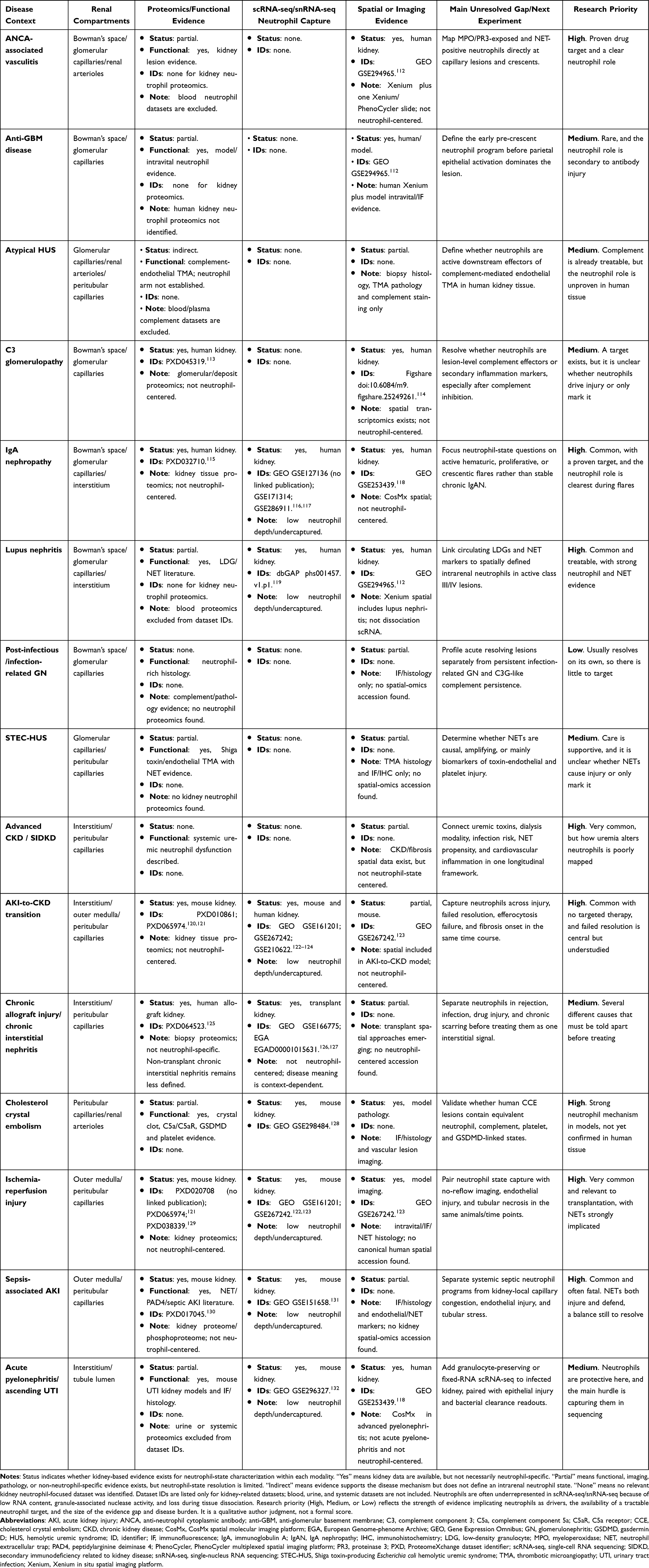 |
Table 1 Evidence Gaps for Kidney Neutrophil-State Biology Across Kidney Diseases |
Together, these approaches support a transition toward mechanism-based classification of kidney inflammation. Rather than treating all patients with the same histologic diagnosis identically, future strategies may identify whether complement activation, NET-driven microvascular injury, persistent neutrophil priming, maladaptive emergency granulopoiesis, or defective inflammatory resolution is the dominant process in a given patient, thereby allowing therapies to be matched more precisely to the underlying neutrophil state. Stratifying patients by activated CD11b, NET marker levels, or complement fragment concentrations could enrich trials for those most likely to respond to C5aR1 antagonism, GSDMD inhibition, or complement inhibition. Early mechanistic endpoints, such as a fall in citrullinated histone H3 or MPO-DNA, could provide pharmacodynamic evidence of target engagement before kidney outcomes become measurable.
Conclusions
Neutrophils contribute to a broad spectrum of acute and chronic kidney diseases, but their role is highly context dependent and ranges from pathogenic effector to secondary inflammatory amplifier. The same neutrophil capable of driving immunothrombotic microvascular injury in cholesterol crystal embolism or IRI is also essential for bacterial clearance in pyelonephritis and urosepsis. What determines these divergent functions is not the neutrophil itself, but the local inflammatory, metabolic, and vascular environment within the kidney. Defining kidney inflammation according to neutrophil state may more accurately capture disease biology than classification by histologic diagnosis alone.
Recent therapeutic advances support this concept. Iptacopan targets factor B within the alternative complement pathway in IgA nephropathy and C3 glomerulopathy, limiting complement-mediated neutrophil recruitment and activation at an upstream level. This strategy targets inflammatory circuits linked to neutrophil activation without directly eliminating neutrophils themselves.
Extending this approach to conditions such as IRI, STEC-HUS, thrombotic microangiopathies, and the AKI-to-CKD transition will require improved methods to define neutrophil state in vivo. Progress will likely depend on the integration of circulating biomarkers, tissue-based imaging, spatial transcriptomics, and translational models that more faithfully reproduce the compartment-specific environments encountered within the human kidney. Ultimately, a neutrophil state–based approach may enable more precise and mechanistically guided therapies that match intervention strategies to patients.
Generative AI Disclosure
OpenAI imaging tools accessed through ChatGPT 5.5 were used to generate the illustration shown in Figure 1, which was manually edited and labeled by the authors. AI was also used to assess formatting requirements according to publication guidelines. The authors confirm the originality and accuracy of the content of Figure 1. They have reviewed the terms of use of the tool and consider its output suitable for publication. They have the right to publish the generated image and have obtained any permissions required. They take full responsibility for the integrity of the whole content, including the accuracy of all references.
Abbreviations
AAV, ANCA-associated vasculitis; AKI, acute kidney injury; ANCA, anti-neutrophil cytoplasmic antibody; anti-GBM, anti-glomerular basement membrane; C3, complement component 3; C5a, complement component 5a; C5aR1, complement component 5a receptor 1; CCE, cholesterol crystal embolism; CKD, chronic kidney disease; DAMP, damage-associated molecular pattern; GSDMD, gasdermin D; HIF-1α, hypoxia-inducible factor 1α; IgA, immunoglobulin A; IRI, ischemia-reperfusion injury; LDG, low-density granulocyte; MPO, myeloperoxidase; NET, neutrophil extracellular trap; PAD4, peptidylarginine deiminase 4; PR3, proteinase 3; ROS, reactive oxygen species; SIDKD, secondary immunodeficiency related to kidney disease; STEC-HUS, Shiga toxin-producing Escherichia coli-associated hemolytic uremic syndrome; TMA, thrombotic microangiopathy; UTI, urinary tract infection.
Data Sharing Statement
No new datasets were generated or analyzed for this review. Publicly available datasets discussed in the article are cited in the references.
Author Contributions
John Ku: conceptualization, data curation, investigation, visualization, writing – original draft, and writing – review and editing.
Kailey K Flora: investigation and writing – review and editing.
Stefanie Steiger: investigation, supervision, and writing – review and editing.
Hans-Joachim Anders: conceptualization, supervision, and writing – review and editing.
All authors gave final approval of the version to be published, have agreed on the journal to which the article has been submitted, and agree to be accountable for all aspects of the work.
Funding
This work was supported by the Deutsche Forschungsgemeinschaft (DFG) collaborative research grant TRR332 (project number 449437943) for this study.
Disclosure
Prof. Dr. Stefanie Steiger reports Support from Deutsche Forschungsgemeinschaft (DFG)TRR332 Project A7, STE2437/4-1 and 4-2, during the conduct of the study; Grants and/or contracts from Novartis; Other interests from Associate Editor of NDT, Board Member of ERA IWG working group, outside the submitted work.
Prof. Dr. Hans-Joachim Anders reports Grants or contracts from DFG; Consulting fees from Industry; Honoraria from Industry; Leadership or fiduciary roles from ERA, outside the submitted work. The authors declare no other competing interests.
References
1. Pallone TL, Zhang Z, Rhinehart K. Physiology of the renal medullary microcirculation. Am J Physiol-Ren Physiol. 2003;284(2):F253–21. doi:10.1152/ajprenal.00304.2002
2. Kida Y. Peritubular capillary rarefaction: an underappreciated regulator of CKD progression. Int J Mol Sci. 2020;21(21):8255. PubMed PMID: 33158122; PubMed Central PMCID: PMC7662781. doi:10.3390/ijms21218255
3. Hommos MS, Glassock RJ, Rule AD. Structural and functional changes in human kidneys with healthy aging. J Am Soc Nephrol JASN. 2017;28(10):2838–2844. PubMed PMID: 28790143; PubMed Central PMCID: PMC5619977. doi:10.1681/ASN.2017040421
4. Anders HJ, Kitching AR, Leung N, Romagnani P. Glomerulonephritis: immunopathogenesis and immunotherapy. Nat Rev Immunol. 2023;23(7):453–471. PubMed PMID: 36635359; PubMed Central PMCID: PMC9838307. doi:10.1038/s41577-022-00816-y
5. Perkovic V, Barratt J, Rovin B, et al. Alternative complement pathway inhibition with iptacopan in IgA nephropathy. N Engl J Med. 2025;392(6):531–543. doi:10.1056/NEJMoa2410316
6. Nester CM, Eisenberger U, Karras A, et al. Iptacopan reduces proteinuria and stabilizes kidney function in C3 glomerulopathy. Kidney Int Rep. 2025;10(2):432–446. PubMed PMID: 39990880; PubMed Central PMCID: PMC11843281. doi:10.1016/j.ekir.2024.10.023
7. Nakazawa D, Masuda S, Nishibata Y, Watanabe-Kusunoki K, Tomaru U, Ishizu A. Neutrophils and NETs in kidney disease. Nat Rev Nephrol. 2025;21(6):383–398. doi:10.1038/s41581-025-00944-3
8. Maier-Begandt D, Alonso-Gonzalez N, Klotz L, et al. Neutrophils-biology and diversity. Nephrol Dial Transplant off Publ Eur Dial Transpl Assoc - Eur Ren Assoc. 2024;39(10):1551–1564. PubMed PMID: 38115607; PubMed Central PMCID: PMC11427074. doi:10.1093/ndt/gfad266
9. Németh T, Sperandio M, Mócsai A. Neutrophils as emerging therapeutic targets. Nat Rev Drug Discov. 2020;19(4):253–275. PubMed PMID: 31969717. doi:10.1038/s41573-019-0054-z
10. Borregaard N. Neutrophils, from marrow to microbes. Immunity. 2010;33(5):657–670. doi:10.1016/j.immuni.2010.11.011
11. Manz MG, Boettcher S. Emergency granulopoiesis. Nat Rev Immunol. 2014;14(5):302–314. doi:10.1038/nri3660
12. Jorch SK, Kubes P. An emerging role for neutrophil extracellular traps in noninfectious disease. Nat Med. 2017;23(3):279–287. PubMed PMID: 28267716. doi:10.1038/nm.4294
13. Brinkmann V, Reichard U, Goosmann C, et al. Neutrophil extracellular traps kill bacteria. Science. 2004;303(5663):1532–1535. doi:10.1126/science.1092385
14. Jennette JC, Falk RJ. Pathogenesis of antineutrophil cytoplasmic autoantibody-mediated disease. Nat Rev Rheumatol. 2014;10(8):463–473. doi:10.1038/nrrheum.2014.103
15. Kettritz R. How anti-neutrophil cytoplasmic autoantibodies activate neutrophils. Clin Exp Immunol. 2012;169(3):220–228. doi:10.1111/j.1365-2249.2012.04615.x
16. Surmiak M, Hubalewska-Mazgaj M, Wawrzycka-Adamczyk K, et al. Neutrophil-related and serum biomarkers in granulomatosis with polyangiitis support extracellular traps mechanism of the disease. Clin Exp Rheumatol. 2016;34(3 Suppl 97):S98–104. PubMed PMID: 27192020.
17. Matsumoto K, Kurasawa T, Yoshimoto K, Suzuki K, Takeuchi T. Identification of neutrophil β2-integrin LFA-1 as a potential mechanistic biomarker in ANCA-associated vasculitis via microarray and validation analyses. Arthritis Res Ther. 2021;23(1):136. PubMed PMID: 33957974; PubMed Central PMCID: PMC8101175. doi:10.1186/s13075-021-02510-1
18. Falk RJ, Jennette JC. Anti-neutrophil cytoplasmic autoantibodies with specificity for myeloperoxidase in patients with systemic vasculitis and idiopathic necrotizing and crescentic glomerulonephritis. N Engl J Med. 1988;318(25):1651–1657. doi:10.1056/NEJM198806233182504
19. Gupta S, Kaplan MJ. The role of neutrophils and NETosis in autoimmune and renal diseases. Nat Rev Nephrol. 2016;12(7):402–413. doi:10.1038/nrneph.2016.71
20. Papayannopoulos V. Neutrophil extracellular traps in immunity and disease. Nat Rev Immunol. 2018;18(2):134–147. doi:10.1038/nri.2017.105
21. Silvestre-Roig C, Braster Q, Wichapong K, et al. Externalized histone H4 orchestrates chronic inflammation by inducing lytic cell death. Nature. 2019;569(7755):236–240. PubMed PMID: 31043745; PubMed Central PMCID: PMC6716525. doi:10.1038/s41586-019-1167-6
22. Wang Y, Li M, Stadler S, et al. Histone hypercitrullination mediates chromatin decondensation and neutrophil extracellular trap formation. J Cell Biol. 2009;184(2):205–213. doi:10.1083/jcb.200806072
23. Denny MF, Yalavarthi S, Zhao W, et al. A distinct subset of proinflammatory neutrophils isolated from patients with systemic lupus erythematosus induces vascular damage and synthesizes type I IFNs. J Immunol. 2010;184(6):3284–3297. doi:10.4049/jimmunol.0902199
24. Villanueva E, Yalavarthi S, Berthier CC, et al. Netting neutrophils induce endothelial damage, infiltrate tissues, and expose immunostimulatory molecules in systemic lupus erythematosus. J Immunol. 2011;187(1):538–552. doi:10.4049/jimmunol.1100450
25. Lood C, Blanco LP, Purmalek MM, et al. Neutrophil extracellular traps enriched in oxidized mitochondrial DNA are interferogenic and contribute to lupus-like disease. Nat Med. 2016;22(2):146–153. doi:10.1038/nm.4027
26. Casanova-Acebes M, Pitaval C, Weiss LA, et al. Rhythmic modulation of the hematopoietic niche through neutrophil clearance. Cell. 2013;153(5):1025–1035. doi:10.1016/j.cell.2013.04.040
27. Adrover JM, Aroca-Crevillén A, Crainiciuc G, et al. Programmed ‘disarming’ of the neutrophil proteome reduces the magnitude of inflammation. Nat Immunol. 2020;21(2):135–144. doi:10.1038/s41590-019-0571-2
28. Adrover JM, Del Fresno C, Crainiciuc G, et al. A neutrophil timer coordinates immune defense and vascular protection. Immunity. 2019;50(2):390–402.e10. PubMed PMID: 30709741. doi:10.1016/j.immuni.2019.01.002
29. Ng LG, Ostuni R, Hidalgo A. Heterogeneity of neutrophils. Nat Rev Immunol. 2019;19(4):255–265. doi:10.1038/s41577-019-0141-8
30. Bolisetty S, Agarwal A. Neutrophils in acute kidney injury: not neutral any more. Kidney Int. 2009;75(7):674–676. doi:10.1038/ki.2008.689
31. Kelly KJ, Williams WW, Colvin RB, et al. Intercellular adhesion molecule-1-deficient mice are protected against ischemic renal injury. J Clin Invest. 1996;97(4):1056–1063. doi:10.1172/JCI118498
32. Kelly KJ, Williams WW, Colvin RB, Bonventre JV. Antibody to intercellular adhesion molecule 1 protects the kidney against ischemic injury. Proc Natl Acad Sci. 1994;91(2):812–816. doi:10.1073/pnas.91.2.812
33. He L, Wang J, Li Y, et al. Mac-1 promotes neutrophil extracellular traps formation via ERK phosphorylation in renal ischemia-reperfusion injury. Inflammation. 2025;48(6):4302–4317. doi:10.1007/s10753-025-02327-z
34. Wang H, Gao T, Zhang R, et al. Neutrophil extracellular traps aggravate contrast-induced acute kidney injury by damaging glomeruli and peritubular capillaries. J Inflamm Res. 2023;16:5629–5646. PubMed PMID: 38046404; PubMed Central PMCID: PMC10693253. doi:10.2147/JIR.S433110
35. Fu Y, Wang W, Gong N, et al. Neutrophil and neutrophil extracellular traps in acute kidney injury: from mechanisms to treatments. Front Immunol. 2025;16:1688207. doi:10.3389/fimmu.2025.1688207
36. Ballermann BJ, Dardik A, Eng E, Liu A. Shear stress and the endothelium. Kidney Int. 1998;54:S100–8. doi:10.1046/j.1523-1755.1998.06720.x
37. Shi C, Zhao D, Lyubenov L, et al. Neutrophil circadian rhythm is associated with different outcomes of acute kidney injury due to cholesterol crystal embolism. Front Cardiovasc Med. 2022;9:974759. doi:10.3389/fcvm.2022.974759
38. Scolari F, Ravani P. Atheroembolic renal disease. The Lancet. 2010;375(9726):1650–1660. doi:10.1016/S0140-6736(09)62073-0
39. Ma Q, Steiger S. Neutrophils and extracellular traps in crystal-associated diseases. Trends Mol Med. 2024;30(9):809–823. doi:10.1016/j.molmed.2024.05.010
40. Mulay SR, Anders HJ. Crystallopathies. N Engl J Med. 2016;374(25):2465–2476. doi:10.1056/NEJMra1601611
41. Watanabe-Kusunoki K, Li C, Bandeira Honda TS, et al. Gasdermin D drives focal crystalline thrombotic microangiopathy by accelerating immunothrombosis and necroinflammation. Blood. 2024;144(3):308–322. PubMed PMID: 38657197. doi:10.1182/blood.2023021949
42. Daehn IS, Duffield JS. The glomerular filtration barrier: a structural target for novel kidney therapies. Nat Rev Drug Discov. 2021;20(10):770–788. doi:10.1038/s41573-021-00242-0
43. Kuligowski MP, Kitching AR, Hickey MJ. Leukocyte recruitment to the inflamed glomerulus: a critical role for platelet-derived P-selectin in the absence of rolling. J Immunol. 2006;176(11):6991–6999. doi:10.4049/jimmunol.176.11.6991
44. Charles Jennette J. Rapidly progressive crescentic glomerulonephritis. Kidney Int. 2003;63(3):1164–1177. doi:10.1046/j.1523-1755.2003.00843.x
45. Anguiano L, Kain R, Anders HJ. The glomerular crescent: triggers, evolution, resolution, and implications for therapy. Curr Opin Nephrol Hypertens. 2020;29(3):302–309. PubMed PMID: 32132388; PubMed Central PMCID: PMC7170443. doi:10.1097/MNH.0000000000000596
46. Smeets B, Uhlig S, Fuss A, et al. Tracing the origin of glomerular extracapillary lesions from parietal epithelial cells. J Am Soc Nephrol. 2009;20(12):2604–2615. doi:10.1681/ASN.2009010122
47. Prendecki M, Gurung A, Pisacano N, Pusey CD. The role of neutrophils in ANCA-associated vasculitis. Immunol Lett. 2024;270:106933. doi:10.1016/j.imlet.2024.106933
48. Park I, Kim S, Um YW, et al. Intravital imaging of peritubular microcirculation impairment in cisplatin-induced acute kidney injury. JCI Insight. 2025;10(9):e178689. PubMed PMID: 40198123; PubMed Central PMCID: PMC12128961. doi:10.1172/jci.insight.178689
49. Bonventre JV, Yang L. Cellular pathophysiology of ischemic acute kidney injury. J Clin Invest. 2011;121(11):4210–4221. doi:10.1172/JCI45161
50. Patschan D, Patschan S, Müller GA. Inflammation and microvasculopathy in renal ischemia reperfusion injury. J Transplant. 2012;2012:764154. PubMed PMID: 22530109; PubMed Central PMCID: PMC3316981. doi:10.1155/2012/764154
51. Freitas F, Attwell D. Pericyte-mediated constriction of renal capillaries evokes no-reflow and kidney injury following ischaemia. eLife. 2022;11:e74211. PubMed PMID: 35285797; PubMed Central PMCID: PMC8947765. doi:10.7554/eLife.74211
52. Nakazawa D, Kumar SV, Marschner J, et al. Histones and neutrophil extracellular traps enhance tubular necrosis and remote organ injury in ischemic AKI. J Am Soc Nephrol. 2017;28(6):1753–1768. doi:10.1681/ASN.2016080925
53. Peerapornratana S, Manrique-Caballero CL, Gómez H, Kellum JA. Acute kidney injury from sepsis: current concepts, epidemiology, pathophysiology, prevention and treatment. Kidney Int. 2019;96(5):1083–1099. doi:10.1016/j.kint.2019.05.026
54. Zarbock A, Nadim MK, Pickkers P, et al. Sepsis-associated acute kidney injury: consensus report of the 28th acute disease quality initiative workgroup. Nat Rev Nephrol. 2023;19(6):401–417. doi:10.1038/s41581-023-00683-3
55. Karpman D, Loos S, Tati R, Arvidsson I. Haemolytic uraemic syndrome. J Intern Med. 2017;281(2):123–148. doi:10.1111/joim.12546
56. Zhao D, Han C, Mammadova-Bach E, et al. Inhibition of complement factor C5a or C5aR for cholesterol crystal embolism-related vascular thrombosis with microvascular injury and its consequences. Kidney Int. 2024;106(5):819–825. PubMed PMID: 39142565. doi:10.1016/j.kint.2024.07.020
57. Walmsley SR, Print C, Farahi N, et al. Hypoxia-induced neutrophil survival is mediated by HIF-1alpha-dependent NF-kappaB activity. J Exp Med. 2005;201(1):105–115. PubMed PMID: 15630139; PubMed Central PMCID: PMC2212759. doi:10.1084/jem.20040624
58. Engelmann B, Massberg S. Thrombosis as an intravascular effector of innate immunity. Nat Rev Immunol. 2013;13(1):34–45. PubMed PMID: 23222502. doi:10.1038/nri3345
59. Chawla LS, Eggers PW, Star RA, Kimmel PL. Acute kidney injury and chronic kidney disease as interconnected syndromes. N Engl J Med. 2014;371(1):58–66. doi:10.1056/NEJMra1214243
60. SMW Y, Bonventre JV. Acute kidney injury and maladaptive tubular repair leading to renal fibrosis. Curr Opin Nephrol Hypertens. 2020;29(3):310–318. PubMed PMID: 32205583; PubMed Central PMCID: PMC7363449. doi:10.1097/MNH.0000000000000605
61. Gewin L, Zent R. How does TGF-β mediate tubulointerstitial fibrosis? Semin Nephrol. 2012;32(3):228–235. PubMed PMID: 22835453; PubMed Central PMCID: PMC4948283. doi:10.1016/j.semnephrol.2012.04.001
62. Soehnlein O, Lindbom L. Phagocyte partnership during the onset and resolution of inflammation. Nat Rev Immunol. 2010;10(6):427–439. PubMed PMID: 20498669. doi:10.1038/nri2779
63. Doran AC, Yurdagul A, Tabas I. Efferocytosis in health and disease. Nat Rev Immunol. 2020;20(4):254–267. doi:10.1038/s41577-019-0240-6
64. Mulay SR, Linkermann A, Anders HJ. Necroinflammation in kidney disease. J Am Soc Nephrol. 2016;27(1):27–39. doi:10.1681/ASN.2015040405
65. Langewisch E, Mannon RB. Chronic allograft injury. Clin J Am Soc Nephrol CJASN. 2021;16(11):1723–1729. PubMed PMID: 33820759; PubMed Central PMCID: PMC8729407. doi:10.2215/CJN.15590920
66. Mulay SR, Kulkarni OP, Rupanagudi KV, et al. Calcium oxalate crystals induce renal inflammation by NLRP3-mediated IL-1β secretion. J Clin Invest. 2013;123(1):236–246. doi:10.1172/JCI63679
67. Leung N, Rajkumar SV. Multiple myeloma with acute light chain cast nephropathy. Blood Cancer J. 2023;13(1):46. PubMed PMID: 36990996; PubMed Central PMCID: PMC10060259. doi:10.1038/s41408-023-00806-w
68. Conger JD. Acute uric acid nephropathy. Med Clin North Am. 1990;74(4):859–871. PubMed PMID: 2195258. doi:10.1016/s0025-7125(16)30522-3
69. Melican K, Sandoval RM, Kader A, et al. Uropathogenic Escherichia coli P and Type 1 fimbriae act in synergy in a living host to facilitate renal colonization leading to nephron obstruction. PLoS Pathog. 2011;7(2):e1001298. PubMed PMID: 21383970; PubMed Central PMCID: PMC3044688. doi:10.1371/journal.ppat.1001298
70. Burg MB, Ferraris JD, Dmitrieva NI. Cellular response to hyperosmotic stresses. Physiol Rev. 2007;87(4):1441–1474. PubMed PMID: 17928589. doi:10.1152/physrev.00056.2006
71. López-Rodríguez C, Antos CL, Shelton JM, et al. Loss of NFAT5 results in renal atrophy and lack of tonicity-responsive gene expression. Proc Natl Acad Sci U S A. 2004;101(8):2392–2397. PubMed PMID: 14983020; PubMed Central PMCID: PMC356961. doi:10.1073/pnas.0308703100
72. Aramburu J, López-Rodríguez C. Regulation of inflammatory functions of macrophages and T lymphocytes by NFAT5. Front Immunol. 2019;10:535. PubMed PMID: 30949179; PubMed Central PMCID: PMC6435587. doi:10.3389/fimmu.2019.00535
73. Mazzitelli I, Bleichmar L, Melucci C, et al. High salt induces a delayed activation of human neutrophils. Front Immunol. 2022;13:831844. PubMed PMID: 35720394; PubMed Central PMCID: PMC9204211. doi:10.3389/fimmu.2022.831844
74. Nadesalingam A, Chen JHK, Farahvash A, Khan MA. Hypertonic saline suppresses NADPH oxidase-dependent neutrophil extracellular trap formation and promotes apoptosis. Front Immunol. 2018;9:359. PubMed PMID: 29593709; PubMed Central PMCID: PMC5859219. doi:10.3389/fimmu.2018.00359
75. Rodríguez-Espinosa O, Rojas-Espinosa O, Moreno-Altamirano MMB, López-Villegas EO, Sánchez-García FJ. Metabolic requirements for neutrophil extracellular traps formation. Immunology. 2015;145(2):213–224. PubMed PMID: 25545227; PubMed Central PMCID: PMC4427386. doi:10.1111/imm.12437
76. Chouchani ET, Pell VR, Gaude E, et al. Ischaemic accumulation of succinate controls reperfusion injury through mitochondrial ROS. Nature. 2014;515(7527):431–435. doi:10.1038/nature13909
77. Nunns GR, Vigneshwar N, Kelher MR, et al. Succinate activation of SUCNR1 predisposes severely injured patients to neutrophil-mediated ARDS. Ann Surg. 2022;276(6):e944–54. PubMed PMID: 33214479; PubMed Central PMCID: PMC8128932. doi:10.1097/SLA.0000000000004644
78. Steiger S, Rossaint J, Zarbock A, Anders HJ. Secondary immunodeficiency related to kidney disease (SIDKD)-definition, unmet need, and mechanisms. J Am Soc Nephrol JASN. 2022;33(2):259–278. PubMed PMID: 34907031; PubMed Central PMCID: PMC8819985. doi:10.1681/ASN.2021091257
79. Vanholder R, De Smet R, Glorieux G, et al. Review on uremic toxins: classification, concentration, and interindividual variability. Kidney Int. 2003;63(5):1934–1943. doi:10.1046/j.1523-1755.2003.00924.x
80. Cohen G, Hörl W. Immune dysfunction in uremia—an update. Toxins. 2012;4(11):962–990. doi:10.3390/toxins4110962
81. Rossaint J, Oehmichen J, Van Aken H, et al. FGF23 signaling impairs neutrophil recruitment and host defense during CKD. J Clin Invest. 2016;126(3):962–974. PubMed PMID: 26878171; PubMed Central PMCID: PMC4767336. doi:10.1172/JCI83470
82. Rossaint J, Spelten O, Kässens N, et al. Acute loss of renal function attenuates slow leukocyte rolling and transmigration by interfering with intracellular signaling. Kidney Int. 2011;80(5):493–503. PubMed PMID: 21562471; PubMed Central PMCID: PMC3156340. doi:10.1038/ki.2011.125
83. Zarbock A, Schmolke M, Spieker T, Jurk K, Van Aken H, Singbartl K. Acute uremia but not renal inflammation attenuates aseptic acute lung injury: a critical role for uremic neutrophils. J Am Soc Nephrol JASN. 2006;17(11):3124–3131. PubMed PMID: 17035612. doi:10.1681/ASN.2006040358
84. Chonchol M. Neutrophil dysfunction and infection risk in end-stage renal disease. Semin Dial. 2006;19(4):291–296. PubMed PMID: 16893406. doi:10.1111/j.1525-139X.2006.00175.x
85. Vanholder R, De Smet R, Waterloos MA, et al. Mechanisms of uremic inhibition of phagocyte reactive species production: characterization of the role of p-cresol. Kidney Int. 1995;47(2):510–517. PubMed PMID: 7723236. doi:10.1038/ki.1995.64
86. Ma Q, Immler R, Pruenster M, et al. Soluble uric acid inhibits β2 integrin-mediated neutrophil recruitment in innate immunity. Blood. 2022;139(23):3402–3417. PubMed PMID: 35303071; PubMed Central PMCID: PMC11022987. doi:10.1182/blood.2021011234
87. Li Q, Anders J, Flora K, et al. Soluble uric acid suppresses neutrophil-mediated host defense in sepsis. Nat Commun. 2026;17(1):4453. PubMed PMID: 42156746; PubMed Central PMCID: PMC13187337. doi:10.1038/s41467-026-73090-4
88. Patole PS, Schubert S, Hildinger K, et al. Toll-like receptor-4: renal cells and bone marrow cells signal for neutrophil recruitment during pyelonephritis. Kidney Int. 2005;68(6):2582–2587. PubMed PMID: 16316333. doi:10.1111/j.1523-1755.2005.00729.x
89. Justice SS, Hunstad DA, Seed PC, Hultgren SJ. Filamentation by Escherichia coli subverts innate defenses during urinary tract infection. Proc Natl Acad Sci U S A. 2006;103(52):19884–19889. PubMed PMID: 17172451; PubMed Central PMCID: PMC1750882. doi:10.1073/pnas.0606329104
90. Czaikoski PG, Mota JMSC, Nascimento DC, et al. Neutrophil extracellular traps induce organ damage during experimental and clinical sepsis. PLoS One. 2016;11(2):e0148142. doi:10.1371/journal.pone.0148142
91. Mai SHC, Khan M, Dwivedi DJ, et al. Delayed but not early treatment with DNase reduces organ damage and improves outcome in a murine model of sepsis. Shock. 2015;44(2):166–172. PubMed PMID: 26009820. doi:10.1097/SHK.0000000000000396
92. Noris M, Remuzzi G. Atypical hemolytic–uremic syndrome. N Engl J Med. 2009;361(17):1676–1687. doi:10.1056/NEJMra0902814
93. Legendre CM, Licht C, Muus P, et al. Terminal complement inhibitor eculizumab in atypical hemolytic–uremic syndrome. N Engl J Med. 2013;368(23):2169–2181. doi:10.1056/NEJMoa1208981
94. Schreiber A, Xiao H, Jennette JC, Schneider W, Luft FC, Kettritz R. C5a receptor mediates neutrophil activation and ANCA-induced glomerulonephritis. J Am Soc Nephrol. 2009;20(2):289–298. doi:10.1681/ASN.2008050497
95. McAdoo SP, Pusey CD. Anti-glomerular basement membrane disease. Clin J Am Soc Nephrol. 2017;12(7):1162–1172. doi:10.2215/CJN.01380217
96. Anders HJ, Saxena R, Zhao M, Parodis I, Salmon JE, Mohan C. Lupus nephritis. Nat Rev Dis Primer. 2020;6(1):7. doi:10.1038/s41572-019-0141-9
97. Garcia-Romo GS, Caielli S, Vega B, et al. Netting neutrophils are major inducers of type I IFN production in pediatric systemic lupus erythematosus. Sci Transl Med. 2011;3(73). doi:10.1126/scitranslmed.3001201
98. Rovin BH, Teng YKO, Ginzler EM, et al. Efficacy and safety of voclosporin versus placebo for lupus nephritis (Aurora 1): a double-blind, randomised, multicentre, placebo-controlled, Phase 3 trial. The Lancet. 2021;397(10289):2070–2080. doi:10.1016/S0140-6736(21)00578-X
99. Furie R, Rovin BH, Houssiau F, et al. Two-year, randomized, controlled trial of belimumab in lupus nephritis. N Engl J Med. 2020;383(12):1117–1128. doi:10.1056/NEJMoa2001180
100. Barratt J, Eren N, Kashihara N, et al. Iptacopan in IgA nephropathy — final 24-month data. N Engl J Med. 2026:NEJMoa2600743. doi:10.1056/NEJMoa2600743
101. Smith RJH, Appel GB, Blom AM, et al. C3 glomerulopathy — understanding a rare complement-driven renal disease. Nat Rev Nephrol. 2019;15(3):129–143. doi:10.1038/s41581-018-0107-2
102. Serhan CN. Pro-resolving lipid mediators are leads for resolution physiology. Nature. 2014;510(7503):92–101. doi:10.1038/nature13479
103. Chalmers JD, Burgel PR, Daley CL, et al. Phase 3 trial of the DPP-1 inhibitor brensocatib in bronchiectasis. N Engl J Med. 2025;392(16):1569–1581. PubMed PMID: 40267423. doi:10.1056/NEJMoa2411664
104. Johnson E, Gilmour A, Chalmers JD. Dipeptidyl peptidase-1 inhibitors in bronchiectasis. Eur Respir Rev off J Eur Respir Soc. 2025;34(176):240257. PubMed PMID: 40533102; PubMed Central PMCID: PMC12175074. doi:10.1183/16000617.0257-2024
105. McNamara LA, Topaz N, Wang X, Hariri S, Fox L, MacNeil JR. High risk for invasive meningococcal disease among patients receiving eculizumab (Soliris) despite receipt of meningococcal vaccine. MMWR Morb Mortal Wkly Rep. 2017;66(27):734–737. PubMed PMID: 28704351; PubMed Central PMCID: PMC5687588. doi:10.15585/mmwr.mm6627e1
106. Mazzieri A, Marcon LMR. Nephroprotective mechanisms of SGLT2i: beyond the glucose-lowering effect. Biomedicines. 2025;13(9):2123. PubMed PMID: 41007686; PubMed Central PMCID: PMC12467186. doi:10.3390/biomedicines13092123
107. Meier M, Cummings J, Abdelsaid M, et al. Glucagon-like peptide-1 receptor agonists in chronic kidney disease: mechanisms and clinical perspectives. Kidney Med. 2026;8(4):101282. PubMed PMID: 41808780; PubMed Central PMCID: PMC12969668. doi:10.1016/j.xkme.2026.101282
108. Wang B, Zhang R, Liu X, et al. Neutrophil extracellular traps in diabetic kidney disease: mechanisms of pathogenesis and emerging therapeutic strategies. Drug Des Devel Ther. 2026;20:583077. PubMed PMID: 41756312; PubMed Central PMCID: PMC12935511. doi:10.2147/DDDT.S583077
109. Thålin C, Aguilera K, Hall NW, et al. Quantification of citrullinated histones: development of an improved assay to reliably quantify nucleosomal H3Cit in human plasma. J Thromb Haemost JTH. 2020;18(10):2732–2743. PubMed PMID: 32654410; PubMed Central PMCID: PMC8722705. doi:10.1111/jth.15003
110. Wang H, Sha LL, Ma TT, Zhang LX, Chen M, Zhao MH. Circulating level of neutrophil extracellular traps is not a useful biomarker for assessing disease activity in antineutrophil cytoplasmic antibody-associated vasculitis. PLoS One. 2016;11(2):e0148197. PubMed PMID: 26840412; PubMed Central PMCID: PMC4739550. doi:10.1371/journal.pone.0148197
111. Aguilar F, Lozano JJ, Clària J, et al. Maladaptive emergency granulopoiesis predicts poor outcomes in patients hospitalized with decompensated cirrhosis. J Hepatol. 2026;84(4):749–765. PubMed PMID: 41314350. doi:10.1016/j.jhep.2025.11.012
112. Wang H, Engesser J, Khatri R, et al. Type I interferon drives T cell cytotoxicity by upregulation of interferon regulatory factor 7 in autoimmune kidney diseases in mice. Nat Commun. 2025;16(1):4686. PubMed PMID: 40393992; PubMed Central PMCID: PMC12092595. doi:10.1038/s41467-025-59819-7
113. Madden B, Singh RD, Haas M, et al. Apolipoprotein E is enriched in dense deposits and is a marker for dense deposit disease in C3 glomerulopathy. Kidney Int. 2024;105(5):1077–1087. PubMed PMID: 38447879. doi:10.1016/j.kint.2024.02.013
114. Koh JH, Kang M, Park S, et al. Spatially resolved transcriptomic profiling for glomerular and tubulointerstitial gene expression in C3 glomerulopathy. Clin Kidney J. 2025;18(5):sfaf139. PubMed PMID: 40416397; PubMed Central PMCID: PMC12102689. doi:10.1093/ckj/sfaf139
115. Chen X, Wang T, Chen L, et al. Cross-species single-cell analysis uncovers the immunopathological mechanisms associated with IgA nephropathy progression. JCI Insight. 2024;9(9):e173651. PubMed PMID: 38716725; PubMed Central PMCID: PMC11141938. doi:10.1172/jci.insight.173651
116. Tang R, Meng T, Lin W, et al. A partial picture of the single-cell transcriptomics of human IgA nephropathy. Front Immunol. 2021;12:645988. PubMed PMID: 33936064; PubMed Central PMCID: PMC8085501. doi:10.3389/fimmu.2021.645988
117. You JP, Cheng GY, Zhang XX, et al. Single nucleus RNA-seq reveals the process from onset to chronic kidney disease in IgA nephropathy. Sci Rep. 2025;15(1):22780. PubMed PMID: 40592877; PubMed Central PMCID: PMC12216285. doi:10.1038/s41598-025-04292-x
118. Reck M, Baird DP, Veizades S, et al. Multiomic analysis of human kidney disease identifies a tractable inflammatory and pro-fibrotic tubular cell phenotype. Nat Commun. 2025;16(1):4745. PubMed PMID: 40399382; PubMed Central PMCID: PMC12095627. doi:10.1038/s41467-025-59997-4
119. Arazi A, Rao DA, Berthier CC, et al. The immune cell landscape in kidneys of patients with lupus nephritis. Nat Immunol. 2019;20(7):902–914. PubMed PMID: 31209404; PubMed Central PMCID: PMC6726437. doi:10.1038/s41590-019-0398-x
120. Pavkovic M, Pantano L, Gerlach CV, et al. Multi omics analysis of fibrotic kidneys in two mouse models. Sci Data. 2019;6(1):92. PubMed PMID: 31201317; PubMed Central PMCID: PMC6570759. doi:10.1038/s41597-019-0095-5
121. Zhang S, Luo H, Wei M, et al. Temporal proteomic and phosphoproteomic profiling deciphers molecular dynamics of acute-to-chronic kidney disease after ischemia-reperfusion injury, with Dock2 emerging as a key regulator. Mol Cell Proteomics MCP. 2026;25(2):101509. PubMed PMID: 41534628; PubMed Central PMCID: PMC12915234. doi:10.1016/j.mcpro.2026.101509
122. Ide S, Kobayashi Y, Ide K, et al. Ferroptotic stress promotes the accumulation of pro-inflammatory proximal tubular cells in maladaptive renal repair. eLife. 2021;10:e68603. PubMed PMID: 34279220; PubMed Central PMCID: PMC8318592. doi:10.7554/eLife.68603
123. Wang C, Zhang Y, Shen A, et al. Mincle receptor in macrophage and neutrophil contributes to the unresolved inflammation during the transition from acute kidney injury to chronic kidney disease. Front Immunol. 2024;15:1385696. PubMed PMID: 38770013; PubMed Central PMCID: PMC11103384. doi:10.3389/fimmu.2024.1385696
124. Hinze C, Kocks C, Leiz J, et al. Single-cell transcriptomics reveals common epithelial response patterns in human acute kidney injury. Genome Med. 2022;14(1):103. PubMed PMID: 36085050; PubMed Central PMCID: PMC9462075. doi:10.1186/s13073-022-01108-9
125. Kania D, Fochtman D, Skoczylas Ł, et al. Biopsy proteome-based classification of T cell-mediated kidney allograft rejection. J Transl Med. 2025;23(1):1139. PubMed PMID: 41121127; PubMed Central PMCID: PMC12539011. doi:10.1186/s12967-025-07116-8
126. Shen Q, Wang Y, Chen J, et al. Single-cell RNA sequencing reveals the immunological profiles of renal allograft rejection in mice. Front Immunol. 2021;12:693608. PubMed PMID: 34367152; PubMed Central PMCID: PMC8340022. doi:10.3389/fimmu.2021.693608
127. Jafree DJ, Stewart BJ, Price KL, et al. Organ-specific features of human kidney lymphatics are disrupted in chronic transplant rejection. J Clin Invest. 2025;135(18):e168962. PubMed PMID: 40663403; PubMed Central PMCID: PMC12435838. doi:10.1172/JCI168962
128. Shi C, Wen Z, Zheng K, et al. CCR5 and CD74 are potential therapeutic targets for necroinflammation in preclinical cholesterol crystal embolism. Sci Transl Med. 2026;18(851):eadv8372. doi:10.1126/scitranslmed.adv8372
129. Burton JB, Silva-Barbosa A, Bons J, et al. Substantial downregulation of mitochondrial and peroxisomal proteins during acute kidney injury revealed by data-independent acquisition proteomics. Proteomics. 2024;24(5):e2300162. PubMed PMID: 37775337. doi:10.1002/pmic.202300162
130. Lin YH, Platt MP, Fu H, et al. Global proteome and phosphoproteome characterization of sepsis-induced kidney injury. Mol Cell Proteomics MCP. 2020;19(12):2030–2047. PubMed PMID: 32963032; PubMed Central PMCID: PMC7710145. doi:10.1074/mcp.RA120.002235
131. Janosevic D, Myslinski J, McCarthy TW, et al. The orchestrated cellular and molecular responses of the kidney to endotoxin define a precise sepsis timeline. eLife. 2021;10:e62270. PubMed PMID: 33448928; PubMed Central PMCID: PMC7810465. doi:10.7554/eLife.62270
132. Hreha TN, Manson AL, Collins CA, et al. Single-nucleus transcriptomics illuminates sex differences during murine Escherichia coli pyelonephritis. Commun Biol. 2026;9(1):711. PubMed PMID: 41917498; PubMed Central PMCID: PMC13201655. doi:10.1038/s42003-026-09946-8
 © 2026 The Author(s). This work is published and licensed by Dove Medical Press Limited. The
full terms of this license are available at https://www.dovepress.com/terms
and incorporate the Creative Commons Attribution
- Non Commercial (unported, 4.0) License.
By accessing the work you hereby accept the Terms. Non-commercial uses of the work are permitted
without any further permission from Dove Medical Press Limited, provided the work is properly
attributed. For permission for commercial use of this work, please see paragraphs 4.2 and 5 of our Terms.
© 2026 The Author(s). This work is published and licensed by Dove Medical Press Limited. The
full terms of this license are available at https://www.dovepress.com/terms
and incorporate the Creative Commons Attribution
- Non Commercial (unported, 4.0) License.
By accessing the work you hereby accept the Terms. Non-commercial uses of the work are permitted
without any further permission from Dove Medical Press Limited, provided the work is properly
attributed. For permission for commercial use of this work, please see paragraphs 4.2 and 5 of our Terms.
Recommended articles

Neutrophil Extracellular Traps Aggravate Contrast-Induced Acute Kidney Injury by Damaging Glomeruli and Peritubular Capillaries
Wang H, Gao T, Zhang R, Hu J, Gao S, Wang Y, Qi X, Zhou Y, Zheng G, Dong H
Journal of Inflammation Research 2023, 16:5629-5646
Published Date: 28 November 2023
Identification and Validation of Hub Genes Related to Neutrophil Extracellular Traps-Mediated Cell Damage During Myocardial Infarction

Ke D, Ni J, Yuan Y, Cao M, Chen S, Zhou H
Journal of Inflammation Research 2024, 17:617-637
Published Date: 1 February 2024
Neutrophil Extracellular Traps Enhance Procoagulant Activity and Predict Poor Prognosis in Patients With Metastatic Breast Cancer
Gong Y, Chen B, Tan Q, Wei W
International Journal of General Medicine 2025, 18:1247-1259
Published Date: 4 March 2025
Mesenchymal Stem Cell-Derived Exosomes in Anti-NET Therapy: Mechanisms, Challenges, and Future Perspectives
Ye Y, Ye Y, Tian M, Zhao Y, Guo Z, Jin C, Duan S, Zheng Y
International Journal of Nanomedicine 2025, 20:14481-14497
Published Date: 4 December 2025
Neutrophil Extracellular Traps in Diabetic Kidney Disease: Mechanisms of Pathogenesis and Emerging Therapeutic Strategies
Wang B, Zhang R, Liu X, Shang Y, Jin T, Gao C, Yang N, Jin J, He Q
Drug Design, Development and Therapy 2026, 20:583077
Published Date: 21 February 2026