
Back to Journals » Drug Design, Development and Therapy » Volume 20
Neutrophil Extracellular Traps in Diabetic Kidney Disease: Mechanisms of Pathogenesis and Emerging Therapeutic Strategies
Authors Wang B, Zhang R  , Liu X, Shang Y, Jin T , Gao C, Yang N, Jin J, He Q
Received 20 November 2025
Accepted for publication 12 February 2026
Published 21 February 2026 Volume 2026:20 583077
DOI https://doi.org/10.2147/DDDT.S583077
Checked for plagiarism Yes
Review by Single anonymous peer review
Peer reviewer comments 4
Editor who approved publication: Dr Tuo Deng
Binqi Wang,1,2,* Ruikai Zhang,1,2,* Xinbo Liu,1,2 Yiwei Shang,1,2 Taian Jin,1,2 Chan Gao,1,2 Nan Yang,1,2 Juan Jin,1,2 Qiang He1,2
1Department of Nephrology, the First Affiliated Hospital of Zhejiang Chinese Medical University (Zhejiang Provincial Hospital of Chinese Medicine), Hangzhou, Zhejiang, 310006, People’s Republic of China; 2Zhejiang Key Laboratory of Research and Translation for Kidney Deficiency-Stasis-Turbidity Disease, Zhejiang-Macau International Joint Laboratory of Integrated Traditional Chinese and Western Medicine for Nephrology and Immunology, Hangzhou, Zhejiang, 310006, People’s Republic of China
*These authors contributed equally to this work
Correspondence: Qiang He, Department of Nephrology, the First Affiliated Hospital of Zhejiang Chinese Medical University (Zhejiang Provincial Hospital of Chinese Medicine), Hangzhou, Zhejiang, 310006, People’s Republic of China, Email [email protected] Juan Jin, Department of Nephrology, the First Affiliated Hospital of Zhejiang Chinese Medical University (Zhejiang Provincial Hospital of Chinese Medicine), Hangzhou, Zhejiang, 310006, People’s Republic of China, Email [email protected]
Abstract: Diabetic kidney disease (DKD) is a major microvascular complication of diabetes mellitus and the leading global cause of end-stage renal disease. While chronic hyperglycemia and classic metabolic pathways such as protein kinase C activation, advanced glycation end product (AGE) accumulation, and renin–angiotensin system overactivity elucidate many features of DKD, they do not fully account for its complex inflammatory pathology. Recent studies have highlighted neutrophil extracellular trap (NET) formation, or NETosis, as a critical mechanism linking metabolic stress to immune dysregulation in DKD. NETosis is a specialized form of programmed cell death in neutrophils that releases web-like chromatin structures decorated with histones, elastase, and myeloperoxidase. Although these structures function as protective barriers against pathogens, they become pathogenic when dysregulated. In the diabetic milieu, hyperglycemia, advanced glycation end products (AGEs), protein kinase C signaling, lipid abnormalities, and pro-inflammatory cytokines all promote excessive NETosis. Once formed, NETs drive renal injury by inducing endothelial dysfunction, podocyte and tubular epithelial cell damage, macrophage activation, and profibrotic signaling, thereby accelerating inflammation, fibrosis, and renal decline. Therapeutic strategies targeting NETosis are emerging, including inhibitors of PAD4, NADPH oxidase, and neutrophil elastase, as well as NET-degrading agents such as DNase I and histone-neutralizing compounds. Classical antidiabetic drugs, notably metformin and GLP-1 receptor agonists, also show potential in modulating NET formation. Collectively, these insights position NETosis as both a biomarker and a therapeutic target in DKD, offering novel opportunities to mitigate diabetes-associated renal complications.
Keywords: diabetic kidney disease, kidney injury, neutrophil extracellular traps, NETosis, inflammation, therapeutic targets
Introduction
Diabetic Kidney Disease (DKD) is a common and severe microvascular complication of diabetes mellitus, characterized by progressive structural and functional damage to the kidneys, primarily driven by chronic hyperglycemia and other metabolic derangements.1 Affecting approximately 40% of all individuals with diabetes, DKD is currently the single leading cause of end-stage renal disease (ESRD) globally, accounting for about 50% of cases in developed countries. The disease typically manifests after 10–20 years of diabetes and disproportionately affects individuals of African, Asian, and Native American descent.2,3 Beyond renal failure, DKD significantly amplifies the risk of cardiovascular disease (CVD), resulting in mortality from cardiovascular events for many patients before they reach ESRD.4 This dual burden of renal and cardiovascular risk imposes immense strain on global healthcare systems and severely impairs the quality of life for millions.3 Therefore, the urgent need for a deeper understanding of its complex pathophysiology is undeniable.
The pathology of DKD is defined by progressive structural damage to the kidney. Hallmarks begin with glomerular basement membrane (GBM) thickening and mesangial matrix expansion, which are intensified by the dysfunction and loss of podocytes (key filtration cells) leading directly to proteinuria.5 These early lesions ultimately progress to irreversible glomerulosclerosis and tubulointerstitial fibrosis, causing a steady decline in renal function.6 Traditionally, these changes are attributed to chronic hyperglycemia, which activates signaling pathways like protein kinase C (PKC) and the polyol pathway. This activation, combined with the accumulation of advanced glycation end products (AGEs) and overactivation of the renin-angiotensin system (RAS), jointly fosters oxidative stress, inflammation, and fibrosis.7
However, these classic mechanisms do not fully explain the complex inflammatory cascade in DKD. More recently, NETosis, a distinct form of programmed cell death in neutrophils, has been identified as a key process in the innate immune response that is closely linked to DKD progression.8 During NETosis, activated neutrophils release web-like structures known as Neutrophil Extracellular Traps (NETs), which are composed of DNA, histones, and antimicrobial proteins like neutrophil elastase. The glucotoxic and lipotoxic environment of diabetes acts as a powerful catalyst for excessive NET formation. These NETs inflict damage both directly, by injuring glomerular endothelial cells and podocytes, and indirectly, by acting as damage-associated molecular patterns (DAMPs) that amplify sterile inflammation in the kidney through pathways like Toll-like receptors (TLRs).9,10 Consequently, NETosis offers a new perspective on how metabolic dysregulation triggers immune dysfunction in DKD and represents a promising novel therapeutic target.
Overview of NETosis
NETosis: Definition and Types
NETs represent a distinctive innate immune defense mechanism wherein neutrophils actively release decondensed chromatin decorated with granular and cytoplasmic proteins into the extracellular space, forming intricate web-like structures. This unique process, termed NETosis, is a specialized form of programmed cell death that differs from traditional apoptosis or necrosis, characterized primarily by the extrusion of genomic material. While critically important for trapping and neutralizing a broad spectrum of pathogens, including bacteria, fungi, viruses, and parasites,11 excessive or dysregulated NETosis contributes significantly to various sterile inflammatory and thrombotic disorders, highlighting its dual role in host defense and disease pathogenesis.12,13
The scientific understanding of NETosis has evolved to recognize distinct mechanistic pathways, broadly encompassing suicidal NETosis, vital NETosis, and mitochondrial NET formation (mtNETs), each exhibiting unique kinetics and cellular outcomes14 (Figure 1). These diverse processes involve the extrusion of DNA and associated proteins into the extracellular space, playing varied roles in both host defense and disease pathogenesis.15
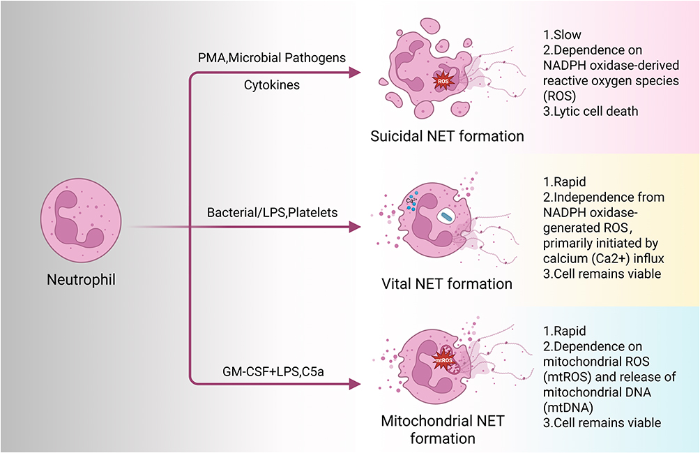 |
Figure 1 Three distinct pathways of NETosis and their key characteristics. Suicidal NET formation can be induced by factors such as PMA, microbial pathogens, and cytokines. It is characterized by a relatively slow response, dependence on NADPH oxidase-derived reactive oxygen species (ROS), and culminates in lytic cell death. Vital NET formation is triggered by stimuli including bacteria, lipopolysaccharide (LPS), and platelets. This pathway is characterized by a rapid response, Independence from NADPH oxidase-derived ROS, and is primarily initiated by calcium (Ca2⁺) influx, with neutrophils retaining their viability. Mitochondrial NET formation is induced by a combination of factors such as GM-CSF plus LPS or C5a. It features a rapid onset, dependence on mitochondrial ROS (mtROS), and the release of mitochondrial DNA, while the neutrophil remains viable. |
Suicidal NETosis
This canonical pathway represents a protracted process, typically taking hours to complete, and ultimately culminates in the lytic death of the neutrophil. It is generally induced by potent stimuli such as phorbol myristate acetate (PMA), certain microbial pathogens, or specific pro-inflammatory cytokines.16 The hallmark of suicidal NETosis involves a series of intricate intracellular events. A critical initial step is the robust generation of reactive oxygen species (ROS), primarily through the activation of NADPH oxidase.17 ROS production is essential for downstream events, including the translocation of neutrophil elastase (NE) and myeloperoxidase (MPO) from azurophilic granules into the nucleus. Within the nucleus, NE and MPO act synergistically to degrade histones, which, along with the action of protein-arginine deiminase 4 (PAD4), leads to extensive chromatin decondensation.18 PAD4, often activated by calcium influx, catalyzes the citrullination of specific arginine residues on histones (particularly histone H3), thereby loosening the chromatin structure and facilitating its unraveling. Subsequently, the nuclear envelope disintegrates, and the decondensed chromatin, now intertwined with various granular proteins, is extruded into the cytoplasm. Finally, the plasma membrane ruptures, releasing the mature NETs into the extracellular milieu.
Vital NETosis
In contrast to its suicidal counterpart, vital NETosis is a more rapid process, typically occurring within minutes, and notably, it does not necessarily lead to the demise of the neutrophil, which can remain viable and capable of performing other immune functions like phagocytosis.19 This pathway is often triggered by specific bacterial stimuli, such as lipopolysaccharide (LPS), or by activated platelets. The defining feature of vital NETosis is the selective extrusion of nuclear or mitochondrial DNA-containing vesicles without overt cell lysis. The neutrophil nucleus undergoes a unique transformation, becoming condensed and lobulated. Portions of the decondensed chromatin are then packaged into membrane-bound vesicles that bud from the nucleus. These vesicles are subsequently released into the extracellular space through exocytosis, where they eventually rupture to unleash their DNA content, forming NETs while preserving the cell’s integrity.20 Although PAD4, MPO, and NE may play roles, the exact molecular machinery differs from suicidal NETosis, often involving less pronounced histone citrullination and a distinct mechanism of chromatin externalization, and is typically independent of NADPH oxidase activity.
Mitochondrial NET Formation
mtNETs represents a distinct pathway of neutrophil extracellular trap release, characterized by the extrusion of mitochondrial DNA (mtDNA) and associated proteins into the extracellular space.21,22 This mechanism was initially reported in 2009, when it was observed that viable neutrophils could release mtDNA to form NETs.21 Unlike the classical “suicidal NETosis” which involves nuclear DNA release and neutrophil lysis, mtNETs are often considered a form of “vital NETosis”, as they typically occur without immediate cell death, allowing neutrophils to maintain their antimicrobial functions.14 This process was initially observed with neutrophils primed by granulocyte-macrophage colony-stimulating factor (GM-CSF) and subsequently stimulated with LPS or complement component C5a.21,22 Mechanistically, mtNET formation is largely dependent on the generation of mitochondrial ROS and can proceed independently of NADPH oxidase activity.23 Extracellular mtDNA, functioning as a potent DAMP, can activate immune responses, notably through TLR9, leading to pro-inflammatory cytokine production.24 While contributing to host defense against pathogens, dysregulated mtNET formation is implicated in various pathological conditions, including sterile inflammation, autoimmune diseases such as systemic lupus erythematosus, and sepsis, where elevated plasma mtDNA levels correlate with disease severity and poor clinical outcomes.25,26 Furthermore, mtNETs have been shown to promote tumor growth and metastasis by enhancing mitochondrial function in cancer cells.27,28
These distinct mechanisms underscore the adaptability of neutrophils in deploying NETs as a defense strategy, tailoring the response to the specific nature and intensity of the triggering stimulus.
Main Components and Biological Activities of NETs
The fundamental scaffold of NETs is comprised of DNA, which includes both nuclear DNA and, to a lesser extent, mitochondrial DNA. Wrapped around this DNA are highly cationic histones, particularly H2A, H2B, H3, and H4, which contribute to the structural integrity and antimicrobial properties of NETs.29 Beyond this nucleoprotein core, NETs are enriched with a diverse array of proteins derived from neutrophil granules and cytoplasm. These include key enzymes such as MPO, vital for the production of hypochlorous acid, a potent antimicrobial agent, and NE, a serine protease that degrades virulence factors of pathogens and contributes to chromatin decondensation. Other important granular proteins found within NETs are cathepsin G, lactoferrin, S100A8/A9, a major component of neutrophil cytosol with antimicrobial and pro-inflammatory properties, and defensins, which are small cationic peptides possessing direct antimicrobial effects.11,30
The intricate composition of NETs enables them to exert a wide range of biological activities, influencing both host defense and pathological processes. NETs physically ensnare and immobilize a broad spectrum of microorganisms, including bacteria, fungi, and viruses, with their antimicrobial proteins and enzymes directly killing or inhibiting trapped pathogens.11 Components of NETs, notably histones and DNA, act as DAMPs that activate innate immune cells and perpetuate inflammation, leading to tissue damage and amplified inflammatory responses.14 Furthermore, NETs serve as a potent pro-coagulant platform, promoting thrombosis by activating coagulation factors, binding to and activating platelets, and stabilizing fibrin clots, thus contributing to microvascular obstruction.31 Beyond pathogen elimination, NETs and their constituents, such as histones, exhibit direct cytotoxic effects on host cells like endothelial cells, leading to tissue damage and organ dysfunction, which is a key driver in various non-infectious diseases.32
Inducers of NETosis (Figure 2)
ROS Generation
Neutrophils, under various pathological conditions, induce NETs generation through the release of ROS.33 Increased levels of ROS lead to the upregulation of citrullinated histone H3 (citH3) expression, which ultimately triggers chromatin decondensation and induces NETosis.34 Furthermore, platelet-induced increase in mtROS is a critical step for NETs formation, and inhibition of mtROS significantly suppresses NETosis.35 Concurrently, NADPH oxidase (NOX) is one of the primary enzymes responsible for ROS generation in neutrophils, and consequently, targeting ROS signaling pathways through the inhibition of NOX activity, direct scavenging of ROS, or inhibition of downstream effectors like MPO, can effectively reduce NETosis.36
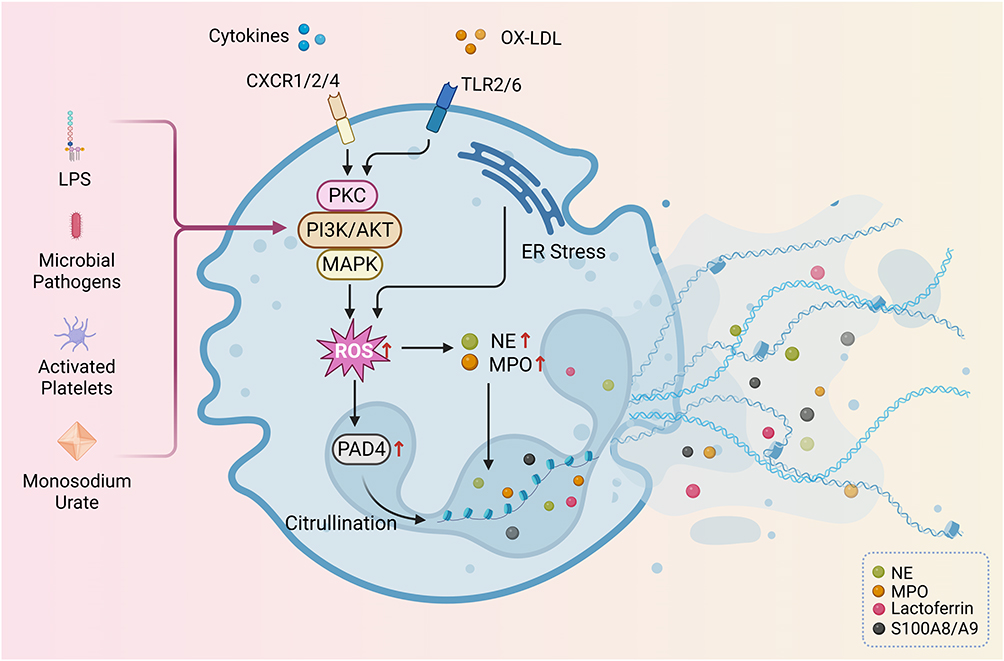 |
Figure 2 Key molecular mechanisms of NETosis. This schematic summarizes the common factors and intracellular signaling cascades driving NETosis. Diverse stimuli, including cytokines, Ox-LDL, microbial pathogens, LPS, activated platelets, and monosodium urate, activate distinct downstream signaling cascades, including PKC, PI3K/AKT, and MAPK, which converge to promote the generation of reactive oxygen species (ROS). Endoplasmic reticulum (ER) stress represents an additional source of ROS. As a central mediator, ROS orchestrates NETosis through two primary mechanisms: 1) facilitating the nuclear translocation of protein arginine deiminase 4 (PAD4), which catalyzes histone citrullination; and 2) stimulating the release of myeloperoxidase (MPO) and neutrophil elastase (NE) from cytoplasmic granules and their subsequent nuclear translocation. These events collectively drive chromatin decondensation and the release of neutrophil extracellular traps (NETs), whose core components include histones, NE, MPO, S100A8/A9, lactoferrin, and other granular/cytoplasmic proteins. ↑: increase or upregulation of the indicated process or marker. |
Endoplasmic Reticulum (ER) Stress
Endoplasmic reticulum (ER) stress is a crucial driver of NETosis, the formation and release of neutrophil extracellular traps, through several intricate molecular mechanisms.37 ER stress can promote the synergistic cleavage of gasdermin D (GSDMD) by caspase-4/11 and caspase-12, contributing to the induction of NETosis.38 Gαq/11 exacerbates NETosis by promoting the activation of the ER stress sensor IRE1α in neutrophils. This activation, coupled with the production of mitochondrial reactive oxygen species (mitoROS), further enhances NETosis.39
Inflammatory Factors
Inflammatory factors are crucial drivers of neutrophil extracellular trap (NETosis) formation, with excessive activation potentially exacerbating pathological damage.40 Interleukin-1β (IL-1β) directly induces NETosis, aggravating vascular inflammation and endothelial cell activation;41 in viral pneumonia, macrophage Hippo pathway activation upregulates NLRP3 and IL-1β, promoting NETosis.40 IL-8 induces NETs formation through the CXCR2-Src/ERK/p38 MAPK signaling pathway on neutrophils, exacerbating atherosclerosis.42 Furthermore, TNF-α and IL-17A are also confirmed inducers of NETosis.43 Inhibiting NF-κB activation significantly suppresses elevated MPO activity and NETosis, thereby mitigating inflammation.44
Lipid Abnormalities
Oxidized Low-Density Lipoprotein (Ox-LDL) can directly act on neutrophils, inducing time- and concentration-dependent ROS generation and subsequent NETs formation, and NOX inhibitors effectively prevent Ox-LDL-induced free radical production and NETs release.45 In terms of signaling pathways, Ox-LDL-induced NETosis requires the synergistic participation of TLR-2 and TLR-6, activating downstream PKC, Interleukin-1 Receptor-Associated Kinase (IRAKs), and Mitogen-Activated Protein Kinase (MAPK) pathways, which collectively drive NETosis.45 Notably, oxidized phospholipids (such as LPC and oxPAPC) within Ox-LDL are identified as the most potent inducers of NETs, playing a critical role in Ox-LDL-mediated NETs release.45
Others
NETosis can be triggered by a wide array of factors, reflecting the neutrophil’s role in diverse physiological and pathological contexts.23 Common stimuli include various microbial pathogens such as bacteria, fungi, and viruses, along with their molecular components like LPS and other pathogen-associated molecular patterns (PAMPs).46 Beyond infection, sterile inflammatory conditions also serve as potent inducers; these encompass endogenous molecules released during tissue damage (eg, DAMPs like histones), activated platelets, and certain crystalline structures like monosodium urate.12 PMA, a potent protein kinase C activator, is a widely used chemical inducer in experimental settings due to its ability to robustly trigger suicidal NETosis.47
Diabetic Kidney Disease-Specific Inducing Factors
In DKD, neutrophils are increasingly primed for NET formation, and various aspects of the diabetic milieu serve as potent stimuli, driving pathological NETosis through distinct molecular pathways48–50 (Figure 3).
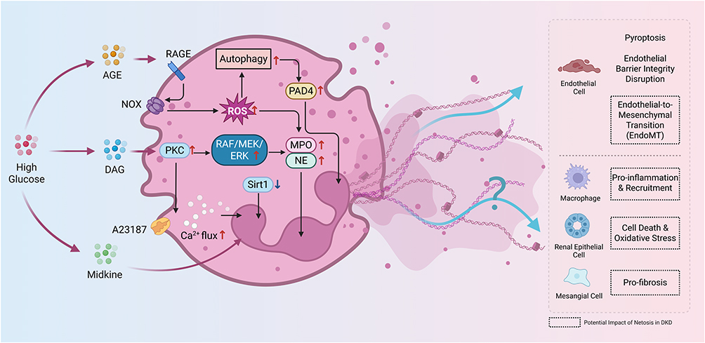 |
Figure 3 Molecular Mechanisms and Cellular Interactions of NETosis in Diabetic Kidney Disease (DKD). In DKD, chronic hyperglycemia drives NETosis through multiple pathways: 1) Accumulation of advanced glycation end products (AGEs) engages RAGE on neutrophils, activating NADPH oxidase to generate reactive oxygen species (ROS) and induce autophagy, thereby promoting NETosis. 2) Hyperglycemia-induced diacylglycerol (DAG) activates PKC, which in turn stimulates downstream pathways including RAF/MEK/ERK and calcium influx, collectively triggering NETosis. 3) High glucose transcriptionally upregulates midkine (MK), which promotes NETosis. 4) Hyperglycemia-induced downregulation of SIRT1 leads to increased NETosis. The deployed NETs subsequently amplify renal injury by targeting various cells. Direct evidence shows that NETs trigger pyroptosis in endothelial cells, compromising vascular integrity. Emerging hypotheses propose additional pathogenic roles, including promoting endothelial-to-mesenchymal transition (EndoMT), recruiting and activating pro-inflammatory macrophages, inducing cell death and oxidative stress in renal epithelial cells, and stimulating pro-fibrotic responses in mesangial cells. ↑: increase or upregulation of the indicated process or marker; ↓: inhibition or suppression of the indicated process or marker. |
Hyperglycemia
Previous studies have demonstrated that a hyperglycemic environment is a critical inducer of neutrophil NETosis in DKD.51,52 High glucose can induce NETosis through multiple pathways.
Advanced Glycation End Products
AGEs are crucial in the pathogenesis and progression of DKD, acting as key mediators that accelerate under hyperglycemic conditions.53,54 The kidney, as the primary organ for AGE clearance, is highly vulnerable to AGE-mediated damage.54 Notably, Corrected serum AGEs (corrected lgAGEs) are novel independent biomarkers for predicting adverse renal outcomes in type 2 DKD.53 Thus, AGEs play a pivotal role in the pathogenesis, progression, and prognosis of DKD.54,55
In the context of diabetes mellitus, AGEs, which accumulate due to chronic hyperglycemia, play a pivotal role in dysregulating neutrophil function and promoting NETosis through a multifaceted cascade involving oxidative stress and enzymatic activation. Specifically, AGEs bind to the receptor for AGEs (RAGE) on neutrophil surfaces, triggering the assembly and activation of the NADPH oxidase complex, which generates excessive ROS within these innate immune cells.56–58 This surge in ROS serves as a priming signal for autophagy, marked by elevated levels of the autophagosomal protein LC3II, which in turn directly drives the downstream activation of NETosis by facilitating chromatin decondensation and nuclear envelope rupture; concurrently, ROS upregulates the expression and activity of key NETosis effectors, including MPO and NE, whose production is directly mediated by the AGE-RAGE interaction.58,59 Consequently, heightened MPO and NE, in synergy with autophagy-mediated pathways, culminate in the extrusion of NETs laden with antimicrobial components, underscoring the integrated role of oxidative stress and autophagic flux in amplifying NET release under hyperglycemic conditions. Inhibition of NADPH oxidase or upstream autophagy pathways has been shown to attenuate this AGE-induced NETosis, highlighting the therapeutic potential of targeting these interconnected mechanisms in diabetic immunopathology.58
Protein Kinase C
In the diabetic environment, chronic hyperglycemia promotes tissue injury by inducing diacylglycerol (DAG) and subsequently activating the PKC pathway, and this phenomenon has also been observed in the kidneys of diabetic rats.60 Mechanistically, elevated extracellular glucose concentrations rapidly induce de novo synthesis of DAG from glycolytic intermediates, particularly through inhibition of glyceraldehyde-3-phosphate dehydrogenase (GAPDH), which diverts upstream metabolites such as dihydroxyacetone phosphate into alternative flux pathways, ultimately forming DAG via stepwise acylation; this process is further amplified by phospholipase D-mediated hydrolysis of phosphatidylcholine and contributions from oxidants or glycated products.61 This hyperglycemia-induced PKC activation can accelerate diabetic nephropathy and vasculopathy.62
PKC is a pivotal signaling molecule that promotes NETosis formation, playing a central role under various physiological and pharmacological stimuli.63 Human neutrophils contain multiple PKC isoforms, including conventional, novel, and atypical PKC (eg, α, βI, βII, δ, and ζ).63
Studies indicate that specific PKC isoforms mediate the occurrence of NETosis. For instance, PKCβ, PKCδ, and PKCζ are involved in the oxidative burst, spreading, and NETosis activated by calcium ionophore A23187, while only PKCβ is implicated in these functions activated by PMA.63 PKC activation typically induces downstream signaling pathways that lead to NETosis. A primary mechanism involves the activation of NOX to generate abundant ROS, ultimately prompting the release of NETs.64,65 For example, the compound EBC-1013 stimulates PKC-dependent neutrophil ROS induction and NETosis.64
PKC activity is subject to various regulations. The mutant peptide mB Box-97 inhibits NETosis, regardless of the NET inducer type, by directly binding to and inhibiting PKC activity, thereby reducing downstream NOX activation, ROS production, and NETosis.65 This suggests that PKC is a viable target for NETosis inhibition.65 Furthermore, the Apolipoprotein M/Sphingosine 1-phosphate (ApoM/S1P) axis, by activating S1P1 and S1P4 receptors, can attenuate PKCδ and PKCα/β activity, thereby suppressing NETosis.66 In diabetic wound management, human β-defensin-2 and ruboxistaurin have been identified as inhibitors of PKC-βII, suggesting a critical role for PKC-βII in NETosis.67,68 Additionally, the PKC/Raf/MEK/ERK signaling cascade functions concomitantly with the PI3K/AKT pathway to potentially regulate the augmented NETs formation induced by heightened glycolytic activity.69
In summary, PKC and its various isoforms serve as central hubs in the NETosis cascade, profoundly influencing the formation and release of neutrophil extracellular traps through regulating ROS generation and crosstalk with other signaling pathways.
Sirtuin 1
Sirtuin 1 (SIRT1), a prominent NAD+-dependent deacetylase, functions as a critical regulator of metabolic homeostasis, yet its expression is frequently downregulated in the progression of Diabetic Kidney Disease (DKD).70,71 In the renal milieu, this deficiency correlates with exacerbated cellular injury, whereas the restoration of SIRT1 activity—mediated by agents such as nicotinamide or melatonin—confers significant renoprotection.71,72 Specifically, SIRT1 signaling has been shown to attenuate glomerular sclerosis and podocyte injury by mitigating oxidative stress, inflammation, and fibrosis, thereby preserving the structural integrity of the kidney.70,72
Beyond its direct cytoprotective effects, SIRT1 exerts a pivotal regulatory influence on neutrophil biology, acting as a potent suppressor of NETosis. Mechanistic investigations reveal that SIRT1 inhibits the formation of NETs by targeting the citrullination of histone H3, a prerequisite step for chromatin decondensation.73 Consequently, pharmacological activation of SIRT1 restricts the release of NET components, whereas specific SIRT1 deficiency in neutrophils significantly exacerbates NETosis and promotes pathological outcomes.73
Diabetes-induced hyperglycemia triggers excessive NETosis, which disrupts the equilibrium between bone formation and resorption.74 Therapeutic interventions, such as the administration of GLP-1 receptor agonists like Liraglutide, have been demonstrated to upregulate SIRT1 expression, which subsequently inhibits the release of NET-associated markers, including myeloperoxidase (MPO) and citrullinated histone H3. The restoration of this SIRT1-NETosis axis effectively alleviates inflammation and reverses the diabetes-induced impairment of osteogenesis.74
Others
In DKD, the expression of Midkine (MK) is often significantly elevated due to the long-term hyperglycemic environment, primarily originating from high glucose stimulation inducing transcriptional activation of MK.75,76 As a novel biomarker, MK can predict the onset of microalbuminuria with high sensitivity and specificity.76 In vitro experiments revealed that under high-glucose conditions, MK stimulated the proliferation of bone marrow-derived neutrophils and drove the assembly of NETs by enhancing the expression of NE, MPO, and H3Cit. Knockdown of MK reduced NETs formation while simultaneously reversing renal tissue fibrosis and pathological damage.77
NETs Involvement in Diabetic Kidney Disease Progression
In DKD, NET formation can be induced by various other cell types, while NETs themselves reciprocally modulate the functions of these cells. Through their inherent pro-inflammatory, pro-coagulant, cytotoxic, and pro-fibrotic characteristics, NETs profoundly contribute to the inflammatory cascade, renal tissue damage, and ultimately, the fibrotic processes observed in DKD.9,52,78 This section will elaborate on how NETs influence the kidney’s resident immune and structural cells, thereby driving the pathophysiological progression of DKD (Figure 3).
Interaction Mechanisms Between NETs and Renal Endothelial Cells
Renal endothelial cells, crucial for maintaining kidney structure and function, are highly susceptible to damage in the diabetic milieu. NETs significantly contribute to this damage, leading to endothelial dysfunction, microvascular complications, and ultimately exacerbating DKD progression.
Endothelial Cell Damage and Dysfunction
In DKD, NETosis promotes glomerular endothelial cell injury by facilitating the deposition of NETs within the glomeruli, as observed in both DKD patients and diabetic mouse models (streptozotocin-induced or db/db), where elevated NETs levels correlate with worsened renal function and histopathological glomerulopathy.51,52 Mechanistically, NETs induce glomerular endothelial cell pyroptosis via a charge-dependent interaction, wherein the positively charged proteins (eg, histones) in NETs bind to the negatively charged glomerular endothelial cell surface, triggering cell membrane pore formation, dysregulation of membrane function-related genes, and upregulation of pyroptosis effectors including cleaved GSDMD and NLRP3.51 Concurrently, under high-glucose stress, NETs drive NLRP3 inflammasome activation in glomerular endothelial cells, culminating in IL-1β-mediated sterile inflammation that impairs endothelial barrier integrity, reduces endothelial nitric oxide synthase (eNOS) phosphorylation, and elevates soluble vascular cell adhesion molecule-1 (sVCAM-1) levels, thereby exacerbating endothelial dysfunction.52 Notably, degrading NETs with DNase I or inhibiting NETosis through PAD4 knockout/pharmacological blockade (eg, GSK484) significantly attenuates glomerular endothelial cell pyroptosis, inflammasome activation, and associated renal injury, underscoring NETosis as a key pathological contributor to DKD progression.51,52
Endothelial-to-Mesenchymal Transition (EndoMT/EndMT)
Evidence from other diseases indicates that excessive formation and impaired clearance of NETs lead to persistent exposure of endothelial cells to NETosis products, which in turn triggers EndoMT. Mechanistically, NET-associated elastase degrades the intercellular junction protein VE-cadherin, disrupting endothelial integrity and promoting the nuclear translocation of β-catenin, which subsequently induces EndoMT in endothelial cells.79 In diabetes, NETosis-derived NETs activate PAK2 via TLR9, leading to phosphorylation of Merlin/NF2 and inhibition of the Hippo-YAP pathway; activated YAP binds to SMAD2 and translocates into the nucleus, inducing EndoMT, which suppresses angiogenesis and delays wound healing.80 In addition, NETs were identified in glomeruli of lupus nephritis patients and correlated with the severity of proteinuria and glomerular EndoMT, further linking NETosis to vascular dysfunction in vivo.79 Complementarily, in a human blood–retinal barrier-on-a-chip model, activated neutrophils interacted with microglia to enhance NETosis, which upregulated EndoMT markers such as MMP2, Notch3, and ICAM1, alongside fibrosis-associated proteins including α-SMA, SMAD2, and SMAD3.81 Moreover, this process was accompanied by elevated inflammatory cytokines (ANG-2, IL-1α, IL-1β, and IL-6), suggesting that NETosis promotes EndoMT not only via junctional protein degradation and β-catenin signaling but also through the activation of pro-fibrotic and pro-inflammatory pathways.81 Although these evidence implicates NETosis in vascular remodeling and fibrosis, its role in DKD remains to be elucidated.
Interaction Mechanisms Between NETs and Other Cells
Macrophages
Macrophages are pivotal immune cells in the kidney, and their infiltration and activation significantly contribute to the progression of DKD.82 While a substantial body of evidence from other studies indicates that NETosis contributes to disease progression through its effects on macrophages, the precise role of this mechanism in DKD remains to be elucidated. Evidence from atherosclerotic studies indicates that NETosis promotes the release of IL-8 by activating the TLR9/NF-κB signaling pathway in macrophages.42 IL-8, as an important chemokine, directly attracts macrophages to the inflammatory area. Specific components within NETs also play crucial roles: for instance, abundant extracellular histones, particularly histone H4, are considered important components in mediating macrophage activation and IL-8 production.83
NETosis is a central driver of renal fibrosis, contributing to both its initiation and progression.84 In a unilateral ureteral obstruction model, GSDMD-dependent NETs induce α-SMA expression in macrophages by promoting NF-κB p65 nuclear translocation and activating the TGF-β1/Smad signaling pathway, thereby enhancing pro-inflammatory cytokine production and facilitating macrophage-to-myofibroblast transition (MMT).85 Consistently, in a folic acid-induced AKI-to-CKD (Acute Kidney Injury-to-Chronic Kidney Disease) transition model, GSDME-mediated pyroptosis of tubular epithelial cells triggered NETs release, which further promoted MMT.86
Renal Epithelial Cells
NETosis contributes to podocyte injury through multiple interconnected pathways. In models of chronic kidney injury, NET-derived proteases degrade slit diaphragm proteins such as nephrin and podocin, thereby impairing the filtration barrier. Moreover, excessive NET deposition induces actin cytoskeleton rearrangement, leading to foot process effacement and loss of podocyte differentiation markers.87 Furthermore, persistent NETosis exacerbates oxidative stress, increases ROS generation, and enhances the expression of adhesion molecules such as VCAM-1, further promoting podocyte dysfunction.52
NETosis also aggravates renal tubular epithelial cell (TEC) injury through several interrelated pathways. In DKD, CASP1 promotes the maturation of IL-1β and IL-18, which drive inflammation and amplify NETosis, excessive NET deposition induces oxidative stress and apoptosis in TECs, leading to impaired cellular proliferation and progressive tubular dysfunction.49 NETs could release additional cytotoxic components—DNA, histones, and proteases—that further damage TECs.88
Mesangial Cell and Fibroblasts
The inflammatory milieu instigated by NETs within diabetic glomeruli may contribute to mesangial cell activation and proliferation, thereby promoting mesangial expansion and extracellular matrix (ECM) accumulation.52,89 Furthermore, in obstructive nephropathy, NETs play a significant role in promoting renal fibrosis by influencing renal fibroblasts and their differentiation into myofibroblasts. Components of NETs, including DNA, histones, and granular proteins, can directly interact with fibroblasts, contributing to their activation90,91 and differentiation into myofibroblasts.85 Whether it plays a similar role in DKD, however, remains to be further investigated.
Potential Therapeutic Strategies for Targeting NETosis in DKD
Inhibiting NETosis Formation (Table 1)
Peptidylarginine Deiminase 4 Inhibitors
 |
Table 1 Emerging Therapeutic Agents Targeting Diabetes-Induced NETosis |
PAD4 inhibitors, such as Cl-amidine, GSK484, and the novel isoform-selective compound JBI-589, have emerged as promising strategies to suppress NET formation. Mechanistically, these agents prevent histone citrullination and chromatin decondensation, thereby blocking NETosis. Cl-amidine has been shown to reduce NET formation and accelerate wound healing in diabetic mice, highlighting its therapeutic potential in hyperglycemia-associated tissue injury.92 Similarly, both Cl-amidine and GSK484 effectively suppress NETosis and ameliorate renal or vascular injury in multiple preclinical models, including lupus nephritis, ischemia-reperfusion injury, and acute kidney injury.9 More recently, JBI-589 was identified as a potent, orally available, PAD4-selective inhibitor that suppresses histone H3 citrullination and NET release in human neutrophils, while in mouse models it attenuates inflammation, reduces organ damage, and enhances the efficacy of immune checkpoint blockade in cancer.93 In the context of DKD, accumulating evidence demonstrates that PAD4 deletion or NET degradation by DNase I alleviates glomerular injury and endothelial pyroptosis in diabetic mice, underscoring NETs as critical drivers of DKD progression.51 Although these findings support PAD4 inhibition as a promising therapeutic avenue, most studies remain at the preclinical or early translational stage, and challenges regarding inhibitor selectivity, long-term safety, and the balance between reducing sterile inflammation and preserving antimicrobial defense remain to be addressed.9
NADPH Oxidase Inhibitors
NOX–derived ROS are increasingly recognized as critical upstream signals driving NOX-dependent NETosis and diabetic complications. Pharmacological inhibition of NOX1/4 with setanaxib (GKT137831) suppresses ROS production, thereby attenuating oxidative stress and potentially reducing NET formation. In preclinical studies, GKT137831 demonstrated renoprotective effects in diabetic mice, lowering albuminuria and renal inflammation even in established nephropathy and vascular disease, supporting NOX as a key source of pathogenic ROS in diabetic kidney disease.94 Translationally, setanaxib has progressed into clinical evaluation, with a multicenter phase 2 trial recruiting patients with type 1 diabetes and persistent albuminuria to assess efficacy and safety.95 While these studies highlight encouraging reno- and vasoprotective effects, current clinical protocols do not yet include NET-related endpoints, and long-term renal outcomes remain to be clarified. Therefore, future trials are warranted to directly assess the role of NOX inhibition in NETosis and to determine whether targeting NOX1/4 can translate into durable clinical benefit in diabetic kidney disease.
Myeloperoxidase Inhibitors
MPO inhibitors such as AZD5904 block MPO-mediated hypochlorous acid (HOCl) generation, thereby attenuating chromatin decondensation and NET stability. Experimental studies have shown that MPO inhibition suppresses extracellular neutrophil activity and alleviates NET-associated tissue injury, including the prevention and reversal of high-fat diet–induced microvascular insulin resistance in rodent models.96 However, in autoimmune settings such as the NOD mouse model of type 1 diabetes, MPO or neutrophil elastase inhibition failed to prevent disease progression, highlighting disease context–dependent efficacy.97 Translationally, MPO inhibition remains at an early stage, with considerations including renal clearance and potential drug–drug interactions requiring further evaluation.
Neutrophil Elastase Inhibitors
NE is a key serine protease that plays a crucial role in inflammation and NETosis. Inhibition of NE has shown promise as a therapeutic strategy in various inflammatory conditions, including DKD. NE exerts its effects by disrupting endothelial cell integrity and promoting NET release, which in turn contributes to tissue injury and vascular permeability. In vitro and in vivo studies have demonstrated that NE inhibition, particularly by drugs such as sivelestat, can prevent the formation of NETs and reduce endothelial permeability, which is often observed in conditions like sepsis and acute respiratory distress syndrome (ARDS).98 Furthermore, NE inhibitors have been proposed as potential treatments for CKD and its progression to ESRD, by mitigating inflammation and oxidative stress, which are pivotal in the disease’s pathogenesis.99 In the context of DKD, NE is implicated in the worsening of retinal vascular permeability and endothelial damage, as seen in animal models of diabetes. This process is partly mediated by IL-17, which regulates NE expression and contributes to the pathophysiology of diabetic retinopathy.100 Therefore, targeting NE could offer a novel approach to limit the inflammatory cascade and protect kidney function in DKD patients.
TLR9 Inhibitor
Targeting the NET–DNA–TLR9 axis has emerged as a promising therapeutic strategy, with antimalarial agents such as chloroquine (CQ) and hydroxychloroquine (HCQ) showing dual functions in both inhibiting autophagy and downregulating NET formation. Mechanistically, CQ and HCQ directly inhibit PAD4 activity, preventing histone citrullination and subsequent chromatin decondensation, thereby suppressing NET release.113 In addition, HCQ has been shown to block intracellular TLR9 signaling, reducing PAD4 and Rac2 expression, and consequently attenuating NET-mediated inflammatory injury in hepatic ischemia/reperfusion models.101 The TLR9 pathway is increasingly recognized in nephrology, where nucleotide-sensing TLR9 and TLR7 contribute to the progression of IgA nephropathy, suggesting that HCQ-mediated inhibition of TLRs may also provide renoprotective benefits by modulating glomerular and tubular inflammation.102
Promoting NETs Degradation
DNase I
Degradation of the extracellular DNA scaffold of NETs represents another therapeutic strategy, with DNase I and its recombinant form dornase alfa facilitating the clearance of NETs. By directly digesting NET-derived chromatin structures, these agents accelerate NET removal and thereby limit downstream inflammatory signaling. In diabetic kidney disease models, treatment with DNase I significantly attenuated glomerular injury and glomerular endothelial cell damage, indicating a protective role against NET-driven microvascular injury.51 More broadly, preclinical evidence across multiple disease contexts supports the capacity of DNase I–mediated NET degradation to reduce disease severity, highlighting its potential as a translational approach in renal and systemic inflammatory disorders.114
Classical Antidiabetic Drugs
Metformin
Metformin, beyond its glucose-lowering properties, has been shown to modulate NETosis through activation of AMPK and subsequent inhibition of the PKC–NADPH oxidase axis, thereby reducing NOX-dependent ROS generation and NETosis. Clinical and experimental evidence indicates that metformin lowers circulating NET components such as neutrophil elastase, proteinase-3, and double-stranded DNA in patients with type 2 diabetes, independently of glycemic control, partly through suppression of PKC-βII membrane translocation and NADPH oxidase activation in neutrophils.103 Consistently, in diabetic bone defect models, metformin reduced high glucose–induced NET formation and reversed NET-driven impairment of osteogenesis.104 However, caution is warranted, as preclinical studies have revealed that in the context of iron overload and acute kidney injury (AKI), metformin may aggravate renal injury by promoting NETosis through a ferroptosis–iron–NET axis.105 These findings suggest that while metformin may alleviate NET-mediated oxidative stress and endothelial injury—pathways also implicated in DKD—direct evidence targeting NETs in human DKD remains lacking, and the current evidence level should be interpreted with caution.
Glucagon-Like Peptide-1 Receptor Agonists (GLP-1RAs)
Glucagon-like peptide-1 receptor agonists (GLP-1RAs), such as liraglutide and semaglutide, have been shown to influence neutrophil function beyond their established metabolic and cardiovascular benefits. Preclinical studies demonstrated that liraglutide reduces NETosis by attenuating ROS generation, thereby lowering circulating NET markers including MPO, NE, and double-stranded DNA in tumor-bearing mice.106 In diabetic bone metabolism models, liraglutide similarly decreased NET-associated proteins (cit-H3, MPO, NE, PAD4) and restored osteogenesis through a SIRT1-dependent mechanism, suggesting a role of the SIRT1 stress-response pathway in NET regulation.74 Moreover, semaglutide was shown to modulate pro-inflammatory neutrophil phenotypes in patients with cardiovascular disease, attenuating oxidative burst capacity and reducing neutrophil adhesion to coronary endothelial cells, thereby potentially limiting endothelial inflammation.107 Although GLP-1RAs have well-established renal protective effects in clinical trials of DKD,108 direct evidence linking their action to suppression of NETosis in human DKD is lacking. At present, the relationship remains biologically plausible and supported by preclinical signals, but requires validation in kidney-specific contexts.
Insulin
In vitro studies suggest that insulin at physiological concentrations can delay NETosis by modulating neutrophil activation pathways, reducing histone citrullination and shifting antimicrobial responses toward enhanced bacterial clearance.109 However, recent data presented at the 2025 ADA meeting indicate that certain commercial insulin formulations containing phenolic preservatives may paradoxically induce NETosis and amplify local inflammatory responses, as shown by both in vitro and in vivo evidence.110 These findings highlight a potential formulation-dependent divergence in insulin’s effects on neutrophils, and the clinical implications of this phenomenon remain to be clarified.
Sodium–Glucose Cotransporter 2 (SGLT2) Inhibitors
Direct evidence on the effects of SGLT2 inhibitors on NET formation remains scarce and inconsistent. In special populations, such as patients with glycogen storage disease type 1b, empagliflozin was shown to restore neutrophil counts and functions, including phagocytosis and chemotaxis, but NET production was only partially improved and did not consistently reach levels seen in healthy controls. Importantly, exploratory studies are currently underway to investigate whether SGLT2 inhibitors, alone or in combination with GLP-1 receptor agonists, can reduce NETosis in broader clinical contexts. While SGLT2 inhibitors such as dapagliflozin and empagliflozin provide robust renal and cardiovascular protection in patients with DKD, a causal link between their clinical benefits and modulation of the NET axis has yet to be established.
Conclusion and Future Perspectives
DKD represents a significant global health burden, persistently advancing to end-stage renal disease and substantially increasing cardiovascular mortality. Despite current therapeutic advancements, a considerable proportion of patients continue to experience disease progression, underscoring the urgent need for novel interventions. This review has meticulously detailed the multifaceted involvement of NETs in the intricate pathophysiology of DKD, from their dysregulated formation in the diabetic milieu to their detrimental interactions with renal resident cells—macrophages, endothelial cells, and epithelial cells—driving inflammation, oxidative stress, thrombotic events, and ultimately, tissue fibrosis. The comprehensive evidence presented herein firmly establishes NETs as a crucial orchestrator of renal injury in DKD, thereby positioning them as a highly promising, yet largely untapped, therapeutic avenue.
Moving forward, applying our understanding of NETs pathology to develop effective clinical strategies for DKD will require careful consideration and further research. The development of highly specific NETs inhibitors, aiming to prevent their detrimental effects while preserving essential host immunity, is paramount.112 This includes targeting key enzymes involved in NETs formation, such as MPO, NE, and PAD4, or pathways leading to ROS production. Simultaneously, strategies to enhance NETs degradation, for instance, via exogenous DNase I administration, offer a direct means to mitigate NET-mediated damage.78 Furthermore, the identification and validation of robust NET biomarkers in both circulation and renal tissue are essential for accurate diagnosis, risk stratification, and effective monitoring of therapeutic responses.115 Given the multifactorial nature of DKD, combination therapies integrating NETs-targeting agents with standard treatments, including the observed NET-modulating effects of existing antidiabetic medications like metformin and SGLT2 inhibitors, may offer synergistic benefits. Ultimately, rigorous, well-designed clinical trials are indispensable to ascertain the safety and efficacy of NETs-targeted strategies, paving the way for personalized therapeutic options that can significantly alter the trajectory of DKD and improve patient outcomes.
Author Contributions
All authors made a significant contribution to the work reported, whether that is in the conception, study design, execution, acquisition of data, analysis and interpretation, or in all these areas; took part in drafting, revising or critically reviewing the article; gave final approval of the version to be published; have agreed on the journal to which the article has been submitted; and agree to be accountable for all aspects of the work.
Funding
This research was supported by the General Program of National Natural Science Foundation of China (No.82374201) and Zhejiang Provincial Public Welfare Research Project (grant. LGF22H050012).
Disclosure
The authors declare that they have no competing interests.
References
1. Shen Z, Fang Y, Xing T, Wang F. Diabetic nephropathy: from pathophysiology to treatment. J Diab Res. 2017;2017:2379432. doi:10.1155/2017/2379432
2. Sagoo MK, Gnudi L. Diabetic nephropathy: an overview. Meth Mol Biol. 2020;2067:3–18.
3. Ong KL, Stafford LK, McLaughlin SA, et al. Global, regional, and national burden of diabetes from 1990 to 2021, with projections of prevalence to 2050: a systematic analysis for the Global Burden of Disease Study 2021. Lancet. 2023;402:203–234.
4. Scilletta S, Di Marco M, Miano N, et al. Update on diabetic kidney disease (DKD): focus on non-albuminuric DKD and cardiovascular risk. Biomolecules. 2023;13(5):752. doi:10.3390/biom13050752
5. Husain S. Role of podocyte in kidney disease. Front Biosci. 2024;29:250. doi:10.31083/j.fbl2907250
6. Tang J, Liu F, Cooper ME, Chai Z. Renal fibrosis as a hallmark of diabetic kidney disease: potential role of targeting transforming growth factor-beta (TGF-β) and related molecules. Expert Opin Ther Targets. 2022;26:721–738. doi:10.1080/14728222.2022.2133698
7. Alicic RZ, Rooney MT, Tuttle KR. Diabetic kidney disease. Clin J Am Soc Nephrol. 2017;12:2032–2045. doi:10.2215/CJN.11491116
8. Zhang Y, Li C, Jiang Q, et al. Unraveling neutrophil extracellular traps: a new frontier in kidney disease therapy. Life Sci. 2025;379:123892. doi:10.1016/j.lfs.2025.123892
9. Juha M, Molnár A, Jakus Z, Ledó N. NETosis: an emerging therapeutic target in renal diseases. Front Immunol. 2023;14. doi:10.3389/fimmu.2023.1253667
10. Njeim R, Ghadieh HE, Nahlawi MI, et al. NETosis contributes to the pathogenesis of diabetic kidney disease: a proposed mechanistic pathway: PO0678. J Am Soc Nephrol. 2021;32(10S):245. doi:10.1681/ASN.20213210S1245d
11. Meier A, Sakoulas G, Nizet V, Ulloa ER. Neutrophil extracellular traps: an emerging therapeutic target to improve infectious disease outcomes. J Infect Dis. 2024;230:514–521. doi:10.1093/infdis/jiae252
12. Monsalve DM, Acosta-Ampudia Y, Acosta NG, et al. NETosis: a key player in autoimmunity, COVID-19, and long COVID. J Transl Autoimmun. 2025;10:100280. doi:10.1016/j.jtauto.2025.100280
13. Li Y, Cao Z, Liu J, et al. Current perspectives and trends of neutrophil extracellular traps in organ fibrosis: a bibliometric and visualization study. Front Immunol. 2025;16:1508909. doi:10.3389/fimmu.2025.1508909
14. Ji Y-M, Li T, Qin Y-H, et al. Neutrophil extracellular traps (NETs) in sterile inflammatory diseases. J Inflamm Res. 2025;18:7989–8004. doi:10.2147/JIR.S526936
15. Tan C, Aziz M, Wang P. The vitals of NETs. J Leukoc Biol. 2021;110:797–808. doi:10.1002/JLB.3RU0620-375R
16. Pai D, Gruber M, Pfaehler S-M, et al. Polymorphonuclear cell chemotaxis and suicidal NETosis: simultaneous observation using fMLP, PMA, H7, and live cell imaging. J Immunol Res. 2020;2020(1):1415947. doi:10.1155/2020/1415947
17. Korba-Mikołajczyk A, Służalska KD, Kasperkiewicz P. Exploring the involvement of serine proteases in neutrophil extracellular traps: a review of mechanisms and implications. Cell Death Dis. 2025;16:535. doi:10.1038/s41419-025-07857-w
18. Ortega-Zapero M, Gomez-Bris R, Pascual-Laguna I, Saez A, Gonzalez-Granado JM. Neutrophils and NETs in pathophysiology and treatment of inflammatory bowel disease. Int J Mol Sci. 2025;26:7098. doi:10.3390/ijms26157098
19. Guillotin F, Fortier M, Portes M, et al. Vital NETosis vs. suicidal NETosis during normal pregnancy and preeclampsia. Front Cell Dev Biol. 2022;10:1099038. doi:10.3389/fcell.2022.1099038
20. Yipp BG, Kubes P. NETosis: how vital is it? Blood. 2013;122:2784–2794. doi:10.1182/blood-2013-04-457671
21. Yousefi S, Mihalache C, Kozlowski E, Schmid I, Simon HU. Viable neutrophils release mitochondrial DNA to form neutrophil extracellular traps. Cell Death Differ. 2009;16:1438–1444. doi:10.1038/cdd.2009.96
22. Stoimenou M, Tzoros G, Skendros P, Chrysanthopoulou A. Methods for the assessment of NET formation: from neutrophil biology to translational research. Int J Mol Sci. 2022;23:15823. doi:10.3390/ijms232415823
23. Huang J, Hong W, Wan M, Zheng L. Molecular mechanisms and therapeutic target of NETosis in diseases. MedComm. 2022;3:e162. doi:10.1002/mco2.162
24. Pastorek M, Konečná B, Janko J, et al. Mitochondria-induced formation of neutrophil extracellular traps is enhanced in the elderly via Toll-like receptor 9. J Leukoc Biol. 2023;114(6):651–665. doi:10.1093/jleuko/qiad101
25. Papayannopoulos V. Neutrophil extracellular traps in immunity and disease. Nat Rev Immunol. 2018;18:134–147. doi:10.1038/nri.2017.105
26. Pignataro G, Gemma S, Petrucci M, et al. Unraveling NETs in sepsis: from cellular mechanisms to clinical relevance. Int J Mol Sci. 2025;26(15):7464. doi:10.3390/ijms26157464
27. Yazdani HO, Roy E, Comerci AJ, et al. Neutrophil extracellular traps drive mitochondrial homeostasis in tumors to augment growth. Cancer Res. 2019;79(21):5626–5639. doi:10.1158/0008-5472.CAN-19-0800
28. Zhang Z, Niu R, Zhao L, Wang Y, Liu G. Mechanisms of neutrophil extracellular trap formation and regulation in cancers. Int J Mol Sci. 2023;24:10265. doi:10.3390/ijms241210265
29. Wang Y, Du C, Zhang Y, Zhu L. Composition and function of neutrophil extracellular traps. Biomolecules. 2024;14:416. doi:10.3390/biom14040416
30. Sprenkeler EGG, Zandstra J, Van Kleef ND, et al. S100A8/A9 is a marker for the release of neutrophil extracellular traps and induces neutrophil activation. Cells. 2022;11:236. doi:10.3390/cells11020236
31. Döring Y, Libby P, Soehnlein O. Neutrophil extracellular traps participate in cardiovascular diseases - recent experimental and clinical insights. Circ Res. 2020;126:1228–1241. doi:10.1161/CIRCRESAHA.120.315931
32. Jorge-Rosas F, Díaz-Godínez C, García-Aguirre S, Martínez-Calvillo S, Carrero JC. Entamoeba histolytica-induced NETs are highly cytotoxic on hepatic and colonic cells due to serine proteases and myeloperoxidase activities. Front Immunol. 2024;15:1493946. doi:10.3389/fimmu.2024.1493946
33. Chavoshinezhad S, Beirami E, Izadpanah E. Neutrophils and NETosis in Alzheimer’s disease: unraveling pathogenic mechanisms and novel therapeutic targets. Biomed Pharmacother. 2025;192:118568. doi:10.1016/j.biopha.2025.118568
34. Li X, Ju Y, Xu C, et al. Cancer cell-derived large extracellular vesicles promote venous thromboembolism by activating NETosis through delivering CYBA. Adv Sci. 2025;12(37):e07867. doi:10.1002/advs.202507867
35. Wu X, Jing H, Jiang T, et al. Platelet-induced NET in liver cirrhosis: mitochondrial ROS -mediated NETosis and its contribution to the hypercoagulable state. Liver Int. 2025;45:e70223. doi:10.1111/liv.70223
36. Zambrano F, Uribe P, Schulz M, et al. Antioxidants as modulators of NETosis: mechanisms, evidence, and therapeutic potential. Int J Mol Sci. 2025;26(11):5272. doi:10.3390/ijms26115272
37. Tumurkhuu G, Laguna DE, Moore RE, et al. Neutrophils contribute to ER stress in lung epithelial cells in the pristane-induced diffuse alveolar hemorrhage mouse model. Front Immunol. 2022;13:790043. doi:10.3389/fimmu.2022.790043
38. Chen H, Yang K, Zhang S, et al. Caspase-12 exhibits non-redundant functions in response to endoplasmic reticulum stress to promote GSDMD-mediated NETosis, leading to thoracic aortic dissection. Transl Res. 2025;278:48–60. doi:10.1016/j.trsl.2025.02.005
39. Xiang Q, Tian Y, Yang K, Du Y, Xie J. Gαq/11 aggravates acute lung injury in mice by promoting endoplasmic reticulum stress-mediated NETosis. Mol Med. 2025;31:67. doi:10.1186/s10020-025-01118-4
40. Luo B, Wang X, Lin J, et al. Hippo pathway and NLRP3-driven NETosis in macrophages: mechanisms of viral pneumoniaaggravation. Cell Death Discov. 2025;11(1):323. doi:10.1038/s41420-025-02556-z
41. Lin S, Zhu P, Jiang L, et al. Neutrophil extracellular traps induced by IL-1β promote endothelial dysfunction and aggravate limb ischemia. Hypertens Res. 2024;47(6):1654–1667. doi:10.1038/s41440-024-01661-3
42. An Z, Li J, Yu J, et al. Neutrophil extracellular traps induced by IL-8 aggravate atherosclerosis via activation NF-κB signaling in macrophages. Cell Cycle. 2019;18(21):2928–2938. doi:10.1080/15384101.2019.1662678
43. Khandpur R, Carmona-Rivera C, Vivekanandan-Giri A, et al. NETs are a source of citrullinated autoantigens and stimulate inflammatory responses in rheumatoid arthritis. Sci Transl Med. 2013;5(178):178ra40. doi:10.1126/scitranslmed.3005580
44. Wang M, Gao F. Suppression of nF-κB by ro 106-9920 alleviates ischemia/reperfusion-induced renal dysfunction and inflammation via modulation of neutrophil extracellular trap formation in acute kidney injury mice. Ren Fail. 2025;47:2545983. doi:10.1080/0886022X.2025.2545983
45. Awasthi D, Nagarkoti S, Kumar A, et al. Oxidized LDL induced extracellular trap formation in human neutrophils via TLR-PKC-IRAK-MAPK and NADPH-oxidase activation. Free Radic Biol Med. 2016;93:190–203. doi:10.1016/j.freeradbiomed.2016.01.004
46. Bhatia N, George B, Masih D, Khan MMU, Malik P. Mechanistic insights into PAMP and DAMP driven activation of NETosis in autoimmune disorders. Int Immunopharmacol. 2025;162:115149. doi:10.1016/j.intimp.2025.115149
47. Inozemtsev V, Sergunova V, Vorobjeva N, et al. Stages of NETosis development upon stimulation of neutrophils with activators of different types. Int J Mol Sci. 2023;24(15):12355. doi:10.3390/ijms241512355
48. Menegazzo L, Ciciliot S, Poncina N, et al. NETosis is induced by high glucose and associated with type 2 diabetes. Acta Diabetol. 2015;52(3):497–503. doi:10.1007/s00592-014-0676-x
49. Liang Y, Lin J, Huang B, et al. NET-related gene as potential diagnostic biomarkers for diabetic tubulointerstitial injury. J Diab Res. 2024;2024(1):4815488. doi:10.1155/2024/4815488
50. Zhu Y, Xia X, He Q, et al. Diabetes-associated neutrophil NETosis: pathogenesis and interventional target of diabetic complications. Front Endocrinol. 2023;14:1202463. doi:10.3389/fendo.2023.1202463
51. Zheng F, Ma L, Li X, et al. Neutrophil extracellular traps induce glomerular endothelial cell dysfunction and pyroptosis in diabetic kidney disease. Diabetes. 2022;71(12):2739–2750. doi:10.2337/db22-0153
52. Gupta A, Singh K, Fatima S, et al. Neutrophil extracellular traps promote NLRP3 inflammasome activation and glomerular endothelial dysfunction in diabetic kidney disease. Nutrients. 2022;14(14):2965. doi:10.3390/nu14142965
53. Wang Z-H, Zhang Z-J, Wang Y-F, et al. Serum advanced glycation end products as a putative biomarker in Type2 DKD patients’ prognosis. Front Physiol. 2025;16:1541198. doi:10.3389/fphys.2025.1541198
54. Fotheringham AK, Gallo LA, Borg DJ, Forbes JM. Advanced glycation end products (AGEs) and chronic kidney disease: does the modern diet AGE the kidney? Nutrients. 2022;14:2675. doi:10.3390/nu14132675
55. Rabbani N, Thornalley PJ. Advanced glycation end products in the pathogenesis of chronic kidney disease. Kidney Int. 2018;93:803–813. doi:10.1016/j.kint.2017.11.034
56. Koike S, Yano S, Tanaka S, et al. Advanced glycation end-products induce apoptosis of vascular smooth muscle cells: a mechanism for vascular calcification. Int J Mol Sci. 2016;17(9):1567. doi:10.3390/ijms17091567
57. Koike S, Yano S, Tanaka S, et al. Advanced glycation end-products stimulate apoptosis and calcium deposition of vascular smooth muscle cells: functional roles of NAD(P)H oxidase. Atherosclerosis. 2017;263:e68. doi:10.1016/j.atherosclerosis.2017.06.228
58. Farhan A, Hassan G, Ali SHL, et al. Spontaneous NETosis in diabetes: a role of hyperglycemia mediated ROS and autophagy. Front Med. 2023;10:1076690. doi:10.3389/fmed.2023.1076690
59. Lu H, Xu S, Liang X, et al. Advanced glycated end products alter neutrophil effect on regulation of CD4+ T cell differentiation through induction of myeloperoxidase and neutrophil elastase activities. Inflammation. 2019;42(2):559–571. doi:10.1007/s10753-018-0913-5
60. Derubertis FR, Craven PA. Activation of protein kinase C in glomerular cells in diabetes. Mechanisms and potential links to the pathogenesis of diabetic glomerulopathy. Diabetes. 1994;43:1–8. doi:10.2337/diab.43.1.1
61. Noh H, King GL. The role of protein kinase C activation in diabetic nephropathy. Kidney Int Suppl. 2007;S49–53. doi:10.1038/sj.ki.5002386
62. Hempel A, Maasch C, Heintze U, et al. High glucose concentrations increase endothelial cell permeability via activation of protein kinase Cα. Circul Res. 1997;81(3):363–371. doi:10.1161/01.RES.81.3.363
63. Vorobjeva N, Dagil Y, Pashenkov M, Pinegin B, Chernyak B. Protein kinase C isoforms mediate the formation of neutrophil extracellular traps. Int Immunopharmacol. 2023;114:109448. doi:10.1016/j.intimp.2022.109448
64. Powell LC, Cullen JK, Boyle GM, et al. Topical, immunomodulatory epoxy-tiglianes induce biofilm disruption and healing in acute and chronic skin wounds. Sci Transl Med. 2022;14(662):eabn3758. doi:10.1126/scitranslmed.abn3758
65. More KR, Devaraj A, Robledo-Avila FH, et al. High-mobility group protein B1 derived mutant peptide mB Box-97 inhibits the formation of neutrophil extracellular traps. Front Immunol. 2025;16:1565252. doi:10.3389/fimmu.2025.1565252
66. Kurano M, Uranbileg B, Yatomi Y. Apolipoprotein M bound sphingosine 1-phosphate suppresses NETosis through activating S1P1 and S1P4. Biomed Pharmacother. 2023;166:115400. doi:10.1016/j.biopha.2023.115400
67. Sanapalli V, Sigalapalli DK, Shaik AB, Bhandare RR, Sanapalli BKR. Computational elucidation of human β-Defensin-2 as a dual inhibitor of MMP-9 and PKC-βII for diabetic wound management. ACS Omega. 2025;10:3575–3584. doi:10.1021/acsomega.4c08292
68. Das SK, Yuan YF, Li MQ. Specific PKC βII inhibitor: one stone two birds in the treatment of diabetic foot ulcers. Biosci Rep. 2018;38:BSR20171459. doi:10.1042/BSR20171459
69. Wei H, Xia D, Li L, et al. Baicalin modulates glycolysis via the PKC/Raf/MEK/ERK and PI3K/AKT signaling pathways to attenuate IFN-I-induced neutrophil NETosis. Mediators Inflamm. 2025;2025:8822728. doi:10.1155/mi/8822728
70. Yacoub R, Lee K, He JC. The role of SIRT1 in diabetic kidney disease. Front Endocrinol. 2014;5:166. doi:10.3389/fendo.2014.00166
71. Yang Y, Huang J, Xie L, et al. Nicotinamide protects against diabetic kidney disease through regulation of Sirt1. Endocrine. 2024;85(2):638–648. doi:10.1007/s12020-024-03721-7
72. Zhang Y, Wang Y, Yang J, Liu G. Melatonin protects against diabetic kidney disease via the SIRT1/NLRP3 signalling pathway. Nephrology. 2025;30:e70073. doi:10.1111/nep.70073
73. Yu W, Wang Z, Dai P, et al. The activation of SIRT1 by resveratrol reduces breast cancer metastasis to lung through inhibiting neutrophil extracellular traps. J Drug Target. 2023;31(9):962–975. doi:10.1080/1061186X.2023.2265585
74. Zhong S, Huang L, Lin T, et al. The glucagon-like peptide-1 (GLP-1) receptor agonist liraglutide regulates sirtuin-1-mediated neutrophil extracellular traps to improve diabetes-induced bone metabolism imbalance. Iran J Pharm Res. 2024;23(1):e148139. doi:10.5812/ijpr-148139
75. Kosugi T, Yuzawa Y, Sato W, et al. Growth factor midkine is involved in the pathogenesis of diabetic nephropathy. Am J Pathol. 2006;168(1):9–19. doi:10.2353/ajpath.2006.050488
76. Metwalley KA, Farghaly HS, Gabri MF, et al. Midkine: utility as a predictor of early diabetic nephropathy in children with type 1 diabetes mellitus. J Clin Res Pediatric Endocrinol. 2021;13(3):293–299. doi:10.4274/jcrpe.galenos.2021.2020.0303
77. Liu G, Ren X, Li Y, Li H. Midkine promotes kidney injury in diabetic kidney disease by increasing neutrophil extracellular traps formation. Ann Transl Med. 2022;10(12):693. doi:10.21037/atm-22-2382
78. Nakazawa D, Masuda S, Nishibata Y, et al. Neutrophils and NETs in kidney disease. Nat Rev Nephrol. 2025;21(6):383–398. doi:10.1038/s41581-025-00944-3
79. Pieterse E, Rother N, Garsen M, et al. Neutrophil extracellular traps drive endothelial-to-mesenchymal transition. Arterioscler Thromb Vasc Biol. 2017;37(7):1371–1379. doi:10.1161/ATVBAHA.117.309002
80. Yang S, Wang S, Chen L, et al. Neutrophil extracellular traps delay diabetic wound healing by inducing endothelial-to-mesenchymal transition via the Hippo pathway. Int J Biol Sci. 2023;19(1):347–361. doi:10.7150/ijbs.78046
81. Yassin S, Monickaraj F, Aladina RK, Smoake J, Das A. Neutrophil-Microglial interaction induces Fibrosis and EndoMT in a 3D Blood-Retinal Barrier-on-a-chip model. Invest. Ophthalmol. Vis. Sci. 2025;66:889.
82. Peng Q-Y, An Y, Jiang -Z-Z, Xu Y. The role of immune cells in DKD: mechanisms and targeted therapies. J Inflamm Res. 2024;17:2103–2118. doi:10.2147/JIR.S457526
83. Nakazawa D, Shida H, Kusunoki Y, et al. The responses of macrophages in interaction with neutrophils that undergo NETosis. J Autoimmun. 2016;67:19–28. doi:10.1016/j.jaut.2015.08.018
84. Salazar-Gonzalez H, Zepeda-Hernandez A, Melo Z, Saavedra-Mayorga DE, Echavarria R. Neutrophil extracellular traps in the establishment and progression of renal diseases. Medicina. 2019;55:431. doi:10.3390/medicina55080431
85. Wang Y, Li Y, Chen Z, et al. GSDMD-dependent neutrophil extracellular traps promote macrophage-to-myofibroblast transition and renal fibrosis in obstructive nephropathy. Cell Death Dis. 2022;13(8):693. doi:10.1038/s41419-022-05138-4
86. Chen Z, Chen C, Lai K, et al. GSDMD and GSDME synergy in the transition of acute kidney injury to chronic kidney disease. Nephrol Dial Transplant. 2024;39(8):1344–1359. doi:10.1093/ndt/gfae014
87. Tong S, Yang S, Li T, et al. Role of neutrophil extracellular traps in chronic kidney injury induced by bisphenol-A. J Endocrinol. 2019;241(2):125–134. doi:10.1530/JOE-18-0608
88. Nakazawa D, Kumar SV, Marschner J, et al. Histones and neutrophil extracellular traps enhance tubular necrosis and remote organ injury in ischemic AKI. J Am Soc Nephrol. 2017;28(6):1753–1768. doi:10.1681/ASN.2016080925
89. Dalla Vestra M, Saller A, Mauer M, Fioretto P. Role of mesangial expansion in the pathogenesis of diabetic nephropathy. J Nephrol. 2001;14 Suppl 4:S51–57.
90. Shafqat A, Abdul Rab S, Ammar O, et al. Emerging role of neutrophil extracellular traps in the complications of diabetes mellitus. Front Med. 2022;9:995993. doi:10.3389/fmed.2022.995993
91. Lin S, Lin R, Zhu P, et al. Neutrophil extracellular traps promoting fibroblast activation and aggravating limb ischemia through Wnt5a pathway. Am J Cancer Res. 2024;14:1866–1879. doi:10.62347/SQOC7984
92. Fadini GP, Menegazzo L, Rigato M, et al. NETosis delays diabetic wound healing in mice and humans. Diabetes. 2016;65(4):1061–1071. doi:10.2337/db15-0863
93. Deng H, Lin C, Garcia-Gerique L, et al. A novel selective inhibitor JBI-589 targets PAD4-mediated neutrophil migration to suppress tumor progression. Cancer Res. 2022;82(19):3561–3572. doi:10.1158/0008-5472.CAN-21-4045
94. Gray SP, Jha JC, Kennedy K, et al. Combined NOX1/4 inhibition with GKT137831 in mice provides dose-dependent Reno- and atheroprotection even in established micro- and macrovascular disease. Diabetologia. 2017;60(5):927–937. doi:10.1007/s00125-017-4215-5
95. De Livera AM, Reutens A, Cooper M, et al. Evaluating the efficacy and safety of GKT137831 in adults with type 1 diabetes and persistently elevated urinary albumin excretion: a statistical analysis plan. Trials. 2020;21(1):459. doi:10.1186/s13063-020-04404-0
96. Chai W, Aylor K, Liu Z, et al. Inhibiting myeloperoxidase prevents onset and reverses established high-fat diet-induced microvascular insulin resistance. Am J Physiol Endocrinol Metab. 2019;317(6):E1063–E1069. doi:10.1152/ajpendo.00203.2019
97. Garciafigueroa Y, Phillips BE, Engman C, Trucco M, Giannoukakis N. Neutrophil-associated inflammatory changes in the pre-diabetic pancreas of early-age NOD mice. Front Endocrinol. 2021;12:565981. doi:10.3389/fendo.2021.565981
98. Okeke EB, Louttit C, Fry C, et al. Inhibition of neutrophil elastase prevents neutrophil extracellular trap formation and rescues mice from endotoxic shock. Biomaterials. 2020;238:119836. doi:10.1016/j.biomaterials.2020.119836
99. Bronze-da-Rocha E, Santos-Silva A. Neutrophil elastase inhibitors and chronic kidney disease. Int J Biol Sci. 2018;14(10):1343–1360. doi:10.7150/ijbs.26111
100. Liu H, Lessieur EM, Saadane A, et al. Neutrophil elastase contributes to the pathological vascular permeability characteristic of diabetic retinopathy. Diabetologia. 2019;62(12):2365–2374. doi:10.1007/s00125-019-04998-4
101. Zhang S, Zhang Q, Wang F, et al. Hydroxychloroquine inhibiting neutrophil extracellular trap formation alleviates hepatic ischemia/reperfusion injury by blocking TLR9 in mice. Clin Immunol. 2020;216:108461. doi:10.1016/j.clim.2020.108461
102. Lee M, Suzuki H, Ogiwara K, et al. The nucleotide-sensing Toll-Like Receptor 9/Toll-Like Receptor 7 system is a potential therapeutic target for IgA nephropathy. Kidney Int. 2023;104(5):943–955. doi:10.1016/j.kint.2023.08.013
103. Menegazzo L, Scattolini V, Cappellari R, et al. The antidiabetic drug metformin blunts NETosis in vitro and reduces circulating NETosis biomarkers in vivo. Acta Diabetol. 2018;55(6):593–601. doi:10.1007/s00592-018-1129-8
104. Zhu W, Xu D, Mei J, et al. Metformin reverses impaired osteogenesis due to hyperglycemia-induced neutrophil extracellular traps formation. Bone. 2023;176:116889. doi:10.1016/j.bone.2023.116889
105. Cai Z, Wu X, Song Z, et al. Metformin potentiates nephrotoxicity by promoting NETosis in response to renal ferroptosis. Cell Discov. 2023;9(1):104. doi:10.1038/s41421-023-00595-3
106. Chen D, Liang H, Huang L, Zhou H, Wang Z. Liraglutide enhances the effect of checkpoint blockade in lung and liver cancers through the inhibition of neutrophil extracellular traps. FEBS Open Bio. 2024;14(8):1365–1377. doi:10.1002/2211-5463.13499
107. Sanchez-Lopez D, Garcia-Vega D, Basdas R, et al. Semaglutide modulates pro-inflammatory neutrophil phenotype induced by supraphysiological levels of the adipokine FABP4 in patients with cardiovascular disease. Eur Heart J. 2024;45(Supplement_1). doi:10.1093/eurheartj/ehae666.3885.
108. Gallwitz B. Glucagon-like peptide-1-based therapies for the treatment of type 2 diabetes mellitus. Treat Endocrinol. 2005;4:361–370. doi:10.2165/00024677-200504060-00005
109. Linnemann C, Şahin F, Li N, et al. Insulin can delay neutrophil extracellular trap formation in vitro-implication for diabetic wound care? Biology. 2023;12(8):1082. doi:10.3390/biology12081082
110. De Souza JG, Wood KA, Kreutzer D, Klueh U. 1585-P: insulin phenolic preservatives initiate NETosis and amplify the inflammatory response. Diabetes. 2025;74:1585–P. doi:10.2337/db25-1585-P
111. Kaczor M, Malicki S, Folkert J, et al. Neutrophil functions in patients with neutropenia due to glycogen storage disease type 1b treated with empagliflozin. Blood Adv. 2024;8(11):2790–2802. doi:10.1182/bloodadvances.2023012403
112. Wigerblad G, Kaplan MJ. Neutrophil extracellular traps in systemic autoimmune and autoinflammatory diseases. Nat Rev Immunol. 2023;23:274–288. doi:10.1038/s41577-022-00787-0
113. Ivey AD, Matthew Fagan B, Murthy P, et al. Chloroquine reduces neutrophil extracellular trap (NET) formation through inhibition of peptidyl arginine deiminase 4 (PAD4). Clin Exp Immunol. 2023;211(3):239–247. doi:10.1093/cei/uxad005
114. Yadav R, Momin A, Godugu C. DNase based therapeutic approaches for the treatment of NETosis related inflammatory diseases. Int Immunopharmacol. 2023;124:110846. doi:10.1016/j.intimp.2023.110846
115. Wang G, Dong Y, Qiao X, et al. Clinical and experimental insights into the role of NETosis in IgA nephropathy pathogenesis. Kidney Dis. 2025;11:450–468. doi:10.1159/000546343
 © 2026 The Author(s). This work is published and licensed by Dove Medical Press Limited. The
full terms of this license are available at https://www.dovepress.com/terms
and incorporate the Creative Commons Attribution
- Non Commercial (unported, 4.0) License.
By accessing the work you hereby accept the Terms. Non-commercial uses of the work are permitted
without any further permission from Dove Medical Press Limited, provided the work is properly
attributed. For permission for commercial use of this work, please see paragraphs 4.2 and 5 of our Terms.
© 2026 The Author(s). This work is published and licensed by Dove Medical Press Limited. The
full terms of this license are available at https://www.dovepress.com/terms
and incorporate the Creative Commons Attribution
- Non Commercial (unported, 4.0) License.
By accessing the work you hereby accept the Terms. Non-commercial uses of the work are permitted
without any further permission from Dove Medical Press Limited, provided the work is properly
attributed. For permission for commercial use of this work, please see paragraphs 4.2 and 5 of our Terms.
Recommended articles

Molecular Pathways of Diabetic Kidney Disease Inferred from Proteomics
Wei L, Han Y, Tu C
Diabetes, Metabolic Syndrome and Obesity 2023, 16:117-128
Published Date: 12 January 2023
Effect of Intestinal Flora on Hyperuricemia-Induced Chronic Kidney Injury in Type 2 Diabetic Patients and the Therapeutic Mechanism of New Anti-Diabetic Prescription Medications
Yan W, Wen S, Zhou L
Diabetes, Metabolic Syndrome and Obesity 2023, 16:3029-3044
Published Date: 29 September 2023
The New Challenge of Obesity - Obesity-Associated Nephropathy
Hao M, Lv Y, Liu S, Guo W
Diabetes, Metabolic Syndrome and Obesity 2024, 17:1957-1971
Published Date: 6 May 2024
Mitochondrial Dysfunction in Diabetic Periodontitis: Mechanisms and Therapeutic Potential
Meng L, Wen W
Journal of Inflammation Research 2025, 18:115-126
Published Date: 10 January 2025
Neutrophil Extracellular Traps in Heart Failure: From Pathophysiological Mechanisms to Therapeutic Targets
Ye S, Dong Z, Jiang X, Li X, Wang X, Li B, Yu R, Fan G, Wang Y, Zhu M, Wei J
Journal of Inflammation Research 2026, 19:603127
Published Date: 23 May 2026