
Back to Journals » Clinical Interventions in Aging » Volume 20
Clinicopathologic and Molecular Characterization of Colorectal Cancer in Patients Aged ≥80 years
Authors Luo R, Shi X, Zhao Z, Lai X, Zhang Z, Wang J, Ma Y, Fu W  , Zhang F, Zhou X
Received 3 September 2025
Accepted for publication 1 December 2025
Published 4 December 2025 Volume 2025:20 Pages 2409—2421
DOI https://doi.org/10.2147/CIA.S565139
Checked for plagiarism Yes
Review by Single anonymous peer review
Peer reviewer comments 3
Editor who approved publication: Dr Zhi-Ying Wu
Renjie Luo,1– 3,* Xiangchao Shi,1– 3,* Zimin Zhao,1– 3,* Xuan Lai,4,* Ziyue Zhang,1– 3 Junwei Wang,1– 3 Yao Ma,1– 3 Wei Fu,1– 3 Fan Zhang,4 Xin Zhou1– 3
1Department of General Surgery, Peking University Third Hospital, Beijing, People’s Republic of China; 2Peking University Third Hospital Cancer Center, Beijing, People’s Republic of China; 3Beijing Key Laboratory of Collaborative Innovation in Gastrointestinal Oncology, Beijing, People’s Republic of China; 4Department of Geriatrics, Peking University Third Hospital, Beijing, People’s Republic of China
*These authors contributed equally to this work
Correspondence: Fan Zhang, Department of Geriatrics, Peking University Third Hospital, No. 49 North Garden Road, Haidian District, Beijing, 100191, People’s Republic of China, Email [email protected] Xin Zhou, Department of General Surgery, Peking University Third Hospital, No. 49 North Garden Road, Haidian District, Beijing, 100191, People’s Republic of China, Email [email protected]
Introduction: With rapid global aging, older colorectal cancer (CRC) patients are increasing, yet their clinical and molecular characteristics remain inadequately characterized. This comparative study systematically analyzed clinical and molecular characteristics of older CRC patients aged 60– 80 years and those aged ≥ 80 years to provide evidence for individualized treatment strategies.
Methods: Older CRC patients who underwent radical surgery at Peking University Third Hospital from October 2015 to June 2023 were retrospectively included. Patients were categorized as older-age group (≥ 80 years, n=214) and younger-age group (≥ 60 and < 80 years, n=958). Clinicopathological characteristics and disease-free survival (DFS) were analyzed. Transcriptome sequencing and analysis was performed on 244 primary CRC tissues (53 older-age and 191 younger-age).
Results: Among 1172 patients, the older-age group demonstrated distinctive clinical features: reduced chemotherapy receipt, elevated CEA levels, more right-sided tumors, more mucinous adenocarcinomas, larger tumor size, and higher mismatch repair deficiency (dMMR) prevalence (all P< 0.05). DFS was significantly shorter in the older-age group (P< 0.001), with age ≥ 80 years identified as an independent risk factor (HR=1.530, 95% CI: 1.022– 2.290, P=0.039). Transcriptomic analysis revealed unique biological characteristics in the older-age group: upregulation of neural regulation and extracellular matrix remodeling pathways, downregulated immune responses with increased M2 macrophage infiltration, and enrichment of CMS1 and CMS4 molecular subtypes.
Conclusion: CRC patients aged ≥ 80 years exhibit higher dMMR rates and shorter DFS, with molecular features of immunosuppression, ECM remodeling, and enhanced neural-tumor interactions, challenging the assumption of slow CRC progression in very older patients. These findings provide basis for personalized treatment strategies in this underrepresented and growing population.
Keywords: older patients, colorectal cancer, mismatch repair deficiency, geriatric oncology, prognosis
Introduction
Colorectal cancer (CRC) ranks as the third most common malignancy and second leading cause of cancer death globally, with incidence rates increasing significantly with age.1,2 As global population aging accelerates, older patients now comprise approximately 60% of newly diagnosed CRC cases.3 However, most published clinical studies investigating chemotherapy and targeted therapies restrict enrollment to patients aged 18–75 years due to concerns about comorbidities, complications, drug toxicity, and limited life expectancy,4 which results in insufficient evidence-based treatment guidelines for older patients. Therefore, investigating the clinical characteristics and unique biological behaviors of older CRC patients is crucial for guiding clinical practice.
Older CRC patients may exhibit distinct molecular characteristics compared to younger patients. Evidence suggests that age-related changes in DNA repair mechanisms, immune senescence, and tumor microenvironment contribute to unique molecular profiles in this population.5–7 However, despite growing interest in age-specific molecular features, current evidence regarding key molecular markers for immunotherapy choice in clinical practice, including tumor mutational burden (TMB), microsatellite instability (MSI), and DNA methylation status, remains inconsistent and contradictory across studies for this polulation8,9 Recent large-scale multinational cohort studies reveal that in hypermutated subgroups, early-onset CRC patients exhibit significantly higher TMB than older patients, whereas in non-hypermutated tumors, early-onset cases show lower TMB.10 This non-linear age-TMB relationship may involve age-stratified distribution of microsatellite instability (MSI) status, mismatch repair deficiency (dMMR), and aging-related epigenetic alterations with reverse regulatory effects.11,12 In DNA hypermethylation studies, p16 promoter methylation silencing is prevalent in normal colonic mucosa of aging individuals, synergizing with Apc mutations to significantly accelerate tumor progression.13 Despite these observations, the molecular characteristics of CRC in patients aged ≥80 years remain poorly defined. Understanding these age-specific molecular changes is therefore essential for developing personalized treatment strategies in this growing population.
Therefore, this study aimed to systematically compare clinicopathologic features, prognostic characteristics, and molecular profiles between older (≥80 years) and younger (60–80 years) CRC patients by analyzing cohort data from a large tertiary hospital and transcriptomic sequencing data from elderly CRC patients, thus providing evidence-based guidance for clinical decision-making, particularly for age-specific treatment strategies.
Methods
Patients
We retrospectively collected clinical data from 2,271 CRC patients who underwent radical surgery at the Department of General Surgery, Peking University Third Hospital, from October 2015 to June 2023. This study was approved by the Medical Science Research Ethics Committee of Peking University Third Hospital (Approval No. IRB00006761-M2024287). Due to the retrospective nature of the study, the requirement for informed consent was waived by the ethics committee. All patient data were anonymized and handled confidentially in accordance with institutional privacy regulations.
Inclusion and Exclusion Criteria
Inclusion criteria: Patients who underwent radical surgery for CRC at the Department of General Surgery, Peking University Third Hospital, with postoperative pathological confirmation of CRC.
Exclusion criteria:1. Age <60 years at diagnosis. 2.Previous neoadjuvant therapy. 3. Pathologically confirmed carcinoma in situ or prior ESD/EMR treatment. 4. Concurrent malignancies.
Definitions
Patients were categorized into two age groups: 60–80 years (younger group) and ≥80 years (older group). TNM staging was determined according to the 8th edition of the AJCC.14 Disease-free survival (DFS) was calculated from CRC surgery to recurrence, metastasis, death or last follow-up.
Data Collection and Follow-Up
Clinical and pathological data were collected from electronic medical records at Peking University Third Hospital. Clinical data included age, gender, adjuvant therapy and survival events. Pathological data included tumor location, tumor differentiation, gross tumor type, tumor size, depth of invasion, lymph node metastasis, distant metastasis, cancer nodules, lymphovascular invasion, and perineural invasion. Follow-up data were obtained through clinic records and telephone interviews conducted at 6-month intervals for the first year, then every year thereafter. The data cutoff date was June 30, 2023, with a median follow-up time of 33 months (interquartile range: 14–59 months) for patients aged 60–80 years and 27.5 months (IQR: 12–46 months) for those aged ≥80 years. The older-age group had a higher rate of non-cancer deaths during follow-up, which were treated as censoring events in the disease-free survival analysis.
Transcriptome Sequencing
Transcriptome sequencing was performed on CRC samples from 244 patients using the Illumina NovaSeq 6000 platform. Fresh tumor tissues were processed on ice and stored in liquid nitrogen. RNA quality and concentration were assessed using the Agilent RNA ScreenTape Assay and Agilent 4200 TapeStation. RNA samples were selected based on the following criteria: RNA Integrity Number (RIN) ≥ 7, 28S/18S ribosomal RNA ratio ≥ 0.8, and concentration ≥ 30 ng/µL. Fastp was used to filter low-quality reads and adapter sequences from FASTQ files. Clean reads were aligned to the human reference genome GRCh38 using HISAT2. SAM files were converted to BAM format using Samtools. Gene expression was quantified using featureCounts with the GRCh38 annotation file (GTF). Differentially expressed genes (DEGs) were identified using DESeq2. DESeq2 uses a negative binomial distribution model for digital gene expression analysis. Genes with P-value < 0.05 and fold change ≥ 2 were defined as significantly differentially expressed. Pathway enrichment analysis was performed using the Metascape platform. Gene Set Enrichment Analysis (GSEA) was conducted based on MSigDB gene sets to identify coordinated pathway changes in phenotypic differences. Consensus molecular subtypes (CMS) classification was performed using the CMScaller R package (version 2.0.1).12
Immune Cell Infiltration Analysis
Immune cell infiltration of cancerous tissues in younger and older age groups was evaluated using algorithms such as “TIMER” and “quanTIseq”. TIMER (Tumor Immune Estimation Resource) employs a deconvolution method based on linear least squares regression. quanTIseq (Quantitative ImmunoCell Population Sequencing) uses a deconvolution algorithm based on constrained least squares regression. It deconvolutes immune cell composition and infers immune cell proportions from RNA-seq transcriptome data. Significance was assessed using the Wilcoxon rank sum test.
Pathological Evaluation
Mismatch repair (MMR) status was assessed by immunohistochemistry (IHC) for four MMR proteins (MLH1, MSH2, MSH6, and PMS2) on formalin-fixed paraffin-embedded tumor sections. Loss of nuclear staining for any of the four proteins in tumor cells (with retained staining in adjacent normal cells serving as internal control) was defined as MMR deficiency (dMMR). All pathological assessments were performed independently by two experienced gastrointestinal pathologists blinded to clinical data.
Statistical Analysis
Continuous variables are expressed as mean ± standard deviation and compared using independent samples t-tests. Categorical variables are expressed as n (%) and compared using chi-square test or Fisher’s exact test. Predictors of DFS were identified by Cox regression analysis. Variables with P < 0.10 in univariate analysis were included in multivariate models. The Kaplan-Meier method was used to compare DFS between age groups. Statistical analysis was performed using Python software. Results were considered statistically significant at P < 0.05.
Results
Characteristics of the Participants
A total of 1,172 patients were included in this study: 958 patients in the younger group (age 68.89±5.56 years) and 214 patients in the older group (age 83.24±2.64 years). The clinicopathologic characteristics are shown in Table 1. Significant differences between the two groups were observed in chemotherapy receipt (P < 0.001), CEA expression level (P = 0.030), tumor site (P = 0.008), histopathological type (P = 0.023), maximum tumor size (P < 0.001), and mismatch repair (MMR) status (P = 0.048).
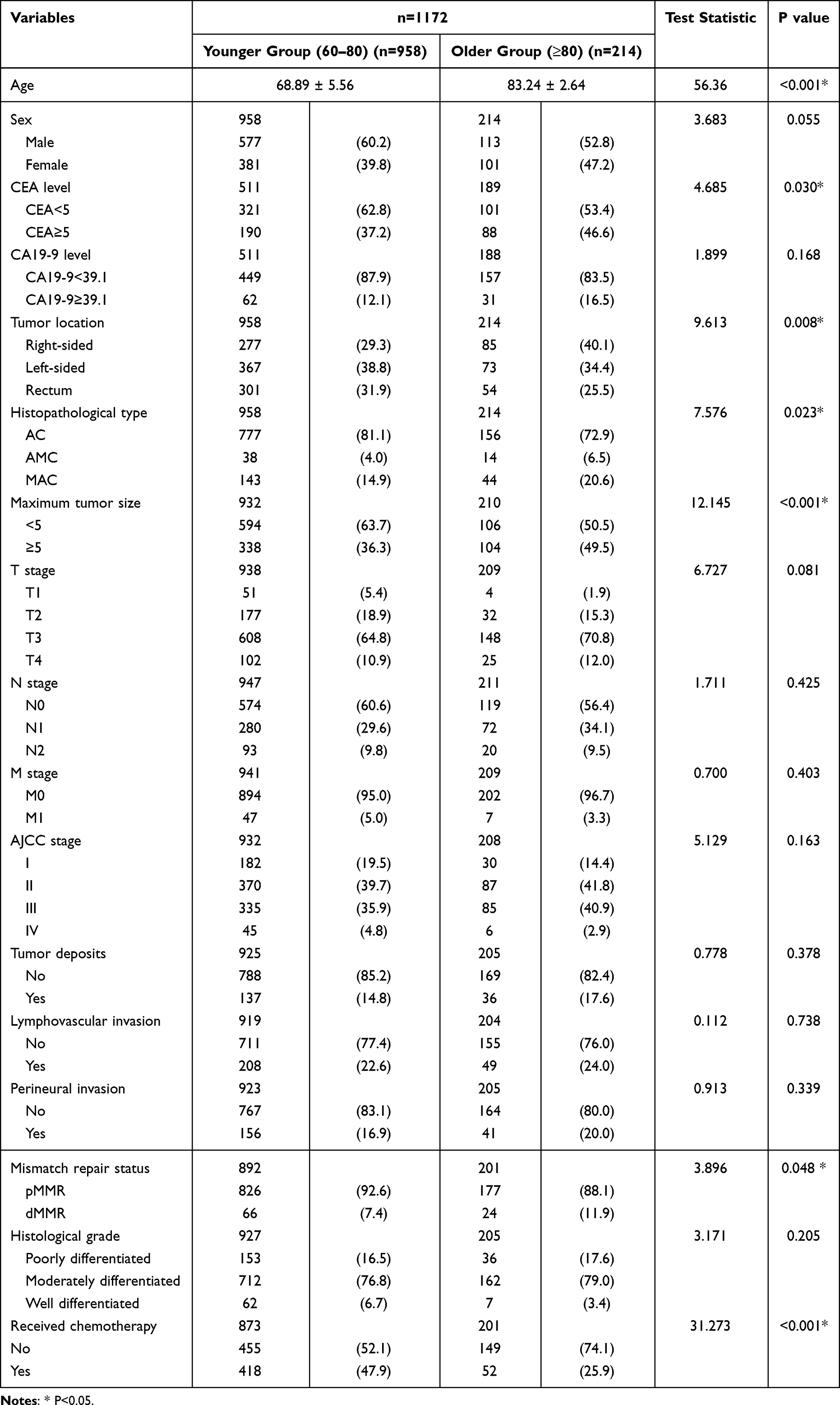 |
Table 1 Baseline and Clinicopathological Characteristics of Older Colorectal Cancer Patients |
Prognostic Factors of DFS in Patients Aged ≥60 years
The Kaplan-Meier curves for DFS in the younger and older groups are shown in Figure 1A. The median follow-up time was 33 (IQR: 14–60) months. During follow-up, the older group (≥80 years) had significantly worse DFS than the younger group (60–80 years) (P < 0.001).
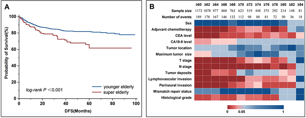 |
Figure 1 Survival analysis and Cox analysis of older patients (≥60 years). (A) DFS survival analysis of the younger and older groups. (B) Summarized p-values from univariate Cox analysis by age group. |
The results of univariate and multivariate Cox analyses of DFS in the overall older population are shown in Table 2. Univariate analysis identified the following risk factors for DFS: age ≥80 years (HR=1.821, 95% CI: 1.316–2.520, P<0.001), CEA level ≥5 ng/mL (HR=2.096, 95% CI: 1.477–2.975, P<0.001), CA19-9 level ≥39 U/mL (HR=1.720, 95% CI: 1.111–2.665, P=0.015), maximum tumor diameter ≥5 cm (HR=1.389, 95% CI: 1.032–1.871, P=0.030), T3-T4 stage (HR=3.115, 95% CI: 1.890–5.135, P<0.001), N1-N2 stage (HR=2.182, 95% CI: 1.627–2.926, P<0.001), presence of tumor deposits (HR=1.928, 95% CI: 1.360–2.734, P<0.001), lymphovascular invasion (HR=2.303, 95% CI: 1.687–3.143, P<0.001), perineural invasion (HR=2.118, 95% CI: 1.527–2.938, P<0.001), poorly differentiated histological grade (HR=2.397, 95% CI: 1.081–5.317, P=0.031) and mucinous adenocarcinoma (HR=1.927, 95% CI: 1.112–3.339, P=0.019).
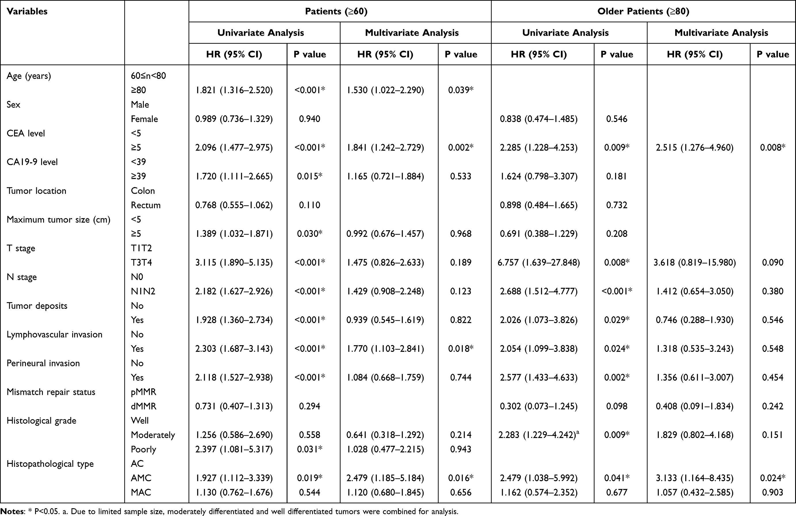 |
Table 2 Univariate and Multivariate Cox Analysis of DFS in Older Colorectal Cancer Patients |
Multivariate analysis showed that age ≥80 years (HR=1.530, 95% CI: 1.022–2.290, P=0.039), CEA level ≥5 ng/mL (HR=1.841, 95% CI: 1.242–2.729, P=0.002), lymphovascular invasion (HR=1.770, 95% CI: 1.103–2.841, P=0.018) and mucinous adenocarcinoma (HR=2.479, 95% CI: 1.185–5.184, P=0.016) were independent risk factors for DFS in older CRC patients.
Prognostic Factors of DFS in Patients Aged ≥80 years
The results of univariate and multivariate Cox analyses of DFS in the older age group (≥80 years) are shown in Table 2.
Univariate analysis showed that the following factors were risk factors for DFS in the over 80 years population: receipt of adjuvant chemotherapy (HR=2.610, 95% CI: 1.473–4.624, P=0.001), CEA level ≥5 ng/mL (HR=2.285, 95% CI: 1.228–4.253, P=0.009), T3-T4 stage (HR=6.757, 95% CI: 1.639–27.848, P=0.008), N1-N2 stage (HR=2.688, 95% CI: 1.512–4.777, P<0.001), presence of tumor deposits (HR=2.026, 95% CI: 1.073–3.826, P=0.029), lymphovascular invasion (HR=2.054, 95% CI: 1.099–3.838, P=0.024), perineural invasion (HR=2.577, 95% CI: 1.433–4.633, P=0.002), poorly to moderately differentiated histological grade (HR=2.283, 95% CI: 1.229–4.242, P=0.009) and mucinous adenocarcinoma (HR=2.479, 95% CI: 1.038–5.922, P=0.041).
Multivariate analysis showed that only CEA level ≥5 ng/mL (HR=2.515, 95% CI: 1.276–4.960, P=0.008) and mucinous adenocarcinoma (HR=3.133, 95% CI: 1.164–8.435, P=0.024) were independent risk factors for DFS in the older population (≥80 years).
To analyze age-related patterns in survival outcomes, we conducted a comprehensive series of univariate Cox analyses across sequential age thresholds. Beginning at age 60 years and incrementing by one-year intervals, we performed independent Cox regression models for each successive age stratum (≥60, ≥61, ≥62 years, continuing sequentially), with patients meeting or exceeding each age threshold included in the respective analysis. Each regression model evaluated the same set of prognostic variables to ensure methodological consistency across age groups. Analysis across different age groups (Figure 1B) demonstrated that CEA level ≥5 ng/mL, advanced T-stage and N-stage, presence of tumor deposits, lymphovascular invasion, and perineural invasion were significant predictors of DFS in the older population (≥80 years). There was a trend toward decreased predictive value of CA19-9 levels for DFS with increasing age. MMR status showed no predictive value for DFS across all age groups. Detailed data figures are shown in Supplementary Figure S1.
Characteristics of Transcriptome in the Older CRC
Transcriptome sequencing was performed on 244 primary tumors, including 53 samples from the older group (≥80 years) and 191 samples from the younger group (60–80 years). Differential gene expression analysis between the two groups revealed 781 upregulated genes and 187 downregulated genes in the older group (≥80 years) group (Figure 2A). GO enrichment analysis showed that upregulated genes were primarily enriched in synaptic signaling, extracellular matrix, GABAergic synapse, and neurotransmitter receptor activity, while downregulated genes were mainly involved in intermediate filament organization, antimicrobial humoral response, and adaptive immune response (Figure 2B). Pathway enrichment analysis demonstrated that upregulated pathways were predominantly associated with neuroactive ligand-receptor interaction, epithelial-mesenchymal transition (EMT), and extracellular matrix organization, whereas downregulated pathways included GPCR ligand binding and KRAS signaling (Figure 2C). Furthermore, GSEA analysis revealed significant upregulation in neuroactive ligand-receptor interaction, ECM-receptor interaction, and signaling angiogenesis, along with downregulation in mismatch repair and CD22-mediated BCR regulation pathways (Figure 2D).
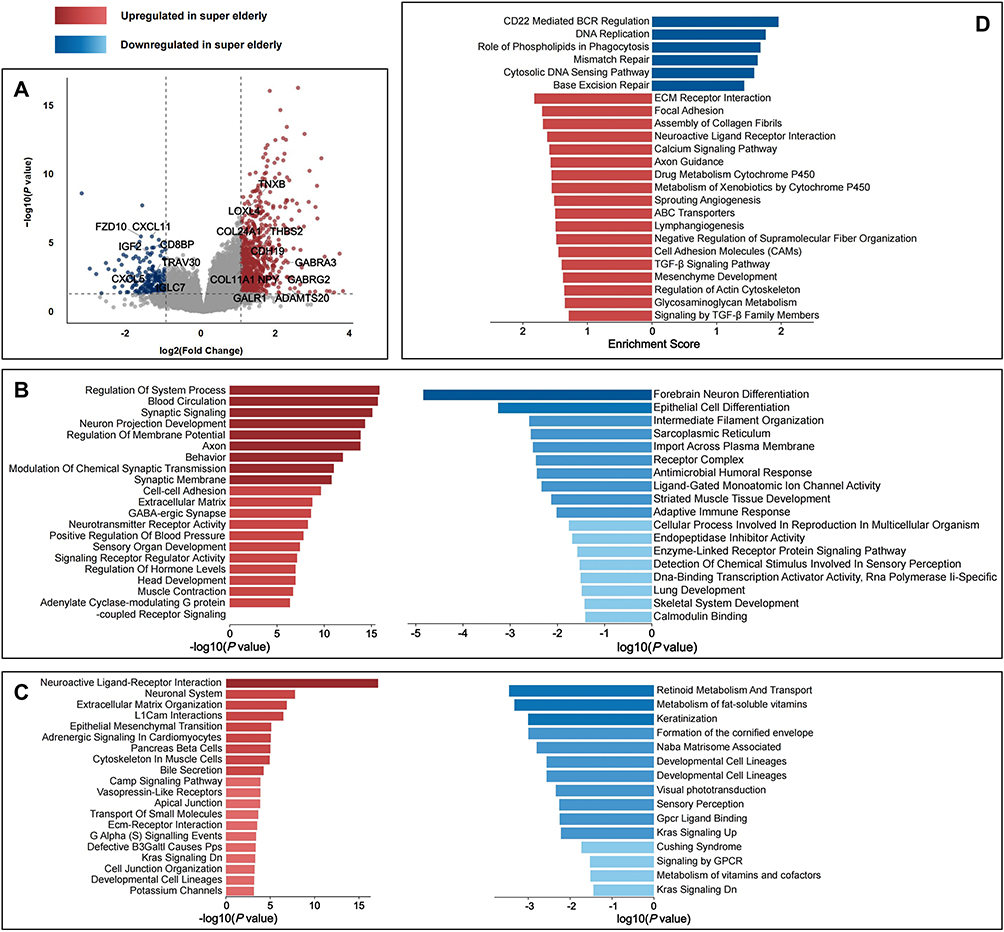 |
Figure 2 Differential gene analysis and enrichment analysis results between younger-group and older-group. (A) Volcano Plot of DEGs between younger-group and older-group. (B) GO enrichment analysis of older patients (≥80 years) in Metascape. (C) Pathway enrichment analysis of older patients (≥80 years) in Metascape. (D) GSEA of younger and older patients (≥80 years) populations. |
The results of the Wilcoxon test for abundance of immune cells in cancer tissues between the older and younger populations are shown in Figure 3A. quanTIseq analysis showed that older group patients (≥80 years) had significantly more M2-type macrophages (P=0.045) compared to younger group patients (60–80 years). Also, TIMER analysis showed significantly more macrophages (P=0.016) in older group patients (≥80 years) compared to younger group patients (60–80 years). The CMS molecular subtypes of older group and younger group are shown in Figure 3B. The older group (≥80 years) showed a higher prevalence of CMS1 and CMS4 subtypes.
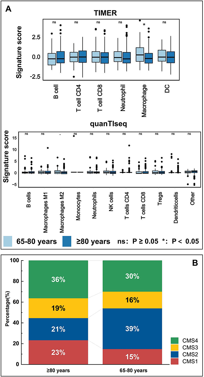 |
Figure 3 Immune infiltration (A) and CMS molecular subtypes (B) between younger-group and older-group. |
Discussion
Our study demonstrated that the clinicopathologic features and molecular characteristics of older patients (≥80 years) were significantly different from those of younger patients (60–80years), particularly in tumor site distribution, DNA mismatch repair status and prognosis. To the best of our knowledge, this represents one of the largest comprehensive analyses of CRC in patients aged ≥80 years (n=214 for clinical analysis; n=53 for transcriptomic profiling), addressing a critical knowledge gap in geriatric oncology where this population is severely underrepresented in clinical research.
In our study the proportion of deficient mismatch repair (dMMR) in older CRC patients (≥80 years) was significantly higher than in younger patients (60–80years). While dMMR is classically associated with hereditary Lynch syndrome in younger individuals,15 the increased prevalence in very older patients is primarily attributed to age-related MLH1 promoter hypermethylation,13,16 an epigenetic mechanism distinct from germline mutations. This finding is also consistent with our transcriptomic results showing an elevated proportion of CMS1-type CRC in the older group (≥80 years), as CMS1 is characterized by high levels of microsatellite instability. The higher dMMR prevalence in octogenarians has important clinical implications. Patients with dMMR tumors are candidates for immune checkpoint inhibitor therapy, which has shown efficacy in older patients comparable to younger cohorts, and the high pathological complete response (pCR) rate after immunotherapy may exempt the operation for older CRC patients who should have undergone high-risk surgery.17 Treatment decisions in this population require integration of comprehensive geriatric assessment (CGA) to balance potential benefits against frailty, comorbidities, and functional status, with systematic dMMR screening guiding personalized treatment strategies.
Notably, our study showed that age ≥80 years is an independent risk factor significantly affecting disease-free survival (DFS), contradicting the common assumption of slow CRC progression in older patients.18,19 This poor outcome in older patients (≥80 years) likely reflects both patient-related factors and aggressive tumor biology. Multiple factors limit treatment delivery and tolerance in these patients. Frailty, characterized by decreased physiologic reserve in physical function, cognition, and nutrition, significantly impacts cancer treatment outcomes.20,21 Performance status alone may underestimate vulnerability in older patients who maintain daily independence yet lack physiologic reserve to tolerate aggressive therapy, emphasizing the importance of comprehensive geriatric assessment (CGA) beyond chronological age.22,23 Additionally, older patients are underrepresented in clinical trials, resulting in insufficient evidence-based medical support for treatment regimens for this population.24,25 Notably, our study found that elevated CA19-9 levels, which typically serve as a poor prognostic marker in younger patients (60–80 years), were not prognostically significant in older patients (≥80 years). This may be because older patients (≥80 years) are often unable to receive effective chemotherapy regardless of CA19-9 levels, thereby weakening the association between CA19-9 levels and prognosis.
In terms of aggressive tumor biology, immunosenescence is an important feature of the aging process, suggesting that anti-tumor immune response capacity is weakened in older patients (≥80 years).26,27 Our study found simultaneous downregulation of cytoplasmic DNA sensing pathways and CD22-mediated B cell responses in older patients (≥80 years), indicating weakened immune surveillance at different levels. Moreover, under the background of immunosenescence, upregulation of TGF-β signaling further enhances immunosuppression. Compared to younger patients (60–80years), the immune microenvironment in older patients (≥80 years) showed enrichment of M2 macrophages, further suppressing anti-tumor immune responses.28,29 Additionally, extracellular matrix remodeling and upregulation of the epithelial-mesenchymal transition pathway may further enhance tumor cell invasion and metastasis.30,31 Extracellular matrix remodeling not only alters the physical barriers of the tumor microenvironment and impedes infiltration of residual immune cells, but may also affect drug distribution and tissue penetration.32 In contrast to immune function decline, patients ≥80 years showed significant activation of pathways related to nervous system regulation, particularly substantial upregulation of GABAergic synaptic transmission and neurotransmitter signaling. The activation of this neuroactive ligand-receptor interaction network suggests that tumor innervation remodeling may occur in older CRC patients (≥80 years).33 Activation of GABAergic inhibitory signaling may regulate tumor cell proliferation and apoptosis,34 while upregulation of neurotransmitter receptors may promote tumor angiogenesis and nutrient supply.35 The establishment of such neural-tumor interactions provides a tumor regulation pattern that differs from younger patients, potentially offering unique growth regulatory advantages and microenvironmental adaptations to tumors.36,37 These molecular changes indicate that older CRC (≥80 years) are characterized by unique neural-tumor interactions, enhanced tumor invasive and metastatic capacity, and significantly weakened immune function, potentially explaining the distinctive clinical manifestations and treatment responses in older CRC patients (≥80 years).
The molecular characteristics identified in our study suggest potential therapeutic strategies tailored for older CRC patients. The enrichment of M2 macrophages indicates that macrophage-targeting approaches, such as CSF-1R inhibitors or CD47 blockade, could restore anti-tumor immunity, complementing immunotherapy in dMMR tumors or overcoming resistance in pMMR tumors.38,39 The pronounced ECM remodeling supports evaluation of TGF-β inhibitors or matrix metalloproteinase inhibitors to disrupt pro-metastatic microenvironments and improve drug penetration.30,31 Additionally, the enhanced neural-tumor interactions represent a novel therapeutic avenue. Our finding of upregulated GABAergic and neurotransmitter signaling suggests that neuromodulatory drugs, such as beta-blockers or GABA receptor modulators, could be explored as repurposed therapies,33–37 with the advantage of established safety profiles facilitating rapid clinical translation. However, translating these insights into clinical practice requires addressing age-specific challenges in older patients (≥80 years), including altered pharmacokinetics, polypharmacy risks, and the need to balance survival with quality of life. Prospective trials in this population should incorporate CGA-based stratification and patient-reported outcomes to provide evidence for individualized treatment decisions.
This study has several limitations. First, this is a single-center retrospective analysis with potential selection and information bias. Although the overall sample size is adequate (n=1,172), the transcriptomic analysis in the older-age group (n=53) remains relatively limited for certain subgroup analyses, which may affect the statistical power for detecting subtle molecular differences. Future multicenter, large-scale prospective studies are needed for validation. Second, the follow-up period for older patients (≥80 years) remains limited, inadequately assessing the long-term potential impacts on prognosis and the higher rate of non-cancer deaths in the older-age group may have influenced the censoring patterns in survival analysis. Although we used disease-free survival as the primary endpoint, competing risks from non-cancer mortality may have affected the interpretation of survival outcomes in very older patients. Future studies incorporating competing risk analysis would provide more accurate survival estimates. Moreover, for older patients (≥80 years), quality of life and functional independence may be as important as disease-free survival. Prospective studies should incorporate patient-reported outcomes and functional assessments alongside traditional oncologic endpoints. Third, cross-sectional transcriptomic analysis cannot fully reflect gene changes and dynamic regulatory processes, necessitating further functional studies.
Conclusion
In conclusion, CRC patients aged ≥80 years exhibit distinctive clinicopathologic and molecular characteristics including higher dMMR prevalence, worse disease-free survival, immunosuppression, ECM remodeling, and enhanced neural-tumor interactions. Contrary to common assumptions, age ≥80 years is an independent adverse prognostic factor driven by both aggressive tumor biology and patient vulnerabilities. These findings suggest potential therapeutic strategies including immune checkpoint inhibitors for dMMR tumors, macrophage-targeting agents, and neuromodulatory drugs. Treatment decisions for older CRC patients (≥80 years) should integrate comprehensive geriatric assessment with molecular profiling beyond chronological age alone. Future trials in this population should incorporate CGA stratification and patient-reported outcomes, and explore interventions targeting immune dysfunction and ECM remodeling to provide evidence-based care for this underrepresented population.
Data Sharing Statement
The datasets analyzed during the current study are not publicly available due to patient privacy protection requirements and institutional data sharing policies but are available from the corresponding authors upon reasonable request and with appropriate ethical approval.
Ethics Approval and Informed Consent
This retrospective study was approved by the Medical Science Research Ethics Committee of Peking University Third Hospital (Approval No. IRB00006761-M2024287) and was conducted in accordance with the Declaration of Helsinki (2013 revision). The Ethics Committee granted a waiver of informed consent. Patient data confidentiality was maintained throughout the study, with all data anonymized and stored securely to ensure patient privacy.
Consent for Publication
This study used de-identified retrospective data from medical records only. No individual patient data, images, or identifiable information requiring specific consent for publication are included.
Funding
This study was funded by the Capital’s Funds for Health Improvement and Research (Grant number:2024-1-4091). The funding organization played no role in the design or conduct of the study; collection, management, analysis, or interpretation of the data; preparation, review, or approval of the manuscript; or the decision to submit the manuscript for publication.
Disclosure
The authors report no conflicts of interest in this work.
References
1. Wang S, Zheng R, Li J, et al. Global, regional, and national lifetime risks of developing and dying from gastrointestinal cancers in 185 countries: a population-based systematic analysis of GLOBOCAN. Lancet Gastroenterol Hepatol. 2024;9(3):229–237. doi:10.1016/s2468-1253(23)00366-7
2. Bray F, Laversanne M, Sung H, et al. Global cancer statistics 2022: GLOBOCAN estimates of incidence and mortality worldwide for 36 cancers in 185 countries. CA Cancer J Clin. 2024;74(3):229–263. doi:10.3322/caac.21834
3. Siegel RL, Kratzer TB, Giaquinto AN, Sung H, Jemal A. Cancer statistics, 2025. CA Cancer J Clin. 2025;75(1):10–45. doi:10.3322/caac.21871
4. Ioffe D, Bhatia-Patel SC, Gandhi S, Hamad EA, Dotan E. Cardiovascular Concerns, Cancer Treatment, and Biological and Chronological Aging in Cancer: JACC Family Series. JACC CardioOncol. 2024;6(2):143–158. doi:10.1016/j.jaccao.2024.02.001
5. Huang Z, Zhuang Y, Li W, et al. Apoptotic vesicles are required to repair DNA damage and suppress premature cellular senescence. J Extracell Vesicles. 2024;13(4):e12428. doi:10.1002/jev2.12428
6. Huang K, Liu X, Zhang Z, et al. AgeAnnoMO: a knowledgebase of multi-omics annotation for animal aging. Nucleic Acids Res. 2024;52(D1):D822–d834. doi:10.1093/nar/gkad884
7. Si F, Liu X, Tao Y, et al. Blocking senescence and tolerogenic function of dendritic cells induced by gammadelta Treg cells enhances tumor-specific immunity for cancer immunotherapy. J Immunother Cancer. 2024;12(4):2. doi:10.1136/jitc-2023-008219
8. Spurr LF, Weichselbaum RR, Pitroda SP. Tumor aneuploidy predicts survival following immunotherapy across multiple cancers. Nat Genet. 2022;54(12):1782–1785. doi:10.1038/s41588-022-01235-4
9. Ritter M, Blume C, Tang Y, et al. Spatially resolved transcriptomics and graph-based deep learning improve accuracy of routine CNS tumor diagnostics. Nat Cancer. 2025;6(2):292–306. doi:10.1038/s43018-024-00904-z
10. Li J, Pan Y, Guo F, et al. Patterns in genomic mutations among patients with early-onset colorectal cancer: an international, multicohort, observational study. Lancet Oncol. 2025;26(8):1055–1066. doi:10.1016/s1470-2045(25)00239-6
11. Matas J, Kohrn B, Fredrickson J, et al. Colorectal Cancer Is Associated with the Presence of Cancer Driver Mutations in Normal Colon. Cancer Res. 2022;82(8):1492–1502. doi:10.1158/0008-5472.Can-21-3607
12. Guinney J, Dienstmann R, Wang X, et al. The consensus molecular subtypes of colorectal cancer. Nature Med. 2015;21(11):1350–1356. doi:10.1038/nm.3967
13. Yang L, Chen X, Lee C, et al. Functional characterization of age-dependent p16 epimutation reveals biological drivers and therapeutic targets for colorectal cancer. J Exp Clin Cancer Res. 2023;42(1):113. doi:10.1186/s13046-023-02689-y
14. Weiser MR. AJCC 8th Edition: colorectal Cancer. Ann Surg Oncol. 2018;25(6):1454–1455. doi:10.1245/s10434-018-6462-1
15. Kastrinos F, Ingram MA, Silver ER, et al. Gene-Specific Variation in Colorectal Cancer Surveillance Strategies for Lynch Syndrome. Gastroenterology. 2021;161(2):453–462.e15. doi:10.1053/j.gastro.2021.04.010
16. Yang J, Wei H, Lin Y, et al. High Soluble Fiber Promotes Colorectal Tumorigenesis Through Modulating Gut Microbiota and Metabolites in Mice. Gastroenterology. 2024;166(2):323–337.e7. doi:10.1053/j.gastro.2023.10.012
17. Saberzadeh-Ardestani B, Jones JC, Hubbard JM, et al. Association Between Survival and Metastatic Site in Mismatch Repair-Deficient Metastatic Colorectal Cancer Treated With First-line Pembrolizumab. JAMA Network Open. 2023;6(2):e230400. doi:10.1001/jamanetworkopen.2023.0400
18. Steup C, Kennel KB, Neurath MF, Fichtner-Feigl S, Greten FR. Current and emerging concepts for systemic treatment of metastatic colorectal cancer. Gut. 2025;74(12):2070–2095. doi:10.1136/gutjnl-2025-335412
19. Yuan C, Zhao X, Wangmo D, Alshareef D, Gates TJ, Subramanian S. Tumor models to assess immune response and tumor-microbiome interactions in colorectal cancer. Pharmacol Ther. 2022;231:107981. doi:10.1016/j.pharmthera.2021.107981
20. Alamgeer M, Ling RR, Ueno R, et al. Frailty and long-term survival among patients in Australian intensive care units with metastatic cancer (FRAIL-CANCER study): a retrospective registry-based cohort study. Lancet Healthy Longev. 2023;4(12):e675–e684. doi:10.1016/s2666-7568(23)00209-x
21. Baltussen JC, de Glas NA, van Holstein Y, et al. Chemotherapy-Related Toxic Effects and Quality of Life and Physical Functioning in Older Patients. JAMA Network Open. 2023;6(10):e2339116. doi:10.1001/jamanetworkopen.2023.39116
22. Dale W, Klepin HD, Williams GR, et al. Practical Assessment and Management of Vulnerabilities in Older Patients Receiving Systemic Cancer Therapy: ASCO Guideline Update. J Clin Oncol. 2023;41(26):4293–4312. doi:10.1200/jco.23.00933
23. Kadambi S, Loh KP, Dunne R, et al. Older adults with cancer and their caregivers - current landscape and future directions for clinical care. Nat Rev Clin Oncol. 2020;17(12):742–755. doi:10.1038/s41571-020-0421-z
24. Chow R, Lage DE, Williams GR, et al. Representation and Outcomes of Older Adults in Practice-Changing Oncology Trials in the Era of Novel Therapies: a Guideline Appraisal. J Natl Compr Canc Netw. 2022;20(1):37–44. doi:10.6004/jnccn.2021.7055
25. Sedrak MS, Freedman RA, Cohen HJ, et al. Older adult participation in cancer clinical trials: a systematic review of barriers and interventions. CA Cancer J Clin. 2021;71(1):78–92. doi:10.3322/caac.21638
26. Lian J, Yue Y, Yu W, Zhang Y. Immunosenescence: a key player in cancer development. J Hematol Oncol. 2020;13(1):151. doi:10.1186/s13045-020-00986-z
27. Borgoni S, Kudryashova KS, Burka K, de Magalhaes JP. Targeting immune dysfunction in aging. Ageing Res Rev. 2021;70:101410. doi:10.1016/j.arr.2021.101410
28. Nalio Ramos R, Missolo-Koussou Y, Gerber-Ferder Y, et al. Tissue-resident FOLR2(+) macrophages associate with CD8(+) T cell infiltration in human breast cancer. Cell. 2022;185(7):1189–1207.e25. doi:10.1016/j.cell.2022.02.021
29. Yin B, Ding J, Liu J, et al. Exosomal CMTM4 Induces Immunosuppressive Macrophages to Promote Ovarian Cancer Progression and Attenuate Anti-PD-1 Immunotherapy. Adv Sci. 2025;2025:e04436. doi:10.1002/advs.202504436
30. Shi X, Askari Rizvi SF, Yang Y, Liu G. Emerging nanomedicines for macrophage-mediated cancer therapy. Biomaterials. 2025;316:123028. doi:10.1016/j.biomaterials.2024.123028
31. Wei Y, Long S, Zhao M, et al. Regulation of Cellular Signaling with an Aptamer Inhibitor to Impede Cancer Metastasis. J Am Chem Soc. 2024;146(1):319–329. doi:10.1021/jacs.3c09091
32. Maiques O, Sallan MC, Laddach R, et al. Matrix mechano-sensing at the invasive front induces a cytoskeletal and transcriptional memory supporting metastasis. Nat Commun. 2025;16(1):1394. doi:10.1038/s41467-025-56299-7
33. Kobayashi H, Iida T, Ochiai Y, et al. Neuro-Mesenchymal Interaction Mediated by a beta2-Adrenergic Nerve Growth Factor Feedforward Loop Promotes Colorectal Cancer Progression. Cancer Discov. 2025;15(1):202–226. doi:10.1158/2159-8290.Cd-24-0287
34. Barron T, Yalcin B, Su M, et al. GABAergic neuron-to-glioma synapses in diffuse midline gliomas. Nature. 2025;639(8056):1060–1068. doi:10.1038/s41586-024-08579-3
35. Hashimoto Y, Campbell M. Eyes on glutamate in angiogenesis and barrier formation. Neuron. 2024;112(12):1895–1897. doi:10.1016/j.neuron.2024.05.020
36. Zhang Y, Liao Q, Wen X, et al. Hijacking of the nervous system in cancer: mechanism and therapeutic targets. Mol Cancer. 2025;24(1):44. doi:10.1186/s12943-025-02246-5
37. Khanmammadova N, Islam S, Sharma P, Amit M. Neuro-immune interactions and immuno-oncology. Trends Cancer. 2023;9(8):636–649. doi:10.1016/j.trecan.2023.05.002
38. Cannarile MA, Weisser M, Jacob W, Jegg AM, Ries CH, Rüttinger D. Colony-stimulating factor 1 receptor (CSF1R) inhibitors in cancer therapy. J Immunother Cancer. 2017;5(1):53. doi:10.1186/s40425-017-0257-y
39. Advani R, Flinn I, Popplewell L, et al. CD47 Blockade by Hu5F9-G4 and Rituximab in Non-Hodgkin’s Lymphoma. N Engl J Med. 2018;379(18):1711–1721. doi:10.1056/NEJMoa1807315
 © 2025 The Author(s). This work is published and licensed by Dove Medical Press Limited. The
full terms of this license are available at https://www.dovepress.com/terms
and incorporate the Creative Commons Attribution
- Non Commercial (unported, 4.0) License.
By accessing the work you hereby accept the Terms. Non-commercial uses of the work are permitted
without any further permission from Dove Medical Press Limited, provided the work is properly
attributed. For permission for commercial use of this work, please see paragraphs 4.2 and 5 of our Terms.
© 2025 The Author(s). This work is published and licensed by Dove Medical Press Limited. The
full terms of this license are available at https://www.dovepress.com/terms
and incorporate the Creative Commons Attribution
- Non Commercial (unported, 4.0) License.
By accessing the work you hereby accept the Terms. Non-commercial uses of the work are permitted
without any further permission from Dove Medical Press Limited, provided the work is properly
attributed. For permission for commercial use of this work, please see paragraphs 4.2 and 5 of our Terms.
Recommended articles

IGFBP5 is Upregulated and Associated with Poor Prognosis in Colorectal Cancer
Deng Y, Yang X, Hua H, Zhang C
International Journal of General Medicine 2022, 15:6485-6497
Published Date: 6 August 2022
Upregulation of Long Noncoding RNA MALAT1 in Colorectal Cancer Promotes Radioresistance and Aggressive Malignance
Shen W, Yu Q, Pu Y, Xing C
International Journal of General Medicine 2022, 15:8365-8380
Published Date: 28 November 2022
Fashionable, but What is Their Real Clinical Usefulness? NLR, LMR, and PLR as a Promising Indicator in Colorectal Cancer Prognosis: A Systematic Review
Misiewicz A, Dymicka-Piekarska V
Journal of Inflammation Research 2023, 16:69-81
Published Date: 7 January 2023
Screening History and 7-Year Survival in 32,099 Colorectal Cancer Patients: A Population-Based Cohort Study
Hsiao BY, Chiang CJ, Yang YW, Lin LJ, Hsu TH, Lee WC
Clinical Epidemiology 2023, 15:1009-1025
Published Date: 3 October 2023
BICDL1 Predicts Poor Prognosis and is Correlated with Methylation and Immune Infiltration in Colorectal Cancer
Luo H, Luo J, Ding N, Zhang T, He Y
Pharmacogenomics and Personalized Medicine 2023, 16:1109-1126
Published Date: 22 December 2023