Back to Journals » Cancer Management and Research » Volume 17

Application of Liquid Biopsy Technology in Lung Cancer: A Bibliometric Study and Visualization Analysis
Authors Xu Y ![]() , Huang B, Zhu G, Chen N, Gao Y, Wu Z
, Huang B, Zhu G, Chen N, Gao Y, Wu Z ![]() , Mo L, Liu L
, Mo L, Liu L
Received 29 June 2025
Accepted for publication 15 September 2025
Published 23 September 2025 Volume 2025:17 Pages 2153—2168
DOI https://doi.org/10.2147/CMAR.S550357
Checked for plagiarism Yes
Review by Single anonymous peer review
Peer reviewer comments 2
Editor who approved publication: Dr Chien-Feng Li
Yueqi Xu,1,2,* Biao Huang,2,* Guoshuang Zhu,1 Nidan Chen,2 Yao Gao,1,2 Zenan Wu,1 Lisha Mo,2 Liangji Liu2
1School of Clinical Medicine, Jiangxi University of Chinese Medicine, Nanchang, People’s Republic of China; 2Affiliated Hospital of Jiangxi University of Chinese Medicine, Nanchang, People’s Republic of China
*These authors contributed equally to this work
Correspondence: Liangji Liu, Affiliated Hospital of Jiangxi University of Chinese Medicine, Nanchang, People’s Republic of China, Email [email protected] Zenan Wu, School of Clinical Medicine, Jiangxi University of Chinese Medicine, Nanchang, People’s Republic of China, Email [email protected]
Background: Lung cancer is a major global health threat, and traditional treatments are hindered by tumor heterogeneity and drug resistance. Liquid biopsy technology offers new opportunities for precise diagnosis and treatment of lung cancer; however, its clinical application still presents challenges. Therefore, we aimed to elucidate the research progress and future trends of liquid biopsy technology through a bibliometric analysis.
Methods: Literature on liquid biopsy technology in lung cancer research spanning from 1990 to 2024 was retrieved from the Web of Science Core Collection. Various bibliometric tools were employed, including VOSviewer (1.6.20), Cite Space (6.2.R3), RStudio (utilizing the R package bibliometrix), and Scimago Graphica, to explore the current situation and research hotspots in this area. The research covered multiple aspects, including publication counts, contributions from different countries and institutions, journals involved, authorship, co-references, and keywords. By generating corresponding visualization maps, an attempt was made to forecast future development trends within this research field.
Results: We identified 363 articles published in 198 journals, authored by 2518 authors from 266 institutions across 54 countries/regions. The journal, country, institution, and author with the highest number of publications were Cancer, China, Harvard University, and ROLFO C. Over the past 34 years, research on the application of liquid biopsy technology in lung cancer have primarily focused on cancer diagnosis, biomarkers, and treatment monitoring.
Conclusion: Liquid biopsy technology is rapidly evolving in the field of lung cancer. Detecting biomarkers such as circulating tumor DNA, circulating tumor cells, and extracellular vesicles, facilitates early detection and monitoring of treatment response, reveals drug resistance mechanisms, and promotes personalized treatment. With the advancement of technology and international cooperation, liquid biopsy is expected to provide more accurate diagnosis and treatment for patients with lung cancer in the future.
Keywords: liquid biopsy technique, tumor, bibliometric tools, bibliometrics, citespace, VOSviewer
Introduction
Lung cancer remains the leading cause of cancer-related mortality worldwide. Patients are often asymptomatic in the early stages, but as the disease progresses, patients may develop persistent symptoms, such as chronic cough, chest pain, and dyspnea.1,2 According to the latest epidemiological data, in 2022, lung cancer ranked first in incidence and mortality among all malignant tumors in China, while in the United States, it remained the leading cause of cancer-related death despite ranking third in incidence.3 Multiple factors influence the prognosis of lung cancer, including histological type, disease stage at diagnosis, patient age, and overall health status.4–6 Consequently, elucidating its pathogenesis is essential for developing novel therapeutic strategies and improving patient outcomes.7,8
Liquid biopsy technology, an emerging non-invasive diagnostic tool, enables early diagnosis, real-time monitoring, and prognostic evaluation of tumors by analyzing biomarkers in the blood, such as circulating tumor cells (CTCs), circulating tumor DNA (ctDNA), and exosomes.9–15 Recent studies have shown that liquid biopsy technology has advanced rapidly in lung cancer research and has become a research hotspot. It can assist in early diagnosis, evaluate treatment effectiveness in real-time, and adjust treatment plans promptly. Its potential application and future development direction in clinical practice have attracted considerable attention.12,13,16–20
Bibliometrics is a method of revealing scientific development trends and research hotspots through quantitative analysis of literature data.21 It can reflect the development trends and research hotspots of a certain field by analyzing data on countries, authors, journals, institutions, and keywords in that field. Considering the compatibility with bibliometric analysis tools, such as CiteSpace and VOSviewer, the Web of Science Core Collection (WoSCC) was selected as the sole data source due to its comprehensive coverage, structured records, and standardized indexing. At present, there is no research on the application of liquid biopsy technology in lung cancer using bibliometric methods. Therefore, we aimed to use bibliometric tools to comprehensively analyze the research and application of liquid biopsy technology in lung cancer from multiple perspectives, as well as the current development status, future research trends, and hotspots in this field.
Materials and Methods
Data Sources and Literature Retrieval
All bibliographic data were obtained from the WoSCC database. The search period spanned January 1, 1990, to September 30, 2024. The retrieval strategy combined subject terms for lung cancer and liquid biopsy techniques, using this search formula: (TS=“Lung Neoplastics” OR “Neoplastics, Pulmonary” OR “Neoplastic, Pulmonary” OR “Pulmonary Neoplastics” OR “Neoplastics, Lung” OR “Lung Neoplastics” OR “Neoplastic, Lung” OR “Lung Cancer” OR “Cancer, Lung” OR “Lung Cancer” OR “Cancer of Lung” OR “Pulmonary Cancer” OR “Cancer, Pulmonary” OR “Cancer, Pulmonary” OR “Cancer, Pulmonary” OR “Cancer, Pulmonary” OR “Cancer, Pulmonary Cancer” or “Cancer of the Lung”) and TS= (liquid biopsy techniques).
Eligibility Criteria and Data Standardization
We included original research articles and review papers that focused on the application of liquid biopsy technology in lung cancer, were written in English, and provided complete bibliographic metadata (title, authors, affiliations, country, keywords, and source, amongst others). We excluded non-research documents, such as conference abstracts, patents, news items, and popular health articles, as well as duplicate records identified during screening. The retrieved records were exported in RefWorks and plain-text formats and subjected to a standardized preprocessing workflow comprising removal of special characters and formatting artifacts, harmonization of synonymous terms and abbreviations (standardizing variants such as “NSCLC” and “non-small cell lung cancer”), and conversion to analytics-ready formats using the Data Import/Export function in CiteSpace.
Data Analysis
We conducted a comprehensive bibliometric analysis using Microsoft Excel, VOSviewer (v1.6.20), CiteSpace (v6.2.R3), RStudio (with the bibliometrix package), and Scimago Graphica across a 34-year period from January 1990 to September 2024, with 1-year time slices. We used Excel to quantify annual publication volumes, track country-level outputs over time, and classify records by research category. RStudio/bibliometrix was used to generate visual mappings of journal sources, national contributions, international collaboration, co-cited references, keyword frequencies, and author activity. CiteSpace was used to examine institutional collaboration networks and centrality as well as to detect keyword clusters and burst terms. Node types included keyword and institution co-occurrence, and pruning was kept at default settings. VOSviewer was used to support the analyses of co-cited references, while Scimago Graphica was used to refine author and keyword frequency visualizations. Co-occurrence, clustering, and burst analyses were integrated to produce optimized knowledge maps for visualization-driven interpretation. The overall retrieval and analysis workflow is summarized in Figure 1.
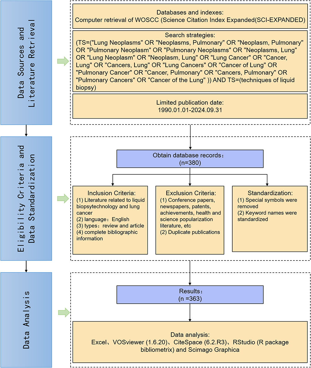 |
Figure 1 Flowchart illustrating the screening process for literature selection. |
Results
Analysis of Publication Year and Journal Source
Publication activity on liquid biopsy in lung cancer remained modest until 2015 and then increased steadily (Figure 2A). The first article appeared in 1990; annual output remained below 10 until 2015, peaking at 51 in 2020. Two growth spurts (2011–2012 and 2014–2016) likely reflect contributions from the Lv Ming and Li Dai-Rong teams. From 2016 to 2024, output maintained an upward trajectory.
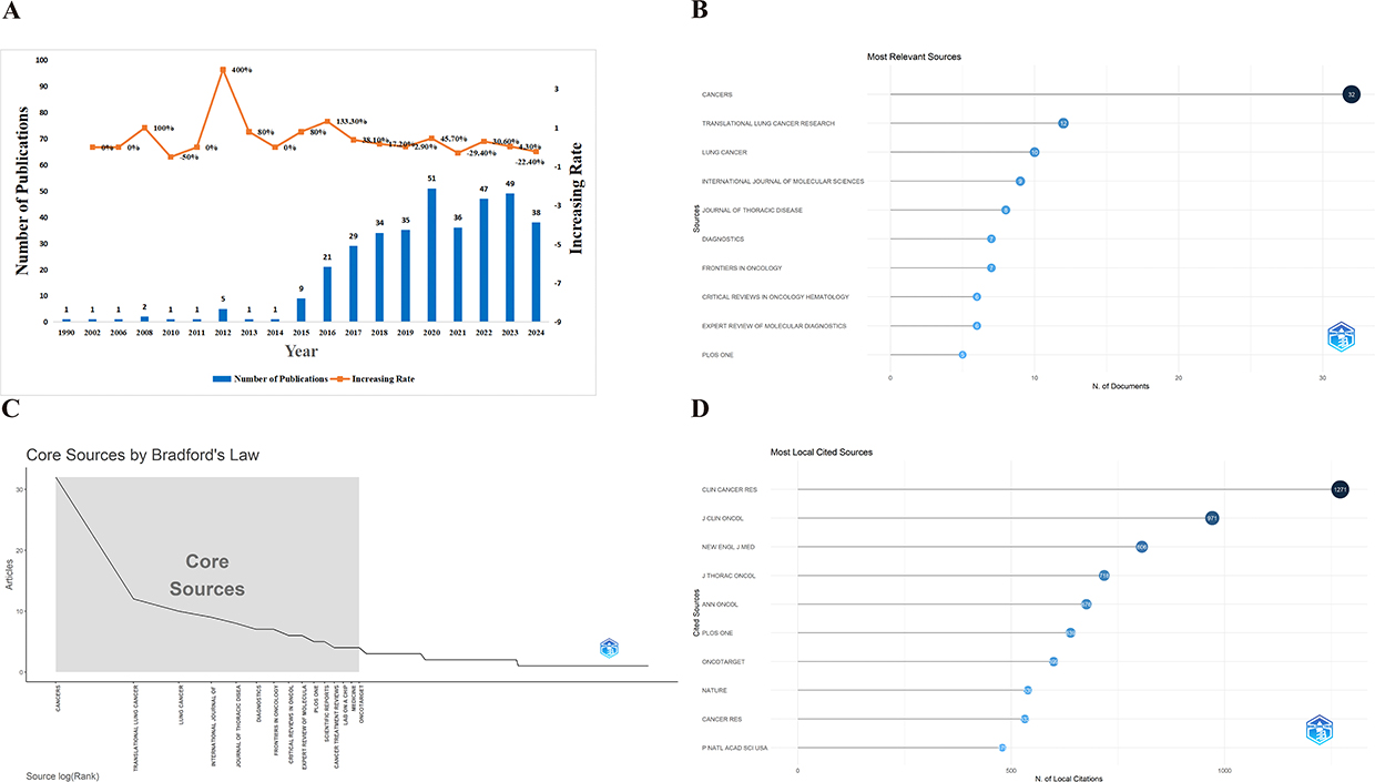 |
Figure 2 (A) Annual distribution of publications; (B) Top 10 journals ranked by the number of published articles; (C) Core journals identified according to Bradford’s Law; (D) Top 10 journals most frequently cited in the included publications. |
Across 198 journals, Cancers (n=32), Translational Lung Cancer Research (n=12), and Lung Cancer (n=10) contributed the largest volumes (Figure 2B). According to Bradford’s law, the core outlets are Cancers, Translational Lung Cancer Research, Lung Cancer, International Journal of Molecular Sciences, and Journal of Thoracic Disease (Figure 2C). Notably, influence is not fully captured by volume alone: Clinical Cancer Research amassed the highest total citations (n=1,271) within our dataset (Figure 2D), illustrating that citation-based indicators (such as citations per paper [CPP] or h-index) provide complementary nuance beyond publication counts. Temporal journal dynamics further show Translational Lung Cancer Research peaking in 2016 (n=4) and Cancers in 2022 (n=4) (Figure 3A). Dual-map overlays indicate cross-disciplinary citation flows from Molecular Biology-Genetics toward Molecular Biology-Immunology and Medicine-Clinical domains (Figure 3B).
 |
Figure 3 (A) Annual publication trends of the top five journals by volume; (B) Dual-map overlay illustrating citation relationships, showing clusters of citing and cited journals. |
Visualization Analysis of International Cooperation
Fifty-four countries/regions contributed to this literature (Figure 4A). By output, the top 10 were China (n=384), USA (n=248), Spain (n=235), Italy (n=159), France (n=94), Belgium (n=61), Germany (n=58), United Kingdom (n=54), India (n=53), and South Korea (n=42) (Table 1). By total citations, the USA ranked first (n=2,683), followed by China (n=1,858) and Italy (n=1,111) (Figure 4B). To avoid conflating productivity with impact, we assessed citation intensity where feasible. For example, CPP was higher for the USA (~10.8; 2,683/248) than for China (~4.8; 1,858/384), underscoring differences in per-article influence that publication volume alone cannot convey.
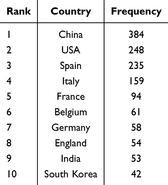 |
Table 1 Top 10 Most Productive Countries/Regions |
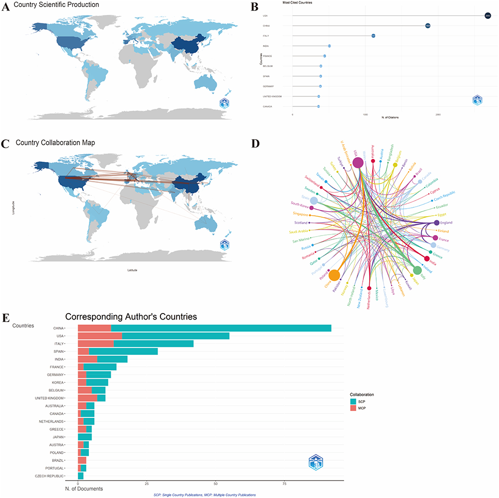 |
Figure 4 (A) Geographic distribution of publications by country; (B) Top 10 most-cited countries ranked by publication count; (C) Distribution of international collaborative research; (D) Chord diagram illustrating global collaboration patterns; (E) Top 20 countries ranked by the number of corresponding authors. |
Collaboration maps (Figures 4C and D) depict international ties; line thickness represents link weight (co-authorship counts). The USA shows multiple high-weight links, indicating broad, intensive collaborations. Figure 4E indicates that China accounts for 25.3% of global output, followed by the USA (15.2%) and Italy (11.6%). By collaboration type, China had the most single-country publications (SCP, n=80), suggesting predominantly domestic teamwork, whereas the USA showed a higher proportion of multiple-country publications (MCP, 29.1%), reflecting stronger international engagement. MCP% should be interpreted in the context of the denominator: countries with small absolute outputs (Brazil at 100% MCP) can exhibit inflated MCP proportions that overstate reliance on foreign partnerships. Where available, network statistics (such as betweenness centrality and weighted degree) provide more objective assessments of structural importance than MCP% alone.
Visualization Analysis of Authors’ and Institutions’ Collaboration
The 363 included publications were authored by 2,518 researchers. Rolfo C (Icahn School of Medicine at Mount Sinai) was the most prolific author (n = 11) and also the most cited (n = 25) (Figures 5A–C). Based on Price’s law (Mp = 0.749 × √Npmax = 2.48), authors with ≥3 publications were designated as core authors, yielding 72 contributors. The co-authorship network (Figure 5E) revealed four major collaborative clusters, led by Rosell Rafel, Rolfo Christian, Provincio Mariano, and Malapele Umberto, with research themes centering on circulating biomarker detection and analysis.22 These collaborations reflect the pivotal role of liquid biopsy in monitoring tumor dynamics, detecting drug resistance, predicting therapeutic response, and supporting personalized treatment, particularly in patients with non-small cell lung cancer (NSCLC).15,23,24
 |
Figure 5 (A) Top 10 authors ranked by the number of publications; (B) Top 10 authors ranked by citation frequency; (C) Annual publication trends of the top 10 authors; (D) Collaboration map of inter-institutional relationships; (E) Network diagram illustrating core author collaborations. |
Institutional collaboration mapping identified 266 nodes and 739 links. Table 2 presents the top 10 institutions in lung cancer liquid biopsy research, ranked by publication quantity and network centrality to reflect research productivity and collaborative influence. Harvard University ranked first in publication volume (n = 14), with research primarily addressing the clinical potential and technical challenges of liquid biopsy.11,25–27 Centro de Investigación Biomédica en Red (CIBER) ranked second (n = 11). The Chinese Academy of Sciences and Brigham & Women’s Hospital each displayed the highest betweenness centrality (0.19), underscoring their bridging roles in international collaboration. Notably, Harvard University, CIBER, CIBERONC, the Chinese Academy of Sciences, and UNICANCER formed a closely connected network, suggesting the presence of sustained and high-impact cross-regional cooperation.
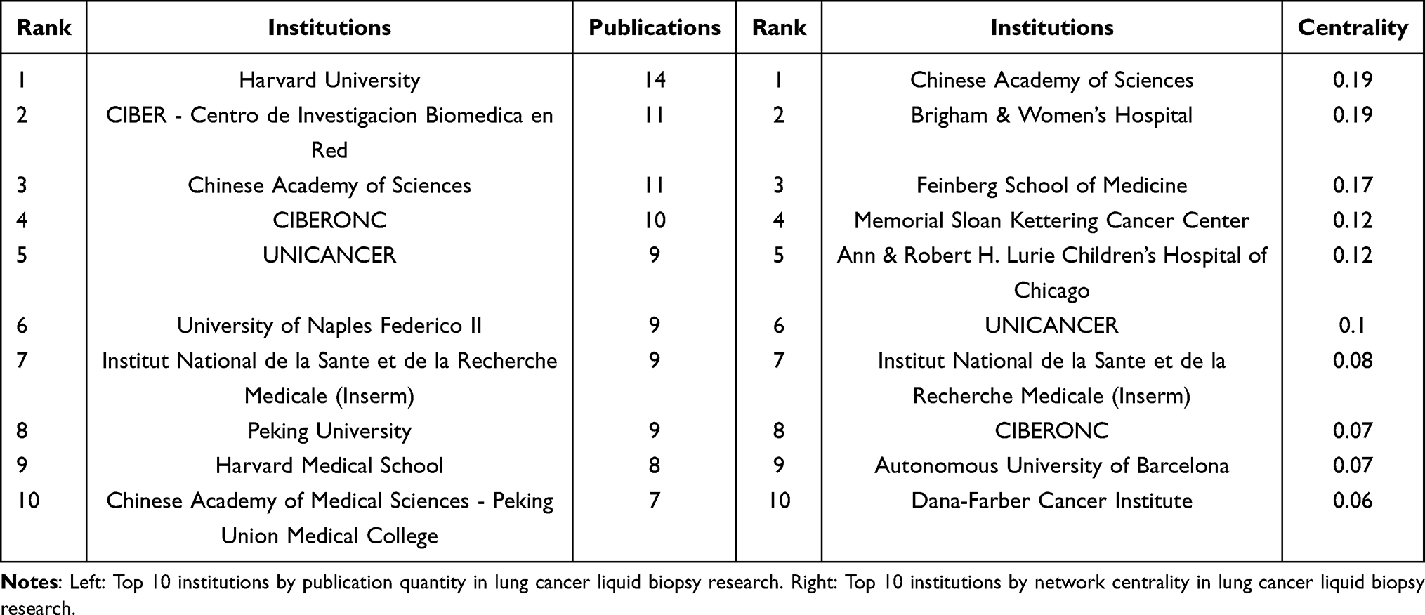 |
Table 2 Leading Institutions in Lung Cancer Liquid Biopsy Research: Top 10 by Publication Quantity and Network Centrality |
Analysis of Cited Literature
The most cited articles were authored by Sorber L (2017, Lung Cancer),28 Kapeleris J (2018, Frontiers in Oncology),29 and Cescon DW (2020, Nature Cancer) (Figure 6A),30 all focusing on ctDNA and CTCs for NSCLC management. Citation burst analysis (Figure 6B) revealed six articles with recent surges, pointing to emerging research priorities including minimal residual disease detection,31 cell-free (cfDNA) fragmentation/methylation profiling,32–34 blood tumor mutational burden (bTMB) prediction for immunotherapy,23,35 and advanced early detection approaches.36
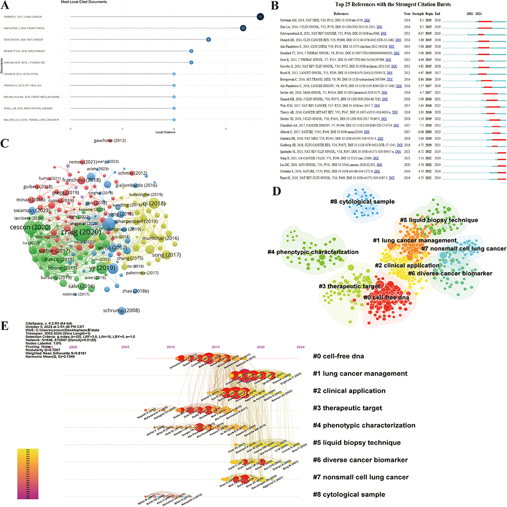 |
Figure 6 (A) Top 10 references ranked by citation frequency; (B) In-depth analysis of citation bursts; (C) Topic coupling analysis of the research literature; (D) Cluster analysis of cited references; (E) Timeline view of citation patterns. |
Thematic coupling (Figure 6C) identified five clusters: biomarkers, ctDNA, molecular detection strategies, CTCs, and treatment monitoring. Citation clustering (Figures 6D–E) confirmed the prominence of cfDNA in treatment response assessment and monitoring. Persistent clusters, such as “Lung cancer management” and “Liquid biopsy technique” reflect ongoing research pillars, while newer clusters on “Clinical application” and “Phenotypic characterization” suggest a shift toward translational and practice-oriented investigations.
Keyword Visualization Analysis
Keyword visualization analysis revealed 48 high-frequency terms (≥10 occurrences) (Figures 7A–D), primarily associated with molecular diagnostics, therapeutic interventions, particularly targeted and immune therapies, tumor biology, and biomarker evaluation, with NSCLC and small cell lung cancer (SCLC) constituting the predominant disease contexts. These co-occurrence patterns underscore the centrality of molecularly driven approaches and biomarker research in shaping the thematic structure of liquid biopsy studies in lung cancer.
 |
Figure 7 (A) Word clouds of supplementary keywords in local clusters; (B) Frequency distribution of keywords; (C) Word clouds of author-provided keywords; (D) Co-occurrence analysis of keywords. |
Keyword burst (Figure 8A) and time-zone analyses (Figures 8B–D) delineated a clear temporal evolution of research priorities. The initial phase (2006–2011) was characterized by exploratory investigations into biopsy methodologies, cfDNA methylation detection, and liquid-based cytology. The subsequent phase (2012–2021) marked a substantial expansion in molecular diagnostics, focusing on epidermal growth factor receptor (EGFR)/Kirsten rat sarcoma viral oncogene homolog mutation detection through next-generation sequencing (NGS) and digital polymerase chain reaction (PCR), thereby advancing precision oncology. In the most recent phase (2022–2024), attention shifted toward integrating liquid biopsy into personalized NSCLC treatment, exemplified by ctDNA-guided targeted therapy selection, with osimertinib and exosomal DNA emerging as focal areas. Time-zone mapping further demonstrated the progressive transition from the development of early diagnostic methodologies (2002–2007) through biomarker discovery and molecular characterization (2008–2014) to clinical translation emphasizing treatment monitoring and trial design (2015–2024).
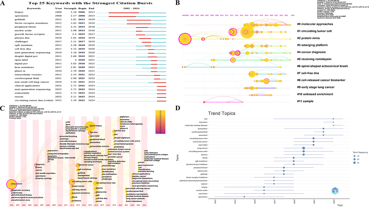 |
Figure 8 Analysis of keyword bursts and temporal trends. (A) Keyword burst analysis; (B) Keyword timeline; (C) Keyword time zone mapping; (D) Emerging research topics in liquid biopsy for lung cancer (1990–2024). |
Discussion
Global Publication Trends
Consistent with our bibliometric findings of a sharp rise in annual publications, research on liquid biopsy in lung cancer has shown an accelerating trajectory over the past three decades, particularly after 2015. This growth mirrors advances in NGS,16,23,37,38 digital PCR,28 and single-cell analysis,11 which have enhanced the sensitivity and specificity of liquid biopsy platforms. Early publications were largely exploratory, focusing on methodological feasibility, whereas recent studies emphasize translational application and clinical integration. This temporal evolution indicates that liquid biopsy has gradually transformed from a conceptual innovation to a critical tool in precision oncology. The increasing contribution of high-impact journals and the expanding citation network suggest that liquid biopsy has achieved sustained recognition as a mainstream research frontier in thoracic oncology.
International and Institutional Collaboration
Our collaboration analysis revealed substantial regional imbalances. China produced the largest volume of publications, reflecting its expanding investment in translational oncology research. However, the CPP for China was lower than those of Western countries, implying that the overall scientific influence of Chinese studies is still developing. In contrast, the United States, with fewer publications, exhibited a higher impact and broader international collaboration, as evidenced by elevated MCP ratios. Institutions such as Harvard University, the Chinese Academy of Sciences, and Icahn School of Medicine at Mount Sinai occupied central network positions, bridging different regions and forming high-density collaborative clusters. These findings underscore that productivity alone does not guarantee influence; rather, international cooperation and network centrality are critical for amplifying global visibility. For countries with high output but limited impact, strengthening cross-regional partnerships and enhancing methodological rigor are necessary to achieve greater international influence.
Biomarkers and Clinical Applications
The bibliometric mapping of keywords, co-cited references, and citation bursts highlights biomarkers as the cornerstone of liquid biopsy research in lung cancer. ctDNA2,9,12,16,30,39,40 and CTCs9–11,19,29,41 remain the most widely investigated analyses, with clinical relevance for early diagnosis, treatment monitoring, and resistance prediction.16,37,38,42 Our results revealed that minimal residual disease (MRD) detection, cfDNA methylation profiling, and bTMB are emerging hotspots, aligning with the global shift toward personalized therapy.13,18,37,43–46 Notably, exosomal nucleic acids,47 although at an earlier stage of investigation, demonstrate complementary potential, expanding the biomarker spectrum beyond ctDNA and CTCs. This diversified biomarker portfolio highlights the capacity of liquid biopsy to provide dynamic, real-time insights into tumor heterogeneity, which is especially crucial for managing NSCLC and SCLC.12,37,41,48
Research Hotspots and Emerging Directions
The evolution of thematic clusters suggests a transition from technical innovation to clinical translation. In the early phase, research prioritized optimization of detection platforms, including digital PCR13,49 and NGS technologies.16,35 More recent studies focus on clinical decision-making, exemplified by ctDNA-guided targeted therapy in EGFR-mutant NSCLC, which validates the translational utility of liquid biopsy in tailoring treatments.12 Furthermore, integrating immunotherapy-related biomarkers, such as bTMB and immune gene signatures, underscores the expanding scope of liquid biopsy beyond targeted therapy toward immuno-oncology.35 Emerging trends identified in our analysis also point to the incorporation of artificial intelligence,17,44 machine learning,17,25 and multi-omics approaches,50 which hold promise for refining predictive algorithms and stratifying patients with greater precision. These findings collectively reflect a field in rapid transition toward holistic and personalized cancer management.
Challenges and Future Prospects
The clinical promise of liquid biopsy is widely recognized; however, several critical challenges remain unresolved. Technical heterogeneity—encompassing pre-analytical variability, inconsistencies in detection sensitivity, and the absence of universally accepted standardized assays—continues to impede its widespread clinical implementation. Moreover, current research has predominantly concentrated on advanced-stage lung cancer,51,52 while applications in early detection and population-based screening remain insufficiently investigated. As indicated by recent citation bursts, expanding investigations into early-stage disease are essential to unlock the full preventive potential of liquid biopsy. In addition, despite the presence of international collaborative networks, establishing a stronger consensus on assay standardization and quality control is urgently required to ensure reproducibility, comparability, and broader clinical translation across diverse research and healthcare settings.
Looking ahead, three research priorities can be distilled from our findings. First, the development of internationally recognized standards for liquid biopsy assays is imperative to ensure methodological consistency and reproducibility across studies and clinical settings. Second, research efforts should be broadened to encompass early diagnosis, longitudinal disease surveillance, and MRD assessment, thereby extending the clinical utility of liquid biopsy beyond advanced-stage management. Third, integrating liquid biopsy with multi-omics platforms, including genomics, transcriptomics, proteomics, and metabolomics, will facilitate the construction of comprehensive biomarker landscapes that more accurately capture tumor heterogeneity. These directions are essential for addressing current limitations and for establishing liquid biopsy as a robust clinical tool that effectively bridges experimental discovery with bedside application, ultimately accelerating the implementation of precision medicine in lung cancer.
Limitations
This study has some limitations. First, the analysis was conducted exclusively using data from the WoSCC, which may have resulted in the omission of pertinent studies indexed in other databases, such as PubMed or Embase. Incorporating multiple databases in future research would improve the comprehensiveness and representativeness of the findings. Second, in the study, we primarily relied on quantitative bibliometric indicators, which are valuable for identifying trends and patterns, but may not fully capture the scientific rigor, methodological quality, or clinical relevance of individual publications. Third, the exclusion of non-English literature may have introduced potential language bias, which could limit the global applicability of the results. Finally, although visualization tools enhance the clarity of bibliometric outputs, interpreting clustering results and temporal trends inevitably involves a degree of subjectivity and may not completely reflect the most recent or ongoing developments in this rapidly evolving field.
Conclusion
We applied bibliometric and visualization tools to systematically map the research landscape, thematic hotspots, and developmental trajectories of liquid biopsy in lung cancer over the past 34 years. This field remains in a dynamic growth phase, with Cancers and Clinical Cancer Research emerging as leading journals in publication output and citation influence, respectively. Current research primarily focuses on cancer diagnosis, biomarker discovery, and treatment monitoring. Liquid biopsy through the detection of ctDNA, CTCs, and extracellular vesicles offers distinct advantages for early detection, real-time therapeutic assessment, elucidation of drug resistance, and personalized treatment. Despite ongoing technical and standardization challenges, rapid methodological advances and expanding global collaborations highlight its substantial potential to reshape clinical practice. Continued multidisciplinary and translational efforts are expected to accelerate the routine integration of this approach, ultimately improving the outcomes of patients with lung cancer.
Abbreviations
bTMB, blood tumor mutational burden; cfDNA, cell-free DNA; CIBER, Centro de Investigación Biomédica en Red; CIBERONC, Centro de Investigación Biomédica en Red de Cáncer; CPP, citations per paper; CTCs, circulating tumor cells; ctDNA, circulating tumor DNA; EGFR, epidermal growth factor receptor; MCP, multiple-country publications; MRD, minimal residual disease; NGS, next-generation sequencing; NSCLC, non-small cell lung cancer; PCR, polymerase chain reaction; SCLC, small cell lung cancer; SCP, single-country publications; WoSCC, Web of Science Core Collection.
Acknowledgment
We thank all team members involved in this study.
Author Contributions
All authors contributed significantly to the work reported, including the conception, study design, execution, data acquisition, analysis, and interpretation. They also participated in drafting, revising, or critically reviewing the article; gave final approval of the version to be published; agreed on the journal to which the article has been submitted; and agreed to be accountable for all aspects of the work.
Funding
This work has received support from the National Natural Science Foundation of China (82405334), the Development Plan for Science and Technology Innovation Teams at Jiangxi University of Traditional Chinese Medicine (No. CXTD22011), the Applied Research Cultivation Plan of Jiangxi Provincial Department of Science and Technology (20212BAG70038), the Natural Science Foundation of Jiangxi Province (No. 20212BAB216061), the Science and Technology Research Project of the Jiangxi Provincial Department of Education (No. GJJ211220); the Natural Science Foundation of Jiangxi Province (No. 20224BAB216098); and the Scientific Research Project of the Jiangxi Administration of Traditional Chinese Medicine (No. 2023A0018).
Disclosure
Yueqi Xu and Biao Huang contributed equally to this work as co-first authors. The authors affirm that the research was conducted without any apparent commercial or financial affiliations that could be construed as potential conflicts of interest.
References
1. Yankelevitz DF, Henschke CI. Overdiagnosis in lung cancer screening. Transl Lung Cancer Res. 2021;10(2):1136–1140. doi:10.21037/tlcr-20-736
2. Chaudhuri AA, Chabon JJ, Lovejoy AF, et al. Early detection of molecular residual disease in localized lung cancer by circulating tumor DNA profiling. Cancer Discov. 2017;7(12):1394–1403. doi:10.1158/2159-8290.C-17-0716
3. Ji YT, Liu SW, Zhang YM, et al. Comparison of the latest cancer statistics, cancer epidemic trends and determinants between China and the United States. Zhonghua Zhong Liu Za Zhi. 2024;46(7):646–656. doi:10.3760/cma.j.cn112152-20240208-00068
4. Riya T, Saurabh G, Gaurav G, et al. Epithelial-mesenchymal transition to mitigate age-related progression in lung cancer. Ageing Res Rev. 2024. doi:10.1016/j.arr.2024.102576
5. Pooja S, Umesh Pratap V, Ajay Kumar V, et al. Periodontal health status in patients with lung cancer: case-control study. Int J Health Sci. 2024;18(1).
6. Barretto AJB, Orda MA, Tsai PW, Tayo LL. Analysis of modular hub genes and therapeutic targets across stages of non-small cell lung cancer transcriptome. Genes. 2024;15(10):1248. doi:10.3390/genes15101248
7. Bao ZH, Hou XB, Li HL, Mao YF, Wang WR. The mechanism and progress of ferroptosis in pancreatic cancer. Acta Histochem. 2022;124(6):151919. doi:10.1016/j.acthis.2022.151919
8. Yin X, Li Y, Wang H, et al. Small cell lung cancer transformation: from pathogenesis to treatment. Semin Cancer Biol. 2022;86(2):595–606. doi:10.1016/j.semcancer.2022.03.006
9. Casagrande GMS, Silva MO, Reis RM, Leal LF. Liquid biopsy for lung cancer: up-to-date and perspectives for screening programs. Int J Mol Sci. 2023;24(3):2505. doi:10.3390/ijms24032505
10. Deng Z, Wu S, Wang Y, Shi D. Circulating tumor cell isolation for cancer diagnosis and prognosis. EBiomedicine. 2022;83:104237. doi:10.1016/j.ebiom.2022.104237
11. Hamza B, Ng SR, Prakadan SM, et al. Optofluidic real-time cell sorter for longitudinal CTC studies in mouse models of cancer. Proc Natl Acad Sci U S A. 2019;116(6):2232–2236. doi:10.1073/pnas.1814102116
12. Duffy MJ. Circulating tumor DNA (ctDNA) as a biomarker for lung cancer: early detection, monitoring and therapy prediction. Tumour Biol. 2024;46(s1):S283–S295. doi:10.3233/-220044
13. Arriola E, Paredes-Lario A, Garcia-Gomez R, et al. Comparison of plasma ctDNA and tissue/cytology-based techniques for the detection of EGFR mutation status in advanced NSCLC: spanish data subset from ASSESS. Clin Transl Oncol. 2018;20(10). doi:10.1007/s12094-018-1855-y
14. Kalluri R, LeBleu VS. The biology, function, and biomedical applications of exosomes. Science. 2020;367(6478):eaau6977. doi:10.1126/science.aau6977
15. Reclusa P, Taverna S, Pucci M, et al. Exosomes as diagnostic and predictive biomarkers in lung cancer. J Thorac Dis. 2017;9(suppl 13):S1373–S1382. doi:10.21037/jtd.2017.10.67
16. Chen M, Zhao H. Next-generation sequencing in liquid biopsy: cancer screening and early detection. Hum Genomics. 2019;13(1):34. doi:10.1186/s40246-019-0220-8
17. Li Y, Wu X, Yang P, Jiang G, Luo Y. Machine learning for lung cancer diagnosis, treatment, and prognosis. Genomics Proteomics Bioinf. 2022;20(5):850–866. doi:10.1016/j.gpb.2022.11.003
18. Maffeo D, Rina A, Serio VB, et al. The evidence base for circulating tumor DNA-methylation in non-small cell lung cancer: a systematic review and meta-analysis. Cancers. 2024;16(21):3641. doi:10.3390/cancers16213641
19. Pailler E, Adam J, Barthélémy A, et al. Detection of circulating tumor cells harboring a unique ALK rearrangement in ALK-positive non-small-cell lung cancer. J Clin Oncol. 2013;31(18):2273–2281. doi:10.1200/.2012.44.5932
20. Schrump DS, Fischette MR, Nguyen DM, et al. Clinical and molecular responses in lung cancer patients receiving romidepsin. Clin Cancer Res. 2008;14(1):188–198. doi:10.1158/1078-0432.C-07-0135
21. Tan Y, Ji L, Mo Y, Huang H, Lei X. Bibliometrics analysis of hotspots research on infertility syndromes and polystyrene. Toxicol Ind Health. 2024;40(8):465–478. doi:10.1177/07482337241257274
22. Molina-Vila MA, Mayo-de-Las-Casas C, Giménez-Capitán A, et al. Liquid biopsy in non-small cell lung cancer. Front Med Lausanne. 2016;3:69. doi:10.3389/fmed.2016.00069
23. Russo A, De Miguel Perez D, Gunasekaran M, et al. Liquid biopsy tracking of lung tumor evolutions over time. Expert Rev Mol Diagn. 2019;19(12):1099–1108. doi:10.1080/14737159.2020.1680287
24. Romero A, Serna-Blasco R, Calvo V, Provencio M. Use of liquid biopsy in the care of patients with non-small cell lung cancer. Curr Treat Options Oncol. 2021;22(10):86. doi:10.1007/s11864-021-00882-9
25. Swanson K, Wu E, Zhang A, Alizadeh AA, Zou J. From patterns to patients: advances in clinical machine learning for cancer diagnosis, prognosis, and treatment. Cell. 2023;186(8):1772–1791. doi:10.1016/j.cell.2023.01.035
26. Sholl LM, Aisner DL, Allen TC, et al. Liquid biopsy in lung cancer: a perspective from members of the pulmonary pathology society. Arch Pathol Lab Med. 2016;140(8):825–829. doi:10.5858/arpa.2016-0163-SA
27. Underwood JJ, Quadri RS, Kalva SP, et al. Liquid biopsy for cancer: review and implications for the radiologist. Radiology. 2020;294(1):5–17. doi:10.1148/radiol.2019182584
28. Sorber L, Zwaenepoel K, Deschoolmeester V, et al. Circulating cell-free nucleic acids and platelets as a liquid biopsy in the provision of personalized therapy for lung cancer patients. Lung Cancer. 2017;107:100–107. doi:10.1016/j.lungcan.2016.04.026
29. Kapeleris J, Kulasinghe A, Warkiani ME, et al. The prognostic role of circulating tumor cells (CTCs) in lung cancer. Front Oncol. 2018;8:311. doi:10.3389/fonc.2018.00311
30. Cescon DW, Bratman SV, Chan SM, Siu LL. Circulating tumor DNA and liquid biopsy in oncology. Nat Cancer. 2020;1(3):276–290. doi:10.1038/s43018-020-0043-5
31. Pantel K, Alix-Panabières C. Liquid biopsy and minimal residual disease—latest advances and implications for cure. Nat Rev Clin Oncol. 2019;16(7):409–424. doi:10.1038/s41571-019-0187-3
32. Sung H, Ferlay J, Siegel RL, et al. Global cancer statistics 2020: GLOBOCAN estimates of incidence and mortality worldwide for 36 cancers in 185 countries. CA Cancer J Clin. 2021;71(3):209–249. doi:10.3322/caac.21660
33. Ignatiadis M, Sledge GW, Jeffrey SS. Liquid biopsy enters the clinic—implementation issues and future challenges. Nat Rev Clin Oncol. 2021;18(5):297–312. doi:10.1038/s41571-020-00457-x
34. Cristiano S, Leal A, Phallen J, et al. Genome-wide cell-free DNA fragmentation in patients with cancer. Nature. 2019;570(7761):385–389. doi:10.1038/s41586-019-1272-6
35. Gandara DR, Paul SM, Kowanetz M, et al. Blood-based tumor mutational burden as a predictor of clinical benefit in non-small-cell lung cancer patients treated with atezolizumab. Nat Med. 2018;24(9):1441–1448. doi:10.1038/s41591-018-0134-3
36. Liu MC, Oxnard GR, Klein EA, et al. Sensitive and specific multi-cancer detection and localization using methylation signatures in cell-free DNA. Ann Oncol. 2020;31(6):745–759. doi:10.1016/j.annonc.2020.02.011
37. Craig AJ, von Felden J, Garcia-Lezana T, Sarcognato S, Villanueva A. Tumour evolution in hepatocellular carcinoma. Nat Rev Gastroenterol Hepatol. 2020;17(3):139–152. doi:10.1038/s41575-019-0229-4
38. Lu J, Han B. Liquid biopsy promotes non-small cell lung cancer precision therapy. Technol Cancer Res Treat. 2018;17:1533033818801809. doi:10.1177/1533033818801809
39. Bettegowda C, Sausen M, Leary RJ, et al. Detection of circulating tumor DNA in early- and late-stage human malignancies. Sci Transl Med. 2014;6(224):224ra24. doi:10.1126/scitranslmed.3007094
40. Chin RI, Chen K, Usmani A, et al. Detection of solid tumor molecular residual disease (MRD) using circulating tumor DNA (ctDNA). Mol Diagn Ther. 2019;23(3):311–331. doi:10.1007/s40291-019-00390-5
41. Ilie M, Long E, Butori C, et al. ALK-gene rearrangement: a comparative analysis on circulating tumour cells and tumour tissue from patients with lung adenocarcinoma. Ann Oncol. 2012;23(11):2907–2913. doi:10.1093/annonc/mds137
42. Lone SN, Nisar S, Masoodi T, et al. Liquid biopsy: a step closer to transform diagnosis, prognosis and future of cancer treatments. Mol Cancer. 2022;21(1):79. doi:10.1186/s12943-022-01543-7
43. Chen G, Huang AC, Zhang W, et al. Exosomal PD-L1 contributes to immunosuppression and is associated with anti-PD-1 response. Nature. 2018;560(7718):382–386. doi:10.1038/s41586-018-0392-8
44. Seijo LM, Peled N, Ajona D, et al. Biomarkers in lung cancer screening: achievements, promises, and challenges. J Thorac Oncol. 2019;14(3):343–357. doi:10.1016/j.jtho.2018.11.023
45. Chen G, Guo P, Zhao H, Zhao D, Yang D. The clinical value of combined detection of seven lung cancer-related autoantibodies in assisting the diagnosis of non-small-cell lung cancer. Biomarker Med. 2024;18(20):917–925. doi:10.1080/17520363.2024.2404379
46. Huang DH, He J, Su XF, et al. The airway microbiota of non-small cell lung cancer patients and its relationship to tumor stage and EGFR gene mutation. Thorac Cancer. 2022;13(6):858–869. doi:10.1111/1759-7714.14340
47. Hao X, Liu Z, Ma F, et al. Exosome-based liquid biopsy in early screening and diagnosis of cancers. Dose Response. 2025;23(2):15593258251344480. doi:10.1177/15593258251344480
48. Francis G, Stein S. Circulating cell-free tumour DNA in the management of cancer. Int J Mol Sci. 2015;16(6):14122–14142. doi:10.3390/ijms160614122
49. Giallombardo M, Chacártegui Borrás J, Castiglia M, et al. Exosomal miRNA analysis in non-small cell lung cancer (NSCLC) patients’ plasma through qPCR: a feasible liquid biopsy tool. J Vis Exp. 2016;(111):53900. doi:10.3791/53900
50. Wang Y, Guo Q, Huang Z, et al. Cell-free epigenomes enhanced fragmentomics-based model for early detection of lung cancer. Clin Transl Med. 2025;15(2):e70225. doi:10.1002/ctm2.70225
51. Akhoundova D, Mosquera Martinez J, Musmann LE, et al. The role of the liquid biopsy in decision-making for patients with non-small cell lung cancer. J Clin Med. 2020;9(11):3674. doi:10.3390/jcm9113674
52. Van der Linden M, Van Gaever B, Raman L, et al. Application of an ultrasensitive NGS-based blood test for the diagnosis of early-stage lung cancer: sensitivity, a hurdle still difficult to overcome. Cancers. 2022;14(8):2031. doi:10.3390/cancers14082031
 © 2025 The Author(s). This work is published and licensed by Dove Medical Press Limited. The
full terms of this license are available at https://www.dovepress.com/terms
and incorporate the Creative Commons Attribution
- Non Commercial (unported, 4.0) License.
By accessing the work you hereby accept the Terms. Non-commercial uses of the work are permitted
without any further permission from Dove Medical Press Limited, provided the work is properly
attributed. For permission for commercial use of this work, please see paragraphs 4.2 and 5 of our Terms.
© 2025 The Author(s). This work is published and licensed by Dove Medical Press Limited. The
full terms of this license are available at https://www.dovepress.com/terms
and incorporate the Creative Commons Attribution
- Non Commercial (unported, 4.0) License.
By accessing the work you hereby accept the Terms. Non-commercial uses of the work are permitted
without any further permission from Dove Medical Press Limited, provided the work is properly
attributed. For permission for commercial use of this work, please see paragraphs 4.2 and 5 of our Terms.
Recommended articles
Knowledge Atlas of the Co-Occurrence of Epilepsy and Autism: A Bibliometric Analysis and Visualization Using VOSviewer and CiteSpace
Wang Y, Huo X, Li W, Xiao L, Li M, Wang C, Sun Y, Sun T
Neuropsychiatric Disease and Treatment 2022, 18:2107-2119
Published Date: 19 September 2022
Bibliometric Analysis of Research Themes and Trends of the Co-Occurrence of Autism and ADHD
Liu A, Lu Y, Gong C, Sun J, Wang B, Jiang Z
Neuropsychiatric Disease and Treatment 2023, 19:985-1002
Published Date: 24 April 2023
Hotspots and Trends in Research on Treating Pain with Electroacupuncture: A Bibliometric and Visualization Analysis from 1994 to 2022
Hu L, Yang J, Liu T, Zhang J, Huang X, Yu H
Journal of Pain Research 2023, 16:3673-3691
Published Date: 3 November 2023
A Bibliometric Analysis of Comorbidity of COPD and Lung Cancer: Research Status and Future Directions
Fang H, Dong T, Li S, Zhang Y, Han Z, Liu M, Dong W, Hong Z, Fu M, Zhang H
International Journal of Chronic Obstructive Pulmonary Disease 2023, 18:3049-3065
Published Date: 22 December 2023
Global Research Status of Maca (Lepidium Meyenii Walp.): A Bibliometric Analysis of Hotspots, Bursts, and Trends
Li K, Zhao C, Dang M, Ren R, Fu M, Bai Y, Wang J, Zhang Q, Luan F
Drug Design, Development and Therapy 2025, 19:2329-2349
Published Date: 27 March 2025