
Back to Journals » Journal of Inflammation Research » Volume 18
The Lipid-Oxidative Stress Axis: Novel Therapeutic Targets for Podocytopathy
Received 15 April 2025
Accepted for publication 25 July 2025
Published 11 September 2025 Volume 2025:18 Pages 12505—12532
DOI https://doi.org/10.2147/JIR.S530737
Checked for plagiarism Yes
Review by Single anonymous peer review
Peer reviewer comments 4
Editor who approved publication: Dr Wenjian Li
Yingxi Liu, Manshu Zou, Yuhong Wang
Academy of Chinese Medical Sciences, Hunan University of Chinese Medicine, Changsha, 410208, People’s Republic of China
Correspondence: Yuhong Wang, Academy of Chinese Medical Sciences, Hunan University of Chinese Medicine, Changsha, 410208, People’s Republic of China, Email [email protected]
Abstract: Podocytes, as terminally differentiated cells within the glomerulus, play a decisive role in maintaining the molecular selectivity of the glomerular filtration barrier (GFB) through structural integrity and functional homeostasis. Podocyte injury not only directly compromises GFB integrity but also serves as a central pathological mechanism underlying the progression of proteinuric nephropathy. Evidence from studies highlights an intricate link between lipid metabolism dysregulation and podocyte dysfunction: Renal ectopic lipid accumulation (ELA) disrupts intracellular homeostasis via lipotoxic effects, inducing mitochondrial oxidative stress, cytoskeletal remodeling, and inflammatory cascades. Concurrently, excessive reactive oxygen species (ROS) generation coupled with compromised antioxidant defense mechanisms establishes a self-perpetuating cycle of redox imbalance. This bidirectional crosstalk within the lipid-oxidative stress axis triggers irreversible pathological alterations. This review summarizes the effects of abnormal signals during lipid synthesis, breakdown, and metabolism on podocytes, as well as the interaction between mitochondria and podocyte dysfunction through signaling mechanisms in lipid metabolism disorders. We also sorted out the key molecular pathways involved in this axis, and the regulation of key nodes of lipid metabolism (SREBP pathway, HMGCR pathway), improvement of mitochondrial function (mitochondrial dynamics and energy metabolism), and activation of antioxidant defenses (AMPK pathway) are highly promising therapeutic targets for intervening in podocyte damage and blocking the progression of the disease.
Keywords: oxidative stress, lipid metabolism, lipotoxicity, podocyte, chronic kidney disease, glomerular filtration barrier
Graphical Abstract:

Introduction
As a highly differentiated cell, podocytes are fundamental to maintaining the integrity of the glomerular filtration barrier and have a key intervention target in chronic kidney disease (CKD).1 Podocytes play a crucial role in maintaining homeostasis in response to diverse physiological and pathological stimuli. However, excessive stress can lead to podocyte maladaptation, resulting in a cascade of complex biological alterations.2 These pathological manifestations include foot process effacement (FPE), cellular vacuolization, microvillus formation, cellular hypertrophy, and ultimately podocyte detachment and loss. Concurrently, these changes trigger the activation and proliferation of parietal epithelial cells (PECs), induce structural modifications in the glomerular basement membrane (GBM) and stimulate excessive extracellular matrix (ECM) production.3 The limited regenerative capacity of podocytes exacerbates these pathological processes, resulting in a critical mismatch between podocyte coverage and the GBM surface area. This disparity leads to the emergence of uncovered GBM regions, ultimately compromising the integrity of the glomerular filtration barrier (GFB) and accelerating podocyte detachment and loss.4,5 Clinically, these pathological changes manifest as proteinuria and its associated clinical manifestations, characteristic of all podocytopathies.6 Furthermore, the migration of activated parietal epithelial cells (PECs) to the glomerular tuft contributes to the formation of sclerotic lesions.7 This process is accompanied by excessive ECM deposition, which progressively disrupts normal renal architecture. The resultant glomerular dysfunction not only impairs kidney function but also promotes the development of glomerulosclerosis and renal fibrosis, thereby accelerating the progression of chronic kidney disease (CKD).8 Therefore, great importance is attached to understanding how the structure and function of podocytes are programmed in many settings and developing techniques for the beneficial therapeutic properties of podocytes.
In studies of CKD, the pathogenic mechanisms of dyslipidemia have been extensively studied.9 Cellular dysfunction caused by accumulation of lipids, ie lipotoxicity,10 is manifested by insulin resistance, actin cytoskeletal rearrangement, mitochondrial oxidative damage and inflammatory responses.11,12 Recent evidence has confirmed that pathological lipid build-up is the result of intracellular disorders of genes or proteins that regulate lipid metabolism, which mainly affects cellular lipid metabolism in terms of lipid synthesis, uptake, storage, utilization and cell output.13,14 Therefore, Lipid metabolism is a critical link between pathology, physiological research, and biochemical communication.
In this review, we will introduce how lipids affect the structure and function of podocytes in pathological environments. We first discuss the basic structure and injury mechanism of podocytes; Then, the significance of intracellular lipid metabolism on podocyte regulation in various immune backgrounds and disease microenvironments is described. Finally, we summarize the effects on lipid metabolism in reaction to oxidative stress. We also emphasized the interaction of intracellular oxidative stress and lipid metabolism at multiple levels, mediating podocyte injury.
Podocyte Structure and Damage
Podocytes are terminally differentiated epithelial cells of the renal glomeruli, characterized by large cell bodies and intricate cytoplasmic extensions. The cell body resides within Bowman’s space, while primary processes branch into finer secondary and tertiary processes, ultimately forming foot processes (FPs).15 These foot processes (FPs) line the outer surface of the glomerular basement membrane (GBM) and anchor tightly to it through adhesion receptors and proteoglycans such as integrins, dystroglycans and agrin.16 A highly organized actin cytoskeleton underpins the structural integrity of FPs. Dynamically regulated by Rho GTPases, this cytoskeletal network not only reinforces FP adhesion to the GBM but also facilitates interdigitation with neighboring podocytes, thereby ensheathing the glomerular capillary wall while maintaining filtration slit architecture.17 In recent years, the fourth critical structural component of podocytes - the Ridge-like Prominence (RLP) - has been identified as an essential element in glomerular filtration. These specialized basal membrane protrusions extend perpendicularly from the podocyte cell body, serving as primary adhesion devices that establish stable connections between the podocyte and glomerular basement membrane (GBM).18 These staggered FPs create filtration slits, which are bridged by an extracellular structure called the slit diaphragms (SDs).19,20 These SDs represent sophisticated extracellular structures that function as ultimate barrier against macromolecular proteinuria.21,22 These specialized podocyte structures cooperate synergistically with glomerular endothelial cells and the GBM to form the tripartite glomerular filtration barrier (GFB).23 (Figure 1) The filtration process involves sequential passage of plasma constituents through Fenestrated endothelial cells (size-selective barrier), The GBM (charge-selective collagenous matrix) and SD-bridged filtration slits (final macromolecular barrier). This coordinated ultrastructural organization enables the production of primary ultrafiltrate while maintaining essential plasma proteins within the vascular compartment.24,25
 |
Figure 1 The normal kidney and glomeruli structure. The podocyte, glomerular basement membrane and capillary endothelium form the GFB and work together to filter blood. |
The study of podocyte function is not a new field; these cells were first observed in the glomerular filtration barrier via electron microscopy in the 1950s.26 In adult kidneys, there are approximately 500–600 podocytes in each glomerular capillary tuft.26 In addition to their adhesive functions essential for maintaining filtration-barrier integrity, podocytes also directly contribute to the synthesis and remodeling of various GBM components.27 Furthermore, it also actively secretes vascular endothelial growth factor (VEGF) as well as a variety of pro-inflammatory and anti-inflammatory cytokines,28,29 wild synthesizing key complement cascade proteins. Through highly dynamic paracrine and autocrine signaling—mediated by endothelin-1 (ET-1), VEGF, TGF-β, BMP-7, latent TGF-β–binding protein-1 (LTBP1), and extracellular vesicles—podocytes modulate mesangial cell and local immune responses.28,30,31 Emerging evidence indicates that podocytes not only perform innate immune roles32—expressing pattern recognition receptors and secreting antimicrobial peptides—but also exhibit adaptive immune functions by presenting antigens and releasing pro-inflammatory or regulatory chemokines,32,33 thus precisely regulating immune homeostasis within the glomerular microenvironment.
However, despite their functional sophistication, podocytes are intrinsically vulnerable to injury due to their unique cytoarchitecture. As terminally differentiated epithelial cells with limited proliferative capacity, they endure substantial hemodynamic forces from glomerular capillary blood flow, primarily comprising two mechanical components: circumferential stress and shear stress.34 When exposed to excessive stress, podocytes undergo a series of complex pathological changes, including foot process effacement (FPE), hypertrophy, cell body attenuation, rapid dynamic changes of pseudocyst filling, emptying, and rupture, pseudocyst formation, and podocyte shedding.5,35–37 In glomerular disease, the “Foot Process Effacement (FPE)” causes a decline in the filtration barrier and is recognized as a pathological indicator of podocyte injury.38,39
In a clinical study of children with idiopathic nephrotic syndrome (INS), researchers found that at least 66% of patients exhibited serum autoantibodies targeting podocytes. Subsequent screening identified 14 specific podocyte autoantigens, with antibody titers showing significant positive correlation with 24-hour urinary protein excretion. Notably, following effective hormone or immunosuppressive therapy, the levels of these autoantibodies decreased markedly, and this decline was highly consistent with the time window for proteinuria resolution. These findings indicate that podocyte autoantibodies may serve as dual-purpose biomarkers for both disease severity assessment and real-time therapeutic monitoring, enabling personalized treatment evaluation.40 Kotaro Haruhara et al demonstrated in a retrospective clinicopathological study that podocyte density outperforms absolute podocyte count in predicting renal outcomes in obesity-related glomerulopathy (ORG), as it more sensitively reflects the vulnerability of the filtration barrier caused by mismatched glomerular enlargement and podocyte spreading. The study results showed that patients with reduced podocyte density exhibited independently associated severe proteinuria, marked glomerulomegaly, and inferior renal survival, indicating that such patients experience rapid disease progression and warrants close clinical attention.41 Another study on INS revealed that loss of nuclear glucocorticoid receptor (GR) expression in podocyte was correlates with delayed therapeutic response to glucocorticoids. By analyzing of podocyte-specific markers in renal biopsies demonstrated potential for predicting glucocorticoid efficacy, supporting targeted investigation of podocyte GR protein in INS patients.42 Although this study shows promise of podocytes as diagnostic biomarkers for kidney diseases, an unequivocal molecular signature for identifying podocyte injury requires validation prior to clinical applications.
Nephrin
As a transmembrane protein, Nephrin is in the SD of mature glomeruli and has eight extracellular immunoglobulin-like structural domains. It is a key protein among the molecules that assemble and strengthen the SD.43 Nephrin is required to keep the glomerular filter in a healthy state, whose downregulated expression is considered an early indication of glomerular damage. Long-term studies have shown that reduced Nephrin levels are associated with increased proteinuria and severity of podocyte injury.44–46
Podocin
Alongside Nephrin, Podocin constitutes another essential component in maintaining slit diaphragm (SD) integrity. This transmembrane protein, encoded by the NPHS2 gene, exhibits podocyte-specific expression during glomerular maturation. Podocin demonstrates a unique bipolar localization flanking the SD,47 where it functions as a critical scaffolding molecule. It facilitates the structural integration of tight junction proteins (TJs) - key regulators of paracellular transport for ions, solutes, and macromolecules48- with the underlying actin cytoskeleton.47,49 Pathogenic mutations in Podocin induce significant cytoskeletal alterations, manifesting as abnormal cortical redistribution of actin filaments and characteristic cytoplasmic depletion.50
Podoplanin
Podoplanin is a 43-kDa transmembrane podocyte glycoprotein that is associated with cell motility and modeling of the actin cytoskeleton. In an in vitro study, the formation of cell extensions increasing with ectopic expression of podoplanin in podocyte, which also enhances cell adhesion and migration, and induces morphological changes in these cells.51,52
These proteins also require an interface molecule, CD2AP—a bridging protein on the cytoplasmic side of the plasma membrane—to bind cytoskeletal proteins. Podocin binds with CD2AP and Nephrin through its C-terminal domain and directly interacts with CD2AP in vivo.53–55 Moreover, podocytes are firmly attached to the GBM, and their function is maintained through podocyte-associated molecules, including α-actinin-4, glomerular epithelial protein 1, Wilms tumor antigen 1, synaptopodin, and dystroglycan. These molecules exhibit a significant positive correlation with acquired proteinuric diseases.56 Initial podocyte injury serves as the pivotal trigger for mesangial expansion and subsequent glomerular albuminuria.57–59
In a genetically engineered rat model of podocyte depletion, 20% podocyte loss resulted in glomerular mesangial expansion and transient proteinuria; 40% depletion led to focal segmental glomerulosclerosis (FSGS) lesions and capsular adhesions, while depletion exceeding 40% caused severe mesangial expansion and global glomerulosclerosis.60 Podocyte injury arises from multiple causes. In addition to genetic variants, environmental factors such as immunological, infectious, toxic, hemodynamic, and obesity-related stressors can damage podocytes.59,61–65
Lipid Metabolism
Lipids are a widely existing class of compounds with multiple important biological functions, acting through the synergistic effects of multiple enzymes, binding proteins, and receptors.66 Lipids display remarkable structural diversity, such as a wide variety of chemical structures of cellular lipids, numerous stereoisomers, and compositional variability. This characteristic demonstrates the multiple roles of lipids in physiology and pathology.67–69 In the Integrated Lipid Classification System (Comprehensive Classification System for LIPID, https://www.lipidmaps. In org), the lipid classification system consists of eight lipid classes, each with its own subclassification hierarchy. Such as: Fatty Acyls (FA), Glycerolipids (GL), Glycerophospholipids (GP), Sphingolipids (SP), Sterol Lipids (ST), Prenol Lipids (PR), Saccharolipids (SL), Polyketides (PK).66
Disorders in lipid metabolism are linked to diseases such as obesity, hyperlipidemia, lipid deposition disease, and metabolic syndrome. They alter gene and protein expression, resulting in cytokine and signaling pathway dysregulation.10 An excess of lipids that accumulate in non-adipose tissue causes cell dysfunction or cell death, an appearance known as Lipotoxic.10,70 The molecular mechanisms of underlying lipotoxicity encompass ER stress, oxidative stress, mitochondrial dysfunction, impaired autophagy, and inflammatory.71,72
Podocytes exhibit a uniquely high membrane cholesterol and sphingolipid content, which is essential for maintaining the structural integrity and dynamic flexibility of their intricate FPs.73 Moreover, podocytes rely heavily on lipid rafts—cholesterol- and sphingolipid-enriched microdomains within the plasma membrane—that serve as critical platforms for organizing signaling molecules and receptors.73,74 These lipid rafts facilitate efficient signal transduction, including insulin signaling and mechanosensitive pathways,75 both of which are vital for podocyte function, survival, and adaptation to mechanical stress.
In addition to their reliance on lipid rafts, a defining aspect of podocyte lipid metabolism is the expression of specific regulatory enzymes, such as sphingomyelin phosphodiesterase acid-like 3b (SMPDL3b),76 which plays a pivotal role in modulating ceramide homeostasis. It is suggested that SMPDL3b enhance podocyte resilience by regulating ceramide-S1P homeostasis76 while also contributing to podocyte injury through the activation of STING.77 This delicate balance underscores the critical need for precise regulation of lipid metabolism to safeguard podocyte integrity and function.
Podocyte Injury Caused by Lipid Metabolism Disorders
The intracellular lipid homeostasis is balanced by regulating the dynamic changes of lipid synthesis, catabolism, and storage (Figure 2). Numerous laboratory studies have shown that intracellular lipid overload in podocytes is an important factor causing proteinuria in chronic kidney disease (CKD), such as diabetic nephropathy (DN) and focal segmental glomerulosclerosis (FSGS).78–81 The disorder of lipid metabolism is not only a consequence of kidney injury but also aggravates the progression of kidney injury.
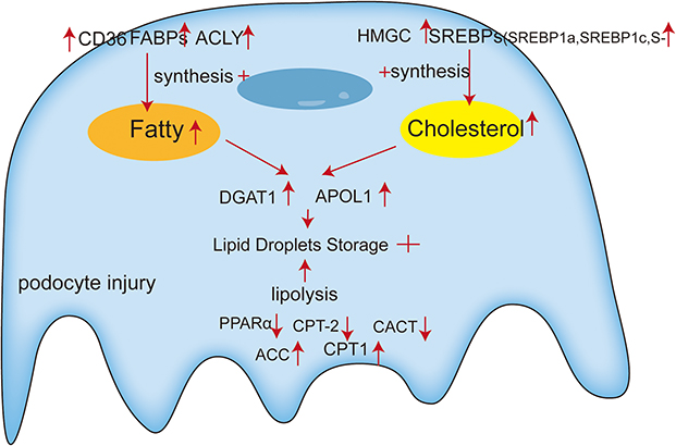 |
Figure 2 Lipid metabolism in the podocyte. In this cartoon, we illustrate the intracellular lipid accumulation is governed by an imbalance between the syntheses, catabolism and storage of FAs and cholesterol. Our results provide insights into the expression of key molecules of lipid metabolism disorders in the podocyte injury. |
Lipid Synthesis
The balance of intracellular lipids is attributed to dynamic lipid synthesis, decomposition and storage. As a part of the glomerular, the podocytes have a huge energy demand to maintain the physiological function of the glomerulus.82 ATP in podocytes is generated mainly through glycolysis and oxidative phosphorylation.82–84 Podocytes exhibit unique cellular structures, such as the integrity of foot processes (FPs), which is critical for glomerular filtration and determine the different regionalization of ATP generation. Mitochondria must be properly localized and mobilized within podocytes to meet cellular energy needs. Usually, mitochondria are absent in the foot processes (FPs) of podocytes, which is likely because mitochondria are larger in volume than the FPs. Therefore, the energy metabolism in the portion of the FPs is provided by glycolysis.82 Studies demonstrate that podocyte energy metabolism relies on both glycolysis and mitochondrial respiration, with their contributions varying under different conditions. Reduced mitochondrial capacity or glycolytic disorder can aggravate abnormal podocyte function.85,86 Beyond their role as essential energy sources, lipids are integral to the structural organization of mitochondrial membranes.87,88 Disruption of mitochondrial oxidative metabolism and elevated ROS levels compromise lipid metabolism in mitochondria, resulting in the lipid excess. In turn, lipid overload can damage mitochondrial DNA, RNA, and respiratory chain proteins, ultimately leading to functional decline.10,89 In an in vitro model of podocyte injury with knockdown PKM2, the expression of pyruvate kinase M2 (a key glycolytic enzyme) rapidly declines, accompanied by reduced glycolytic flux, glomerular and podocyte injury, loss of foot processes, and proteinuria. Inhibition of the glycolytic pathway leads to ATP insufficiency, resulting in cytoskeletal remodeling and podocyte apoptosis.90 Furthermore, reduced expression of pyruvate kinase M2 in podocytes was also found in renal biopsies from patients with hypertensive nephropathy and diabetic kidney disease (DKD).86 Abnormal mitochondrial energy metabolism in podocytes is closely related to lipid metabolism disorders. The roles of lipid in podocytes metabolic and signaling will be discussed below.
Fatty Acids (FAs)
FAs are important nutrients stored in adipose tissue as triglycerides, enabling humans to tolerate prolonged starvation, fasting, and metabolic diseases (such as febrile illnesses) and serve as the main substrates for podocyte mitochondria.91,92 Podocyte injury upregulates de novo lipogenesis (DNL) programs, leading to increased production of FAs. Fatty acid synthesis (FAS) is coordinated by several key enzymes, including acetyl-CoA citrate lyase (ACLY), Acetyl-CoA carboxylase (ACC), fatty acid synthase (FASN), and Stearyl-CoA desaturase 1 (SCD 1). Podocyte-specific ACCβ overexpression brings about serious podocyte damage in animal models of diabetic nephropathy,93 whereas inhibition of ACC reduces gene expression of fibrosis markers and apoptosis in podocytes.94 In addition, ACLY serves as a key enzyme regulating FAS and fatty acid β-oxidation (FAO), which is located in the cytoplasm and nucleus and responsible for converting citric into acetyl-CoA in the cytoplasm.95 Studies on renal reperfusion injury evidence that suppressing ACLY transcription reduces renal fibrosis, accompanied by enhanced fatty acid oxidation (FAO) and decreased lipid accumulation.96 Overall, these studies highlight the significant role of FAs in the controlling podocyte injury and renal dysfunction.
Fatty Acid Translocase CD36
The CD36 is a class B scavenger receptor and lipid sensor that is widely present in many immune and non-immune cells including macrophages,97 microvascular endothelial cells98 and podocytes.99 It serves as a multifunctional receptor, mediating signaling in response to damage-associated molecular patterns (DAMPs) and pathogen-associated molecular patterns (PAMPs), as well as functioning as a transporter for long-chain free fatty acids.97 The transport of FAs by CD36 involves a complex internalization process. During this process, FAs bind to CD36 on the cell membrane, activating its downstream kinase LYN. LYN phosphorylates and inactivates CD36’s palmitoyl transferase DHHC5 at the Tyr91 site, leading to the depalmitoylation of CD36 by acyl protein thioesterase 1 (APT1). This modification enables CD36 to recruit another tyrosine kinase, spleen tyrosine kinase (SYK), which phosphorylates c-Jun N-terminal kinase (JNK) and VAV proteins, initiating the endocytic uptake of FAs.100 Disruption of CD36-dependent FA uptake can be achieved by interrupting CD36 internalization, which is mediated by blocking either LYN or SYK.101,102 In podocytes, CD36 plays a dual role in lipid metabolism and inflammation. Beyond its function in FA transport, CD36 has been implicated in promoting podocyte injury through the activation of the NLRP3 inflammasome. Recent studies have shown that CD36-mediated NLRP3 inflammasome activation leads to the release of pro-inflammatory cytokines such as IL-1β and IL-18, exacerbating podocyte injury and contributing to glomerular damage.103 Knockdown of CD36 in experimental models has been shown to significantly reduce inflammation, attenuate NLRP3 inflammasome activation, and alleviate podocyte injury, highlighting its potential as a therapeutic target in kidney diseases.104 Moreover, CD36’s role in podocyte injury is further complicated by its interaction with oxidative stress and lipid peroxidation products. In conditions such as diabetic nephropathy, CD36 facilitates the uptake of oxidized low-density lipoprotein (oxLDL), leading to intracellular lipid accumulation, mitochondrial dysfunction, and podocyte apoptosis.105 This process is exacerbated by the generation of reactive oxygen species (ROS), further amplifies CD36-dependent inflammatory signaling and podocyte damage.61
ATP Citrate Lyase (ACLY)
The ACLY is a key rate-limiting cytosolic enzyme involved in de novo fat synthesis by catalyzing the conversion of citrate to oxaloacetate (OAA) and Acetyl-CoA, while hydrolyzing ATP to ADP and phosphate.106 The role of Acetyl-CoA in metabolism represents a key node as it facilitates crucial biochemical reactions, which includes the synthesis of FAs, cholesterol and acetylcholine, along with the acetylation of protein substrates including histones.107,108 Under most physiological and nutritional conditions, Acetyl-CoA serves as the primary means through which carbon enters the tricarboxylic acid (TCA) cycle109 - a series of chemical reactions that produce energy by oxidising carbohydrates, FAs and Acetyl-CoA. ACLY plays a role in acetyl-CoA production by cleaving citrate exported from the mitochondrial TCA cycle.110
In the renal tissue of overweight or obese patients (ORG) and ob/ob BTBR mice, ACLY is highly expressed the enzyme is linked with ELA increasing, glomerulosclerosis, and albuminuria.111 Administration of the ACLY inhibitor BMS-303141 reduces serum lipids level and renal ELA, ameliorates renal injury, and tubulointerstitial fibrosis. Moreover, it decreases the levels of various lipogenic enzymes such as ACC, FAS and HMGCR in db/db mice.112 In mouse embryonic fibroblasts, ACLY deletion resulted in Acetyl-CoA synthetase 2 (ACSS2) upregulation.113 ACSS2 regulates histone acetylation, produces acetyl-CoA in the promoter region of TFEB target genes, and enhances the transcriptional control of genes associated with lysosomal biogenesis and autophagy.114 For example, ACSS2 epigenetically induces raptor expression through histone H3K9 acetylation, thereby facilitating activation of raptor/mTORC1 pathway.
Fatty Acid-Binding Proteins (FABPs)
The FABPs were initially found to be intracellular proteins that are key mediators of local and systemic metabolic and inflammatory processes, and are therefore important therapeutic targets for immune and metabolic diseases.115 Functionally, FABPs regulate intracellular lipid homeostasis by modulating FA transport and metabolism between nuclear and extranuclear compartments.116,117 There are many isoforms of FABPs. To date, scholars have identified 12 FABP genes. Tissue-specific deletion of FABP genes (eg, FABP4 in adipocytes) can impair lipid storage or mobilization, leading to altered adipose tissue homeostasis.118 In normal kidneys, FABP 4 is mainly expressed in glomerular mesangial cells, peritubular capillary endothelial cells, and cortical and medullary venous,119 which was found to be a potential vector in mediating the inflammatory response to renal interstitial fibrosis.120 Induction of ectopic FABP4 expression occurs in glomerular injury and the level of ectopic FABP4 expression is relevantly linked with proteinuria and renal insufficiency.121 In recent reports, Heart-type fatty acid binding protein (H-FABP) exacerbates FA-induced metabolic disorders, inflammation, and oxidative stress markers in podocytes and aggravates FA-induced damage.122 The level of H-FABP was upregulated in both glomeruli of ORG and obese db/db mice, and the expression of H-FABP was more pronounced in the glomeruli of ORG patients. It was hypothesized that glomerular hypertrophy might be related to the abnormal expression level of H-FABP in glomeruli.123 Although human liver-type fatty acid-binding protein (hL-FABP) is only expressed in the proximal renal tubules, it can serve as a predictor of renal injury by binding excess FFA in proteinuric renal disease.124,125 Urinary hL-FABP is elevated in ischemic renal disease.126 Increased level of L-FABP in tubular cells protects anti-GBM GN mice from worsening tubulointerstitial and glomerular injury.127 Together, these studies established the significant role played by FABPs in regulating FA metabolism within podocytes.
Cholesterol
Synthesized by nearly all cell types, serves dual physiological roles as a critical component of cell membrane stability and the essential precursor for steroid hormone biosynthesis. These cholesterol-derived signaling molecules - including glucocorticoids, mineralocorticoids, sex hormones, and vitamin D- constitute a vital regulatory network governing systemic homeostasis through carbohydrate metabolism regulation, sodium balance maintenance, and modulation of reproductive and skeletal development.128,129 The endoplasmic reticulum (ER) functions as a primary organelle for protein folding regulation, secretion, Ca2 + storage and release, as well as lipid synthesis in eukaryotic cells.130 A multitude of studies have validated the pivotal function of the ER in cholesterol metabolism.131 Cholesterol has the capacity to interact with multitudes transmembrane proteins, continue contributing to the maintenance or alteration of their conformation. In addition to this, Cholesterol also engages with numerous sterol transport proteins to promote cholesterol transport and control its subcellular distribution.132 The biosynthesis of cholesterol is an energy-demanding process that requires substantial inputs of acetyl-CoA, ATP, oxygen, and the reducing agents NADPH and NADH. The synthesis process of this key substance involves five basic steps:133–135 1) condensation of three Acetyl-CoA units to form HMG-CoA (3-hydroxy-3-methylglutaryl-CoA); 2) The reduction of HMG-CoA to mevalonate, catalyzed by HMG-CoA reductase (HMGCR), requires NADPH as a reducing agent; 3) The mevalonate is consecutively phosphorylated by three ATP-dependent phosphorylation reactions in the presence of three kinases. It is then decarboxylated to form the active isopentenyl unit, isopentenyl diphosphate; 4) Six Isoprenoid units to produce Squalene; 5) Then, The Squalene is converted to squalene 2,3-epoxide by squalene epoxidase in the ER. Squalene 2,3-epoxide is then cyclized by lanosterol cyclase to form lanosterol. Finally, the conversion of lanosterol to cholesterol occurs within the membranes of the ER.132,136–138 This process is regulated by three key factors: Sterol Regulatory Element Binding Protein 2 (SREBP2),139 3-Hydroxy-3-methylglutaryl-CoA reductase (HMGCR)140 and Squalene monooxygenase (SM).141
Sterol Regulatory Element Binding Proteins (SREBPs, Including SREBP1a, SREBP1c and SREBP2)
SREBPs are members of the bHLH-Zip family, function as key transcriptional regulators in cholesterol and fatty acid metabolism, with their activation and subsequent induction of cholesterol synthesis dependent on the assistance of SCAP (SREBPs cleavage activating protein).142,143 Escorted by SCAP, SREBPs are transported from the ER to the Golgi for proteolytic cleavage, releasing their NH2-terminal domain. This domain then activates lipid synthesis genes, promoting cholesterol production.144,145 SREBPs can regulate the transcription activation of HMG coenzyme A reductase, as well as the transcription of genes encoding other key enzymes in the cholesterol synthesis pathway, such as farnesyl diphosphate synthase (FPPS) and squalene synthase (SQS).146,147 Studies have shown that the increased cholesterol in podocyte is accompanied by increased expression of LDL Receptor (LDLR), SREBPs (SREBP1 and SREBP2), and 3-hydroxy-3-methylglutaryl-CoA reductase (HMGCR).148 This dysregulation leads to the intracellular accumulation of cholesterol and triglycerides (TGs), which disrupts podocyte function and contributes to glomerular injury. For example, SREBP2 activation in renal tissue mediates the accumulation of triglycerides and cholesterol, leading to podocyte damage and proteinuria.149 Podocyte-specific lipid rafts, characterized as dynamic microdomains enriched in cholesterol and sphingolipids, are essential for preserving the structural stability of the SD. These specialized membrane domains are involved in nephron phosphorylation and organization of the Glomerular SD. They serve as dynamic platforms that recruit a diverse array of molecules, including receptors and signaling proteins, essential for cellular recognition and signal transduction processes.150–152 This activation is associated with the upregulation of glomerulosclerosis-associated factors, including VEGF, PAI-1, collagen type IV, and fibronectin.153 For instance, studies in db/db mice, a model of type 2 diabetes, have demonstrated that podocyte injury is accompanied by elevated SREBP1 and SREBP2 levels, along with increased cholesterol and TG deposition in glomeruli.154 This lipid overload exacerbates podocyte injury by inducing oxidative stress, mitochondrial dysfunction, and apoptosis.155 Furthermore, Pharmacological inhibitors of SREBP activation, such as fatostatin and betulin, have shown efficacy in reducing lipid accumulation and preserving glomerular function in preclinical models.156–158 Therefore, podocyte-specific insights into SREBP pathways, including their effects on lipid rafts, autophagy, and oxidative stress, provide a deeper understanding of the mechanisms underlying lipid-induced podocyte damage.
3-Hydroxy-3-Methylglutaryl Coenzyme A Reductase (HMGCR or HMG-CoA Reductase)
HMGCR is a key enzyme in the mevalonate pathway, catalyzing the rate-limiting step of converting HMG-CoA to mevalonate. Mevalonate serves as a critical intermediate for the synthesis of isoprenoids, such as cholesterol, bile acids, and steroid hormones,159 and non-sterol isoprenoids.160 As the rate-limiting enzyme in cholesterol biosynthesis, HMGCR is a primary target of statins, which are widely used to lower serum cholesterol levels.161 Beyond their lipid-lowering effects, statins exhibit renoprotective properties, including anti-inflammatory and immunomodulatory effects, which have been demonstrated in various kidney disease models. For instance, studies have demonstrated that fluvastatin significantly lowers mortality in experimental deoxycorticosterone-acetate (DOCA) salt-treated rats. This protective effect is achieved through multiple mechanisms, including reducing urinary protein excretion, glomerular hyperplasia, macrophage infiltration, and suppressing glomerulosclerosis. Studies have demonstrated that fluvastatin significantly lowers.162
In podocytes, statins exert significant protective effects against oxidative stress and injury, primarily through the modulation of key signaling pathways and epigenetic mechanisms. Research has demonstrated that statins prevent oxidative LDL-induced glomerular podocyte injury by activating the phosphatidylinositol 3-kinase/AKT signaling pathway.163 Additionally, Atorvastatin improves renal pathology by suppressing HDAC activity, enhancing H3 and H4 acetylation in glomerular mesangial cells,164 and downregulating MALAT1 and miR-200c to protect MPC-5 cells from pyroptosis while reducing renal oxidative stress levels.165
Hyperglycemia-induced podocyte injury is another critical area where statins demonstrate protective effects. High glucose levels disrupt F-actin cytoskeletal organization, reduce the expression of podocyte-specific markers such as synaptopodin and Wilms tumor-1 (WT-1), and decrease the production of bone morphogenetic protein-7 (BMP-7). Pitavastatin has been shown to mitigate these effects by inhibiting Rho kinase activation, thereby preserving podocyte structure and function under hyperglycemic conditions.166 Clinical studies further support the renoprotective effects of statins, particularly in patients with chronic kidney disease (CKD). For example, a 6-month treatment with simvastatin in hypercholesterolemic patients with chronic glomerulonephritis, which resulted in significant reductions in total cholesterol, LDL cholesterol, triglycerides, urinary protein excretion, and urinary podocyte excretion.167 Moreover, statin therapy has been associated with reduced risks of all-cause mortality, cancer mortality, and cardiac mortality in hyperlipidemic patients with CKD and end-stage renal disease (ESRD). Notably, hydrophilic statins appear to offer superior benefits compared to lipophilic statins, suggesting that statin selection may influence clinical outcomes.168 In podocytes, the protective mechanisms of statins extend beyond cholesterol reduction. Statins modulate key signaling pathways, including RhoA/ROCK, PI3K/AKT, and Wnt/β-catenin. Fluvastatin exerts its protective effects by inhibiting the RhoA, thereby preventing its membrane localization and activation.169 This inhibition suppresses downstream Rho-associated kinase (ROCK) activity. The suppression of the ROCK pathway plays a crucial role in maintaining the stability of the podocyte cytoskeleton, preventing the reorganization and disruption of actin filaments (F-actin). This mechanism is particularly significant in diabetic nephropathy, where hyperglycemia leads to the overactivation of the RhoA/ROCK pathway, resulting in podocyte injury and detachment.170 By inhibiting this pathway, statins reduce podocyte apoptosis and the occurrence of proteinuria, thereby preserving glomerular function.
In summary, HMGCR and the mevalonate pathway are central to podocyte biology, with statins providing multifaceted protection against podocyte injury through lipid-lowering, anti-inflammatory, and cytoprotective effects. Targeting cholesterol metabolism and its downstream pathways offers a promising therapeutic strategy to preserve podocyte function and slow glomerular disease progression. Future research should focus on podocyte-specific mechanisms to optimize statin-based treatments for kidney diseases.
Lipid Storage
In mammals, there exist two primary types of adipose tissue: White adipose tissue (WAT) and Brown adipose tissue (BAT). Brown adipose tissue (BAT) functions as a thermogenic organ, producing heat via mitochondrial uncoupling protein 1 (UCP1). It relies on multiple metabolites, such as glucose, lipids, and amino acids, to fuel thermogenic processes, while triglycerides (TG) act as a stored energy source.171 White adipose tissue (WAT) is the main form of fat storage in mammals, storing lipids in the form of TG. In addition to this, WAT is also a major secretory and endocrine organ, such as leptin.172 WAT consists of adipocytes, adipocyte progenitors, leukocytes and endothelial cells. Although adipocytes constitute only 20–40% of the total cell population, they account for more than 90% of the tissue volume.173,174 WAT takes up excess lipids and stores them as triglycerides (TG) to prevent ectopic lipid deposition175 and adverse metabolic complications, while on the other hand, it acts as a source of energy, releasing lipids as non-esterified fatty acids (NEFA) when energy is required.176
Lipid Droplets (LDs) are the main storage forms of neutral lipids in cells, which act as central agents, coordinating the pathway of intracellular lipid uptake, distribution, storage and use, and release FAs to replenish energy during energy shortage.177 The biogenesis and breakdown of LDs are closely linked to cellular metabolism, which is essential for buffering the content of toxic lipid species. Thus, LDs promote coordination and communication between different organelles, which are an important hub for cellular metabolism.178 As a highly dynamic organelles, changes in LDs closely reflect the cellular metabolism and nutrient availability cycles: When nutrients are sufficient, lipids are stored and later mobilized to provide energy during starvation or to synthesis phospholipid when membrane production is required. Lipid droplets also sequester potentially toxic lipids, thereby playing a crucial role in preventing lipotoxicity and oxidative stress.179,180 It is currently accepted that lipid droplet biosynthesis occurs mainly in the ER: 1) Neutral lipid synthesis and lens formation. Riacylglycerol (TAG) synthesis and the activity of cholesteryl ester synthase deposit neutral lipids between the lobules of ER bilayers.181,182 After exceeding a specific concentration threshold, neutral lipids delaminate and aggregate to form a lens.183,184 However, it remains unclear whether neutral lipid crystals form randomly throughout the endoplasmic reticulum or if their formation is localized to specific sites. 2) The crystalline structure promotes the growth of nascent LD in response to a variety of LD biogenesis factors.176 3) Swelling of the neutral lipid lattice leads to the emergence of LD derived from the ER membrane.185 After budding, LD swell and grow by fusion or localized lipid synthesis.186
LD formation is mediated by diacylglycerol-acyltransferase 1 (DGAT1). DGAT1 upregulation promotes fatty acid (FA) storage as triglycerides and LD. Conversely, DGAT1 inhibition disrupts lipid homeostasis, leading to excessive FA oxidation in mitochondria. This process generates high levels of reactive oxygen species (ROS), triggers cytochrome c release, and induces apoptosis.187 APOL1 usually locate in the ER and podocytes plasma membrane, of which overexpression can cause podocyte toxicity in the kidney.188 However, specific APOL1 variants promote LD formation, reducing cytotoxicity, enhancing autophagic flux, and decreasing cell death.189 These variants may protect cells by modulating lipid metabolism or interacting with LD-associated proteins.
Thus, the role of LDs in podocytes and the impact of aberrant LD metabolism on cellular function are key research focuses on CKD.
Lipid Catabolism
Lipid catabolism is defined as the metabolic process by which triacylglycerols, stored in lipid droplets (LDs) of cells, undergo sequential hydrolysis to produce FAs and glycerol.190 FAs are not only efficient energy substrates but also essential components for the synthesis of membrane lipids and intracellular signaling molecules.191
Intracellular lipolysis generates energy through the release of FAs, which are then activated as Acyl-CoA into the mitochondria for β-oxidation cycle. The main process for FAs degradation is Mitochondrial Fatty Acid β-oxidation (FAO)91 as well as the primary source of ATP. After FAs enter the cells, Fatty Acyl-CoA synthase catalyze the thioesterification reaction of FAs with coenzyme A to synthesize Fatty acyl-CoA thioesters. Next, Fatty acyl-CoA esters enter the mitochondrial matrix via the carnitine transport cycle. The L-carnitine system enables long- and medium-chain fatty acyl carboxylic acids to pass through the mitochondrial membrane. Carnitine palmitoyltransferase-1 facilitates the transfer of acyl groups from coenzyme A to L-carnitine, creating acyl-carnitine esters at the outer mitochondrial membrane. Acyl-carnitine esters are converted back to Acyl-CoA esters by Carnitine palmitoyltransferase-2. Finally, Acyl-CoA esters undergo the β-oxidation pathway, generating acetyl-CoA and a Fatty acyl-CoA ester that is two carbons shorter.192,193 Impairment of FAO leads to abnormal lipolysis, resulting in abnormal lipid concentration and lipotoxicity.177
Peroxisome Proliferator-Activated Receptor α (PPARα)
The PPARα plays an indispensable role in podocytes FAO process.194 PPARα is a transcription factor that forms a complex with the Retinoid X receptor α (RXRα) as a heterodimer and binds to the PPAR response elements (PPREs) sequence in the promoter region of target genes through the PPARα DNA-binding domain (DBD) and control of the transcription of many FAO-related which participate in lipid regulation.195 Transcription initiation of PPARα requires several lipophilic molecules to activate it, include Natural Saturated FAs, Unsaturated FAs, Polyunsaturated FAs (PUFAs) and synthetic ligands, collectively known as PPARα activator.196 After PPARβ/δ activation, the mRNA expression of transcription factors such as carnitine palmitoyl transferase-1b (CPT-1b), 2-CPT-1b, CPT-2, PGC-1α and nuclear respiratory factor 1 (NRF-1) also increases, which is associated with mitochondrial respiratory function,197 thus participating in the FAO process. Besides, AMPK, Sirtuins, HIF-1 and TGF-α/SMAD3 signaling shown to play a key role in the regulation of FAO in kidney diseases and restoring FAO by regulating these molecules ameliorates the developing of such diseases. One of the major reasons increasing the genes expression of FAO was associated with increased AMP-activated protein kinase (AMPK) activity. AMPK is an important cellular energy sensor involved in insulin signaling and therefore controls glucose uptake and podocyte insulin sensitivity.198,199 AMPK directly catalyzes the phosphorylation of specific serine (Ser) or threonine (Thr) residues (eg, Thr177 and Ser538) on the PGC-1α protein.200 This phosphorylation induces conformational changes in PGC-1α, enhancing its transcriptional coactivator activity, which subsequently activates the expression of genes involved in mitochondrial biogenesis, fatty acid oxidation, and antioxidant defense mechanisms.201 Moreover, AMPK and Sirtuin 1 (SIRT1) reciprocally activate each other, synergistically suppressing PPARγ activity to attenuate lipid storage and enhance catabolic processes. Conversely, PPARγ activation exerts inhibitory effects on both AMPK and SIRT1 signaling pathways, thereby sustaining lipogenesis and adipocyte differentiation.202
Acetyl-CoA Carboxylase (ACC)
ACC is the enzyme that catalyzes the ATP-dependent carboxylation of Acetyl CoA to form Malonyl-CoA, serving as both an intermediate in fatty acid synthesis and a negative regulator of fatty acid oxidation, and it can regulate mitochondrial oxidation of long-chain fatty acids (LCFAs) by inhibiting carnitine palmitoyltransferases 1A and 1B (CPT1A, CPT1b).203,204 Long-chain fatty acids (LCFAs) are transported into mitochondria through the carnitine shuttle. The carnitine shuttle system consists of carnitine palmitoyltransferase 1 (CPT1), carnitine-acylcarnitine translocase (CACT), and carnitine palmitoyltransferase 2 (CPT2), which allows LCFA-CoA to be converted into acylcarnitine for mitochondrial entry via an ester-exchange reaction, followed by FAO.205
In FA metabolism, ACC exists in two isoforms, ACC1 and ACC2. Among them, the ACCA gene encodes ACC1, while the ACCB gene encodes ACC2.206 ACC1 is a cytoplasmic protein that is the first rate-limiting enzyme in the de novo fatty acid synthesis pathway. ACC2, found in the outer mitochondrial membrane, generates malonyl coenzyme A to inhibit CPT1 activity and is primarily expressed in the liver, heart, and skeletal muscle.207–209 The expression of ACC2 is associated with type 2 diabetes. Specifically, the inhibition of the ACC-encoding genes Acaca and Acacb can reduce fibrosis and programmed cell death, and even attenuate hyperglycaemia-induced upregulation of DNL in podocytes and renal tubular cells.94 On the contrary, Overexpression of ACC2 gene (ACACB, rs2268388) will exacerbate podocyte injury. In a study of STZ-induced diabetic mice, those with podocyte-specific overexpression of the ACACB gene exhibited a significant increase in urinary albumin excretion, along with reduced synaptophysin expression and mislocalization of podocin in podocytes.93
Targeted Lipid Metabolism in Primary and Secondary Diseases
These studies have definitively established lipid metabolism as a central driver of podocyte pathology. Consequently, researchers are expanding investigations of lipid metabolism–related targets in clinical studies of glomerular diseases, while also evaluating their potential relevance in primary and secondary glomerular diseases to advance translational research.
Among various secondary glomerular diseases, a recent clinical and experimental study on diabetic nephropathy has revealed that Dock5 possesses previously unrecognized functions and mechanisms in two pathological states: podocyte lipotoxicity and proteinuria nephropathy. This molecule can regulate the uptake of fatty acids by modulating the LXRα/CD36 signaling pathway, thereby further participating in the regulation of podocyte uptake of FAs, suggesting that Dock5 regulation of lipid metabolism may serve as a promising therapeutic target for proteinuria nephropathy.210 Meanwhile, in glomeruli isolated from DKD patient renal biopsy specimens, PCSK9 mRNA expression was markedly downregulated, and this downregulation was associated with mitochondrial dysfunction, apoptosis, and renal injury, and was accompanied by cellular lipid overload.211
In addition to DKD, significant effects of lipid metabolism have also been confirmed in studies of primary glomerular diseases such as FSGS. He Chao’s team conducted comparative transcriptomic analysis of glomeruli from patients with FSGS, minimal change disease (MCD), and healthy controls, identifying a set of differentially expressed lipid metabolism-related genes (DELMRGs). These genes are primarily involved in fatty acid synthesis and degradation and many exhibit oxidoreductase activity. Among these, ECHS1 was significantly upregulated in FSGS glomeruli and is considered a potential lipid metabolism biomarker for focal segmental glomerulosclerosis (FSGS) in children. This study not only elucidates the role of lipid metabolism disorders in the pathogenesis of FSGS but also provides new candidate molecules for the early diagnosis and targeted treatment of this disease.212 In Col4a3 knockout mouse models, the expression of SMPDL3b increased threefold in both glomeruli and tubules isolated from the kidneys, as well as in podocytes. Elevated SMPDL3b levels were closely associated with disrupted sphingolipid metabolism in the kidneys, primarily manifested as significant fluctuations in glomerular S1P levels. Further experiments demonstrated that specific knockout of Smpdl3b in podocytes effectively restored glomerular S1P levels, significantly reduced proteinuria, and improved podocyte loss, thereby highlighting the central role of lipid metabolism regulation in the progression of Alport syndrome.213 Notably, recent studies have found that patients with podocyte injury-associated kidney disease (KD) exhibit significantly elevated concentrations of podocyte-derived vesicles in their urine, and these vesicles are rich in phospholipase A2 receptor (PLA2R) protein. Further analysis indicates that quantitative detection of urinary vesicles can accurately assess serum levels of anti-PLA2R autoantibodies in patients with membranous nephropathy. These findings validate the potential of podocyte-derived vesicles in urine as a diagnostic biomarker for podocyte injury, not only reflecting the extent of podocyte damage but also providing a non-invasive diagnostic tool for clinical practice.214
Shojiro Watanabe’s research found that compared with the non-proteinuria control group, patients with pediatric idiopathic nephrotic syndrome (INS), proteinuria-associated IgA nephropathy, and lupus nephritis all exhibited significantly reduced urinary SMPDL-3b excretion levels (most pronounced in the INS group), suggesting downregulation of SMPDL-3b expression on podocyte surfaces. Notably, serum SMPDL-3b concentrations and glomerular tissue immunostains in the same cohort showed no significant differences, further confirming that the expression changes exhibit podocyte-specific localization.215 Not only that, Shojiro Watanabe also found that SMPDL-3b had the same effect in clinical sample tests for nephrotic syndrome (NS).216
In patients with FSGS, serum levels of soluble urokinase-type plasminogen activator receptor (suPAR) are significantly elevated. suPAR binds to αvβ3 integrins on podocyte surfaces, inducing αvβ3-dependent cell migration and disrupting gap junction membrane homeostasis. Notably, FSGS renal biopsy tissues showed that the activation level of αvβ3 integrins was negatively correlated with reduced expression of SMPDL3b, suggesting that downregulation of SMPDL3b may enhance suPAR-αvβ3 signaling-mediated podocyte migration and injury. However, in DKD patients and human podocyte models treated with DKD serum, despite elevated suPAR levels, podocyte SMPDL3b expression was paradoxically increased. Its overexpression can antagonize suPAR-mediated activation of integrin αvβ3, inhibit excessive activation of the RhoA pathway, thereby maintaining cytoskeletal stability and reducing apoptosis. These differences reveal the bidirectional regulatory role of SMPDL3b in glomerular disease: its downregulation promotes pathological podocyte migration in FSGS, while its upregulation in DKD resists suPAR-mediated damage through a negative feedback mechanism.217
In a Mendelian randomization analysis study of ACLY, it was found that ACLY is constitutively expressed in all cell types. A 34% reduction in ACLY expression score was associated with a reduced risk of CKD.218
Bempedoic acid (BA), an innovative oral lipid-lowering drug and a prodrug of ACLY inhibitors, was approved by the FDA in 2020 for the treatment of hypercholesterolemia. It works by inhibiting cholesterol synthesis, with a mechanism of action similar to that of other statins.219 In a study of angiotensin II–induced renal hypertension in rats, BA demonstrated significant activation of the cellular energy sensor AMPK, enhancing its downstream pathways to counteract Ang II–induced podocyte and endothelial stress; it also exhibited antioxidant effects, upregulating eNOS and alleviating ER stress. Furthermore, it mitigates glomerular and interstitial fibrosis by inhibiting the ERK/TGF-β signaling pathway, preserving renal microvascular structure, and preventing remodeling. Animal models demonstrate that these mechanisms translate into improved renal function and reduced proteinuria, highlighting its potential value in the prevention and treatment of kidney diseases.220 Although BA has gradually attracted significant attention in kidney disease, several research gaps remain in the regulation of podocyte lipid metabolism, such as the lack of long-term renal safety and adverse event monitoring data in patients with severe renal insufficiency and dialysis, insufficient studies on the synergistic or antagonistic effects of BA with commonly used medications such as SGLT2 inhibitors, statins, or PPAR agonists; and the optimization of multi-target combination therapy regimens and their validation in primary podocytes and animal models remain to be further explored.
Improve Podocyte Injury by Regulating Oxidative Stress
Oxidative stress (OS) refers to a state where the balance between prooxidants and antioxidants is disrupted under pathological conditions. Specifically, excessive prooxidant activity triggers overproduction of reactive oxygen species (ROS) and reactive nitrogen species (RNS) metabolites, while simultaneously impairing the antioxidant enzyme system. Consequently, this imbalance induces structural damage to cellular components, including membranes, lipids, proteins, DNA, and RNA.221 Among organs affected by OS, the kidneys are particularly vulnerable due to their high mitochondrial density, which renders them susceptible to ROS-mediated injury. In this context, the NADPH oxidase (NOX) family emerges as the predominant source of renal ROS, with pathological oxidative stress primarily driven by excessive NOX activation.222 Within the NOX family, NADPH oxidase 4 (Nox4) is uniquely prominent in renal tissues. Notably, Nox4 acts as a critical enzymatic driver in kidney pathologies such as diabetic kidney disease (DKD) and renal carcinoma.223 For instance, its overexpression exacerbates cisplatin-induced nephrotoxicity by amplifying programmed cell death and inflammatory responses. Conversely, targeted downregulation of Nox4 effectively suppresses pathological ROS elevation in podocytes.224
Beyond NOX-dependent mechanisms, ROS generation is tightly regulated by transcription factors Nrf1 and Nrf2. Intriguingly, aberrant Nrf2 upregulation enhances glycolysis to partially compensate for cellular energy demands yet paradoxically aggravates mitochondrial stress. In contrast, Nrf1 deficiency disrupts basal redox homeostasis, resulting in unchecked ROS accumulation and oxidative damage.225 Moreover, Nrf1 and Nrf 2 also differentially regulate ROS production through the UCP 2 pathway mediated by miR-195 and miR-497.225 Expanding on regulatory networks, multiple signaling pathways—including MAPK, HIF-1α, TRPML1 channels, NLRP3 inflammasomes, and TGF-β1—have been implicated in ROS modulation.226–230 Expanding on regulatory networks, multiple signaling pathways—including MAPK, HIF-1α, TRPML1 channels, NLRP3 inflammasomes, and TGF-β1—have been implicated in ROS modulation (Figure 3).
 |
Figure 3 Mechanism of lipid metabolism disorders and oxidative stress in podocyte. |
Lipids are Metabolic Rheostats of Oxidative Stress in Podocytes
Free fatty acids (FFAs), particularly palmitic acid (PA), serve as a primary energy source for ATP synthesis through mitochondrial fatty acid oxidation (FAO). In experimental studies, exposure to high concentrations of palmitic acid induces lipotoxicity in podocytes, characterized by mitochondrial superoxide overproduction, oxidative stress, and impaired proteostasis. Prolonged PA exposure reduces immunoproteasome expression, thereby disrupting protein homeostasis (proteostasis)—a critical cellular process involving protein synthesis, folding, and degradation.231 The primary function of the immunoproteasome involves selectively degrading misfolded, damaged, or oxidized proteins, which prevents toxic aggregate accumulation and maintains cellular integrity.232 It is particularly important in immune cells and stressed cells, where it generates peptides for antigen presentation and enhances cellular responses to oxidative stress and inflammation. To ensure proteome stability and functionality, cells depend on three core processes: protein synthesis (translation), proper folding or assembly, and efficient clearance, which operate in a coordinated manner.233–235 In response to protein damage, an intricate system comprising molecular chaperones and quality control pathways actively monitors and preserves proteome integrity. This system addresses damage either by facilitating the refolding of misfolded proteins or by targeting them for degradation, thus averting the accumulation of harmful cellular aggregates.236–238 Within this framework, the immunoproteasome serves as a critical component, playing an essential role in the regulation of protein homeostasis.232
As the largest membrane-bound organelle in the cell, the ER is a central hub for the regulation of metabolic homeostasis. Dysregulation of ER structure and function impairs its capacity to process folded proteins, leading to the accumulation of misfolded proteins and disruption of calcium homeostasis. These changes trigger ER stress, which is characterized by the activation of the unfolded protein response (UPR).239,240 The primary systems for protein degradation include the ubiquitin-proteasome system (UPS) and autophagy-lysosomal pathway (ALP). Several studies have found that ER stress and UPR can be strongly associated with lipotoxicity.241
Studies using C57BL/6 mice revealed that dysregulated protein degradation during ER stress promotes lipid synthesis, leading to lipid deposits and impaired kidney function. Following tunicamycin treatment, metabolic changes in the mice were assessed at 8 and 24 hours. At the 8-hour mark, phosphorylation of renal UPR markers and upregulation of the adipogenic transcription factor SREBP1 were observed. By 24 hours, SREBP-1 levels further increased, accompanied by a significant rise in triglyceride (TG) content and a reduction in AMP-activated protein kinase (AMPK) expression.242
Furthermore, under conditions of ER stress, glomerular α5 collagen IV was observed to colocalize with RTN3L and autophagosomes, suggesting a potential interaction between ER stress and autophagy in podocytes. In murine models, the expression levels of Sec23B, RTN3L, and LC3-II were significantly upregulated in podocytes, indicating enhanced autophagic activity.243 Moreover, palmitic acid (PA) was shown to exacerbate ER stress and elevate reactive oxygen species (ROS) production, ultimately leading to podocyte apoptosis.244 In the renal cortex of type 2 diabetic mice, lipotoxicity aggravated podocyte injury and impaired expression of immune protectors.231
Under conditions of energy stress, the AMP-activated protein kinase (AMPK) complex acts as a central sensor of intracellular ATP levels, orchestrating cellular responses to mitochondrial dysfunction and metabolic imbalance.245
The reduction in AMPKα activation leads to an increase in the activity of its downstream target, SREBP-1c. SREBP-1c is a transcription factor whose enhanced nuclear localization promotes the accumulation of FFA and Fatty acid-binding protein 4 (FABP4).246
Conversely, enhanced AMPK phosphorylation suppresses renal SREBP-1c upregulation, reducing acetyl-CoA carboxylase and fatty acid synthase activity, as well as adipose differentiation-related protein expression. This decreases renal triglyceride accumulation, potentially preserving renal function in diabetes.247 In addition to its role in lipid metabolism, AMPK is crucial for mitigating oxidative stress (OS) in podocytes. By inhibiting glycogen synthase kinase-3β (GSK3β), AMPK prevents GSK3β from suppressing Nrf2 activity through nuclear export and degradation. This Nrf2-mediated antioxidant response neutralizes reactive oxygen species (ROS) generated during oxidative stress, thereby enhancing cellular defenses against oxidative damage.248
AMPK and PPARα can inhibit FA uptake and lipogenesis, promote β-oxidation of FAs and reduce intracellular lipid accumulation. It also stimulates Nrf2 nuclear translocation and increases the expression of Nrf2-reactive enzymes HO-1 and Nqo-1, thus ameliorating OS.249 Moreover, AMPK collaborates with peroxisome proliferator-activated receptor alpha (PPARα) to regulate fatty acid metabolism and oxidative stress in podocytes. AMPK and PPARα synergistically inhibit fatty acid uptake and lipogenesis while promoting fatty acid β-oxidation, thereby reducing intracellular lipid accumulation. On the other hand, AMPK stimulates Nrf2 nuclear translocation and upregulates the expression of Nrf2-target genes, such as HO-1 and Nqo-1. These findings highlight the central role of AMPK in integrating lipid metabolism and oxidative stress responses, making it a promising therapeutic target for podocyte-related kidney diseases.
Oxidative Stress Leads to Cellular Lipotoxicity
While excessive oxidative stress and mitochondrial dysfunction have been proven to cause podocyte injury, the underlying mechanisms and the physiological energy metabolism of podocytes are still a matter of debate. Abe et al demonstrated that in mouse podocytes, energy metabolism is primarily driven by mitochondrial respiration, with restricted glycolytic activity, and that these cells can metabolize multiple substrates.250 Conversely, other studies have shown that ATP distribution in podocytes exhibits regional heterogeneity: glycolysis primarily maintains ATP supply in the cortical region, while mitochondrial oxidative phosphorylation (OXPHOS) is responsible for energy generation in the perinuclear cytosol. Glycolysis and mitochondrial OXPHOS regulate energy metabolism in distinct cellular regions, with glycolysis playing a dominant role in early differentiation and cell survival, while OXPHOS becomes the primary energy supply pathway after differentiation.82 Brinkkoetter et al pointed out that anaerobic glycolysis serves as the primary energy source for podocytes, while mitochondria may predominantly function in signaling rather than energy supply in these cells.118 In addition to glycolysis and OXPHOS, lactate has also been demonstrated to serve as an alternative substrate for maintaining podocyte energy homeostasis and glycolytic flux, indicating that podocytes possess an efficient lactate transport system.251 Lactate stimulation can modulate mitochondrial dynamics and respiratory efficiency in primary rat podocytes, further supporting the role of lactate as a critical precursor for cellular energy metabolism.84 Thus, elucidating the impact of specific energy metabolism pathways and mitochondrial function on podocyte biology could pave the way for innovative clinical interventions.
Mitochondrial structural disorder may be one of the mechanisms underlying podocyte damage. Specifically, ischemia-reperfusion injury induces mitochondrial fragmentation and impairs ATP synthesis, contributing to foot process effacement through the disruption of actin dynamics. Furthermore, pharmacological inhibition of DRP1, which prevents mitochondrial fission, has been shown to ameliorate podocyte injury.252 In diabetic nephropathy, increased mitochondria-associated endoplasmic reticulum membranes (MAMs) promote excessive mitochondrial fission by regulating the phosphorylation and mitochondrial translocation of dynamin-related protein 1 (Drp1) via A-kinase anchoring protein 1 (AKAP1), ultimately leading to podocyte injury.253 However, how mitochondrial dysfunction affects the long-term viability of podocytes remains unclear. Additionally, whether the signaling function of podocyte mitochondria in communication with endothelial cells plays a role, as well as the specific signaling pathways involved, requires further investigation.
Oxidative stress also activates signaling pathways that exacerbate podocyte injury. For instance, Sirt6 significantly reduces oxidative stress in cellular, because its overexpression effectively reduces apoptosis, which is associated with AMPK phosphorylation.254 Upregulation of Sirt6 expression markedly reduces the level of mitochondrial superoxide and intracellular ROS production, also promotes increased AMPK phosphorylation. This suggests that Sirt 6 protects podocyte mitochondria and exerts anti-oxidative stress, anti-apoptotic effects by activating the AMPK pathway. AMPK regulates lipid synthesis, lipolysis and FAO by inhibits the ACC.255 ACC is an important regulatory site in the FA synthesis and oxidation pathways because it can catalyze the carboxylation of acetyl-CoA to malonyl-coenzyme during FA synthesis. ACC suppresses the lipogenic in terminating podocytes.94
The final step of the glycolysis process also requires catalysis by a specific enzyme--Pyruvate kinase M2 (PKM2). Under oxidative stress, PKM2 translocates to the mitochondria, where it interacts with and phosphorylates Bcl2 at threonine 69. This phosphorylation prevents the integration of the Cul3-based E3 ligase with Bcl2, thereby inhibiting its following degradation and regulating oxidative stress-induced apoptosis.256 Activation of PKM2 can increase the metabolic flux caused by glycolysis, increased glycolytic flux in podocytes to induce mitochondrial dysfunction, upregulate glycolytic flux through the polyol pathway, and reinforce the accumulation of methylglyoxal and diacylglycerol (DAG) of synthetic.257,258 DAG is a lipid second messenger that induces aberrant activation of protein kinase C (PKC).259 Activated PKC phosphorylates a variety of intracellular target proteins, which can mediate multiple pathways to ameliorate podocyte injury.260,261 The following is a summary of the targets, mechanisms of action, and representative modulators (Table 1).
 |
Table 1 Multi-Signaling Pathway Targets in Podocyte Injury Amelioration |
Mitochondria are a Double-Edged Sword
As mentioned earlier, podocytes, due to their highly specialized and complex foot process structure, have an extremely high demand for a continuous supply of local ATP. As a double-edged sword, mitochondria are both the primary generators of ROS and responsible for maintaining energy and clearing damaged mitochondria. Mitochondrial dynamics and mitochondrial autophagy are key processes in maintaining mitochondrial homeostasis to protect podocyte function.276 A recent study found that ROS-driven membrane lipid peroxidation increases ferroptosis, directly leading to damage and shedding of cystinosis podocytes, identifying lipid peroxidation as a new priority target for breaking the cycle of cystinosis podocyte damage and shedding.277
Yiqun Hao’s clinical research on hypertensive nephropathy (HN) also confirmed the role of mitochondrial function in podocyte lipid metabolism. In the glomeruli of HN patients, the levels of Sirt6 and mitochondrial outer membrane phospholipase PLD6 were significantly decreased. Similarly, in vivo and in vitro studies also demonstrated the same results. Further studies revealed that the Sirt6–PLD6 axis achieves its preventive effect against lipid nephrotoxicity by regulating the hydrolysis and remodeling of characteristic phospholipids—cardiolipin—on the mitochondrial inner membrane.278 In another study on lipid toxicity in podocytes with Alport syndrome, reduced contact between LDs and mitochondria was identified as one of the primary causes of inefficient fatty acid transfer in AS, exacerbating both mitochondrial dysfunction and podocyte damage in AS. Based on this, enhancing LD-mitochondrial contact formation is a promising direction for effectively preventing lipotoxicity.275 It is evident that mitochondrial homeostasis is a decisive factor in podocyte survival and functional maintenance.
Mitochondrial Dynamics
Mitochondrial dynamics maintain dynamic homeostasis by regulating mitochondrial mass and function. Restoring this balance reverses podocyte apoptosis in db/db mice, improves glucose metabolism disorders, reduces basement membrane thickening, alleviates mesangial expansion, and inhibits glomerular fibrosis.279 Further studies indicate that this process also reshapes the lipid profile within podocytes. In diabetic nephropathy patients and DKD mouse models, the expression of ABCA1, which is responsible for cholesterol and phospholipid efflux in podocytes, is significantly downregulated. In human podocytes lacking ABCA1, excessive accumulation of cardiolipin occurs, accompanied by mitochondrial respiratory chain complex dysfunction and structural reorganization.280 Additionally, in podocytes, ChREBP is a glucose-responsive transcription factor and a major regulator of lipid biogenesis, significantly increasing mitochondrial fission in podocytes of db/db mice. It also modulates the transcription of the Gnpat gene, thereby influencing the expression of its encoded protein, further inducing mitochondrial remodeling.281 PGC-1α is a key regulator of mitochondrial biogenesis, and its reduced expression leads to mitochondrial and podocyte damage. In a study by Minmin Gong, pyruvate kinase M2 (PKM2) activates the PGC-1α/Opa1 pathway to reduce mitochondrial fragmentation and protect podocytes, which may represent a potential early strategy for DKD.282
Mitochondrial Autophagy
In addition to mitochondrial dynamics, mitochondrial autophagy is another critical component in maintaining mitochondrial homeostasis. A recent targeted lipidomics analysis and podocyte-specific measurement study identified cholesterol ester 20:4 as a key lipid metabolite accumulated in podocytes, which is associated with mitochondrial morphological changes, mitchondrial respiration, glycolysis, and reduced ATPase activity, accompanied by autophagy activation, indicating impaired mitochondrial function.283 As described earlier, PGC-1α not only maintains mitochondrial homeostasis by regulating mitochondrial fission and fusion, but in vitro experiments also confirm that it can activate mitochondrial autophagy in podocytes, thereby clearing damaged mitochondria and alleviating kidney damage in the DKD model.284 Xu-shun Jiang et al clearly demonstrated that PA increases the formation of autophagosomes and autophagolysosomes in podocytes, promoting podocyte autophagy. This suggests that the activation of autophagy serves as a self-protective mechanism for podocytes in response to PA-induced stress, attempting to clear damaged organelles and excess proteins through autophagy to maintain normal cellular function.285 Yiqun Hao also confirmed this finding.264 Existing studies have confirmed that mitochondrial-lipid metabolism axis dysfunction is a common pathological basis for podocyte injury, but its regulatory network exhibits significant disease-dependent contradictions: molecular functional heterogeneity: the same molecule (SMPDL3b) has opposite effects in different kidney diseases; pathway spatiotemporal specificity: the upstream and downstream effects of key nodes (the Sirt6-PLD6) exhibit upstream and downstream effects that dynamically change with the disease microenvironment. While targeted interventions targeting mitochondria have shown efficacy in animal models, the lack of a disease-specific precise regulatory framework hinders clinical translation. These bottlenecks urgently require breakthroughs.
Conclusion
Lipid metabolism and oxidative stress are intricately linked in the pathogenesis of podocyte injury. Dysregulated lipid metabolism leads to mitochondrial dysfunction, increased ROS production, and subsequent podocyte damage, contributing to proteinuria and glomerular disease progression. Therapeutic strategies targeting lipid metabolism, mitochondrial function, and oxidative stress pathways hold promise for mitigating podocyte injury and improving outcomes in chronic kidney disease. Future research should focus on elucidating the specific molecular mechanisms underlying these interactions and developing targeted therapies to restore podocyte homeostasis.
The current focus on the interplay between lipid metabolism disorders and oxidative stress offers a novel perspective. However, several critical issues and research directions warrant further exploration.
First, how does oxidative stress mediate podocyte injury and regulate lipid uptake, synthesis, breakdown, and storage both intracellularly and extracellularly? The complexity of metabolic pathways is evident, as lipid metabolism is not only directly linked to fat synthesis and degradation, but also intricately connected to glucose metabolism, amino acid metabolism, and protein synthesis. The interactions and feedback mechanisms among these pathways remain incompletely understood, particularly regarding how different fat types (white adipose tissue vs brown adipose tissue) interact. Additionally, individual variability—driven by factors such as age, gender, race, and genetic background—leads to diverse lipid metabolism patterns. Current research predominantly relies on laboratory models or specific populations, leaving a gap in comprehensive studies across broader human demographics.
Second, studies have demonstrated a strong link between oxidative stress and lipid metabolism, particularly in how oxidative stress exacerbates lipid metabolic disorders by impairing fatty acid oxidation and altering fat storage. However, the precise molecular mechanisms, such as the role of oxidative stress in regulating fatty acid transport and adipose tissue inflammation, remain unclear and require further investigation.
Third, there is a pressing need for effective and specific biomarkers to diagnose and predict these diseases. Many studies focus on general oxidative stress markers (MDA, 8-OHdG) and lipid metabolism-related indicators (fatty acids, triglycerides, cholesterol). However, these biomarkers often lack sufficient specificity and sensitivity for early diagnosis. The intertwined nature of oxidative stress and lipid metabolism disruption complicates biomarker detection, requiring consideration of multiple factors, such as oxidant types and metabolic product concentrations. Moreover, many oxidative stress biomarkers may not be detectable in the early stages of disease, posing challenges for timely diagnosis.
Lastly, it is crucial to delineate the specific signaling networks governing material metabolism in podocytes. This includes understanding how signal expression and transmission regulate lipid metabolic reorganization, as well as downstream responses controlled by lipids at transcriptional, epigenetic, and post-translational levels. Current therapeutic strategies for addressing lipid metabolism disruption and oxidative stress primarily involve pharmaceutical interventions (eg, antioxidants, lipid-lowering drugs) and lifestyle modifications (eg, diet, exercise). However, these approaches have limitations. For example, while some antioxidants (eg, vitamin C, vitamin E, selenium) have shown promise in clinical trials, many studies have failed to demonstrate significant improvements in disease outcomes. This may stem from antioxidants interfering with other physiological processes, potentially causing adverse effects. Furthermore, significant individual differences in lipid metabolism and oxidative stress highlight the need for personalized interventions, which current treatment protocols often fail to address. Developing tailored treatment strategies for specific patient groups will require extensive clinical data and research efforts.
In recent years, the combination of Gene-edited286 and induced pluripotent stem cell (iPSC)287,288 technology has enabled researchers to simulate critical developmental stages in the process of human organ formation. By inducing iPSCs to undergo stepwise differentiation along embryological milestones, three-dimensional organoid models containing multiple cell types have been constructed, such as the cerebral cortex, intestine, and stomach. This strategy has also been successfully extended to the construction of kidney organoids, which partially recreate the tissue architecture and cellular composition of nephrons. This provides an unprecedented experimental model and research platform for studying the development, functional maintenance, and underlying mechanisms of human foot cells and related diseases.
High Light
Lipids play a critical role in maintaining podocyte structure and function, with dysregulated lipid metabolism leading to mitochondrial dysfunction and oxidative stress.
Oxidative stress exacerbates podocyte injury by disrupting mitochondrial function and promoting lipid accumulation, creating a vicious cycle in glomerular disease.
Targeting lipid metabolism and oxidative stress pathways offers therapeutic potential for podocyte-related kidney diseases, including diabetic nephropathy and focal segmental glomerulosclerosis.
Abbreviation
ACC, Acetyl-CoA carboxylase; ACLY, Acetyl-CoA citrate lyase; AMPK, AMP-activated protein kinase; BAT, Brown adipose tissue; CKD, Chronic kidney disease; DKD, Diabetic kidney disease; DN, Diabetic nephropathy; DNL, de novo lipogenesis; ELA, Ectopic lipid accumulation; ER, Endoplasmic reticulum; FA, Fatty Acyls; FABPs, Fatty Acid-Binding Proteins; FAO, Fatty Acid β-oxidation; FAS, Fatty acid synthase; FFAs, Free fatty acids; FPE, Foot Process Effacement; FPs, Foot processes; FSGS, Focal segmental glomerulosclerosis; GBM, Glomerular Basement Membrane; GFB, Glomerular filtration barrier; GL, Glycerolipids; GP, Glycerophospholipids; HMGCR, 3-Hydroxy-3-methylglutaryl coenzyme A reductase; LDs, Lipid Droplets; PK, Polyketides; PPARα, Peroxisome Proliferator-activated receptor α; PR, Prenol Lipids; RLP, Ridge-like Prominence; ROS, Reactive oxygen species; SD, Slit diaphragm.
Acknowledgments
The authors express their gratitude to the researchers, laboratory staffs, and students from Institute of Innovation and Applied Research of Hunan University of Chinese Medicine who have made valuable contributions to the projects.
Author Contributions
All authors made a significant contribution to the work reported, whether that is in the conception, study design, execution, acquisition of data, analysis and interpretation, or in all these areas; took part in drafting, revising or critically reviewing the article; gave final approval of the version to be published; have agreed on the journal to which the article has been submitted; and agree to be accountable for all aspects of the work.
Funding
This work was supported by the National Natural Science Foundation of China (NSFC, grant numbers 82174357), and Scientific research projects of Chinese medicine in Hunan Province of China (grant numbers B2023141), and Natural Science Foundation of Hunan Provincial in China (grant numbers 2023JJ60476), and Discipline Building Project of Hunan University of Chinese Medicine in China (grant numbers 22JBZ06), and 2023 Scientific Research Program of Furong Laboratory, Hunan Province (grant numbers 2024PT5107), and 2023 Talent Recruitment Special Program (JBGS) of Hunan University of Chinese Medicine (grant numbers Z2023JBGS03).
Disclosure
The authors declare that they have no conflicts of interest in this work.
References
1. Haydak J, Azeloglu EU. Role of biophysics and mechanobiology in podocyte physiology. Nat Rev Nephrol. 2024;20(6):371–385. doi:10.1038/s41581-024-00815-3
2. Nagata M. Podocyte injury and its consequences. Kidney Int. 2016;89(6):1221–1230. doi:10.1016/j.kint.2016.01.012
3. Husain S. Role of Podocyte in Kidney Disease. Front Biosci. 2024;29(7):250. doi:10.31083/j.fbl2907250
4. Kopp JB, Anders HJ, Susztak K, et al. Podocytopathies. Nat Rev Dis Primers. 2020;6(1):68. doi:10.1038/s41572-020-0196-7
5. Burford JL, Gyarmati G, Shirato I, Kriz W, Lemley KV, Peti-Peterdi J. Combined use of electron microscopy and intravital imaging captures morphological and functional features of podocyte detachment. Pflugers Arch. 2017;469(7–8):965–974. doi:10.1007/s00424-017-2020-0
6. Laurens W, Battaglia C, Foglieni C, et al. Direct podocyte damage in the single nephron leads to albuminuria in vivo. Kidney Int. 1995;47(4):1078–1086. doi:10.1038/ki.1995.154
7. Jayne D, Herbert C, Anquetil V, Teixeira G. Exploring the critical role of tight junction proteins in kidney disease pathogenesis. Nephron. 2024;1–11. doi:10.1159/000542498
8. Lopes TG, de Souza ML, da Silva VD, et al. Markers of renal fibrosis: how do they correlate with podocyte damage in glomerular diseases? PLoS One. 2019;14(6):e0217585–e0217605. doi:10.1371/journal.pone.0217585
9. Sun Y, Ge X, Li X, et al. High-fat diet promotes renal injury by inducing oxidative stress and mitochondrial dysfunction. Cell Death Dis. 2020;11(10):914–927. doi:10.1038/s41419-020-03122-4
10. Engin AB. What Is Lipotoxicity? Adv Exp Med Biol. 2017;960:197–220. doi:10.1007/978-3-319-48382-5_8
11. Zheng P, Xie Z, Yuan Y, et al. Plin5 alleviates myocardial ischaemia/reperfusion injury by reducing oxidative stress through inhibiting the lipolysis of lipid droplets. Scientific Reports. 2017;7(1):42574. doi:10.1038/srep42574
12. JI K, Park J, Ji Y, et al. During adipocyte remodeling, lipid droplet configurations regulate insulin sensitivity through F-actin and G-actin reorganization. Mol Cell Biol. 2019;39(20):e00210–19. doi:10.1128/MCB.00210-19
13. Guaragna MS, Souza ML, Torsoni AS, Belangero VMS, de Mello MP. Expression evaluation of TRPC6 and PODXL genes in podocyte cell culture after albumin overload with and without puromycin-aminoglycoside damage: TH-PO783. J Am Soc Nephrol. 2023;34(11S):308. doi:10.1681/ASN.20233411S1308b
14. Zhou Y, Liu L, Jin B, et al. Metrnl alleviates lipid accumulation by modulating mitochondrial homeostasis in diabetic nephropathy. Diabetes. 2023;72(5):611–626. doi:10.2337/db22-0680
15. Ichimura K, Miyazaki N, Sadayama S, et al. Three-dimensional architecture of podocytes revealed by block-face scanning electron microscopy. Sci Rep. 2015;5:8993. doi:10.1038/srep08993
16. Ichimura K, Kakuta S, Kawasaki Y, et al. Morphological process of podocyte development revealed by block-face scanning electron microscopy. J Cell Sci. 2017;130(1):132–142. doi:10.1242/jcs.187815
17. Qu C, Roth R, Puapatanakul P, et al. Three-dimensional visualization of the podocyte actin network using integrated membrane extraction, electron microscopy, and machine learning. J Am Soc Nephrol. 2022;33(1):155–173. doi:10.1681/ASN.2021020182
18. Ichimura K, Miyaki T, Kawasaki Y, Kinoshita M, Kakuta S, Sakai T. Morphological processes of foot process effacement in puromycin aminonucleoside nephrosis revealed by FIB/SEM tomography. J Am Soc Nephrol. 2019;30(1):96–108. doi:10.1681/ASN.2018020139
19. Fukasawa H, Bornheimer S, Kudlicka K, Farquhar MG. Slit diaphragms contain tight junction proteins. J Am Soc Nephrol. 2009;20(7):1491–1503. doi:10.1681/ASN.2008101117
20. Gagliardini E, Conti S, Benigni A, Remuzzi G, Remuzzi A. Imaging of the porous ultrastructure of the glomerular epithelial filtration slit. J Am Soc Nephrol. 2010;21(12):2081–2089. doi:10.1681/ASN.2010020199
21. Rodewald R, Karnovsky MJ. Porous substructure of the glomerular slit diaphragm in the rat and mouse. J Cell Biol. 1974;60(2):423–433. doi:10.1083/jcb.60.2.423
22. Kawachi H, Fukusumi Y. New insight into podocyte slit diaphragm, a therapeutic target of proteinuria. Clin Exp Nephrol. 2020;24(3):193–204. doi:10.1007/s10157-020-01854-3
23. Blaine J, Dylewski J. Regulation of the actin cytoskeleton in podocytes. Cells. 2020;9(7):1700. doi:10.3390/cells9071700
24. Remuzzi A, Conti S, Ene-Iordache B, et al. Role of ultrastructural determinants of glomerular permeability in ultrafiltration function loss. JCI Insight. 2020;5(13):e137249. doi:10.1172/jci.insight.137249
25. Fissell WH, Miner JH. What is the glomerular ultrafiltration barrier? J Am Soc Nephrol. 2018;29(9):2262–2264. doi:10.1681/ASN.2018050490
26. Yamada E. The fine structure of the renal glomerulus of the mouse. J Biophys Biochem Cytol. 1955;1(6):551–566. doi:10.1083/jcb.1.6.551
27. Abrahamson DR. Role of the podocyte (and glomerular endothelium) in building the GBM. Semin Nephrol. 2012;32(4):342–349. doi:10.1016/j.semnephrol.2012.06.005
28. Datta K, Li J, Karumanchi SA, Wang E, Rondeau E, Mukhopadhyay D. Regulation of vascular permeability factor/vascular endothelial growth factor (VPF/VEGF-A) expression in podocytes. Kidney Int. 2004;66(4):1471–1478. doi:10.1111/j.1523-1755.2004.00910.x
29. Shahzad K, Fatima S, Khawaja H, et al. Podocyte-specific Nlrp3 inflammasome activation promotes diabetic kidney disease. Kidney Int. 2022;102(4):766–779. doi:10.1016/j.kint.2022.06.010
30. Guan F, Villegas G, Teichman J, Mundel P, Tufro A. Autocrine VEGF-A system in podocytes regulates podocin and its interaction with CD2AP. Am J Physiol Renal Physiol. 2006;291(2):F422–8. doi:10.1152/ajprenal.00448.2005
31. Tossidou I, Schiffer M. TGF-beta/BMP pathways and the podocyte. Semin Nephrol. 2012;32(4):368–376. doi:10.1016/j.semnephrol.2012.06.008
32. Bruno V, Muhlig AK, Oh J, Licht C. New insights into the immune functions of podocytes: the role of complement. Mol Cell Pediatr. 2023;10(1):3. doi:10.1186/s40348-023-00157-3
33. Burke GW 3rd, Mitrofanova A, Fontanella A, et al. The podocyte: glomerular sentinel at the crossroads of innate and adaptive immunity. Front Immunol. 2023;14:1201619. doi:10.3389/fimmu.2023.1201619
34. Endlich N, Kress KR, Reiser J, et al. Podocytes respond to mechanical stress in vitro. J Am Soc Nephrol. 2001;12(3):413–422. doi:10.1681/ASN.V123413
35. Deegens JK, Dijkman HB, Borm GF, et al. Podocyte foot process effacement as a diagnostic tool in focal segmental glomerulosclerosis. Kidney Int. 2008;74(12):1568–1576. doi:10.1038/ki.2008.413
36. Saga N, Sakamoto K, Matsusaka T, Nagata M. Glomerular filtrate affects the dynamics of podocyte detachment in a model of diffuse toxic podocytopathy. Kidney Int. 2021;99(5):1149–1161. doi:10.1016/j.kint.2020.12.034
37. Petermann AT, Pippin J, Durvasula R, et al. Mechanical stretch induces podocyte hypertrophy in vitro. Kidney Int. 2005;67(1):157–166. doi:10.1111/j.1523-1755.2005.00066.x
38. Basgen JM, Wong JS, Ray J, Nicholas SB, Campbell KN. Podocyte foot process effacement precedes albuminuria and glomerular hypertrophy in CD2-associated protein deficient mice. Front Med. 2021;8:745319. doi:10.3389/fmed.2021.745319
39. Tewari R, Nada R, Rayat CS, et al. Correlation of proteinuria with podocyte foot process effacement in IgA nephropathy: an ultrastructural study. Ultrastruct Pathol. 2015;39(2):147–151. doi:10.3109/01913123.2014.960543
40. Ye Q, Zhou C, Wang D, Fu H, Wang J, Mao J. Seven novel podocyte autoantibodies were identified to diagnosis a new disease subgroup-autoimmune podocytopathies. Clin Immunol. 2021;232:108869. doi:10.1016/j.clim.2021.108869
41. Haruhara K, Okabayashi Y, Sasaki T, et al. Podocyte density as a predictor of long-term kidney outcome in obesity-related glomerulopathy. Kidney Int. 2024;106(3):496–507. doi:10.1016/j.kint.2024.05.025
42. van den Berge BT, Jansen J, Wetzels JFM, Smeets B, Maas RJ. Podocyte glucocorticoid receptor expression and treatment outcome in idiopathic nephrotic syndrome. Kidney Int Rep. 2025;10(7):2450–2452. doi:10.1016/j.ekir.2025.04.034
43. Ruotsalainen V, Ljungberg P, Wartiovaara J, et al. Nephrin is specifically located at the slit diaphragm of glomerular podocytes. Proc Natl Acad Sci U S A. 1999;96(14):7962. doi:10.1073/pnas.96.14.7962
44. Verma R, Venkatareddy M, Kalinowski A, et al. Nephrin is necessary for podocyte recovery following injury in an adult mature glomerulus. PLoS One. 2018;13(6):e0198013. doi:10.1371/journal.pone.0198013
45. Kandasamy Y, Smith R, Lumbers ER, Rudd D. Nephrin - a biomarker of early glomerular injury. Biomark Res. 2014;2:1. doi:10.1186/2050-7771-2-21
46. Kawachi H, Koike H, Kurihara H, et al. Cloning of rat nephrin: expression in developing glomeruli and in proteinuric states. Kidney Int. 2000;57(5):1949. doi:10.1046/j.1523-1755.2000.00044.x
47. Roselli S, Gribouval O, Boute N, et al. Podocin localizes in the kidney to the slit diaphragm area. Am J Pathol. 2002;160(1):131. doi:10.1016/S0002-9440(10)64357-X
48. Gonzalez-Mariscal L, Betanzos A, Nava P, Jaramillo BE. Tight junction proteins. Prog Biophys Mol Biol. 2003;81(1):1. doi:10.1016/s0079-6107(02)00037-8
49. Shono A, Tsukaguchi H, Yaoita E, et al. Podocin participates in the assembly of tight junctions between foot processes in nephrotic podocytes. J Am Soc Nephrol. 2007;18(9):2525. doi:10.1681/ASN.2006101084
50. Fan Q, Zhang H, Ding J, et al. R168H and V165X mutant podocin might induce different degrees of podocyte injury via different molecular mechanisms. Genes Cells. 2009;14(9):1079. doi:10.1111/j.1365-2443.2009.01336.x
51. Tanaka K, Tanaka M, Watanabe N, et al. C-type lectin-like receptor (CLEC)-2, the ligand of podoplanin, induces morphological changes in podocytes. Sci Rep. 2022;12(1):22356. doi:10.1038/s41598-022-26456-9
52. Koop K, Eikmans M, Wehland M, et al. Selective loss of podoplanin protein expression accompanies proteinuria and precedes alterations in podocyte morphology in a spontaneous proteinuric rat model. Am J Pathol. 2008;173(2):315. doi:10.2353/ajpath.2008.080063
53. Shih NY, Li J, Cotran R, Mundel P, Miner JH, Shaw AS. CD2AP localizes to the slit diaphragm and binds to nephrin via a novel C-terminal domain. Am J Pathol. 2001;159(6):2303. doi:10.1016/S0002-9440(10)63080-5
54. Schwarz K, Simons M, Reiser J, et al. Podocin, a raft-associated component of the glomerular slit diaphragm, interacts with CD2AP and nephrin. J Clin Invest. 2001;108(11):1621. doi:10.1172/JCI12849
55. Li C, Ruotsalainen V, Tryggvason K, Shaw AS, Miner JH. CD2AP is expressed with nephrin in developing podocytes and is found widely in mature kidney and elsewhere. Am J Physiol Renal Physiol. 2000;279(4):F785. doi:10.1152/ajprenal.2000.279.4.F785
56. Schmid H, Henger A, Cohen CD, et al. Gene expression profiles of podocyte-associated molecules as diagnostic markers in acquired proteinuric diseases. J Am Soc Nephrol. 2003;14(11):2958. doi:10.1097/01.asn.0000090745.85482.06
57. Chen Y, Lin L, Tao X, Song Y, Cui J, Wan J. The role of podocyte damage in the etiology of ischemia-reperfusion acute kidney injury and post-injury fibrosis. BMC Nephrol. 2019;20(1):106. doi:10.1186/s12882-019-1298-x
58. Ren J, Xu Y, Lu X, et al. Twist1 in podocytes ameliorates podocyte injury and proteinuria by limiting CCL2-dependent macrophage infiltration. JCI Insight. 2021;6(15):e148109. doi:10.1172/jci.insight.148109
59. Lee J-H, Jang S-H, Cho N-J, et al. Severity of foot process effacement is associated with proteinuria in patients with IgA nephropathy. Kidney Res Clin Pract. 2020;39(3):295–304. doi:10.23876/j.krcp.20.017
60. Wharram BL, Goyal M, Wiggins JE, et al. Podocyte depletion causes glomerulosclerosis: diphtheria toxin-induced podocyte depletion in rats expressing human diphtheria toxin receptor transgene. J Am Soc Nephrol. 2005;16(10):2941. doi:10.1681/ASN.2005010055
61. Zhao J, Rui H-L, Yang M, Sun L-J, Dong H-R, Cheng H. CD36-mediated lipid accumulation and activation of NLRP3 inflammasome lead to podocyte injury in obesity-related glomerulopathy. Med Inflamm. 2019;2019(1):3172647. doi:10.1155/2019/3172647
62. Fan L, Gao W, Nguyen BV, et al. Impaired renal hemodynamics and glomerular hyperfiltration contribute to hypertension-induced renal injury. Am J Physiol Renal Physiol. 2020;319(4):F624. doi:10.1152/ajprenal.00239.2020
63. Li L, Tang W, Zhang Y, et al. Targeting tissue-resident memory CD8(+) T cells in the kidney is a potential therapeutic strategy to ameliorate podocyte injury and glomerulosclerosis. Mol Ther. 2022;30(8):2746. doi:10.1016/j.ymthe.2022.04.024
64. Qi YY, Zhou XJ, Cheng FJ, et al. Increased autophagy is cytoprotective against podocyte injury induced by antibody and interferon-alpha in lupus nephritis. Ann Rheum Dis. 2018;77(12):1799. doi:10.1136/annrheumdis-2018-213028
65. Durand A, Winkler CA, Vince N, et al. Identification of Novel Genetic Risk Factors for Focal Segmental Glomerulosclerosis in Children: results From the Chronic Kidney Disease in Children (CKiD) Cohort. Am J Kidney Dis. 2023;81(6):635. doi:10.1053/j.ajkd.2022.11.003
66. Fahy E, Subramaniam S, Brown HA, et al. A comprehensive classification system for lipids. J Lipid Res. 2005;46(5):839. doi:10.1194/jlr.E400004-JLR200
67. Harayama T, Riezman H. Understanding the diversity of membrane lipid composition. Nat Rev Mol Cell Biol. 2018;19(5):281–296. doi:10.1038/nrm.2017.138
68. Muro E, Atilla-Gokcumen GE, Eggert US. Lipids in cell biology: how can we understand them better? Mol Biol Cell. 2014;25(12):1819–1823. doi:10.1091/mbc.E13-09-0516
69. Hashiba K, Sato Y, Taguchi M, et al. Branching ionizable lipids can enhance the stability, fusogenicity, and functional delivery of mRNA. Small Science. 2023;3(1):2200071. doi:10.1002/smsc.202200071
70. Schaffer JE. Lipotoxicity: when tissues overeat. Curr Opin Lipidol. 2003;14(3):281–287. doi:10.1097/00041433-200306000-00008
71. CC H, Puchalska P, PA C. Integrating the contributions of mitochondrial oxidative metabolism to lipotoxicity and inflammation in NAFLD pathogenesis. Biochim Biophys Acta Mol Cell Biol Lipids. 2022;1867(11):159209. doi:10.1016/j.bbalip.2022.159209
72. Yoon H, Shaw JL, Haigis MC, Greka A. Lipid metabolism in sickness and in health: emerging regulators of lipotoxicity. Mol Cell. 2021;81(18):3708–3730. doi:10.1016/j.molcel.2021.08.027
73. Tolerico M, Merscher S, Fornoni A. Normal and dysregulated sphingolipid metabolism: contributions to podocyte injury and beyond. Cells. 2024;13(11):890. doi:10.3390/cells13110890
74. Karyu H, Niki T, Sorimachi Y, et al. Collaboration between a cis-interacting natural killer cell receptor and membrane sphingolipid is critical for the phagocyte function. Front Immunol. 2024;15:1401294. doi:10.3389/fimmu.2024.1401294
75. Staruschenko A, Ma R, Palygin O, Dryer SE. Ion channels and channelopathies in glomeruli. Physiol Rev. 2023;103(1):787–854. doi:10.1152/physrev.00013.2022
76. Sakic Z, Atic A, Potocki S, Basic-Jukic N. Sphingolipids and chronic kidney disease. J Clin Med. 2024;13(17):5050. doi:10.3390/jcm13175050
77. Fontanella AM, Semenova V, David JTM, et al. SMPDL3b modulates podocyte innate immunity via Stimulator of Interferon Genes (STING) activation in CKD. J Am Soc Nephrol. 2023;34(11S):57. doi:10.1681/ASN.20233411S157b
78. Muller-Deile J, Sarau G, Kotb AM, et al. Novel diagnostic and therapeutic techniques reveal changed metabolic profiles in recurrent focal segmental glomerulosclerosis. Sci Rep. 2021;11(1):4577–4596. doi:10.1038/s41598-021-83883-w
79. Lanzon B, Martin-Taboada M, Castro-Alves V, et al. Lipidomic and metabolomic signature of progression of chronic kidney disease in patients with severe obesity. Metabolites. 2021;11(12):836. doi:10.3390/metabo11120836
80. Jung MH, Ihm SH. Obesity-related hypertension and chronic kidney disease: from evaluation to management. Kidney Res Clin Pract. 2023;42(4):431–444. doi:10.23876/j.krcp.23.072
81. Wan EYF, EYT Y, Chin WY, et al. Greater variability in lipid measurements associated with kidney diseases in patients with type 2 diabetes mellitus in a 10-year diabetes cohort study. Sci Rep. 2021;11(1):8047. doi:10.1038/s41598-021-87067-4
82. Ozawa S, Ueda S, Imamura H, et al. Glycolysis, but not mitochondria, responsible for intracellular ATP distribution in cortical area of podocytes. Sci Rep. 2015;5:18575. doi:10.1038/srep18575
83. Brinkkoetter PT, Bork T, Salou S, et al. Anaerobic glycolysis maintains the glomerular filtration barrier independent of mitochondrial metabolism and dynamics. Cell Rep. 2019;27(5):1551–1566. doi:10.1016/j.celrep.2019.04.012
84. Audzeyenka I, Szrejder M, Rachubik P, et al. Lactate regulates respiratory efficiency and mitochondrial dynamics in primary rat podocytes. Free Radic Biol Med. 2024;220:312–323. doi:10.1016/j.freeradbiomed.2024.05.022
85. Ma Y, Chen Z, Tao Y, et al. Increased mitochondrial fission of glomerular podocytes in diabetic nephropathy. Endocr Connect. 2019;8(8):1206–1212. doi:10.1530/EC-19-0234
86. Chen Z, Zhu Z, Liang W, et al. Reduction of anaerobic glycolysis contributes to angiotensin II-induced podocyte injury with foot process effacement. Kidney Int. 2023;103(4):735–748. doi:10.1016/j.kint.2023.01.007
87. Mayr JA. Lipid metabolism in mitochondrial membranes. J Inherit Metab Dis. 2015;38(1):137–144. doi:10.1007/s10545-014-9748-x
88. Dores-Silva PR, Cauvi DM, Kiraly VTR, Borges JC, De Maio A. Human HSPA9 (mtHsp70, mortalin) interacts with lipid bilayers containing cardiolipin, a major component of the inner mitochondrial membrane. Biochim Biophys Acta Biomembr. 2020;1862(11):183436. doi:10.1016/j.bbamem.2020.183436
89. Geng Y, Faber KN, de Meijer VE, Blokzijl H, Moshage H. How does hepatic lipid accumulation lead to lipotoxicity in non-alcoholic fatty liver disease? Hepatol Int. 2021;15(1):21–35. doi:10.1007/s12072-020-10121-2
90. Yuan Q, Miao J, Yang Q, et al. Role of pyruvate kinase M2-mediated metabolic reprogramming during podocyte differentiation. Cell Death Dis. 2020;11(5):355. doi:10.1038/s41419-020-2481-5
91. Houten SM, Violante S, Ventura FV, Wanders RJ. The biochemistry and physiology of mitochondrial fatty acid beta-oxidation and its genetic disorders. Annu Rev Physiol. 2016;78:23–44. doi:10.1146/annurev-physiol-021115-105045
92. Enkler L, Szentgyorgyi V, Pennauer M, et al. Arf1 coordinates fatty acid metabolism and mitochondrial homeostasis. Nat Cell Biol. 2023;25(8):1157–1172. doi:10.1038/s41556-023-01180-2
93. Tanaka Y, Kume S, Maeda S, et al. Overexpression of acetyl CoA carboxylase beta exacerbates podocyte injury in the kidney of streptozotocin-induced diabetic mice. Biochem Biophys Res Commun. 2018;495(1):1115–1121. doi:10.1016/j.bbrc.2017.11.145
94. Kayampilly P, Roeser N, Rajendiran TM, Pennathur S, Afshinnia F. Acetyl Co-A carboxylase inhibition halts hyperglycemia induced upregulation of de novo lipogenesis in podocytes and proximal tubular cells. Metabolites. 2022;12(10):940. doi:10.3390/metabo12100940
95. Kuna RS, Kumar A, Wessendorf-Rodriguez KA, et al. Inter-organelle cross-talk supports acetyl-coenzyme A homeostasis and lipogenesis under metabolic stress. Sci Adv. 2023;9(18):eadf0138. doi:10.1126/sciadv.adf0138
96. Wu H, Wang L, Kang P, Zhou X, Li W, Xia Z. The SP1/SIRT1/ACLY signaling axis mediates fatty acid oxidation in renal ischemia-reperfusion-induced renal fibrosis. Int Immunopharmacol. 2024;132:112002. doi:10.1016/j.intimp.2024.112002
97. Huang W, Li R, Zhang J, Cheng Y, Ramakrishnan DP, Silverstein RL. A CD36 transmembrane domain peptide interrupts CD36 interactions with membrane partners on macrophages and inhibits atherogenic functions. Transl Res. 2023;254:68–76. doi:10.1016/j.trsl.2022.10.005
98. Son NH, Basu D, Samovski D, et al. Endothelial cell CD36 optimizes tissue fatty acid uptake. J Clin Invest. 2018;128(10):4329–4342. doi:10.1172/JCI99315
99. Hua W, Peng L, Chen XM, et al. CD36-mediated podocyte lipotoxicity promotes foot process effacement. Open Med. 2024;19(1):20240918. doi:10.1515/med-2024-0918
100. Hao JW, Wang J, Guo H, et al. CD36 facilitates fatty acid uptake by dynamic palmitoylation-regulated endocytosis. Nat Commun. 2020;11(1):4765. doi:10.1038/s41467-020-18565-8
101. Zhao L, Zhang C, Luo X, et al. CD36 palmitoylation disrupts free fatty acid metabolism and promotes tissue inflammation in non-alcoholic steatohepatitis. J Hepatol. 2018;69(3):705–717. doi:10.1016/j.jhep.2018.04.006
102. Wieczorek-Szukala K, Markiewicz M, Walczewska A, Zgorzynska E. Docosahexaenoic acid (DHA) reduces LPS-induced inflammatory response via ATF3 transcription factor and stimulates src/syk signaling-dependent phagocytosis in microglia. Cell Physiol Biochem. 2023;57(6):411–425. doi:10.33594/000000668
103. Lv F, He Y, Xu H, et al. CD36 aggravates podocyte injury by activating NLRP3 inflammasome and inhibiting autophagy in lupus nephritis. Cell Death Dis. 2022;13(8):729. doi:10.1038/s41419-022-05179-9
104. Hou Y, Wang Q, Han B, Chen Y, Qiao X, Wang L. CD36 promotes NLRP3 inflammasome activation via the mtROS pathway in renal tubular epithelial cells of diabetic kidneys. Cell Death Dis. 2021;12(6):523. doi:10.1038/s41419-021-03813-6
105. Wang Y, Liu T, Wu Y, et al. Lipid homeostasis in diabetic kidney disease. Int J Biol Sci. 2024;20(10):3710–3724. doi:10.7150/ijbs.95216
106. Wellen KE, Hatzivassiliou G, Sachdeva UM, Bui TV, Cross JR, Thompson CB. ATP-citrate lyase links cellular metabolism to histone acetylation. Science. 2009;324(5930):1076–1080. doi:10.1126/science.1164097
107. Paik WK, Pearson D, Lee HW, Kim S. Nonenzymatic acetylation of histones with acetyl-CoA. Biochim Biophys Acta. 1970;213(2):513–522. doi:10.1016/0005-2787(70)90058-4
108. Wang Y, Yang H, Geerts C, et al. The multiple facets of acetyl-CoA metabolism: energetics, biosynthesis, regulation, acylation and inborn errors. Mol Genet Metab. 2023;138(1):106966. doi:10.1016/j.ymgme.2022.106966
109. Jones JG, Sherry AD, Jeffrey FM, Storey CJ, Malloy CR. Sources of acetyl-CoA entering the tricarboxylic acid cycle as determined by analysis of succinate 13C isotopomers. Biochemistry. 1993;32(45):12240–12244. doi:10.1021/bi00096a037
110. Verschueren KHG, Blanchet C, Felix J, et al. Structure of ATP citrate lyase and the origin of citrate synthase in the Krebs cycle. Nature. 2019;568(7753):571–575. doi:10.1038/s41586-019-1095-5
111. Chen Y, Deb DK, Fu X, et al. ATP-citrate lyase is an epigenetic regulator to promote obesity-related kidney injury. FASEB J. 2019;33(8):9602–9615. doi:10.1096/fj.201900213R
112. Zhan Z, Li A, Zhang W, et al. ATP-citrate lyase inhibitor improves ectopic lipid accumulation in the kidney in a db/db mouse model. Front Endocrinol. 2022;13:914865. doi:10.3389/fendo.2022.914865
113. Zhao S, Torres A, Henry RA, et al. ATP-citrate lyase controls a glucose-to-acetate metabolic switch. Cell Rep. 2016;17(4):1037–1052. doi:10.1016/j.celrep.2016.09.069
114. Li X, Qian X, Lu Z. Local histone acetylation by ACSS2 promotes gene transcription for lysosomal biogenesis and autophagy. Autophagy. 2017;13(10):1790–1791. doi:10.1080/15548627.2017.1349581
115. Hotamisligil GS, Bernlohr DA. Metabolic functions of FABPs--mechanisms and therapeutic implications. Nat Rev Endocrinol. 2015;11(10):592–605. doi:10.1038/nrendo.2015.122
116. Schroeder F, Petrescu AD, Huang H, et al. Role of fatty acid binding proteins and long chain fatty acids in modulating nuclear receptors and gene transcription. Lipids. 2008;43(1):1–17. doi:10.1007/s11745-007-3111-z
117. Zhang Y, Zhang J, Ren Y, Lu R, Yang L, Nie G. Tracing the evolution of fatty acid-binding proteins (FABPs) in organisms with a heterogeneous fat distribution. FEBS Open Bio. 2020;10(5):861–872. doi:10.1002/2211-5463.12840
118. Shinoda Y, Wang Y, Yamamoto T, Miyachi H, Fukunaga K. Analysis of binding affinity and docking of novel fatty acid-binding protein (FABP) ligands. J Pharmacol Sci. 2020;143(4):264–271. doi:10.1016/j.jphs.2020.05.005
119. Wu YW, Chang TT, Chang CC, Chen JW. Fatty-acid-binding protein 4 as a novel contributor to mononuclear cell activation and endothelial cell dysfunction in atherosclerosis. Int J Mol Sci. 2020;21(23):9245. doi:10.3390/ijms21239245
120. Shi M, Guo F, Liao D, et al. Pharmacological inhibition of fatty acid-binding protein 4 alleviated kidney inflammation and fibrosis in hyperuricemic nephropathy. Eur J Pharmacol. 2020;887:173570. doi:10.1016/j.ejphar.2020.173570
121. Tanaka M, Furuhashi M, Okazaki Y, et al. Ectopic expression of fatty acid-binding protein 4 in the glomerulus is associated with proteinuria and renal dysfunction. Nephron Clin Pract. 2014;128(3–4):345–351. doi:10.1159/000368412
122. Gao Q, Sarkar A, Chen Y, et al. Overexpression of heart-type fatty acid binding protein enhances fatty acid-induced podocyte injury. Exp Ther Med. 2018;15(2):2054–2061. doi:10.3892/etm.2017.5643
123. Chen HM, Zheng CX, Gao Q, Ge YC, Liu ZH. Heart-type fatty acid binding protein is associated with proteinuria in obesity. PLoS One. 2012;7(9):e45691. doi:10.1371/journal.pone.0045691
124. Tanaka T, Doi K, Maeda-Mamiya R, et al. Urinary L-type fatty acid-binding protein can reflect renal tubulointerstitial injury. Am J Pathol. 2009;174(4):1203–1211. doi:10.2353/ajpath.2009.080511
125. Oyama Y, Takeda T, Hama H, et al. Evidence for megalin-mediated proximal tubular uptake of L-FABP, a carrier of potentially nephrotoxic molecules. Lab Invest. 2005;85(4):522–531. doi:10.1038/labinvest.3700240
126. Yanishi M, Kinoshita H. Urinary L-type fatty acid-binding protein is a predictor of cisplatin-induced acute kidney injury. BMC Nephrol. 2022;23(1):125. doi:10.1186/s12882-022-02760-4
127. Kanaguchi Y, Suzuki Y, Osaki K, Sugaya T, Horikoshi S, Tomino Y. Protective effects of L-type fatty acid-binding protein (L-FABP) in proximal tubular cells against glomerular injury in anti-GBM antibody-mediated glomerulonephritis. Nephrol Dial Transplant. 2011;26(11):3465–3473. doi:10.1093/ndt/gfr110
128. Schade DS, Shey L, Eaton RP. Cholesterol review: a metabolically important molecule. Endocrine Pract. 2020;26(12):1514–1523. doi:10.4158/EP-2020-0347
129. Bao C, Wu T, Zhu S, et al. Regulation of cholesterol homeostasis in osteoporosis mechanisms and therapeutics. Clinical Science. 2023;137(15):1131–1143. doi:10.1042/CS20220752
130. Widenmaier SB, Snyder NA, Nguyen TB, et al. NRF1 is an ER membrane sensor that is central to cholesterol homeostasis. Cell. 2017;171(5):1094–1109e15. doi:10.1016/j.cell.2017.10.003
131. Wang T, Zhao Y, You Z, et al. Endoplasmic reticulum stress affects cholesterol homeostasis by inhibiting LXRalpha expression in hepatocytes and macrophages. Nutrients. 2020;12(10):3088. doi:10.3390/nu12103088
132. Shi Q, Chen J, Zou X, Tang X. Intracellular Cholesterol Synthesis and Transport. Front Cell Dev Biol. 2022;10:819281. doi:10.3389/fcell.2022.819281
133. Brown AJ, Coates HW, Sharpe LJ. Cholesterol synthesis. Biochemistry of lipids, Lipoproteins and Membranes. 2021;317–355.
134. Rosenhouse-Dantsker A, Gazgalis D, Logothetis DE. PI(4,5)P(2) and cholesterol: synthesis, regulation, and functions. Adv Exp Med Biol. 2023;1422:3–59. doi:10.1007/978-3-031-21547-6_1
135. Scott NA, Sharpe LJ, Capell-Hattam IM, Gullo SJ, Luu W, Brown AJ. The cholesterol synthesis enzyme lanosterol 14alpha-demethylase is post-translationally regulated by the E3 ubiquitin ligase MARCH6. Biochem J. 2020;477(2):541–555. doi:10.1042/BCJ20190647
136. Kim JY, Garcia-Carbonell R, Yamachika S, et al. ER stress drives lipogenesis and steatohepatitis via caspase-2 activation of S1P. Cell. 2018;175(1):133–145e15. doi:10.1016/j.cell.2018.08.020
137. Zhong S, Li L, Liang N, et al. Acetaldehyde Dehydrogenase 2 regulates HMG-CoA reductase stability and cholesterol synthesis in the liver. Redox Biol. 2021;41:101919. doi:10.1016/j.redox.2021.101919
138. Coates HW, Capell-Hattam IM, Brown AJ. The mammalian cholesterol synthesis enzyme squalene monooxygenase is proteasomally truncated to a constitutively active form. J Biol Chem. 2021;296:100731. doi:10.1016/j.jbc.2021.100731
139. Sakakura Y, Shimano H, Sone H, et al. Sterol regulatory element-binding proteins induce an entire pathway of cholesterol synthesis. Biochem Biophys Res Commun. 2001;286(1):176–183. doi:10.1006/bbrc.2001.5375
140. Sharpe LJ, Brown AJ. Controlling cholesterol synthesis beyond 3-hydroxy-3-methylglutaryl-CoA reductase (HMGCR). J Biol Chem. 2013;288(26):18707–18715. doi:10.1074/jbc.R113.479808
141. Gill S, Stevenson J, Kristiana I, Brown AJ. Cholesterol-dependent degradation of squalene monooxygenase, a control point in cholesterol synthesis beyond HMG-CoA reductase. Cell Metab. 2011;13(3):260–273. doi:10.1016/j.cmet.2011.01.015
142. Shimano H, Yahagi N, Amemiya-Kudo M, et al. Sterol regulatory element-binding protein-1 as a key transcription factor for nutritional induction of lipogenic enzyme genes. J Biol Chem. 1999;274(50):35832–35839. doi:10.1074/jbc.274.50.35832
143. Madison BB. Srebp2: a master regulator of sterol and fatty acid synthesis. J Lipid Res. 2016;57(3):333–335. doi:10.1194/jlr.C066712
144. Sakai J, Nohturfft A, Goldstein JL, Brown MS. Cleavage of sterol regulatory element-binding proteins (SREBPs) at site-1 requires interaction with SREBP cleavage-activating protein. Evidence from in vivo competition studies. J Biol Chem. 1998;273(10):5785–5793. doi:10.1074/jbc.273.10.5785
145. Rawson RB, DeBose-Boyd R, Goldstein JL, Brown MS. Failure to cleave sterol regulatory element-binding proteins (SREBPs) causes cholesterol auxotrophy in Chinese hamster ovary cells with genetic absence of SREBP cleavage-activating protein. J Biol Chem. 1999;274(40):28549–28556. doi:10.1074/jbc.274.40.28549
146. Ericsson J, Jackson SM, Lee BC, Edwards PA. Sterol regulatory element binding protein binds to a cis element in the promoter of the farnesyl diphosphate synthase gene. Proc Natl Acad Sci U S A. 1996;93(2):945–950. doi:10.1073/pnas.93.2.945
147. Vallett SM, Sanchez HB, Rosenfeld JM, Osborne TF. A direct role for sterol regulatory element binding protein in activation of 3-hydroxy-3-methylglutaryl coenzyme A reductase gene. J Biol Chem. 1996;271(21):12247–12253. doi:10.1074/jbc.271.21.12247
148. Yang Y, Yang Q, Yang J, Ma Y, Ding G. Angiotensin II induces cholesterol accumulation and injury in podocytes. Sci Rep. 2017;7(1):10672. doi:10.1038/s41598-017-09733-w
149. Wang Z, Jiang T, Li J, et al. Regulation of renal lipid metabolism, lipid accumulation, and glomerulosclerosis in FVB db/db mice with type 2 diabetes. Diabetes. 2005;54(8):2328–2335. doi:10.2337/diabetes.54.8.2328
150. Ouweneel AB, Thomas MJ, Sorci-Thomas MG. The ins and outs of lipid rafts: functions in intracellular cholesterol homeostasis, microparticles, and cell membranes: thematic review series: biology of lipid rafts. J Lipid Res. 2020;61(5):676–686. doi:10.1194/jlr.TR119000383
151. Cho YY, Kwon OH, Chung S. Preferred endocytosis of amyloid precursor protein from cholesterol-enriched lipid raft microdomains. Molecules. 2020;25(23):5490. doi:10.3390/molecules25235490
152. Bakillah A, Hejji FA, Almasaud A, et al. Lipid Raft integrity and cellular cholesterol homeostasis are critical for SARS-CoV-2 entry into cells. Nutrients. 2022;14(16):3417. doi:10.3390/nu14163417
153. Jiang T, Wang Z, Proctor G, et al. Diet-induced obesity in C57BL/6J mice causes increased renal lipid accumulation and glomerulosclerosis via a sterol regulatory element-binding protein-1c-dependent pathway. J Biol Chem. 2005;280(37):32317–32325. doi:10.1074/jbc.M500801200
154. Wu M, Yang Z, Zhang C, et al. Inhibition of NLRP3 inflammasome ameliorates podocyte damage by suppressing lipid accumulation in diabetic nephropathy. Metabolism. 2021;118:154748. doi:10.1016/j.metabol.2021.154748
155. Elseweidy MM, Asker ME, El-Zeiky RR, Elmaghraby AM, Elrashidy RA. Sitagliptin alleviates renal steatosis and endoplasmic reticulum stress in high fat diet-induced obese rats by targeting SREBP-1/CD36 signaling pathway. Eur J Pharmacol. 2024;977:176745. doi:10.1016/j.ejphar.2024.176745
156. Tang JJ, Li JG, Qi W, et al. Inhibition of SREBP by a small molecule, betulin, improves hyperlipidemia and insulin resistance and reduces atherosclerotic plaques. Cell Metab. 2021;33(1):222. doi:10.1016/j.cmet.2020.11.013
157. Zheng ZG, Zhou YP, Zhang X, et al. Anhydroicaritin improves diet-induced obesity and hyperlipidemia and alleviates insulin resistance by suppressing SREBPs activation. Biochem Pharmacol. 2016;122:42–61. doi:10.1016/j.bcp.2016.10.016
158. Van Krieken R, Marway M, Parthasarathy P, et al. Inhibition of SREBP with fatostatin does not attenuate early diabetic nephropathy in male mice. Endocrinology. 2018;159(3):1479–1495. doi:10.1210/en.2018-00093
159. Parker TS, McNamara DJ, Brown CD, et al. Plasma mevalonate as a measure of cholesterol synthesis in man. J Clin Invest. 1984;74(3):795–804. doi:10.1172/JCI111495
160. Lasuncion MA, Martinez-Botas J, Martin-Sanchez C, Busto R, Gomez-Coronado D. Cell cycle dependence on the mevalonate pathway: role of cholesterol and non-sterol isoprenoids. Biochem Pharmacol. 2022;196:114623. doi:10.1016/j.bcp.2021.114623
161. Agur T, Wedel J, Bose S, et al. Inhibition of mevalonate metabolism by statins augments the immunoregulatory phenotype of vascular endothelial cells and inhibits the costimulation of CD4(+) T cells. Am J Transplant. 2022;22(3):947–954. doi:10.1111/ajt.16872
162. Hartner A, Klanke B, Cordasic N, et al. Statin treatment reduces glomerular inflammation and podocyte damage in rat deoxycorticosterone-acetate-salt hypertension. J Hypertens. 2009;27(2):376–385. doi:10.1097/hjh.0b013e32831997d6
163. Bussolati B, Deregibus MC, Fonsato V, et al. Statins prevent oxidized LDL-induced injury of glomerular podocytes by activating the phosphatidylinositol 3-kinase/AKT-signaling pathway. J Am Soc Nephrol. 2005;16(7):1936–1947. doi:10.1681/ASN.2004080629
164. Singh RS, Chaudhary DK, Mohan A, et al. Greater efficacy of atorvastatin versus a non-statin lipid-lowering agent against renal injury: potential role as a histone deacetylase inhibitor. Sci Rep. 2016;6:38034. doi:10.1038/srep38034
165. Zuo Y, Chen L, He X, et al. Atorvastatin regulates MALAT1/miR-200c/NRF2 activity to protect against podocyte pyroptosis induced by high glucose. Diabetes Metab Syndr Obes. 2021;14:1631–1645. doi:10.2147/DMSO.S298950
166. Ohigashi M, Kobara M, Takahashi T, Toba H, Wada T, Nakata T. Pitavastatin suppresses hyperglycaemia-induced podocyte injury via bone morphogenetic protein-7 preservation. Clin Exp Pharmacol Physiol. 2017;44(3):378–385. doi:10.1111/1440-1681.12716
167. Nakamura T, Ushiyama C, Hirokawa K, et al. Effect of cerivastatin on proteinuria and urinary podocytes in patients with chronic glomerulonephritis. Nephrol Dial Transplant. 2002;17(5):798–802. doi:10.1093/ndt/17.5.798
168. Sung FC, Jong YC, Muo CH, Hsu CC, Tsai WC, Hsu YH. Statin therapy for hyperlipidemic patients with chronic kidney disease and end-stage renal disease: a retrospective cohort study based on 925,418 adults in Taiwan. Front Pharmacol. 2022;13:815882. doi:10.3389/fphar.2022.815882
169. Shibata S, Nagase M, Fujita T. Fluvastatin ameliorates podocyte injury in proteinuric rats via modulation of excessive rho signaling. J Am Soc Nephrol. 2006;17(3):754–764. doi:10.1681/ASN.2005050571
170. Huang TS, Wu T, YD W, et al. Long-term statins administration exacerbates diabetic nephropathy via ectopic fat deposition in diabetic mice. Nat Commun. 2023;14(1):390. doi:10.1038/s41467-023-35944-z
171. Villarroya F, Gavalda-Navarro A, Peyrou M, Villarroya J, Giralt M. The lives and times of brown adipokines. Trends Endocrinol Metab. 2017;28(12):855–867. doi:10.1016/j.tem.2017.10.005
172. Trayhurn P, Beattie JH. Physiological role of adipose tissue: white adipose tissue as an endocrine and secretory organ. Proc Nutr Soc. 2001;60(3):329–339. doi:10.1079/pns200194
173. Knittle JL, Timmers K, Ginsberg-Fellner F, Brown RE, Katz DP. The growth of adipose tissue in children and adolescents. Cross-sectional and longitudinal studies of adipose cell number and size. J Clin Invest. 1979;63(2):239–246. doi:10.1172/JCI109295
174. Parlee SD, Lentz SI, Mori H, MacDougald OA. Quantifying size and number of adipocytes in adipose tissue. Methods Enzymol. 2014;537:93–122. doi:10.1016/B978-0-12-411619-1.00006-9
175. Schweiger M, Schreiber R, Haemmerle G, et al. Adipose triglyceride lipase and hormone-sensitive lipase are the major enzymes in adipose tissue triacylglycerol catabolism. J Biol Chem. 2006;281(52):40236–40241. doi:10.1074/jbc.M608048200
176. Perry WF, Bowen HF. Factors affecting the in vitro production of non-esterified fatty acid from adipose tissue. Canad J Biochem Physiol. 1962;40(1):749–755. doi:10.1139/y62-087
177. Kloska A, Wesierska M, Malinowska M, Gabig-Ciminska M, Jakobkiewicz-Banecka J. Lipophagy and lipolysis status in lipid storage and lipid metabolism diseases. Int J Mol Sci. 2020;21(17):6113. doi:10.3390/ijms21176113
178. Olzmann JA, Carvalho P. Dynamics and functions of lipid droplets. Nat Rev Mol Cell Biol. 2019;20(3):137–155. doi:10.1038/s41580-018-0085-z
179. Hariri H, Rogers S, Ugrankar R, Liu YL, Feathers JR, Henne WM. Lipid droplet biogenesis is spatially coordinated at ER-vacuole contacts under nutritional stress. EMBO Rep. 2018;19(1):57–72. doi:10.15252/embr.201744815
180. Athenstaedt K, Daum G. Lipid storage: yeast we can! Eur J Lipid Sci Technol. 2011;113(10):1188–1197. doi:10.1002/ejlt.201100051
181. Kuerschner L, Moessinger C, Thiele C. Imaging of lipid biosynthesis: how a neutral lipid enters lipid droplets. Traffic. 2008;9(3):338–352. doi:10.1111/j.1600-0854.2007.00689.x
182. Connerth M, Grillitsch K, Kofeler H, Daum G. Analysis of lipid particles from yeast. Methods Mol Biol. 2009;579:359–374. doi:10.1007/978-1-60761-322-0_18
183. Duelund L, Jensen GV, Hannibal-Bach HK, et al. Composition, structure and properties of POPC-triolein mixtures. Evidence of triglyceride domains in phospholipid bilayers. Biochim Biophys Acta. 2013;1828(8):1909–1917. doi:10.1016/j.bbamem.2013.03.020
184. Choudhary V, Ojha N, Golden A, Prinz WA. A conserved family of proteins facilitates nascent lipid droplet budding from the ER. J Cell Biol. 2015;211(2):261–271. doi:10.1083/jcb.201505067
185. Kim S, Chung J, Arlt H, et al. Seipin transmembrane segments critically function in triglyceride nucleation and lipid droplet budding from the membrane. Elife. 2022:
186. Deng Y, Zhou C, Mirza AH, et al. Rab18 binds PLIN2 and ACSL3 to mediate lipid droplet dynamics. Biochim Biophys Acta Mol Cell Biol Lipids. 2021;1866(7):158923. doi:10.1016/j.bbalip.2021.158923
187. Cheng X, Geng F, Pan M, et al. Targeting DGAT1 Ameliorates Glioblastoma by Increasing Fat Catabolism and Oxidative Stress. Cell Metab. 2020;32(2):229–242e8. doi:10.1016/j.cmet.2020.06.002
188. Scales SJ, Gupta N, De Maziere AM, et al. Apolipoprotein L1-specific antibodies detect endogenous APOL1 inside the endoplasmic reticulum and on the plasma membrane of podocytes. J Am Soc Nephrol. 2020;31(9):2044–2064. doi:10.1681/ASN.2019080829
189. Chun J, Zhang JY, Wilkins MS, et al. Recruitment of APOL1 kidney disease risk variants to lipid droplets attenuates cell toxicity. Proc Natl Acad Sci U S A. 2019;116(9):3712–3721. doi:10.1073/pnas.1820414116
190. Schweiger M, Eichmann TO, Taschler U, Zimmermann R, Zechner R, Lass A. Measurement of lipolysis. Methods Enzymol. 2014;538:171–193. doi:10.1016/B978-0-12-800280-3.00010-4
191. Zamboni M, Rossi AP, Fantin F, et al. Predictors of ectopic fat in humans. Curr Obes Rep. 2014;3(4):404–413. doi:10.1007/s13679-014-0126-7
192. Adeva-Andany MM, Carneiro-Freire N, Seco-Filgueira M, Fernandez-Fernandez C, Mourino-Bayolo D. Mitochondrial beta-oxidation of saturated fatty acids in humans. Mitochondrion. 2019;46:73–90. doi:10.1016/j.mito.2018.02.009
193. Goepfert S, Poirier Y. Beta-oxidation in fatty acid degradation and beyond. Curr Opin Plant Biol. 2007;10(3):245–251. doi:10.1016/j.pbi.2007.04.007
194. Chen Q, Xie C, Tang K, et al. The E3 ligase Trim63 promotes podocyte injury and proteinuria by targeting PPARalpha to inhibit fatty acid oxidation. Free Radic Biol Med. 2023;209(Pt 1):40–54. doi:10.1016/j.freeradbiomed.2023.09.039
195. Tahri-Joutey M, Andreoletti P, Surapureddi S, Nasser B, Cherkaoui-Malki M, Latruffe N. Mechanisms mediating the regulation of peroxisomal fatty acid beta-oxidation by PPARalpha. Int J Mol Sci. 2021;22(16):8969. doi:10.3390/ijms22168969
196. Bocos C, Gottlicher M, Gearing K, et al. Fatty acid activation of peroxisome proliferator-activated receptor (PPAR). J Steroid Biochem Mol Biol. 1995;53(1–6):467–473. doi:10.1016/0960-0760(95)00093-f
197. Papatheodorou I, Makrecka-Kuka M, Kuka J, Liepinsh E, Dambrova M, Lazou A. Pharmacological activation of PPARbeta/delta preserves mitochondrial respiratory function in ischemia/reperfusion via stimulation of fatty acid oxidation-linked respiration and PGC-1alpha/NRF-1 signaling. Front Endocrinol. 2022;13:941822. doi:10.3389/fendo.2022.941822
198. Rogacka D, Piwkowska A, Audzeyenka I, Angielski S, Jankowski M. Involvement of the AMPK-PTEN pathway in insulin resistance induced by high glucose in cultured rat podocytes. Int J Biochem Cell Biol. 2014;51:120–130. doi:10.1016/j.biocel.2014.04.008
199. Chopra I, Li HF, Wang H, Webster KA. Phosphorylation of the insulin receptor by AMP-activated protein kinase (AMPK) promotes ligand-independent activation of the insulin signalling pathway in rodent muscle. Diabetologia. 2012;55(3):783–794. doi:10.1007/s00125-011-2407-y
200. Jager S, Handschin C, St-Pierre J, Spiegelman BM. AMP-activated protein kinase (AMPK) action in skeletal muscle via direct phosphorylation of PGC-1alpha. Proc Natl Acad Sci U S A. 2007;104(29):12017–12022. doi:10.1073/pnas.0705070104
201. Sung JY, Kim SG, Park SY, Kim JR, Choi HC. Telomere stabilization by metformin mitigates the progression of atherosclerosis via the AMPK-dependent p-PGC-1alpha pathway. Exp Mol Med. 2024;56(9):1967–1979. doi:10.1038/s12276-024-01297-w
202. Jiang S, Wang W, Miner J, Fromm M. Cross regulation of sirtuin 1, AMPK, and PPARgamma in conjugated linoleic acid treated adipocytes. PLoS One. 2012;7(11):e48874. doi:10.1371/journal.pone.0048874
203. McGarry JD, Mannaerts GP, Foster DW. A possible role for malonyl-CoA in the regulation of hepatic fatty acid oxidation and ketogenesis. J Clin Invest. 1977;60(1):265–270. doi:10.1172/JCI108764
204. Foster DW. Malonyl-CoA: the regulator of fatty acid synthesis and oxidation. J Clin Invest. 2012;122(6):1958–1959. doi:10.1172/jci63967
205. Schlaepfer IR, Joshi M. CPT1A-mediated fat oxidation, mechanisms, and therapeutic potential. Endocrinology. 2020;161(2):bqz046. doi:10.1210/endocr/bqz046
206. Bailey A, Keon J, Owen J, Hargreaves J. The ACC1 gene, encoding acetyl-CoA carboxylase, is essential for growth in Ustilago maydis. Mol Gen Genet. 1995;249(2):191–201. doi:10.1007/BF00290366
207. Mao J, Chirala SS, Wakil SJ. Human acetyl-CoA carboxylase 1 gene: presence of three promoters and heterogeneity at the 5’-untranslated mRNA region. Proc Natl Acad Sci U S A. 2003;100(13):7515–7520. doi:10.1073/pnas.1332670100
208. Abu-Elheiga L, Almarza-Ortega DB, Baldini A, Wakil SJ. Human acetyl-CoA carboxylase 2. Molecular cloning, characterization, chromosomal mapping, and evidence for two isoforms. J Biol Chem. 1997;272(16):10669–10677. doi:10.1074/jbc.272.16.10669
209. Abu-Elheiga L, Brinkley WR, Zhong L, Chirala SS, Woldegiorgis G, Wakil SJ. The subcellular localization of acetyl-CoA carboxylase 2. Proc Natl Acad Sci U S A. 2000;97(4):1444–1449. doi:10.1073/pnas.97.4.1444
210. Qu H, Liu X, Zhu J, et al. Dock5 deficiency promotes proteinuric kidney diseases via modulating podocyte lipid metabolism. Adv Sci. 2024;11(11):e2306365. doi:10.1002/advs.202306365
211. Wu M, Yoon CY, Park J, et al. The role of PCSK9 in glomerular lipid accumulation and renal injury in diabetic kidney disease. Diabetologia. 2024;67(9):1980–1997. doi:10.1007/s00125-024-06191-8
212. He C, Peng W, Li S, Xu C, Chen X, Qin Y. ECHS1 as a lipid metabolism biomarker for pediatric focal segmental glomerulosclerosis. PLoS One. 2025;20(3):e0319049. doi:10.1371/journal.pone.0319049
213. Mitrofanova A, Fontanella AM, Molina J, et al. The enzyme SMPDL3b in podocytes decouples proteinuria from chronic kidney disease progression in experimental Alport Syndrome. Kidney Int. 2025;30. doi:10.1016/j.kint.2025.04.024
214. Yang R, Zhang H, Chen S, et al. Quantification of urinary podocyte-derived migrasomes for the diagnosis of kidney disease. J Extracell Vesicles. 2024;13(6):e12460. doi:10.1002/jev2.12460
215. Watanabe S, Hirono K, Aizawa T, et al. Podocyte sphingomyelin phosphodiesterase acid-like 3b decreases among children with idiopathic nephrotic syndrome. Clin Exp Nephrol. 2021;25(1):44–51. doi:10.1007/s10157-020-01970-0
216. Watanabe S, Tsugawa K, Tsuruga K, Imaizumi T, Tanaka H. Urinary excretion of sphingomyelinase phosphodiesterase acid-like 3b in children with intractable nephrotic syndrome. Pediatr Int. 2017;59(10):1112–1115. doi:10.1111/ped.13355
217. Yoo TH, Pedigo CE, Guzman J, et al. Sphingomyelinase-like phosphodiesterase 3b expression levels determine podocyte injury phenotypes in glomerular disease. J Am Soc Nephrol. 2015;26(1):133–147. doi:10.1681/ASN.2013111213
218. Mohammadi-Shemirani P, Chong M, Perrot N, et al. ACLY and CKD: a mendelian randomization analysis. Kidney Int Rep. 2022;7(7):1673–1681. doi:10.1016/j.ekir.2022.04.013
219. Supplee JG, Marmorstein R, Wellen KE. Molecular targets of bempedoic acid and related decoy fatty acids. Trends Endocrinol Metab. 2025. doi:10.1016/j.tem.2025.04.002
220. Ahmed AA, Mohamed SK, Nofal S, El Morsy EM, Ahmed AAE. Effect of bempedoic acid on angiotensin-II induced hypertension and vascular tissue remodelling in renal hypertensive rats through AMPK multiple signalling pathways modulation. Life Sci. 2023;320:121573. doi:10.1016/j.lfs.2023.121573
221. Sies H. Oxidative stress: concept and some practical aspects. Antioxidants. 2020;9(9):852. doi:10.3390/antiox9090852
222. Urner S, Ho F, Jha JC, Ziegler D, Jandeleit-Dahm K. NADPH oxidase inhibition: preclinical and clinical studies in diabetic complications. Antioxid Redox Signal. 2020;33(6):415–434. doi:10.1089/ars.2020.8047
223. Kaushik D, Ashcraft KA, Wang H, et al. Nuclear NADPH oxidase-4 associated with disease progression in renal cell carcinoma. Transl Res. 2020;223:1–14. doi:10.1016/j.trsl.2020.05.009
224. Liang Y, Liu H, Fang Y, et al. Salvianolate ameliorates oxidative stress and podocyte injury through modulation of NOX4 activity in db/db mice. J Cell Mol Med. 2021;25(2):1012–1023. doi:10.1111/jcmm.16165
225. Hu S, Feng J, Wang M, et al. Nrf1 is an indispensable redox-determining factor for mitochondrial homeostasis by integrating multi-hierarchical regulatory networks. Redox Biol. 2022;57:102470. doi:10.1016/j.redox.2022.102470
226. Ma P, He Y, Wang B, Qiu D, Xu Q. CircGAB1 facilitates podocyte injury through sponging miR-346 and activating MAPK6 in diabetic nephropathy. Appl Biochem Biotechnol. 2024;196(4):1863–1875. doi:10.1007/s12010-023-04645-0
227. Li G, Huang D, Li N, Ritter JK, Li PL. Regulation of TRPML1 channel activity and inflammatory exosome release by endogenously produced reactive oxygen species in mouse podocytes. Redox Biol. 2021;43:102013. doi:10.1016/j.redox.2021.102013
228. Zhang M, Liu W, Liu Y, et al. Astragaloside IV inhibited podocyte pyroptosis in diabetic kidney disease by regulating SIRT6/HIF-1alpha axis. DNA Cell Biol. 2023;42(10):594–607. doi:10.1089/dna.2023.0102
229. Lv C, Cheng T, Zhang B, Sun K, Lu K. Triptolide protects against podocyte injury in diabetic nephropathy by activating the Nrf2/HO-1 pathway and inhibiting the NLRP3 inflammasome pathway. Ren Fail. 2023;45(1):2165103. doi:10.1080/0886022X.2023.2165103
230. Jia M, Lin L, Xun K, et al. Indoxyl sulfate aggravates podocyte damage through the TGF-beta1/Smad/ROS signalling pathway. Kidney Blood Press Res. 2024;49(1):385–396. doi:10.1159/000538858
231. Lee HS, Suh JY, Kang BC, Lee E. Lipotoxicity dysregulates the immunoproteasome in podocytes and kidneys in type 2 diabetes. Am J Physiol Renal Physiol. 2021;320(4):F548–F558. doi:10.1152/ajprenal.00509.2020
232. Basler M, Groettrup M. On the role of the immunoproteasome in protein homeostasis. Cells. 2021;10(11):3216. doi:10.3390/cells10113216
233. Chee NT, Lohse I, Brothers SP. mRNA-to-protein translation in hypoxia. Mol Cancer. 2019;18(1):49. doi:10.1186/s12943-019-0968-4
234. Kim YE, Hipp MS, Bracher A, Hayer-Hartl M, Hartl FU. Molecular chaperone functions in protein folding and proteostasis. Annu Rev Biochem. 2013;82:323–355. doi:10.1146/annurev-biochem-060208-092442
235. Hanna J, Guerra-Moreno A, Ang J, Micoogullari Y. Protein degradation and the pathologic basis of disease. Am J Pathol. 2019;189(1):94–103. doi:10.1016/j.ajpath.2018.09.004
236. Walter S, Buchner J. Molecular chaperones--cellular machines for protein folding. Angew Chem Int Ed Engl. 2002;41(7):1098–1113. doi:10.1002/1521-3773(20020402)41:7<1098::aid-anie1098>3.0.co;2-9
237. Bukau B, Weissman J, Horwich A. Molecular chaperones and protein quality control. Cell. 2006;125(3):443–451. doi:10.1016/j.cell.2006.04.014
238. McClellan AJ, Tam S, Kaganovich D, Frydman J. Protein quality control: chaperones culling corrupt conformations. Nat Cell Biol. 2005;7(8):736–741. doi:10.1038/ncb0805-736
239. Read A, Schroder M. The Unfolded Protein Response: an Overview. Biology. 2021;10(5):384. doi:10.3390/biology10050384
240. Bhattarai KR, Riaz TA, Kim HR, Chae HJ. The aftermath of the interplay between the endoplasmic reticulum stress response and redox signaling. Exp Mol Med. 2021;53(2):151–167. doi:10.1038/s12276-021-00560-8
241. Qin J, Ru S, Wang W, et al. Long-term bisphenol S exposure aggravates non-alcoholic fatty liver by regulating lipid metabolism and inducing endoplasmic reticulum stress response with activation of unfolded protein response in male zebrafish. Environ Pollut. 2020;263(Pt B):114535. doi:10.1016/j.envpol.2020.114535
242. Figueroa-Juarez E, Noriega LG, Perez-Monter C, et al. The role of the unfolded protein response on renal lipogenesis in C57BL/6 mice. Biomolecules. 2021;11(1):73. doi:10.3390/biom11010073
243. Navarro-Betancourt JR, Papillon J, Guillemette J, Chung CF, Iwawaki T, Cybulsky AV. The unfolded protein response transducer IRE1alpha promotes reticulophagy in podocytes. Biochim Biophys Acta Mol Basis Dis. 2022;1868(6):166391. doi:10.1016/j.bbadis.2022.166391
244. Xiang XY, Liu T, Wu Y, et al. Berberine alleviates palmitic acid‑induced podocyte apoptosis by reducing reactive oxygen species‑mediated endoplasmic reticulum stress. Mol Med Rep. 2021;23(1):1–10. doi:10.3892/mmr.2020.11641
245. Herzig S, Shaw RJ. AMPK: guardian of metabolism and mitochondrial homeostasis. Nat Rev Mol Cell Biol. 2018;19(2):121–135. doi:10.1038/nrm.2017.95
246. Attal N, Marrero E, Thompson KJ, McKillop IH. Role of AMPK-SREBP signaling in regulating fatty acid binding-4 (FABP4) expression following ethanol metabolism. Biology. 2022;11(11):1613. doi:10.3390/biology11111613
247. Soetikno V, Sari FR, Sukumaran V, et al. Curcumin decreases renal triglyceride accumulation through AMPK-SREBP signaling pathway in streptozotocin-induced type 1 diabetic rats. J Nutr Biochem. 2013;24(5):796–802. doi:10.1016/j.jnutbio.2012.04.013
248. Lv H, Liu Q, Wen Z, Feng H, Deng X, Ci X. Xanthohumol ameliorates lipopolysaccharide (LPS)-induced acute lung injury via induction of AMPK/GSK3beta-Nrf2 signal axis. Redox Biol. 2017;12:311–324. doi:10.1016/j.redox.2017.03.001
249. Sharma A, Anand SK, Singh N, Dwivedi UN, Kakkar P. Berbamine induced AMPK activation regulates mTOR/SREBP-1c axis and Nrf2/ARE pathway to allay lipid accumulation and oxidative stress in steatotic HepG2 cells. Eur J Pharmacol. 2020;882:173244. doi:10.1016/j.ejphar.2020.173244
250. Abe Y, Sakairi T, Kajiyama H, Shrivastav S, Beeson C, Kopp JB. Bioenergetic characterization of mouse podocytes. Am J Physiol Cell Physiol. 2010;299(2):C464–76. doi:10.1152/ajpcell.00563.2009
251. Szrejder M, Typiak M, Pikul P, et al. Role of L-lactate as an energy substrate in primary rat podocytes under physiological and glucose deprivation conditions. Eur J Cell Biol. 2023;102(2):151298. doi:10.1016/j.ejcb.2023.151298
252. Takahashi M, Yamamoto S, Yamamoto S, et al. ATP dynamics as a predictor of future podocyte structure and function after acute ischemic kidney injury in female mice. Nat Commun. 2024;15(1):9977. doi:10.1038/s41467-024-54222-0
253. Li X, Yang Q, Liu S, Song S, Wang C. Mitochondria-associated endoplasmic reticulum membranes promote mitochondrial fission through AKAP1-Drp1 pathway in podocytes under high glucose conditions. Exp Cell Res. 2023;424(2):113512. doi:10.1016/j.yexcr.2023.113512
254. Fan Y, Yang Q, Yang Y, et al. Sirt6 suppresses high glucose-induced mitochondrial dysfunction and apoptosis in podocytes through AMPK activation. Int J Biol Sci. 2019;15(3):701–713. doi:10.7150/ijbs.29323
255. Lepropre S, Kautbally S, Octave M, et al. AMPK-ACC signaling modulates platelet phospholipids and potentiates thrombus formation. Blood. 2018;132(11):1180–1192. doi:10.1182/blood-2018-02-831503
256. Liang J, Cao R, Wang X, et al. Mitochondrial PKM2 regulates oxidative stress-induced apoptosis by stabilizing Bcl2. Cell Res. 2017;27(3):329–351. doi:10.1038/cr.2016.159
257. Gordin D, Shah H, Shinjo T, et al. Characterization of glycolytic enzymes and pyruvate kinase M2 in type 1 and 2 diabetic nephropathy. Diabetes Care. 2019;42(7):1263–1273. doi:10.2337/dc18-2585
258. Feng J, Ma Y, Chen Z, Hu J, Yang Q, Ding G. Mitochondrial pyruvate carrier 2 mediates mitochondrial dysfunction and apoptosis in high glucose-treated podocytes. Life Sci. 2019;237:116941. doi:10.1016/j.lfs.2019.116941
259. Oancea E, Meyer T. Protein kinase C as a molecular machine for decoding calcium and diacylglycerol signals. Cell. 1998;95(3):307–318. doi:10.1016/s0092-8674(00)81763-8
260. Goncalves GL, Costa-Pessoa JM, Thieme K, Lins BB, Oliveira-Souza M. Intracellular albumin overload elicits endoplasmic reticulum stress and PKC-delta/p38 MAPK pathway activation to induce podocyte apoptosis. Sci Rep. 2018;8(1):18012. doi:10.1038/s41598-018-36933-9
261. Cardoso VG, Goncalves GL, Costa-Pessoa JM, et al. Angiotensin II-induced podocyte apoptosis is mediated by endoplasmic reticulum stress/PKC-delta/p38 MAPK pathway activation and trough increased Na(+)/H(+) exchanger isoform 1 activity. BMC Nephrol. 2018;19(1):179. doi:10.1186/s12882-018-0968-4
262. Rogacka D, Audzeyenka I, Rychlowski M, et al. Metformin overcomes high glucose-induced insulin resistance of podocytes by pleiotropic effects on SIRT1 and AMPK. Biochim Biophys Acta Mol Basis Dis. 2018;1864(1):115–125. doi:10.1016/j.bbadis.2017.10.014
263. Szrejder M, Rachubik P, Rogacka D, et al. Metformin reduces TRPC6 expression through AMPK activation and modulates cytoskeleton dynamics in podocytes under diabetic conditions. Biochim Biophys Acta Mol Basis Dis. 2020;1866(3):165610. doi:10.1016/j.bbadis.2019.165610
264. Hao Y, Fan Y, Feng J, et al. ALCAT1-mediated abnormal cardiolipin remodelling promotes mitochondrial injury in podocytes in diabetic kidney disease. Cell Commun Signal. 2024;22(1):26. doi:10.1186/s12964-023-01399-4
265. Qin X, Jiang M, Zhao Y, et al. Berberine protects against diabetic kidney disease via promoting PGC-1alpha-regulated mitochondrial energy homeostasis. Br J Pharmacol. 2020;177(16):3646–3661. doi:10.1111/bph.14935
266. Yin T, Yang L, Tang L, et al. Podocyte FFAR4 deficiency aggravated glomerular diseases and aging. Mol Ther. 2025;30. doi:10.1016/j.ymthe.2025.05.035
267. Li L, Feng Y, Zhang J, et al. Microtubule associated protein 4 phosphorylation-induced epithelial-to-mesenchymal transition of podocyte leads to proteinuria in diabetic nephropathy. Cell Commun Signal. 2022;20(1):115. doi:10.1186/s12964-022-00883-7
268. Yang X, Wu Y, Li Q, et al. CD36 Promotes podocyte apoptosis by activating the pyrin domain-containing-3 (NLRP3) inflammasome in primary nephrotic syndrome. Med Sci Monit. 2018;24:6832–6839. doi:10.12659/MSM.909810
269. Tanaka Y, Kume S, Araki S, et al. Fenofibrate, a PPARalpha agonist, has renoprotective effects in mice by enhancing renal lipolysis. Kidney Int. 2011;79(8):871–882. doi:10.1038/ki.2010.530
270. Knafl D, Winnicki W, Zimprich A, et al. FP247The urinary microbiome during acute kidney injury in renal transplant recipients versus non-transplant recipients. Nephrol Dial Transplant. 2019;34(Supplement_1). doi:10.1093/ndt/gfz106.FP247
271. Zhou G, Wang Y, He P, Li D. Probucol inhibited Nox2 expression and attenuated podocyte injury in type 2 diabetic nephropathy of db/db mice. Biol Pharm Bull. 2013;36(12):1883–1890. doi:10.1248/bpb.b12-00634
272. He P, Kawamura H, Takemoto M, et al. Combination of cilostazol and probucol protected podocytes from lipopolysaccharide-induced injury by both anti-inflammatory and anti-oxidative mechanisms. J Nephrol. 2017;30(4):531–541. doi:10.1007/s40620-016-0361-y
273. Yasuda M, Tanaka Y, Kume S, et al. Fatty acids are novel nutrient factors to regulate mTORC1 lysosomal localization and apoptosis in podocytes. Biochim Biophys Acta. 2014;1842(7):1097–1108. doi:10.1016/j.bbadis.2014.04.001
274. Ma M, Pan Y, Zhang Y, et al. Metformin combined with rapamycin ameliorates podocyte injury in idiopathic membranous nephropathy through the AMPK/mTOR signaling pathway. J Cell Commun Signal. 2023;17(4):1405–1415. doi:10.1007/s12079-023-00781-8
275. Kim JJ, Yang EJ, Molina DJ, et al. Ezetimibe enhances lipid droplet and mitochondria contact formation, improving fatty acid transfer and reducing lipotoxicity in alport syndrome podocytes. Int J Mol Sci. 2024;25(23):13134. doi:10.3390/ijms252313134
276. Huang F, Huang S, Sun K, et al. Protective effect of compound K against podocyte injury in chronic kidney disease by maintaining mitochondrial homeostasis. Sci Rep. 2025;15(1):435. doi:10.1038/s41598-024-84704-6
277. Berlingerio SP, Bondue T, Tassinari S, et al. Targeting oxidative stress-induced lipid peroxidation enhances podocyte function in cystinosis. J Transl Med. 2025;23(1):206. doi:10.1186/s12967-024-05996-w
278. Hao Y, Hu J, Zhang Z, et al. Sirt6 deficiency exacerbates angiotensin II-induced lipid nephrotoxicity by affecting PLD6-derived cardiolipin metabolism in podocytes. Cell Signal. 2025;133:111858. doi:10.1016/j.cellsig.2025.111858
279. Audzeyenka I, Rachubik P, Rogacka D, Saleem MA, Piwkowska A. Insulin induces bioenergetic changes and alters mitochondrial dynamics in podocytes. J Endocrinol. 2024;261(3). doi:10.1530/JOE-23-0357
280. Ducasa GM, Mitrofanova A, Mallela SK, et al. ATP-binding cassette A1 deficiency causes cardiolipin-driven mitochondrial dysfunction in podocytes. J Clin Invest. 2019;129(8):3387–3400. doi:10.1172/JCI125316
281. Li L, Long J, Mise K, et al. The transcription factor ChREBP links mitochondrial lipidomes to mitochondrial morphology and progression of diabetic kidney disease. J Biol Chem. 2023;299(9):105185. doi:10.1016/j.jbc.2023.105185
282. Gong M, Guo Y, Dong H, et al. Modified Hu-lu-ba-wan protects diabetic glomerular podocytes via promoting PKM2-mediated mitochondrial dynamic homeostasis. Phytomedicine. 2024;123:155247. doi:10.1016/j.phymed.2023.155247
283. Oh SH, Kim YJ, Bae S, et al. High-fat diet promotes lipotoxicity in the podocytes of uninephrectomized mice: a targeted lipidomics and kidney podocyte-specific analysis. Cell Death Discov. 2025;11(1):193. doi:10.1038/s41420-025-02419-7
284. Rui Y, Guo Y, He L, Wang ME, Wu H. SIRT1/PGC-1alpha-mediated mitophagy participates the improvement roles of BMAL1 in podocytes injury in diabetic nephropathy: evidences from in vitro experiments. Eur J Med Res. 2025;30(1):29. doi:10.1186/s40001-025-02280-5
285. Jiang XS, Chen XM, Wan JM, Gui HB, Ruan XZ, Du XG. Autophagy protects against palmitic acid-induced apoptosis in podocytes in vitro. Sci Rep. 2017;7:42764. doi:10.1038/srep42764
286. Kim YK, Refaeli I, Brooks CR, et al. Gene-edited human kidney organoids reveal mechanisms of disease in podocyte development. Stem Cells. 2017;35(12):2366–2378. doi:10.1002/stem.2707
287. Hale LJ, Howden SE, Phipson B, et al. 3D organoid-derived human glomeruli for personalised podocyte disease modelling and drug screening. Nat Commun. 2018;9(1):5167. doi:10.1038/s41467-018-07594-z
288. Tran T, Lindstrom NO, Ransick A, et al. In vivo developmental trajectories of human podocyte inform in vitro differentiation of pluripotent stem cell-derived podocytes. Dev Cell. 2019;50(1):102–116e6. doi:10.1016/j.devcel.2019.06.001
 © 2025 The Author(s). This work is published and licensed by Dove Medical Press Limited. The
full terms of this license are available at https://www.dovepress.com/terms
and incorporate the Creative Commons Attribution
- Non Commercial (unported, 4.0) License.
By accessing the work you hereby accept the Terms. Non-commercial uses of the work are permitted
without any further permission from Dove Medical Press Limited, provided the work is properly
attributed. For permission for commercial use of this work, please see paragraphs 4.2 and 5 of our Terms.
© 2025 The Author(s). This work is published and licensed by Dove Medical Press Limited. The
full terms of this license are available at https://www.dovepress.com/terms
and incorporate the Creative Commons Attribution
- Non Commercial (unported, 4.0) License.
By accessing the work you hereby accept the Terms. Non-commercial uses of the work are permitted
without any further permission from Dove Medical Press Limited, provided the work is properly
attributed. For permission for commercial use of this work, please see paragraphs 4.2 and 5 of our Terms.
Recommended articles

Effects of Semaglutide on Cardiac Protein Expression and Cardiac Function of Obese Mice
Pan X, Yue L, Ban J, Ren L, Chen S
Journal of Inflammation Research 2022, 15:6409-6425
Published Date: 24 November 2022
Therapeutic Approaches for Nonalcoholic Fatty Liver Disease: Established Targets and Drugs
Huang X, Chen H, Wen S, Dong M, Zhou L, Yuan X
Diabetes, Metabolic Syndrome and Obesity 2023, 16:1809-1819
Published Date: 21 June 2023
Metabolic Crosstalk in Diabetic Kidney Disease: Synergistic Effects of Glucotoxicity and Lipotoxicity
Cao J, Dun Z, Tian D, Qiao Z, Wang Y
Diabetes, Metabolic Syndrome and Obesity 2026, 19:580866
Published Date: 23 March 2026
Biomaterials Promote the Regression of Atherosclerotic Plaque by Regulating Cell Behavior
Wang C, Sun C, Wu X, Ding Z, Liu K, Cao J
International Journal of Nanomedicine 2026, 21:574592
Published Date: 2 May 2026
Correlation Between Superoxide Dismutase and Very-Low-Density Lipoprotein Cholesterol in the Blood of Patients with Uterine Fibroids
Dai J, Man Y, Zhang L, Bo C, Chen C, Wang Y
International Journal of Women's Health 2026, 18:608011
Published Date: 9 July 2026