
Back to Journals » Journal of Multidisciplinary Healthcare » Volume 18
Research Hotspots and Prospects of Artificial Intelligence in Cardiovascular Disease: A Bibliometric Analysis
Received 15 July 2025
Accepted for publication 18 December 2025
Published 24 December 2025 Volume 2025:18 Pages 8209—8223
DOI https://doi.org/10.2147/JMDH.S553225
Checked for plagiarism Yes
Review by Single anonymous peer review
Peer reviewer comments 3
Editor who approved publication: Dr Jacqueline Dunbar-Jacob
Shuhao He, Zihan Shen
College of Information Engineering, Liaoning University of Traditional Chinese Medicine, Shenyang, 110847, People’s Republic of China
Correspondence: Zihan Shen, Email [email protected]
Objective: To analyze the current status, research hotspots, and trends in the application of artificial intelligence (AI) in cardiovascular disease (CVD) using bibliometric methods, providing a reference for future research.
Methods: A systematic search was conducted in the WoSCC for relevant literature published from database inception to March 5, 2025. VOSviewer v.1.6.20 was used for co-occurrence analysis of institutions (≥ 10 publications) and authors (≥ 5 publications), and Scimago Graphica V1.0.25 was used to visualize collaboration networks among countries/regions. CiteSpace 6.3.R1 was employed for institutional co-occurrence analysis (≥ 5 publications), keyword co-occurrence, and clustering analysis.
Results: A total of 1738 relevant articles were included, with a gradual increase in annual publications, especially after 2018. The United States led in both publication volume and total citations. Harvard Medical School was the most prolific institution. Saba, Luca, and Suri, Jasjit S. were the most productive authors. 《IEEE ACCESS》was the journal with the most publications. High-frequency keywords included machine learning, coronary heart disease, and CVD, forming 10 clusters. Main research areas included AI in disease diagnosis, classification, biomarker discovery, and AI system design. Co-cited literature clusters into four AI-CVD directions: classification, risk prediction, algorithm refinement, imaging. In addition, issues such as the interpretability and clinical acceptance of AI data quality and patient privacy protection models cannot be ignored.
Conclusion: Research on AI in the field of CVD is still in a stage of rapid development. Currently, the hotspots in this field focus on the application of AI in CVD diagnosis and classification, the application of AI in CVD risk prediction, and the precise utilization of AI in CVD imaging. How to develop explainable AI models is a hot topic of research in the coming period.
Keywords: artificial intelligence, cardiovascular disease, citespace, VOSviewer, visualization analysis
Introduction
Cardiovascular disease (CVD) is one of the leading causes of death and disease burden globally, with rising incidence and mortality rates.1 Aging populations and lifestyle changes pose significant challenges to CVD prevention and treatment.2 Although traditional diagnostic and treatment methods continue to improve, limitations remain, such as insufficient diagnostic accuracy and limited risk prediction capabilities. In recent years, the rapid development of Artificial intelligence (AI) technologies has brought new opportunities for CVD diagnosis, treatment, and management. AI, particularly machine learning (ML)3 and deep learning (DL),4 excels in handling large-scale data, pattern recognition, and automated analysis. At the 2020 European Society of Cardiology Congress, AI was recognized as a frontier in cardiovascular medicine, poised to transform diagnosis, prevention, and treatment.5 In 2024, American Heart Association (AHA) proposed in its scientific statement on the application of AI in CVD published in Circulation that AI The application of AI in medicine has been promoted by the academic community and global government agencies. Departments at all levels are investing a large amount of resources to utilize AI to transform healthcare services.6 With a surge in academic research on AI and CVD, in-depth studies are crucial for advancing precision medicine and improving patient outcomes.
Bibliometrics can be used to quantify the impact of independent research findings and the development of literature in a field of study.7 Specifically, the evaluation systematically quantifies the scholarly contributions and impact of individual authors, countries/regions, institutions, disciplines, and journals, while simultaneously delineating the current status, evolving trends, and emerging frontiers of the field. CiteSpace8 and Vosviewer9 are relatively cutting-edge bibliometric analysis software. WoS is one of the commonly used bibliometric analysis databases. Although bibliometric has proliferated across disciplines in recent years, to our knowledge, so far, there is no bibliometric study in the field of AI and CVD.
This present study utilizes Citespace, VOSviewer, Scimago Graphica and Microsoft Office Excel 2021, tools such as AI were used to analyze relevant literature in the CVD field, sort out the research hotspots and development trends in this field, explore its advantages and limitations, and provide valuable insights for future research in this field.
Data and Methods
Literature Sources and Search Strategy
1) Literature Source: Web of Science Core Collection (WOSCC) database (https://www.webofscience.com/wos/woscc/basic-search). 2) Retrieval Strategies: (TS=(“Artificial Intelligence” OR “Machine Intelligence” OR “AI” OR “Cognitive Computing” OR “Machine Learning” OR “Automated Reasoning” OR “Neural Networks” OR “Computational Intelligence” OR “Smart Systems”)) AND TS=(“Cardiovascular disease” OR “Heart disease” OR “Cardiac disease” OR “Circulatory disease Heart disease” OR “Heart and blood vessel disease”) 3) Retrieval timeframe: From the establishment of the database to March 5, 2025. 4) Citation index: SCI-EXPANDED. 5) Language: English 6) Literature Type: Article OR Review. 7) Search date: March 5, 2025. To avoid deviations caused by daily database updates, the search was completed within one day.
Data Processing
Microsoft Office (Excel 2021, PowerPoint) was used to create a graph of the annual number of published articles. Scimago Graphica V1.0.25 (https://graphica.app/) was employed to generate a geographical distribution map of AI-related publications in the field of CVD. The bibliometric analysis software VOSviewer v.1.6.20 (https://www.vosviewer.com/) was utilized to visualize the collaborative relationships among institutions, authors and acquire the Total LinkStrength (TLS). CiteSpace 6.3.R1 (http://cluster.cis.drexel.edu/~cchen/citespace/) was used to create keyword co-occurrence, keyword burst graphs, and keyword clusters and literature co-citation cluster mapping to identify research hotspots in different periods and future research trends.
In the graph, the size of each node reflects the frequency of the corresponding item: the larger the node, the higher the frequency. Nodes are connected by edges that vary in thickness and color. Edge color encodes the time at which the connection first emerged, whereas edge thickness and color saturation jointly indicate the strength of the interaction: the thicker and darker the edge, the stronger the association.
Keywords with the same meaning were merged. For example, “artificial intelligence (ai)” and “AI” were unified as “artificial intelligence”; “coronary heart disease (chd)” was standardized as “coronary heart disease”; “convolutional neural networks” and “convolutional neural network (cnn)” were combined into “convolutional neural network”.
Software-Parameter Configuration
To ensure a clear and interpretable visualization, the CiteSpace parameters were configured as follows: the literature was retrieved for the period January 1987–March 2025, the time slice was set to 1 year, the node type was defined as “keyword”, no pruning method was selected, the g-index threshold was set to k = 25, and all remaining options were kept at their default values. In VOSviewer, the minimum number of publications required for a journal or an author to be included was set to 5.
Results
Search Results and Annual Trends in Literature Publication
After screening, a total of 1,738 articles on the application of AI in the field of CVD were obtained. The specific search process is shown in Figure 1. The term “artificial intelligence” was first proposed as early as 1956, and the possibility of simulating intelligence with machines was explored. However, the first relevant literature on AI in the field of CVD did not appear until 1987. The number of publications was relatively low in the 30 years from 1987 to 2017. Since 2018, the number of publications has shown exponential growth, indicating that with the development and maturation of AI technology, a trend of AI medical models has emerged in the cardiovascular field, and this trend is still ongoing (Figure 2).
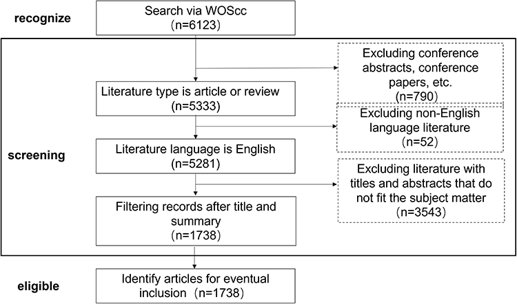 |
Figure 1 Literature Screening Flowchart. |
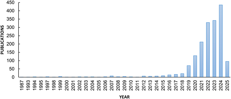 |
Figure 2 Trends in the growth of publications. |
Analysis of Country
A total of 99 countries have conducted research on the application of AI in the field of CVD. Table 1 shows the top 10 countries in terms of the number of publications and citation frequency. The three countries with the highest output are the United States (432 articles), China (372 articles), and India (334 articles). Figure 3 shows that the United States, the United Kingdom, and India have relatively close collaborative relationships with other countries.
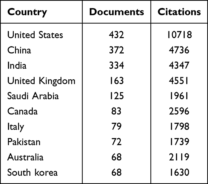 |
Table 1 Influential Countries/Regions in the Field of AI in CVD Related Research |
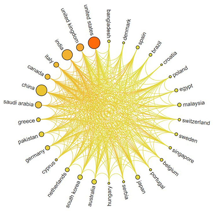 |
Figure 3 The global number of publications related to AI in CVD research. |
Analysis of Institutions
A total of 2,985 institutions have participated in research on AI in the CVD field. Table 2 lists the top 10 institutions with the highest output in relevant research fields. These institutions are located in the United States (3), the United Kingdom (3), Canada (2), India (1), and Saudi Arabia (1). Harvard Medical School (43 articles) has published the most papers in this field; Mayo Clinic (30 articles) and Leland Stanford Junior University (29 articles) follow closely. The top three institutions in terms of citation frequency are Mayo Clinic (1,225 citations), University of Oxford (932 citations), and Icahn School of Medicine at Mount Sinai (913 citations). Figure 4 shows the collaborative situation among 58 institutions with more than 10 publications, indicating relatively close collaborative relationships among these institutions. TLS refers to the sum of the strength of a node’s relationships with other nodes in a network, which can used to evaluate the collaborative relationships. In terms of TLS, the top three institutions are Queen’s University, Harvard Medical School, and Icahn School of Medicine at Mount Sinai.
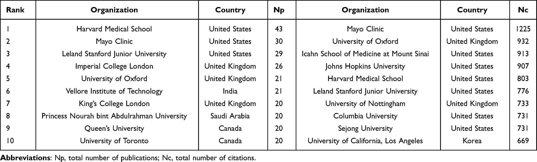 |
Table 2 Influential Institutions in the Field of AI in CVD Related Research |
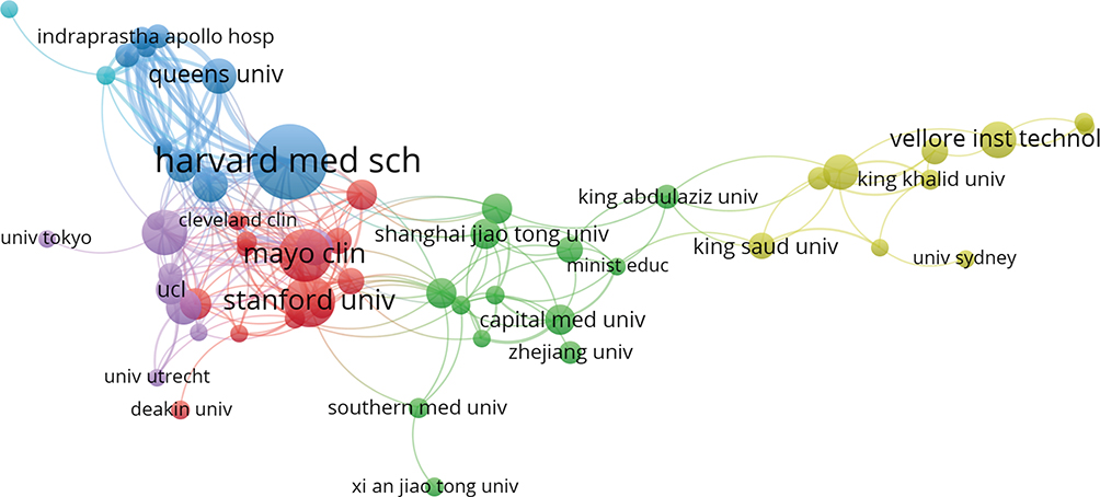 |
Figure 4 Visualization of organizations related to AI in CVD publications. |
Analysis of Author Co-Occurrence Network
A total of 8,945 authors have participated in research related to AI and CVD fields. On average, approximately five authors are involved in each article. Among them, 35 authors have published more than five research papers, and their collaborative relationships are shown in Figure 5. Saba, Luca, and Suri, Jasjit S. have the highest number of publications, with 21 articles each. Their main research areas focus on the application of AI combined with imaging in CVD. Next is Laird, John R., whose research direction centers on the application of AI in CVD risk stratification. Besides, among the top 10 authors, there are Khanna, Narendra N. (15 articles), Johri, Amer M. (12 articles), and Nicolaides, Andrew (10 articles). Detailed information on the top 10 authors by publication count is presented in Table 3.
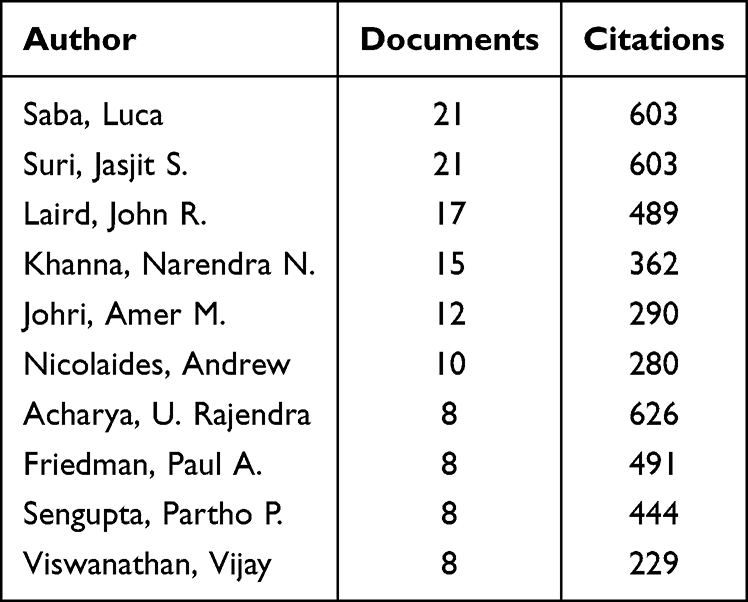 |
Table 3 Author Publication Details |
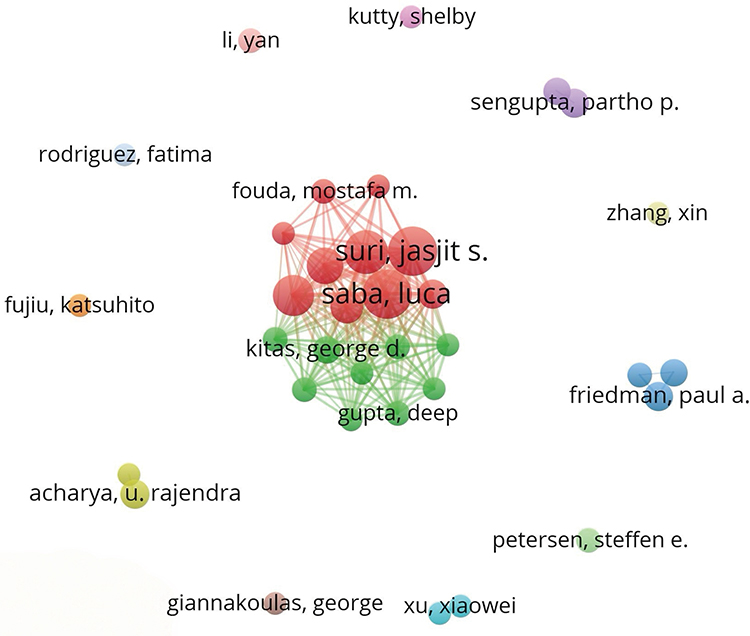 |
Figure 5 Cooperation network of authors. |
Analysis of Publishing Journals and Literature
A total of 695 journals have published research in this field, with 75 journals having published more than five articles. Table 4 shows the names of the top 10 journals by publication count, most of which are in JCR Q1 or Q2. IEEE ACCESS is the journal with the highest number of publications in the field of AI in CVD (66 articles), mainly focusing on computer science research. It is followed by FRONTIERS IN CARDIOVASCULAR MEDICINE, which mainly focuses on the heart and cardiovascular system, and SCIENTIFIC REPORTS, which mainly focuses on the application of computer science in the cardiovascular system. Combined with the double figure superimposed journals (Figure 6) can be inferred from the analysis that the field involves research in clinical medicine, computer, mathematics, health medicine, molecular.
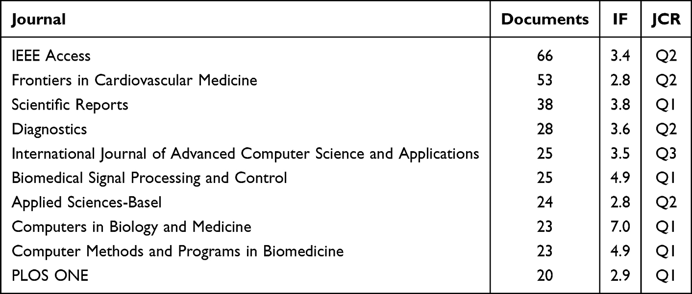 |
Table 4 Journal Publication Details |
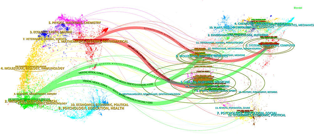 |
Figure 6 Journal overlay map related to AI in CVD research. |
Analysis of Keyword
Keywords in literature summarize the content and themes of the articles. Hence, research and analysis of hot keywords can help us identify research hotspots in a particular field. Using CiteSpace to analyze the keywords in 1,738 papers, a total of 1,221 keywords were identified. The top 10 most frequent keywords include: machine learning (618), coronary heart disease (240), cardiovascular disease (336), heart disease (270), injury (42), myocardial deep learning (216), classification (186), diagnosis (175), prediction (166), and risk (154) (Figure 7A). Keywords such as “cardiovascular disease,” “neural networks,” “feature extraction,” “cardiovascular diseases,” and “myocardial infarction” are located at the center, acting as hubs connecting other nodes, reflecting the hotspots and general direction of AI research in the CVD field in recent years.
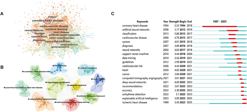 |
Figure 7 (A) High-frequency keywords. (B) Cluster diagram of keywords. (C) Top 20 keywords with the strongest citation bursts. |
The purpose of keyword clustering analysis is to display the distribution of core content by classifying keywords based on similarity.10 Figure 7B shows the CiteSpace keyword cluster and hotspot identification map, forming 10 research hotspots: “#0: electrocardiogram-based deep learning”, “#1: polymorphism-based biomarker”, “#2: metabolic syndrome”, “#3: classification identification”, “#4: artificial intelligence framework”, “#5: screening native american elder”, “#6: preclinical myxomatous mitral valve disease”, “#7: artificial intelligence”, “#8: deep learning”, “#9: expert system”. Table 5 presents the specific characteristics of each cluster in Figure 7B, including cluster number, cluster label, average publication year of cluster literature, research focus of cluster literature, and number of cluster members. Except for clusters #2, #5, and #6, the other clusters demonstrate research hotspot themes in the application of AI in CVD diagnosis and treatment, reflecting the positive role of AI in assisting disease diagnosis, classification, and the identification of emerging biomarkers, as well as the design and research of AI systems.
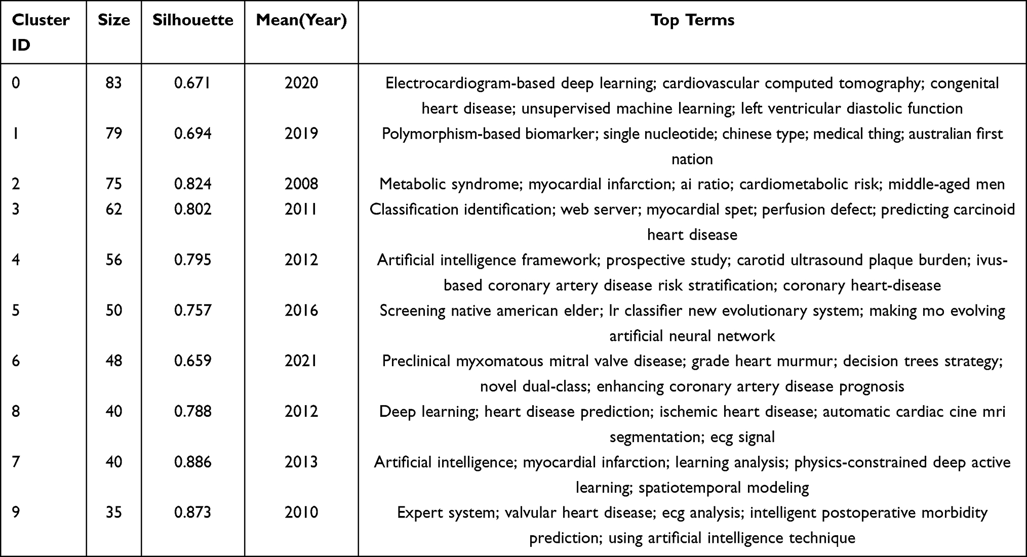 |
Table 5 Keyword Clustering Features |
Keyword bursts refer to keywords with a high rate of frequency change detected at corresponding time nodes, indicating hot research topics during a specific period. Figure 7C shows the top 20 keywords with the strongest citation bursts. Keywords with high burst intensity include “coronary heart disease,” “classification,” and “cardiovascular disease.” In the early stages, the keywords “coronary heart disease” and “artificial neural networks” had long burst durations, indicating their significant impact on current research in the field. Since 2019, keywords such as “cardiovascular risk,” “deep neural networks,” and “accuracy” have increased, suggesting a growing number of articles exploring this field and the deepening of research methods. The current research hotspot keywords that persist to this day are “explainable artificial intelligence” and “ischemic heart disease.” Further analysis reveals that improving the algorithm performance of AI and ischemic heart disease may be among the future research hotspots of interest.
Cluster Analysis of Co-Cited Literature
The number of citations can be used to measure the influence and academic value of a research result. Co-cited literature refers to two articles that are cited together, and the frequency of co-citation is calculated to determine co-citation relationships.7 The most co-cited literature is Effective Heart Disease Prediction Using Hybrid Machine Learning Techniques written by Senthilkumar Mohan and published in IEEE Access. This present study adopts hybrid machine learning techniques, combining the advantages of multiple algorithms, to provide new technical ideas for CVD prediction. Besides, by constructing a high-performance prediction model, it offers new tools and methods for CVD risk assessment, prompting researchers to pay more attention to how to optimize risk assessment models using machine learning techniques to better identify high-risk patients and achieve early intervention and precise treatment. In general, the common research focuses among the top 10 co-cited articles mainly center on data-driven prediction, multimodal data fusion, model accuracy and generalization ability, the potential and challenges of clinical applications, as well as auxiliary decision-making and personalized treatment.
Subject clustering of co-cited literature can reflect the main research content and development history of a field.11 The co-cited literature network identified eight clusters with significant modularity (Q = 0.8398) and a high silhouette score (S = 0.9083), indicating the credibility and uniqueness of the clusters11 (Figure 8).Based on co-citation relationships for clustering and in-depth analysis of cluster literature and their research focuses, we can summarize the current research themes of AI in the CVD field: the application of AI in the classification and diagnosis of coronary artery disease (clusters 3 and 8); the application of AI in CVD risk prediction (cluster 2); the improvement of AI algorithms (clusters 0 and 6); and the precise use of AI in imaging (clusters 1, 4, and 15).
 |
Figure 8 Literature co-citation cluster mapping. |
Discussion
Current Research Status of AI in the CVD Field
The first application of AI technology in CVD research in 1987 marked an important starting point in this field. At that time, computer technology was in a stage of rapid development, and the concept of AI was gradually transitioning from theory to practical application. As one of the major global health threats, the complexity and diversity of CVD pose numerous challenges to traditional diagnostic and treatment methods. The introduction of AI has provided new ideas and methods for the diagnosis, treatment, and management of CVD, opening a new chapter in precision medicine for CVD. Analysis of the annual publication volume and highly cited literature reveals that, in the early stages of research, the number of articles on the application of AI in the CVD field was relatively small. Research directions were mostly based on simple machine learning algorithms, such as support vector machines and data mining, which were used to extract features from electrocardiogram (ECG) and cardiovascular imaging data for simple disease diagnosis and classification. Nonetheless, large-scale, systematic application practices had not yet been carried out. These studies preliminarily demonstrated the feasibility of AI technology in CVD diagnosis, laying a foundation for subsequent in-depth research. Since 2018, with the popularization and promotion of the concept of intelligent health management worldwide, research on the application of AI in the CVD field has rapidly heated up. The number of articles on AI and CVD published after 2018 has shown exponential growth, with the main research directions focusing on the extraction of coronary artery CT and electrocardiac activity-related data based on deep neural networks, with a greater emphasis on the diagnosis of arrhythmias and ischemic heart disease.
The top three countries with the highest number of publications in this field are the United States, China, and India. The United States surpasses other countries in terms of citation frequency and the number of international collaborations, and most of the institutions ranking high in terms of publication volume and citation frequency are from the United States, indicating that the United States is a global leader in this field. Among the top 10 journals in terms of publication volume, the journal with the highest impact factor is COMPUTERS IN BIOLOGY AND MEDICINE (IF = 7.0), whose main research area is the application of computers in biomedicine. Nevertheless, overall, the proportion of AI-related research in CVD published in high-impact journals is relatively low, possibly because the interdisciplinary field of AI and CVD science is still in its early stages of development. In the future, it is necessary to strengthen cooperation and communication among different authors, institutions, and even countries to promote the production of high-quality scientific research results and drive the further development of AI in the CVD field.
Research Hotspots
Based on keyword frequency and cluster analyses, current research is mainly focused on four hotspots:① AI-assisted disease diagnosis: employing machine learning, data mining, deep learning, and other advanced techniques to enhance diagnostic accuracy.② AI-assisted disease classification: facilitating the stratification of various CVD, with acute myocardial infarction and arrhythmias as the primary targets.③ Emerging biomarkers: developing sophisticated pipelines that integrate traditional bioinformatics, classical statistics, and multimodal machine learning to identify novel CVD biomarkers with high predictive value.④ AI system design: constructing predictive and diagnostic models for CVD through AI technologies to improve clinical efficiency.
Research Directions
Co-citation Clustering can be used to reveal the knowledge structure and research hotspots within a certain disciplinary field. This method identifies which documents share similarities in terms of topic or research content by analyzing the co-citation relationships among them (ie, when two documents are simultaneously cited by the same subsequent document), and then classifies these documents into different clusters. Based on co-citation clustering, the principal research directions can be summarized as follows:
Application of AI in CVD Diagnosis and Classification
ECG, echocardiography, coronary artery CT, and cardiac MRI are the most commonly used auxiliary examinations for the clinical diagnosis of CVD. However, these methods still have limitations, such as difficulty in diagnosing complex cases and subjective deviations in the interpretation of results by different clinicians, which may affect diagnostic accuracy. Plus, the vast amount of complex data also poses significant challenges for data organization and analysis.12 Exploratory work in AI-assisted risk prediction, diagnosis, treatment and clinical research of CVD has achieved many results, promoting the deep integration of AI and digital health tools in the field of CVD. Deep learning algorithms can automatically learn features from large amounts of complex data, greatly improving diagnostic efficiency and accuracy. For instance, convolutional neural networks (CNNs) are widely used in CVD imaging analysis and can automatically identify features of diseases such as coronary artery disease and heart failure. Wu et al13 applied a model combining CNN and long short-term memory (LSTM) to ECG data, achieving an area under the curve (AUC) value of 0.99 for the diagnosis of acute ST-segment elevation myocardial infarction. Ahmed et al14 used a one-dimensional CNN-based deep learning method for the classification of arrhythmias, with the model achieving an accuracy of 0.99 on the test dataset, serving as an alternative for rapid and automatic arrhythmia diagnosis. Cho et al15 developed a CNN model that demonstrated good efficacy in identifying infarcted vessels such as the left anterior descending artery (LAD), left circumflex artery (LCX), and right coronary artery (RCA). In the future, with the development of AI and imaging technology, more accurate and efficient diagnostic and classification tools can be expected to help improve the accuracy of diagnosis and classification for various types of CVD.
Application of AI in CVD Risk Prediction
Globally, approximately half of CVD deaths occur in adults without obvious symptoms,16 and CVD is the leading cause of morbidity and mortality among the elderly.17 Early identification of individuals at high risk of CVD and the implementation of active intervention measures can significantly reduce the incidence of CVD events. In this regard, there is an urgent need for early and accurate prediction of high-risk populations. Currently, AI prediction models have been widely applied in various fields of CVD, such as coronary heart disease,18 atrial fibrillation,19 and heart failure,20 enabling early screening and risk prediction of patients at different risk levels. Golas et al21 made use of a machine learning model to predict the risk of 30-day readmission in patients with heart failure and provided remote monitoring interventions for patients at high risk of readmission, achieving potential cost savings in healthcare utilization.21 Muhammad Tayyeb et al22 proposed a simple multilayer perceptron (MLP) model for heart disease prediction to reduce computational complexity. B Mohan Rao et al23 proposed the use of the ResNet-50 model for ECG prioritization in CVD detection in clinical and out-of-hospital settings. Tadashi Araki et al24 developed a machine learning-based CADx system for the automatic classification of coronary artery images in high-risk and low-risk patients. Tseng et al25 used retinal data from the UK Biobank to develop a new CVD risk stratification model, which demonstrated greater accuracy in identifying individuals at high risk of CVD within 10 years compared to the traditional model QRISK3, and could identify high-risk individuals who would benefit earlier.26 In addition, wearable devices can guide the management of patients with heart failure. A multicenter study showed that the application of AI to a wearable multi-sensor chest patch on a smartphone had high predictive value for the exacerbation of heart failure (sensitivity of 88%, specificity of 85%).27 The application of AI technology to CVD prediction models can significantly improve the accuracy of identifying individuals at high risk of CVD, facilitate the development of personalized treatment plans for patients, and further reduce mortality and the occurrence of adverse events. But the widespread application of AI in CVD risk assessment still faces challenges, as many healthcare professionals currently have insufficient understanding and acceptance of AI. Hence, it is crucial to promote education on the benefits and applications of AI in CVD risk prediction and accelerate its integration into clinical practice.
Precise Application of AI in CVD Imaging
Cardiac imaging methods (such as echocardiography, cardiac CT, and cardiac MRI) are susceptible to the operator’s skill level and individual variations, resulting in certain limitations in the reproducibility and consistency of results. The introduction of AI technology has significantly enhanced image acquisition, measurement, and diagnostic capabilities. Currently, in multiple fields including heart failure and heart valve diseases, AI has been developed for automated measurement and assessment, demonstrating notable advantages in terms of reproducibility and speed.28 Relevant research published in Nature has shown that AI models based on deep learning can automatically extract multiple key parameters from ultrasound images and identify low-quality images, thereby improving the accuracy and consistency of measurements.29 Holste et al30 developed a deep learning (DL) model based on two-dimensional parasternal long-axis ultrasound videos. Through self-supervised contrastive pre-training and integration with a three-dimensional convolutional neural network, the model achieved automated detection of aortic stenosis (AS), with significantly better accuracy in diagnosing severe AS compared to traditional methods. Coronary CT is an important means of evaluating coronary artery stenosis, with current reports relying on the subjective visual assessment of clinicians. Kelm et al31 used machine learning (ML) algorithms to automatically identify, grade, and classify coronary artery stenosis caused by calcified and non-calcified plaques. Cardiac MRI is often used to assess cardiac function parameters and diagnose CVD, and is regarded as the “gold standard” for evaluating cardiac structure and function. But its drawback is the long imaging time, typically requiring extended scan durations. Based on CNN technology, image reconstruction and denoising can be performed, enabling the acquisition of higher-quality images in a shorter period.28 Recent years have also witnessed research demonstrating the superior performance of DL technology in image reconstruction.28 Non-invasive techniques such as carotid artery ultrasound, arterial hardness assessment, echocardiography and coronary artery calcification score can identify subclinical atherosclerosis and ventricular dysfunction, providing insights that complement conventional risk factors.32 With the gradual application of AI in CVD imaging assessment, it will help simplify the traditional image acquisition and analysis process, and is expected to achieve more accurate and repeatable measurements. At present, however, most studies are retrospective studies and lack the support of large-scale clinical research.33 Their application in clinical practice still faces challenges such as insufficient generalization and lagging ethical supervision.
Improvements in AI Algorithms Drive the Intelligent Upgrade of CVD Diagnosis and Treatment
Continuous optimization of AI algorithms has become pivotal for enhancing diagnostic accuracy, predictive power, and clinical applicability. The diagnosis of CVD not only relies on static images but also requires capturing the dynamic functional changes of the heart. Wang et al introduced spatio-temporal feature-modeling mechanisms namely 3D W-MSA and 3D SW-MSA modules, to encode temporal dependencies within cardiac image sequences. This design enables AI models to detect early myocardial motion abnormalities and regional wall-motion disorders, facilitating timely intervention for heart failure and cardiomyopathies.34 Yi et al35 used machine-learning techniques to develop comparative models predicting individual blood-pressure responses to different antihypertensive agents, with extreme gradient boosting (XGBoost) exhibiting the best performance. Collectively, advances in model architecture, multimodal fusion, spatio-temporal modeling, and interpretability are driving the intelligent transformation of risk prediction, cardiovascular image analysis, and personalized therapeutics.
Challenges and Prospects of AI in CVD Applications
Data Quality and Privacy Protection
Despite the significant progress made in the application of AI in CVD, several challenges remain, such as data quality and privacy protection, which are urgent issues to be addressed. AI models have high requirements for data quality. Currently, the varying quality of medical data and the lack of unified data standards across different medical institutions pose difficulties for the generalization of CVD prediction models.36 This necessitates that training data must include images of varying quality to improve the model’s generalization ability. For the same type of data, a unified data format and standard should be followed to facilitate data sharing, analysis, and cross-platform compatibility, ensuring the accuracy, comprehensiveness, and representativeness of the data. Additionally, we recommend establishing national-or regional-level multicenter cardiovascular AI data alliances to harmonize data formats, acquisition workflows, and quality-control standards. “Data lineage tracking” and version-control mechanisms should be embedded to guarantee full traceability across collection, cleaning, annotation, and modeling stages. An independent data-audit and quality-assessment body should be instituted to issue periodic data-quality reports, which must be met as a prerequisite for AI model certification and peer-reviewed publication. Medical data related to CVD often involves a large amount of personal privacy information. How to fully utilize this data while ensuring data security is a critical issue. Federated learning is a distributed machine learning technique that allows users to train models on local devices. Through decentralized privacy protection technologies, it enables group model training without transmitting raw data, thereby protecting data security and privacy.37 Furthermore, developers must deploy appropriate firewalls and other essential cybersecurity measures, with regular updates, to ensure data integrity. Stakeholders may define an acceptable risk threshold below which data sharing is permitted, thereby fostering global interoperability of medical knowledge. In summary, the deployment of AI in cardiovascular medicine must evolve from “ata availability” to “data trustworthiness”. Future initiatives should advance standardized governance and privacy-preserving computation in concert, guaranteeing that AI models are not only technically viable but also ethically and legally sustainable, thereby achieving a true bench-to-clinical closed-loop translation.
Precise Screening with AI Models
Given the complex pathogenesis, rapid disease progression, and high heterogeneity of CVD, AI models need to incorporate a wider range of data. They often require the joint consideration of various types of multimodal data, such as laboratory biochemical tests, electrocardiograms, and imaging data, to achieve precise screening and diagnosis of CVD. At present, general AI models for CVD are mainly limited to unimodal data.34 To this end, future research should place greater emphasis on the fusion of multimodal data, such as combining electrocardiogram and imaging data, electronic health records, and data from wearable devices, to explore the integration capabilities of AI in “multi-omics” data to enhance the understanding of disease mechanisms, and to improve the accuracy and reliability of diagnosis. This fusion can provide a more comprehensive perspective for the early screening and precise treatment of CVD.38 The performance of AI models is highly dependent on the quality and diversity of training data. If the training dataset does not adequately represent different populations and clinical scenarios, the model may perform poorly in practical applications. Most current AI models are trained on region- or ethnicity-specific datasets. To enhance generalizability, future efforts must extend to diverse racial groups, geographic regions, and health-care systems. Prospective, multicenter clinical trials should be initiated to rigorously evaluate the real-world robustness and accuracy of these models. In parallel, a consensus framework for AI-assisted CVD screening encompassing standardized workflows, performance metrics, and ethical guidelines should be established through multidisciplinary collaboration among cardiologists, radiologists, and AI experts.
Model Interpretability and Clinical Acceptance
The lack of interpretability of AI models is also an important factor limiting their wide application, which has become the core bottleneck from “laboratory accuracy” to “bedside decision-making”. Interpretability may refer to the degree to which a human can understand the internal mechanisms and decision-making processes of an AI model.39 As a “black box” algorithm, the prediction process of AI is opaque. In real-world diagnostic and treatment scenarios, clinicians need to understand the internal mechanisms underlying the model’s decisions.40 Developing interpretable AI models and enhancing the interpretability of model decisions are particularly important for the clinical translation of AI models, and this will remain a hot topic of research in the foreseeable future. The challenge of interpretability in machine learning models hinders healthcare practitioners’ trust in the diagnostic results generated by these models. Interpretable Machine Learning (IML) models pave the way for automated diagnostic tools by building trust with doctors and providing evidence-based diagnoses.41 Murdoch et al42 defined the focus of IML as “extracting knowledge about relationships contained in the data or learned by the model from ML models.” In recent years, large language models (LLMs), represented by generative pre-trained models and bidirectional encoder representations from transformers, have garnered widespread attention.43 Multimodal LLMs can process data from different modalities (such as images and text) and provide more comprehensive and accurate diagnostic results by fusing information from these diverse modalities. Based on the chain-of-thought technology of LLMs, the thought processes and decision-making mechanisms of medical experts can also be understood, endowing the model with greater credibility, transparency, and interpretability.44
Limitations
The main limitation of this present study is that the data were downloaded from the WoSCC database and could not be extended to other databases such as Elsevier or Springer. Nonetheless, WoSCC is the most commonly used database in scientometrics analysis, containing most of the information from related articles. Visuality-based literature analysis lays the foundation for researchers to understand the hotspots and potential trends of AI in CVD.
Conclusion
In conclusion, this present study employed bibliometric methods to explore the application of AI in CVD. The results showed that AI technology is gradually being introduced into the CVD field and has been developing rapidly in recent years. The main research directions are the auxiliary diagnosis and classification of AI in CVD, risk prediction and imaging assessment, as well as the improvement of AI algorithms. In addition, issues such as the interpretability and clinical acceptance of AI data quality and patient privacy protection models cannot be ignored. Future researches should establish standards for medical privacy protection. On the premise of ensuring data security, it is essential to fully integrate clinical diagnostic and treatment processes and their characteristics, and to build high-quality, standardized multimodal databases. This tend to promote the development of AI in CVD diagnosis and treatment toward standardization and intelligence. Despite the current challenges, with ongoing technological advancements and increasingly sophisticated research, AI is anticipated to become an indispensable tool in the diagnosis and management of CVD, contributing to improved clinical outcomes for affected patients.
Author Contributions
All authors made a significant contribution to the work reported, whether that is in the conception, study design, execution, acquisition of data, analysis and interpretation, or in all these areas; took part in drafting, revising or critically reviewing the article; gave final approval of the version to be published; have agreed on the journal to which the article has been submitted; and agree to be accountable for all aspects of the work.
Disclosure
The authors declare that the research was conducted in the absence of any commercial or financial relationships that could be construed as a potential conflict of interest.
References
1. Roth GA, Mensah GA, Johnson CO, et al. Global burden of cardiovascular diseases and risk factors, 1990-2019: update from the GBD 2019 study. J Am Coll Cardiol. 2020;76(25):
2. Magnussen C, Alegre-Diaz J, Al-Nasser LA, et al. Global effect of cardiovascular risk factors on lifetime estimates. N Engl J Med. 2025;2025.
3. Esteva A, Robicquet A, Ramsundar B, et al. A guide to deep learning in healthcare. Nat Med. 2019;25(1):
4. Krittanawong C, Zhang H, Wang Z, et al. Artificial intelligence in precision cardiovascular medicine. J Am Coll Cardiol. 2017;69(21):
5. Antoniades C, Asselbergs FW, Vardas P. The year in cardiovascular medicine 2020: digital health and innovation. Eur Heart J. 2021;42(7):
6. Armoundas AA, Narayan SM, Arnett DK, et al. Use of artificial intelligence in improving outcomes in heart disease: a scientific statement from the american heart association. Circulation. 2024;149(14):
7. Hood WW, Wilson CS. The literature of bibliometrics, scientometrics, and informetrics. Scientometrics. 2001;52(2):
8. Chen C. CiteSpace II: detecting and visualizing emerging trends and transient patterns in scientific literature. J Am Soc Inform Sci Technol. 2006;57(3):
9. van Eck NJ, Waltman L. Software survey: vOSviewer, a computer program for bibliometric mapping. Scientometrics. 2010;84(2):
10. Song L, Zhang J, Ma D, et al. A bibliometric and knowledge-map analysis of macrophage polarization in atherosclerosis from 2001 to 2021. Front Immunol. 2022;13:910444. doi:10.3389/fimmu.2022.910444
11. Yue C, Chaomei C, Zeyuan L, et al. The methodological functions of the CiteSpace knowledge graph. Res Sci Sci. 2015;33(02):
12. Narula S, Shameer K, Salem OA, et al. Machine-learning algorithms to automate morphological and functional assessments in 2D echocardiography. J Am Coll Cardiol. 2016;68(21):
13. Wu L, Huang G, Yu X, et al. Deep learning networks accurately detect ST-segment elevation myocardial infarction and culprit vessel. Front Cardiovasc Med. 2022;9:797207. doi:10.3389/fcvm.2022.797207
14. Ahmed AA, Ali W, Abdullah TAA, et al. Classifying cardiac arrhythmia from ECG signal using 1D CNN deep learning model. Mathematics. 2023;11.
15. Cho Y, Kwon JM, Kim KH, et al. Artificial intelligence algorithm for detecting myocardial infarction using six-lead electrocardiography. Sci Rep. 2020;10(1):20495. doi:10.1038/s41598-020-77599-6
16. Kumar A, Avishay DM, Jones CR, et al. Sudden cardiac death: epidemiology, pathogenesis and management. Rev Cardiovasc Med. 2021;22(1):
17. Kumar M, Yan P, Kuchel GA, et al. Cellular senescence as a targetable risk factor for cardiovascular diseases: therapeutic implications: JACC family series. JACC Basic Transl Sci. 2024;9(4):
18. Dutta A, Batabyal T, Basu M, et al. An efficient convolutional neural network for coronary heart disease prediction. Exp Sys Applicat. 2020;159:113408. doi:10.1016/j.eswa.2020.113408
19. Hill NR, Ayoubkhani D, Mcewan P, et al. Predicting atrial fibrillation in primary care using machine learning. PLoS One. 2019;14(11):e224582. doi:10.1371/journal.pone.0224582
20. Segar MW, Jaeger BC, Patel KV, et al. Development and validation of machine learning-based race-specific models to predict 10-year risk of heart failure: a multicohort analysis. Circulation. 2021;143(24):
21. Golas SB, Shibahara T, Agboola S, et al. A machine learning model to predict the risk of 30-day readmissions in patients with heart failure: a retrospective analysis of electronic medical records data. BMC Med Inform Decis Mak. 2018;18(1):44. doi:10.1186/s12911-018-0620-z
22. Tayyeb M, Umer M, Alnowaiser K, et al. Deep learning approach for automatic cardiovascular disease prediction employing ECG signals. CMES. 2023;137(2):
23. Mohan Rao B, Kumar A. Detection of heart arrhythmia based on UCMFB and deep learning technique. Sādhanā. 2022;47(4):252. doi:10.1007/s12046-022-02027-6
24. Araki T, Ikeda N, Shukla D, et al. A new method for IVUS-based coronary artery disease risk stratification: a link between coronary & carotid ultrasound plaque burdens. Comput Methods Programs Biomed. 2016;124:
25. Tseng R, Rim TH, Shantsila E, et al. Validation of a deep-learning-based retinal biomarker (Reti-CVD) in the prediction of cardiovascular disease: data from UK Biobank. BMC Med. 2023;21(1):28. doi:10.1186/s12916-022-02684-8
26. Flores AM, Schuler A, Eberhard AV, et al. Unsupervised learning for automated detection of coronary artery disease subgroups. J Am Heart Assoc. 2021;10(23):e21976. doi:10.1161/JAHA.121.021976
27. Shah AJ, Isakadze N, Levantsevych O, et al. Detecting heart failure using wearables: a pilot study. Physiol Meas. 2020;41(4):44001. doi:10.1088/1361-6579/ab7f93
28. Elias P, Jain SS, Poterucha T, et al. Artificial intelligence for cardiovascular care—part 1: advances. JACC. 2024;83(24):
29. He B, Kwan AC, Cho JH, et al. Blinded, randomized trial of sonographer versus AI cardiac function assessment. Nature. 2023;616(7957):
30. Holste G, Oikonomou EK, Mortazavi BJ, et al. Severe aortic stenosis detection by deep learning applied to echocardiography. Eur Heart J. 2023;44(43):
31. Kelm BM, Mittal S, Zheng Y, et al. Detection, grading and classification of coronary stenoses in computed tomography angiography. Med Image Comput Comput Assist Interv. 2011;14(Pt 3):
32. Carerj ML, Restelli D, Poleggi C, et al. The role of imaging in cardiovascular prevention: a comprehensive review. J Cardiovasc Echogr. 2025;35(1):
33. Biondi-Zoccai G, D’Ascenzo F, Giordano S, et al. Artificial intelligence in cardiology: general perspectives and focus on interventional cardiology. Anatol J Cardiol. 2025;29(4):
34. Wang YJ, Yang K, Wen Y, et al. Screening and diagnosis of cardiovascular disease using artificial intelligence-enabled cardiac magnetic resonance imaging. Nat Med. 2024;30(5):
35. Yi J, Wang L, Song J, et al. Development of a machine learning-based model for predicting individual responses to antihypertensive treatments. Nutr Metab Cardiovasc Dis. 2024;34(7):
36. Li Y, Sperrin M, Martin GP, et al. Examining the impact of data quality and completeness of electronic health records on predictions of patients’ risks of cardiovascular disease. Int J Med Inform. 2020;133:104033. doi:10.1016/j.ijmedinf.2019.104033
37. Uddin MI, Mashwani W. Federated learning: unlocking the power of collaborative intelligence. 2024.
38. Kanagaraj S. Leveraging cloud-based deep learning for cardiovascular diseases prediction: challenges and solutions. 2025.
39. Grote T. Allure of simplicity: on interpretable machine learning models in healthcare. Philosophy Med. 2023;4.
40. Xu H, Shuttleworth KMJ. Medical artificial intelligence and the black box problem: a view based on the ethical principle of “do no harm”. Intelligent Medicine. 2024;4(1):
41. Abdullah TAA, Zahid MS, Ali W. A review of interpretable Ml in healthcare: taxonomy, applications, challenges, and future directions. Symmetry. 2021;13.
42. Murdoch WJ, Singh C, Kumbier K, et al. Definitions, methods, and applications in interpretable machine learning. Proc Natl Acad Sci U S A. 2019;116(44):
43. Lee S, Kim WJ, Chang J, et al. LLM-CXR instruction-finetuned LLM for CXR image understanding and generation. 2024.
44. Liu Y, Yao Y, Ton J, et al. Trustworthy LLMs a survey and guideline for evaluating large language models’. Alignment. 2024;2024.
 © 2025 The Author(s). This work is published and licensed by Dove Medical Press Limited. The
full terms of this license are available at https://www.dovepress.com/terms
and incorporate the Creative Commons Attribution
- Non Commercial (unported, 4.0) License.
By accessing the work you hereby accept the Terms. Non-commercial uses of the work are permitted
without any further permission from Dove Medical Press Limited, provided the work is properly
attributed. For permission for commercial use of this work, please see paragraphs 4.2 and 5 of our Terms.
© 2025 The Author(s). This work is published and licensed by Dove Medical Press Limited. The
full terms of this license are available at https://www.dovepress.com/terms
and incorporate the Creative Commons Attribution
- Non Commercial (unported, 4.0) License.
By accessing the work you hereby accept the Terms. Non-commercial uses of the work are permitted
without any further permission from Dove Medical Press Limited, provided the work is properly
attributed. For permission for commercial use of this work, please see paragraphs 4.2 and 5 of our Terms.
Recommended articles

Stem Cell Therapy for Neuropathic Pain: A Bibliometric and Visual Analysis
Kan H, Fan L, Gui X, Li X, Yang S, Huang Y, Chen L, Shen W
Journal of Pain Research 2022, 15:1797-1811
Published Date: 23 June 2022
Knowledge Atlas of the Co-Occurrence of Epilepsy and Autism: A Bibliometric Analysis and Visualization Using VOSviewer and CiteSpace
Wang Y, Huo X, Li W, Xiao L, Li M, Wang C, Sun Y, Sun T
Neuropsychiatric Disease and Treatment 2022, 18:2107-2119
Published Date: 19 September 2022
Bibliometric and Visualization Analysis of Biomechanical Research on Lumbar Intervertebral Disc
Hou Z, Wang W, Su S, Chen Y, Chen L, Lu Y, Zhou H
Journal of Pain Research 2023, 16:3441-3462
Published Date: 17 October 2023
Bibliometric and Visualization Analysis of Research Hotspots and Frontiers in Endoscopic Lumbar Discectomy
Zhong Y, Wang J, Liang Z, Han T, Lu H, Hou Z
Journal of Pain Research 2024, 17:2165-2190
Published Date: 18 June 2024
Global Research Status of Maca (Lepidium Meyenii Walp.): A Bibliometric Analysis of Hotspots, Bursts, and Trends
Li K, Zhao C, Dang M, Ren R, Fu M, Bai Y, Wang J, Zhang Q, Luan F
Drug Design, Development and Therapy 2025, 19:2329-2349
Published Date: 27 March 2025