Back to Journals » Journal of Pain Research » Volume 16
Bibliometric and Visualization Analysis of Biomechanical Research on Lumbar Intervertebral Disc
Authors Hou Z ![]() , Wang W, Su S, Chen Y, Chen L
, Wang W, Su S, Chen Y, Chen L ![]() , Lu Y, Zhou H
, Lu Y, Zhou H
Received 4 July 2023
Accepted for publication 28 September 2023
Published 17 October 2023 Volume 2023:16 Pages 3441—3462
DOI https://doi.org/10.2147/JPR.S428991
Checked for plagiarism Yes
Review by Single anonymous peer review
Peer reviewer comments 2
Editor who approved publication: Dr Alaa Abd-Elsayed
Zhaomeng Hou,1– 3,* Wei Wang,4,* Shaoting Su,1 Yixin Chen,4 Longhao Chen,1,5 Yan Lu,1,5,6 Honghai Zhou1,5
1Faculty of Orthopedics and Traumatology, Guangxi University of Chinese Medicine, Nanning, People’s Republic of China; 2Department of Orthopedics and Traumatology, Yancheng TCM Hospital Affiliated to Nanjing University of Chinese Medicine, Yancheng, People’s Republic of China; 3Department of Orthopedics and Traumatology, Yancheng TCM Hospital, Yancheng, People’s Republic of China; 4Department of Rehabilitation Medicine, The First Affiliated Hospital of Guangxi University of Chinese Medicine, Nanning, People’s Republic of China; 5Guangxi Key Laboratory of Biomechanics and Injury Repair in Traditional Chinese Medicine Orthopedics and Traumatology, Nanning, People’s Republic of China; 6Department of Orthopedics and Traumatology, The First Affiliated Hospital of Guangxi University of Chinese Medicine, Nanning, People’s Republic of China
*These authors contributed equally to this work
Correspondence: Honghai Zhou; Yan Lu, Faculty of Orthopedics and Traumatology, Guangxi University of Chinese Medicine, Nanning, People’s Republic of China, Email [email protected]; [email protected]
Background: Biomechanical research on the lumbar intervertebral disc (IVD) provides valuable information for the diagnosis, treatment, and prevention of related diseases, and has received increasing attention. Using bibliometric methods and visualization techniques, this study investigates for the first time the research status and development trends in this field, with the aim of providing guidance and support for subsequent research.
Methods: The Science Citation Index Expanded (SCI-Expanded) within the Web of Science Core Collection (WoSCC) database was used as the data source to select literature published from 2003 to 2022 related to biomechanical research on lumbar IVD. VOSviewer 1.6.19 and CiteSpace 6.2.R2 visualization software, as well as the online analysis platform of literature metrology, were utilized to generate scientific knowledge maps for visual display and data analysis.
Results: The United States is the most productive country in this field, with the Ulm University making the largest contribution. Wilke HJ is both the most prolific author and one of the highly cited authors, while Adams MA is the most cited author. Spine, J Biomech, Eur Spine J, Spine J, and Clin Biomech are not only the journals with the highest number of publications, but also highly cited journals. The main research topics in this field include constructing and validating three-dimensional (3D) finite element model (FEM) of lumbar spine, measuring intradiscal pressure, exploring the biomechanical effects and related risk factors of lumbar disc degeneration, studying the mechanical responses to different torque load combinations, and classifying lumbar disc degeneration based on magnetic resonance images (MRI), which are also the hot research themes in recent years.
Conclusion: This study systematically reviews the knowledge system and development trends in the field of biomechanics of lumbar IVD, providing valuable references for further research.
Keywords: lumbar intervertebral disc, biomechanics, bibliometric, visualization analysis, VOSviewer, CiteSpace
Introduction
The lumbar intervertebral disc (IVD) is an essential component of the spinal system, playing a crucial role in maintaining the biomechanical balance of the spine.1–3 With the advancements in modern medicine and the increasing aging population worldwide, diseases related to lumbar IVD degeneration have become a global health challenge, significantly impacting people’s quality of life and physical and mental health.4–6 The biomechanical characteristics of the lumbar IVD are a crucial factor in its degeneration, and an in-depth understanding of these characteristics can provide more scientific theoretical basis for the prevention, treatment, and rehabilitation of relevant diseases caused by IVD degeneration.7–9 In recent years, with the deepening of biomechanical research, people have gained a more profound understanding of the biomechanical characteristics of the lumbar IVD, and research on the biomechanics of the lumbar IVD has received increasing attention. Bibliometrics, as a research method that can conduct quantitative analysis and visualization of large-scale literature data, can reveal the development trends and evolution rules of specific research fields.10–12 By adopting bibliometric visualization technology, a comprehensive and in-depth analysis of the literature related to lumbar IVD biomechanics can be conducted, which can not only contribute to a deeper understanding of the research history, current status, and future development trends in this field, but also reveal the weak areas and directions of research, providing new insights and guidance for further research. However, to the best of our knowledge, there is currently no bibliometric report on the study of lumbar IVD biomechanics.
The present study aims to utilize the data of relevant literature on biomechanical research of lumbar IVD, published in the Science Citation Index Expanded (SCI-Expanded) of Web of Science Core Collection (WoSCC) database over the past 2 decades, as the research object. Visual analysis software such as VOSviewer 1.6.19 and CiteSpace 6.2.R2, as well as the online analysis platform of literature metrology (https://bibliometric.com/) were used to generate scientific knowledge maps, displaying research hotspots and future development trends from multiple perspectives. Based on big data and visualization techniques, this study provides important guidance and insights for the research and academic development of the biomechanics of lumbar IVD, offering creative perspectives and ideas for the future development of relevant disciplines.
Materials and Methods
Data Source and Search Strategy
The SCI-Expanded dataset from the WoSCC database was selected as the data source for this study. To ensure accuracy and consistency of the data, both literature search and data extraction were conducted on the same day. To improve the accuracy of the search, we obtained the subject headings from MeSH and constructed the search strategy using a combination of subject headings and free words. The specific search formula was as follows: (((((TS = (lumbar)) AND TS = (intervertebral disc OR intervertebral disk)) AND TS = (biomechanical OR biomechanics OR biomechanic OR finite element)) AND DT = (Article OR Review)) AND LA = (English)) AND DOP = (2003-01-01/2022-12-31). Following screening, a total of 1,409 relevant publications that met the inclusion criteria were obtained.
Bibliometric Analysis
Export the publications that meet the inclusion criteria in a plain text format with “full record and cited references”, and name the file “download_xxx.txt.” Import this file to VOSviewer 1.6.19 and CiteSpace 6.2.R2 software for scientific knowledge map visualization. Simultaneously, export the publications in a tab delimited file and import it into the online analysis platform of document metrology for country/region collaboration analysis. In VOSviewer, set the normalization method to association strength and set the minimum thresholds for country, institution, and author publication to 15, 10, and 10, as well as for author, journal, and reference citation frequency to 100, 200, and 70, respectively. The minimum threshold for keyword frequency is set to 15. In CiteSpace, set the time span to January 2003 to December 2022, with a year per slice of 2. Select “keyword” and “reference” as node types and “top 50 each slice” as selection criteria. Use pathfinder, pruning sliced networks, and pruning the merged network in pruning, while keeping other settings as default.
Results
Analysis of Annual Publications and Citations
This study incorporates a total of 1,409 literature pieces related to biomechanical research on lumbar IVD, including 1,314 research papers (93.26% of the total) and 95 review papers (6.74% of the total). The total citations (TC) are 36,850, with an average of 26.15 citations per paper and an H-index of 86. As illustrated in Figure 1A, both the annual publications and citations have demonstrated a steady upward trend over the past two decades. Specifically, the number of publications increased nearly 3.5 times from 34 in 2003 to 118 in 2022, while the citations increased by over 300 times from 13 in 2003 to 4,335 in 2022. These findings indicate a growing research interest and recognition in this field, particularly in the last 3 years, when the citation frequency experienced a rapid rise. Figure 1B shows the changing trends in annual publications among the top 10 countries/regions in terms of publication, which have shown varying degrees of growth from 2003 to 2022. Notably, China has experienced the greatest increase in annual publications, with relatively low publication numbers in earlier years, but with rapid increases in later years, particularly in the past 5 years. In 2019, China surpassed the United States for the first time, ranking first in the annual publications.
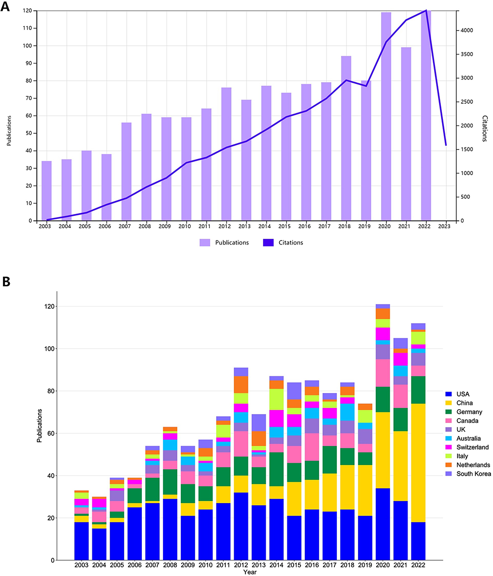 |
Figure 1 (A) Annual publications and citations trend chart of biomechanical research on lumbar IVD. (B) Stack bar plot of top 10 countries/regions by total papers from 2003 to 2022. |
Analysis of Countries/Regions
Figure 2A illustrates a knowledge graph of national/regional cooperation network. where each colored block represents a country/region and its size is proportional to the number of publications. Lines indicate the cooperation relationships among countries/regions, with the thickness of the lines representing the frequency of cooperation.13,14 As shown, the United States has published the most papers in this field and has close collaborations with China, Canada, Switzerland, Australia, and Germany. Figure 2B displays the cooperation relationships and the average publication time among countries/regions. Node size represents the number of publications, and the lines and thickness indicate the cooperation relationships and strength, respectively. The warmer colors of the nodes signify later average publication times.15–18 It appears that the United States made outstanding contributions in the early research of this field with earlier average publication times, while China has recently published a large number of academic achievements. Additionally, Table 1 lists relevant information of the top 10 countries/regions in terms of publication volume, with the United States (484, 34.35%), China (272, 19.30%), and Germany (174, 12.35%) accounting for 66% of the total publications. The United States ranks first in TC (16,258), h-index (66), and total link strength (TLS) (154), while Switzerland, despite ranking seventh in publication output (61), exhibits the highest average citation per publication (ACPP) (38.26). The h-index is an indicator of academic influence, while TLS reflects the strength of relationships with other nodes.19–22
 |
Figure 2 (A) National/regional cooperative network knowledge graph. (B) Time superposition diagram of national/regional cooperation network. |
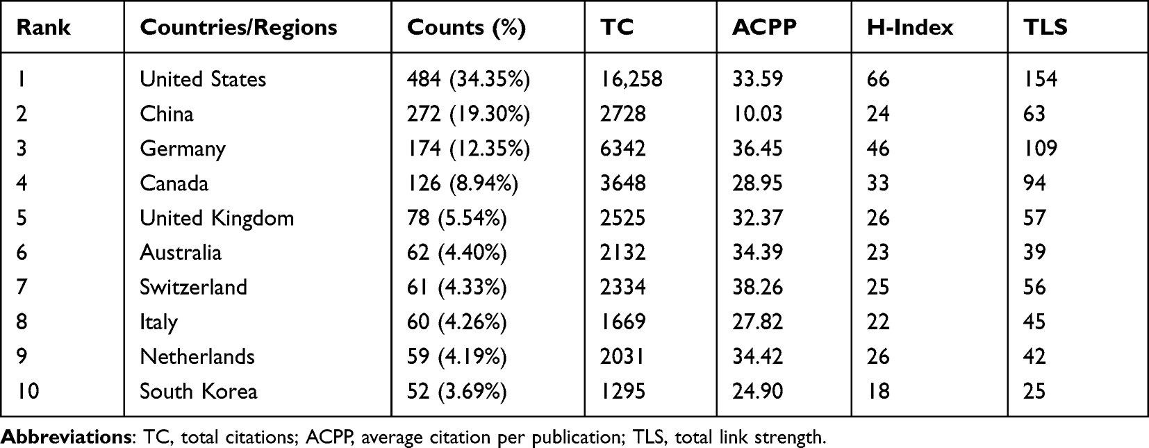 |
Table 1 Top 10 Countries/Regions Ranked by Number of Publications |
Analysis of Institutions
In Figure 3, the collaboration and average publishing time among institutions with a publication count of at least 10 are presented. It can be observed that Ulm University, University of Pennsylvania, and University of Bern have earlier average publishing times, while Icahn School of Medicine at Mount Sinai has shown increased activity in this field in recent years. Table 2 lists the top 10 institutions in terms of publication count, with Ulm University (53, 3.76%) being the most prolific institution, followed by University of Pennsylvania (41, 2.91%), University of California San Francisco (28, 1.99%), Northeastern University (28, 1.99%), and University of Bern (28, 1.99%). Ulm University also leads in TC (3,367) and h-index (32), while Rush University (23, 1.63%) and University of Vermont (23, 1.63%) have the highest TLS (29) and ACPP (66.43), respectively.
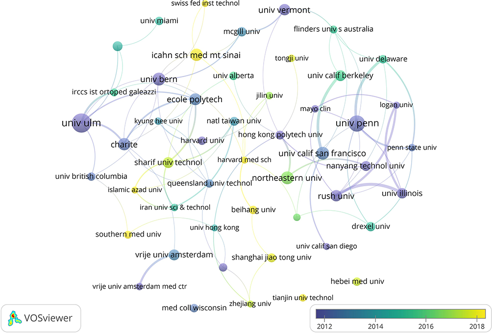 |
Figure 3 Time superposition diagram of institutional cooperation network. |
 |
Table 2 Top 10 Institutions Ranked by Number of Publications |
Analysis of Authors
Figure 4A depicts the collaboration network and average publication time for authors who have published no fewer than 10 papers in this field. Notably, early research in this area featured contributions from distinguished authors such as Wilke HJ, Schmidt H, Elliott DM, and Heuer F, while research in recent years has seen prolific contributions from authors such as Iatridis JC, Shirazi-adl A, and Guo LX. Figure 4B presents a co-citation network of authors who have been cited no less than 100 times. Each node represents a cited author, with node size indicative of frequency of citation, and links between nodes indicating co-citation relationships. Different colors denote distinct clusters.23,24 Table 3 reveals that Wilke HJ not only has the most publications (61, 4.33%), but also boasts the highest TC (3,613), h-index (33) and TLS (98), whereas Rohlmann A’s ACPP (88.80) ranks at the top. Additionally, Table 4 lists information about the top 10 authors with the highest citation frequency, including Adams MA (1065), Wilke HJ (919), and Panjabi MM (741), who are ranked in the top 3 for both citation frequency and TLS.
 |
Figure 4 (A) Time superposition diagram of author’s cooperative network. (B) Author co-citation network knowledge graph. |
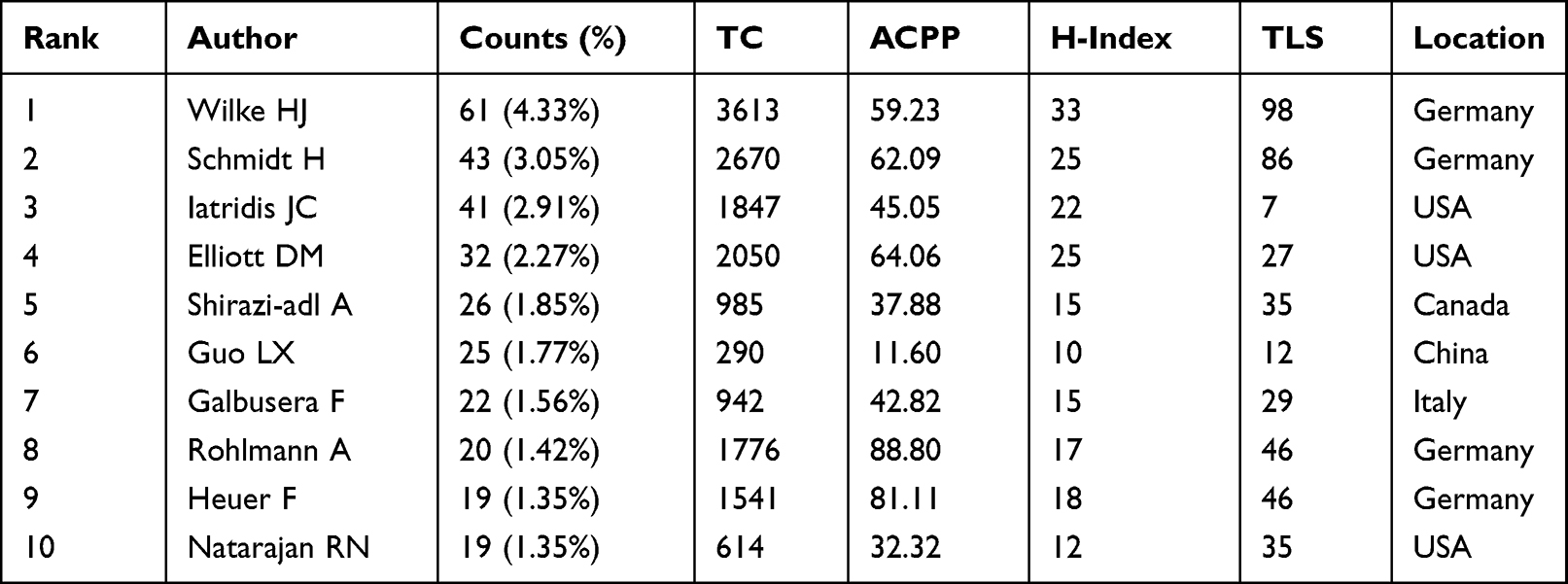 |
Table 3 Top 10 Authors Ranked by Number of Publications |
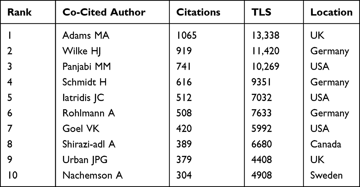 |
Table 4 Top 10 Co-Cited Authors Ranked by Citation Frequency |
Analysis of Journals
Table 5 illustrates the relevant information of the top 10 journals in the research field of biomechanics of lumbar IVD, ranked by publication count. Spine is the highest-ranked journal in terms of publications (163, 11.57%), TC (7,939), and h-index (228). Its impact factor (IF) and journal citation reports (JCR) quartile are 3.241 and Q2, respectively. J Biomech and Eur Spine J follow closely behind, with Eur Spine J having the highest ACPP (49.52). Notably, Spine J is the highest-ranked journal in terms of IF (4.297) and JCR quartile (Q1). Figure 5 displays the co-citation relationships among these journals, and Spine, J Biomech, and Eur Spine J are the top 3 journals in terms of citation frequency and TLS. J Bone Joint Surg Am has an IF of 6.558 and JCR quartile of Q1, making it a highly cited journal of high quality. Further information can be found in Table 6. The dual-map overlay analysis in Figure 6 visually displays the distribution of journals and their referencing relationships. The left side of the figure shows the citing journals, while the right side shows the cited journals, with colored paths representing citation relationships.25–27 Five main citation paths are identified, indicating that papers published in journals related to molecular/biology/genetics, health/nursing/medicine, sports/rehabilitation/sport, and psychology/education/social are primarily cited by papers published in medicine/medical/clinical and neurology/sports/ophthalmology journals.
 |
Figure 5 Journal co-citation network knowledge graph. |
 |
Figure 6 Double graph superposition of journals. |
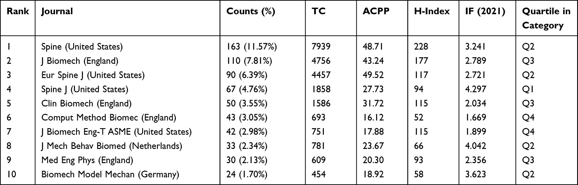 |
Table 5 Top 10 Journals Ranked by Number of Publications |
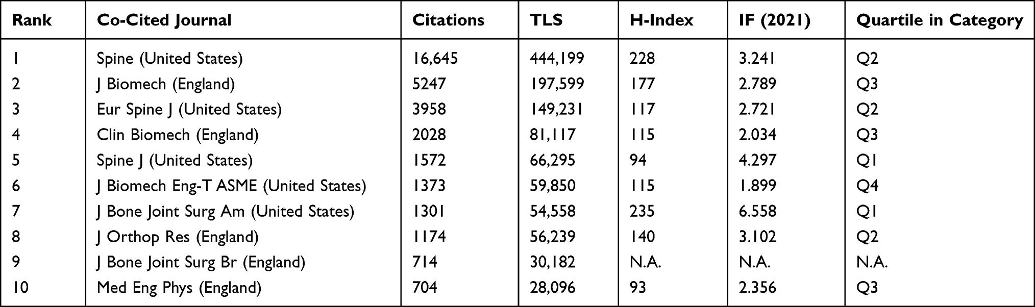 |
Table 6 Top 10 Co-Cited Journals Ranked by Citation Frequency |
Analysis of References
Figure 7 illustrates the co-citation relationships among 35 publications that have been cited no less than 70 times, whereas Table 7 presents relevant information on the top 10 most cited publications. The study conducted by Wilke et al published in Spine in 1999 stands out as the most cited document, with the highest TLS. By defining the minimum burst duration as 3 years, a total of 30 references with high burst strength were detected, of which 13 had a burst duration that ended in 2022 or later, as shown in Figure 8. The blue lines in the figure represent time intervals, the red lines indicate burstiness durations, while “Strength” denotes burst strength, and “Begin” and “End” respectively signify the start and end time of the burstiness.28–31 Citation bursts refer to papers that are frequently cited within a period.32–34 By analyzing these burst papers, researchers can gain insight into the research hotspots and future development trends in a particular field.
 |
Figure 7 Literature co-citation network knowledge graph. |
 |
Figure 8 Top 30 references ranked by burst strength. |
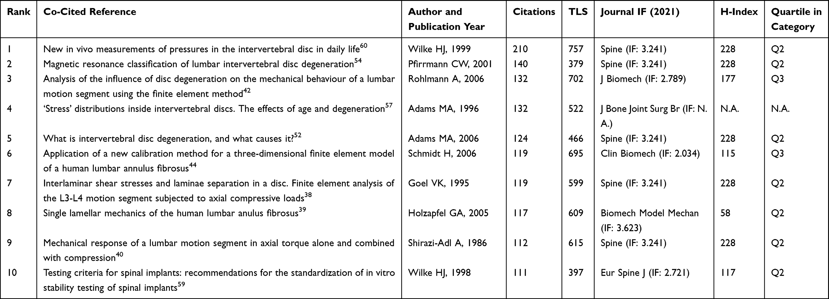 |
Table 7 Top 10 Co-Cited References Ranked by Citation Frequency |
Analysis of Keywords
The succinct summary of research contents using keywords allows for an analysis of high-frequency keywords, providing insight into the main research content and hot topics within a particular field.86–88 Figure 9 depicts the co-occurrence relationship and average appearance time of keywords appearing no less than 15 times. It can be observed that keywords such as biomechanics, lumbar spine, and IVD appear earlier due to the focus of the study, while keywords such as finite element analysis (FEA), low back pain, and IVD degeneration occur frequently in recent years. Additionally, Table 8 presents the top 20 high-frequency keywords and their TLS, which to some extent reflect the primary research content and hot topics within the field. Through keyword clustering, 12 clustering labels were obtained and presented in the form of a keyword timeline map (Figure 10) to facilitate the clear observation of the time evolution characteristics of each clustering field. Burst words are keywords that frequently appear in a certain period, and analyzing them can reveal the research hotspots and development trends of a field.89–91 By setting the shortest burst duration to 3 years, 30 keywords with the highest burst strength were detected, of which 8 will continue to burst until 2022 or later (Figure 11), thereby reflecting recent hot research topics to some extent.
 |
Figure 9 Keyword co-occurrence network knowledge graph. |
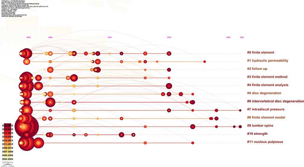 |
Figure 10 Keyword timeline graph. |
 |
Figure 11 Top 30 keywords ranked by burst strength. |
 |
Table 8 Top 20 Keywords Ranked by Frequency of Occurrence |
Discussion
This study employs bibliometric analysis and visualization techniques to comprehensively and systematically dissect the field of biomechanical research on lumbar IVD from multiple perspectives. By constructing scientific knowledge maps, the knowledge framework and development trajectory of this field were visually presented, and research hotspots and future trends were explored to some extent, offering valuable insights for further investigation.
The United States, as the most prolific country, has made significant contributions in this field, constituting 34.35% of the total publications. Furthermore, it ranks first in TC, h-index, and TLS, signifying its notable academic influence and standing in this research sphere, as well as its close collaborations with other countries/regions. Conversely, China, while being the second-highest producer of publications, and surpassing the US in annual paper output since 2019, ranks last in terms of ACPP, and only seventh in terms of h-index. This indicates that China, while having a significant edge in publication quantity, has not achieved extensive recognition and citation by researchers in the same field due to its research quality. Therefore, while pursuing publication volume, China needs to focus on improving research quality and strengthening international exchange and cooperation in order to produce high-quality research results and enhance its academic influence in this field. Meanwhile, Switzerland, despite having relatively fewer publications, has the highest ACPP, indicating that its research results have gained widespread recognition and citations from scholars in the same field, reflecting its high-quality research level and academic prestige. Moreover, it is noteworthy that 70% of the top 10 high-productivity countries/regions come from North America and Europe, indicating that they are the main research hubs in this field.
Ulm University, located in Germany, is the institution with the most publications, highest TC, and highest h-index, indicating its exceptional academic research caliber and the attention its research achievements receive. Among these, a highly cited study published by the institution investigated the impact of IVD degeneration on the mechanics of the motion segment by constructing a three-dimensional (3D) nonlinear finite element model (FEM) of L3/L4 functional units. The results showed that the predicted trends of intersegmental rotation and intradiscal pressure were consistent with the results of in vitro studies. Mildly degenerated IVD increased intersegmental rotation under all loading conditions, while with further degeneration, intersegmental rotation decreased. Regarding axial rotation, the decrease occurred in the final stages. In comparison with healthy discs, degenerated discs showed lower intradiscal pressure but higher facet joint forces and maximum von Mises stresses in the annulus fibrosus (AF).42 The Ulm University has close collaborations with institutions such as the University of Bern, Charite, Ecole Polytechnique, the University of British Columbia, and the Eindhoven University of Technology. The University of Vermont, located in the United States, ranks 10th in terms of publications, but has the highest ACPP, reflecting its outstanding research level and high academic influence. One of the most highly cited publications from this institution reviewed literature evidence in the fields of biomechanics, epidemiology, animal models, and IVD physiology to explore the mechanical conditions that accelerate IVD degeneration. The conclusion was that any abnormal loading conditions, including overload and immobilization, could cause tissue trauma and/or adaptive changes, leading to IVD degeneration.92 Although Northeastern University in China ranks high in terms of publications, it has the lowest citation frequency and h-index and lacks international communication and cooperation. Therefore, its ability to conduct high-level research should be improved and international communication should be strengthened. Furthermore, it is evident that the majority of highly productive and influential institutions are situated in Europe and the United States, with the United States alone accounting for 50% of these institutions. This indicates that the primary research forces in this field are distributed in Western countries, which is consistent with the analysis of countries/regions.
Wilke HJ from Ulm University in Germany is the most prolific author with the highest TC, h-index and TLS, which attests to his exceptional academic accomplishments and influence in the field, and has established extensive collaborations with Schmidt H, Galbusera F, Heuer F, Shirazi-adl A, Rohlmann A, Clses L, and Iatridis JC. One of his highly cited papers used a 3D nonlinear FEM of the lumbar L4-L5 segment to investigate intradiscal pressure, shear strains between the AF and adjacent endplate, and fiber strains of the AF under pure and combined moments. The results showed that combined moments may lead to higher stresses in the disc, especially in the posterior-lateral region, which may be more susceptible to disc injury and herniation.93 Rohlmann A from Charite Universitatsmedizin Berlin in Germany is the author with the highest ACPP, indicating that his research findings are frequently cited by scholars in the same field and he has a high academic reputation. One of his highly cited papers used 3D FEMs of the lumbar L4-L5 segment with different IVD degeneration grades (healthy, mild, moderate, and severe) to investigate the loading conditions resulting in the highest internal stresses in healthy and degenerated discs. The results showed that intradiscal pressure is highest during flexion in all degeneration grades, and shear and fiber strains increased significantly in healthy discs during lateral bending + flexion and in all degenerated discs during axial rotation and lateral bending + axial rotation, mainly in the posterior-lateral AF, with mild degeneration exhibiting increased intradiscal pressure and fiber strains when compared to healthy discs.77 Guo LX from Northeastern University in China has relatively lower citation frequency and h-index, indicating the need to continuously improve research capability, strengthen international cooperation and communication, and publish high-quality academic research. Adams MA from the University of Bristol in the UK has the highest citation frequency and TLS among cited authors, indicating his high academic influence and the broad referencing of his research results, as well as active co-citation relationships with researchers such as Wilke HJ, Panjabi MM, Schmidt H, Iatridis JC, Rohlmann A, Goel VK, Shirazi-adl A, Urban JPG, and Nachemson A.
Spine, J Biomech, Eur Spine J, Spine J, and Clin Biomech are renowned high productivity and highly cited journals that hold significant academic influence and exhibit strong co-citation relationships in the field. Thus, when publishing research findings or searching for relevant literature, these five journals should be considered as a priority. Notably, 80% of the top 10 high productivity journals are from the United States and the United Kingdom, while all of the top 10 highly cited journals are from these two countries as well. This indicates that journals from these countries make significant contributions to research in this field, attracting broad attention and recognition from global researchers. Therefore, it is crucial to recognize the importance of these journals and regard them as valuable channels for publishing and retrieving high-level research achievements.
To a significant extent, highly cited publications can reflect the main research themes and knowledge structure of a given field, and are considered to be high-quality research with significant academic influence.94–96 Among them, the most cited publication is a study by Wilke et al published in Spine in 1999, which measured intradiscal pressure in a volunteer and found that sitting may actually result in lower intradiscal pressure than standing, and that muscle activity increases pressure. The study also emphasized the importance of continuously changing positions to promote fluid (nutrient) flow to the disc, and suggested that some of the physical therapy methods studied may need to be re-evaluated.60 Secondly, Pfirrmann et al54 developed and optimized an algorithm for assessing and grading lumbar disc degeneration based on conventional magnetic resonance imaging (MRI), and tested the reliability of the grading system. The results showed that the grading system and algorithm can reliably grade disc degeneration on MRI. Additionally, Adams et al57 researched the distribution of compression stress in cadaveric IVD and found that age and degeneration have a greater effect on the L4/L5 segment than on the L2/L3 segment, with the posterior AF being more affected than the anterior AF. Adams et al52 also discovered through literature review that structural defects, such as endplate fractures, radial fissures, and herniation, are clear indicators of IVD dysfunction. The reason for biological progress is that structural failure decouples the local mechanical environment of the IVD cells from the overall load on the IVD, resulting in potentially inappropriate or abnormal responses by the IVD cells. Excessive mechanical load damages the structure of the IVD, triggering a series of cell-mediated reactions that lead to further damage. Schmidt et al44 developed a method for calibrating the double composite structure of the AF, the ground substance, and collagen fibers by constructing a 3D nonlinear FEM of the L4-L5 segment. They found that the combinations of infinite material parameters for collagen fibers and ground substance resulted in the same range of motion (ROM), but different ROM for each bending moment. However, there was only one combination that was applicable to all bending moments and in all loading directions. Goel et al38 found through FEA that the interlaminar shear stress was higher in the posterolateral regions of the intact IVD, further demonstrating that tears originate from the posterior-lateral part of the IVD. The significant inter-laminar shear stress caused by the asymmetry of the IVD structure due to damage and the chemical and structural changes of the IVD with age may be important reasons for the further degeneration via laminae separation. Holzapfel et al39 conducted an in vitro study of single lamellar anulus lamellae, showing that the single layer of AF lamellae could be regarded as the basic structural unit of the AF, displaying significant anisotropy and clear regional variation of tensile properties and fiber angles. Shirazi-Adl et al40 analyzed the response of the L2-3 lumbar motion segment to pure axial torque and combined compression using a nonlinear 3D finite element program, finding that with increasing torque, the axis of rotation moved backward in the IVD of the intact segment, such that at the maximum torque, the axis of rotation was behind the IVD itself. The loss of IVD pressure increased this backward movement, while resection of the facets reduced it. Torque itself did not cause fiber damage in the IVD, but when combined with other types of loads (such as flexion), it increased the vulnerability of fibers located in the posterior-lateral and posterior regions. The posterior bony structure was the most fragile part of this segment in torque. The aforementioned highly cited literature serves as the primary research foundation in the field, carrying considerable academic influence and reference significance. These works represent significant objects of focus for gaining a comprehensive understanding of the field.
The phenomenon of literature burst refers to specific publications that have garnered substantial attention within a given time frame. By analyzing burst literature during this period, one can gain insight into the current hot topics and trends within a research field.97–99 Accordingly, this study will focus on the analysis of literature that has remained in the spotlight until 2022 or later, in order to better reflect recent research hotspots and future development trends within this field. Wilke et al73 implanted pressure sensors into the nucleus pulposus (NP) of a non-degenerative L4-5 IVD in a volunteer to measure intra-disc pressure data as a basis for validating predicted spinal load models. Results showed that intra-disc pressure is dependent on the type of prior activity, posture, external load, and muscle activity. Dreischarf et al53 conducted a study on eight established lumbar (L1-5) FEMs from various research centers globally, using pure and combined loading modes, and compared in vivo and in vitro measurement results of intervertebral rotations, intra-disc pressures, and facet joint forces. Results indicated that in pure moment loading, almost all models predicted L1-5 rotation angles within the reported range of in vitro measurements, with median values differing by only 2° for flexion/extension, 1° for lateral bending, and 5° for axial rotation, and predicted facet joint forces and disc pressures median values also corresponded well with published in vitro medians. Schmidt et al76 performed FEA on multi-level total disc arthroplasty (TDA) by establishing a complete human lumbar spine 3D FEM and implanting multiple Charité IVDs in the L1-L5 region from level two to four. The results indicated that the more artificial IVD are implanted, the stronger the predicted flexion and extension motion increased compared to the intact state. Deviation from the optimal implantation location could lead to adverse kinematics, high facet joint forces, and even delamination. Therefore, multi-level TDA should only be performed in suitable patients with good muscle condition and surgical doctors who can ensure optimal implantation positions. Park et al78 conducted an analysis of how degeneration of IVD affects the biomechanics of the lumbar spine and established and verified lumbar spine FEM with different grades of degeneration in L4-L5 functional spinal unit (FSU). Results found that as degeneration progressed, intersegmental rotation in the degenerated FSU decreased during flexion/extension and lateral bending, while adjacent FSU’s intra-disc pressure increased during flexion and lateral bending. Additionally, facet joint forces in the degenerated FSU increased during lateral bending and axial rotation. Newell et al79 conducted a comprehensive review of testing techniques and outcomes related to human IVD biomechanics. They found that the mechanical properties of the entire IVD can be conveniently assessed by testing “motion segment”, consisting of two vertebrae and the intervening IVD and ligaments. However, the accuracy of these evaluations may be influenced by various factors, including the testing environment, preprocessing methodology, loading rate, sample age and degeneration, and the spinal level being tested. Schmidt et al80 conducted a study to examine the accuracy disparities of calibrated and uncalibrated FEM of lumbar segments with various clinical defects. The results found that both FEM performed well in simulating the intact state. However, in cases of defects, the calibrated model demonstrated exceptional consistency in predicting movement behavior, while the uncalibrated model exhibited significant deviation. A comprehensive review conducted by Iatridis et al explored the role of biomechanics in IVD degeneration and regeneration therapy, including the specific mechanical characteristics that need to be repaired and how to evaluate and achieve such reparative processes using biomaterials. The study concluded that IVD height, neutral zone features, and torsional biomechanics are sensitive to specific changes in the compression of the NP and the integrity of the AF, which require effective functional repair. Synthetic and natural biomaterials hold promise for NP replacement, AF repair, AF sealant, or total IVD replacement. To attain optimal efficacy and durability of the repair, it is critical that the biomaterials meet both mechanical and biocompatibility criteria.81 Ayturk et al82 employed a FEM based on the human lumbar L1-L5 to verify the mesh convergence for every component of the tissue, and extensively validated their model using kinematic/kinetic and stress/strain data. Meanwhile, Renner et al83 utilized a 3D FEM of the lumbar L1-S1 to analyze the effects of a significant compressive follower preload on the ROM in all three planes. The results indicated that the compressive follower preload led to a reduction in both the total and segmental ROM during flexion-extension by 18%, lateral bending by 42%, and torsion by 26%. Xu et al84 employed the same modeling approach to develop five FEMs of human lumbar (L1-L5) based on five healthy live subjects, and conducted extensive validation using literature data as well as mesh convergence and material sensitivity analysis. The results demonstrated that the results generated by the five FEMs developed by the authors were in alignment with both experimental data and simulation outcomes reported within the current literature. Adams et al85 reviewed evidence for two types of IVD degeneration. The “endplate-driven” type involves endplate defects and inward buckling of the AF, has a higher genetic predisposition, and primarily affects IVDs of the upper lumbar and thoracic regions, whereas the “annulus-driven” type involves radial fissures and/or disc herniation, has a lower heritability, and mainly affects IVDs of the lower lumbar region. Structural defects triggering both processes play a role in decompressing the IVD nucleus, reducing the likelihood of subsequent defect occurrence, hence, these two phenotypes of IVD degeneration can be considered distinct. Based on the analysis of highly cited and burst literature, it can be inferred that the main research topics in this field include the construction and validation of 3D FEM of the lumbar spine, measurement of intradiscal pressure, biomechanical effects of lumbar IVD degeneration, biomechanical risk factors of lumbar IVD degeneration, mechanical response to different torque load combinations, and MRI classification of lumbar IVD degeneration.
Analysis of keyword co-occurrence, clustering, and burst reveals that the primary research topics in this field includes biomechanics, IVD, lumbar spine, and FEA, which have high frequency of occurrence and strong TLS. The FEA, low back pain, IVD degeneration, AF, MRI, tissue engineering, and endplate are relatively new research subjects that have gained attention in recent years. The 12 clustering labels, including #0 finite element, #1 hydraulic permeability, #2 follow up, #3 finite element method, #4 FEA, #5 disc degeneration, #6 IVD degeneration, #7 intradiscal pressure, #8 FEM, #9 lumbar spine, #10 strength, and #11 NP, to some extent reflect the main research themes in this field. Meanwhile, the burst keywords, such as classification, risk factors, herniation, validation, stepwise reduction, disc, and loads, have duration that ends in 2022 or later, indicating the research hotspots and future development trends in this field.
Strengths and Limitations
This study conducted a comprehensive and systematic analysis of the literature pertaining to the biomechanics of lumbar IVD using bibliometrics and visualization techniques. Unlike traditional reviews that rely on subjective screening and incorporate a limited sample size of research objects, bibliometrics, as a big data analysis methodology, can thoroughly evaluate research objects from multiple perspectives while presenting the results in a more comprehensive and intuitive manner through visualization.100,101 Nevertheless, this study also has some limitations. Firstly, our study focused solely on the SCI-Expanded in WoSCC database, which may have potentially excluded relevant studies, although WoSCC database is the most commonly used database in bibliometrics.102–105 Secondly, some high-quality studies published recently may have been underestimated in our analysis due to their limited citation frequency. Finally, due to the constantly updating nature of databases, the dataset for this year is still incomplete and therefore not included in this study. Despite these limitations, our study results still hold significant reference value, particularly in facilitating the development of research areas and guiding future research directions.
Conclusion
The field of biomechanics of lumbar IVD has seen steady growth in the number of publications and citations over the past 20 years, attracting increasing attention from researchers. The United States has emerged as the most productive country, with Ulm University making the largest contribution. Wilke HJ is both the most prolific author and one of the highly cited authors, with extremely high academic impact and prestige, while Adams MA has the highest citation frequency. Spine, J Biomech, Eur Spine J, Spine J, and Clin Biomech are not only the journals with the most publications, but also highly cited journals, making outstanding contributions to this field and having high academic influence. The main research topics in this field include the construction and validation of 3D FEM of the lumbar spine, measurement of intradiscal pressure, exploration of the biomechanical effects and related risk factors of lumbar IVD degeneration, study of the mechanical response to different moment load combinations, and MRI classification of lumbar IVD degeneration, which are also the hot research topics in recent years. Future research trends are expected to focus on the establishment of more sophisticated FEM of the lumbar spine for multidimensional biomechanical analysis of IVD. Additionally, based on the biomechanical characteristics of IVD, research will explore the effective repair and regenerative treatment of disc degeneration and injuries using biomaterials. These trends will bring new concepts and methods to the research and clinical treatment of lumbar IVD, promoting faster development in this field.
Data Sharing Statement
The original contributions presented in the study are included in the article, further inquiries can be directed to the corresponding authors.
Funding
The study was supported by the Innovation Project of Guangxi Graduate Education (No. YCBXJ2022004), the Guangxi Construction Project of Studio for Famous TCM Experts (GZYKJF [2021] No.6, No.2022004-005-008), and Research Center for Academic Thought and Clinical Diagnosis and Treatment Inheritance and Development of Chinese Medicine Master Wei Guikang at Guangxi University of Chinese Medicine Sponsored Project (No. 2022V001).
Disclosure
The authors declare that there are no conflicts of interest in this work.
References
1. Wang S, Park WM, Gadikota HR, et al. A combined numerical and experimental technique for estimation of the forces and moments in the lumbar intervertebral disc. Comput Methods Biomech Biomed Eng. 2013;16(12):1278–1286. doi:10.1080/10255842.2012.668537
2. Li H, Wang Z. Intervertebral disc biomechanical analysis using the finite element modeling based on medical images. Comput Med Imaging Graph. 2006;30(6–7):363–370. doi:10.1016/j.compmedimag.2006.09.004
3. Wu Y, Wang Y, Wu J, et al. Study of double-level degeneration of lower lumbar spines by finite element model. World Neurosurg. 2016;86:294–299. doi:10.1016/J.WNEU.2015.09.038
4. Wu J, Yang D, Han Y, et al. Application of dual-trajectory screws in revision surgery for lumbar adjacent segment disease: a finite element study. J Orthop Surg Res. 2022;17(1):427. doi:10.1186/s13018-022-03317-9
5. Huang J, Zeng W, Li M, et al. Biomechanical effects of cement discoplasty on the lumbar spinal unit. Front Surg. 2022;9:951141. doi:10.3389/fsurg.2022.951141
6. Höflsauer S, Bonnaire FC, Bamberger CE, Danalache M, Feierabend M, Hofmann UK. Changes in stiffness of the extracellular and pericellular matrix in the anulus fibrosus of lumbar intervertebral discs over the course of degeneration. Front Bioeng Biotechnol. 2022;10:1006615. doi:10.3389/fbioe.2022.1006615
7. Techens C, Montanari S, Bereczki F, Eltes PE, Lazary A, Cristofolini L. Biomechanical consequences of cement discoplasty: an in vitro study on thoraco-lumbar human spines. Front Bioeng Biotechnol. 2022;10:1040695. doi:10.3389/fbioe.2022.1040695
8. Linders DR, Nuckley DJ. Deduction of spinal loading from vertebral body surface strain measurements. Exp Mech. 2007;47:303–310. doi:10.1007/s11340-006-9008-6
9. Park WM, Kim YH, Lee S. Effect of intervertebral disc degeneration on biomechanical behaviors of a lumbar motion segment under physiological loading conditions. J Mech Sci Technol. 2013;27:483–489. doi:10.1007/s12206-012-1264-z
10. Xu Q, Zhou Y, Zhang H, Li H, Qin H, Wang H. Bibliometric analysis of hotspots and frontiers of immunotherapy in pancreatic cancer. Healthcare. 2023;11:304. doi:10.3390/healthcare11030304
11. Zhang J-H, Wang M-J, Tan Y-T, Luo J, Wang S-C. A bibliometric analysis of apoptosis in glaucoma. Front Neurosci. 2023;17:1105158. doi:10.3389/fnins.2023.1105158
12. Ding Z, Tang N, Huang J, Cao X, Wu S. Global hotspots and emerging trends in 3D bioprinting research. Front Bioeng Biotechnol. 2023;11:1169893. doi:10.3389/fbioe.2023.1169893
13. Lu H, Han T, Li F, Yang J, Hou Z. Global trends and hotspots in research of robotic surgery in oncology: a bibliometric and visual analysis from 2002 to 2021. Front Oncol. 2022;12:1055118. doi:10.3389/fonc.2022.1055118
14. Yang J, Wu J, Han T, et al. Global research hotspots and frontiers of myasthenia gravis from 2002 to 2021: a bibliometric study. Medicine. 2023;102:e34002. doi:10.1097/MD.0000000000034002
15. Zhang D, Li X, Jia N, Chen W, Hu Y. Bibliometric and visual analysis of cerebral revascularization from 1999 to 2022. Front Neurosci. 2023;16:1088448. doi:10.3389/fnins.2022.1088448
16. He L, Guo Q-F, Hu Y, et al. Bibliometric and visualised analysis on non-invasive cerebellar stimulation from 1995 to 2021. Front Neurosci. 2023;17:1047238. doi:10.3389/fnins.2023.1047238
17. Yang Z, Fan Z, Wang D, et al. Bibliometric and visualization analysis of stem cell therapy for meniscal regeneration from 2012 to 2022. Front Bioeng Biotechnol. 2023;11:1107209. doi:10.3389/fbioe.2023.1107209
18. Zhu M-X, Sun S-Q, Fan G-B, et al. Knowledge mapping of research on the mitochondrial unfolded protein response: a bibliometric and visual analysis. Ann Transl Med. 2023;11:64. doi:10.21037/atm-22-6423
19. Hou Z, Jiang P, Su S, Zhou H. Hotspots and trends in multiple myeloma bone diseases: a bibliometric visualization analysis. Front Pharmacol. 2022;13:1003228. doi:10.3389/fphar.2022.1003228
20. Huang P, Feng Z, Shu X, et al. A bibliometric and visual analysis of publications on artificial intelligence in colorectal cancer (2002). Front Oncol. 2023;13:1077539. doi:10.3389/fonc.2023.1077539
21. Jiang F, Sun T, Cheng P, Wang J, Gong W. A summary on tuberculosis vaccine development-where to go? J Pers Med. 2023;13:408. doi:10.3390/jpm13030408
22. Zhang J-Y, Xiao C-F, Wang C, Yao Y-B. Bibliometric insights in fournier’s gangrene: research landscapes, turning points, and global trends. Front Surg. 2023;10:1057486. doi:10.3389/fsurg.2023.1057486
23. Tang F, Jiang C, Chen J, Wang L, Zhao F. Global hotspots and trends in myofascial pain syndrome research from 1956 to 2022: a bibliometric analysis. Medicine. 2023;102. doi:10.1097/MD.0000000000033347
24. Chen M, Zhang Y, Dong L, Guo X. Bibliometric analysis of stroke and quality of life. Front Neurol. 2023;14:1143713. doi:10.3389/fneur.2023.1143713
25. Li Y, Wang H, Jiang L, Chen L, Zhao K, Li X. A bibliometric analysis of chronic obstructive pulmonary disease and COVID-19. Medicine. 2023;102. doi:10.1097/MD.0000000000033240
26. Wang J, Cao B, Lin S, et al. A bibliometric analysis of urologic chronic pelvic pain syndrome from 2000 to 2022. J Pain Res. 2023;16:1225–1241. doi:10.2147/JPR.S396009
27. Wang J, Gong Z, Yu M. Bibliometric study on the knowledge graph of immunotherapy for head and neck cancer. Front Oncol. 2023;13:942777. doi:10.3389/fonc.2023.942777
28. Lin Y, Ren X, Chen D. Steroid treatment in macular edema: a bibliometric study and visualization analysis. Front Pharmacol. 2022;13:824790. doi:10.3389/fphar.2022.824790
29. Zhang T, Zhang B, Tian W, et al. A bibliometric analysis of atrophic gastritis from 2011 to 2021. Front Med. 2022;9:843395. doi:10.3389/fmed.2022.843395
30. Guo Y, Xu Z-Y-R, Cai M-T, Gong W-X, Shen C-H. Epilepsy with suicide: a bibliometrics study and visualization analysis via CiteSpace. Front Neurol. 2021;12:823474. doi:10.3389/fneur.2021.823474
31. Xiong J-Q, Fu Y-F, Qiu J-H, Liao W-D, Luo L-Y, Chen S-H. Global research trends of immunotherapy and biotherapy for inflammatory bowel disease: a bibliometric analysis from 2002 to 2021. Biomed Eng Online. 2022;21:42. doi:10.1186/s12938-022-01011-9
32. Liu Z, Wang M, Luo J, Tan Y, Hou M, Wang S. A bibliometric analysis of hotpots and trends for the relationship between skin inflammation and regeneration. Front Surg. 2023;10:1180624. doi:10.3389/fsurg.2023.1180624
33. Wang Y, Zhang S, Zhi J, Huang M, Pei F. A bibliometric analysis: current status and frontier trends of Schwann cells in neurosciences. Front Molec Neurosci. 2023;15:1087550. doi:10.3389/fnmol.2022.1087550
34. Zhang J, Song L, Jia J, Tian W, Lai R. Knowledge mapping of necroptosis from 2012 to 2021: a bibliometric analysis. Front Immunol. 2022;13:917155. doi:10.3389/fimmu.2022.917155
35. Shirazi-Adl SA, Shrivastava SC, Ahmed AM. Stress analysis of the lumbar disc-body unit in compression. A three-dimensional nonlinear finite element study. Spine. 1984;9(2):120–134. doi:10.1097/00007632-198403000-00003
36. Marchand F, Ahmed AM. Investigation of the laminate structure of lumbar disc anulus fibrosus. Spine. 1990;15(5):402–410. doi:10.1097/00007632-199005000-00011
37. Sharma M, Langrana NA, Rodriguez J. Role of ligaments and facets in lumbar spinal stability. Spine. 1995;20(8):887–900. doi:10.1097/00007632-199504150-00003
38. Goel VK, Monroe BT, Gilbertson LG, Brinckmann P. Interlaminar shear stresses and laminae separation in a disc. Finite element analysis of the L3-L4 motion segment subjected to axial compressive loads. Spine. 1995;20:689–698.
39. Holzapfel GA, Schulze-Bauer C, Feigl G, Regitnig P. Single lamellar mechanics of the human lumbar anulus fibrosus. Biomech Model Mechanobiol. 2005;3:125–140. doi:10.1007/s10237-004-0053-8
40. Shirazi-Adl A, Ahmed AM, Shrivastava SC. Mechanical response of a lumbar motion segment in axial torque alone and combined with compression. Spine. 1986;11:914–927. doi:10.1097/00007632-198611000-00012
41. Argoubi M, Shirazi-Adl A. Poroelastic creep response analysis of a lumbar motion segment in compression. J Biomech. 1996;29(10):1331–1339. doi:10.1016/0021-9290(96)00035-8
42. Rohlmann A, Zander T, Schmidt H, Wilke H-J, Bergmann G. Analysis of the influence of disc degeneration on the mechanical behaviour of a lumbar motion segment using the finite element method. J Biomech. 2006;39:2484–2490. doi:10.1016/j.jbiomech.2005.07.026
43. Iatridis JC, Setton LA, Foster RJ, Rawlins BA, Weidenbaum M, Mow VC. Degeneration affects the anisotropic and nonlinear behaviors of human anulus fibrosus in compression. J Biomech. 1998;31(6):535–544. doi:10.1016/s0021-9290(98)00046-3
44. Schmidt H, Heuer F, Simon U, et al. Application of a new calibration method for a three-dimensional finite element model of a human lumbar annulus fibrosus. Clin Biomech. 2006;21:337–344. doi:10.1016/j.clinbiomech.2005.12.001
45. Beckstein JC, Sen S, Schaer TP, Vresilovic EJ, Elliott DM. Comparison of animal discs used in disc research to human lumbar disc: axial compression mechanics and glycosaminoglycan content. Spine. 2008;33(6):E166–E173. doi:10.1097/BRS.0b013e318166e001
46. White AA, Panjabi MM. Clinical Biomechanics of the Spine.
47. Heuer F, Schmidt H, Klezl Z, Claes L, Wilke HJ. Stepwise reduction of functional spinal structures increase range of motion and change lordosis angle. J Biomech. 2007;40(2):271–280. doi:10.1016/j.jbiomech.2006.01.007
48. Urban JP, Roberts S. Degeneration of the intervertebral disc. Arthritis Res Ther. 2003;5(3):120–130. doi:10.1186/ar629
49. Mimura M, Panjabi MM, Oxland TR, Crisco JJ, Yamamoto I, Vasavada A. Disc degeneration affects the multidirectional flexibility of the lumbar spine. Spine. 1994;19(12):1371–1380. doi:10.1097/00007632-199406000-00011
50. Thompson JP, Pearce RH, Schechter MT, Adams ME, Tsang IK, Bishop PB. Preliminary evaluation of a scheme for grading the gross morphology of the human intervertebral disc. Spine. 1990;15(5):411–415. doi:10.1097/00007632-199005000-00012
51. Panjabi MM, Oxland TR, Yamamoto I, Crisco JJ. Mechanical behavior of the human lumbar and lumbosacral spine as shown by three-dimensional load-displacement curves. J. Bone Joint Surg Am. 1994;76(3):413–424. doi:10.2106/00004623-199403000-00012
52. Adams MA, Roughley PJ. What is intervertebral disc degeneration, and what causes it? Spine. 2006;31:2151–2161. doi:10.1097/01.brs.0000231761.73859.2c
53. Dreischarf M, Zander T, Shirazi-Adl A, et al. Comparison of eight published static finite element models of the intact lumbar spine: predictive power of models improves when combined together. J Biomech. 2014;47:1757–1766. doi:10.1016/j.jbiomech.2014.04.002
54. Pfirrmann CW, Metzdorf A, Zanetti M, Hodler J, Boos N. Magnetic resonance classification of lumbar intervertebral disc degeneration. Spine. 2001;26:1873–1878. doi:10.1097/00007632-200109010-00011
55. Brinckmann P, Grootenboer H. Change of disc height, radial disc bulge, and intradiscal pressure from discectomy. An in vitro investigation on human lumbar discs. Spine. 1991;16(6):641–646. doi:10.1097/00007632-199106000-00008
56. Adams MA, Freeman BJ, Morrison HP, Nelson IW, Dolan P. Mechanical initiation of intervertebral disc degeneration. Spine. 2000;25(13):1625–1636. doi:10.1097/00007632-200007010-00005
57. Adams MA, McNally DS, Dolan P. “Stress” distributions inside intervertebral discs. The effects of age and degeneration. J Bone Joint Surg Br. 1996;78:965–972. doi:10.1302/0301-620x78b6.1287
58. Wilke HJ, Rohlmann A, Neller S, et al. Is it possible to simulate physiologic loading conditions by applying pure moments? A comparison of in vivo and in vitro load components in an internal fixator. Spine. 2001;26(6):636–642. doi:10.1097/00007632-200103150-00014
59. Wilke HJ, Wenger K, Claes L. Testing criteria for spinal implants: recommendations for the standardization of in vitro stability testing of spinal implants. Eur Spine J. 1998;7(2):148–154. doi:10.1007/s005860050045
60. Wilke HJ, Neef P, Caimi M, Hoogland T, Claes LE. New in vivo measurements of pressures in the intervertebral disc in daily life. Spine. 1999;24:755–762. doi:10.1097/00007632-199904150-00005
61. Patwardhan AG, Havey RM, Meade KP, Lee B, Dunlap B. A follower load increases the load-carrying capacity of the lumbar spine in compression. Spine. 1999;24(10):1003–1009. doi:10.1097/00007632-199905150-00014
62. Lotz JC, Colliou OK, Chin JR, Duncan NA, Liebenberg E. Compression-induced degeneration of the intervertebral disc: an in vivo mouse model and finite-element study. Spine. 1998;23(23):2493–2506. doi:10.1097/00007632-199812010-00004
63. Lee CK. Accelerated degeneration of the segment adjacent to a lumbar fusion. Spine. 1988;13(3):375–377. doi:10.1097/00007632-198803000-00029
64. Cunningham BW, Kotani Y, McNulty PS, Cappuccino A, McAfee PC. The effect of spinal destabilization and instrumentation on lumbar intradiscal pressure: an in vitro biomechanical analysis. Spine. 1997;22(22):2655–2663. doi:10.1097/00007632-199711150-00014
65. McNally DS, Adams MA. Internal intervertebral disc mechanics as revealed by stress profilometry. Spine. 1992;17(1):66–73. doi:10.1097/00007632-199201000-00011
66. Osti OL, Vernon-Roberts B, Fraser RD. 1990 Volvo Award in experimental studies. Anulus tears and intervertebral disc degeneration. An experimental study using an animal model. Spine. 1990;15(8):762–767. doi:10.1097/00007632-199008010-00005
67. Dooris AP, Goel VK, Grosland NM, Gilbertson LG, Wilder DG. Load-sharing between anterior and posterior elements in a lumbar motion segment implanted with an artificial disc. Spine. 2001;26(6):E122–E129. doi:10.1097/00007632-200103150-00004
68. Gu WY, Mao XG, Foster RJ, Weidenbaum M, Mow VC, Rawlins BA. The anisotropic hydraulic permeability of human lumbar anulus fibrosus. Influence of age, degeneration, direction, and water content. Spine. 1999;24(23):2449–2455. doi:10.1097/00007632-199912010-00005
69. Ghiselli G, Wang JC, Bhatia NN, Hsu WK, Dawson EG. Adjacent segment degeneration in the lumbar spine. J Bone Joint Surg Am. 2004;86(7):1497–1503. doi:10.2106/00004623-200407000-00020
70. Rohlmann A, Zander T, Rao M, Bergmann G. Applying a follower load delivers realistic results for simulating standing. J Biomech. 2009;42(10):1520–1526. doi:10.1016/j.jbiomech.2009.03.048
71. Urban JP, Smith S, Fairbank JC. Nutrition of the intervertebral disc. Spine. 2004;29(23):2700–2709. doi:10.1097/01.brs.0000146499.97948.52
72. Schmidt H, Shirazi-Adl A, Galbusera F, Wilke HJ. Response analysis of the lumbar spine during regular daily activities--a finite element analysis. J Biomech. 2010;43(10):1849–1856. doi:10.1016/j.jbiomech.2010.03.035
73. Wilke H, Neef P, Hinz B, Seidel H, Claes L. Intradiscal pressure together with anthropometric data--a data set for the validation of models. Clin Biomech. 2001;16(Suppl 1):S111–126. doi:10.1016/s0268-0033(00)00103-0
74. Niemeyer F, Wilke HJ, Schmidt H. Geometry strongly influences the response of numerical models of the lumbar spine--a probabilistic finite element analysis. J Biomech. 2012;45(8):1414–1423. doi:10.1016/j.jbiomech.2012.02.021
75. Alini M, Eisenstein SM, Ito K, et al. Are animal models useful for studying human disc disorders/degeneration? Eur Spine J. 2008;17(1):2–19. doi:10.1007/s00586-007-0414-y
76. Schmidt H, Galbusera F, Rohlmann A, Zander T, Wilke H-J. Effect of multilevel lumbar disc arthroplasty on spine kinematics and facet joint loads in flexion and extension: a finite element analysis. Eur Spine J. 2012;21(Suppl 5):S663–674. doi:10.1007/s00586-010-1382-1
77. Schmidt H, Kettler A, Rohlmann A, Claes L, Wilke H-J. The risk of disc prolapses with complex loading in different degrees of disc degeneration - a finite element analysis. Clin Biomech. 2007;22:988–998. doi:10.1016/j.clinbiomech.2007.07.008
78. Park WM, Kim K, Kim YH. Effects of degenerated intervertebral discs on intersegmental rotations, intradiscal pressures, and facet joint forces of the whole lumbar spine. Comput Biol Med. 2013;43:1234–1240. doi:10.1016/j.compbiomed.2013.06.011
79. Newell N, Little JP, Christou A, Adams MA, Adam CJ, Masouros SD. Biomechanics of the human intervertebral disc: a review of testing techniques and results. J Mech Behav Biomed Mater. 2017;69:420–434. doi:10.1016/j.jmbbm.2017.01.037
80. Schmidt H, Heuer F, Drumm J, Klezl Z, Claes L, Wilke H-J. Application of a calibration method provides more realistic results for a finite element model of a lumbar spinal segment. Clin Biomech. 2007;22:377–384. doi:10.1016/j.clinbiomech.2006.11.008
81. Iatridis JC, Nicoll SB, Michalek AJ, Walter BA, Gupta MS. Role of biomechanics in intervertebral disc degeneration and regenerative therapies: what needs repairing in the disc and what are promising biomaterials for its repair? Spine J. 2013;13:243–262. doi:10.1016/j.spinee.2012.12.002
82. Ayturk UM, Puttlitz CM. Parametric convergence sensitivity and validation of a finite element model of the human lumbar spine. Comput Methods Biomech Biomed Engin. 2011;14:695–705. doi:10.1080/10255842.2010.493517
83. Renner SM, Natarajan RN, Patwardhan AG, et al. Novel model to analyze the effect of a large compressive follower pre-load on range of motions in a lumbar spine. J Biomech. 2007;40:1326–1332. doi:10.1016/j.jbiomech.2006.05.019
84. Xu M, Yang J, Lieberman IH, Haddas R. Lumbar spine finite element model for healthy subjects: development and validation. Comput Methods Biomech Biomed Engin. 2017;20:1–15. doi:10.1080/10255842.2016.1193596
85. Adams MA, Dolan P. Intervertebral disc degeneration: evidence for two distinct phenotypes. J Anat. 2012;221:497–506. doi:10.1111/j.1469-7580.2012.01551.x
86. Xiao L, Huo X, Wang Y, et al. A bibliometric analysis of global research status and trends in neuromodulation techniques in the treatment of autism spectrum disorder. BMC Psychiatry. 2023;23:183. doi:10.1186/s12888-023-04666-3
87. Zhang G, Song J, Feng Z, et al. Artificial intelligence applicated in gastric cancer: a bibliometric and visual analysis via CiteSpace. Front Oncol. 2023;12:1075974. doi:10.3389/fonc.2022.1075974
88. Ji L, Li F. Potential markers of neurocognitive disorders after cardiac surgery: a bibliometric and visual analysis. Front Aging Neurosci. 2022;14:868158. doi:10.3389/fnagi.2022.868158
89. Li R, Wang Y, Zhao Z, Li X, Liu Z. A bibliometric analysis based on Web of Science from 2012 to 2021: current situation, hot spots, and global trends of medullary thyroid carcinoma. Front Oncol. 2023;13:1119915. doi:10.3389/fonc.2023.1119915
90. Ling F, Qi W, Li X, et al. Bibliometric analysis of acupuncture therapy for cancer pain over the past 10 years. J Pain Res. 2023;16:985–1003. doi:10.2147/JPR.S395421
91. Tan Y, Yu Y, Liu W, Ma X, Shi D. Bibliometric evaluation of publications on inflammasomes in atherosclerosis from 2002 to 2022. Front Cardiovasc Med. 2023;10:1067226. doi:10.3389/fcvm.2023.1067226
92. Stokes IAF, Iatridis JC. Mechanical conditions that accelerate intervertebral disc degeneration: overload versus immobilization. Spine. 2004;29:2724–2732. doi:10.1097/01.brs.0000146049.52152.da
93. Schmidt H, Kettler A, Heuer F, Simon U, Claes L, Wilke H-J. Intradiscal pressure, shear strain, and fiber strain in the intervertebral disc under combined loading. Spine. 2007;32:748–755. doi:10.1097/01.brs.0000259059.90430.c2
94. Chen S, Sun D, Wang N, et al. Current status and trends in quantitative MRI study of intervertebral disc degeneration: a bibliometric and clinical study analysis. Quant Imaging Med Surg. 2023;13:2953–2974. doi:10.21037/qims-22-1219
95. Zhu X, Hu J, Deng S, et al. Comprehensive bibliometric analysis of the Kynurenine pathway in mood disorders: focus on gut microbiota research. Front Pharmacol. 2021;12:687757. doi:10.3389/fphar.2021.687757
96. Wu H, Zhou Y, Wang Y, et al. Current state and future directions of intranasal delivery route for central nervous system disorders: a scientometric and visualization analysis. Front Pharmacol. 2021;12:717192. doi:10.3389/fphar.2021.717192
97. Zhu G, Fu Z, Su S, Tang Y, Liu F, Yu W. Global trends and hotspots in trigeminal neuralgia research from 2001 to 2021: a bibliometric analysis. Front Neurol. 2022;13:894006. doi:10.3389/fneur.2022.894006
98. Guo Y, Yang Y, Xu M, et al. Trends and developments in the detection of pathogens in central nervous system infections: a bibliometric study. Front Cell Infect Microbiol. 2022;12:856845. doi:10.3389/fcimb.2022.856845
99. Cheng K, Guo Q, Yang W, Wang Y, Sun Z, Wu H. Mapping knowledge landscapes and emerging trends of the links between bone metabolism and diabetes mellitus: a bibliometric analysis from 2000 to 2021. Front Public Health. 2022;10:918483. doi:10.3389/fpubh.2022.918483
100. You Y, Li W, Liu J, Li X, Fu Y, Ma X. Bibliometric review to explore emerging high-intensity interval training in health promotion: a new century picture. Front Public Health. 2021;9:697633. doi:10.3389/fpubh.2021.697633
101. Qiu H, Guo R, Zhang Y, Ying J, Yan Y, Xiong J. A bibliometric analysis of the hotspots concerning stem cell extracellular vesicles for diabetes in the last 5 years. Front Public Health. 2022;10:868440. doi:10.3389/fpubh.2022.868440
102. Li M, Jiang Z, Wen R, Liu C, Wang J. A bibliometric analysis of the application of imaging in sleep in neurodegenerative disease. Front Aging Neurosci. 2023;15:1078807. doi:10.3389/fnagi.2023.1078807
103. Wang L, Deng S, Meng F, et al. Comprehensive analysis of global research on overactive bladder: a scientometric approach. Front Surg. 2023;9:1078052. doi:10.3389/fsurg.2022.1078052
104. Li J, Gong X. Bibliometric and visualization analysis of kidney repair associated with acute kidney injury from 2002 to 2022. Front Pharmacol. 2023;14:1101036. doi:10.3389/fphar.2023.1101036
105. Carhuallanqui-Ciocca EI, Echevarria-Quispe JY, Hernandez-Vasquez A, Diaz-Ruiz R, Azanedo D. Bibliometric analysis of the scientific production on inguinal hernia surgery in the web of science. Front Surg. 2023;10:1138805. doi:10.3389/fsurg.2023.1138805
 © 2023 The Author(s). This work is published and licensed by Dove Medical Press Limited. The
full terms of this license are available at https://www.dovepress.com/terms
and incorporate the Creative Commons Attribution
- Non Commercial (unported, 3.0) License.
By accessing the work you hereby accept the Terms. Non-commercial uses of the work are permitted
without any further permission from Dove Medical Press Limited, provided the work is properly
attributed. For permission for commercial use of this work, please see paragraphs 4.2 and 5 of our Terms.
© 2023 The Author(s). This work is published and licensed by Dove Medical Press Limited. The
full terms of this license are available at https://www.dovepress.com/terms
and incorporate the Creative Commons Attribution
- Non Commercial (unported, 3.0) License.
By accessing the work you hereby accept the Terms. Non-commercial uses of the work are permitted
without any further permission from Dove Medical Press Limited, provided the work is properly
attributed. For permission for commercial use of this work, please see paragraphs 4.2 and 5 of our Terms.
Recommended articles
Knowledge Atlas of the Co-Occurrence of Epilepsy and Autism: A Bibliometric Analysis and Visualization Using VOSviewer and CiteSpace
Wang Y, Huo X, Li W, Xiao L, Li M, Wang C, Sun Y, Sun T
Neuropsychiatric Disease and Treatment 2022, 18:2107-2119
Published Date: 19 September 2022
Bibliometric and Visualization Analysis of Research Hotspots and Frontiers in Endoscopic Lumbar Discectomy
Zhong Y, Wang J, Liang Z, Han T, Lu H, Hou Z
Journal of Pain Research 2024, 17:2165-2190
Published Date: 18 June 2024
Global Trends on β-Thalassemia Research Over 10 Years: A Bibliometric Analysis
Lv A, Li J, Chen M, Wang W, Xu L, Huang H
International Journal of General Medicine 2024, 17:3989-4001
Published Date: 11 September 2024
Research Progress and Trends in Exercise Interventions for Mild Cognitive Impairment: A Bibliometric Visualization Analysis Using CiteSpace
Han Q, Kim SM
Journal of Multidisciplinary Healthcare 2025, 18:505-529
Published Date: 31 January 2025
Comprehensive Analysis of the Biomechanical Research of Pelvic Organ Prolapse: A Scientometric Approach
Du H, Yang M, Qi X, Yang L, Wang Z, Yang T, Xu S, Fu L
Journal of Multidisciplinary Healthcare 2025, 18:1249-1268
Published Date: 1 March 2025