
Back to Journals » International Journal of Nanomedicine » Volume 21
Nanomaterial-Based Photothermal Therapy for Inflammatory Diseases: Intervention Strategies, Synergistic Effects, and Future Challenges
Authors Wang C  , Yu S, Yang J, Zhang H, Wu C, Lai D, Zhou R, Chen T
Received 30 April 2026
Accepted for publication 10 July 2026
Published 17 July 2026 Volume 2026:21 621265
DOI https://doi.org/10.2147/IJN.S621265
Checked for plagiarism Yes
Review by Single anonymous peer review
Peer reviewer comments 4
Editor who approved publication: Prof. Dr. RDK Misra
Chenglong Wang,1,* Shiyu Yu,1,* Jian Yang,1,* Hao Zhang,2 Chengxi Wu,3 Dixuan Lai,1 Ruiou Zhou,4 Tao Chen1
1Department of Pharmacy, Yibin Hospital Affiliated to Children’s Hospital of Chongqing Medical University, Yibin, Sichuan, People’s Republic of China; 2Department of Pharmacy, Zigong First People’s Hospital, Zigong, Sichuan, People’s Republic of China; 3Department of Vascular Surgery, The Third People’s Hospital of Yibin, Yibin, Sichuan, People’s Republic of China; 4Department of Pharmacy, Children’s Hospital of Chongqing Medical University, National Clinical Research Center for Children and Adolescents’ Health and Diseases, Ministry of Education Key Laboratory of Child Development and Disorders, Chongqing Key Laboratory of Pediatric Metabolism and Inflammatory Diseases, Chongqi, People’s Republic of China
*These authors contributed equally to this work
Correspondence: Ruiou Zhou, Email [email protected] Tao Chen, Email [email protected]
Abstract: As a fundamental host defense mechanism, the transition from acute to chronic inflammation serves as the foundation for numerous major diseases. Conventional anti-inflammatory interventions are frequently hindered by inadequate bioavailability, instability, and undesirable side effects. High-temperature photothermal therapy (PTT) based on nanomaterials inhibits inflammatory cascades via the precise ablation of activated inflammatory cells, induction of apoptosis, bacterial elimination, and controlled drug release at lesion sites. Meanwhile, mild PTT exerts anti-inflammation through immunomodulation, including regulation of M1/M2 macrophage polarization and upregulation of heat shock proteins (HSPs). These two PTT modalities are complementary in anti‑inflammatory applications, providing more adaptable strategies for precision therapy of inflammatory diseases. This review systematically summarizes strategies to enhance PTT-mediated anti-inflammatory therapy through photothermal nanomaterials, with a particular focus on an emerging paradigm designed to achieve synergistic effects. This review begins with a categorical assessment of current research on inorganic and organic photothermal nanomaterials for anti-inflammatory applications. Building on this, we provide an in-depth analysis of disease-oriented PTT-based anti-inflammatory therapies (e.g. for vascular, articular, and periodontal inflammation), focusing on how engineering design, particularly targeted delivery and multimodal synergistic strategies, enables precise treatment in specific pathological models. Finally, we critically analyze the key challenges in this field, including photodynamic therapy (PDT) side effects, standardization of laser parameters, insufficient light penetration depth, clearance mechanisms, biosafety, and clinical translation. Prospective insights are also provided to guide the development of the next-generation intelligent and efficient photothermal nanoplatforms for anti-inflammatory therapy.
Keywords: photothermal therapy, nanomaterials, inflammation, macrophage, synergistic strategy
Introduction
Inflammation represents a complex yet essential defensive response triggered by external chemical or physical stimuli, as well as internal biological signals.1,2 During the acute phase, a programmed cascade directs numerous immune cells to migrate to the injury site,3–5 orchestrated by various inflammatory mediators, chemokines, and acute-phase proteins. This precisely regulated response eliminates harmful stimuli and activates healing processes, thereby promoting the return to tissue homeostasis.6,7 Nevertheless, persistent exposure to inflammatory triggers or dysregulated immune responses can drive the transition from acute to chronic inflammation. Subsequent tissue injury and fibrotic changes underpin a range of chronic diseases, including arthritis, atherosclerosis (AS), inflammatory bowel disease (IBD), and various autoimmune disorders.8–10 Conventional anti-inflammatory pharmacotherapies are frequently limited by inadequate targeting, suboptimal biocompatibility, multidrug resistance, and systemic accumulation.11,12 These shortcomings result in reduced therapeutic outcomes and significant off-target effects, ultimately diminishing patient quality of life and worsening clinical outcomes.
Advances in nanomaterial science have facilitated a host of advanced inflammation suppression strategies noted for their enhanced efficacy and safety. Notably, non-invasive phototherapies such as photothermal therapy (PTT) and photodynamic therapy (PDT) have been extensively investigated for modulating inflammatory progression across diverse disorders.13–15 As a minimally invasive technique, PTT achieves therapeutic effects via photosensitizers or nanoparticles (NPs) that generate localized heat upon near-infrared (NIR) laser irradiation.16,17 Initially applied in oncology for tumor ablation due to its deep tissue penetration and thermal efficacy,18,19 subsequent research has revealed that PTT effectively eliminates inflammatory cells at injury sites and curbs pathological development. Notably, some studies have reported that the thermal tolerance of certain inflammatory cells may be inferior to that of normal cells.20,21 Specifically, temperatures in the range of 42–45°C can effectively eliminate inflammatory cells, whereas normal cells remain viable under such conditions for prolonged periods.22 This differential thermal sensitivity suggests the potential for selective ablation or apoptosis induction in target cell populations (eg, macrophages, neutrophils (NEs), and synovial cells) through localized hyperthermia,23 although the extent to which this applies across different tissues remains to be established. Nonetheless, targeted delivery of photothermal nanomaterials for PTT offers new approaches for precise inflammatory disease management.
Thermal ablation of inflammatory cells disrupts the vicious cycle of pathological cell recruitment, diminishing the influx of additional immune cells to inflamed tissues. Meanwhile, this intervention inhibits several downstream pro-inflammatory signaling axes, exemplified by the nuclear factor-κB (NF-κB) pathway,24,25 and substantially reduces the expression of interleukin-1β (IL-1β), interleukin-6 (IL-6), and tumor necrosis factor-α (TNF-α).26 These effects collectively alleviate local inflammation and retard disease progression. Unlike the direct killing effect of thermal ablation, mild PTT exerts anti-inflammatory activity primarily through immunomodulatory mechanisms. Evidence indicates that mild heating (<45 °C) can modulate the M1/M2 macrophage balance in the inflammatory microenvironment. For instance, by modulating the inducible nitric oxide synthase/arginase 1 (iNOS/Arg1) balance, mild PTT promotes the transition of macrophages from the pro-inflammatory M1 phenotype to the anti-inflammatory M2 phenotype, thereby enhancing the production of anti-inflammatory cytokines IL-10 and IL-4.27 Additionally, mild heating re-equilibrates macrophage polarization through the cGAS-STING pathway, alleviating inflammation and ameliorating the pro-inflammatory osteoimmune microenvironment in periodontitis.28 Moreover, mild PTT upregulates heat shock proteins (HSPs), which primarily enhance cellular stress tolerance and protect against inflammatory mediator-induced damage.29,30 HSPs may also suppress pro-inflammatory gene expression by inhibiting NO synthase, a process linked to NF-κB activity.31 Additionally, HSP-induced regulatory T cells promote inflammation resolution by secreting anti-inflammatory cytokines and suppressing or eliminating effector T cells.32 Emerging evidence indicates that mild PTT mitigates cell injury and secondary inflammation by preserving mitochondrial integrity and TCA cycle function, as well as regulating metabolic pathways.33 Consequently, mild PTT-based strategies may restore tissue homeostasis, offering a more balanced and durable solution for inflammatory diseases. Significantly, PTT contributes to inflammation control by addressing resistance mechanisms, enabling spatially controlled drug delivery, and effectively eradicating bacteria. Collectively, these attributes, validated in numerous rigorous studies, establish PTT-centric approaches have been recognized as promising and sustainable alternatives for anti-inflammatory therapy.
With its non-invasive nature, excellent biosafety, versatility, and precise spatial control, PTT has emerged as a prospective therapeutic strategy for diverse inflammatory conditions, including vascular, articular, periodontal, and neural inflammation. Currently reported photothermal agents fall broadly into inorganic and organic nanomaterials.34 In the context of inflammation treatment, they are further differentiated into categories such as precious metal nanomaterials, metal compound nanocomposites, carbon-based materials, polymer NPs, and small-molecule organic nanomaterials.35,36 Nanomaterials enabling photothermal conversion have propelled the development of PTT-based anti-inflammatory strategies. Under high-temperature conditions, they can directly ablate inflammatory cells and pathogens, whereas mild hyperthermia triggers autophagy in inflammatory cells, ultimately inducing apoptosis or necrosis.37
Nanomaterials administered without specific guidance seldom achieve satisfactory targeting to pathological tissues, leading to reduced therapeutic outcomes. Consequently, rational nanoparticle modification or the use of advanced delivery systems is essential for boosting cellular internalization and anti-inflammatory potency.38,39 In addition, PTT as a monotherapy faces inherent limitations, particularly in precisely regulating the range and degree of temperature increase within affected tissues. In many cases, superior outcomes are attained through multimodal synergistic approaches. Consequently, developing well-designed engineered nanocarriers is of key importance. As an example, chemo-photothermal integrated nanosystems leverage the intrinsic properties of their components to overcome common limitations of conventional pharmacotherapy, such as multidrug resistance and poor selectivity.40 Furthermore, integrating PTT with other modalities, including PDT, sonodynamic therapy (SDT), gene therapy, or gas therapy, achieves superior anti-inflammatory effects relative to monotherapies, fulfilling treatment objectives more effectively.41–43 Leveraging their intrinsic photothermal imaging properties, these nanomaterials can also be combined with other imaging modalities to enable integrated diagnosis and treatment of inflammatory disorders, underscoring their unique clinical potential. In this review, keywords including “photothermal therapy”, “nanomaterials”, “inflammation”, “macrophage”, and “synergistic therapy” were searched in PubMed, Web of Science, and Google Scholar from database inception to 2025, focusing mainly on the last five years but including earlier seminal work.
This review provides a categorized overview of photothermal nanomaterials currently employed in inflammatory disease research, highlighting their key advantages. More importantly, it details the management of inflammation achieved through engineered nanomaterial-mediated PTT, organized by disease category, and emphasizes practical combination treatment strategies. We critically assess the current state of the field, including strengths, shortcomings, and both near-term and ongoing challenges, while suggesting potential improvements and key research directions to inform the evolution of therapeutic paradigms for inflammatory disorders.
Photothermal Nanomaterials for Inflammation Treatment
Photothermal nanomaterials, well-established in antitumor therapy, are now emerging as promising tools for inflammatory disease treatment. Anti-inflammatory photothermal agents encompass diverse categories, such as precious metal nanomaterials, metal compound nanocomposites, carbon-based materials, polymer NPs, and small-molecule organic nanomaterials, covering both inorganic and organic systems (Figure 1). Each class possesses unique attributes that enable versatile and dependable nanodelivery platforms. Their potent photothermal properties, in particular, constitutes a key mechanism in mitigating inflammatory development.
 |
Figure 1 Various photothermal nanomaterials exhibiting excellent photothermal properties for the investigation of inflammatory disease therapy. |
Precious Metal Nanomaterials
Precious metal nanomaterials, particularly gold (Au), silver (Ag), palladium (Pd), and platinum (Pt), offer significant advantages in PTT due to their excellent biocompatibility, facile surface functionalization, strong optical absorption, and pronounced localized surface plasmon resonance (LSPR) effects.44–46 These properties enable outstanding photothermal conversion efficiency, establishing them as widely utilized photothermal agents or carriers in anti-inflammatory nanoplatforms. Au NPs, with their diverse morphologies, are extensively utilized in PTT strategies.47,48 As an illustration, a dissolvable microneedle patch comprising a gelatin-based flexible substrate loaded with tacrolimus and Au nanorods was developed for combined photothermal and chemotherapy against rheumatoid arthritis (RA).49 Upon skin implantation, the microneedles dissolved and then released their cargo. The resulting photothermal and pharmacological intervention suppressed pro-inflammatory cytokine production and blocked osteoclast differentiation, achieving pronounced local anti-inflammatory and immunosuppressive effects. Hybrid nanostructures combining Au with inorganic components offer improved anti-inflammatory effects relative to single-component systems. A representative design by Lu et al featured silica-coated Au nanorods (Au@SiO2) that served dual roles as photothermal agents and nanocarriers (Figure 2).22 The surface was covalently grafted with dextran sulfate (DS) to enable targeting of macrophage SR-A receptors. These NPs exhibited potent anti-inflammatory and ROS-scavenging properties, enabling precise recognition and ablation of inflammatory macrophages within arterial plaques. Moreover, laser-triggered nitric oxide (NO) releasing mitigated PTT-induced inflammation, contributing to the inhibition of plaque development.
 |
Figure 2 GSNPD NPs for AS treatment via synergistic macrophage ablation, NO generation, and ROS scavenging. (A) Schematic illustration of the preparation of GSNPD NPs and their combined photothermal and anti-inflammatory therapy against AS. DS-modified GSNPD NPs recognize and target SR-A overexpressed on activated macrophages within plaques, competitively inhibiting the internalization of ox-LDL, thereby reducing foam cell formation. These NPs scavenge intracellular ROS and enable precise ablation of inflammatory macrophages under PTT. Meanwhile, laser irradiation triggers localized NO, which counteracts the PTT-induced inflammatory response. The red downward arrows indicate inhibition of ox-LDL uptake, suppression of foam cell formation, and downregulation of ROS levels, respectively. (B) Transmission electron microscope (TEM) images of different NPs. (C) Thermal images of GSNPD NPs (200 μg/mL) irradiated at different laser power densities. (D) Zeta potentials of different NPs. (E) Photothermal heating curves of GSNPD NPs at various concentrations under NIR light irradiation (2.0 W/cm2). (F and G) NO release profiles of GSNPD NPs under varying laser power densities, and with or without irradiation (200 μg/mL). (H) Fluorescence images of live/dead co-stained macrophages revealed markedly increased cell death in GSNPD + Laser group compared to GSNO + Laser group. (I) Optical microscopy images showing a marked reduction in foam cell numbers in the bottom chamber for GSNO + Laser and GSNPD + Laser groups compared to other treatment conditions. Reproduced with permission.22 Copyright 2025 Lu et al. |
Ag NPs share with Au NPs the advantages of size controllability, morphological diversity, and strong optoelectronic characteristics, attracting considerable research interest.50 Their photothermal performance has been well documented across various disease models.51,52 Nevertheless, in vivo oxidative environments render Ag NPs susceptible to oxidation, causing morphological alterations. Such instability reduces LSPR strength, compromises photothermal efficacy, and may introduce toxicity via Ag⁺ release.53 Surface coating is therefore critical for stabilizing Ag NPs. In a representative approach, Zhu et al developed Ag@Au core–shell NPs via Au encapsulation, which mitigates the instability of bare Ag.54 This theranostic nanoplates combined the robust stability of Au with the strong LSPR response of Ag, supporting the clearance of persistent inflammation-related toxicity. Additionally, assembling Janus-type heterodimeric photothermal materials by pairing Ag NPs with functional semiconductors reinforced the LSPR effect and elevated the photothermal conversion efficiency.55 Upon uptake by inflammatory macrophages, the potent photothermal capability of Ag/Ag2S directly eliminated these cells, suppressing inflammation propagation at the onset of arterial stenosis.
Pd NPs feature robust NIR absorption, high photothermal stability, and excellent conversion efficiency.56,57 Th Widely investigated in oncological PTT with favorable results,58,59 they have recently been explored in inflammatory contexts. For instance, Chen et al leveraged hexagonal Pd nanosheets to potentiate methotrexate therapy via photothermal effects, leading to marked inhibition of pro-inflammatory cytokines and amelioration of RA symptoms.60 Although reports on Pd NPs for PTT in inflammatory diseases remain limited, emerging evidence suggests a promising direction for future research.
Metal Compound Nanocomposites
Despite their therapeutic potential, precious metal nanomaterials face significant hurdles in inflammatory therapy, primarily due to cytotoxicity risks from metal ion leakage and high, often unpredictable costs. As an attractive alternative, metal compound nanocomposites (eg, metal sulfides and oxides) offer favorable biocompatible, straightforward fabricated, and robust photothermal conversion capabilities. Copper sulfide (CuS) NPs are attractive candidates for inflammatory disease therapy due to their biodegradability, excellent photothermal performance, and stability.61,62 In periodontitis, where plaque biofilm drives progressive destruction of periodontal structures and tooth loss, CuS-based PTT acted in concert with enzymatic biofilm disruption. This synergistic approach markedly alleviated gingival inflammation and curbs bone resorption, presenting a targeted and safe therapeutic option.63 Integrating CuS-mediated PTT with chemotherapy has demonstrated markedly enhanced therapeutic effects against AS. In one representative strategy, CuS-capped mesoporous silica NPs loaded with heparin and coated with hyaluronic acid (HA) enabled targeted and controlled drug release while providing synergistic chemo-photothermal activity.64 Through specifically targeting CD44-positive inflammatory macrophages, this platform achieves precise drug delivery and controlled release. With the perfect combination of photothermal ablation and anticoagulant thrombolysis, this nanodelivery system underscores its strong translational prospects for AS management.
Molybdenum disulfide (MoS2), a two-dimensional nanomaterial with a graphene-like layered structure, offers high specific surface area and facile surface modification, making it an effective photothermal agent.65–67 Its favorable biocompatibility, biodegradability, and efficient photothermal conversion have attracted increasing interest in inflammatory disease therapeutics. Surface modification of MoS2 nanosheets with hyperbranched polyglycerol has been shown to enhance their aqueous dispersibility, biocompatibility, and photothermal performance, supporting their potential in photothermal applications.68 In a related approach, Zhao et al developed chitosan-functionalized MoS2 nanosheets as a light-responsive platform enabling photothermal-triggered dexamethasone release, which enhanced the therapeutic efficacy of the anti-inflammatory drug while reducing systemic exposure.69 Under NIR light irradiation, this nanoplatform attenuated osteoarthritis (OA)-related cartilage degradation caused by excessive inflammatory factors, with minimal off-target organ toxicity. Notably, the combination of metal compound nanocomposites with noble metal nanomaterials offers a strategy to boost photothermal performance and therapeutic efficacy. For instance, Au nanorods modified with MoS2 nanosheets enabled active targeting of painful knee joints through specific recognition of nerve growth factor (NGF).70 Upon NIR light exposure, this platform attenuated OA-related mechanical pain and improved motor function, without causing local tissue damage or systemic side effects.
Beyond metal sulfides, metal oxides also exhibit properties favorable for PTT in inflammatory diseases, including good biocompatibility, chemical stability, and high photothermal conversion efficiency. Titanium dioxide (TiO2) NPs, characterized by excellent hydrophilicity and low toxicity, have been extensively studied in antimicrobial and anticancer applications.71,72 Their favorable properties render them promising platforms for anti-inflammatory PTT. For example, Dai et al constructed a dual-functional nanoprobe for combined PTT and PDT by loading the photosensitizer porphyrin onto HA-modified TiO2 NPs (Figure 3).73 Upon NIR laser irradiation, the lesion site reached a mild hyperthermia (44.5 °C), enhancing ABCA1-dependent cholesterol efflux and attenuating cholesterol uptake through the SREBP2/LDLR pathway. MoO2 NPs demonstrated strong NIR absorption and maintained high photothermal conversion efficiency even under low laser power density.74,75 Their favorable biocompatibility and pH-triggered degradation further supported their potential in inflammatory disease therapy. A MoO2 nanocluster-based platform has been established for PTT of macrophage-driven AS.76 This system preferentially ablated inflammatory macrophages with minimal endothelial toxicity, yet further efforts were required to overcome long-term retention and localized aggregation, which may lead to organ damage.
 |
Figure 3 Schematic diagram illustrating the construction of bTiO2 nanoprobes and their regulation of foam cell lipid metabolism under 808 nm NIR laser irradiation. Mild PTT (≤ 45 °C) mediated by bTiO2-HA-porphine NPs upregulates HSP27 expression, which in turn promotes ABCA1-mediated cholesterol efflux and protects cells from apoptosis. The combination of mild PTT and PDT limits cholesterol influx via the SREBP2/LDLR pathway and concurrently augments ABCA1‑dependent efflux, thereby substantially attenuating lipid accumulation in foam cells. The red upward arrow indicates upregulation of HSP27 and ABCA1, respectively, while the cross symbol denotes inhibition of apoptosis. Reproduced with permission.73 Copyright 2022 The Authors. |
Fe3O4 NPs offer inherent biodegradability, excellent biocompatibility, and versatile surface modifiability, enabling targeted delivery and MRI-guided therapy.77,78 Their strong superparamagnetic properties, combined with notable photothermal performance allow them to function as either magnetothermal or photothermal agents under corresponding external stimulation, making them well-suited for therapeutic applications in inflammatory conditions.79 Ruan et al utilized living macrophages as delivery vehicles for Fe3O4 and sulfadiazine to achieve combined PTT and ferroptosis in RA, avoiding rapid nanoparticle clearance.80 NIR light irradiation triggered heating of internalized Fe3O4, disrupting macrophage carriers and releasing their cargo. The synergistic photothermal and ferroptosis effects eradicated resident inflammatory cells and proliferating synovial fibroblasts, significantly improving arthritis treatment outcomes.
Although several metal compound nanomaterials (eg, FeS, TiS2, MnO2, and CeO2 NPs) have demonstrated photothermal potential in prior studies,81–84 their utility in inflammatory disease PTT requires more in-depth exploration. Future research should prioritize addressing concerns regarding thermal damage to healthy tissues and long-term off-target effects, which are crucial for ensuring clinical safety.
Carbon-Based Materials
With outstanding photothermal conversion, excellent electrical and thermal conductivity, and large surface-to-volume ratio, carbon-based nanomaterials have attracted widespread interest in antimicrobial and anticancer studies.85,86 Their low cytotoxicity and spatially controllable features further enabled precise ablation of inflammatory cells, expanding their potential in treating inflammatory diseases. Notably, their diverse nanostructures and high drug-loading capacity facilitate integration with multiple anti-inflammatory strategies for optimized therapeutic outcomes. These materials encompass carbon nanotubes, carbon NPs, graphene-based nanostructures, and carbon dots (CDs).87,88
Carbon nanotubes, composed of carbon atoms arranged in condensed aromatic rings and rolled into cylindrical structures, feature a high surface-to-volume ratio and exist in both single-walled and multi-walled forms. Their broad NIR absorption, strong drug delivery capacity, and excellent biocompatibility make them highly suitable for anti-inflammatory applications,89,90 establishing them as the most extensively studied carbon-based nanomaterials in inflammatory disease research. Functionalization on the sidewalls or tips of carbon nanotubes enhances their water solubility, targeting specificity, and biocompatibility. For instance, Cy5.5-labeled, PL-PEG-modified single-walled carbon nanotubes were fabricated to investigate their photothermal macrophage ablation capacity in blood vessels.91 Immunofluorescence analysis revealed co-localization of these nanotubes with plaque macrophages. Upon NIR laser irradiation, these nanotubes induced apoptosis of inflammatory macrophages via localized heating in the left carotid artery, demonstrating a promising platform for integrated inflammatory imaging and PTT. Furthermore, Han et al functionalized single-walled carbon nanotubes with phenoxylated dextran through noncovalent π–π stacking, which preserved their intrinsic photothermal properties while conferring specific targeting ability toward inflammatory macrophages.92 This design enabled specifically target inflammatory macrophages by binding to scavenger receptors, without adding extra receptor molecules. Functionalized carbon nanotubes selectively penetrate inflammatory macrophages and do not damage other cells, providing an instructive framework for developing targeted therapies against macrophage-associated inflammatory disorders.
Carbon-based nanomaterials such as mesoporous carbon NPs (MCNs) and hollow carbon nanospheres (HCNs) offer abundant porous channels and large surface areas, driving growing interest in biomedical applications.93,94 Their unique structural features provide high loading capacity, while favorable surface tailorability and photothermal performance underscore their promise for monitoring and treating inflammatory conditions. Leveraging these advantages, Wang et al fabricated a metal-free nanozyme using DS-modified HCNs, constructing a multifunctional platform against AS capable of multimodal photothermal/photoacoustic imaging (PAI).37 By generating a mild photothermal effect, these nanozymes activated autophagy and induced apoptosis in macrophages and foam cells, thereby promoting cholesterol efflux and suppressed inflammation. This cascade effectively inhibited plaque rupture and reduced AS risk. Leveraging their excellent biocompatibility and chemical stability, MCNs have been developed for inflammatory disease treatment. For instance, a gadolinium-doped MCNs platform deposited with Pt NPs was engineered to ameliorate the inflammatory milieu in AS.95 Anti-CD36 surface modification conferred macrophage targeting, enabling site-specific delivery of rapamycin. This nanoplatform integrated photothermal macrophage ablation, blockade of oxidized low-density lipoprotein uptake, and induction of autophagy and proliferation arrest, thereby enabling targeted imaging and synergistic AS therapy.
Graphene, a two-dimensional honeycomb lattice of monolayer sp2-hybridized carbon atoms, exhibits favorable NIR absorption and a large surface area.96,97 Recently, an innovative microneedle patch composed of graphene oxide was developed for chronic wound management.98 This system allowed controlled drug release upon NIR exposure, thereby suppressing inflammation and promoting tissue repair. In another study treating subcutaneous abscesses infected by multidrug-resistant bacteria, researchers developed functionalized glycol chitosan-conjugated carboxyl graphene.99 This modified graphene exhibited stable NIR-induced heating, generating a confined thermal effect that directly eliminated bacterial pathogens and facilitates wound recovery while sparing surrounding healthy tissue from thermal damage. CDs, as zero-dimensional carbon nanomaterials, are attractive for materials science due to their high biocompatibility, photostability, and straightforward synthesis.100 By optimized synthesis and functional modifications, CDs have been explored for various inflammatory disease applications.101,102 However, their optical absorption is typically confined to the short wavelengths due to π–π* transitions associated with C=C bonds, constraining their standalone efficacy in PTT.103 Consequently, CD-based PTT strategies for inflammation remain underexplored, warranting further research to assess their translational potential.
Polymer Nanomaterials
Polymer nanomaterials such as polypyrrole (PPy), polydopamine (PDA), and polyaniline (PANI) exhibit excellent photothermal stability and conversion efficiency, making them promising photothermal agents.104,105 Their intrinsic biodegradability facilitates efficient metabolic clearance in vivo, enhancing their biosafety profile for biomedical applications. Leveraging tunable design, scalable production, and reliable photothermal performance, these nanomaterials enable precise ablation of inflammatory cells in PTT for inflammatory diseases, thereby reducing off-target tissue injury.
Building on their proven photothermal performance in cancer cell ablation,106,107 PPy nanomaterials have recently gained attention for treating inflammatory conditions. For instance, Peng et al developed biodegradable PPy NPs to alleviate arterial inflammation by photothermally eliminating infiltrated macrophages.108 Under 915 nm NIR laser irradiation, locally injected PPy NPs effectively killed inflammatory macrophages in vivo, induced significant apoptosis and cell death, ameliorated arterial inflammation and stenosis, and exhibited no obvious toxic side effects. To counteract the cytoprotective autophagy that hinders M1 macrophage removal, researchers developed a multifunctional PPy-based nanoplatform decorated with iron phosphate and loaded with methotrexate.109 This nanocomposite synergistically combined the pro-apoptotic action of methotrexate, photothermal ablation, and ferroptosis induction, collectively promoting M1 macrophage apoptosis and death. Consequently, this platform effectively suppressed synovial inflammation and potentiated the anti-RA response. Additionally, the photothermal effect of PPy can enhance transdermal drug absorption. To overcome the skin barrier, Weng et al designed a strontium ranelate-loaded alginate phototherapeutic hydrogel incorporating PPy NPs (Figure 4).110 NIR-triggered photothermal conversion ameliorated joint degeneration and markedly enhanced patient compliance through the reduction of inflammation, upregulation of HSP expression, and promotion of M2 macrophage polarization.
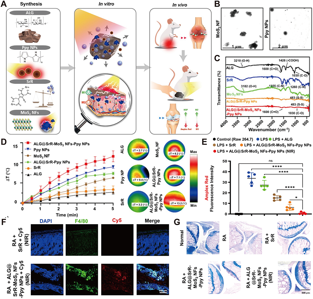 |
Figure 4 A phototherapeutic hydrogel with superior photothermal and photoelectric conversion properties for RA treatment. (A) Schematic illustration of the construction of the ALG@SrR-MoS2 NFs-Ppy phototherapeutic hydrogel and its proposed mechanism against RA. (B) TEM images of MoS2 nanoflowers and Ppy NPs. (C) FTIR spectroscopy revealed the chemical structures and bonding relationships among the various NPs. (D) Evaluation of the thermal behavior of various nanomaterials under NIR light irradiation (808 nm, 1.0 W/cm2). (E) Fluorescence imaging indicated that treatment with ALG@SrR-MoS2 NFs-Ppy NPs under NIR light irradiation significantly reduced ROS levels in LPS-stimulated cells. Data are presented as mean ± standard deviation (SD). *P < 0.05, ****P < 0.0001, ns, not significant. (F) Fluorescence imaging revealed significantly increased Cy5 fluorescence and enhanced macrophage accumulation in the skin of RA mice treated with ALG@SrR-MoS2 NFs-Ppy NPs (Cy5-labeled) under NIR light irradiation. (G) Alcian blue staining of knee joint cartilage revealed that the ALG@SrR-MoS2 NFs-Ppy NPs + NIR group prevented cartilage erosion. Reproduced with permission.110 Copyright 2024 The Authors. |
PDA, structurally analogous to eumelanin, exhibits excellent biocompatibility, outstanding photothermal conversion efficiency, and strong drug-binding capacity, making it well-suited for PTT.111,112 Given the pivotal role of excessive ROS and inflammatory cell infiltration in RA pathogenesis, a PDA-coated, CeO2-doped zeolitic imidazolate framework-8 (ZIF-8) nanocomposite was developed.113 This platform ablated overproliferated inflammatory cells at the lesion site via photothermal effect and eliminated excessive ROS. Through light-triggered decomposition of the acid-responsive structure within ZIF-8, the nanocomposite released CeO2, which decomposed hydrogen peroxide (H2O2) to generate oxygen, thereby alleviating the hypoxic environment. PDA is also recognized as a suitable option for anti-inflammatory nanodelivery due to its function as a photo-switchable medium for controlled drug release. For instance, a photo-triggered platform was constructed to enable on-demand release of diclofenac sodium.114 NIR irradiation elevated the temperature above the gatekeeper’s melting point, triggering intelligent cargo release. This system effectively mitigated temporomandibular joint cartilage deterioration and improved OA outcomes.
A novel conjugated polymer, poly-5,5′-[2,5-bis(2-octyldodecyl)-3,6-di-(thiophen-2-yl)-2,5-dihydropyrrolo[3,4-c]pyrrole-1,4-dione] (PDPP), demonstrates exceptional photothermal conversion efficiency reaching 57.48%,115 enabling effective PTT at reduced doses and irradiation power. Leveraging this property, Li et al reported that NIR-responsive PDPP NPs mitigate neuroinflammatory responses by modulating ROS and Ca2+ signaling pathways.116 Upon laser irradiation, the NPs efficiently crossed the blood–brain barrier, decreased amyloid-β peptide aggregation, and facilitated partial disruption of aggregates outside the cell, thereby alleviating neuroinflammation through the reduction of oxidative stress-induced cellular damage. As a conjugated polymer with high stability and strong NIR absorption, PANI has been widely explored for PTT.117 Under NIR laser irradiation, an amphiphilic PANI-conjugated glycosylated chitosan polymer generated substantial heat, elevating local temperature by approximately 5 °C. This ensured direct ablation of target bacteria while minimizing tissue damage and promoting wound healing.118 Despite these encouraging results, the application of polymeric nanomaterial-mediated PTT in inflammatory diseases remains in its early stages, warranting further expansion and investigation.
Small-Molecule Organic Nanomaterials
Clinical translation of metal-based and carbon-based nanomaterials is often hindered by high cost and poor biodegradability. Small-molecule organic nanomaterials have thus emerged as promising alternatives, offering lower cost, favorable biodegradability, and good biocompatibility.119,120 Notable classes such as cyanine dyes, porphyrins, and phthalocyanines exhibit strong photothermal conversion and excellent biosafety, achieving significant therapeutic outcomes in PTT. However, their application is constrained by low water solubility and insufficient targeting specificity, necessitating strategies such as molecular engineering or encapsulation within nanoscale carriers to improve biodistribution, enhance localized accumulation, and ultimately strengthen PTT-mediated anti-inflammatory efficacy.
Indocyanine green (ICG), an FDA-approved NIR photosensitizer of the cyanine family, can be delivered to the inflammatory sites via encapsulation strategies that also enhance its photostability. In a representative approach, ICG was encapsulated with epigallocatechin-3-gallate through self-polymerization and intermolecular interactions,121 resulting in enhanced photothermal conversion. Following cRGD modification, the nanocomposite specifically targeted activated platelets within thrombi. Upon NIR exposure, this system elicited robust clot dissolution and markedly alleviated vascular inflammation through effective free-radical scavenging. Moreover, Kim et al co-loaded ICG and metformin into poly(lactic-co-glycolic acid) (PLGA)-based microspheres to assess therapeutic efficacy and investigated the thermoresponsive behavior of the IL-22 receptor.122 This work offered relatively detailed mechanistic insight, showing that PTT application significantly reduced IL-22 receptor levels and suppressed Th17 cell accumulation. Concurrently, released metformin promoted M2 macrophage polarization, synergistically enhancing RA therapeutic outcomes through downregulation of pro-inflammatory mediators and upregulation of anti-inflammatory molecules. IR820, an NIR-responsive heptamethine cyanine dye derived from ICG, offers stronger photobleaching resistance and improved aqueous solubility, rendering it valuable for anti-inflammatory therapy.123 Encapsulation within PDA yielded PDA/IR820 nanocomposites capable of targeted keratinocyte ablation in psoriatic lesions.124 Under NIR light irradiation, these NPs elevated skin temperature by 11.7 °C. By inducing keratinocyte apoptosis via both the caspase and poly ADP-ribose polymerase pathways, this nanoparticle decreased excessive cytokine production and restored barrier function, leading to effective elimination of psoriatic lesions in mice.
Porphyrins are distinguished by their excellent chemical and thermal stability relative to many other organic materials. However, their π-conjugated architecture also predisposes them to aggregation and precipitation.125 To address this, porphyrin-based materials can be enhanced via coordination complex formation or covalent crosslinking, capitalizing on the intrinsic modifiability of porphyrin molecules.126 As an example, Wang et al developed a heteronuclear copper-porphyrin complex featuring a copper-coordinated tetraphenylporphyrin core (Figure 5).127 The resulting NPs integrated NIR chemiluminescence imaging, PTT, and PDT. This system allowed for precise real-time thrombus monitoring, non-invasive thrombolysis, and scavenging of surplus ROS, thereby promoting safe reperfusion and exerting anti-inflammatory effects. With a donor–acceptor–donor architecture, diketopyrrolopyrrole (DPP) and its derivatives offer high molar extinction coefficients and excellent photothermal stability, making them versatile photothermal agents for fluorescence imaging and PTT.128 Their facile functionalization further facilitates their application as NIR fluorescence imaging probes and photothermal platforms. Notably, three self-assembling DPP derivatives with varying donor-acceptor units and polyethylene glycol (PEG) side chains have been reported, all exhibiting high biocompatibility.129 These derivatives demonstrated excellent photothermal performance under 635 nm laser irradiation, enabling effective NIR-guided in vivo PTT. To improved fabrication approaches and photothermal properties, Wu et al constructed a novel donor-acceptor organic nanomaterial based on porphyrin-DPP.130 Owing to the donor-acceptor interaction that enables efficient energy conversion and non-radiative heat production, this self-assembled nanomaterial exhibited excellent theranostic performance with a photothermal conversion efficiency of 62.5%. Moreover, the broadened and red-shifted absorption spectrum of Por-DPP relative to other organic molecules underlay its potent PTT effect. These attributed underpinned its pronounced photothermal effect and suggested considerable promise for anti-inflammatory applications.
 |
Figure 5 Cu2Ir NPs integrating long-lived NIR chemiluminescence (CL) imaging, PTT, and PDT for multimodal thrombolysis. (A) Schematic illustration of Cu2Ir NPs for dual-mode PTT/PDT thrombolytic therapy. Cu2Ir NPs possess potent antioxidant properties that enable efficient scavenging of RONS and suppression of pro‑inflammatory cytokine production, thus preventing re-embolism. The downward arrows indicate the reduction of RONS levels and pro-inflammatory cytokines (eg, IL-6 and TNF-α), while the upward arrow represents the enhancement of antithrombotic capacity. (B) Dynamic light scattering analysis and TEM images of Cu2Ir NPs. Scale bars = 200 mm. (C) The ultraviolet-visible (UV-vis) absorption spectra of different NPs in water. (D) Kinetics of 1O2 generation. (E) EPR signals of 5,5-dimethyl-1-pyrrolineN-oxide (DMPO) under different treatment conditions. (F) Photothermal profiles of different NPs under 635 nm laser irradiation. (G) Concentration-dependent photothermal effect of Cu2Ir NPs under 635 nm laser irradiation. (H and I) Photographic and quantitative clot-dissolution results across treatment groups, indicating PTT as the primary role and the superior efficacy of Cu2Ir NPs in the presence of hydrogen peroxide. Scale bars = 5 mm. Data are presented as mean ± SD. ***p < 0.001. (J and K) Representative in vivo CL images of thrombosed arteries thrombotic artery at various time points post-treatment and quantitative analysis of relative CL intensity from mouse neck thrombi, indicating precise thrombus localization by Cu2Ir NPs. Scale bar = 5 mm. Data are presented as mean ± SD. **p < 0.01, ***p < 0.001. Reproduced with permission.127 Copyright 2025 The Authors. Advanced Science published by Wiley-VCH GmbH. |
Others
With its robust photothermal performance and potent multienzyme-mimetic activities, Prussian blue (PB) NPs enable efficient photothermal conversion alongside ROS scavenging and inhibition of inflammatory cytokine, making them attractive for anti-inflammatory applications.131,132 Their high NIR absorption across 650–900 nm is attributed to intervalence charge transfer between Fe2+ and Fe3+.133 Surface modification of PB NPs represents an effective strategy to overcome inherent limitations such as short circulation time, insufficient targeting, and poor solubility. Deng et al employed a folic acid (FA)-modified biomimetic PB nanoparticle to selectively deliver Xuetongsu into inflammatory macrophages and osteoclasts.24 Under NIR light irradiation, the nanocomposite exhibited a robust photothermal effect that promoted site-specific drug release. By suppression the NF-κB pathway in inflammatory macrophages and the RANK/RANKL/NFATc1 signaling pathway in osteoclasts, this platform achieved synergistic anti-inflammatory and ROS scavenging effects, offering a novel perspective for clinical RA management. Separately, PB-mediated photothermal stimulation has been shown to upregulate ABCA1 and ABCG1 levels in foam cells, enhancing lipid efflux and reducing plaque instability driven by apoptotic and necrotic processes.134
As a natural polyphenolic compound with critical biological functions, melanin offers efficient photothermal conversion.135 Its structural richness in catechol moieties also facilitates straightforward surface modification. Combined with its inherent biocompatibility and biodegradability, melanin and melanin-based NPs have attracted considerable interest in PTT. Chen et al constructed a multimodal anti-inflammatory platform incorporating NIR-II fluorescence and photoacoustic imaging to explore synovial fibroblasts involvement in the RA microenvironment.136 This nanocomposite exhibited rapid, lesion-responsive degradation, achieving synergistic antioxidant and anti-inflammatory effects via fibroblasts ablation and hydrogen (H2) release.
Black phosphorus (BP), a layered semiconductor with a folded honeycomb structure, offers excellent biocompatibility and high drug-loading capacity. Leveraging its superior photothermal effect, BP is well suited for photo-triggered drug delivery and PTT. Hou et al incorporated rutin and BP nanosheets into a composite hydrogel for mitigating RA-associated inflammation.137 Under NIR exposure, the photothermal conversion of BP nanosheets cooperated with the anti-inflammatory drug rutin to effectively suppress inflammation and reduce joint damage. A thermoresponsive hydrogel integrating BP nanosheets with platelet-rich plasma (PRP) has also been developed for RA management.138 In this system, BP nanosheets enabled photothermal ablation of hyperplastic synovial cells, while PRP provided bioactive support and biomineralization-enhanced bone regeneration, collectively achieving effective protection of articular cartilage.
PTT-Boosted Anti-Inflammatory Therapy
Rapid diversification of photothermal nanomaterials has significantly advanced the biomedical application of PTT. Harnessing photo-controlled drug release, thermal eradication of inflammatory cells, and direct antibacterial effects (Figure 6),28,139 these nanomaterial-boosted anti-inflammatory approaches have substantial advancement. Through targeted modifications and delivery vehicles, these photothermal agents can be directed to specific cell types, enabling precise treatment under external laser illumination. A growing body of research on PTT-mediated anti-inflammatory therapies underscores their significant promise across a range of inflammatory diseases, from vascular and articular to periodontal inflammation (Tables 1 and 2). By enabling efficient and precise photothermal intervention, these strategies expand the therapeutic landscape and hold potential for advancing targeted in vivo efficacy.
 |
Figure 6 Schematic illustration of PTT-based photothermal nanomaterials demonstrate great potential for the treatment of multiple inflammatory diseases including AS, RA, OA, and periodontitis through effective inflammation management (controlled drug release, thermal ablation of inflammatory cells, disruption of bacterial biofilms, and induction of inflammatory cell apoptosis) and feasible combination therapy modalities (SDT, PDT, gene therapy, chemotherapy, and NO gas therapy). |
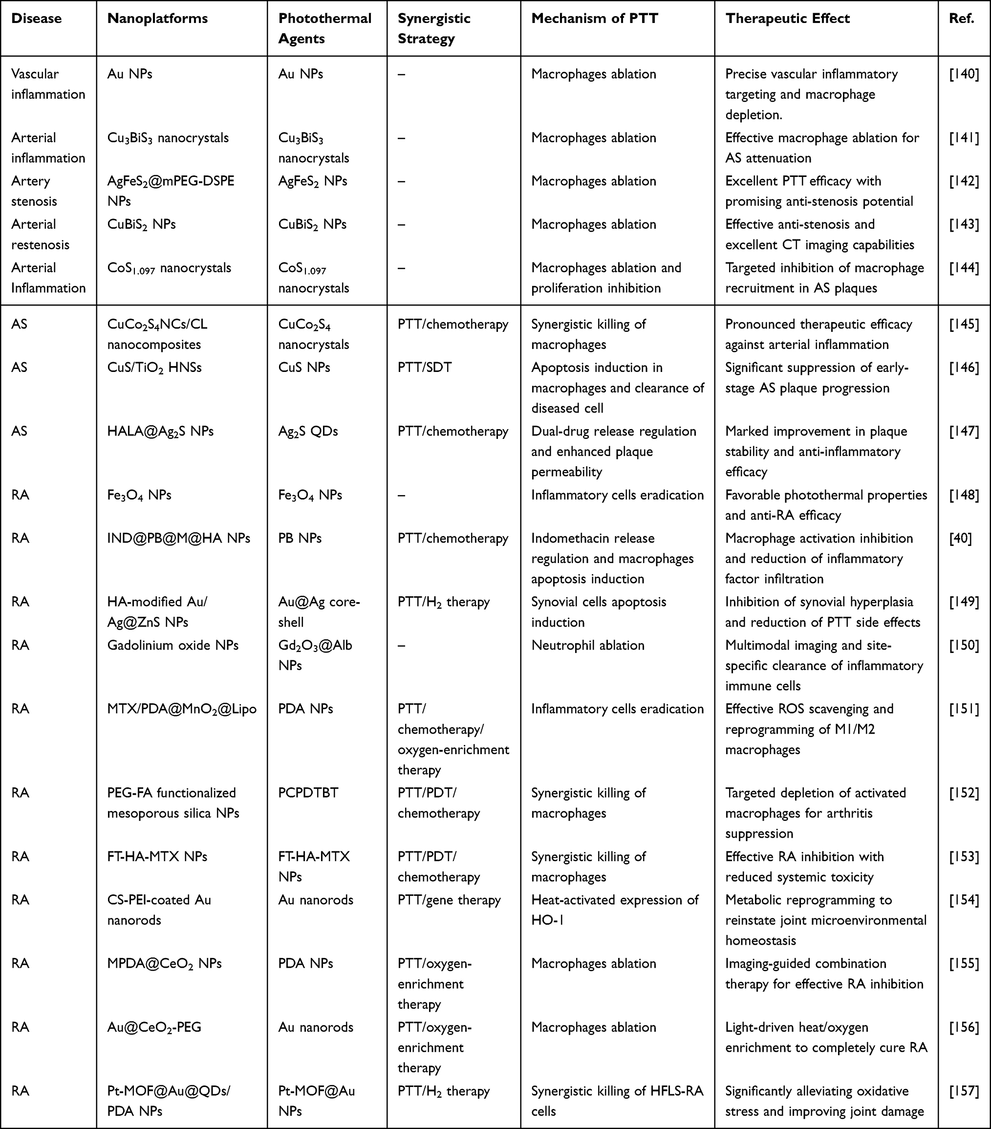 |
Table 1 Enhanced Anti-Inflammatory PTT for Treating AS (Encompassing Associated Vascular Inflammation) and RA |
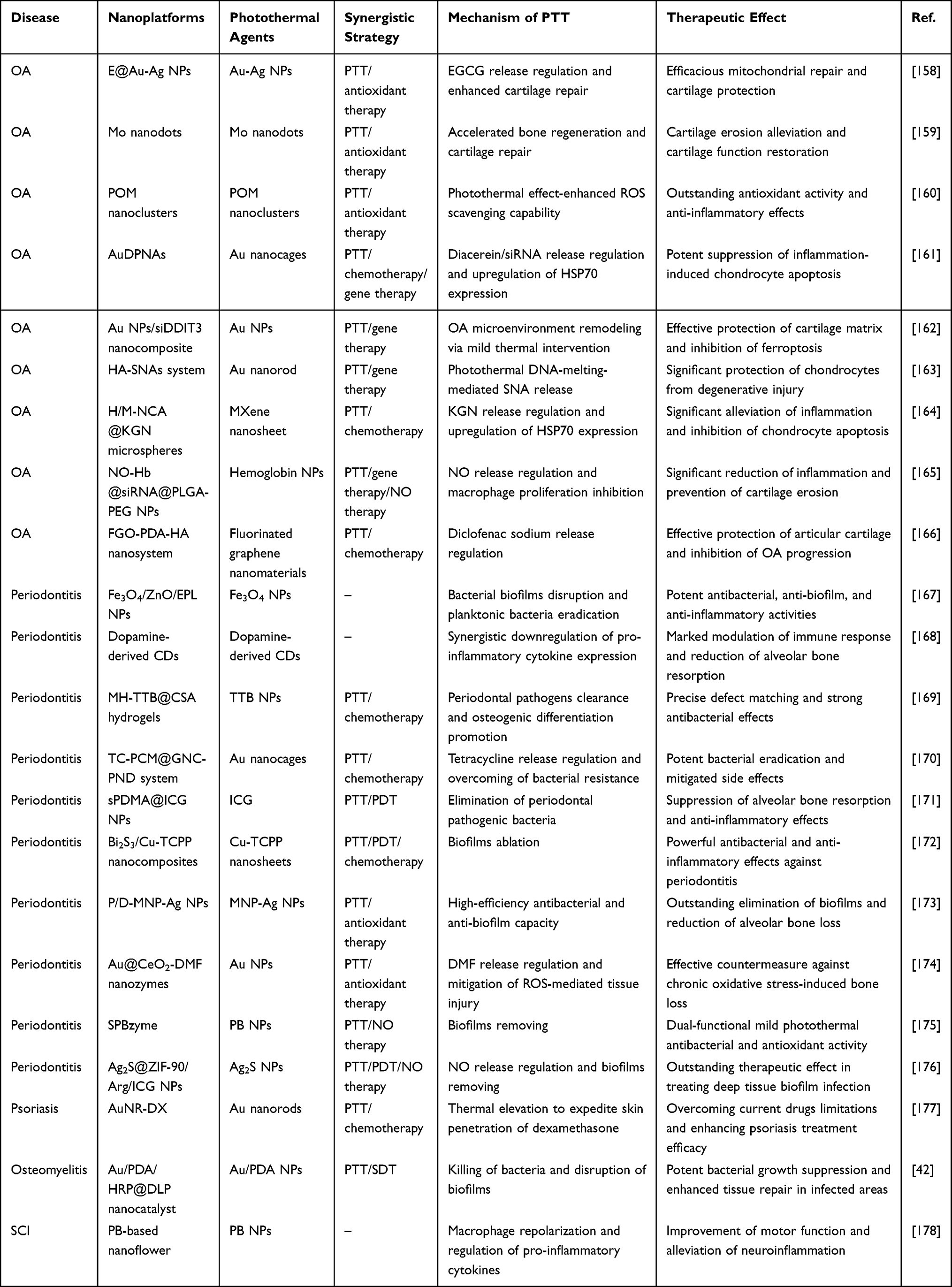 |
Table 2 Enhanced Anti-Inflammatory PTT for Treating OA, Periodontitis and Other Inflammatory Diseases |
AS
Sustained systemic vascular inflammation and arterial restenosis are key drivers of AS progression.179 Plaque formation involves the enrichment, activation, and infiltration of macrophages, coupled with smooth muscle cell migration and proliferation.180 Therefore, suppressing macrophage activation and infiltration, along with blocking inflammatory cytokine secretion, represents a viable approach to attenuate AS. Importantly, the non-invasive nature of PTT allows it to target the underlying mechanism by directly inducing macrophage apoptosis or exerting a killing effect. Elevated temperatures are well documented to suppress and eliminate tumor cells.181 By analogy, harnessing the photothermal effect of PTT to precisely inhibit proliferation or ablate inflammatory cells holds considerable promise for AS therapy.
As vascular inflammation accompanies all phases of AS progression, developing effective mitigation strategies remains crucial. Studies have shown that under NIR light irradiation, macrophages incubated with Au NPs exhibit reduced viability.140 Computed tomography (CT) imaging further revealed enhanced attenuation in perivascular areas following nanoparticle administration, suggesting localization at inflammatory vascular sites. Nevertheless, further studies are required to confirm their in vivo anti-vascular inflammatory activity. In another study, Lu et al synthesized 12 nm Cu3BiS3 nanocrystals via a hydrothermal method for CT-guided PTT of arterial inflammation.141 Leveraging their excellent photothermal conversion performance, these nanocrystals enabled effective macrophage elimination in cellular and animal models under NIR laser exposure, thereby attenuating AS progression. Additionally, serving as a CT contrast agent, they enabled visualization of carotid inflammatory and supported precise image-guided PTT.
PTT holds considerable promise for inflammatory diseases such as arterial stenosis. Peng et al developed AgFeS2 NPs with favorable biocompatibility and low biotoxicity, positioning them as suitable photothermal agents.142 After mPEG-DSPE modification, these NPs showed superior photothermal efficacy in vitro and successfully eliminated macrophages at low concentrations in mouse models, representing a promising approach for managing arterial inflammation and stenosis. Intervening in macrophage infiltration-driven arterial inflammation is crucial for mitigating restenosis following therapeutic vascular procedures. Incorporating bismuth (Bi) as a radiocontrast agent into copper chalcogenide nanocrystals improved the accuracy of CT-guided PTT.143 CuBiS2 NPs, under optimized dosing and conditions, combined biosafety with efficient macrophage ablation to secure therapeutic outcomes. Their superior photothermal properties effectively suppressed stenosis in a carotid artery injury model, positioning these cost-effective, high-performance nanomaterials as promising candidates for preventing restenosis following endovascular treatment.
Early diagnosis and intervention are critical in AS. PB, with their high photothermal performance, enable both plaque detection and subsequent in vivo treatment.182 Nanomaterial-mediated photothermal strategies counteract AS progression by inducing macrophage ablation and inhibiting proliferation. Employing a facile hydrothermal approach, Lu et al developed CoS1.097 nanocrystals as a novel photothermal nanoplatform for AS.144 With strong NIR absorption, these nanocrystals generated a potent photothermal effect that effectively eradicates macrophages, resulting in marked attenuation of AS progression. Integrating chemotherapy with PTT represents a strategy to enhance vascular anti-inflammatory effects. For instance, self-assembled CuCo2S4 nanocrystals with outstanding photothermal performance were employed to deliver the anti-inflammatory agent chloroquine, achieving improved combined therapeutic efficacy in vivo.145 Under NIR light irradiation, the photothermal and chemotherapeutic actions were concurrently activated, synergizing to achieve effective macrophage elimination. Combining SDT with PTT offers an effective approach for treating AS plaque. Cao et al constructed CuS/TiO2 heterostructure nanosheets (HNSs) for low-intensity sonodynamic and mild photothermal combination therapy targeting early-stage plaques.146 Surface functionalization with HA and PEG endowed these nanosheets with targeting capability toward inflammatory macrophages within plaques. The nanosheets exhibited outstanding sonodynamic activity that downregulates HSP90 expression, along with pronounced NIR-II absorption that augments the sonocatalytic process. Together, these properties enabled the combined SDT/PTT strategy to synergistically induce macrophage apoptosis, thereby alleviating inflammation and inhibiting early AS plaque progression. Designing controllable nanosystems that actively respond to the disease microenvironment is essential for effective AS treatment. In one study, Liang et al fabricated a triple-stimuli synergistic nanocomposite responsive to enzyme, light, and multiple drugs, integrating enzyme-responsive dual-drug delivery with NIR-triggered PTT (Figure 7).147 HA modification conferred the nanocomposite with enhanced targeting, controlled release, and anti-inflammatory properties. In parallel, the photothermal action of Ag2S quantum dots (QDs) boosted localized anti-inflammatory responses and augmented overall therapeutic outcome by facilitating drug release and enhancing permeability within plaques. This green, multimodal strategy holds substantial potential for application in complex cardiovascular disease therapy.
 |
Figure 7 Enzyme-responsive dual-drug delivery with NIR-II imaging-guided PTT using HALA@Ag2S NPs for anti-AS research. (A) Schematic illustration of the therapeutic mechanism of HALA@Ag2S NPs within AS plaques. Under 808 nm irradiation, PTT induces apoptosis of inflammatory macrophages, while pharmacotherapy promotes plaque stabilization. Both processes contribute to anti-inflammatory effects, resulting in reduced expression of pro-inflammatory cytokines. Additionally, HALA@Ag2S NPs modulate cholesterol metabolism via SREBP2 downregulation and ABCA1 upregulation, reducing foam cell formation. The upward arrows indicate enhancement of endothelial function and upregulation of ABCA1, while the downward arrows denote reduction of platelet aggregation, downregulation of SREBP2, decreased foam cell formation, and suppression of pro-inflammatory cytokines (TNF-α, IL-1, and IL-6), respectively. (B) Hydrodynamic diameter variation of Ag2S QDs and HALA@Ag2S NPs evaluated over 72 h. (C) TEM image of HALA@Ag2S NPs. (D) Photothermal heating curves of HALA@Ag2S NPs under 808 nm laser irradiation at different power densities. (E) Thermogram distribution of HALA@Ag2S NPs during 10 minutes of irradiation (F) Photothermal cycling stability of HALA@Ag2S NPs over repeated heating/cooling cycles (G) Photothermal imaging of cells after treatment with different irradiation durations. (H) Positive ORO staining of whole aortas and quantitative lesion area (%) histogram from different treatment groups, demonstrating a marked plaque reduction in ApoE−/− mice with HALA@Ag2S NPs + Laser group compared to PBS group. Data are presented as mean ± SD. *P < 0.05, ***P < 0.001. Reproduced with permission.147 Copyright 2025 The Authors. |
RA
RA is a chronic autoimmune disease marked by persistent synovial inflammation, joint swelling and destruction, and bone injury.183 Inflammatory cell infiltration into injured joints drives substantial pro-inflammatory cytokine production, leading to progressive cartilage degradation.184 Current pharmacological management is constrained by limited efficacy and pronounced adverse effects. In this context, PTT offers a novel anti-inflammatory approach. By leveraging nano-functionalization or targeted delivery strategies, photothermal nanomaterials can be precisely localized to specific tissues and cells to ablate inflammatory cells. For instance, Zhang et al synthesized small-sized Fe3O4 NPs that are readily phagocytosed by inflammatory cells, demonstrating enhanced cytotoxicity upon NIR laser irradiation.148 In vivo, Fe3O4 NPs approximately 220 nm in diameter showed improved accumulation at inflammatory sites, resulting in a greater temperature increase and superior therapeutic outcomes. In another study, HA-modified PB NPs loaded with indomethacin (IND) achieved targeted delivery to inflamed joints, with additional coating of a hybrid cell membrane further enhancing immune evasion.40 Localizing at the disease site, these nanocomposites raised intracellular temperature via efficient photothermal conversion, thereby reducing activated macrophage viability and mitigating inflammatory pathology.
PTT also enables remodeling of the synovial microenvironment by promoting synovial cell apoptosis and curbing pathological proliferation. As an illustration, a HA-functionalized Au/Ag@ZnS yolk–shell heterostructure functionalized was engineered to remodel the synovial microenvironment and improve RA symptoms.149 This system concurrently countered inflammation, modulated macrophage polarization, and induced synovial apoptosis through integrated photocatalytic hydrogen evolution and mild PTT, thereby minimizing potential side effects of photothermal treatment. As the earliest immune cells recruited to inflammatory sites, NEs show recruitment levels that correlate positively with the disease stage. Photothermal ablation of NEs to downregulate pro-inflammatory cytokines and interrupt the immune cascade thus represents a crucial therapeutic approach. To this end, Yu et al fabricated a cypate-grafted bioinspired gadolinium oxide nanoplatform for dynamic monitoring of inflammatory cell recruitment using combined MRI and photoacoustic imaging.150 Following rapid accumulation at inflammatory lesions, these NPs enabled imaging-guided PTT that effectively disrupted NE nuclei and significantly decreased pro-inflammatory cytokine levels.
Notably, integration PTT with complementary anti-inflammatory approaches promotes drug release, increases inflammatory cell death, and modulates immune cascades, collectively enhancing RA treatment. A representative example was a multifunctional liposomal PDA nanoparticle system developed to remodel the RA microenvironment by combining chemotherapy with photothermal ablation of inflammatory cells (Figure 8).151 In this system, hyperthermia from PTT disassembled thermosensitive liposomes, allowing site-specific methotrexate release. By concurrently clearing ROS, promoting M2 macrophage polarization, and eradicating proliferating inflammatory cells, this nanosystem effectively preserved cartilage integrity and retarded RA advancement. Extensive studies have demonstrated the favorable therapeutic potential of PDT in oncology, where it induces tumor cell apoptosis via ROS generation, offering precise and controllable treatment outcomes.185 Similarly, PDT can further boost the PTT-mediated elimination of inflammatory cells in the RA, thereby alleviating disease symptoms. For example, mesoporous silica NPs hybridized with polymer QDs were used to encapsulate the prodrug tirapazamine, establishing a triple-combination PTT/PDT/chemotherapy platform against RA.152 PEG-FA surface modification enabled targeted accumulation of nanocomposites in activated macrophages. Upon NIR light irradiation, the platform simultaneously elevated intracellular temperature, generated substantial singlet oxygen (1O2), and hypoxia-triggered release of cytotoxic tirapazamine, synergistically eliminating inflammatory macrophages. Furthermore, Zhao et al constructed a HA-modified metal-organic framework loaded with methotrexate to suppress RA progression through combined regimen of PTT/PDT/chemotherapy.153 With a photothermal conversion efficiency of 36.3%, the nanocomposite also facilitated hydroxyl radical (·OH) production via a Fenton reaction, synergistically inducing macrophage death. In vivo evaluation confirmed its anti-inflammatory efficacy, with significant reductions in synovial TNF-α and IL-1β levels. Combining PTT with gene therapy also offers a promising approach for treating inflammatory diseases. One platform utilized Au nanorods functionalized with chondroitin sulfate (CS)-grafted polyethyleneimine (PEI) to deliver the heme oxygenase-1 (HO-1) plasmid containing HSP70 promoter.154 Local hyperthermia activated the promoter to drive HO-1 expression with spatial precision. Through metabolic reprogramming and macrophage polarization, this platform inhibited glycolytic activity, reduced inflammatory responses, and promoted joint homeostasis, underscoring the promise of gene therapy in inflammatory conditions.
 |
Figure 8 Schematic illustration of the fabrication of MPM@Lipo NPs and their proposed therapeutic mechanism against RA. The thermal effect of PTT disrupts the structure of MPM@Lipo, leading to the release of methotrexate, PDA, and MnO2. PDA-mediated PTT directly induces apoptosis of inflammatory cells and suppresses the production of pro-inflammatory cytokines. Meanwhile, MnO2 catalyzes H2O2 decomposition to O2, relieving local joint hypoxia and suppressing HIF-1α levels, which synergizes with PDA’s ROS scavenging activity to promote M2 macrophage repolarization. The red downward arrows indicate downregulation of pro-inflammatory cytokines (IL-1β and TNF-α), scavenging of ROS, and inhibition of HIF-1α levels, respectively. Reproduced with permission.151 Copyright 2024 Shenyang Pharmaceutical University. Published by Elsevier B.V. |
Hypoxia and elevated ROS levels are additional hallmarks of RA that exacerbate synovial inflammation.186 This arises from a mismatch between the increased oxygen demand of heightened metabolism and the insufficient oxygen supply due to vascular dysfunction. The resulting hypoxic microenvironment further aggravates abnormal synovial hyperplasia and intensifies inflammatory responses.187 Therefore, synergistic strategies combining PTT with oxygen-enrichment regimens hold considerable promise for RA treatment. In one study, mesoporous PDA was combined with CeO2 NPs (MPDA@CeO2) to establish a synergistic strategy coupling PTT, oxygen delivery, and ROS scavenging under PAI guidance.155 Upon reaching the inflammatory site, MPDA@CeO2 initiated photothermal ablation of inflammatory cells, while thermal activation triggered CeO2 release to scavenge ROS and supplied oxygen. These alterations were detectable by PAI, enabling image-directed combined therapy for RA. Furthermore, a composite material integrating Au nanorods with a PEG-functionalized CeO2 shell was developed for photothermal and oxygen-augmenting therapy.156 The nanocomposite exhibited substantially amplified LSPR-driven photothermal effect, enabling effective removal of hyperplastic inflammatory cells. Importantly, localized temperature elevation accelerated H2O2 decomposition to generate considerable O2. These dual effects contributed to significant recovery from RA-associated tissue damage. By crossing cell membranes and eliminating intracellular ROS, hydrogen mitigated the cytotoxic effects of ·OH making H2 therapy highly promising for inflammatory disorders.188 To address the targeting and solubility challenges of H2 therapy, a smart MOF-derived H2 nano-generator incorporating dopamine and perovskite QDs was developed.157 Leveraging Au-mediated LSPR and a Pt-MOF Schottky junction, the platform achieved pronounced photothermal performance and considerable H2 production. This PTT/H2 combination effectively alleviated both articular injury and systemic inflammation in RA.
OA
As a heterogeneous degenerative disease with diverse etiologies, OA is characterized by joint deformity, cartilage deterioration, and sustained pain.189,190 Current interventions primarily target ROS elimination, inflammation reduction, anti-aging effects, and cartilage regeneration. However, these monotherapies often result in significant side effects, inadequate anti-inflammatory efficacy, and unsatisfactory therapeutic outcomes. Recently, PTT has emerged as a promising alternative for OA, offering the potential to reshape the joint microenvironment.
By modulating the regenerative microenvironment and elevating HSP70 expression, low-intensity PTT facilitates cartilage formation under inflammatory conditions.191 It also exerts bone-protective effects by suppressing synovial inflammation, downregulating inflammatory cytokines, and enhancing bone density. Accordingly, combining PTT with other modalities can achieve superior therapeutic outcomes for OA. One example is its combination with antioxidant regimens, which aids in restoring chondrocyte function, delaying senescence, and inhibiting inflammation. A representative design featured Au-Ag nano-jars coated with epigallocatechin gallate (EGCG) as an NIR-responsive nano-platform for OA therapy.158 NIR laser activation increased the intra-articular temperature to 46.6 °C, promoting EGCG release and cartilage repair. In parallel, this system synergistically enhanced chondrocyte migration and proliferation by ameliorating mitochondrial dysfunction and suppressing apoptotic pathways, offering a promising approach for OA management. To mimic antioxidant synthase activity and eliminate ROS and reactive nitrogen species (RNS) in OA chondrocytes, Lu et al developed a biomimetic photothermal system based on Mo nanodots.159 The induced photothermal effect boosted the osteogenic potential of bone marrow mesenchymal stem cells (BMSCs) in vitro, thereby contributing to subchondral bone repair. Importantly, by disrupting the ROS-fueled senescence cycle and ameliorating mitochondrial dysfunction, this nanoplatform effectively retarded OA progression, highlighting the feasibility of integrating PTT with antioxidant defense strategies. Moreover, Mo-based polyoxometalate (POM) nanoclusters exhibit an intrinsic photothermal effect that enhances ROS scavenging. Building on this, Shi et al constructed NIR-responsive POM nanoclusters with potent antioxidant and anti-inflammatory activities.160 By downregulating inflammatory cytokine secretion and suppressing catabolic protease activity, this system significantly slowed OA progression.
Nanodelivery-based gene therapy has recently enabled localized, sustained, and controllable genetic interventions in diseased joints, offering protection and repair of articular cartilage in OA. When combined with PTT, this approach holds promise for preserving cartilage and alleviate OA symptoms. To overcome persistent pain and inadequate anti-inflammatory effects, Qiao et al designed a photosensitive nanotherapeutic system comprising polymer-coated Au nanocages co-encapsulating diacerein and siRNA targeting NGF (siNGF).161 Marked articular accumulation of the photosensitive nanodrug was observed in OA mouse models. The induced photothermal response enabled localized release of diacerein and siNGF, resulting in potent anti-inflammatory and analgesic effects, accompanied by increased HSP70 expression to protect chondrocytes from apoptosis. DDIT3 gene is known to induce ferroptosis and accelerate articular cartilage degeneration, contributing to OA progression. Targeting this pathway, Wang et al designed a star-shaped Au nanoparticle platform for siDDIT3 delivery, achieving improved therapeutic outcomes via combined PTT.162 The star architecture facilitated efficient intracellular siRNA delivery and DDIT3 suppression, while Raman scattering enabled real-time nanoparticle tracking. Mild hyperthermia generated by these Au NPs modulated the OA microenvironment, working synergistically with gene therapy to enhance therapeutic outcomes.
PTT-mediated drug release further contributes to combinatorial OA therapy. In one study, Spherical nucleic acids (SNAs) were constructed by conjugating interference oligonucleotides onto Au nanorods and subsequently delivered via a HA carrier grafted with complementary oligonucleotides, forming a DNA-grafted HA complex through specific base pairing interactions (Figure 9).163 Leveraging the photothermal effect, the Au nanorods disrupted the DNA duplex through hydrogen bond breakage, enabling precise photo-controlled release. This system successfully silenced IL-1β mRNA and reduced catabolic protease expression in cartilage, thereby retarding OA progression. To achieve photothermal-triggered dual cargo delivery, Chen et al fabricated homogeneous nanospheres by encapsulating kartogenin (KGN) in nano cationic amylose (NCA), followed by conjugation with HA methacrylate and MXene via a microfluidic approach.164 These nanospheres, carrying BMSCs, demonstrated favorable biocompatibility and cartilage-adhesive properties. Upon NIR light irradiation, they enabled controlled KGN release and efficient BMSC delivery, promoting chondrogenic differentiation. Additionally, mild thermal stimulation induced HSP70 expression in chondrocytes, exerting a chondroprotective effect.
 |
Figure 9 NIR-regulated release of SNAs constructed via DNA-conjugated HA for managing OA. (A) NIR-light-induced intracellular SNA release from intra-articularly injected HA-SNAs system for IL-1β gene silencing via mRNA interference. This process proceeds via the following steps: (i) cellular internalization; (ii) binding of SNAs to the target mRNA; (iii) inhibition of protein synthesis; (iv) transcription of DNA to mRNA; and (v) translation of mRNA into IL-1β protein. (B) TEM images of (i) Au nanorods and (ii) SNAs nanorods, showing that pure Au nanorods tend to aggregate, whereas DNA conjugation significantly reduces this phenomenon. (C) Photothermal images of various NPs under 808 nm laser irradiation (1.5 W/cm2), where the values annotated on each image represent the maximum temperature captured at that time point. The rate of temperature increase for the HA-SNAs solution within 14 minutes was higher than that of the HA + SNAs and SNAs groups. (D) Temperature distribution profiles of different NPs exposed to 808 nm laser light (1.5 W/cm2). (E) Representative micro-CT scanning and reconstructions of mouse knee joints showing improved joint space width and reduced osteophyte volume in the HA-SNAs + NIR group relative to PBS group. (F) Relative articular space width in the medial compartment of the mouse knee at 12 weeks post-surgery across experimental groups. Data are presented as mean ± SD. ***P < 0.001 vs PBS. (G) Relative osteophyte volume following different treatments. Reproduced with permission.163 Data are presented as mean ± SD. **P < 0.01 vs PBS. Copyright 2021 The Authors. Advanced Science published by Wiley-VCH GmbH. |
Elevated NO levels alleviate inflammation by regulating vascular tone and activating the AMPK pathway.192,193 To harness this, hemoglobin NPs have been employed as NO carriers for NIR-triggered, precise NO release within inflammatory cells.165 The nanoplatform was further modified with Notch1-siRNA to target macrophage proliferation, enabling synergistic combination of NO gas therapy, gene therapy, and PTT. By inhibiting macrophage activity and reducing inflammatory cytokine levels, this NO-controlled photothermal nanosystem modulated the inflammatory microenvironment and protected against cartilage degradation. OA progression is driven by persistent inflammation and impaired cartilage lubrication. Combination therapies that leverage PTT for both anti-inflammatory and lubrication enhancement thus hold significant promise. Li et al developed a nanoplatform based on PDA-coated, HA-grafted fluorinated graphene nanosheets, offering durable lubrication and photothermal-responsive drug release.166 Controlled release of diclofenac sodium from this system promoted anabolic gene expression and suppressed catabolic protease activity, effectively attenuating OA deterioration.
Periodontitis
As an inflammatory disease resulting from bacterial infection in periodontal tissues, periodontitis involves robust immune activation and elevated ROS concentrations.194,195 These factors contribute to connective tissue degradation, alveolar bone resorption, and eventual tooth loss, significantly impairing patients’ quality of life. However, current antibacterial strategies lack selectivity against pathogenic bacteria, limiting clinical efficacy. PTT offers a precise approach to periodontitis treatment with robust antibacterial effects, holding great appeal for clinical translation. By leveraging efficient photothermal conversion, such agents can compromise biofilm integrity and achieve complete bacterial eradication. Illustratively, composite NPs featuring a spherical ZnO shell around an Fe3O4 core was developed to facilitate adsorption and deep biofilm penetration, allowing photothermal ablation at periodontal sites.167 The positive surface charge enabled selective binding to negatively charged bacterial membrane, synergizing with PTT to produce robust antibacterial activity and effectively suppress the inflammatory response. Furthermore, mild PTT suppresses pro-inflammatory cytokines production in RAW264.7 cells, contributing to reduced periodontal inflammation. For instance, Zhao et al fabricated dopamine-derived CDs using hydrothermal synthesis to remodel the immunomodulatory landscape of periodontal tissues.168 Under mild PTT, the material displayed notable anti-inflammatory properties and simultaneously attenuated alveolar bone resorption via immune modulation.
PTT-based synergistic strategies contribute to enhance periodontitis management and favorable treatment outcomes. Combining PTT with chemotherapy is a widely adopted regimen that offers superior therapeutic performance along with reduced side effects. To overcome the limited antibacterial activity of conventional guided tissue regeneration membranes while promoting bone repair, Li et al designed a hydrogel loaded with minocycline and TTB NPs that adapts to bone defect morphology.169 This hydrogel demonstrated strong antimicrobial activity upon NIR laser exposure. Mild photothermal treatment alongside the antibacterial properties of minocycline elevated alkaline phosphatase expression, thereby accelerating mineralization and promoting bone formation. Additionally, PTT integration may a lower the risk of drug resistance and reduce antibiotic-associated side effects. To this end, a photoactivatable nano-antibiotic system was constructed using Au nanocages and dual thermosensitive gatekeepers, enabling NIR-modulated drug release kinetics.170 The localized hyperthermia generated by this nanoplatform enabled simultaneous bacterial eradication and on-demand drug release. Importantly, the two thermosensitive gatekeepers formed an injectable hydrogel in situ, enhancing periodontal retention of tetracycline and achieving potent antibacterial efficacy.
Integrating PTT with PDT presents considerable benefits for antimicrobial applications. This combined strategy enables precise spatiotemporal control and excellent bacterial penetration, yielding potent activity against both drug-sensitive and drug-resistant bacteria. ICG, a NIR-absorbing agent with an established safety profile in tumor therapy196 demonstrates strong synergistic photothermal and photodynamic effects, underscoring its promise for antibacterial therapy. However, the strong hydrophilicity and negative surface charge of ICG impede its penetration through the negatively charged lipid bilayer of bacterial biofilms. Bust PTT/PDT performance, leading to potent bactericidal activity against Porphyromonas gingivalis. Furthermore, the platform markedly inhibited alveolar bone resorption and alleviated inflammatory responses. Porphyrin-based PDT is a promising strategy for periodontitis, leveraging laser irradiation to generate ROS, including •OH, 1O2, and superoxide anion (O2−), that disrupt bacterial DNA, proteins, and lipids.197 In addition, combining CDT to generate highly reactive ROS via Fenton-like reactions could further enhance the overall antibacterial efficacy of nanomaterials,198 building upon the photothermal ablation of inflammatory cells. Building on these mechanisms, Kong et al constructed a Z-scheme heterojunction nanomaterial (Bi2S3/Cu-TCPP) capable of triple-modality therapy combining PTT, PDT, and CDT.172 Capable of efficiently capturing O2 and •OH, the nanocomposite facilitated rapid ROS production. Meanwhile, PTT stimulated Cu2+ release, which depleted intracellular glutathione and compromised bacterial antioxidant defenses, thereby amplifying CDT effect. With robust antimicrobial activity, this triple-modal strategy presented a prospective approach for periodontitis management.
Excessive ROS can exert cytotoxic effects on periodontal ligament cells and disrupt tissue homeostasis. ROS surges also trigger mitochondrial fragmentation, exacerbating oxidative stress in the periodontal microenvironment. Additionally, the interplay between ROS and M1 macrophages promotes the destruction of periodontal fibers and alveolar bone, resulting in significant tissue injury. A dual strategy combining PTT with antioxidant therapy has therefore emerged, aiming to deliver antimicrobial activity in the initial stage of disease a followed by ROS elimination and inflammation reduction. As an example, melanin-coated Ag NPs functionalized with dual polymers were developed for periodontal treatment.173 This design imparted an increased positive surface charge and efficient Ca2+ chelation, thereby enhancing biofilm penetration and binding to hydroxyapatite surface. Triggered by the acidic biofilm environment and NIR light irradiation, the nanocomposite rapidly released Ag+ to exert strong antibacterial effects while simultaneously clearing ROS, attenuating inflammation, and preventing alveolar bone loss. Separately, Li et al constructed a yolk-shell nanoplatform incorporating the multifunctional nanozyme Au@CeO2 and the antioxidant agent dimethyl fumarate (DMF), providing a combined therapeutic approach for periodontitis.174 Driven by photothermal activation, this nanoplatform enabled controlled delivery of DMF, which ameliorated mitochondrial function, reduced oxidative stress via antioxidant enzyme-like activity, and reprogramed macrophage polarization to remodel the local immune microenvironment. By concurrently targeting multiple key pathological mechanisms, this combined therapy achieved durable and stable treatment of periodontitis.
NO gas therapy exhibits potent bactericidal efficacy by inducing lipid peroxidation, disrupting protein function, and causing DNA damage, thereby effectively addressing biofilm-associated resistance.199 When combined with PTT, it synergistically disrupts bacterial biofilms, leading to substantially enhanced sterilization outcomes. Achieving effective NO loading and precise delivery is essential for optimal therapeutic performance. Accordingly, Li et al incorporated sodium nitroprusside into PB NPs with intrinsic antioxidant activity for combined PTT and NO gas therapy.175 NO release synergistically amplified the photothermal antibacterial effect, leading to efficient biofilm clearance, inhibition of bacterial proliferation, and marked antioxidant and anti-inflammatory activity. Another study further integrated PTT, PDT and NO gas therapy to maximize synergistic antibacterial efficacy (Figure 10).176 In this work, L-arginine, serving as an NO donor, was loaded into a nanocomposite comprising silver sulfide (Ag2S) nanocrystals as the core and ZIF-90 as the shell. NIR laser irradiation activated both PTT and PDT, generating sufficient heat and ROS to promote NO release. The released NO further facilitated biofilm disruption via interference with the c-di-AMP pathway. This platform thus enabled efficient management of deep-seated biofilm infections, offering a promising approach for addressing diverse biofilm-associated pathologies.
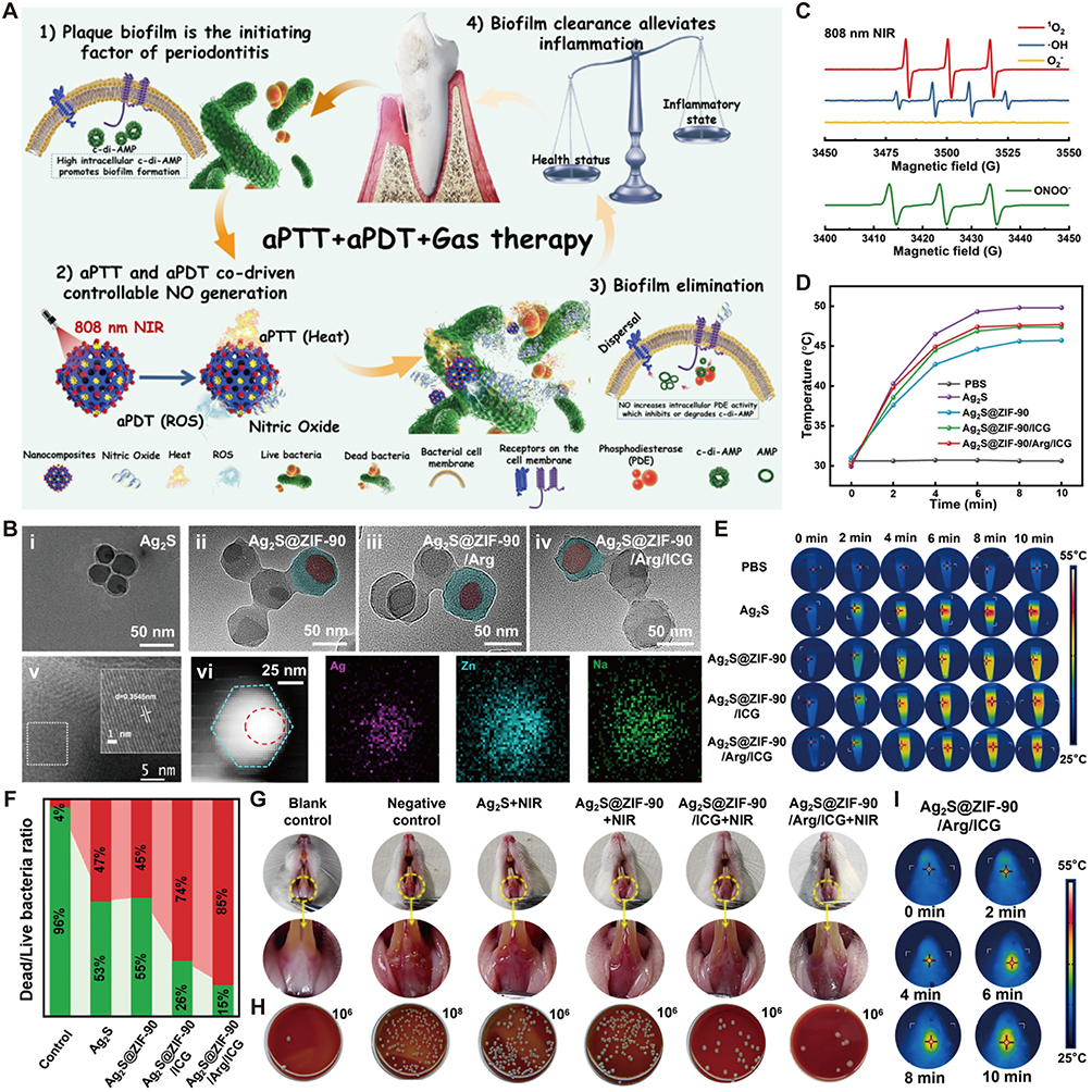 |
Figure 10 A synergistic PTT/PDT/NO gas therapy platform based on Ag2S@ZIF-90/Arg/ICG nanocomposites for effective periodontal biofilm eradication. (A) Schematic illustration of the mechanism for biofilm inhibition and periodontitis alleviation by Ag2S@ZIF-90/Arg/ICG nanocomposites. (B) TEM images of (i) Ag2S, (ii) Ag2S@ZIF-90, (iii) Ag2S@ZIF-90/Arg, and (iv) Ag2S@ZIF-90/Arg/ICG, along with (v) the HR-TEM image of Ag2S and (vi) the elemental (Ag, Zn, Na) distribution mappings within the red dotted circle (indicating the scanned region) of the Ag2S@ZIF-90/Arg/ICG. The magnified area in the dotted square reveals a lattice spacing of 0.3545 nm for Ag2S. (C) EPR spectra of reactive species (1O2, •OH, O2−, and ONOO−) under 808 nm NIR light irradiation. (D and E) Infrared thermal photos and corresponding temperature curves of various NPs upon NIR light irradiation (808 nm, 1.0 W/cm2) for 10 min. (F) Bacterial viability proportion (live/dead) in P. gingivalis biofilms upon treatment with various nanocomposites. (G) Intraoral photographs showing superior gingival recovery in the Ag2S@ZIF-90/Arg/ICG + NIR group versus the Ag2S + NIR and Ag2S@ZIF-90 + NIR groups. (H) Nearly 3-log CFU reduction (vs negative control group) demonstrating the antibacterial activity of Ag2S@ZIF-90/Arg/ICG nanocomposite from gingival tissue. (I) Photothermal images of Ag2S@ZIF-90/Arg/ICG nanocomposites under 808 nm NIR excitation in vivo. Reproduced with permission.176 Copyright 2023 The Authors. |
Others
Beyond the aforementioned applications, PTT has been widely employed as an anti-inflammatory approach for various inflammatory disorders. Psoriasis, a chronic inflammatory dermatosis, is characterized by erythematous plaques and scales, with recurrent episodes and immune-related pathology.200 To address the poor penetration and significant side effects of conventional therapeutic drugs, researchers have employed Au nanorods as a delivery vehicle.177 This strategy enhanced the permeability and cellular uptake of dexamethasone, thereby reducing the required dosage. Photothermal heating from the Au nanorods promoted deeper skin penetration of dexamethasone and restored peroxisome proliferator-activated receptor-γ (PPAR-γ) levels. The synergy between PTT and localized drug delivery therefore offers a viable approach for managing inflammatory dermatoses.
Osteomyelitis, an inflammatory disease of the bone marrow, usually arises from a local infectious focus and progresses to affect the cortical bone and periosteum.201,202 The emergence of drug resistance has rendered antibiotic treatments less effective, leading to recurrent and persistent infections.203 PTT thus presents a promising avenue for osteomyelitis management. Beyond potentiating ROS attack via bacterial membrane disruption, PTT also modulates local immune cell recruitment and activation, contributing to immune regulation. In response, Song et al encapsulated horseradish peroxidase (HRP)-loaded Au/PDA NPs within cationic liposomes for combined PTT and SDT against osteomyelitis.42 Through ROS overproduction and photothermal effects, the nanocomposite effectively eradicated bacteria and induced the release of pathogen antigen, thereby activating innate and adaptive immune responses to promote tissue healing. This integration of immune activation within an antimicrobial strategy introduces a new paradigm for treating chronic osteomyelitis.
Excessive ROS production and aberrant M1 macrophage infiltration are key contributors to spinal cord injury (SCI), driving neuroinflammation and neuronal death.204 To address this, Wang et al designed a PB-based nanoflower that combines photothermal and intrinsic antioxidant activities to synergistically counteract SCI-induced neuroinflammation,178 leading to substantial improvement in locomotor function. The nanoflowers were further functionalized with an optimized CKLVFFAED peptide sequence to enabled blood-brain barrier penetration and macrophage phenotype modulation, effectively alleviating neuroinflammation in the SCI mouse model. In conclusion, the growing diversity of photothermal nanomaterials and an improved understanding of PTT mechanisms have positioned PTT as a viable option for addressing inflammatory disease. Furthermore, rationally integrating PTT with complementary therapeutic modalities based on specific therapeutic objectives can broaden the anti-inflammatory toolkit and provide practical guidance for managing inflammatory conditions.
Conclusion and Future Perspective
Chronic inflammation, arising from the progression of acute inflammation, leads to extensive tissue damage and fibrosis, underpinning the pathogenesis of many chronic diseases. With rapid advances in photothermal nanomaterials and nanotechnology, PTT-based strategies tailored to specific inflammatory microenvironments have achieved marked progress, addressing key limitations of conventional therapies, including poor targeting and adverse side effects. Non-invasive PTT penetrates deeply into tissues to deliver efficient heating, allowing direct ablation or induced apoptosis of inflammatory cells, as well as biofilm disruption for thorough bacterial eradication. In contrast, mild PTT exerts anti-inflammatory effects primarily through immunomodulatory mechanisms rather than direct ablation effect, including the regulation of M1/M2 macrophage polarization, promotion of anti-inflammatory cytokine production, and upregulation of HSPs. By remodeling the inflammatory immune microenvironment and restoring tissue homeostasis, this strategy offers a more balanced and sustainable therapeutic approach for inflammatory diseases.
This review systematically examines photothermal nanomaterial-enhanced PTT across diverse inflammatory diseases, illustrating an innovative engineering paradigm for achieving synergistic therapeutic amplification. Distinct inflammatory diseases vary greatly in immune cell infiltration, cytokine types, and signaling pathways. Disease progression drives dynamic changes in the lesional microenvironment as acute inflammation shifts to chronic stages. Current research on photothermal nanomaterials focuses mainly on overall anti-inflammatory efficacy, but pays little attention to inflammation heterogeneity or disease-stage specificity. Most experiments using single-stage animal models cannot provide sufficient evidence for broad applicability. Future studies should develop personalized photothermal strategies that adapt to different stages or target specific inflammation types, thus addressing these gaps. Integrating nanomaterials with targeted delivery and multimodal synergistic treatment modalities yields engineered nanosystems capable of high spatiotemporal precision in regulation and eliminating inflammatory cells and bacteria within lesions, with favorable biosafety and efficacy. These compelling findings firmly support the broad applicability and validity of this core concept.
Notably, non-invasive PTT offers a drug-independent treatment paradigm for directly targeting inflammatory cells, enabling precise control of inflammatory responses while circumventing common pharmacological limitations such as drug resistance and systemic toxicity. Advances in photothermal nanomaterials with diverse architectures and functionalities further facilitate the integration of PTT with other therapeutic modalities, including chemotherapy, PDT, SDT, gene therapy, and gas therapy, while also supporting theranostic applications through intrinsic photothermal imaging or fluorescent probe doping. When applied within complex inflammatory microenvironments, these combinatorial therapies are designed to achieve potentiated anti-inflammatory effects.
The flexibility, high biocompatibility, and superior spatial precision of PTT rely on diverse photothermal nanomaterials that offer colloidal stability, tunable size and structure, and facile surface modification. Importantly, their abundant material sources, low cost, scalable production, and convenient storage further support the applicability of PTT in inflammatory diseases. Nevertheless, despite evidence that inflammatory cells are less heat-resistant than neighboring healthy cells, the risk of thermal injury to normal tissues remains unavoidable. Importantly, surface engineering or advanced delivery approaches can reduce dosage, prevent premature metal ion leakage, and improve targeting specificity to inflammatory cells. Moreover, mild PTT promotes apoptosis of inflammatory cells while triggering beneficial immune responses, which greatly minimizes collateral thermal damage. These refinements are critical for improving nanocomposite stability and addressing the difficulties of selective photothermal ablation.
Despite these encouraging advances, the clinical translation of PTT based on photothermal nanomaterials still faces considerable challenges. Here, we highlight critical obstacles that require attention and propose new investigative avenues for deeper exploration. First, although the photothermal effect predominates in certain nanomaterials, the concomitant photodynamic activity, however minor, cannot be ignored, as it may provoke a substantial increase in ROS, intensify local oxidative stress, and impair the overall anti-inflammatory efficacy. Consequently, it is essential to prioritize the main therapeutic mechanism when constructing nanosystems. Therefore, integrating antioxidants or nanozymes into photothermal nanoplatforms offers a strategy to scavenge excess ROS at the inflammation site or to alleviate the oxidative injury arising from the secondary PDT component,205 thereby reducing off-target effects on healthy cells. Second, the lack of standardized laser parameters (wavelength, power density, irradiation time, laser type, and beam profile) across studies complicates cross-comparison of therapeutic outcomes.206,207 Even with the same nanomaterial, different laser settings produce distinct thermal effects: high temperatures may cause necrosis and release of harmful molecules, whereas mild PTT induces apoptosis and beneficial immune responses. This variability highlights the urgent need for standardized irradiation protocols and uniform reporting guidelines to facilitate consistent evaluation of PTT results across laboratories. Third, although many photothermal nanomaterials possess favorable photothermal conversion performance, a portion of laser energy is inevitably absorbed by overlying tissues, underscoring the need for nanomaterials with enhanced extinction coefficients. Minimally invasive interventional strategies represent a promising alternative. Image-guided fiber optic interventional methods, ranging from the conventional sequential delivery-and-irradiation approach to the integrated nanoparticle-coated fiber surfaces,208,209 can greatly improve therapeutic efficacy and dramatically reduce damage to adjacent normal tissues. Deeper-penetrating light sources (chemiluminescence-, X-ray-, and ultrasound-activated light therapy) also exhibit substantial potential to resolve this issue.210 However, more research is needed to address existing limitations such as the absence of external control, radiation-induced injury, and inadequate control over luminescence generation patterns. Fourth, incorporating activation mechanisms triggered by light, thermal, pH, or ultrasonic signals can improve therapeutic precision within inflammatory microenvironments and potentiate PTT outcomes. Finally, while photothermal nanomaterials facilitate convenient multimodal synergy, further investigation into the underlying mechanisms of combined therapeutic action and continued refinement of nanosystem composition remain essential.
Numerous preclinical studies have validated the efficacy of nanomaterial-based PTT in suppressing inflammatory progression. Chronic inflammatory conditions, in particular, require repeated administration of metal nanomaterials and multiple exposures to light. While the reported pharmacokinetic profiles and safety outcomes are generally positive, showing no apparent acute toxicity or histopathological abnormalities, the challenges associated with long-term nanoparticle accumulation and their resistance to degradation have yet to be resolved. Studies have shown that, following systemic circulation, the majority of NPs accumulate preferentially in the liver and spleen, while a minor portion undergoes renal clearance. A widely held view is that controlling nanoparticle size within an appropriate range promotes efficient excretion and removal. Conversely, larger-sized NPs tend to exhibit prolonged in vivo retention, raising concerns about their potential long-term toxicity. While surface functionalization extends nanoparticle circulation and improves lesion targeting, it may simultaneously impede renal excretion. Adopting organic or biodegradable nanomaterials offers a practical approach to reducing potential toxicity in vivo. Furthermore, while pursuing favorable nanoparticle clearance, it is essential to weigh the in vivo stability and circulation time to avoid compromised therapeutic efficacy due to inadequate inflammatory cell uptake. Prior to clinical translation, standardized protocols should be established for nanoparticle characterization, scalable manufacturing, storage procedures, and immunogenicity assessment. Potential long-term safety remains a major hurdle for clinical application. The observation windows of most current studies are insufficiently long (typically only a few months) to enable systematic assessment of nanoparticle degradation mechanisms, metabolic pathways, chronic biosafety, and retention-related side effects. Hence, longer-term studies are required to enable thorough and stringent assessments. Most preclinical models remain substantially different from the authentic inflammatory milieu. Therefore, prior to clinical application, appropriate in vitro and in vivo models that recapitulate human physiological architecture and behavioral characteristics should be developed allowing for more precise forecasting of nanoparticle in vivo fate, including their delivery, tissue distribution, degradation, and elimination routes. Looking ahead, deeper mechanistic investigations are anticipated to fuel continued advancements in photothermal multimodal anti-inflammatory strategies based on photothermal nanomaterials, substantially enhancing therapeutic outcomes and pioneering new directions for the management of inflammatory diseases.
Abbreviations
PTT, photothermal therapy; AS, atherosclerosis; IBD, inflammatory bowel disease; PDT, photodynamic therapy; NPs, nanoparticles; NIR, near-infrared; NEs, neutrophils; NF-κB, nuclear factor-κB; IL-1β, interleukin-1β; IL-6, interleukin-6; TNF-α, tumor necrosis factor-α; HSPs, heat shock proteins; SDT, sonodynamic therapy; Au, gold; Ag, silver; Pd, palladium; Pt, platinum; LSPR, localized surface plasmon resonance; RA, rheumatoid arthritis; DS, dextran sulfate; NO, nitric oxide; CuS, Copper sulfide; MoS2, molybdenum disulfide; OA, osteoarthritis; HA, hyaluronic acid; NGF, nerve growth factor; TiO2, titanium dioxide; CDs, carbon dots; MCNs, mesoporous carbon NPs; HCNs, hollow carbon nanospheres; PAI, photoacoustic imaging; PPy, polypyrrole; PDA, polydopamine; PANI, polyaniline; ZIF-8, zeolitic imidazolate framework-8; H2O2, hydrogen peroxide; PDPP, poly-5,5′-[2,5-bis(2-octyldodecyl)-3,6-di-(thiophen-2-yl)-2,5-dihydropyrrolo[3,4-c]pyrrole-1,4-dione]; ICG, Indocyanine green; PLGA, poly(lactic-co-glycolic acid); DPP, diketopyrrolopyrrole; PEG, polyethylene glycol; PB, Prussian blue; FA, folic acid; BP, black phosphorus; H2, hydrogen; PRP, platelet-rich plasma; CT, computed tomography; Bi, bismuth; HNSs, heterostructure nanosheets; QDs, quantum dots; IND, indomethacin; 1O2, singlet oxygen; ·OH, hydroxyl radical; CS, chondroitin sulfate; PEI, polyethyleneimine; HO-1, heme oxygenase-1; EGCG, epigallocatechin gallate; RNS, reactive nitrogen species; BMSCs, bone marrow mesenchymal stem cells; POM, polyoxometalate; siNGF, siRNA targeting NGF; SNAs, spherical nucleic acids; KGN, kartogenin; NCA, nano cationic amylose; sPDMA, poly(2-(dimethylamino)ethyl methacrylate); O2−, superoxide anion; DMF, dimethyl fumarate; Ag2S, silver sulfide; PPAR-γ, peroxisome proliferator-activated receptor-γ; HRP, horseradish peroxidase; SCI, spinal cord injury; Alb, albumin; FGO, fluorinated graphene oxide; EPL, epsilon-polylysine; CSA, chitosan/sodium alginate; PCM, phase-change materials; GNC, gold nanocages; PND, poly(N-isopropylacrylamide-co-diethylaminoethyl methacrylate); DLP, DOTAP/DOPE cationic liposome; SD, standard deviation.
Data Sharing Statement
Data sharing not applicable – no new data generated, or the article describes entirely theoretical research.
Author Contributions
All authors made a significant contribution to the work reported, whether that is in the conception, study design, execution, acquisition of data, analysis and interpretation, or in all these areas; took part in drafting, revising or critically reviewing the article; gave final approval of the version to be published; have agreed on the journal to which the article has been submitted; and agree to be accountable for all aspects of the work.
Funding
This work was supported by the Yibin Science and Technology Plan Project (No. 2025MZ008), which was awarded solely to the institution of the corresponding author, Chen Tao.
Disclosure
The authors declare no conflicts of interest in this work.
References
1. Stewart AG, Beart PM. Inflammation: maladies, models, mechanisms and molecules. Br J Pharmacol. 2016;173:631–35. doi:10.1111/bph.13389
2. Kiss AL. Inflammation in focus: the beginning and the end. Pathol Oncol Res. 2021;27:1610136. doi:10.3389/pore.2021.1610136
3. Varela ML, Mogildea M, Moreno I, Lopes A. Acute inflammation and metabolism. Inflammation. 2018;41:1115–1127. doi:10.1007/s10753-018-0739-1
4. Ross EA, Devitt A, Johnson JR. Macrophages: the good, the bad, and the gluttony. Front Immunol. 2021;12:708186. doi:10.3389/fimmu.2021.708186
5. Luo M, Zhao F, Cheng H, Su M, Wang Y. Macrophage polarization: an important role in inflammatory diseases. Front Immunol. 2024;15:1352946. doi:10.3389/fimmu.2024.1352946
6. Panigrahy D, Gilligan MM, Serhan CN, Kashfi K. Resolution of inflammation: an organizing principle in biology and medicine. Pharmacol Ther. 2021;227:107879. doi:10.1016/j.pharmthera.2021.107879
7. Soliman AM, Barreda DR. Acute Inflammation in Tissue Healing. Int J Mol Sci. 2022;24:641. doi:10.3390/ijms24010641
8. Germolec DR, Shipkowski KA, Frawley RP, Evans E. Markers of Inflammation. Methods Mol Biol. 2018;1803:57–79.
9. Arulselvan P, Fard MT, Tan WS, et al. Role of antioxidants and natural products in inflammation. Oxid Med Cell Longev. 2016;2016:5276130. doi:10.1155/2016/5276130
10. Feehan KT, Gilroy DW. Is resolution the end of inflammation? Trends Mol Med. 2019;25:198–214. doi:10.1016/j.molmed.2019.01.006
11. Mao L, Wu W, Wang M, et al. Targeted treatment for osteoarthritis: drugs and delivery system. Drug Deliv. 2021;28:1861–1876. doi:10.1080/10717544.2021.1971798
12. Chen W, Schilperoort M, Cao Y, Shi J, Tabas I, Tao W. Macrophage-targeted nanomedicine for the diagnosis and treatment of atherosclerosis. Nat Rev Cardiol. 2022;19:228–249. doi:10.1038/s41569-021-00629-x
13. Zhang D, Yang J, Ye S, et al. Combination of photothermal therapy with anti-inflammation therapy attenuates the inflammation tumor microenvironment and weakens immunosuppression for enhancement antitumor treatment. Small. 2022;18:e2107071. doi:10.1002/smll.202107071
14. Wang W, Gao Y, Zhang M, Li Y, Tang BZ. Neutrophil-like biomimic AIE nanoparticles with high-efficiency inflammatory cytokine targeting enable precise photothermal therapy and alleviation of inflammation. ACS Nano. 2023;17:7394–7405. doi:10.1021/acsnano.2c11762
15. Mytych W, Bartusik-Aebisher D, Łoś A, Dynarowicz K, Myśliwiec A, Aebisher D. Photodynamic therapy for atherosclerosis. Int J Mol Sci. 2024;25:1958. doi:10.3390/ijms25041958
16. Ren Y, Yan Y, Qi H. Photothermal conversion and transfer in photothermal therapy: from macroscale to nanoscale. Adv Colloid Interface Sci. 2022;308:102753. doi:10.1016/j.cis.2022.102753
17. Hu X, Chen J, Qiu Y, et al. Bacteria-based cascade in situ near-infrared nano-optogenetically induced photothermal tumor therapy. Theranostics. 2024;14:4933–4947. doi:10.7150/thno.98097
18. Li C, Cheng Y, Li D, et al. Antitumor applications of photothermal agents and photothermal synergistic therapies. Int J Mol Sci. 2022;23:7909. doi:10.3390/ijms23147909
19. Shen S, Qiu J, Huo D, Xia Y. Nanomaterial-enabled photothermal heating and its use for cancer therapy via localized hyperthermia. Small. 2024;20:e2305426.
20. Zhang R, Qu J. The mechanisms and efficacy of photobiomodulation therapy for arthritis: a comprehensive review. Int J Mol Sci. 2023;24:14293. doi:10.3390/ijms241814293
21. Heintzman DR, Sinard RC, Fisher EL, et al. Subset-specific mitochondrial stress and DNA damage shape T cell responses to fever and inflammation. Sci Immunol. 2024;9:eadp3475. doi:10.1126/sciimmunol.adp3475
22. Lu Y, Wang Y, Li Y, Li Y, Jiang YW, Li J. Golden tandem of photothermal ablation and simultaneous anti-inflammation in one nanoparticle for activated macrophage-targeted atherosclerosis treatment. Int J Nanomed. 2025;20:1731–1746. doi:10.2147/IJN.S503774
23. Zheng X, Zhang Z, Zheng Y, et al. Thermosensitive light-driven smart platform induces apoptosis of fibroblast-like synovial cells and macrophages for enhanced rheumatoid arthritis therapy. ACS Appl Mater Interfaces. 2025;17:25035–25047. doi:10.1021/acsami.5c01723
24. Deng Y, Li B, Zheng H, et al. Multifunctional Prussian blue nanoparticles loading with Xuetongsu for efficient rheumatoid arthritis therapy through targeting inflammatory macrophages and osteoclasts. Asian J Pharm Sci. 2025;20:101037. doi:10.1016/j.ajps.2025.101037
25. Huang Y, He S, Yu S, et al. MXene-decorated nanofibrous membrane with programmed antibacterial and anti-inflammatory effects via steering NF-κB pathway for infectious cutaneous regeneration. Small. 2024;20:e2304119. doi:10.1002/smll.202304119
26. Quni S, Zhang Y, Liu L, et al. NF-κB-signaling-targeted immunomodulatory nanoparticle with photothermal and quorum-sensing inhibition effects for efficient healing of biofilm-infected wounds. ACS Appl Mater Interfaces. 2024;16:25757–25772. doi:10.1021/acsami.4c03142
27. Zhao J, Luo Y, Zhang L, et al. Mild hyperthermia accelerates bone repair by dynamically regulating iNOS/Arg1 balance in the early stage. Adv Sci. 2025;12:e2409882. doi:10.1002/advs.202409882
28. Pan S, Lan Y, Yang Y, et al. Metal-polyphenol network coated bilirubin nanoparticles for the alleviation of periodontitis via mild photothermal therapy and immunomodulatory therapy. ACS Nano. 2025;19:23919–23944. doi:10.1021/acsnano.5c05266
29. Zheng Z, Hu Y, Zhu Y, et al. On-demand mild photothermal cascade platform reprogramming mitochondrial immunity for tendon rejuvenation. Bioact Mater. 2026;59:642–661. doi:10.1016/j.bioactmat.2026.01.004
30. Wu S-H, Lu H-T, Nguyen HT, Lin P-Y, Pan W-Y, Chuang AEY. Microwave-orchestrated phototherapeutic polypyrrole/soy extraction micro-complex for burn wound healing in a rodent model. J Drug Deliv Sci Technol. 2025;108:106911. doi:10.1016/j.jddst.2025.106911
31. Singh MK, Han S, Ju S, et al. Hsp70: a multifunctional chaperone in maintaining proteostasis and its implications in human disease. Cells. 2025;14:509. doi:10.3390/cells14070509
32. Zhang B, Qi R. The dual-function of HSP70 in immune response and tumor immunity: from molecular regulation to therapeutic innovations. Front Immunol. 2025;16:1587414.
33. Yuan H, Zeng B, Shen P, et al. Macrophage-mimetic photothermal nanotherapeutics regulate mitochondrial homeostasis and inflammatory cascades in lung ischemia-reperfusion injury. Cell Rep Med. 2026;7:102768. doi:10.1016/j.xcrm.2026.102768
34. Li J, Zhang W, Ji W, et al. Near infrared photothermal conversion materials: mechanism, preparation, and photothermal cancer therapy applications. J Mat Chem B. 2021;9:7909–7926.
35. Zhu H, Cheng P, Chen P, Pu K. Recent progress in the development of near-infrared organic photothermal and photodynamic nanotherapeutics. Biomater Sci. 2018;6:746–765. doi:10.1039/C7BM01210A
36. Zhu H, Li B, Chan CY, et al. Advances in Single-component inorganic nanostructures for photoacoustic imaging guided photothermal therapy. Adv Drug Deliv Rev. 2023;192:114644. doi:10.1016/j.addr.2022.114644
37. Wang L, Zhang X, Zhang H, et al. Novel metal-free nanozyme for targeted imaging and inhibition of atherosclerosis via macrophage autophagy activation to prevent vulnerable plaque formation and rupture. ACS Appl Mater Interfaces. 2024;16:51944–51956. doi:10.1021/acsami.4c08671
38. Yu P, Li Y, Sun H, et al. Mimicking antioxidases and hyaluronan synthase: a Zwitterionic nanozyme for photothermal therapy of osteoarthritis. Adv Mater. 2023;35:e2303299. doi:10.1002/adma.202303299
39. Liu D, Chen J, Gao L, et al. Nano Sim@ZIF8@PDA modified injectable temperature sensitive nanocomposite hydrogel for photothermal/drug therapy for peri-implantitis. Carbohydr Polym. 2025;354:123327. doi:10.1016/j.carbpol.2025.123327
40. Hu S, Lin Y, Tong C, et al. A pH-Driven indomethacin-loaded nanomedicine for effective rheumatoid arthritis therapy by combining with photothermal therapy. J Drug Targeting. 2022;30:737–752. doi:10.1080/1061186X.2022.2053539
41. Hou YJ, Yang XX, Liu RQ, et al. Pathological mechanism of photodynamic therapy and photothermal therapy based on nanoparticles. Int J Nanomed. 2020;15:6827–6838.
42. Song Y, Li H, Yuan Y, et al. Synergistic photothermal-sonodynamic therapy for antibacterial and immune reprogramming in chronic osteomyelitis. J Control Release. 2025;381:113612. doi:10.1016/j.jconrel.2025.113612
43. Li L, Liang X, He T, et al. Multifunctional light-activatable nanocomplex conducting temperate-heat photothermal therapy to avert excessive inflammation and trigger augmented immunotherapy. Biomaterials. 2022;290:121815. doi:10.1016/j.biomaterials.2022.121815
44. Wang J, Duan Q, Yang M, et al. Rapid controlled synthesis of gold-platinum nanorods with excellent photothermal properties under 808 nm excitation. Beilstein J Nanotechnol. 2021;12:462–472. doi:10.3762/bjnano.12.37
45. Wang B, Xu Y, Shao D, et al. Inorganic nanomaterials for intelligent photothermal antibacterial applications. Front Bioeng Biotechnol. 2022;10:1047598. doi:10.3389/fbioe.2022.1047598
46. Duan Q, Wang J, Zhang B, et al. Polydopamine coated Au-Pt nanorods: enhanced photothermal properties and efficient reactive oxygen scavengers. Colloids Surf B. 2022;210:112247. doi:10.1016/j.colsurfb.2021.112247
47. Wang Y, Meng HM, Li Z. Near-infrared inorganic nanomaterial-based nanosystems for photothermal therapy. Nanoscale. 2021;13:8751–8772.
48. Koc SNT, Benam SR, Aral IP, Shahbazi R, Ulubayram K. Gold nanoparticles-mediated photothermal and photodynamic therapies for cancer. Int J Pharm. 2024;655:124057.
49. Shen S, Wan A, Wang Y, et al. Flexible microneedles incorporating gold nanorods and tacrolimus for effective synergistic photothermal-chemotherapy of rheumatoid arthritis. Int J Biol Macromol. 2024;276:133797.
50. Sears J, Swanner J, Fahrenholtz CD, et al. Combined photothermal and ionizing radiation sensitization of triple-negative breast cancer using triangular silver nanoparticles. Int J Nanomed. 2021;16:851–865. doi:10.2147/IJN.S296513
51. Levi-Polyachenko N, Jacob R, Day C, Kuthirummal N. Chitosan wound dressing with hexagonal silver nanoparticles for hyperthermia and enhanced delivery of small molecules. Colloids Surf B. 2016;142:315–324. doi:10.1016/j.colsurfb.2016.02.038
52. Liu S, Phillips S, Northrup S, Levi N. The impact of silver nanoparticle-induced photothermal therapy and its augmentation of hyperthermia on breast cancer cells harboring intracellular bacteria. Pharmaceutics. 2023;15:2466. doi:10.3390/pharmaceutics15102466
53. Manshian BB, Pfeiffer C, Pelaz B, et al. High-content imaging and gene expression approaches to unravel the effect of surface functionality on cellular interactions of silver nanoparticles. ACS Nano. 2015;9:10431–10444. doi:10.1021/acsnano.5b04661
54. Zhu J, Wang Y, Huo D, Ding Q, Lu Z, Hu Y. Epitaxial growth of gold on silver nanoplates for imaging-guided photothermal therapy. Mater Sci Eng C Mater Biol Appl. 2019;105:110023. doi:10.1016/j.msec.2019.110023
55. Peng X, Liu J, Li B, et al. Janus Ag/Ag2S beads as efficient photothermal agents for the eradication of inflammation and artery stenosis. Nanoscale. 2019;11:20324–20332. doi:10.1039/C9NR04804A
56. Gu W, Hua Z, Li Z, et al. Palladium cubes with Pt shell deposition for localized surface plasmon resonance enhanced photodynamic and photothermal therapy of hypoxic tumors. Biomater Sci. 2021;10:216–226. doi:10.1039/D1BM01406D
57. Li S, Xu B, Lu M, et al. Tensile-strained palladium nanosheets for synthetic catalytic therapy and phototherapy. Adv Mater. 2022;34:e2202609. doi:10.1002/adma.202202609
58. Tang S, Chen M, Zheng N. Sub-10-nm Pd nanosheets with renal clearance for efficient near-infrared photothermal cancer therapy. Small. 2014;10:3139–3144. doi:10.1002/smll.201303631
59. Yin X, Fan T, Zheng N, et al. Palladium nanoparticle based smart hydrogels for NIR light-triggered photothermal/photodynamic therapy and drug release with wound healing capability. Nanoscale Adv. 2023;5:1729–1739. doi:10.1039/D2NA00897A
60. Chen X, Zhu X, Xu T, et al. Targeted hexagonal Pd nanosheet combination therapy for rheumatoid arthritis via the photothermal controlled release of MTX. J Mat Chem B. 2019;7:112–122. doi:10.1039/C8TB02302F
61. Tong F, Hu H, Xu Y, et al. Hollow copper sulfide nanoparticles carrying ISRIB for the sensitized photothermal therapy of breast cancer and brain metastases through inhibiting stress granule formation and reprogramming tumor-associated macrophages. Acta Pharmaceutica Sinica B. 2023;13:3471–3488. doi:10.1016/j.apsb.2022.11.003
62. Chen D, Chen Z, Wang Z, Yang Y, Jiang Y, Hu C. [Photothermal effect of nano-copper sulfide against tongue squamous cell carcinoma]. Nan fang Yi Ke da Xue Xue bao. 2021;41:1843–1849. Chinese. doi:10.12122/j.issn.1673-4254.2021.12.13
63. Gao P, Li G, Wang Z, et al. Protease-loaded CuS nanoparticles with synergistic photothermal/dynamic therapy against F. nucleatum-induced periodontitis. ACS Appl Mater Interfaces. 2023;15:32215–32225. doi:10.1021/acsami.3c04534
64. Liu S, Zhao Y, Shen M, et al. Hyaluronic acid targeted and pH-responsive multifunctional nanoparticles for chemo-photothermal synergistic therapy of atherosclerosis. J Mat Chem B. 2022;10:562–570. doi:10.1039/D1TB02000E
65. Zhang W, Kuang Z, Song P, et al. Synthesis of a two-dimensional molybdenum disulfide nanosheet and ultrasensitive trapping of staphylococcus aureus for enhanced photothermal and antibacterial wound-healing therapy. Nanomaterials. 2022;12:1865. doi:10.3390/nano12111865
66. Li X, Kong L, Hu W, et al. Safe and efficient 2D molybdenum disulfide platform for cooperative imaging-guided photothermal-selective chemotherapy: a preclinical study. J Adv Res. 2022;37:255–266. doi:10.1016/j.jare.2021.08.004
67. Xin L, Zhao H, Peng M, Zhu Y. Roles of two-dimensional materials in antibiofilm applications: recent developments and prospects. Pharmaceuticals. 2024;17:950. doi:10.3390/ph17070950
68. Huang B, Wang D, Wang G, Zhang F, Zhou L. Enhancing the colloidal stability and surface functionality of molybdenum disulfide (MoS2) nanosheets with hyperbranched polyglycerol for photothermal therapy. J Colloid Interface Sci. 2017;508:214–221. doi:10.1016/j.jcis.2017.08.062
69. Zhao Y, Wei C, Chen X, et al. Drug delivery system based on near-infrared light-responsive molybdenum disulfide nanosheets controls the high-efficiency release of dexamethasone to inhibit inflammation and treat osteoarthritis. ACS Appl Mater Interfaces. 2019;11:11587–11601. doi:10.1021/acsami.8b20372
70. Au MT, Shi J, Fan Y, Ni J, Wen C, Yang M. Nerve growth factor-targeted molecular theranostics based on molybdenum disulfide nanosheet-coated gold nanorods (MoS2-AuNR) for osteoarthritis pain. ACS Nano. 2021;15:11711–11723. doi:10.1021/acsnano.1c02454
71. Asrar A, Sobhani Z, Behnam MA. Melanoma cancer therapy using PEGylated nanoparticles and semiconductor laser. Adv Pharm Bull. 2022;12:524–530. doi:10.34172/apb.2022.055
72. Behnam MA, Emami F, Sobhani Z, Dehghanian AR. The application of titanium dioxide (TiO2) nanoparticles in the photo-thermal therapy of melanoma cancer model. Iran J Basic Med Sci. 2018;21:1133–1139. doi:10.22038/IJBMS.2018.30284.7304
73. Dai T, He W, Tu S, et al. Black TiO2 nanoprobe-mediated mild phototherapy reduces intracellular lipid levels in atherosclerotic foam cells via cholesterol regulation pathways instead of apoptosis. Bioact Mater. 2022;17:18–28. doi:10.1016/j.bioactmat.2022.01.013
74. Chen S, Zhang P, Bai H, Yi W. Recent advances in nano-molybdenum oxide for photothermal cancer therapy. Nanomedicine. 2025;20:883–901. doi:10.1080/17435889.2025.2476386
75. Saravanan N, Bajhal S, Thinakaran J, Sundaramurthy A. Multifunctional nanozymes based on MoS2 for synergistic catalytic activity and cancer photothermal therapy. Nanomedicine. 2025;20:1621–1635. doi:10.1080/17435889.2025.2510891
76. Wang X, Wu X, Qin J, et al. Differential phagocytosis-based photothermal ablation of inflammatory macrophages in atherosclerotic disease. ACS Appl Mater Interfaces. 2019;11:41009–41018. doi:10.1021/acsami.9b12258
77. Xu Y, Li W, Chen S, Huang B, Pei W, Niu C. Near-Infrared Responsive Phase-Shifted Nanoparticles for Magnetically Targeted MR/US Imaging and Photothermal Therapy of Cancer. Front Bioeng Biotechnol. 2020;8:599107. doi:10.3389/fbioe.2020.599107
78. Xu Z, Jiang J, Song C, Fan L, Xi J, Guo R. Flower-like Fe3O4@FPDA@Pt composite nanozyme for catalytic-photothermal tumor therapy. Colloids Surf B. 2025;253:114739. doi:10.1016/j.colsurfb.2025.114739
79. Zhang Q, Shan W, Ai C, et al. Construction of multifunctional Fe3O4-MTX@HBc nanoparticles for MR imaging and photothermal therapy/chemotherapy. Nanotheranostics. 2018;2:87–95. doi:10.7150/ntno.21942
80. Ruan L, Cai X, Qian R, et al. Live macrophages loaded with Fe(3)O(4) and sulfasalazine for ferroptosis and photothermal therapy of rheumatoid arthritis. Mater Today Bio. 2024;24:100925. doi:10.1016/j.mtbio.2023.100925
81. Li Y, Deng Q, Chen Z, et al. Supramolecular hydrogel microneedles achieve a three-in-one antibacterial wound treatment. Adv Healthc Mater. 2025;14:e03143. doi:10.1002/adhm.202503143
82. Perini G, Minopoli A, Zambrano D, et al. Impact of different 2D materials on the efficacy of photothermal and photodynamic therapy in 3D-bioprinted breast cancer. Nanoscale. 2025;17:3221–3235. doi:10.1039/D4NR05026F
83. Yan D, Cao G, Gao Y, et al. Photothermal-controlled microneedle for transdermal delivery of metal-phenolic nanozyme with staged multifunctions to accelerate healing of infected diabetic wounds. Mater Today Bio. 2025;31:101554. doi:10.1016/j.mtbio.2025.101554
84. Feng N, Liu Y, Dai X, Wang Y, Guo Q, Li Q. Advanced applications of cerium oxide based nanozymes in cancer. RSC Adv. 2022;12:1486–1493. doi:10.1039/D1RA05407D
85. Algarra M, Vinacua S, Gil-Korilis A, Gil A. Recent developments in the use of carbon-based nanomaterials in cancer therapy. J Control Release. 2025;386:114100. doi:10.1016/j.jconrel.2025.114100
86. Yang S, Liu J, Yuan H, et al. Synergistic photothermal therapy and chemotherapy enabled by tumor microenvironment-responsive targeted SWCNT delivery. Int J Mol Sci. 2024;25:9177. doi:10.3390/ijms25179177
87. Feng S, Wang J, Mu X, et al. Mesoporous carbon nanoenzyme as nano-booster for photothermal-enhanced photodynamic therapy compared with graphene oxide. Colloids Surf B. 2023;222:113095. doi:10.1016/j.colsurfb.2022.113095
88. Naderi N, Lalebeigi F, Sadat Z, Eivazzadeh-Keihan R, Maleki A, Mahdavi M. Recent advances on hyperthermia therapy applications of carbon-based nanocomposites. Colloids Surf B. 2023;228:113430. doi:10.1016/j.colsurfb.2023.113430
89. Brito CL, Silva JV, Gonzaga RV, La-Scalea MA, Giarolla J, Ferreira EI. A review on carbon nanotubes family of nanomaterials and their health field. ACS Omega. 2024;9:8687–8708. doi:10.1021/acsomega.3c08824
90. Li H, Tursun M, Aihemaiti A, Yan P, Liu X, Abdukayum A. A multifunctional nanoplatform based on carbon nanotubes loaded with persistent luminescent nanoparticles for photocatalysis, photothermal therapy, and drug delivery applications. ACS Appl Mater Interfaces. 2025;17:13592–13604. doi:10.1021/acsami.4c21734
91. Kosuge H, Sherlock SP, Kitagawa T, et al. Near infrared imaging and photothermal ablation of vascular inflammation using single-walled carbon nanotubes. J Am Heart Assoc. 2012;1:e002568. doi:10.1161/JAHA.112.002568
92. Han S, Kwon T, Um JE, Haam S, Kim WJ. Highly selective photothermal therapy by a phenoxylated-dextran-functionalized smart carbon nanotube platform. Adv Healthc Mater. 2016;5:1147–1156. doi:10.1002/adhm.201600015
93. Tian H, Zhang R, Li J, et al. A novel yolk-shell Fe3O4@ Mesoporous carbon nanoparticle as an effective tumor-targeting nanocarrier for improvement of chemotherapy and photothermal therapy. Int J Mol Sci. 2022;23:1623. doi:10.3390/ijms23031623
94. Zhao Q, Lin Y, Han N, et al. Mesoporous carbon nanomaterials in drug delivery and biomedical application. Drug Deliv. 2017;24:94–107. doi:10.1080/10717544.2017.1399300
95. Wei M, Jiang Q, Bian S, et al. Dual-mode-driven nanomotors targeting inflammatory macrophages for the MRI and synergistic treatment of atherosclerosis. J Nanobiotechnol. 2025;23:54. doi:10.1186/s12951-025-03136-0
96. Shailendrakumar AM, Tippavajhala VK. Gold and carbon-based nano-theranostics: an overview on the developments and applications for cancer phototherapy. Adv Pharm Bull. 2022;12:673–685. doi:10.34172/apb.2022.071
97. Dar MS, Tabish TA, Thorat ND, Swati G, Sahu NK. Photothermal therapy using graphene quantum dots. APL Bioeng. 2023;7:031502. doi:10.1063/5.0160324
98. Zeng J, Lu M, Wang Y, Zhao X, Zhao Y. Photothermal fish gelatin-graphene microneedle patches for chronic wound treatment. Small. 2024;20:e2405847. doi:10.1002/smll.202405847
99. Qian W, Yan C, He D, et al. pH-triggered charge-reversible of glycol chitosan conjugated carboxyl graphene for enhancing photothermal ablation of focal infection. Acta Biomater. 2018;69:256–264. doi:10.1016/j.actbio.2018.01.022
100. Wang J, Fu Y, Gu Z, et al. Multifunctional carbon dots for biomedical applications: diagnosis, therapy, and theranostic. Small. 2024;20:e2303773. doi:10.1002/smll.202303773
101. Zhang R, Lin X, Lin R, et al. Effectively alleviate rheumatoid arthritis via maintaining redox balance, inducing macrophage repolarization and restoring homeostasis of fibroblast-like synoviocytes by metformin-derived carbon dots. J Nanobiotechnol. 2025;23:58. doi:10.1186/s12951-025-03159-7
102. Xia J, Wang J, Liu F, et al. Red/NIR-I-fluorescence carbon dots based on rhein with active oxygen scavenging and colitis targeting for UC therapeutics. Adv Healthc Mater. 2024;13:e2304674. doi:10.1002/adhm.202304674
103. Shi X, Tian Y, Liu Y, et al. Research progress of photothermal nanomaterials in multimodal tumor therapy. Front Oncol. 2022;12:939365. doi:10.3389/fonc.2022.939365
104. Tekade M, Pingale P, Gupta R, Pawar B, Tekade RK, Sharma MC. Recent advances in polymer-based nanomaterials for non-invasive photothermal therapy of arthritis. Pharmaceutics. 2023;15:735. doi:10.3390/pharmaceutics15030735
105. Pierini F, Nakielski P, Urbanek O, et al. Polymer-based nanomaterials for photothermal therapy: from light-responsive to multifunctional nanoplatforms for synergistically combined technologies. Biomacromolecules. 2018;19:4147–4167. doi:10.1021/acs.biomac.8b01138
106. Wang M. Emerging multifunctional NIR photothermal therapy systems based on polypyrrole nanoparticles. Polymers. 2016;8:373. doi:10.3390/polym8100373
107. Sun X, Wang J, Wang Z, et al. Gold nanorod@void@polypyrrole yolk@shell nanostructures: synchronous regulation of photothermal and drug delivery performance for synergistic cancer therapy. J Colloid Interface Sci. 2022;610:89–97. doi:10.1016/j.jcis.2021.11.189
108. Peng Z, Qin J, Li B, et al. An effective approach to reduce inflammation and stenosis in carotid artery: polypyrrole nanoparticle-based photothermal therapy. Nanoscale. 2015;7:7682–7691. doi:10.1039/C5NR00542F
109. Liu H, Wan H, Zhang A, et al. Polypyrrole-ferric phosphate-methotrexate nanoparticles enhance apoptosis/ferroptosis of M1 macrophages via autophagy blockage for rheumatoid arthritis treatment. J Nanobiotechnol. 2025;23:428. doi:10.1186/s12951-025-03501-z
110. Weng PW, Lu HT, Rethi L, et al. Alleviating rheumatoid arthritis with a photo-pharmacotherapeutic glycan-integrated nanogel complex for advanced percutaneous delivery. J Nanobiotechnol. 2024;22:646. doi:10.1186/s12951-024-02877-8
111. Salehi N, Mohammadi A, Alinezhad V, et al. Synergistic photothermal and photodynamic therapy to promote bacteria-infected wound healing using ZnO@PDA/Ag-integrated waterborne polyurethane films. J Mat Chem B. 2025;13:6177–6198. doi:10.1039/D5TB00277J
112. Yu YL, Wu JJ, Lin CC, et al. Elimination of methicillin-resistant Staphylococcus aureus biofilms on titanium implants via photothermally-triggered nitric oxide and immunotherapy for enhanced osseointegration. Military Med Res. 2023;10:21. doi:10.1186/s40779-023-00454-y
113. Chen MW, Lu QJ, Chen YJ, et al. NIR-PTT/ROS-scavenging/oxygen-enriched synergetic therapy for rheumatoid arthritis by a pH-responsive hybrid CeO2-ZIF-8 coated with polydopamine. ACS Biomater Sci Eng. 2022;8:3361–3376. doi:10.1021/acsbiomaterials.2c00592
114. Li Q, Hou Y, Cao P, Bi R, Zhu S. Near-infrared light-activated mesoporous polydopamine for temporomandibular joint osteoarthritis combined photothermal-chemo therapy. Int J Mol Sci. 2023;24:9055. doi:10.3390/ijms24109055
115. Cui Q, Yuan H, Bao X, Ma G, Wu M, Xing C. Synergistic photodynamic and photothermal antibacterial therapy based on a conjugated polymer nanoparticle-doped hydrogel. ACS Appl Bio Mater. 2020;3:4436–4443. doi:10.1021/acsabm.0c00423
116. Li B, Li N, Chen L, et al. Alleviating neuroinflammation through photothermal conjugated polymer nanoparticles by regulating reactive oxygen species and Ca2+ signaling. ACS Appl Mater Interfaces. 2022;14:48416–48425. doi:10.1021/acsami.2c13322
117. Lei X, Meng J, Gao T, et al. pH-responsive photothermal effect and heterojunction formation for tumor-specific pyroelectrodynamic and nanozyme-catalyzed starvation therapy. Acta Biomater. 2025;197:444–459. doi:10.1016/j.actbio.2025.03.031
118. Korupalli C, Huang CC, Lin WC, et al. Acidity-triggered charge-convertible nanoparticles that can cause bacterium-specific aggregation in situ to enhance photothermal ablation of focal infection. Biomaterials. 2017;116:1–9. doi:10.1016/j.biomaterials.2016.11.045
119. Zou Y, Zhang Q, Chen J, Wang W, Zheng X. Metal-organic frameworks-loaded indocyanine green for enhanced phototherapy: a comprehensive review. Front Bioeng Biotechnol. 2025;13:1601476. doi:10.3389/fbioe.2025.1601476
120. Guo S, Gu D, Yang Y, Tian J, Chen X. Near-infrared photodynamic and photothermal co-therapy based on organic small molecular dyes. J Nanobiotechnol. 2023;21:348. doi:10.1186/s12951-023-02111-x
121. Wang H, Tang C, Xiang Y, et al. Tea polyphenol-derived nanomedicine for targeted photothermal thrombolysis and inflammation suppression. J Nanobiotechnol. 2024;22:146. doi:10.1186/s12951-024-02446-z
122. Kim DK, Park JY, Kang YJ, Khang D. Drug repositioning of metformin encapsulated in PLGA combined with photothermal therapy ameliorates rheumatoid arthritis. Int J Nanomed. 2023;18:7267–7285. doi:10.2147/IJN.S438388
123. Leitão MM, de Melo-Diogo D, Alves CG, Lima-Sousa R, Correia IJ. Prototypic heptamethine cyanine incorporating nanomaterials for cancer phototheragnostic. Adv Healthc Mater. 2020;9:e1901665. doi:10.1002/adhm.201901665
124. Nirmal GR, Lin ZC, Lin CH, Sung CT, Liao CC, Fang JY. Polydopamine/IR820 nanoparticles as topical phototheranostics for inhibiting psoriasiform lesions through dual photothermal and photodynamic treatments. Biomater Sci. 2022;10:6172–6189. doi:10.1039/D2BM00835A
125. Chen J, Zhu Y, Kaskel S. Porphyrin-based metal-organic frameworks for biomedical applications. Angew Chem. 2021;60:5010–5035. doi:10.1002/anie.201909880
126. Qi C, Chen J, Qu Y, Luo X, Wang W, Zheng X. Recent advances in porphyrin-based covalent organic frameworks for synergistic photodynamic and photothermal therapy. Pharmaceutics. 2024;16:1625. doi:10.3390/pharmaceutics16121625
127. Wang Z, Zhu B, Nie W, et al. Endogenous near-infrared chemiluminescence: imaging-guided non-invasive thrombolysis and anti-inflammation based on a heteronuclear transition metal complex. Adv Sci. 2025;12:e2501257. doi:10.1002/advs.202501257
128. Wang L, Lai B, Ran X, Tang H, Cao D. Recent advances of diketopyrrolopyrrole derivatives in cancer therapy and imaging applications. Molecules. 2023;28:4097. doi:10.3390/molecules28104097
129. Zheng X, Bian S, Liu W, et al. Amphiphilic diketopyrrolopyrrole derivatives for efficient near-infrared fluorescence imaging and photothermal therapy. ACS Omega. 2021;6:26575–26582. doi:10.1021/acsomega.1c03947
130. Wu F, Chen L, Yue L, et al. Small-molecule porphyrin-based organic nanoparticles with remarkable photothermal conversion efficiency for in vivo photoacoustic imaging and photothermal therapy. ACS Appl Mater Interfaces. 2019;11:21408–21416. doi:10.1021/acsami.9b06866
131. Busquets MA, Estelrich J. Prussian blue nanoparticles: synthesis, surface modification, and biomedical applications. Drug Discovery Today. 2020;25:1431–1443. doi:10.1016/j.drudis.2020.05.014
132. Wang C, He Y, Tang J, et al. Chondroitin sulfate functionalized nanozymes inhibit the inflammation feedback loop for enhanced atherosclerosis therapy by regulating intercellular crosstalk. Int J Biol Macromol. 2024;282:136918. doi:10.1016/j.ijbiomac.2024.136918
133. Gao X, Wang Q, Cheng C, et al. The application of prussian blue nanoparticles in tumor diagnosis and treatment. Sensors. 2020;20:6905. doi:10.3390/s20236905
134. Xu M, Ran D, Hu J, et al. Multifunctional Prussian blue nanozymes alleviate atherosclerosis through inhibiting the inflammation feedback loop. J Mat Chem B. 2025;13:1459–1473. doi:10.1039/D4TB01926A
135. Mavridi-Printezi A, Menichetti A, Mordini D, Montalti M. Functionalization of and through Melanin: strategies and Bio-Applications. Int J Mol Sci. 2023;24:9689. doi:10.3390/ijms24119689
136. Chen L, Zhao M, Kang W, et al. Endogenous melanin and hydrogen-based specific activated theranostics nanoagents: a novel multi-treatment paradigm for rheumatoid arthritis. Adv Sci. 2024;11:e2401046. doi:10.1002/advs.202401046
137. Hou J, Yin S, Jiao R, et al. The combination of hydrogels and rutin-loaded black phosphorus nanosheets treats rheumatoid arthritis. Mater Today Bio. 2024;29:101264. doi:10.1016/j.mtbio.2024.101264
138. Pan W, Dai C, Li Y, et al. PRP-chitosan thermoresponsive hydrogel combined with black phosphorus nanosheets as injectable biomaterial for biotherapy and phototherapy treatment of rheumatoid arthritis. Biomaterials. 2020;239:119851. doi:10.1016/j.biomaterials.2020.119851
139. Chen C, Wei Q, Zhu Z, et al. A thermoresponsive magnetic nanocarrier with the DNA-mimetic base pairing-guided controlled release of methotrexate in the treatment of rheumatoid arthritis. J Control Release. 2025;384:113952. doi:10.1016/j.jconrel.2025.113952
140. Kosuge H, Nakamura M, Oyane A, et al. Potential of gold nanoparticles for noninvasive imaging and therapy for vascular inflammation. Mol Imaging Biol. 2022;24:692–699. doi:10.1007/s11307-021-01654-5
141. Lu R, Zhu J, Yu C, Nie Z, Gao Y. Cu3BiS3 nanocrystals as efficient nanoplatforms for CT imaging guided photothermal therapy of arterial inflammation. Front Bioeng Biotechnol. 2020;8:981. doi:10.3389/fbioe.2020.00981
142. Peng X, Liu J, Ming C, et al. AgFeS2 nanoparticles as a novel photothermal platform for effective artery stenosis therapy. Nanoscale. 2020;12:11288–11296. doi:10.1039/D0NR01587C
143. Wu X, Liu K, Wang R, Yang G, Lin J, Liu X. Multifunctional CuBiS2 nanoparticles for computed tomography guided photothermal therapy in preventing arterial restenosis after endovascular treatment. Front Bioeng Biotechnol. 2020;8:585631. doi:10.3389/fbioe.2020.585631
144. Lu R, Ge Z, Guan Z, Sun Y, Wang X, Liu B. CoS1.097 nanocrystals as new nanoplatforms for photothermal therapy of arterial inflammation. RSC Adv. 2024;14:21241–21249. doi:10.1039/D4RA04006F
145. Lu R, Wang W, Dong B, et al. Self-assembled CuCo2S4 nanoparticles for efficient chemo-photothermal therapy of arterial inflammation. Molecules. 2022;27:8134. doi:10.3390/molecules27238134
146. Cao Z, Yuan G, Zeng L, et al. Macrophage-targeted sonodynamic/photothermal synergistic therapy for preventing atherosclerotic plaque progression using CuS/TiO2 heterostructured nanosheets. ACS Nano. 2022;16:10608–10622. doi:10.1021/acsnano.2c02177
147. Liang H, Ma Y, Wang M, et al. Intelligent hyaluronidase-responsive supramolecular nanoassemblies for programmable dual-drug delivery and NIR-II photothermal therapy of atherosclerosis. Mater Today Bio. 2025;34:102242. doi:10.1016/j.mtbio.2025.102242
148. Zhang S, Wu L, Cao J, et al. Effect of magnetic nanoparticles size on rheumatoid arthritis targeting and photothermal therapy. Colloids Surf B. 2018;170:224–232. doi:10.1016/j.colsurfb.2018.06.016
149. Chao M, Huang Y, Zhou P, et al. Au/Ag@ZnS yolk-shell photocatalysts enhanced with noble metals and hyaluronic acid for efficient hydrogen production in rheumatoid arthritis therapy. Int J Biol Macromol. 2024;280:135929. doi:10.1016/j.ijbiomac.2024.135929
150. Yu H, Wu Y, Xu J, et al. Neutrophils-mediated bioinspired nanoagents for noninvasive monitoring of inflammatory recruitment dynamics and navigating phototherapy in rheumatoid arthritis. Biomater Adv. 2024;158:213764. doi:10.1016/j.bioadv.2024.213764
151. Fu X, Song Y, Feng X, et al. Synergistic chemotherapy/PTT/oxygen enrichment by multifunctional liposomal polydopamine nanoparticles for rheumatoid arthritis treatment. Asian J Pharm Sci. 2024;19:100885. doi:10.1016/j.ajps.2024.100885
152. Li X, Zhang S, Zhang X, et al. Folate receptor-targeting semiconducting polymer dots hybrid mesoporous silica nanoparticles against rheumatoid arthritis through synergistic photothermal therapy, photodynamic therapy, and chemotherapy. Int J Pharm. 2021;607:120947. doi:10.1016/j.ijpharm.2021.120947
153. Zhao H, Wei J, He Y, Wu Y, Ge L, Zheng C. A novel treatment modality for rheumatoid arthritis: inflammation-targeted multifunctional metal-organic frameworks with synergistic phototherapy and chemotherapy. Colloids Surf B. 2024;239:113952. doi:10.1016/j.colsurfb.2024.113952
154. Zhang H, Jia J, Liu H, Han H, Li Q. Near-infrared light-driven metabolic reprogramming of synoviocytes for the treatment of rheumatoid arthritis. Nat Commun. 2025;16:6590. doi:10.1038/s41467-025-61923-7
155. Liu H, Wang J, Zhao C, et al. Mesoporous polydopamine@CeO2 nanoparticles for photoacoustic-guided photothermal therapy in suppressing arthritis. Iscience. 2025;28:112576. doi:10.1016/j.isci.2025.112576
156. Wang S, Chen R, Yu Q, et al. Near-infrared plasmon-boosted heat/oxygen enrichment for reversing rheumatoid arthritis with metal/semiconductor composites. ACS Appl Mater Interfaces. 2020;12:45796–45806. doi:10.1021/acsami.0c13261
157. Pan W, Li Z, Qiu S, et al. Octahedral Pt-MOF with Au deposition for plasmonic effect and Schottky junction enhanced hydrogenothermal therapy of rheumatoid arthritis. Mater Today Bio. 2022;13:100214. doi:10.1016/j.mtbio.2022.100214
158. Xu S, Chang L, Zhao X, et al. Preparation of epigallocatechin gallate decorated Au-Ag nano-heterostructures as NIR-sensitive nano-enzymes for the treatment of osteoarthritis through mitochondrial repair and cartilage protection. Acta Biomater. 2022;144:168–182. doi:10.1016/j.actbio.2022.03.038
159. Shi G, Lan S, Zhang Q, et al. Molybdenum nanodots act as antioxidants for photothermal therapy osteoarthritis. Biomaterials. 2025;315:122909.
160. Shi G, Jiang H, Yang F, et al. NIR-responsive molybdenum (Mo)-based nanoclusters enhance ROS scavenging for osteoarthritis therapy. Pharmacol Res. 2023;192:106768. doi:10.1016/j.phrs.2023.106768
161. Qiao L, Li Z, Li B, et al. Combination of anti-inflammatory therapy and RNA interference by light-inducible hybrid nanomedicine for osteoarthritis treatment. Acta Pharmaceutica Sinica B. 2024;14:5008–5025. doi:10.1016/j.apsb.2024.06.009
162. Wang C, Hua S, Feng T, et al. Starlike Au nanoparticle unleashing siDDIT3 and photothermal power to combat ferroptosis - driven osteoarthritis. J Nanobiotechnol. 2025;23:487. doi:10.1186/s12951-025-03563-z
163. Chen Z, Zhang F, Zhang H, et al. DNA-grafted hyaluronic acid system with enhanced injectability and biostability for photo-controlled osteoarthritis gene therapy. Adv Sci. 2021;8:2004793. doi:10.1002/advs.202004793
164. Chen F, Wang W, Zhao H, et al. MXene-based cartilage-adhesive microspheres for photothermal-controlled hydrophobic drug release and mesenchymal stem cell delivery in osteoarthritis. ACS Nano. 2025;19:20502–20515. doi:10.1021/acsnano.4c16918
165. Chen X, Liu Y, Wen Y, et al. A photothermal-triggered nitric oxide nanogenerator combined with siRNA for precise therapy of osteoarthritis by suppressing macrophage inflammation. Nanoscale. 2019;11:6693–6709. doi:10.1039/C8NR10013F
166. Li C, Gong P, Chao M, et al. A biomimetic lubricating nanosystem with responsive drug release for osteoarthritis synergistic therapy. Adv Healthc Mater. 2023;12:e2203245. doi:10.1002/adhm.202203245
167. Li Y, Wang P, Liu Y, et al. Fe3O4-based nanospheres with high photothermal conversion efficiency for dual-effect and mild biofilm eradication against periodontitis. ACS Appl Mater Interfaces. 2025;17:14832–14845. doi:10.1021/acsami.4c17966
168. Zhao T, Mu M, Yin H, Yang Y, Bu W, Sun H. Dopamine carbon dots synergize with mild photothermal therapy for anti-inflammatory treatment of periodontitis. Bioconjugate Chem. 2025;36:2247–2256. doi:10.1021/acs.bioconjchem.5c00327
169. Li C, Yue X, Gao H, et al. Injectable composite hydrogels based on minocycline and NIR photothermal therapy for antimicrobial and bone regeneration. Biomater Adv. 2025;177:214397. doi:10.1016/j.bioadv.2025.214397
170. Zhang L, Wang Y, Wang C, et al. Light-activable on-demand release of nano-antibiotic platforms for precise synergy of thermochemotherapy on periodontitis. ACS Appl Mater Interfaces. 2020;12:3354–3362. doi:10.1021/acsami.9b17335
171. Shi E, Bai L, Mao L, et al. Self-assembled nanoparticles containing photosensitizer and polycationic brush for synergistic photothermal and photodynamic therapy against periodontitis. J Nanobiotechnol. 2021;19:413.
172. Kong Q, Qi M, Li W, et al. A novel Z-scheme heterostructured Bi2S3/Cu-TCPP nanocomposite with synergistically enhanced therapeutics against bacterial biofilm infections in periodontitis. Small. 2023;19:e2302547. doi:10.1002/smll.202302547
173. Cao B, Zhang J, Ma Y, et al. Dual-polymer functionalized Melanin-AgNPs nanocomposite with hydroxyapatite binding ability to penetrate and retain in biofilm sequentially treating periodontitis. Small. 2024;20:e2400771. doi:10.1002/smll.202400771
174. Li T, Shu M, Zhu C, et al. Triple-combination therapy with a multifunctional yolk-shell nanozyme Au@CeO2 loaded with dimethyl fumarate for periodontitis. Adv Sci. 2025;12:e2413891. doi:10.1002/advs.202413891
175. Li Z, Fan X, Liu Y, et al. Engineering mild-photothermal responsive and NO donor prussian blue nanozymes using mild synthesis for inflammation regulation and bacterial eradication in periodontal disease. Adv Mater. 2025;37:e2409840. doi:10.1002/adma.202409840
176. Wu X, Qi M, Liu C, et al. Near-infrared light-triggered nitric oxide nanocomposites for photodynamic/photothermal complementary therapy against periodontal biofilm in an animal model. Theranostics. 2023;13:2350–2367. doi:10.7150/thno.83745
177. Kim N, Jeong YG, Lee S, et al. Ameliorated skin inflammation through the synergistic effect of gold nanorod-dexamethasone and photothermal therapy. ACS Appl Mater Interfaces. 2024;16:12217–12231. doi:10.1021/acsami.3c16919
178. Wang W, Zhao H, Yang H, Lin S, Gao K. NIR-responsive functionalized nanoflower for acute spinal cord injury. ACS Appl Mater Interfaces. 2025;17:10380–10386. doi:10.1021/acsami.4c19064
179. Ajoolabady A, Pratico D, Lin L, et al. Inflammation in atherosclerosis: pathophysiology and mechanisms. Cell Death Dis. 2024;15:817. doi:10.1038/s41419-024-07166-8
180. Hou P, Fang J, Liu Z, et al. Macrophage polarization and metabolism in atherosclerosis. Cell Death Dis. 2023;14:691. doi:10.1038/s41419-023-06206-z
181. Yi J, Liu L, Gao W, et al. Advances and perspectives in phototherapy-based combination therapy for cancer treatment. J Mat Chem B. 2024;12:6285–6304. doi:10.1039/D4TB00483C
182. Chen X, Dai C, Hu R, Yu L, Chen Y, Zhang B. Engineering ROS-scavenging Prussian blue nanozymes for efficient atherosclerosis nanotherapy. J Mat Chem B. 2023;11:1881–1890. doi:10.1039/D2TB02661A
183. Radu AF, Bungau SG. Management of Rheumatoid Arthritis: an Overview. Cells. 2021;10:2857. doi:10.3390/cells10112857
184. Lin YJ, Anzaghe M, Schülke S. Update on the pathomechanism, diagnosis, and treatment options for rheumatoid arthritis. Cells. 2020;9:880. doi:10.3390/cells9040880
185. Toh IJ, Ly JP, Sun B, et al. Photodynamic therapy management of canine mammary gland tumour with wireless upconversion implant. VIEW. 2025;6:20250020. doi:10.1002/VIW.20250020
186. Hu F, Mu R, Zhu J, et al. Hypoxia and hypoxia-inducible factor-1α provoke toll-like receptor signalling-induced inflammation in rheumatoid arthritis. Ann Rheumatic Dis. 2014;73:928–936. doi:10.1136/annrheumdis-2012-202444
187. Quiñonez-Flores CM, González-Chávez SA, Pacheco-Tena C. Hypoxia and its implications in rheumatoid arthritis. J Biomed Sci. 2016;23:62. doi:10.1186/s12929-016-0281-0
188. Ohsawa I, Ishikawa M, Takahashi K, et al. Hydrogen acts as a therapeutic antioxidant by selectively reducing cytotoxic oxygen radicals. Nat Med. 2007;13:688–694. doi:10.1038/nm1577
189. Glyn-Jones S, Palmer AJ, Agricola R, et al. Osteoarthritis. Lancet. 2015;386:376–387.
190. Jiang Y. Osteoarthritis year in review 2021: biology. Osteoarthritis Cartilage. 2022;30:207–215. doi:10.1016/j.joca.2021.11.009
191. Zhang Y, Song J, Li Y, Ji Q. Diagnostic and therapeutic potential of photo-responsive nanomaterials in osteoarthritis. Aging Dis. 2025;17:625. doi:10.14336/AD.2025.0166
192. Jin P, Wiraja C, Zhao J, Zhang J, Zheng L, Xu C. Nitric oxide nanosensors for predicting the development of osteoarthritis in rat model. ACS Appl Mater Interfaces. 2017;9:25128–25137. doi:10.1021/acsami.7b06404
193. Liu Y, Ma L, Zhou H, et al. Polypeptide nano-Se targeting inflammation and theranostic rheumatoid arthritis by anti-angiogenic and NO activating AMPKα signaling pathway. J Mat Chem B. 2018;6:3497–3514. doi:10.1039/C8TB00080H
194. Heitz-Mayfield LJA. Conventional diagnostic criteria for periodontal diseases (plaque-induced gingivitis and periodontitis). Periodontology. 2024;95:10–19. doi:10.1111/prd.12579
195. Xu Y, Yan J, Cui C, et al. Nanozymes empower periodontitis treatment: new strategies and clinical application prospects. Biomater Res. 2025;29:0210. doi:10.34133/bmr.0210
196. Wang C, Li T, Wang Z, et al. Nano-modulators with the function of disrupting mitochondrial Ca2+ homeostasis and photothermal conversion for synergistic breast cancer therapy. J Nanobiotechnol. 2023;21:465.
197. Yamada T, Komatsu T. Protein-porphyrin complex photosensitizers for anticancer and antimicrobial photodynamic therapies. ChemMedChem. 2023;18:e202300373. doi:10.1002/cmdc.202300373
198. Liu C, Li H, Sun G, et al. Triggering self-supplied nanocatalytic reaction on Ti3C2 MXene@CaO2-TiO2-x for synergistic oral cancer therapy, antibacterial infection, and bone regeneration. VIEW. 2025;6:20250031. doi:10.1002/VIW.20250031
199. Backlund CJ, Sergesketter AR, Offenbacher S, Schoenfisch MH. Antibacterial efficacy of exogenous nitric oxide on periodontal pathogens. J Dent Res. 2014;93:1089–1094. doi:10.1177/0022034514529974
200. Kanda N. Psoriasis: pathogenesis, comorbidities, and therapy updated. Int J Mol Sci. 2021;22:2979. doi:10.3390/ijms22062979
201. Lew DP, Waldvogel FA. Osteomyelitis. Lancet. 2004;364:369–379. doi:10.1016/S0140-6736(04)16727-5
202. Zhang G, Li Z, Lu Y, et al. A herbal-piezoelectric heterojunction strategy to potentiate bacterial cuproptosis-like death and remodel the inflammatory microenvironment in infection-associated implant osteomyelitis. Adv Sci. 2025;12:e06362. doi:10.1002/advs.202506362
203. Li L, Wu J, Lin Y, et al. Self-reinforcing nanosonosensitizer boosts cascade immune activation for refractory osteomyelitis treatment and recurrence prevention. ACS Nano. 2025;19:37722–37737. doi:10.1021/acsnano.5c10295
204. Clifford T, Finkel ZS, Rodriguez B, Joseph A, Cai L. Current advancements in spinal cord injury research-glial scar formation and neural regeneration. Cells. 2023;12:853. doi:10.3390/cells12060853
205. Ding Z, Sang Q, Wang Y, Qian K. Advancement and perspective of nanozyme in cancer diagnosis and treatment applications. ACS Appl Mater Interfaces. 2025;17:57742–57767.
206. Henriksen-Lacey M, de la Encarnación C, Renero-Lecuna C, et al. Best-practice guidelines for reporting the photothermal effects of plasmonic nanoheaters. ACS Nano. 2025;19:41923–41936. doi:10.1021/acsnano.5c13978
207. Sheng W, He S, Seare WJ, Almutairi A. Review of the progress toward achieving heat confinement-the holy grail of photothermal therapy. J Biomed Opt. 2017;22:80901. doi:10.1117/1.JBO.22.8.080901
208. Parchur AK, Sharma G, Jagtap JM, et al. Vascular interventional radiology-guided photothermal therapy of colorectal cancer liver metastasis with theranostic gold nanorods. ACS Nano. 2018;12:6597–6611. doi:10.1021/acsnano.8b01424
209. Xu K, Ning X, Yang Y, et al. Integration of dual functions of cytosensing and highly efficient photothermal therapy using a cascaded Ω-shaped fiber optic localized surface plasmon resonance sensor. ACS Sens. 2026;11:1686–1696. doi:10.1021/acssensors.5c04218
210. Tsang CY, Zhang Y. Nanomaterials for light-mediated therapeutics in deep tissue. Chem Soc Rev. 2024;53:2898–2931. doi:10.1039/D3CS00862B
 © 2026 The Author(s). This work is published and licensed by Dove Medical Press Limited. The
full terms of this license are available at https://www.dovepress.com/terms
and incorporate the Creative Commons Attribution
- Non Commercial (unported, 4.0) License.
By accessing the work you hereby accept the Terms. Non-commercial uses of the work are permitted
without any further permission from Dove Medical Press Limited, provided the work is properly
attributed. For permission for commercial use of this work, please see paragraphs 4.2 and 5 of our Terms.
© 2026 The Author(s). This work is published and licensed by Dove Medical Press Limited. The
full terms of this license are available at https://www.dovepress.com/terms
and incorporate the Creative Commons Attribution
- Non Commercial (unported, 4.0) License.
By accessing the work you hereby accept the Terms. Non-commercial uses of the work are permitted
without any further permission from Dove Medical Press Limited, provided the work is properly
attributed. For permission for commercial use of this work, please see paragraphs 4.2 and 5 of our Terms.
Recommended articles

Shensong Yangxin Capsule Reduces the Susceptibility of Arrhythmia in db/db Mice via Inhibiting the Inflammatory Response Induced by Endothelium Dysfunction
Zhang J, Li H, Wang D, Gu J, Hou Y, Wu Y
Drug Design, Development and Therapy 2023, 17:313-330
Published Date: 5 February 2023
Thymosin β4 Regulates Tissue Inflammatory Response in Mouse Nonalcoholic Fatty Liver Disease by Promoting Macrophage M2-Type Polarization
Zhu Z, Liao Y, Mou Q, Liu H, Shen Y, Zhu L, Cong S
Journal of Inflammation Research 2025, 18:5791-5809
Published Date: 29 April 2025
Immune Mechanisms and Pain in Intervertebral Disc Degeneration
Cheng H, Wang L, Huang C, Yang Z, Wu H, Fang Z
Journal of Inflammation Research 2025, 18:16843-16855
Published Date: 1 December 2025
The Bioinformatics Analysis of Publicly Available Datasets and Validation in a Mouse Model of Myocardial Infarction Through Coronary Artery Ligation
Sun H, Liu F, Sun M, Wang W, Wang J, Wang H, Jiang X, Ding X, Wang C, Zhong L
Journal of Inflammation Research 2026, 19:558274
Published Date: 6 January 2026

Interleukin-Mediated Macrophage Polarization in Allergic Rhinitis Inflammation: A Systematic Review
Luan H, Gao S, Ma Y, Yu Z, Li X
Journal of Inflammation Research 2026, 19:607167
Published Date: 7 July 2026