Back to Journals » Breast Cancer: Targets and Therapy » Volume 17
Functional Analysis and Experimental Validation of the Prognostic and Immune Effects of the Oncogenic Protein CDC45 in Breast Cancer
Authors Zhang JN, Li LW, Cao MQ, Liu X, Yi ZL, Liu SS ![]() , Liu H
, Liu H ![]()
Received 17 October 2024
Accepted for publication 20 December 2024
Published 9 January 2025 Volume 2025:17 Pages 11—25
DOI https://doi.org/10.2147/BCTT.S497975
Checked for plagiarism Yes
Review by Single anonymous peer review
Peer reviewer comments 3
Editor who approved publication: Professor Pranela Rameshwar
Jia-Ning Zhang,1,2,* Lin-Wei Li,1,2,* Man-Qing Cao,1,2,* Xin Liu,1,2 Zi-Lu Yi,1,2 Sha-Sha Liu,1,2 Hong Liu1,2
1The Second Surgical Department of Breast Cancer, Tianjin Medical University Cancer Institute & Hospital, National Clinical Research Center for Cancer, Key Laboratory of Cancer Prevention and Therapy, Tianjin, People’s Republic of China; 2Tianjin’s Clinical Research Center for Cancer, Key Laboratory of Breast Cancer Prevention and Therapy, Tianjin Medical University, Ministry of Education, Tianjin, People’s Republic of China
*These authors contributed equally to this work
Correspondence: Hong Liu, Email [email protected]
Purpose: Cell division cycle protein 45 (CDC45) plays a crucial role in DNA replication. This study investigates its role in breast cancer (BC) and its impact on tumor progression.
Methods: We utilized the GEO database to screen differentially expressed genes (DEGs) and conducted enrichment analysis on these genes. We established a Nomogram model based on CDC45 and other clinical indicators. Additionally, we performed protein-protein interaction (PPI) network construction, drug sensitivity analysis, and immune correlation analysis of CDC45. The function of CDC45 was further verified through cell and animal experiments.
Results: CDC45 is highly expressed in most tumors, including BC. The expression level of CDC45 was significantly associated with age, sex, race, cancer stage, and molecular subtypes (all p < 0.05). CDC45 was incorporated into a Nomogram model, which showed moderate accuracy in predicting patient prognosis. We also analyzed the co-expression genes of CDC45, including TOPBP1, GINS2, MCM5, GINS1, GINS4, POLE2, MCM2, MCM6, MCM4, and MCM7. Furthermore, CDC45 expression was closely linked to immune infiltration levels, immune checkpoint inhibitors, and the therapeutic response to small molecule drugs. Finally, both in vitro and in vivo experiments confirmed the cancer-promoting effect of CDC45 in BC.
Conclusion: The expression level of CDC45 is linked to the prognosis, immune infiltration, and drug sensitivity of BC. In vitro and in vivo experiments have confirmed that CDC45 acts as a cancer-promoting protein in breast cancer.
Keywords: breast cancer, CDC45, prognosis, immunity, animal experiment
Introduction
Breast tumor is the most common malignancy among women.1–3 While early-stage patients are typically treated with surgical resection, advanced-stage patients often lose the opportunity for surgery.4 Consequently, identifying more effective therapeutic targets for inoperable cases has become a primary focus in BC treatment.
In addition, compared with other tumors, such as liver cancer and pancreatic cancer, the overall survival prognosis of BC patients is better than that of these tumors.5–7 But considering the overall number of patients and the increasing incidence, it is also necessary to find some effective tumor markers or therapeutic targets to help the prevention or treatment of BC.8
CDC45 is a protein that plays a key role in DNA replication and is part of the CDC45-MCM-Gins helicase complex (CMG). As one of the important components of CMG, CDC45 plays an important role in the regulation of DNA replication.9,10 Of course, we are more concerned about whether CDC45 can perform some specific biological functions in tumor cells. In previous studies, we found that CDC45 is involved in tumor genesis and development in a variety of tumors. For example, CDC45 inhibits the development of acute myeloid leukemia by regulating MCM7 expression and inhibiting cell proliferation, which is achieved by inhibiting the PI3K/AKT pathway11; In non-small cell lung cancer, downregulation of CDC45 inhibits the proliferation of non-small cell lung cancer cells in vitro and in vivo and keeps the cells in the G2/M phase of the cell cycle.12 Therefore, the various functions involved in CDC45 are worth investigating in different cancers. Especially in the field of breast tumor research, the number of studies on CDC45 is limited, and its biological functions remain unclear.13,14
In this investigation, we used bioinformatics methods to screen out the differential molecules and finally determined CDC45 as the object of study. Furthermore, we not only carried out a series of complex analyses for CDC45 such as prognosis, immunity, and drug sensitivity, but also carried out biological function verification in vitro and vivo experiments. Overall, the biological function of CDC45 in BC has been explored from a comprehensive perspective.
Materials and Methods
Data Source and Processing
The raw data was downloaded from the GEO database. We analyzed both tumor tissue and normal breast tissue in the GSE113865 dataset.
Identification of Differentially Expressed Genes (DEGs)
The DEGs were analyzed with the limma package in R software. The threshold of DEGs was determined using a | log2-fold change|>2.0 and adj. p value <0.05.
GO and KEGG Analysis
GO and KEGG pathway enrichment was conducted based on the DEGs via the clusterProfiler package of R.
Screening and Survival Analysis of Candidate Genes
In order to further identify a gene worth studying, we analyzed the top 10 differential genes. The expression of these 10 genes in the dataset samples is shown in the form of heat maps using R statistics. To further investigate the mutations of candidate genes in breast tumors, we utilized the online platform (https://www.bioinformatics.com.cn) based on TCGA data. In addition, the Kaplan-Meier plotter was used for prognosis analysis.In addition, we relied on TCGA data to conduct correlation analysis between CDC45 and different clinical indicators through with the Xiantao Academic web server using R software.
Construction of the Nomogram Model
To predict patients’ overall survival, a nomogram model was developed using independent prognostic factors. This model was constructed through the Xiantao tool online database (https://www.xiantao.love/), an integrated platform that utilizes R software to obtain analytical data from the TCGA database. To evaluate the performance of the nomogram model, calibration plots were employed. Subsequently, the concordance index (C-index) was used to assess the model’s discrimination capability.
The Expression of CDC45 in Different Tumors
A standardized pan-cancer dataset was downloaded from the UCSC database, and the expression difference between tumor and normal tissue samples was identified by the standard of p-value < 0.05. R software (Version 3.6.4) was used for data analysis. In addition, TCGA and HPA databases were further used to verify the expression level of CDC45 in breast tumors.15,16
Protein-Protein Interaction (PPI) Network Analysis of CDC45
The PPI network construction and gene prediction were performed using the STRING database.17 TCGA and GEPIA database was used for further analysis of interacting genes in BC.18
Analysis of the Immune Function of CDC45
Tumor immune estimation resource (TIMER) 2.0 (http://timer.cistrome.org/) was performed for an analysis of the immune function of CDC45 in BC, including the somatic copy number alterations and the correlation of CDC45 expression with immune infiltration level.
Furthermore, we explored the CDC45 expression levels across breast tumor models and ICB treatments (as well as Cytokine treatment) using the TISMO database.
Finally, the relations between the CDC45 expression levels and chemokines were explored by the TISIDB database (http://cis.hku.hk/TISIDB/index.php).
Drug Sensitivity Analysis
The expression of the CDC45 gene in the gene set was performed by the GSCA (Gene Set Cancer Analysis) database with the small molecule/drug sensitivity.19
Cell Culture
The human breast tumor cell lines MCF-7 and MDA-MB-231 all were purchased from the National Collection of Authenticated Cell Cultures, Chinese Academy of Sciences (Shanghai, China). Breast tumor cells are in DMEM medium supplemented with 10% FBS, 1% penicillin-streptomycin grown under standard cell culture conditions (37°C, 95% humidity with 5% CO2).
Cell Proliferation, Migration and Invasion Assays
A clone formation assay was used to observe the proliferation of tumor cells. The cells with an adherent rate of >90% were taken out and planted in 6-well plates. 1000 cells were added to each hole and cultured in a CO2 incubator. After 1 week of culture, the culture medium was discarded, 2mL methanol was added to each well and fixed for 30min. The methanol was discarded, and 2mL0.1% crystal violet was added to each well for 3min, and then the crystal violet was cleaned, photographed, and counted.
As for migration and invasion assays, we first starved the breast cells, removed the influence of serum, digested the cells with pancreatic enzymes, washed the cells with PBS, took 200µL suspension, and added it to the Transwell chamber. For specific procedures, refer to this article.20 The cells were cultured for 12~48 h, and the number of cells passed through was counted.
Cell Transfection and Efficiency Verification
Breast tumor cell lines were incubated 1 day in advance for subsequent cell transfection. Prepare plasmid DNA for overexpression of CDC45 or siRNA for knockdown CDC45. In addition, lentivirus-mediated short hairpin (sh) RNA was used in animal experiments, and all these were purchased from Genomeditech Company (Shanghai, China). Refer to this article for specific transfection procedures.21
The knockdown and overexpression efficiency were verified by using Western blot. Please refer to the following for specific steps.22 Antibodies for CDC45 were purchased from Zen Bio, and the GAPDH antibody was purchased from Abcam.
Animal Experiments
To investigate the involvement of CDC45 in tumorigenesis, Stable CDC45-knockdown BC cells and control cells were injected with 5% Matrigel into 4-week-old immunodeficient mice. The specific procedural details can be found in the referenced document.23 Following this, The volume of the tumor was measured every 5 days and the subcutaneous tumors were collected within 4 weeks. It is important to note that all animal experiments were approved by the Animal Ethics Committee of Tianjin Medical University Cancer Institute and Hospital (approved No. 2024034) and were performed according to the animal welfare guidelines in cancer research.
Statistical Analysis
The R software was used to perform statistical data analysis from the TCGA or GEO database. The overall survival of CDC45 was determined by Kaplan–Meier. A survival package was used to test the proportional risk hypothesis and perform Cox regression analysis. Nomogram-related models were constructed and visualized using the rms package. In addition, Wilcox or Kruskal was used to explore the relationship between the expression level of CDC45 and the clinical characteristics.The Spearman correlation analysis was used to explore the relationship between CDC45 expression and different immune cells.Some results are expressed as mean ± SD from at least three independent experiments (P < 0.05 was considered statistically significant).
Results
Screening and Functional Analysis of Differentially Expressed Genes (DEGs)
Firstly, we downloaded the GSE113865 dataset from GEO and performed a DEG analysis between tumor tissue and normal tissue. The results (Figure 1A) showed that there were 49 up-regulated genes and 20 down-regulated genes. Furthermore, we conducted GO analysis for these DEGs, and the results included the Biological Process (Figure 1B), Cellular Component (Figure 1C), and Molecular Function (Figure 1D) analyses. However, due to the excessive number of functional pathways involved in the analysis, we conducted statistics on the top results with statistical differences in each analysis.
 |
Figure 1 The Screening of differentially expressed genes (DEGs) and the GO function enrichment analyses of overlapping DEGs. (A), The volcano map shows the DEGs (B), The GO term, biological processes of DEGs. (C), The GO term, cell components of DEGs. (D), The GO term, molecular functions of DEGs. |
For example, the Biological Process most meaningful is hemidesmosome assembly; the Cellular Component most meaningful is cytosolic ribosome; the Molecular Function most meaningful is the structural constituent of ribosome (Supplementary Figure 1A). As for the KEGG pathways analyses, the results (Supplementary Figure 1B) suggested that these pathways are involved, including Ribosome, Coronavirus disease - COVID-19, and Vascular smooth muscle contraction.
The Screening Process of the CDC45 Gene
We first selected the top ten genes with the most statistical differences as candidate genes for analysis. The expression levels of these genes in the samples are shown in (Figure 2A). We can find that the CDC45 gene is highly expressed in the tumor, while other genes are all lowly expressed in the tumor compared with normal tissues. In addition, we analyzed the prognostic outcomes of these candidate genes (Figure 2B–K) and found that the genes with statistical differences in OS and RFS were as follows: EE1A1, CDC45, INPP1, SDPR, and KCNMB1. Based on the above results, although all of these molecules have potential for further study, we are more focused on oncogenes, so CDC45 is the object of further study.
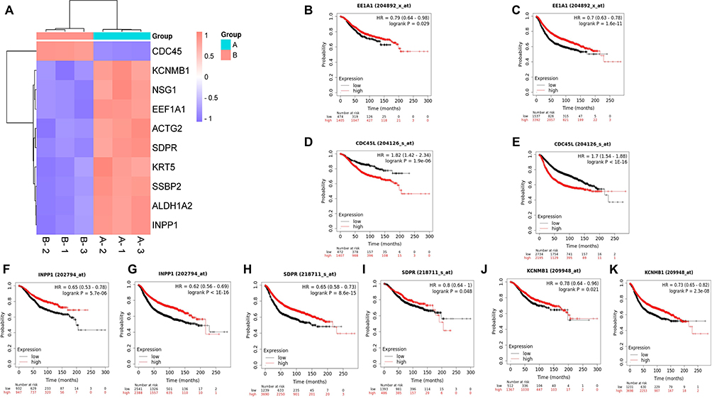 |
Figure 2 The screening process for the objective gene. (A), A heat map shown the top ten differential genes. (B-K), The prognostic analyses of these candidate genes. |
Analysis of the Expression Level of CDC45 in Tumors and Its Correlation with Clinical Indicators
We first analyzed the expression level of CDC45 in pan-cancer and found that CDC45 was highly expressed in most tumors (Figure 3A), such as lung adenocarcinoma (LUAD), Prostate adenocarcinoma (PRAD), Ovarian serous cystadenocarcinoma (OV). Only a small number of tumors have low expression levels of CDC45, such as Testicular germ cell tumors (TGCT). As for BC, CDC45 is highly expressed in tumor tissues compared to normal tissues, both at the transcriptome level (Figure 3B) and at the protein level (Figure 3C). We further analyzed the relationship between CDC45 expression and various clinical indicators. Take indicator age as an example, CDC45 expression is lower in normal tissues than in tumor tissues, and its expression level shows a decreasing trend in tumors with the increase of age (Figure 3D). All the results (Figure 3D–J) indicated that the CDC45 was significantly differentially expressed in age, sex, race, pathologic T stage, pathologic N stage, pathologic M stage, and different subtypes of breast tumors.
 |
Figure 3 The analysis of CDC45 expression and clinical characteristics. (A), The CDC45 expression levels in pan-cancer. (B), The transcriptome expression level expression of CDC45 in BC. (C), The protein expression level expression of CDC45 in BC. (D-J), The results indicated that the CDC45 was significantly differentially expressed existed in age, gender, race, pathologic M stage, pathologic N stage, pathologic T stage and different subtypes of breast tumors. (**P < 0.01, ***P < 0.001, ****P < 0.0001). |
Construction of a Nomogram Based on the Different Clinical Factors
To predict the OS possibility of patients with BC, different clinical features were integrated into the Nomogram model (Figure 4A), and the C-index was 0.723 (95% CI=0.698–0.747), indicating that the model had a moderately accurate prediction effect for patients’ prognosis. In addition, calibration curves (Figure 4B–D) were used to evaluate the prediction efficacy of the nomogram.
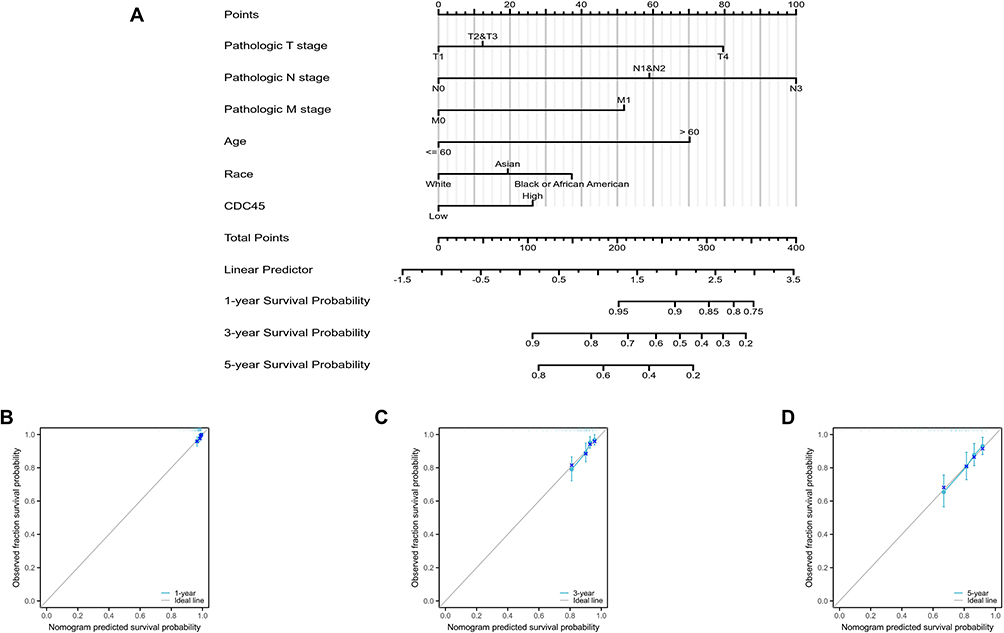 |
Figure 4 Construction a Nomogram for prediction overall survival rates of patients with BC. (A), A nomogram for prediction of different survival time period of breast cancer patients. (B–D), Calibration curves of the nomogram in 1, 3 and 5 year overall survival rates of BC patients. |
Protein-Protein Interaction (PPI) Network Analysis of CDC45
We used the STRING database to analyze the genes that interact with CDC45, and PPI Network analysis was shown in (Figure 5A). We further analyzed the association of these genes with CDC45 in BC. Not surprisingly, similar results (Figure 5B) were found in breast tumor. In addition, the correlation coefficient between CDC45 and each associated gene (Supplementary Figure 2A-J) was analyzed, including TOPBP1, GINS2, MCM5, GINS1, GINS4, POLE2, MCM2, MCM6, MCM4 and MCM7 (all P<0.05).
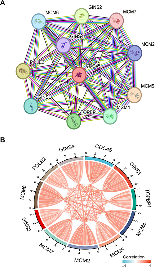 |
Figure 5 Analysis of co-expression genes of CDC45. (A), The PPI network of CDC45 gene was constructed by using string website. (B), Correlation analysis of co-expressed genes in BC. |
The Immune Function Analysis of CDC45
To further explore the immune-related functions of CDC45, we first used the TIMER database to analyze the relationship between the expression level of CDC45 and the somatic copy number alterations in BC (Figure 6A). CDC45 CNV was significantly associated with the infiltration levels of B cells, CD8+ T cells, CD4+ T cells, macrophages, neutrophils, and dendritic cells. In addition, CDC45 expression was significantly correlated with tumor purity (Spearman’s r=0.196, p=4.32E-10) and various immune cells in BC, such as B cell (Figure 6B).
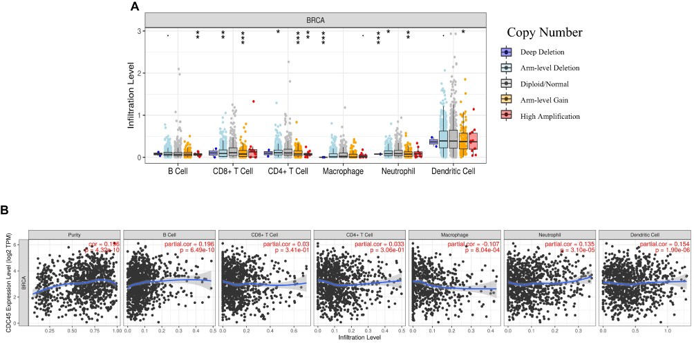 |
Figure 6 The correlation analysis of CDC45 expression levels and various immune cells. (A), The relationship between expression level of CDC45 and the somatic copy number alterations in BC. (B), The relationship between expression level of CDC45 and the Immune cell infiltration level in BC.(*P < 0.05, **P < 0.01, ***P < 0.001). |
We further explored CDC45 gene expression levels across breast tumor mice models and Immune Checkpoint Blockade (ICB)-treatment modules. The ICB treatments included in this module: are anti-PD1, anti-PDL1, anti-PDL2, and anti-CTLA4.The results (Figure 7A and B) indicated that anti-PDL1 and anti-CTLA4 may play an important role in the treatment of breast tumor, with significant statistical significance(all p<0.01). In addition, we also investigate the CDC45 gene expression levels across breast cell lines applying Cytokine treatment (Cytokine treatments included in this module: IFNγ, IFNβ, TNFα, and TGFb1). The results (Figure 7C) showed that TGFb1 had remarkable statistical significance (p<0.01).
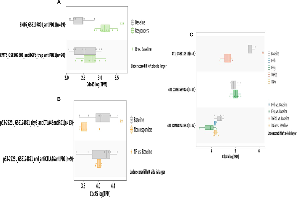 |
Figure 7 The relationship between CDC45 gene expression levels and Immune Checkpoint Blockade (ICB)-treatments and Cytokine response. (A), The relationship between expression level of CDC45 and antiPDL1 response. (B), The relationship between expression level of CDC45 and antiCTLA4 and antiPD1 response. (C), The relations between the CDC45 expression levels and Cytokine treatment.(*P < 0.05, **P < 0.01, ***P < 0.001). |
Finally, the relations between the CDC45 expression levels and chemokines were investigated in breast tumors. As the (Figure 8A–S) shows, it suggested that CDC45 expression is positively correlated with these chemokines with statistical difference. On the contrary, the (supplementary Figure 3A-H) shows that CDC45 expression is negatively correlated with these chemokines with a statistical difference.
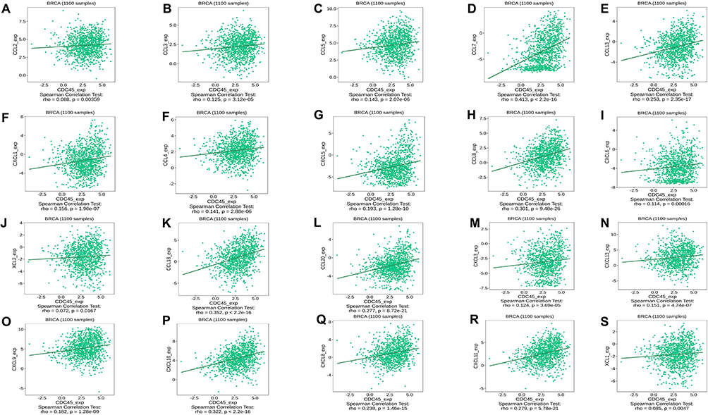 |
Figure 8 The relationship between CDC45 gene expression levels and chemokines were investigated in breast tumor. (A-S), The results shown CDC45 expression was positively correlated with these chemokines with statistical difference. |
Prediction of Drug Sensitivity of CDC45 Target Gene
We performed correlation analysis between various molecule drugs and CDC45 gene expression among breast tumors in different databases (GDSC and CTRP). These results (Figure 9A and B) indicated that many drugs (such as I-BET-762, methotrexate, GSK1070916, NPK76-II-72-1, Vorinostat, and WZ3105) were negatively correlated with the expression level of CDC45.
 |
Figure 9 The correlation analysis between various molecule drugs and CDC45 target gene among breast cancer in different database. (A), The analysis results shown from GDSC database. (B), The analysis results shown from CTRP database. |
CDC45 Mediates the Proliferation, Migration and Invasion of Breast Cancer
We analyzed CDC45 expression levels in common BC cell lines using the Cancer Cell Line Encyclopedia (CCLE) database (supplementary Figure 4A). MDA-MB-231, which displayed relatively high CDC45 expression, was chosen for knockdown, whereas MCF-7 was selected for overexpression. The overexpression or knockdown efficiency was well (supplementary Figure 4B-C). Firstly, we performed clone formation and Transwell assays to verify cell proliferation, migration, and invasion functions. Firstly, we conducted overexpression and knockdown of CDC45 in breast tumor cells and found that knockdown of CDC45 inhibited the proliferation of tumor cells (Figure 10A), while overexpression of CDC45 promoted the growth of tumor cells (supplementary Figure 4D). Next, we explored whether CDC45 could regulate the migration and invasion of BC. The results showed that the knockdown of CDC45 inhibited the migration and invasion of cancer cells (Figure 10B and C). Similarly, overexpression of CDC45 promoted the migration and invasion of tumor cells (supplementary Figure 4E and F).
 |
Figure 10 Functional experiments and molecular mechanism exploration of knockdown CDC45. (A), The knockdown of CDC45 in MDA-MB-231 cells verified by clone formation. (B and C), The knockdown of CDC45 in MDA-MB-231 cells verified by Transwell migration and invasion assay respectively.(D), The knockdown of CDC45 in MDA-MB-231 cells verified by animal experiments.(*P < 0.05, **P<0.01,***P < 0.001, ****P < 0.0001). |
Animal Experiments
To further investigate the potential role of CDC45 in vivo, a Cell line-derived xenograft (CDX) was established using MDA-MB-231 cells. Following the construction of the CDX model and the subsequent removal of the tumor, it was observed that the knockdown of CDC45 in MDA-MB-231 cells inhibited the tumor progression, which was evidenced by a statistically significant difference in tumor volume and weight (Figure 10D). The results of in vivo experiments further confirmed the carcinogenic effect of CDC45 in BC.
Discussion
Previous studies have shown that CDC45 plays a decisive role in DNA replication, ensuring that chromosomal DNA is replicated only once per cell cycle.24,25 Moreover, the expression of CDC45 is closely related to the function of cancer cell proliferation in a variety of tumors, and CDC45 appears to be a valuable new biomarker in tumor cell biology.10,26 In pan-cancer research, the study of CDC45 in breast tumors is briefly involved, there are few studies on CDC45 in breast tumors and no in-depth exploration of its biological function and related pathways.26 In the current study, we used bioinformatics analysis to uncover the multifaceted biological role of CDC45 as a facilitator of cancer progression in BC. Our findings not only highlighted its potential as a reliable prognostic marker but also underscored its association with immune response modulation and its impact on the efficacy of various small-molecule drug treatments. Subsequently, experimental validation was conducted to corroborate CDC45’s pro-carcinogenic properties.
We first screened differential genes using samples of breast tumor tissue and normal tissue. The GO analysis performed by differentially expressed genes was mainly enriched in hemidesmosome assembly, cytosolic ribosome, and structural constituent of ribosome. While the KEGG pathway analysis is mainly enriched in the Ribosome, Coronavirus disease - COVID-19, and Vascular smooth muscle contraction. These analyses shed light on the functions of these differential genes and the important biological processes involved.
We conducted rigorous screening of candidate genes from different dimensions, including their basic expression and survival prognosis differences in BC. The result showed that CDC45 not only has a relatively high expression in breast tumors, but its high expression implies a poor prognosis for BC patients. Based on these analysis results, we finally selected CDC45 as the gene for our subsequent in-depth analysis and functional exploration in vitro experiments. First, we conducted an overall analysis of CDC45 expression in pan-cancer, and the results suggest that it is abnormally expressed in a variety of tumors, and is highly expressed in most tumors. These results seem to indicate that CDC45 plays more of an oncogene role in tumors. In fact, many previous investigations have confirmed this view. For example, CDC45 can promote the progression of melanoma, and hsa_circ_0062270 can regulate CDC45 and thus play the opposite role27; in hepatocellular carcinoma (HCC), CDC45 may promote the development of HCC through co-expression with NUDT1, E2F1, CCNE2, MCM5 and CENPM.28 Not surprisingly, we also found high expression of CDC45 in BC, both at transcriptomic and protein levels. Further, we analyzed the relationship between CDC45 expression levels and clinical characteristics. We found a close correlation between the expression level of CDC45 and a variety of clinical indicators, including age, sex, race, cancer stage and different subtypes of breast tumors. These analyses reveal great potential for future studies of CDC45 as a clinical prognostic indicator.
In recent years, the construction of disease models has been widely used in the medical field, especially since the models built based on different algorithms are constantly updated, and some specific coding genes or non-coding genes can be used as one of the indicators of model construction.29–33 In recent years, researchers have increasingly emphasized the importance of the clinical translational value of their studies, with the ultimate goal of benefiting clinical patients. In our study, we developed a Nomogram model that includes CDC45 and additional clinical indicators to forecast the prognosis of BC patients. The advantage of the model we constructed is that its analytical results have been shown to help guide subsequent treatment strategies and improve patient outcomes. Additionally, the identification of CDC45 as a crucial parameter for constructing a nomogram has enabled accurate prognosis prediction for patients, marking a significant advancement toward achieving clinical transformation in the future.
Furthermore, PPI is fundamental to sustaining these life activities, so we performed an analysis of CDC45 interacting proteins.34 Further research was performed to explore the PPI network of CDC45, and the top ten hub genes (TOPBP1, GINS2, MCM5, GINS1, GINS4, POLE2, MCM2, MCM6, MCM4 and MCM7) were screened out. Immunotherapy, as a new way to treat cancer, has become an important anti-cancer weapon in addition to surgery, radiotherapy, endocrine therapy, chemotherapy, and targeted therapy.35 Immunotherapy plays an important role in some subtypes of BC, such as the clinical use of immune checkpoint inhibitors to treat different subtypes of breast tumors.36 Therefore, we investigated the relationship between CDC45 expression levels and immune function in BC. The results indicated that the expression of CDC45 was related to the infiltration level of various immune cells, including B cells, macrophages, neutrophils, and dendritic cells. Studying the immune cells linked to CDC45 expression offers a novel approach to personalized immunotherapy for diverse patient populations in the future, aligning with the concept of precision medicine. Of course, we also investigated the relationship between CDC45 expression levels and Immune Checkpoint Blockade. Anti-PDL1 and anti-CTLA4 showed therapeutic potential in breast tumors. Finally, given that the multifactorial function of cytokines and chemokines in tumorigenesis has gradually emerged as potential anticancer strategies, some valuable chemokines and cytokines associated with CDC45 have been analyzed.37 All these results have provided important research evidence for the treatment of BC.
Moreover, drug resistance in BC is a frequently encountered and challenging issue in clinical settings.38,39 Numerous studies have explored the mechanisms of drug resistance and developed new treatments to combat it.40,41 Concomitantly, an analysis was conducted on the expression levels of CDC45 in relation to potential involvement in drug resistance alongside small molecule drugs. We used the tumor drug susceptibility multiomics database for drug susceptibility analysis. These small molecule drugs represented by I-BET-762, methotrexate, GSK1070916, NPK76-II-72-1, Vorinostat, and WZ3105 have shown poor therapeutic efficacy in breast tumors with high CDC45 expression and this provided a theoretical basis for future targeted therapy when drug resistance occurs. In the future, we can tailor therapeutic drugs or programs based on CDC45 expression levels to help to individualize the treatment of the patients. These findings provide strong evidence support for future clinical transformation. In addition, we also conducted cell experiments to explore the biological function of CDC45 in BC. In vitro and vivo experiments, we found that CDC45 may promote the progress of tumor cells. Our study is a systematic and multi-dimensional exploration of the biological function of CDC45 in BC, a topic that has received limited attention in previous research. Although our experiment uncovered preliminary insights into the molecular pathway mechanism involving CDC45, it is crucial to acknowledge the limitations of our study. The complexity of the molecular mechanisms underlying CDC45 necessitates further exploration, which will be the focus of our future research efforts. We have conducted a preliminary investigation on the biological function of CDC45. Although it shows great research potential in BC, there is still a lack of in-depth mechanism exploration. In addition, the number of immunodeficient mice in our animal experiments is relatively small, and it is worth adding more numbers for further verification.
In conclusion, we screened various differential proteins from breast tumors and normal tissues, ultimately selecting the CDC45 protein for further study. This selection was based on its potential as a prognostic biomarker and its significant biological functions. CDC45 expression levels are closely associated with immune response and drug resistance. By examining the expression characteristics of CDC45, we uncovered its relationship with BC. This provides important insights into the mechanisms underlying BC development.
Abbreviations
CDC45, Cell division cycle protein 45; Differentially expressed genes, DEGs; Protein-protein interaction, PPI; Breast cancer, BC; Immune Checkpoint Blockade (ICB), Lung adenocarcinoma, LUAD; Prostate adenocarcinoma, PRAD; Ovarian serous cystadenocarcinoma, OV; Cancer Cell Line Encyclopedia; CCLE, Cell line-derived xenograft (CDX); Hepatocellular carcinoma, HCC.
Data Sharing Statement
The datasets generated or analyzed during the current study are available from the corresponding author on reasonable request.
Ethics Statement
TCGA, HPA and GEO belong to public databases. The patients involved in the database have obtained ethical approval. Users can download relevant data for free for research and publish relevant articles. Our study is based on open-source data, so there are no ethical issues.
All human research involved in our study was sourced entirely from public databases and anonymized information, which is in line with items 1 and 2 of Article 32 of the Measures for the Ethical Review of Life Science and Medical Research Involving Human Subjects issued by China on February 18, 2023, so it can be exempted from applying for ethics. All animal experiments conducted were in compliance with the regulations set forth by the Ethics Committee of Tianjin Medical University Cancer Institute and Hospital (NO.2024034).
Funding
This study was supported by Tianjin Key Medical Discipline (Specialty) Construction Project (TJYXZDXK-009A).
Disclosure
The authors declare they have no competing interests in this work.
References
1. Lei S, Zheng R, Zhang S, et al. Global patterns of breast cancer incidence and mortality: a population-based cancer registry data analysis from 2000 to 2020. Cancer Commun. 2021;41:1183–1194. doi:10.1002/cac2.12207
2. Sancho-Garnier H, Colonna M. Breast cancer epidemiology. Presse Med. 2019;48:1076–1084. doi:10.1016/j.lpm.2019.09.022
3. DeSantis C, Siegel R, Bandi P, et al. Breast cancer statistics, 2011. CA Cancer J Clin. 2011;61:409–418. doi:10.3322/caac.20134
4. Wang J, Wu S-G. Breast Cancer: an Overview of Current Therapeutic Strategies, Challenge, and Perspectives. Breast Cancer. 2023;15:721–730. doi:10.2147/BCTT.S432526
5. Rezaianzadeh A, Jalali M, Maghsoudi A, et al. The overall 5-year survival rate of breast cancer among Iranian women: a systematic review and meta-analysis of published studies. Breast Dis. 2017;37:63–68. doi:10.3233/BD-160244
6. Aguayo A, Patt YZ. Liver cancer. Clin Liver Dis. 2001;5:479–507. doi:10.1016/S1089-3261(05)70175-6
7. Jd M, S R, Jw V, et al. Pancreatic cancer. Lancet. 2020;395.
8. Deng J, Liu M, Xiao R, et al. Risk, Incidence, and Mortality of Breast Cancer in Primary Sjögren’s Syndrome: a Systematic Review and Meta-Analysis. Front Immunol. 2022;13:904682. doi:10.3389/fimmu.2022.904682
9. Broderick R, Nasheuer H-P. Regulation of Cdc45 in the cell cycle and after DNA damage. Biochem Soc Trans. 2009;37(4):926–930. doi:10.1042/BST0370926
10. Pollok S, Bauerschmidt C, Sänger J, et al. Human Cdc45 is a proliferation-associated antigen. FEBS J. 2007;274:3669–3684. doi:10.1111/j.1742-4658.2007.05900.x
11. Zhang R, Liu Z, Zhang G. CDC45 modulates MCM7 expression and inhibits cell proliferation by suppressing the PI3K/AKT pathway in acute myeloid leukemia. Am J Transl Res. 2021;13:1183–1194.
12. Huang J, Li Y, Lu Z, et al. Analysis of functional hub genes identifies CDC45 as an oncogene in non-small cell lung cancer - A short report. Cell Oncol Dordr. 2019;42:571–578. doi:10.1007/s13402-019-00438-y
13. Oyola SO, Bringaud F, Melville SE. A kinetoplastid BRCA2 interacts with DNA replication protein CDC45. Int J Parasitol. 2009;39:59–69. doi:10.1016/j.ijpara.2008.07.002
14. Schmidt U, Wollmann Y, Franke C, et al. Characterization of the interaction between the human DNA topoisomerase IIbeta-binding protein 1 (TopBP1) and the cell division cycle 45 (Cdc45) protein. Biochem J. 2008;409:169–177. doi:10.1042/BJ20070872
15. Chandrashekar DS, Bashel B, Balasubramanya SAH, et al. UALCAN: a Portal for Facilitating Tumor Subgroup Gene Expression and Survival Analyses. Neoplasia. 2017;19:649–658. doi:10.1016/j.neo.2017.05.002
16. Thul PJ, Lindskog C. The human protein atlas: a spatial map of the human proteome. Protein Sci. 2018;27:233–244. doi:10.1002/pro.3307
17. Szklarczyk D, Kirsch R, Koutrouli M, et al. The STRING database in 2023: protein-protein association networks and functional enrichment analyses for any sequenced genome of interest. Nucleic Acids Res. 2023;51:D638–46. doi:10.1093/nar/gkac1000
18. Tang Z, Kang B, Li C, et al. GEPIA2: an enhanced web server for large-scale expression profiling and interactive analysis. Nucleic Acids Res. 2019;47:W556–60. doi:10.1093/nar/gkz430
19. Liu C-J, Hu -F-F, Xie G-Y, et al. GSCA: an integrated platform for gene set cancer analysis at genomic, pharmacogenomic and immunogenomic levels. Brief Bioinform. 2023;24(1):bbac558. doi:10.1093/bib/bbac558
20. Ruan Q, Wang C, Wu Y, et al. Exosome microRNA-22 inhibiting proliferation, migration and invasion through regulating Twist1/CADM1 axis in osteosarcoma. Sci Rep. 2024;14:761. doi:10.1038/s41598-023-50612-4
21. Sun S, Fan Z, Liu X, et al. Microglia TREM1-mediated neuroinflammation contributes to central sensitization via the NF-κB pathway in a chronic migraine model. J Headache Pain. 2024;25:3. doi:10.1186/s10194-023-01707-w
22. Wang Q, Huang C, Ding Y, et al. Inhibition of CCCTC Binding Factor-Programmed Cell Death Ligand 1 Axis Suppresses Emergence of Chemoresistance Induced by Gastric Cancer-Derived Mesenchymal Stem Cells. Front Immunol. 2022;13:884373. doi:10.3389/fimmu.2022.884373
23. Chen Z-H, Tian Y, Zhou G-L, et al. CMTM7 inhibits breast cancer progression by regulating Wnt/β-catenin signaling. Breast Cancer Res. 2023. doi:10.1186/s13058-023-01620-9
24. Yadav A, Sharma V, Pal J, et al. DNA replication protein Cdc45 directly interacts with PCNA via its PIP box in Leishmania donovani and the Cdc45 PIP box is essential for cell survival. PLoS Pathog. 2020;16:e1008190. doi:10.1371/journal.ppat.1008190
25. Srinivasan SV, Dominguez-Sola D, Wang LC, et al. Cdc45 is a critical effector of myc-dependent DNA replication stress. Cell Rep. 2013;3:1629–1639. doi:10.1016/j.celrep.2013.04.002
26. Lu Y, Chen X, Liu F, et al. Systematic pan‑cancer analysis identifies CDC45 as having an oncogenic role in human cancers. Oncol Rep. 2022;48:185. doi:10.3892/or.2022.8400
27. Hao T, Yang Y, He J, et al. Knockdown of circular RNA hsa_circ_0062270 suppresses the progression of melanoma via downregulation of CDC45. Histol Histopathol. 2022;37:373–383. doi:10.14670/HH-18-412
28. Lu H-P, Du X-F, Li J-D, et al. Expression of Cell Division Cycle Protein 45 in Tissue Microarrays and the CDC45 Gene by Bioinformatics Analysis in Human Hepatocellular Carcinoma and Patient Outcomes. Med Sci Monit. 2021;27:e928800. doi:10.12659/MSM.928800
29. Balachandran VP, Gonen M, Smith JJ, et al. Nomograms in oncology: more than meets the eye. Lancet Oncol. 2015;16:e173–180. doi:10.1016/S1470-2045(14)71116-7
30. Huang L, Zhang L, Chen X. Updated review of advances in microRNAs and complex diseases: taxonomy, trends and challenges of computational models. Brief Bioinform. 2022;23:bbac358. doi:10.1093/bib/bbac358
31. Yu L, Shen N, Shi Y, et al. Characterization of cancer-related fibroblasts (CAF) in hepatocellular carcinoma and construction of CAF-based risk signature based on single-cell RNA-seq and bulk RNA-seq data. Front Immunol. 2022;13:1009789. doi:10.3389/fimmu.2022.1009789
32. Zhang B, Xie L, Liu J, et al. Construction and validation of a cuproptosis-related prognostic model for glioblastoma. Front Immunol. 2023;14:1082974. doi:10.3389/fimmu.2023.1082974
33. Yu B, Xing Z, Tian X, et al. A Prognostic Risk Signature of Two Autophagy-Related Genes for Predicting Triple-Negative Breast Cancer Outcomes. Breast Cancer. 2024;16:529–544. doi:10.2147/BCTT.S475007
34. Bouhaddou M, Eckhardt M, Chi Naing ZZ, et al. Mapping the protein-protein and genetic interactions of cancer to guide precision medicine. Curr Opin Genet Dev. 2019;54:110–117. doi:10.1016/j.gde.2019.04.005
35. Lee S, Kim T-D. Breakthroughs in Cancer Immunotherapy: an Overview of T Cell, NK Cell, Mφ, and DC-Based Treatments. Int J Mol Sci. 2023;24:17634. doi:10.3390/ijms242417634
36. Anayyat U, Ahad F, Muluh TA, et al. Immunotherapy: constructive Approach for Breast Cancer Treatment. Breast Cancer. 2023;15:925–951. doi:10.2147/BCTT.S424624
37. Amedei A, Prisco D, Elios MM D. The use of cytokines and chemokines in the cancer immunotherapy. Recent Pat Anticancer Drug Discov. 2013;8:126–142. doi:10.2174/1574892811308020002
38. Zhang Y. The root cause of drug resistance in HER2-positive breast cancer and the therapeutic approaches to overcoming the resistance. Pharmacol Ther. 2021;218:107677. doi:10.1016/j.pharmthera.2020.107677
39. Ou X, Tan Y, Xie J, et al. Methylation of GPRC5A promotes liver metastasis and docetaxel resistance through activating mTOR signaling pathway in triple negative breast cancer. Drug Resist Updat. 2024;73:101063. doi:10.1016/j.drup.2024.101063
40. Ye F, Dewanjee S, Li Y, et al. Advancements in clinical aspects of targeted therapy and immunotherapy in breast cancer. Mol Cancer. 2023;22:105. doi:10.1186/s12943-023-01805-y
41. Wu Q, Yang Z, Nie Y, et al. Multi-drug resistance in cancer chemotherapeutics: mechanisms and lab approaches. Cancer Lett. 2014;347:159–166. doi:10.1016/j.canlet.2014.03.013
 © 2025 The Author(s). This work is published and licensed by Dove Medical Press Limited. The
full terms of this license are available at https://www.dovepress.com/terms
and incorporate the Creative Commons Attribution
- Non Commercial (unported, 3.0) License.
By accessing the work you hereby accept the Terms. Non-commercial uses of the work are permitted
without any further permission from Dove Medical Press Limited, provided the work is properly
attributed. For permission for commercial use of this work, please see paragraphs 4.2 and 5 of our Terms.
© 2025 The Author(s). This work is published and licensed by Dove Medical Press Limited. The
full terms of this license are available at https://www.dovepress.com/terms
and incorporate the Creative Commons Attribution
- Non Commercial (unported, 3.0) License.
By accessing the work you hereby accept the Terms. Non-commercial uses of the work are permitted
without any further permission from Dove Medical Press Limited, provided the work is properly
attributed. For permission for commercial use of this work, please see paragraphs 4.2 and 5 of our Terms.
Recommended articles
Establishment and Validation of a Model for Disease-Free Survival Rate Prediction Using the Combination of microRNA-381 and Clinical Indicators in Patients with Breast Cancer
Shen J, Wang M, Li F, Yan H, Wang R, Zhou J
Breast Cancer: Targets and Therapy 2022, 14:375-389
Published Date: 30 November 2022
Establishment and Validation of a Predictive Model for Post-Treatment Anxiety Based on Patient Attributes and Pre-Treatment Anxiety Scores
Sun W, Shen J, Sun R, Zhou D, Li H
Psychology Research and Behavior Management 2023, 16:3883-3894
Published Date: 19 September 2023
Early Warning of Axillary Lymph Node Metastasis in Breast Cancer Patients Using Multi-Omics Signature: A Machine Learning-Based Retrospective Study
Ke Z, Shen L, Shao J
International Journal of General Medicine 2024, 17:6101-6114
Published Date: 12 December 2024
A Prognostic Nutritional Index-Based Nomogram to Predict Breast Cancer Metastasis: A Retrospective Cohort Validation
Chen Z, Gao H, Cheng M, Song C
Breast Cancer: Targets and Therapy 2025, 17:497-510
Published Date: 10 June 2025
Development and Validation of a Radiomics-Based Nomogram for Predicting HER-2 Status in Breast Cancer: A Retrospective Study with Small Validation Cohort
Qiu Q, Chen C, Chen J, Liao C, Tang L
Breast Cancer: Targets and Therapy 2025, 17:1119-1132
Published Date: 2 December 2025