Back to Journals » International Journal of Nanomedicine » Volume 21
From Barrier to Gateway: Nanomaterials Reshaping the Tumor Microenvironment for Therapy
Authors Xie M, Sun W, Hu D, Liu L, Han C, Liu W, Liu L, Bao X, Zhang W, Hao X, Zhou Y
Received 11 October 2025
Accepted for publication 25 February 2026
Published 10 March 2026 Volume 2026:21 573490
DOI https://doi.org/10.2147/IJN.S573490
Checked for plagiarism Yes
Review by Single anonymous peer review
Peer reviewer comments 6
Editor who approved publication: Dr Kamakhya Misra
Mina Xie,1,2 Wanyu Sun,1,2 Dongping Hu,3– 5 Lu Liu,6 Caiwen Han,3– 5 Wenhan Liu,3– 5 Lede Liu,3– 5 Xiaojiang Bao,3– 5 Weisheng Zhang,3– 5 Xiangyong Hao,2 Yongqiang Zhou3– 5
1First Clinical Medical College, Gansu University of Chinese Medicine, Lanzhou, Gansu, 730000, People’s Republic of China; 2Department of General Surgery, Gansu Provincial Hospital, Lanzhou, Gansu, 730000, People’s Republic of China; 3Department of Anorectal Surgery, Gansu Provincial Hospital, Lanzhou, Gansu, 730000, People’s Republic of China; 4The Third School of Clinical Medicine, Lanzhou University, Lanzhou, Gansu, 730000, People’s Republic of China; 5Gansu Provincial Clinical Medical Research Center for Anorectal Diseases, Lanzhou, Gansu, 730000, People’s Republic of China; 6Department of Pediatrics, The First Hospital of Lanzhou University, Lanzhou, Gansu, 730000, People’s Republic of China
Correspondence: Xiangyong Hao, Department of General Surgery, Gansu Provincial Hospital, Lanzhou, Gansu, 730000, People’s Republic of China, Email [email protected] Yongqiang Zhou, Department of Anorectal Surgery, Gansu Provincial Hospital, Lanzhou, Gansu, 730000, People’s Republic of China, Email [email protected]
Abstract: The tumor microenvironment constitutes the external condition that supports the survival and development of tumor cells. It promotes tumor cell proliferation and survival by secreting various cytokines and the provision of nutritional support, thereby driving tumor advancement. However, its structural density and complex composition pose significant barriers to drug delivery and therapeutic intervention, necessitating the development of advanced techniques for effective penetration. In recent years, nanotechnology, characterized by its distinctive physicochemical properties and excellent targeting and regulatory capabilities, has shown tremendous potential in overcoming the tumor microenvironment barriers, garnering significant research interest. This paper systematically summarizes the formation mechanisms of various TME subtypes, including immunosuppressive, metabolic, acidic, hypoxic, stromal, mechanical, microbial, inflammatory, and neural TME. It analyzes the principal challenges faced by nanomaterials in regulating these microenvironments, focuses on research strategies and application prospects of nanomaterials across different subtype microenvironments, and proposes novel directions for future investigation. The objective is to facilitate breakthroughs in the translational application of nanomaterials from mechanistic innovation to clinical practice.
Keywords: tumor microenvironment, nanomaterials, immune suppression, metabolism, hypoxia, stroma, mechanics
Introduction
Tumor microenvironment (TME)1,2 refers to the surrounding environment of tumor cells, which is primarily composed of tumor cells, immune cells, neovasculature, diverse stromal cells, and a range of cytokines. It is distinguished by elevated density, reduced permeability, hypoxia, and acidity. Tumor cells remodel external conditions through various mechanisms to evade immune surveillance or enhance immune suppression, thereby creating favorable conditions for tumor proliferation and survival. Consequently, the complexity and density of TME present a substantial obstacle to effective pharmacological intervention.
Nanomaterials,3–5 representing an emerging frontier technology, are defined as molecular materials with dimensions ranging from 1 to 100 nanometers. Their inherent physicochemical properties enable them to effectively penetrate the TME barrier. Nanomaterials, constructed according to different strategies, such as inorganic/metallic nanoparticles, lipids, polymer micelles, and protein-based carriers, have the potential to assist in the diagnosis, treatment, and modulation of TME in tumors.
From the standpoint of composition and function, the TME can be categorized into several aspects, including immunosuppressive, metabolism, acidity, hypoxia, stroma, mechanics, microbiota, inflammation, and neural TME. Among them, the immunosuppressive microenvironment,6,7 as one of the most prevalent types, is characterized by an abundance of regulatory T cells (Tregs), inhibitory stromal cells, and immunosuppressive factors such as interleukin-10 (IL-10), which collectively induce changes in the phenotype and function of immune cells and promote the formation of immunosuppressive cell lineages. The metabolic microenvironment,8,9 through the accumulation of abnormal metabolic products, suppresses T cell function and downregulates the expression of inhibitory receptors such as programmed cell death protein 1 and cytotoxic T-lymphocyte-associated protein 4. There is a close interrelationship among the metabolic, acidic, and hypoxic microenvironments. The acidic microenvironment10 is associated with metabolic dysregulation and abnormal angiogenesis, further enhancing the immune-suppressive functions of Tregs and Myeloid-Derived Suppressor Cells (MDSCs). The hypoxic microenvironment11 originates from the elevated metabolic demands of tumor cells combined with increased interstitial pressure, which impedes effective oxygen delivery. The principal regulatory factor, Hypoxia-Inducible Factor (HIF), facilitates tumor angiogenesis and immune evasion. The stromal microenvironment12,13 mainly includes various stromal cells and related fibrous components, which participate in regulating tumor cell adhesion, migration, and signal transduction. The mechanical microenvironment14,15 involves the physical properties within tumor tissues, including tissue stiffness, mechanical stress, and interstitial pressure, with related signaling pathways affecting tumor cell proliferation and migration. The microbiota microenvironment16,17 regulates tumor progression and immune response through interactions between the microbiome and tumor cells. Inflammatory mediators within the inflammatory microenvironment18,19 participate in cascade reactions that promote immune suppression and stromal remodeling. Lastly, the neural TME20 supports angiogenesis and tumor metastasis while exerting a regulatory role in immune exhaustion and escape. The submicroenvironments described above are closely interconnected in a multidimensional and dynamic manner, which has a certain impact on tumor progression. Existing nanomaterials, with their inherent programmability, responsiveness, physical and chemical properties, as well as excellent permeability, are capable of selectively targeting the differences in these microenvironments to influence interaction mechanisms, intervene at multiple points, weaken the barrier synergistic effect, and bring the microenvironment back to a controllable state, thus laying the foundation for subsequent treatments.
Unlike previous reviews, this paper explores the mechanisms of action of nanomaterials on various subtypes of the TME and therapeutic approaches. It primarily discusses synergistic therapeutic strategies such as phototherapy, sonotherapy, and magnetotherapy, novel research concepts like cuproptosis and ferroptosis, and aspects like mitochondrial targeting, enzyme regulation, and gene editing, to elucidate effective research materials. The paper also highlights challenges such as the single-action nature, mass production of materials, and tumor heterogeneity. It aims to furnish researchers and clinicians in related fields with strategies for the application of nanomaterials in tumor treatment. Furthermore, the article offers potential future research directions to facilitate clinical translation, thereby contributing novel therapeutic opportunities and hope for patients (Figure 1).
 |
Figure 1 Currently, nanomaterials, due to their multifunctionality, have become an effective strategy for modulating various types of TME. These strategies not only involve precise modulation of the TME through mechanisms such as multimodal combination therapies, but also include the direct induction of tumor cell death using techniques such as cuproptosis, ferroptosis, and gene silencing. This figure was created by Adobe illustrator and Biorender (https://app.biorender.com/). Abbreviations: cGAS-STING, cyclic GMP-AMP synthase pathway; IDO1, indoleamine 2,3-dioxygenase 1. |
Immunosuppressive Tumor Microenvironment
The immunosuppressive TME is one of the key mechanisms by which tumors escape immune surveillance. It is composed of various immune-suppressive cells, stromal components, and interactions with tumor cells themselves,21 collectively establishing a complex immunosuppressive network. These cells secrete immunosuppressive cytokines that regulate the functions of CD8+ T cells and natural killer (NK) cells.22 Among these, the cyclic GMP-AMP synthase-stimulator of interferon genes (cGAS-STING) pathway23 and the remodeling of the immune microenvironment are critical strategies for enhancing anti-tumor responses. These pathways are critically involved in multiple cancers and represent pivotal targets in current tumor therapies.
Synergistic Effects of Nanomaterials with Multimodal Therapies
The combination of nanomaterials with various tumor treatment methods has become a significant research direction in this field, including photothermal therapy (PTT), sonodynamic therapy (SDT), radiotherapy (RT), chemotherapy, immunotherapy, and targeted therapy24–36 (see Additional Table 1). With in phototherapy, Near-Infrared Regions (NIR) I and NIR II are distinct spectral regions. NIR I light is cost-effective and widely used in early research, whereas NIR II light has superior tissue penetration depth, which is crucial for treating deep-seated tumors. Although the majority of nanomaterials focus on lasers and fluorescence, other light sources, such as organic light-emitting diodes, quantum dot light sources, and supercontinuum light sources, due to their tunable spectral properties and potential clinical applications, should be considered in nanomaterial design. The selection of an appropriate light source should comprehensively consider factors such as tumor anatomical location, depth, volume, and treatment goals to achieve precise and personalized treatment.
Phototherapy utilizes light energy to activate immune responses, while radiofrequency (RF) ablates tumors through electromagnetic waves. Low-intensity radiofrequency uses nanomaterials37 to produce reduced energy output compared to traditional RF, minimizing thermal damage to surrounding tissues, providing a gentler new strategy for tumor treatment. Future studies may focus on other modalities, such as pulsed radiofrequency or combining RF with magnetic therapy and microwaves for multi-physical field synergy, which could expand the application dimensions and treatment precision of RF therapy. Similarly, microwaves also use electromagnetic waves through the microwave thermal effect for tumor ablation. Nanomaterials based on microwave thermal effects38 demonstrate enhanced capabilities for deep tissue heating and accelerated thermal response compared to traditional PTT, which also avoids the accumulation issues associated with photosensitizers. During the treatment process, real-time imaging and monitoring are crucial for evaluating therapeutic efficacy and guiding precise intervention.
Common imaging methods include fluorescence imaging (FLI), photoacoustic imaging (PAI), and photothermal imaging (PTI). FLI reflects molecular activity and, when combined with NIR II, enables the monitoring of drug distribution and cellular metabolism,39 which is crucial for the precise evaluation of drug efficacy and optimization of treatment plans. PAI provides high spatial resolution and deep tumor information,40 helping to better understand tumor morphology, dimensions, and boundaries. PTI measures temperature changes and monitors photothermal effects in real-time,41 ensuring accurate temperature control during treatment and preventing excessive thermal damage. FLI-PAI-PTI tri-modal imaging is one of the most advanced imaging methods. Guided by this multimodal imaging, nanomaterials42 are capable of providing real-time, multi-dimensional, and comprehensive imaging information. Researchers may consider this strategy when technical and cost conditions permit, to further improve the precision and efficacy of tumor therapy.
Exosomes are small vesicles secreted by various cells,43,44 which have excellent drug delivery and tumor-targeting capabilities. When combined with multimodal imaging, exosomes facilitate precise tumor treatment.45 They can also be applied in other TME or drug delivery nanomaterials, with broad application prospects after being combined with multimodal therapy.
The cavitation effect46 is an important mechanism in SDT. Under the action of ultrasound, bubbles in liquids undergo vibration, growth, shrinkage, and rupture, generating mechanical stress, localized elevated temperatures, and free radicals that induce tumor cell death. Nanomaterials utilizing the cavitation effect47 not only have excellent energy transfer capabilities but also efficiently generate reactive oxygen species (ROS), enhance tumor vascular permeability, and minimize tissue fibrosis and hardening, avoiding the exacerbation of the microenvironment. A similar effect, termed photocavitation, relies on photosensitizers to chemically generate bubbles, aiding drug delivery. However, this area remains in early stages, and its mechanisms are yet to be fully elucidated. Therefore, the synergy of cavitation effects with other therapies can produce strong anti-tumor immune responses, making related nanomaterials promising for tumor treatment.
Cyclic GMP-AMP Synthase-Stimulator of Interferon Genes Pathway
The cGAS-STING pathway activates the cGAS enzyme to recognize cytoplasmic DNA, subsequently catalyzing the synthesis of the second messenger cyclic GMP-AMP. It activates the STING protein and induces robust induction of type I interferons (IFN-I) through the downstream TANK-binding kinase 1 - Interferon Regulatory Factor 3 signaling axis, promoting dendritic cell (DC) maturation and the activation of cytotoxic T lymphocytes (CTLs). Nanomaterials based on this pathway enhance CD8+ T cell activity and inhibit tumor cell proliferation by targeting the delivery of cGAS-STING agonists (such as cyclic dinucleotide analogs), encapsulating DNA-damaging drugs, or carrying cytoplasmic DNA release inducers.48–56 The materials described above primarily utilize double-stranded DNA, such as mitochondrial DNA (mtDNA) leakage or DNA damage, while metal ions enhance the ability of cGAS to recognize DNA, thereby synergistically activating the cGAS-STING signaling pathway.
Nonetheless, differences are observed in the composition, mechanisms of synergistic stimulation, and the timing control of administration. For instance, OSA@CMCS Gel52 serves as a localized implantable hydrogel delivering cisplatin and the demethylating agent DAC in combination with MnCl2. DAC mediates the demethylation that upregulates Gasdermin E expression, triggering pyroptosis in tumor cells and subsequently releasing damage-associated molecular patterns (DAMPs). This process diminishes the proportion of Tregs and promotes the formation of central memory T cells and effector memory T cells. The hydrogel enables localized and sustained drug release, thereby mitigating systemic toxicity, with a degradation period of approximately 15 days in the post-operative tumor cavity. It demonstrates favorable biocompatibility and potential anti-tumor metastatic properties. However, the assessment of circulation, maximum tolerated dose (MTD), and long-term safety remains to be thoroughly evaluated.
Furthermore, Cu-ZnO2@PDA49 exhibits selective release of Cu2⁺ and Zn2⁺ along with Hydrogen Peroxide (H2O2) under acidic conditions, inducing cuproptosis and resulting in mitochondrial damage, which in turn releases mtDNA. Zn2⁺ cooperatively activates the cGAS-STING pathway with mtDNA, leading to a significant amplification of the immune cascade following treatment with 808 nm PTT. In vivo safety evaluations indicated an MTD of ≥ 50 mg/kg, evidencing its excellent biocompatibility. However, this study has not yet provided comprehensive pharmacokinetic parameters, such as plasma elimination half-life and dose-response curves. Therefore, further detailed toxicokinetic and pharmacokinetic studies are required in alignment with preclinical research guidelines to support subsequent preclinical investigations.
Similar to the cGAS-STING pathway, the Programmed Death-1/Programmed Death-Ligand 1 (PD1/PD-L1) pathway also regulates T cell-mediated anti-tumor immune responses, but it primarily relies on the blockage of inhibitory signals to restore T cell anti-tumor effects.57–62 Although both JS-20162 and aPD-1-siRNA@NP58 stimulate immune responses by inhibiting the PD-1 pathway, the former alleviates immune suppression through combination with TGF-β neutralization, while the latter modulates the microenvironment more precisely via MDK-siRNA, suggesting that a combination of classical targets with emerging delivery systems holds significant potential for application. Meanwhile, nano-drugs targeting PD-L1, such as SPS-NPs,59 CPPD1,61 CXNP-CeBM,57 and PML@Len,60 intervene in the PD-1/PD-L1 axis through direct blockade, lysosomal degradation, or occupancy shielding, thus providing diverse approaches for immune therapy. In the future, the combination of nanomaterials targeting the PD-1/PD-L1 axis with advanced technologies like CRISPR-dCas9 presents a compelling avenue for development, although careful consideration must be given to minimizing biotoxicity and enhancing delivery specificity.
The excessive activation of other pathways, such as the Nuclear Factor κ-light-chain-enhancer of activated B cells (NF-κB) and Signal Transducer and Activator of Transcription 3 (STAT3) pathways, often mediates the formation of an immunosuppressive microenvironment. In response, one researcher has developed a nanoparticle-based DNAzyme53 designed to selectively degrade miR-19a in the miR-19a-PTEN-STAT3 immunosuppressive loop. This degradation alleviates the post-transcriptional suppression of PTEN by miR-19a, restores PTEN protein levels, and subsequently inhibits STAT3 phosphorylation. As a result, this intervention reverses the immunosuppressive microenvironment, enabling T cells and NK cells to recover their cytotoxic function. Similarly, another researcher employed the proteasome inhibitor Bortezomib to prevent the degradation of IκBα, thereby inhibiting the nuclear translocation of NF-κB.63 This approach reduces the expression of downstream pro-inflammatory factors, including IL-6, thereby mitigating the inflammatory and immunosuppressive state of the TME and enhancing the efficacy of immunotherapy. However, there remains a limited number of nanoparticle-based materials that utilize these two mechanisms.
Another pathway that induces IFN production is the Retinoic acid-inducible Gene I (RIG-I) pathway, which has garnered increasing research interest. This pathway activates the RIG-I receptor upon recognizing double-stranded RNA, initiating a Mitochondrial Antiviral Signaling Protein-mediated signaling cascade that induces IFN-I production and promotes immunogenic cell death (ICD). Phung64 used antisense oligonucleotides (ASOs) of Kirsten Rat Sarcoma Viral Oncogene Homolog (KRAS) in combination with RIG-I agonists to simultaneously suppress mutated KRAS expression and activate RIG-I, significantly enhancing anti-tumor immunity and directly inducing tumor cell apoptosis. Although the downstream effector molecules and dynamic regulatory networks of the RIG-I pathway remain incompletely elucidated, the combination of RIG-I agonists with novel delivery systems, such as nanovesicles, hydrogels, and engineered bacteria, may offer new directions for tumor immunotherapy.
Despite the relatively mature clinical translation of PD-1/PD-L1, the cGAS-STING pathway, which has emerged as a star player in innate immunity over the past decade, still presents significant opportunities for exploration. While classical pathways such as NF-κB, STAT3, and RIG-I are relatively well-understood at the molecular level, their spatiotemporal regulation within the TME and their synergistic effects with nanomaterials have yet to be fully investigated. These pathways continue to offer promising opportunities for integration with the aforementioned materials and technological platforms. The integration of immune modulation with hypoxia or acidity targeting has become a prevalent method for TME-responsive nanocarriers. Compared to single-function nanoplatforms, this combined approach demonstrates clearer clinical translation potential in enhancing drug delivery, overcoming immune suppression, and promoting systemic anti-tumor immunity (Figure 2).
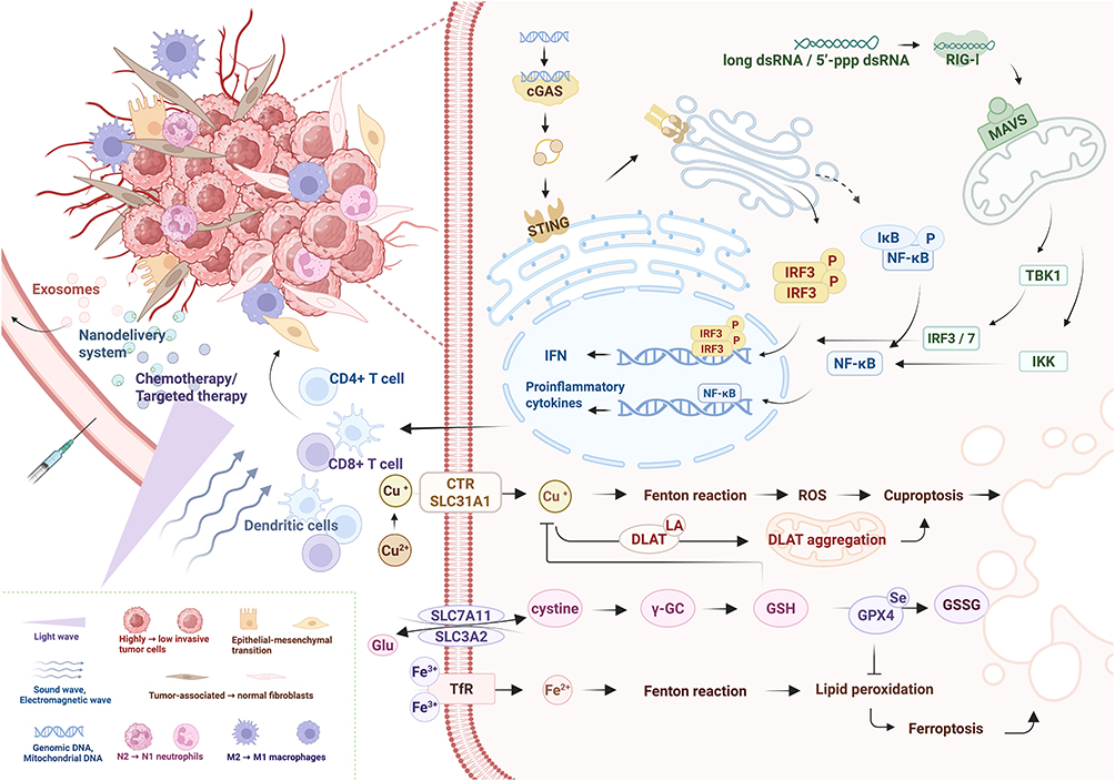 |
Figure 2 In the immunosuppressive TME, various therapeutic strategies can modulate immune regulatory mechanisms, such as nanotechnology-based delivery systems, exosome-based drug delivery, chemotherapy, targeted therapy, phototherapy, and sonotherapy. These interventions help diminish the invasiveness of tumor cells, induce polarization of macrophages and neutrophils, promote the phenotypic transformation of TAMs, inhibit EMT, and reduce tumor angiogenesis, thereby suppressing tumor invasion and metastasis. Additionally, DNA derived from bacteria, viruses, mitochondria, and genomes can activate the cGAS-STING signaling pathway, while double-stranded RNA triggers the secretion of IFN and other pro-inflammatory cytokines via the RIG-I pathway, thereby promoting DC maturation and enhancing the tumor-targeted killing effect of CD8⁺ T cells and CD4⁺ T cells. Furthermore, copper and iron ions can induce tumor cell death through the Fenton reaction, with glutamine metabolism plays a regulatory role in copper-induced and iron-induced cell death. This figure was created by Biorender (https://app.biorender.com/). Abbreviations: cGAS, cyclic GMP-AMP Synthase; STING, Stimulator of Interferon Genes; IRF3, Interferon Regulatory Factor 3; IκB, Inhibitor of Nuclear Factor κ-light-chain-enhancer of Activated B Cells; NF-κB, Nuclear Factor κ-light-chain-enhancer of Activated B Cells; IFN, Interferon; dsRNA, double-stranded RNA; RIG-I, Retinoic Acid-Inducible Gene I; MAVS, Mitochondrial Antiviral Signaling Protein; TBK1, TANK-binding Kinase 1; IKK, IκB Kinase; CTR, Copper Transporter; SLC31A1, Solute Carrier Family 31 Member 1; DLAT, Dihydrolipoamide S-Acetyltransferase; ROS, Reactive Oxygen Species; Glu, Glutamate; γ-GC, γ-Glutamylcysteine; GSH, Glutathione; GPX4, Glutathione Peroxidase 4; GSSG, Oxidized Glutathione; TfR, Transferrin Receptor. |
Multitargeted Strategy for Immune Microenvironment Remodeling
Reshaping the immune microenvironment represents a critical therapeutic strategy for reversing immune suppression.65–71 Currently, nanomaterials exhibit a broad range of regulatory potential in this field, mainly by promoting the repolarization of M2 tumor-promoting macrophages to the M1 anti-tumor phenotype,31,47,58,60,72–77 transforming N2 immune-suppressive neutrophils into the N1 phenotype,78 degrading extracellular matrix (ECM)79 to improve tumor tissue permeability, and inhibiting epithelial-mesenchymal transition (EMT)80 to promote T cell infiltration, thereby enhancing anti-tumor immune responses. While the study of macrophage polarization is relatively mature, the polarization mechanism of N2 neutrophils requires further exploration, and the signaling pathways and metabolic regulation associated with them are not yet clear. These cells play a complex role in establishing the inflammatory microenvironment and immune suppression. Targeted regulation strategies are still in the exploratory stage, and in-depth research is urgently needed from the perspectives of microenvironment interactions and molecular mechanisms.
Other Emerging Therapeutic Strategies
Cuproptosis and ferroptosis, along with mitochondrial-targeted cell death mechanisms, are gradually becoming effective strategies for inducing tumor cell death.49,50,81–85 Cuproptosis involves the abnormal aggregation and stress of mitochondrial respiration-related proteins triggered by copper ions, while ferroptosis is driven by iron-dependent lipid peroxidation. In comparison, arsenic and vanadium-related nanomaterials86 exhibit distinct mechanisms and cutting-edge value. Arsenic compounds efficiently induce apoptosis, whereas vanadium promotes ferroptosis by promoting lipid peroxidation. Their synergistic effects can simultaneously activate both apoptosis and ferroptosis pathways. Despite their promising potential, these agents exhibit certain systemic toxicity, and long-term use may lead to liver and kidney damage, as well as neurotoxicity. Therefore, careful design of dosing regimens and dose control strategies is required for safe clinical application within the therapeutic window.
The FasL-Fas pathway, a classical exogenous apoptosis pathway, has also been applied in nanomaterials to enhance anti-tumor effects.87 However, it is limited by instability and non-specificity in inducing apoptosis, susceptibility to interference from multiple factors in the TME, and faces challenges such as unstable activation and off-target effects. Luo88 designed an innovative nanomaterial combining gas therapy with PANoptosis to alleviate the immunosuppressive microenvironment. Hydrogen sulfide (H2S) is an emerging gas therapy that helps induce ICD and relieves immune suppression, as well as modulates pathological conditions like acidity and hypoxia, offering potential for multi-mechanistic synergistic treatment. PANoptosis, which combines pyroptosis, necrosis, and apoptosis, is associated with various molecular mechanisms, including inflammasomes, Gasdermin family proteins, and Caspase cascades. This comprehensive approach induces cell death and effectively circumvents drug resistance problems related to single-cell death pathways, making it a prominent focus in recent research.
In metabolic regulation, inhibition of Indoleamine 2,3-dioxygenase 1 (IDO1) expression and its mediation of the kynurenine metabolic pathway can enhance immune responses,88–91 alleviating tryptophan depletion-induced T cell dysfunction, reducing Nicotinamide Adenine Dinucleotide levels, inhibiting glycolysis, and reversing immune suppression. This strategy can break the immune-metabolic cycle and reverse immune suppression. In future research, this treatment strategy could be considered in combination with other metabolic pathways or multi-modal treatments to address the limitations of single therapy.
Gene silencing technology, with its high programmability and precise targeting ability, has become an important tool in regulating the TME.92,93 The use of small interfering RNA (siRNA) to silence mRNA or transcription factors can affect various cellular functions and protein expression in the TME, enhancing the activity of CD8+ T cells. Its designability and efficient targeting make it a popular option for nanomaterials. Future research could explore additional options, such as ASOs,64 CRISPR-Cas systems, and RNA interference (RNAi), which further expand the dimensions and depth of gene regulation, providing diverse tools for nanomaterial design.
Despite significant progress in immune microenvironment regulation research, the clinical translation of related nanomaterials still faces challenges. Future efforts should promote interdisciplinary collaboration and evaluate their safety, pharmacokinetic characteristics, and therapeutic efficacy through carefully designed clinical trials to facilitate the transition of high-translational-value strategies from basic research to clinical practice.
Metabolic Tumor Microenvironment
Metabolic reprogramming in the TME is a key mechanism for maintaining tumor malignancy and immune suppression. Metabolites such as glucose, lactate, lipids, glutamine, or glutathione (GSH), and adenosine94–96 provide bioenergy and synthetic precursors for tumor cells, while also directly regulating immune cell functions, forming metabolic barriers that support tumor growth. Nanomaterials targeting specific metabolic pathways have become an important strategy for reversing immune suppression and enhancing anti-tumor effects.
Glucose Metabolism
In terms of glucose metabolism, nanomaterials targeting aerobic glycolysis can reduce ATP production and lactate accumulation97–99 by inhibiting key enzymes or transport proteins, such as Pyruvate Kinase M2 (PKM2) and Lactate Dehydrogenase A (see Additional Table 2). More precise strategies can also be considered, such as metabolic-specific therapies based on local glucose levels, or combining with cuproptosis, ferroptosis, and disulfide-induced apoptosis to synergistically induce tumor cell death.100 By regulating the tricarboxylic acid cycle or pentose phosphate pathway, and integrating multimodal imaging, further understanding of the metabolic-immunity coupling mechanisms in the TME can be achieved.
Beyond the direct inhibition of aerobic glycolysis, targeting glucose transport and pivotal metabolic proteins has also shown significant therapeutic potential, such as mannose competing with glucose for the same transport proteins, and inhibiting the expression of glycolysis-related proteins to indirectly affect energy supply and carbon source replenishment.101,102 Furthermore, glucose metabolism reprogramming not only involves energy supply but is also closely related to epigenetic modifications, redox homeostasis, and immune microenvironment regulation. Inhibiting the expression of IDO191 can simultaneously regulate the immune suppression mediated by tryptophan depletion and the glucose metabolism process. Suppressing PKM2103 can redirect the metabolic flux toward biosynthetic pathways, enhancing oxidative stress sensitivity. Downregulating Glucose Transporter 1104 and Hexokinase 2 (HK2)105 directly limits glucose influx and glucose phosphorylation, while targeting Pyruvate Dehydrogenase Kinase (PDK)106 can restore the activity of the pyruvate dehydrogenase complex, promoting pyruvate entry into the tricarboxylic acid cycle, and reversing the aerobic glycolysis phenotype. Further exploration of the downstream regulatory nodes of glycolysis, such as Phosphofructokinase-1, Phosphoglycerate Kinase, and Enolase, which are integral to branching metabolic pathways, is needed. Changes in the activity of these enzymes may affect nucleotide synthesis, serine-glycine metabolism, and redox balance, providing new dimensions for the development of multi-target metabolic intervention nanomaterials (Figure 3).
 |
Figure 3 Normal cells primarily rely on HK and PKM1 for glycolytic metabolism. In contrast, tumor cells tend to favor the high expression of HK2 and PKM2, engaging in aerobic glycolysis (Warburg effect) and generating large amounts of lactate. Lactate is subsequently exported to the microenvironment through monocarboxylate transporters, leading to an increase in extracellular acidity and the formation of an acidic TME, which in turn promotes tumor progression and weakens anti-tumor immune responses. Furthermore, lipid metabolism can cause immune cell dysfunction, such as impairing T cell activation and macrophage polarization. Conversely, a decrease in intracellular glutathione (GSH) synthesis compromises the antioxidant defenses of tumor cells, thereby inhibiting their proliferation and survival. This figure was created by Biorender (https://app.biorender.com/). Abbreviations: Glc, Glucose; HK2, Hexokinase 2; Glc-6-P, Glucose-6-Phosphate; Fru-6-P, Fructose-6-Phosphate; PFK2, Phosphofructokinase 2; Fru-2,6-3, Fructose-2,6-Bisphosphate; Fru-1,6-BP, Fructose-1,6-Bisphosphate; G3P, Glyceraldehyde-3-Phosphate; DHAP, Dihydroxyacetone Phosphate; PEP, Phosphoenolpyruvate; PKM2, Pyruvate Kinase M2; LDH-A, Lactate Dehydrogenase A; Lac, Lactate; MCT-4, Monocarboxylate Transporter 4; FABPs, Fatty Acid Binding Proteins; CD36, Cluster of Differentiation 36; ACSL4, Acyl-CoA Synthetase Long-chain Family Member 4; Acyl-CoA, Acyl-Coenzyme A; CPT1-M, Carnitine Palmitoyltransferase 1-Mitochondrial; FAO, Fatty Acid Oxidation; Acetyl-CoA, Acetyl-Coenzyme A; TCA, Tricarboxylic Acid Cycle; ACC1, Acetyl-CoA Carboxylase 1; Malonyl-CoA, Malonyl-Coenzyme A; FASN, Fatty Acid Synthase; α-KG, α-Ketoglutarate; IDH1, Isocitrate Dehydrogenase 1; ACLY, ATP-Citrate Lyase; GLS, Glutaminase; ASCT2, Alanine, Serine, Cysteine Transporter 2; GCLC, Glutamate-Cysteine Ligase Catalytic Subunit; γ-Glu-cys, γ-Glutamylcysteine; GSS, Glutathione Synthetase; GSH, Glutathione; GSSG, Oxidized Glutathione. |
Lactate Metabolism
Nanomaterials that influence lactate metabolism have been shown to reduce lactate efflux by depleting lactate or inhibiting Monocarboxylate Transporters (such as MCT-1), thereby reversing the pH of the TME.107–111 Compared to the well-researched MCT-1, MCT-4 as the main channel for lactate efflux in high glycolysis tumors, remains underexplored in terms of its mechanistic research and targeting strategies.112 Integrating lactate metabolism regulation with mitochondrial-targeting strategies can further disrupt redox homeostasis, enhancing the anti-tumor efficacy.113,114 When combined with ultrasound, PTT, cuproptosis, apoptosis, and other therapies, it holds the potential for synergistic enhancement of metabolic intervention and physical treatment,115–117 although its potential toxicity and metabolic adaptability still require systematic evaluation.
Glycolysis and lactate metabolism are closely coupled in the TME, where increased glycolytic flux directly leads to increased lactate production, driving microenvironment acidification and immune suppression. Researchers have designed nanomaterials that are capable of concurrently inhibiting glucose uptake and lactate production by inhibiting HK2, 3-Phosphoinositide-Dependent Protein Kinase-1, and Glucose Oxidase to reduce lactate generation.105,106,118,119 Moreover, combining lactate metabolism regulation with multiple metabolic pathway interventions, such as glucose and glutamine, or synergizing with photothermal, chemodynamic, and immunotherapy strategies, holds promise for achieving multidimensional and precise control over tumor acidity. Such strategies have the potential to reverse the immune-suppressive microenvironment and enhance therapeutic responses.
Other Metabolism
Beyond the metabolism of glucose and lactate, other metabolism-related nanomaterials also play a pivotal role. Tumor cells, through the reprogramming of lipid metabolism, facilitate their proliferation, metastasis, preservation of cancer stemness, and chemotherapy resistance, while concurrently inhibiting anti-tumor immune responses in a lipid-enriched microenvironment. Lipid droplets not only serve as energy reserves to support tumor cell metabolic adaptation but also promote the formation and maintenance of an immune evasion microenvironment by reshaping the lipid metabolic pathways of immunosuppressive cells. Studies have shown that triglycerides and glycerophospholipids can abnormally accumulate in lipid droplets of macrophages,120 and the accumulation of succinates promotes the activation of lipid generation-related signaling pathways.121 Similar lipid metabolism abnormalities can lead to the dysfunction of related immune cells and lactate accumulation, forming a vicious cycle of metabolic suppression. Therefore, developing nanomaterials targeting lipid metabolism has become a promising strategy to counteract immune suppression, such as inhibiting Fatty Acid Synthase expression to reduce lipid droplet formation and free fatty acid accumulation, thus enhancing immune cell activity.122 Additionally, regulating key metabolic intermediates like citrate to inhibit fatty acid synthesis and energy metabolism, succinate to influence hypoxia signaling and inflammation response, inhibiting Fatty Acid Oxidation, and using energy stress inducers to intervene in lipid metabolism balance have shown great potential for translational applications. Future research may further explore the interactions between lipid metabolism and glucose or glutamine metabolism, and design smart nanomaterials capable of achieving coordinated regulation across multiple metabolic pathways to more effectively reshape the immune TME.
GSH and glutamine metabolism serve as nitrogen and carbon source supplementation pathways. The regulation strategies mainly involve inhibiting Glutaminase 1 to prevent the conversion of glutamine to glutamate, thereby reducing GSH formation and inducing oxidative damage to suppress tumor progression.123 Moreover, reprogramming the glutamine metabolic pathway can affect the regeneration capacity of Nicotinamide Adenine Dinucleotide Phosphate (NADPH), disrupting the tumor’s redox homeostasis.124 Combined with glucose metabolism and glutamine metabolism can induce energy metabolic crises and biosynthesis disorders, enhancing the anti-tumor properties of materials.104 Future research can further explore the interplay between glutamine and lactate metabolism, such as targeting Glutamine-derived α-ketoglutarate’s metabolic feedback on lactate production or inhibiting Alanine-serine-cysteine transporter 2 to restrict glutamine uptake, enhancing the spatial specificity of metabolic interference.
Adenosine metabolism also holds research potential. Extracellular adenosine accumulation can inhibit DC maturation and T cell function via the Adenosine A2A receptor signaling pathway and promote M2 macrophage polarization. Lowering adenosine levels, promoting its conversion to inosine, or blocking the breakdown of ATP and ADP to adenosine helps restore T cell function.125,126 Therefore, future nanomaterials can further explore the optimal balance of adenosine or inosine, aiming to precisely reshape a T-cell-friendly immune microenvironment in terms of spatiotemporal dimensions.
Despite the promising prospects of metabolism-targeting nanomaterials in correcting tumor metabolic reprogramming and augmenting immune activity, they face complex challenges such as metabolic pathway redundancy, intercellular nutrient competition, and metabolite crosstalk. Integrating multi-omics technologies such as metabolomics, fluxomics, and proteomics will systematically reveal the interaction mechanisms between tumors and immune cells, facilitating the rational design of metabolic intervention strategies. Additionally, the combined use of specific metabolic small molecule inhibitors like 2-Deoxyglucose, 3-Phospho-Pyruvate, and glutaminase inhibitors like CB-839 with responsive nanocarriers holds the promise of achieving more selective and synergistic anti-tumor therapies.
Acidic Tumor Microenvironment
The formation of the acidic TME is closely related to the Warburg effect. Tumor cells rely on aerobic glycolysis to produce large amounts of lactate, which is expelled from the cells via Monocarboxylate transporters, resulting in a significant decrease in extracellular pH.127 The acidic TME not only directly inhibits the function of immune effector cells, such as CTLs and NK cells, but also promotes the polarization of Tregs and M2-tumor-associated macrophages (TAMs), creating a niche that supports tumor progression and metastasis.128,129 Current primary therapies for the acidic TME include phototherapy, ultrasound therapy, cuproptosis, and ferroptosis, which indirectly regulate the acid-base balance and help improve immune cell function (Figure 4).
 |
Figure 4 The hypoxic microenvironment promotes tumor malignant progression through multiple mechanisms. On one hand, it induces functional changes in vascular smooth muscle cells, facilitating tumor angiogenesis, and concurrently activates TAFs, driving extracellular matrix remodeling. Additionally, hypoxia significantly suppresses immune cell function, collectively creating conditions favorable for tumor cell survival and invasion. On the other hand, hypoxia activates multiple signaling pathways, including PI3K/AKT, Ras/Raf, and JAK/STAT3, stabilizing the expression of HIF-α. HIF further regulates the expression of critical glycolytic enzymes, such as GLUT1, HK2, PGI, PFK1, enhancing aerobic glycolysis and lactate production, thereby promoting the formation of the acidic TME and ultimately supporting tumor cell survival and adaptation. This figure was created by Biorender (https://app.biorender.com/). Abbreviations: TCR, T Cell Receptor; PI3K, Phosphoinositide 3-Kinase; AKT, Protein Kinase B; PHDs, Proline Hydroxylases; mTOR, Mammalian Target of Rapamycin; P70S6K, p70 Ribosomal S6 Kinase; HIF-α, Hypoxia-Inducible Factor α; 4E-BP1, Eukaryotic Initiation Factor 4E-Binding Protein 1; eIF-4E, Eukaryotic Initiation Factor 4E; eNOS, endothelial Nitric Oxide Synthase; NO, Nitric Oxide; GFR, Growth Factor Receptor; Ras, Rat Sarcoma; Raf, Rapidly Accelerated Fibrosarcoma; Erk, Extracellular Signal-Regulated Kinase; MNK, Mitogen-Activated Protein Kinase-Interacting Kinase; IL-6R, Interleukin-6 Receptor; JAK, Janus Kinase; STAT3, Signal Transducer and Activator of Transcription 3; TLR, Toll-Like Receptor; IKK, IκB Kinase; NF-κB, Nuclear Factor κB; Glc, Glucose; GLUT1, Glucose Transporter 1; HK2, Hexokinase 2; Glc-6-P, Glucose-6-Phosphate; PGI, Phosphoglucose Isomerase; Fru-6-P, Fructose-6-Phosphate; PFK1, Phosphofructokinase 1; Fru-1,6-BP, Fructose-1,6-Bisphosphate; G3P, Glyceraldehyde-3-Phosphate; GAPDH, Glyceraldehyde-3-Phosphate Dehydrogenase; BPG, 1,3-Bisphosphoglycerate; PGK1, Phosphoglycerate Kinase 1; PEP, Phosphoenolpyruvate; PKM2, Pyruvate Kinase M2; LDH-A, Lactate Dehydrogenase A; Lac, Lactate; MCT, Monocarboxylate Transporter. |
Combination Therapy Strategies for Acidic Tumor Microenvironment and Applications of Nanomaterials
The treatment strategies for acidic TME mainly focus on locally modulating pH and combining various therapies. Acid-responsive and drug-related nanomaterials are widely used in combined applications such as phototherapy, ultrasound therapy, chemotherapy, and immune targeting due to their advantages in targeted delivery and controlled drug release130–157 (see Additional Table 3). Magnetic hyperthermia has excellent tissue penetration and efficient heat generation performance under acidic conditions.158 In the future, it can be combined with irreversible electroporation, targeted gene delivery, and other therapies.
In terms of material design, proton doping, which involves introducing protons (H⁺) into certain conjugated materials, alters the electrical and optical properties of the material. For example, Hu159 incorporated protons into polypyrrole (PPy), enhancing its light absorption and photothermal conversion capabilities. This strategy is not limited to PPy and can be extended to other conjugated polymers and metallic nanomaterials. Nevertheless, the dynamic instability of proton doping, such as proton transition and desorption, limits its transformation application. Therefore, optimizing surface modification of nanomaterials and controlling appropriate temperature and humidity can help address this issue.
Proteolysis Targeting Chimera technology, a cutting-edge method for targeted protein degradation, significantly improves targeted degradation capability and bioavailability when combined with nanomaterials,160 and reduces off-target toxicity. However, since it relies on reversible protein degradation rather than gene silencing, there remains a risk of tumor recurrence, underscoring the necessity for multifaceted therapeutic regimens. Biomimetic nanotechnology, which mimics the composition or structure of biological systems, significantly enhances drug biocompatibility and tumor-targeting efficiency.161 When combined with cryoablation and nanocatalytic treatment, it shows considerable research potential,162 but it is still in the concept verification stage and faces challenges in preparation complexity and large-scale production, which is time-consuming.
Exploration of Emerging Therapies and Regulatory Strategies
Cuproptosis and ferroptosis, as metabolism-related forms of cell death, have gained attention in the regulation of the acidic TME.163–168 The Fenton reaction, as one of the sources of ROS in ferroptosis, serves as an inducer of hydroxyl radical generation, promoting the accumulation of lipid peroxides that culminate in cell death. Fenton-like therapy utilizes non-iron metal ions to catalyze the conversion of H2O2 into hydroxyl radicals. For instance, CCDRH167 and Lipo@CP@DQ165 utilize the redox activity of Cu2⁺ to mediate the production of hydroxyl radicals from H2O2. Compared to traditional Fenton therapy, Fenton-like therapy has a broader acidic microenvironment suitability and reduces the probability of Hydroxyl radical scavenging. However, it has indirect reaction pathways and incomplete oxidative damage, which could potentially be combined with SO2 gas therapy, phototherapy, and acoustic therapy. It can also be applied to other TMEs to extend its treatment window and application scenarios. Disulfide death,169 through the depletion of NADPH and GSH, destroys cytoskeletal proteins to trigger cell death. However, its multi-pathway regulation and tumor heterogeneity limit clinical translation and require further clarification of the mechanisms and optimization of regulation strategies.
SO2, as an emerging gas therapy,170 has good acid-response and tissue penetration properties. It can reduce GSH levels and achieve regional treatment effects through uniform diffusion. Despite this, it releases inflammatory mediators, and long-term treatment may lead to chronic inflammation in the TME. Therefore, further exploration in combined anti-inflammatory therapy or controllable release systems is needed.
Enzyme regulation strategies, such as Dihydrolipoamide S-acetyltransferase,171,172 Peroxidase (POD) and Catalase (CAT),164,167 and Serine/Threonine Protein Phosphatase 2A,173 can indirectly regulate lactate metabolism and proton production, alleviating acidosis. Carbonic anhydrase IX (CA9) is markedly upregulated in hypoxic microenvironments, and it not only participates in pH regulation but also indirectly influences immune cell function through mechanisms such as adenosine signaling pathways. Deng174 developed corresponding nanomaterials to address this issue. Due to the heterogeneity of CA9 expression levels across tumor types and stages, future research should focus on developing stratified treatment strategies based on its expression patterns. He also highlighted pH-regulating proteins such as Sodium-Hydrogen Exchanger 1, MCT-4, and V-type Proton ATPase, since these molecules help reverse the acidity of the TME. Therefore, developing inhibitors targeting pH-regulating proteins and their nanodelivery platforms represents a promising avenue for overcoming tumor acidity and reversing immune suppression in the future.
Mitochondrial targeting strategies, which disturb mitochondrial membrane potential and oxidative stress levels, indirectly regulate intracellular pH and induce apoptosis.175,176 Analogous to cuproptosis, calcium overload can induce collapse of the mitochondrial membrane potential,177 but its cytotoxic effect is lower than cuproptosis and may result in non-specific toxicity. Nevertheless, its unique advantages, such as good biocompatibility and more established clinical applications, remain attractive. Interestingly, Zhang178 designed nanomaterials — named FeFKC — that can switch morphology based on different acidity levels, facilitating lysosomal escape and mitochondrial targeting, providing novel insights for developing environment-adaptive drug delivery systems. However, it still faces challenges related to synthesis complexity and cost-effectiveness.
Gene Technology and Precise Regulation of Acidic Tumor Microenvironment
Gene technologies, such as RNAi and engineered bacteria, present opportunities for precise regulation of acidic TME.179–181 Guo182 took an innovative approach, focusing on the regulatory function of m6A in TME, and downregulated YTH domain family protein 2 and MYC oncogene protein to suppress MDSC function via siRNA. Although this approach does not directly alter the pH of the tumor, it offers a new perspective on epitranscriptomic regulation of acidic TME. Future research should further explore m6A and its associated gene expression mechanisms in acidic microenvironments, as well as investigate silencing strategies for m6A-related lactate metabolism genes and their biomarker development.
Overall, the acidic microenvironment promotes tumor progression by inducing immune suppression, and most current nanomedicines targeting this environment remain at the preclinical stage. The targeting specificity and therapeutic efficacy require further improvement. Given the multiple interactions between the metabolic, acidity, and hypoxia microenvironment, developing multi-functional nanomaterials that can simultaneously respond to multiple microenvironmental features will be essential for achieving efficient anti-tumor therapies. Nonetheless, the regulation of the acidic TME still lacks highly specific molecular targets, and most current strategies rely on indirect interventions. The development of truly effective, precise, and clinically translatable combined therapy platforms needs continued exploration and innovation.
Hypoxic Tumor Microenvironment
Hypoxic TME not only significantly enhances the malignant phenotype and metastatic capacity of tumors but also mediates resistance to RT, chemotherapy, and immunotherapy through various mechanisms. It establishes a positive feedback loop with the acidic microenvironment, collectively promoting the formation of an immune-suppressive state. Combination therapies involving nanomaterials, such as phototherapy and sonotherapy, as well as CAT to help normal cells resist excessive oxidative stress,183 HIF-1α to assist tumor angiogenesis and immune modulation,184 and the antioxidant function of GSH, offer targeted interventions within the TME and enhance treatment effects.
Combination Therapy Strategies for Hypoxic Microenvironment and Applications of Nanomaterials
In combination therapy strategies, hypoxia-responsive nanomaterials utilize multimodal regulatory approaches, including PTT, sonotherapy, magnetic therapy, and chemodynamic therapy, to reverse the hypoxic microenvironment and facilitate immune cell activation185–198 (see Additional Table 4). Photodynamic therapy relies on photosensitizers that generate ROS under specific wavelength excitation, but its efficacy is frequently constrained by oxygen dependence, intersystem crossing (ISC) instability, low photon conversion efficiency, and phototoxicity. Integrating other treatment modalities can mitigate the shortcomings of single phototherapy. Ultrasound-targeted microbubble destruction based on nanobubbles can enhance vascular permeability and local drug release through cavitation effects, thereby improving tumor oxygenation.199 However, multifunctional nanoplatforms that can synergize various physical effects and biochemical regulation remain insufficient, impeding their clinical translation.
Magnetic nanomaterials200 generate heat through magnetic hysteresis losses and eddy current effects under alternating magnetic fields, which not only induce tumor cell apoptosis but also promote vasodilation and blood flow reperfusion, alleviating tissue hypoxia. Subsequent optimization of magnetic field parameters, size distribution, and surface functionalization of nanomaterials can enhance targeting and thermal conversion efficiency. Similar to magnetic therapy, electrodynamic therapy enhances the delivery and efficacy of nanomaterials through electric fields or currents. Although it does not directly generate ROS, it can promote photosensitizer electron transfer through electric field regulation, helping to resolve ISC limitations and enhance singlet oxygen yield.201 It has not yet been fully explored in combination with PTT, sonotherapy, and magnetic therapy, requiring systematic evaluation of optimal combinations and the most suitable biological toxicity to improve biocompatibility and treatment windows for tumor therapy. Inducing tumor cell pyroptosis elicits inflammatory responses and releases a large number of Damage-Associated Molecular Patterns (DAMPs). When combined with phototherapy,202 it has shown promising potential, and further incorporation of magnetic therapy and electrodynamic therapy is expected to reduce off-target effects and normal cell injury by spatially selective activation mechanisms. Exosomes, as important carriers for tumor immune modulation, transport PD-L1 and induce Treg expansion alongside DC functional inhibition. Therefore, inhibiting exosome production203 or using biomimetic exosome nanomaterials204 has become an effective strategy to enhance immunotherapy. However, the instability of exosome secretion, complex composition, and functional diversity impose higher requirements for material design, necessitating comprehensive consideration by researchers.
Nanom Enzyme Technology and Redox Balance Regulation
The development of nanenzyme technology provides a new approach for sustainably regulating tumor hypoxia. In addition to simulating POD and CAT to directly decompose H2O2 and generate oxygen,205–208 Oxidase-type nanomaterials209 can indirectly consume hypoxia-related metabolites through substrate oxidation. Targeting critical enzymes in the redox balance, such as Glutathione Peroxidase 4210 and Glutathione Oxidase,211 can disrupt the ROS equilibrium in tumor cells, indirectly improving oxygen utilization. Lactate Oxidase212 catalyzes the conversion of lactate into pyruvate and hydrogen peroxide, which not only reduces the acidity but also provides a substrate for CAT to sustain an oxygen-generating cycle. Inhibiting the CD39/CD73/adenosine pathway213 reduces the accumulation of immunosuppressive adenosine, augments T cell function, and improves oxygenation. Future research can combine these various oxidases to design multi-enzyme cascade systems, forming self-sustaining catalytic cycles, as well as exploring additional enzymes such as Superoxide Dismutase, Lactate Dehydrogenase, and Glutathione Reductase to regulate the hypoxic microenvironment.
HIF-1α Regulation and Gene Therapy Strategies
HIF-1α, as the central transcription factor of the oxygen-sensing pathway, serves as a critical integrator connecting metabolic reprogramming, acidity, and the hypoxic microenvironment. It facilitates the production of lactate, which subsequently stabilizes HIF-1α, establishing a positive feedback loop that exacerbates acidosis. This acidic environment inhibits mitochondrial oxidative phosphorylation, further reducing oxygen utilization efficiency, thereby sustaining immune suppression within the TME. Downregulating HIF-1α has been demonstrated to effectively block downstream angiogenesis, glycolysis, and immune suppression gene expression.207,214–217 Nonetheless, HIF-1α plays an important role in normal tissues, and the use of nanomaterials to target HIF-1α requires precise control of targeting performance and biocompatibility. Alternative gene regulation strategies mainly utilize siRNA technology or silence genes such as Vascular Endothelial Growth Factor (VEGF),218–222 but their efficacy is limited by tumor heterogeneity and gene compensation effects and needs to be combined with oxygen-economization strategies or other treatments.
Mitochondrial Targeting and Oxygen Economy Strategies
Targeting the antioxidant defense system is another important direction. GSH is a key intracellular antioxidant that protects mitochondria from oxidative stress damage and mainly exists in the reduced form (GSH) and oxidized form (GSSG), participating in cellular antioxidant defense through dynamic conversion between these states. The GSH/GSSG ratio has gradually attracted attention in recent years, with tumor cells exhibiting a significantly increased GSH/GSSG ratio, indicating heightened activation of the antioxidant system. Direct depletion of GSH reduces the antioxidant defense capacity of tumor cells to some extent.217,223–228 Nevertheless, since normal cells also rely on the GSH system to preserve redox homeostasis, this strategy lacks selectivity and may induce oxidative damage and mitochondrial dysfunction in healthy tissues.
In contrast, indirectly regulating the GSH/GSSG ratio229,230 or increasing GSSG levels, such as promoting the oxidation of GSH to GSSG or inhibiting its recycling, can specifically disrupt redox balance within tumors while reducing off-target effects on normal cells. Furthermore, inhibiting Glutamate-Cysteine Ligase Catalytic Subunits, GSSG Reductase, or using RNAi technology can further regulate this ratio. However, single-targeting of the GSH system still faces challenges, mainly because tumor cells activate compensatory antioxidant pathways, such as the Thioredoxin System, leading to treatment resistance. Therefore, combining this strategy with RT, chemotherapy, immunotherapy, or novel metal drugs to induce oxidative stress through multiple mechanisms has become a promising direction to overcome resistance and improve therapeutic efficacy.
The use of cuproptosis or ferroptosis induction strategies and the Fenton reaction to achieve tumor-specific cell death presents notable advantages.217,231–234 But the classic Fenton reaction faces bottlenecks such as stringent pH requirements, limited efficiency in radical generation, and insufficient substrate H2O2 concentration, which restrict its biological application efficacy.
To overcome these limitations, nanocatalytic materials have been developed by doping with metal ions to enhance the kinetics of Fenton-like reactions. Manganese-based nanomaterials206 are widely used due to their multiple valence states (Mn2⁺/Mn3⁺/Mn4⁺), favorable biocompatibility, and relatively low cost. They can effectively catalyze H2O2 decomposition and promote oxygen-free radical generation under mildly acidic conditions. Similarly, silver-doped materials235 also show excellent catalytic activity. By accelerating metal cycling (Ag0/Ag⁺), they significantly enhance free radical production and maintain reaction activity at lower H2O2 concentrations. Manganese-based systems offer better cost-effectiveness and long-term safety, while silver-based catalysts provide stronger catalytic aggressiveness. Future optimization directions include the construction of multi-metal oxides, regulation of material surface electronic structures, and the design of responsive delivery systems to achieve more efficient and controllable ROS generation in complex physiological environments, ultimately enhancing the precision and intensity of antitumor effects.
Mitochondria, as central organelles for oxygen consumption and ROS generation, are important targets for reversing hypoxic microenvironments. Researchers reduce oxygen consumption and indirectly alleviate hypoxia by disrupting mitochondrial membrane potential,217,227,236–240 mediating mtDNA damage,241 and increasing mitochondrial oxidative stress.228,242,243 The Mitochondrial Membrane Potential (MMP) is an indicator of mitochondrial function and energy status, and its collapse is often accompanied by the opening of mitochondrial permeability transition pores and the release of Cytochrome C, thereby initiating the apoptosis program. In addition to targeting MMP, emerging mitochondrial gene editing technologies (such as mitoCRISPR) provide more precise tools for specifically inducing mtDNA mutations and functional defects. However, a single mitochondrial-targeting mechanism is difficult to completely eradicate tumors and needs to be combined with other therapeutic approaches.
Oxygen economization is an emerging strategy to reduce oxygen consumption or improve oxygen efficiency to enhance oxygen availability, and inhibiting mitochondrial Oxidative Phosphorylation (OXPHOS)227 is one of the critical strategies. In addition, inhibiting other oxygen-consuming enzymes, such as Monoamine Oxidase, or improving oxygen efficiency through hyperbaric oxygen therapy, also contributes to oxygen economization. These strategies can be combined with nanomaterials and various therapies for multidimensional treatment to alleviate tumor hypoxia. Besides mitochondria, other organelles, such as lysosomes, are gradually becoming regulatory targets. Targeting lysosomal membrane stability or inducing protein misfolding and aggregation244 may cooperate with mitochondrial intervention strategies to reshape the tumor metabolic microenvironment, enhancing the breadth and effectiveness of treatments.
Although most current nanomaterials focus on catalytically generating ROS to alleviate hypoxia, significant challenges persist due to the high heterogeneity and dynamic evolution of the tumor hypoxic microenvironment. Specific issues include: spatiotemporal heterogeneity of hypoxia, non-oxygen-dependent alternative pathways induced by metabolic reprogramming, and acquired resistance under therapeutic stress. The integration of cutting-edge technologies such as spatial transcriptomics, single-cell proteomics, and metabolomics will help to analyze the regulatory network of the hypoxic microenvironment at the system level, providing a theoretical foundation for the development of the next generation of intelligent responsive nanodrugs.
Stromal Tumor Microenvironment
The tumor stromal microenvironment plays a pivotal role in tumor progression and therapy resistance, primarily consisting of tumor-associated fibroblasts (TAFs), ECM, endothelial cells, immune cells, and various factors. TAFs promote the excessive deposition and remodeling of the ECM, contributing to the construction of physical and biochemical barriers. Abnormally activated endothelial cells promote the malformed growth of tumor-associated vasculature,245 which collectively restricts T-cell infiltration and drug penetration. Studies have shown246 that nanodrugs exhibit significant regional heterogeneity in distribution within tumors, with higher concentrations found in the tumor periphery or ECM-rich areas with macrophage infiltration, while the central tumor region has a lower concentration. This highlights the critical regulatory role of the stromal microenvironment in the delivery of nanodrugs. The stromal TME has only recently gained attention, and preliminary progress has been made in tumor types with highly enriched stroma, such as breast cancer, pancreatic cancer, and melanoma. However, the underlying mechanisms and broad applicability remain to be systematically elucidated (see Additional Table 5).
Targeting Tumor-Associated Fibroblasts and Their Signaling Pathways to Improve Drug Delivery and the Immune Microenvironment
Targeting TAFs and their activation signaling pathways has become a critical direction for improving drug delivery and the immune microenvironment. TAFs contribute to the reinforcement of the ECM by promoting the synthesis of collagen and hyaluronic acid through the Transforming Growth Factor-β/Smads signaling pathway (TGF-β/Smads). Therefore, inhibiting the TGF-β/Smads pathway as well as the synthesis of collagen and hyaluronic acid can significantly reduce the stiffness and density of the stroma, thereby enhancing the permeability of nanodrugs.247–252 Reprogramming TAFs from an activated state to a quiescent state, for example, by downregulating α-smooth muscle actin (α-SMA) expression, or in combination with PTT, RT, chemotherapy, and immunotherapy, shows potential for synergistic anti-tumor effects.253–257 Targeting specific genes in TAFs, such as SLC7A11, or organelles like mitochondria, has broadened the scope of research.258,259 Traditional Chinese medicine, the baicalin, has also been found to have potential for regulating TAFs.260 However, the targeting specificity and systemic side effects of these nanomaterials still require further improvement. Platelet-Derived Growth Factor Receptor β-positive stromal fibroblasts, due to their high targeting ability and internalization capacity, have become excellent targets. Nanomaterials targeting these cells261 have also achieved good anti-tumor effects. Consequently, the development of highly specific nanomaterials cannot be overlooked. However, challenges remain in their preparation complexity, in vivo stability, and targeting efficiency.
Extracellular Matrix Regulation and Multimodal Therapy Strategies
ECM, as the physical scaffold of the tumor, not only increases the hardness and viscosity of the tumor stromal network due to excessive deposition, but also promotes the formation of an immune-suppressive microenvironment. Nanomaterials combined with phototherapy, acoustic therapy, enzyme-targeted degradation, metabolic interventions, and other multimodal treatment strategies can regulate ECM dynamic balance,262–264 such as through the NO → ONOO− → Matrix Metalloproteinase signaling pathway265 to activate this enzyme, promoting collagen degradation, effectively reducing tumor stiffness, and enhancing nanodrug penetration. By combining advanced technologies such as confocal microscopy and laser ablation, the cell-matrix mechanical interactions can be observed from both optical imaging and mechanical operation perspectives, providing important tools for understanding the regulatory mechanisms of stromal TME. Some nanomaterials can also influence ECM synthesis and assembly by intervening in glucose and lipid metabolism reprogramming.266,267 This strategy uses metabolic crosstalk to regulate stromal cell functions, reverse immune-suppressive states. Based on this, further combine metabolic regulation with gene silencing technologies (such as siRNA or CRISPR systems), which can synergistically enhance ECM regulation and expand therapeutic responses through the bystander effect.268,269 However, phenotypic differences and functional heterogeneity may result in varying therapeutic effects of gene silencing, and effective ECM regulation often requires combined RT, PTT, or drug interventions to enhance the bystander effect and therapeutic depth.
Tumor Vasculature Targeting and Epithelial-Mesenchymal Transition Intervention
The abnormal structure of the tumor vasculature results in elevated stromal pressure and hypoxic microenvironments. On the one hand, promoting tumor vessel normalization, such as anti-VEGF strategies, can reduce stromal pressure and improve drug delivery efficiency.270–272 On the other hand, reducing tumor vascular density and disrupting vascular structures can inhibit tumor growth by cutting off nutrient supply.273,274 However, the potential risks and therapeutic efficacy balance of these strategies need to be carefully evaluated. Future vascular-targeting research should extend beyond vascular normalization to include stromal normalization strategies, utilizing the enhanced permeability and retention (EPR) effect,275 enhanced transcytosis and retention to optimize nanodrug distribution, and emerging directions such as inhibiting angiogenesis.
Although EMT is not strictly part of the stromal components, it plays a key role in promoting stromal fibrosis, tumor invasion, and metastasis. Targeting molecular markers of EMT, including Epithelial cadherin, Neuronal cadherin, and Matrix Metalloproteinase-9,276 is a good approach. Using gene silencing technologies, such as siRNA to silence TGF-β and α-SMA expression,277,278 effectively suppresses the activation of the EMT signaling pathway and related transcriptional regulatory programs. However, this strategy still faces challenges, including low in vivo delivery efficiency, instability, and off-target effects. In addition to the classical targets mentioned above, targeting dynamic mesenchymal markers, cell polarity-regulating genes (such as Par3, Crumbs complex members), and their underlying signaling networks in the EMT process shows great exploratory potential. Moreover, reversing the EMT phenotype through specific signaling axes allows tumor cells to regain epithelial characteristics, which is becoming an emerging therapeutic concept. For example, Zhang279 successfully demonstrated this method through the DDR2/p-ERK1/2/Snail1 signaling axis, providing a mechanistic basis and intervention window for targeting EMT plasticity, showing good translational prospects.
TAFs, vascular abnormalities, and EMT processes are the three most active directions in stromal microenvironment research, and most nanoregulation strategies also revolve around these areas. In addition to conventional targeting strategies, vitamin C280 has also been found to influence ECM mechanical signal transduction by regulating collagen deposition and crosslinking. High-dose vitamin C can inhibit tumor angiogenesis and induce mitochondrial ROS bursts, promoting oxidative stress and providing new directions for metabolic intervention of the ECM. To achieve more comprehensive and dynamic stromal remodeling, future research urgently needs to integrate multimodal regulatory methods. For example, cryoablation can induce coagulative necrosis of the tumor vascular network and enhance antigen exposure, whereas microwave ablation causes protein denaturation and coagulative necrosis through thermal effects. Both methods can significantly alter the physical structure of the stroma and the immune microenvironment. When used in combination with nanodrugs, they are expected to synergistically enhance targeting and therapeutic efficacy. Additionally, gas molecule therapies, including H2S and SO2, can regulate redox and sulfur metabolism pathways to influence stromal cell functions, offering a novel dimension for nanomaterial microenvironment regulation, especially in adjusting tumor metabolism and inflammatory responses.
Mechanical Tumor Microenvironment
The complex dynamics of cell-nanoparticle interactions play an important role in shaping cell behavior through the mechanical microenvironment. The mechanical properties of the mechanical TME, such as tissue stiffness, interstitial fluid pressure (IFP), and solid stress, all have significant effects on drug targeting and cancer cell uptake. For example, the elevated density and rigidity of the ECM limit T cell infiltration, and the mechanical forces generated by it lead to tumor vessel compression, occlusion, and reduced perfusion, which in turn hinders nanoparticle drug penetration. This phenomenon281 is also one of the main reasons for the heterogeneity of drug permeability and the EPR effect (see Additional Table 5).
Mechanical Microenvironment Improvement Strategies and Vascular Regulation
Strategies to improve the mechanical microenvironment have shown clear therapeutic potential. Reducing IFP, solid stress, and matrix stiffness can directly promote drug diffusion,282 while reducing collagen content may indirectly relieve tumor vessel compression and improve perfusion efficiency.283,284 Based on these nanoparticles, future research could further enhance vascular permeability and perfusion to promote drug penetration. Inducing vascular occlusion to minimize interstitial fluid leakage represents a viable therapeutic strategy.285,286 However, vascular occlusion may be more suitable for hepatic artery chemoembolization, and it may exacerbate tumor hypoxia and acidic conditions, affecting chemotherapy and immunotherapy efficiency. Therefore, in most solid tumors, vascular normalization strategies have more promising applications than vascular destruction.
Cross-Physical Field Regulation Strategies and Applications of Nanomaterials
Cross-physical-field regulation strategies offer new approaches for reprogramming the mechanical microenvironment. Nanomaterials combined with phototherapy, acoustic therapy, such as high-intensity focused ultrasound, can destroy ECM structures and decrease tumor interstitial pressure through photomechanical or acousto-mechanical effects.287–290 However, these methods still face issues such as limited penetration depth or potential damage to normal tissues. Emerging thermomechanical strategies, such as explosive vaporization of thin water layers or mechanical decomposition induced by overheated lipid layers,291 may help alleviate the issues with photoacoustic mechanical effects. Mpekris292 explored a specific parameter combination (MI = 0.6 and NoC = 32) where acoustic permeability significantly increased tumor perfusion and reduced IFP. The ultrasound-mediated microbubble drug delivery technology they used holds broad clinical application prospects, but the targeting accuracy and stability of microbubbles still need systematic optimization.
Piezoelectric Effect and Physical Property Design of Nanoparticles
Piezoelectric effects, as a special mechanical-electrical signal conversion mechanism, not only regulate tumor cells’ ability to perceive mechanical stimuli from the microenvironment, but also indirectly reshape the physical properties of the mechanical TME by altering ECM assembly and rigidity.293 Mechanistically, the surface charge changes induced by piezoelectric nanomaterials upon stress can influence ion channel activity and mechanosensitive signaling pathways, such as PI3K–Akt–mTOR, Wnt/β-catenin, which are closely related to cell proliferation, differentiation, and invasion. Therefore, research into the intersection of piezoelectric effects and mechanical signal transduction networks is expected to open novel directions for application.
In the design of the physical properties of nanoparticles, surface charge and mechanical properties are of critical biological significance. Positively charged nanomaterials294 enhance interactions with cell membranes through electrostatic forces, promoting endocytosis and transcellular transport. Whereas modulating the softness of nanoparticles290 contributes to improved drug penetration. Thus, transformable nanoparticles can respond to microenvironmental signals — such as pH, enzymes, or mechanical stimuli — at different delivery stages within the body, dynamically adjusting surface chemical or mechanical properties to achieve more precise drug penetration and release. Concurrently, tumor cell softness is related to invasiveness. Zhang295 proposed an innovative mechanical regulation strategy in which they used nanomaterials to intervene in lowering membrane cholesterol levels and induce F-actin rearrangement, thereby increasing tumor cell rigidity. This helps enhance T cell cytotoxicity and provides new avenues for mechanical immune therapeutic strategies. This method, if further combined with immune checkpoint inhibitors, molecular-targeted drugs, or exosomes, may generate synergistic anti-tumor effects.
Enhancing mechanical forces often strengthens physical barrier functions, while relieving mechanical microenvironment compression helps promote immune cell infiltration and improve nanoparticle drug permeability, also improving the hypoxic and acidic conditions within the tumor.
Looking ahead, strategies for intervening in the mechanical TME urgently need to integrate multidisciplinary expertise spanning materials science, biomechanics, cell immunology, and clinical medicine. Treatments targeting the mechanical microenvironment could involve inhibiting core mechanotransduction pathways such as FAK, Rho/ROCK, and YAP/TAZ, which have demonstrated significant potential in reversing fibrosis. Furthermore, these approaches could be integrated with molecular targets, biomaterials such as Microporous Annealed Particle Hydrogel, and other disciplines to form an interdisciplinary approach. By employing such integrative methodologies, it is possible to systematically regulate the mechanical properties of tumors, thus opening new paradigms for tumor treatment.
Microbial Tumor Microenvironment
Microbial TME refers to the complex interaction network formed between microbial communities, such as bacteria, fungi, and viruses, planted in tumor tissues and tumor cells along with their surrounding stroma.296,297 It plays a crucial role in tumor initiation, progression, and immune evasion. In recent years, the intervention of microbial TME using microorganism-derived or microorganism-mimicking nanomaterials has become an emerging strategy for cancer treatment, especially showing promising application prospects in fields such as breast cancer, colorectal cancer, and melanoma (see Additional Table 5).
Current research focuses on the construction and application of engineered microbial nanomaterials. Engineered bacterial systems, such as those based on salmonella and cyanobacteria, have been used to precisely regulate the TME.298–302 For example, the engineered bacterium LR-S-CD/CpG@LNP303 can disrupt mitochondrial membrane structures, directly damaging the energy metabolism homeostasis of tumor cells. Furthermore, combining microorganism-derived nanomaterials with photobiological therapy, chemotherapy drugs, or metabolic intervention strategies can achieve multi-modal synergistic regulation of the microbial TME.303–306 Photobiological therapy, in particular, has shown distinct advantages in personalized treatment due to its non-invasive nature, precise spatiotemporal control, and favorable compatibility with immunotherapy.
Another strategy focuses on biomimetic immune regulation, which activates pattern recognition receptors, such as Dectin-2 and Toll-Like Receptor 4 (TLR-4) on DC, by mimicking Pathogen-Associated Molecular Patterns (PAMPs) in microbial cell walls using nanomaterials.307 These biomimetic nanomaterials not only retain the immune activation properties of natural ligands but also possess programmable physicochemical properties and delivery efficiency. Future research should extend beyond PAMPs mimicry and to the development of nanomimics targeting DAMPs receptors, Nucleotide-binding oligomerization domain-like receptors, C-type lectin receptors, and other immune recognition pathways, aiming to achieve multi-target and synergistic regulation of the innate immune system. Compared to inorganic nanomaterials, microorganism-derived nanomaterials usually have better biocompatibility and inherent targeting ability. Nonetheless, when combined with other treatment modalities, such as RT and chemotherapy, these materials encounter challenges such as insufficient stability and limitations in physiological tolerance. Consequently, improving the stability and biological tolerance of these materials is a critical avenue for their clinical translation. Optimizing manufacturing processes to enhance batch-to-batch consistency and promoting large-scale production306 will substantially expedite their clinical implementation.
It is worth noting that Tertiary Lymphoid Structures (TLS), as important markers of immune activation within tumors, are closely related to the LIGHT-HVEM signaling pathway.308–310 Studies have demonstrated that engineered bacteria-mediated drug delivery may influence the development of TLS, involving complex regulation by various cytokines such as Innate Lymphoid Cells Type 3 and IFN-γ. However, the impact of drug-loaded engineered bacteria on the behavior of the bacteria themselves, as well as how the cytokine network synergistically regulates TLS formation, remains unclear and warrants further in-depth investigation.
In summary, although microorganism-related nanomaterials have shown therapeutic potential in regulating microbial TME through multi-species and multi-mechanism approaches, there remains a notable deficiency in systematic analysis of specific molecular pathways and precise molecular targets. Given the significant heterogeneity in the composition and abundance of the microbiota between patients and tumor types, future research should prioritize the development of personalized treatment strategies and integrate high-throughput technologies such as metagenomics, transcriptomics, and proteomics to advance the systematic study of microbial-nanomaterial interaction mechanisms and clinical translation.
Inflamed Tumor Microenvironment
The formation of the inflammatory TME is closely associated with adipose tissue and chronic inflammatory states. Adipose tissue contributes to the release of pro-inflammatory factors and chemokines by secreting various hormones and cytokines (such as IL-6, CCL2/5, CXCL8),311 thus participating in the shaping of the inflamed TME in tissues like the breast. Similarly, chronic hepatitis induces the formation of an inflammatory microenvironment in the liver, indicating that tumors from different tissue sources share common inflammatory regulation mechanisms. Currently, investigations into nanomaterials targeting the inflammatory microenvironment predominantly use breast cancer as the primary model, and their underlying mechanisms and translational value need to be verified in more cancer types (see Additional Table 5).
Macrophage Polarization and Nanomaterial Regulation
In the TME, M2 macrophages typically dominate, promoting tumor immune evasion by secreting anti-inflammatory factors and suppressing immune responses. Recent studies have shown that inducing macrophages to polarize from the immune-suppressive M2 phenotype to the pro-inflammatory M1 phenotype using nanomaterials can effectively enhance anti-tumor immune responses.312–314 Existing strategies are mainly based on the activation of signaling pathways, such as TLR, NF-κB, or lysosomal pathways, to indirectly regulate the polarization state. Future research could focus on directly enhancing macrophage phagocytic function, for instance, by promoting the expression of surface phagocytic receptors or regulating phagocytosis-related genes, thereby augmenting their ability to eliminate tumor cells.
Myeloid-Derived Suppressor Cells and Granulocytes in the Tumor Microenvironment
MDSCs comprise two subtypes: monocyte-derived (M-MDSCs) and polymorphonuclear cell-derived (PMN-MDSCs), both of which regulate the immune-suppressive state in the TME through various mechanisms. Studies show that the reduction of PMN-MDSCs mediated by nanomaterials can promote CD8⁺ T cell infiltration and enhance anti-tumor immunity.315,316 Although research on PMN-MDSCs has been more extensive, M-MDSCs and spleen-derived granulocytic MDSCs (G-MDSCs) also play significant roles. In particular, M-MDSCs have pronounced phagocytic capabilities, and the regulation of their immune-suppressive function will become an important direction for future research, such as reducing M-MDSCs or regulating G-MDSCs to control immunosuppression and promote anti-tumor immune responses.
Neutrophils also exhibit dual functions in the TME, with the N1 phenotype showing anti-tumor activity while the N2 phenotype promotes tumor progression. Although there have been attempts to drive the conversion of N2 to N1 using nanomaterials,267,298 the specific mechanisms remain inadequately elucidated, and critical toxic granules such as Myeloperoxidase, Elastase, and others have not been included in the studies. Eosinophils contribute to the immune status of the TME by mediating the release of inflammatory factors through pyroptosis.317 The roles of additional factors, such as Eosinophil Cationic Proteins, Eosinophil-Derived Neurotoxins, and Basophil Major Basic Protein in the TME, warrant further in-depth exploration, suggesting that granulocytes have broad research prospects in the inflammatory microenvironment.
Signal Pathway Regulation and Gene Targeting Strategies
Signal pathway regulation is another important strategy for reshaping the inflammatory microenvironment. Inhibiting the TGF-β, PI3K/AKT/mTOR pathways, or activating the AMPK, cGAS-STING pathways, and TLR-4/9 pathways can elevate pro-inflammatory factor levels. However, single pathway regulation often fails to continuously suppress tumor progression and should be combined with other treatments like PTT, cuproptosis, ferroptosis, IFN-γ, and stress induction.318–321 Gao322 designs nanomaterials based on the TLS generation mechanism, providing new ideas for combination immunotherapy, but issues such as the side effects and complexity of dosing regimens320 in combination treatments still need to be addressed.
Nanomaterials with gene-targeting functions offer possibilities for precise regulation of the inflammatory microenvironment. For example, downregulating pro-inflammatory genes such as Tim-3, IL-6, IL-1β, or upregulating anti-inflammatory gene IL-10 expression323–325 has been shown to significantly alter the cytokine profile. Lin323 discovered that mitochondrial OXPHOS-related genes might influence the inflammatory TME through the TLR9/MyD88-AMPK/mTORC1 axis, suggesting that future approaches could extend beyond DNA to include non-coding RNAs and other mitochondrial genes in influencing tumor cells. Furthermore, gene silencing technologies contribute to this regulatory framework. For instance, siRNA liposomes326 targeting Chemokine-like Motif Containing 4 (CMTM4) can potentiate anti-tumor immunity by inhibiting the AKT/mTOR and NF-κB pathways. It is noteworthy that CMTM family members exhibit functional heterogeneity across different tumors and developmental stages, and future research should focus on stage-specific and tumor type-specific designs.
RNAi can act synergistically with other combination therapies to adjust the inflammatory microenvironment, such as by interfering with lactate metabolism327 to regulate lactic acid levels and block the inflammation-lactate positive feedback loop. This shows that gene silencing technologies can influence tumor progression by interfering with glucose, lipid, and glutamine metabolism or combining with therapies like PTT and RT, thus reprogramming the TME in multiple dimensions.
Although research on the tumor inflammatory microenvironment remains in the developmental stage, its mechanistic complexity and therapeutic potential are increasingly evident. From understanding the mechanisms of inflammation formation, identifying associated biomarkers, to integrating targeted drugs and nanomaterials, this domain is expected to provide breakthroughs in cancer treatment. Future efforts should further combine multi-omics analysis, advanced nanomedicines, and immune regulation strategies to promote the clinical translation of inflammation-targeted therapies.
Neural Tumor Microenvironment
Neuropeptides in the TME serve as critical regulatory components,328 significantly promoting the evolution of malignant tumor phenotypes through localized neurosecretion or systemic regulatory mechanisms, including sympathetic nerve activity regulation and circadian rhythm influences.328 Despite this, anticancer strategies targeting neurocomponents remain in the early stages, and the field lacks a comprehensive framework, resulting in a limited number of nanomaterials available for neuro-targeted therapy.
Current nanomaterials mainly focus on the intervention of neurotransmitter signaling, such as regulating neuroimmune cross-talk in the microenvironment through neurotransmitter inhibitors329,330 (see Additional Table 5). This type of intervention can indirectly affect the functional status of TAMs. However, this strategy has not been fully extended to a wider range of neuroendocrine components, such as argentophilic cells, chromaffin cells, and neurotrophic factors. These elements are highly pathophysiologically related to the neural TME, and further in-depth investigation is expected to reveal new microenvironmental regulatory targets.
The neural TME, a previously underappreciated but functionally crucial subunit of TME, involves various types of cells, including schwann cells, neurons, glial cells, and others, that participate in immune regulation, angiogenesis, and tumor invasion through multiple mechanisms. The heterogeneity of the neural TME across different tumor types, the bidirectional signaling exchange between nerves and tumor cells, and the potential role of nanomaterials in intervening in these interactions constitute significant scientific issues in the field. By integrating multi-omics analysis, bioinformatics exploration, and high-throughput in vitro model construction, a systematic understanding of the neuro-TME cross-network will be promoted.
Discussion
Despite significant progress in the design and application of nanomaterials in disease treatment, ongoing research continues to introduce innovative therapeutic strategies. This review summarizes various emerging therapeutic approaches based on nanomaterials, which hold potential for integration with novel therapies such as chimeric antigen receptor therapies, genetically engineered mesenchymal stem cells, growth factors, ionic liquids, and hydrogen therapy, potentially further enhancing therapeutic outcomes. However, despite the extensive clinical promise of nanomaterials, their inherent limitations remain a critical issue that warrants careful consideration.
Optimization of General Properties and Scalability Challenges
Upon entry into the bloodstream, nanoparticles are influenced by parameters including particle size, surface charge, and ligand modification, which collectively dictate the structure of the protein corona, thereby preventing immune cell attacks. These parameters not only regulate the drug loading efficiency, release kinetics, and bioavailability but also establish the basis for subsequent passive targeting. However, passive targeting relies on the EPR effect at the pathological sites and external triggers such as ultrasound, temperature, or pH, lacking the ability to actively recognize tumor cells, leading to non-specific targeting. In contrast, active targeting, through specific interactions such as ligand-receptor or antibody-antigen binding, enables directional delivery at the cellular level. However, it involves considerable design and technical thresholds, and the number of active targeting nanomaterials undergoing preclinical validation remains limited. To expand the application scope of active targeting, future investigations may focus on identifying tumor-specific antigens, novel peptides, biomimetic membranes, biomarkers, and heterogeneity targets revealed by spatial transcriptomics.
Moreover, it is imperative to consider the spatial gradient of target expression within the individual tumor to avoid off-target toxicity caused by insufficient recognition in regions exhibiting low expression levels. Inappropriate design of surface properties may lead to protein corona remodeling, premature drug degradation, or accelerated immune clearance, thereby amplifying non-specific distribution and systemic toxicity. Therefore, identifying low-abundance targets, improving the biosafety of carrier materials, and balancing toxicity and efficacy remain critical challenges in drug design. Furthermore, after the nanocarrier releases the drug, its degradation products or residual components often accumulate in metabolic organs like the liver and kidneys over extended periods, and high-dose accumulation could interfere with the body’s detoxification and excretion functions. Clarifying the degradation pathways, metabolic fate, and organ clearance kinetics of the carriers constitutes a fundamental consideration for researchers in this field.
In terms of large-scale production, nanomaterials frequently encounter challenges related to inter-batch variability. Variations in raw materials, process parameter control, and preparation complexity may lead to significant discrepancies in particle size, surface characteristics, and pharmacodynamics between different batches, ultimately affecting clinical consistency and reproducibility. Therefore, a more stringent quality control system needs to be established to enhance preparation precision and product uniformity. Meanwhile, the long-term stability of nanomaterials during storage and transportation has not been guaranteed, and the degradation mechanisms and stability assessments under diverse environmental conditions still require systematic research.
Crucially, the current preparation standards and regulatory policies for nanomaterials remain insufficiently developed, with an absence of unified industry norms and regulatory guidance, becoming one of the significant barriers limiting their clinical translation. This issue has attracted widespread attention from researchers, and establishing a scientific, rational, standardized system and regulatory framework is essential to facilitate the transition of nanomaterials from the laboratory to clinical applications.
Material Improvement Strategies Targeting Different TME
Although significant progress has been made in TME research, numerous aspects remain poorly understood. Regarding the immune-suppressive microenvironment, it may represent the ultimate result of the tumor regulation network interactions. Investigating the fundamental mechanisms is essential for improving regulation and treatment. Optimizing metabolic reprogramming can help reverse immune suppression and reduce tumor cell supply. However, the numerous interfering factors involved in metabolism and the metabolic differences among different tumors make it crucial to identify specific receptors and conduct precise treatments. Metabolism, hypoxia, and acidic microenvironments have complex interactions and synergistic effects, and gene silencing, along with combination therapies, may yield promising results. Additionally, clinical applications face challenges including tumor differentiation, hypoxia or acidity evaluation, specific signaling pathways, and systemic toxicity. Techniques such as epigenetics and spatial transcriptomics can serve as supportive tools. Stromal cells contribute to the formation of adverse environments, and targeting TAFs, EMT transition, and the ECM are current research hotspots. The associated signaling pathways and potential biomarkers have good research potential. Moreover, physical stress has been shown to enhance barrier functions, and interdisciplinary collaboration can help address this issue. The presence of microorganisms, such as bacteria and viruses, in tumors plays a significant role. Researchers can promote tumor treatment by interacting with microbial communities, regulating bacterial abundance, and altering metabolism. Chronic inflammation can induce changes in extracellular conditions, with related factors further promoting tumor invasion. Targeting pro-inflammatory factors and IFN, as well as constructing organoids, may offer deeper insights. The interaction between neurons and tumors is gradually being understood. The differences between different nerves and tumors, the release of neurotransmitters, and the connections between nerves and tumors with other microenvironments may further advance drug research. In summary, using nanomaterials to modulate the TME is one of the critical strategies for achieving precise cancer treatment.
Conclusion
This article systematically reviews the regulatory strategies of various nanocarriers targeting distinct subtypes of TME. By integrating multimodal therapies, key signaling pathway interventions, microenvironment remodeling, novel forms of cell death induction, organelle targeting, enzyme activity regulation, signal molecule delivery, and gene editing techniques, nanocarriers and drugs can synergistically promote the conversion of “cold” tumors to “hot” tumors. This enhances immune cell infiltration and activation, improves the microenvironment of different subtypes, thereby inhibiting tumor immune escape and augmenting the efficacy of immunotherapy. Numerous studies have demonstrated that the interactions between multiple microenvironments provide a favorable environment for tumor growth, forming a complex and dynamic network. Therefore, the strategic importance of the TME is clearly established. This review aims to promote the clinical translation of nanomaterials and provide novel insights for subsequent material research.
Abbreviations
TME, Tumor microenvironment; Tregs, Regulatory T cells; IL-10, Interleukin-10; MDSCs, Myeloid-Derived Suppressor Cells; HIF-1α, Hypoxia-Inducible Factor; NK, Natural killer cells; cGAS-STING, cyclic GMP-AMP synthase-stimulator of interferon genes; PTT, Photothermal therapy; SDT, Sonodynamic therapy; RT, Radiotherapy; NIR, Near-Infrared Regions; RF, Radiofrequency; FLI, Fluorescence imaging; PAI, Photoacoustic imaging; PTI, Photothermal imaging; ROS, Reactive oxygen species; IFN, Interferons; DC, Dendritic cell; CTLs, Cytotoxic T lymphocytes; PD1/PD-L1, Programmed Death-1/Programmed Death-Ligand 1; NF-κB, Nuclear Factor κ-light-chain-enhancer of activated B cells; STAT3, Signal Transducer and Activator of Transcription 3; RIG-I, Retinoic acid-inducible Gene I; ICD, Immunogenic cell death; ASOs, Antisense oligonucleotides; KRAS, Kirsten Rat Sarcoma Viral Oncogene Homolog; ECM, Extracellular matrix; EMT, Epithelial-mesenchymal transition; H2S, Hydrogen sulfide; IDO1, Indoleamine 2,3-dioxygenase 1; siRNA, Small interfering RNA; RNAi, RNA interference; GSH, Glutathione; GSSG, Glutathione Disulfide; PKM2, Pyruvate Kinase M2; HK2, Hexokinase 2; PDK, Pyruvate Dehydrogenase Kinase; NADPH, Nicotinamide Adenine Dinucleotide Phosphate; TAMs, Tumor-associated macrophages; PPy, Polypyrrole; POD, Peroxidase; CAT, Catalase; CA9, Carbonic anhydrase IX; MDSC, Myeloid-Derived Suppressor Cell; ISC, Intersystem crossing; DAMPs, Damage-Associated Molecular Patterns; H2O2, Hydrogen Peroxide; VEGF, Vascular Endothelial Growth Factor; mtDNA, Mitochondrial DNA; MMP, Mitochondrial Membrane Potential; OXPHOS, Oxidative Phosphorylation; TAFs, Tumor-associated fibroblasts; TGF-β/Smads, Transforming Growth Factor-β/Smads signaling pathway; α-SMA, α-smooth muscle actin; EPR, Enhanced permeability and retention effect; IFP, Interstitial fluid pressure; TLR, Toll-Like Receptor 4; PAMPs, Pathogen-Associated Molecular Patterns; TLS, Tertiary Lymphoid Structures; CMTM4, Chemokine-like Motif Containing 4.
Data Sharing Statement
No datasets were generated or analysed during the current study.
Acknowledgments
We thank Adobe Illustrator and Biorender (https://www.biorender.com/) for the assistance with the illustration.
Author Contributions
All authors made a significant contribution to the work reported, whether that is in the conception, study design, execution, acquisition of data, analysis and interpretation, or in all these areas; took part in drafting, revising or critically reviewing the article; gave final approval of the version to be published; have agreed on the journal to which the article has been submitted; and agree to be accountable for all aspects of the work.
Funding
This work was funded by the Gansu Provincial Hospital Fund (24GSSYB-12), the Natural Science Foundation of Gansu Province (23JRRA0924), the Lanzhou University First Hospital Fund (ldyyyn2022-36), and the Natural Science Foundation of Gansu Province (23JRRA1313).
Disclosure
The authors declare no competing interests.
References
1. Zhang H, Fan J, Kong D, et al. Immunometabolism: crosstalk with tumor metabolism and implications for cancer immunotherapy. Mol Cancer. 2025;24(1):249. doi:10.1186/s12943-025-02460-1
2. Jiang L, Fu Z, Ye B, et al. Metal nanoparticles in cancer theranostics: from synthesis to tumor microenvironment-responsive applications. Drug Delivery. 2025;32(1):2565480. doi:10.1080/10717544.2025.2565480
3. Li S, Wang X, Han H, et al. Metallic nanomedicine in cancer immunotherapy. Acta Pharm Sin B. 2025;15(9):4614–34. doi:10.1016/j.apsb.2025.07.017
4. Zhuang L, Lian Y, Zhu T. Multifunctional gold nanoparticles: bridging detection, diagnosis, and targeted therapy in cancer. Mol Cancer. 2025;24(1):228. doi:10.1186/s12943-025-02431-6
5. Karimi S, Bakhshali R, Bolandi S, et al. For and against tumor microenvironment: nanoparticle-based strategies for active cancer therapy. Mater Today Bio. 2025;31:101626. doi:10.1016/j.mtbio.2025.101626
6. Chen H, Yang H, Guo L, Sun Q. The role of immune checkpoint inhibitors in cancer therapy: mechanism and therapeutic advances. MedComm. 2025;6(10):e70412. doi:10.1002/mco2.70412
7. Wei XY, Feng HJ, Zhu YY, et al. The immune microenvironment of pathogen-associated cancers and current clinical therapeutics. Mol Cancer. 2025;24(1):232.
8. Chen J, Fei S, Cao Z, et al. Tumor microenvironment metabolism-modulating nanomedicines for enhancing anti-tumor immunity. Small. 2025;21:e09685. doi:10.1002/smll.202509685
9. Li S, Han H, Yang K, et al. Exosome-mediated metabolic reprogramming: effects on thyroid cancer progression and tumor microenvironment remodeling. Mol Cancer. 2025;24(1):247. doi:10.1186/s12943-025-02470-z
10. Gong Q, Song X, Tong Y, et al. Recent advances of anti-tumor nano-strategies via overturning pH gradient: alkalization and acidification. J Nanobiotechnol. 2025;23(1):42. doi:10.1186/s12951-025-03134-2
11. Wu C, Xu T, Zhang H, et al. Hypoxia and immunometabolism in the tumor microenvironment: insights into mechanisms and therapeutic potential. Cancer Lett. 2025;631:217913. doi:10.1016/j.canlet.2025.217913
12. Thakur R, Mullen NJ, Mehla K, Singh PK. Tumor-stromal metabolic crosstalk in pancreatic cancer. Trends Cell Biol. 2025;35:1068–1083. doi:10.1016/j.tcb.2025.04.007
13. Srinivasan D, Balakrishnan R, Chauhan A, et al. Epithelial-mesenchymal transition in cancer: insights into therapeutic targets and clinical implications. MedComm. 2025;6(9):e70333. doi:10.1002/mco2.70333
14. Linke JA, Munn LL, Jain RK. Compressive stresses in cancer: characterization and implications for tumour progression and treatment. Nat Rev Cancer. 2024;24(11):768–791. doi:10.1038/s41568-024-00745-z
15. Nia HT, Munn LL, Jain RK. Probing the physical hallmarks of cancer. Nat Methods. 2025;22(9):1800–1818. doi:10.1038/s41592-024-02564-4
16. Xia S, Jia D, Wang L. Microbiota: a Dawn for cancer metastasis therapy. Trends Mol Med. 2025. doi:10.1016/j.molmed.2025.09.008
17. Green Buzhor M, Longobardi G, Kandli O, et al. Harnessing next-generation 3D cancer models to elucidate tumor-microbiome crosstalk. Adv Healthcare Mater. 2025;15:e03198. doi:10.1002/adhm.202503198
18. Lamabadusuriya DA, Jayasena H, Bopitiya AK, De Silva AD, Jayasekera P. Obesity-driven inflammation and cancer risk: a comprehensive review. Semi Cancer Biol. 2025;114:256–266. doi:10.1016/j.semcancer.2025.07.007
19. Arrè V, De Luca R, Mrmić S, et al. Gastrointestinal inflammation and cancer: viral and bacterial interplay. Gut Microbes. 2025;17(1):2519703. doi:10.1080/19490976.2025.2519703
20. Vanden Abeele F, Salzet M. The neuro-immune oncology axis. Cancer Lett. 2025;634:218070. doi:10.1016/j.canlet.2025.218070
21. Wang Q, Yu Y, Zhuang J, Liu R, Sun C. Demystifying the cGAS-STING pathway: precision regulation in the tumor immune microenvironment. Mol Cancer. 2025;24(1):178. doi:10.1186/s12943-025-02380-0
22. He L, Tam PK, Deng CX. Orchestration of tumor-associated macrophages in the tumor cell-macrophage-CD8(+) T cell loop for cancer immunotherapy. Int J Bio Sci. 2025;21(9):4098–4116. doi:10.7150/ijbs.115932
23. Li WJ, Dong GH, Bi Y, et al. The cGAS‒STING pathway in colorectal cancer: bridging innate immunity and therapeutic strategies. J Exp Clin Cancer Res. 2025;44(1):286. doi:10.1186/s13046-025-03544-y
24. Huang H, Xie Z, Li N, et al. Biomimetic gold nano-modulator for deep-tumor NIR-II photothermal immunotherapy via gaseous microenvironment remodeling strategy. J Nanobiotechnol. 2025;23(1):220. doi:10.1186/s12951-025-03304-2
25. Ding X, Miao H, Duan C, et al. Construction of polydopamine nanomedicine for dual inhibition and degradation of histone deacetylases in cancer cells. Int J Biol Macromol. 2025;313:144340. doi:10.1016/j.ijbiomac.2025.144340
26. Ren S, Zhang M, Cai C, et al. A carrier-free ultrasound-responsive polyphenol nanonetworks with enhanced sonodynamic-immunotherapy for synergistic therapy of breast cancer. Biomaterials. 2025;317:123109. doi:10.1016/j.biomaterials.2025.123109
27. Qian X, Yi W, Yan W, et al. Cryo-shocked tumor-reprogrammed sonosensitive antigen-presenting cells improving sonoimmunotherapy via T cells and NK cells immunity. Adv Mater. 2025;37(12):e2413289. doi:10.1002/adma.202413289
28. Guo Y, Pan J, Wang J, et al. Engineering of sono-activatable immunogels for immunometabolism disorder normalization therapy of breast cancer. J Am Chem Soc. 2025;147(26):22412–22426. doi:10.1021/jacs.5c00124
29. Liang J, Cheng G, Qiu L, et al. Activatable sulfur dioxide nanosonosensitizer enables precisely controllable sono-gaseous checkpoint trimodal therapy for orthotopic hepatocellular carcinoma. Adv Sci. 2025;12(5):e2409442. doi:10.1002/advs.202409442
30. Conte M, Carofiglio M, Vander Pol RS, et al. Acoustically driven hybrid nanocrystals for in vivo pancreatic cancer treatment. ACS Appl Mater Interfaces. 2025;17(8):11873–11887. doi:10.1021/acsami.4c21975
31. Chen P, Liu Y, Huang H, et al. Genetically engineered IL12/CSF1R-macrophage membrane-liposome hybrid nanovesicles for NIR-II fluorescence imaging-guided and membrane-targeted mild photothermal-immunotherapy of glioblastoma. Adv Sci. 2025;12(23):e2500131. doi:10.1002/advs.202500131
32. Meng J, Zuo J, Li L, Zhang Y, Zhao M, Xiong P. Sonodynamic therapy induces pyroptosis and recruits CAR-NK cells to enhance the treatment of oral squamous cell carcinoma. ACS Appl Mater Interfaces. 2025;17(20):29352–29363. doi:10.1021/acsami.5c03584
33. He H, Zheng Y, Ji J, et al. Liposomal all-trans retinoic acid boosts anti-tumor immunity of radiotherapy via mitigating cancer stemness and remedying tumor microenvironment. J Control Release. 2025;385:113995. doi:10.1016/j.jconrel.2025.113995
34. Fan J, Qin Y, Qiu W, et al. Gamabufotalin loaded micro-nanocomposites for multimodal therapy of metastatic TNBC by efficiently inducing ICD. Biomaterials. 2025;314:122851. doi:10.1016/j.biomaterials.2024.122851
35. Ramos-Valle A, Domínguez A, Navarro N, et al. Targeted tumor microenvironment delivery of floxuridine prodrug via soluble silica nanoparticles in malignant melanoma as a model for aggressive cancer treatment. Small. 2025;21(20):e2407752. doi:10.1002/smll.202407752
36. Liu F, Howard CB, Huda P, et al. Immune-modulating nanomedicines for enhanced drug delivery to non-small-cell lung cancer. Biomaterials. 2025;317:123089. doi:10.1016/j.biomaterials.2025.123089
37. Fang Y, Hu F, Ren W, et al. Nanomedicine-unlocked radiofrequency dynamic therapy dampens incomplete radiofrequency ablation-arised immunosuppression to suppress cancer relapse. Biomaterials. 2025;317:123087. doi:10.1016/j.biomaterials.2025.123087
38. Liu Z, Liu F, Feng D, et al. Microwave-responsive engineered platelet microneedle patch for deep tumor penetration and precision therapy. ACS Appl Mater Interfaces. 2025;17(7):10457–10469. doi:10.1021/acsami.4c20896
39. Feng X, Wei L, Liu Y, Chen X, Tian R. Orchestrated strategies for developing fluorophores for NIR-II imaging. Adv Healthcare Mater. 2023;12(24):e2300537. doi:10.1002/adhm.202300537
40. Pan Y, Cheng J, Zhu Y, Zhang J, Fan W, Chen X. Immunological nanomaterials to combat cancer metastasis. Chem Soc Rev. 2024;53(12):6399–6444. doi:10.1039/d2cs00968d
41. Huang P, Tang Q, Li M, et al. Manganese-derived biomaterials for tumor diagnosis and therapy. J Nanobiotechnol. 2024;22(1):335. doi:10.1186/s12951-024-02629-8
42. Wang Y, Chen P, Wen H, et al. Advanced nanoplatform mediated by CRISPR-Cas9 and aggregation-induced emission photosensitizers to boost cancer theranostics. ACS Nano. 2024;18(48):33168–33180. doi:10.1021/acsnano.4c11757
43. Zhang A, Zheng X, Yan G, et al. Sonodynamic biomimetic-nanomedicine fight cancers. J Nanobiotechnol. 2025;23(1):548. doi:10.1186/s12951-025-03583-9
44. Ko H, Kim CH, Son S, Shin JM, Park JH. Unlocking the therapeutic potential of tumor-derived small extracellular vesicles in cancer immunotherapy: a multifaceted approach. Biomaterials. 2025;327:123733. doi:10.1016/j.biomaterials.2025.123733
45. Ning S, Shangguan P, Zhu X, et al. Pyridinium rotor strategy toward a robust photothermal agent for STING activation and multimodal image-guided immunotherapy for triple-negative breast cancer. J Am Chem Soc. 2025;147(9):7433–7444. doi:10.1021/jacs.4c15534
46. Kumar VB. Design and development of molten metal nanomaterials using sonochemistry for multiple applications. Adv Colloid Interface Sci. 2023;318:102934. doi:10.1016/j.cis.2023.102934
47. Chen S, Li Y, Zhou Z, et al. Macrophage hitchhiking nanomedicine for enhanced β-elemene delivery and tumor therapy. Sci Adv. 2025;11(21):eadw7191. doi:10.1126/sciadv.adw7191
48. Song J, Wang H, Meng X, Li W, Qi J. A hypoxia-activated and microenvironment-remodeling nanoplatform for multifunctional imaging and potentiated immunotherapy of cancer. Nat Commun. 2024;15(1):10395. doi:10.1038/s41467-024-53906-x
49. Zhou B, Chen M, Hao Z, et al. Zinc-copper bimetallic nanoplatforms trigger photothermal-amplified cuproptosis and cGAS-STING activation for enhancing triple-negative breast cancer immunotherapy. J Nanobiotechnol. 2025;23(1):137. doi:10.1186/s12951-025-03186-4
50. Aishajiang R, Liu Z, Liang Y, et al. Concurrent amplification of ferroptosis and immune system activation via nanomedicine-mediated radiosensitization for triple-negative breast cancer therapy. Adv Sci. 2025;12(7):e2407833. doi:10.1002/advs.202407833
51. Chu Z, Zheng W, Fu W, et al. Implanted microneedles loaded with sparfloxacin and zinc-manganese sulfide nanoparticles activates immunity for postoperative triple-negative breast cancer to prevent recurrence and metastasis. Adv Sci. 2025;12(16):e2416270. doi:10.1002/advs.202416270
52. Jin G, Liu H, Mei Z, et al. Polymeric immunogel prevents tumor recurrence and metastasis by dual activation of innate and adaptive immunity. Bioact Mater. 2025;45:102–114. doi:10.1016/j.bioactmat.2024.11.008
53. Zhao Z, Zhou J, Li X, et al. Manganese-based virus-mimicking nanomedicine with triple immunomodulatory functions inhibits breast cancer brain metastasis. Biomaterials. 2025;320:123262. doi:10.1016/j.biomaterials.2025.123262
54. Cheng W, Peng X, He L, et al. Bimetallic MnZnS(X) nanotheranostics for self-activatable chemo-immunotherapy of hepatocellular carcinoma via H2S-triggered arsenic prodrug activation and binary cGAS-STING pathway modulation. Adv Healthcare Mater. 2025;14(10):e2404238. doi:10.1002/adhm.202404238
55. Liu L, Fu S, Gu H, et al. Platinum(IV)-backboned polymer prodrug-functionalized manganese oxide nanoparticles for enhanced lung cancer chemoimmunotherapy via amplifying stimulator of interferon genes activation. ACS Nano. 2025;19(2):2726–2741. doi:10.1021/acsnano.4c15115
56. Xia Y, Shi B, Wang K, et al. A trinity STING-activating nanoparticle harnesses cancer cell STING machinery for enhanced immunotherapy. J Control Release. 2025;377:256–266. doi:10.1016/j.jconrel.2024.11.035
57. Liu Y, Chen X, Zhang W, et al. A CXCR4-targeted immunomodulatory nanomedicine for photodynamic amplified immune checkpoint blockade therapy against breast cancer. Acta Biomater. 2025;197:400–415. doi:10.1016/j.actbio.2025.03.049
58. Xu H, Li S, Liu Y, Sung YY, Zhou Y, Wu H. A novel pH-sensitive nanoparticles encapsulating anti-PD-1 antibody and MDK-siRNA overcome immune checkpoint blockade resistance in HCC via reshaping immunosuppressive TME. J Exp Clin Cancer Res. 2025;44(1):148. doi:10.1186/s13046-025-03396-6
59. Wang D, Nie T, Fang Y, et al. Tailored liposomal nanomedicine suppresses incomplete radiofrequency ablation-induced tumor relapse by reprogramming antitumor immunity. Adv Healthcare Mater. 2025;14(9):e2403979. doi:10.1002/adhm.202403979
60. Guo H, Huang G, Long H, et al. Harnessing PD-1-overexpressing macrophage membrane for preparation of lenvatinib-loaded vesicles to boost immunotherapy against HCC recurrence after radiofrequency ablation. Biomaterials. 2025;323:123433. doi:10.1016/j.biomaterials.2025.123433
61. Lee JH, Yang SB, Park SJ, et al. Cell-penetrating peptide like anti-programmed cell death-ligand 1 peptide conjugate-based self-assembled nanoparticles for immunogenic photodynamic therapy. ACS Nano. 2025;19(2):2870–2889. doi:10.1021/acsnano.4c16128
62. Wang S, Xu D, Wang Y, et al. A bifunctional antibody targeting PD-1 and TGF-β signaling has antitumor activity in combination with radiotherapy and attenuates radiation-induced lung injury. Cancer Immunol Res. 2025;13(5):767–784. doi:10.1158/2326-6066.CIR-23-0903
63. Shan X, Cai Y, Zhu B, et al. Computer-aided design of self-assembled nanoparticles to enhance cancer chemoimmunotherapy via dual-modulation strategy. Adv Healthcare Mater. 2025;14(7):e2404261. doi:10.1002/adhm.202404261
64. Phung CD, Tran TTT, Yeo BZJ, et al. Combination of KRAS ASO and RIG-I agonist in extracellular vesicles transforms the tumor microenvironment towards effective treatment of KRAS-dependent cancers. Theranostics. 2025;15(14):6818–6838. doi:10.7150/thno.105519
65. Lin W, Wei R, Lai S, et al. Acid-responsive disassembly of nanomedicines for extracellular drug delivery reversing glioblastoma immunosuppressive microenvironment by targeting the adenosine-A2AR pathway. Small. 2025;21(24):e2411689. doi:10.1002/smll.202411689
66. Ji S, Xu X, Li A, Liu H, Zhu J, Fei H. GSH-activable and cytolytic iPep-coupled immune nanoagonist for cancer synergetic therapy. Biomaterials. 2025;322:123402. doi:10.1016/j.biomaterials.2025.123402
67. Tang Y, Yu X, He L, et al. A high-valence bismuth(V) nanoplatform triggers cancer cell death and anti-tumor immune responses with exogenous excitation-free endogenous H(2)O(2)- and O(2)-independent ROS generation. Nat Commun. 2025;16(1):860. doi:10.1038/s41467-025-56110-7
68. Yan H, Liu Y, Wang M, Shu Z, Fang X, Li Z. Reactive oxygen species-responsive pyroptosis nanoinitiators promote immune cell infiltration and activate anti-tumor immune response. Int J Nanomed. 2025;20:4069–4084. doi:10.2147/IJN.S503580
69. Xian J, Xiao F, Zou J, et al. Elemene hydrogel modulates the tumor immune microenvironment for enhanced treatment of postoperative cancer recurrence and metastases. J Am Chem Soc. 2024;146(51):35252–35263. doi:10.1021/jacs.4c12531
70. Liu N, Wang X, Wang Z, et al. Nanomaterials-driven in situ vaccination: a novel frontier in tumor immunotherapy. J Hematol Oncol. 2025;18(1):45.
71. Kong X, Xie X, Wu J, et al. Combating cancer immunotherapy resistance: a nano-medicine perspective. Cancer Commun. 2025;45(7):813–840. doi:10.1002/cac2.70025
72. Rodella G, Ma Z, Ucakar B, et al. Repurposing chemotherapeutics in a hyaluronic acid-conjugate combination treatment approach for the local immunomodulation of the glioblastoma microenvironment. Int J Pharm. 2025;676:125612. doi:10.1016/j.ijpharm.2025.125612
73. Yu L, Liu J, Fan Y, et al. The radiosensitizing effect of tumor-derived microparticles Co-Loaded with Sorafenib and gold nanoparticles on hepatocellular carcinoma. Int J Nanomed. 2025;20:5489–5508. doi:10.2147/IJN.S509936
74. Park N, Kim KS, Lee S, Choi JH, Na K. Enhanced stem cell-mediated therapeutic immune modulation with zinc oxide nanoparticles in liver regenerative therapy. Biomaterials. 2025;320:123232.
75. Lee GA, Hsu JB, Chang YW, et al. IL-19 as a promising theranostic target to reprogram the glioblastoma immunosuppressive microenvironment. J Biomed Sci. 2025;32(1):34. doi:10.1186/s12929-025-01126-w
76. Zhang JG, Zhang XM, Wu X, et al. Covalent organic frameworks-delivered reuterin drives trained immunity in tumor-associated macrophages to enhance melanoma immunotherapy via glycerophospholipid metabolism. Adv Sci. 2025;12:e04784. doi:10.1002/advs.202504784
77. Filipová M, Tavares MR, Hovorková M, et al. Selective glycopolymer inhibitors of Galectin-3: supportive anti-cancer agents protecting monocytes and preserving interferon-gamma function. Int J Nanomed. 2025;20:6591–6609. doi:10.2147/IJN.S503381
78. Guo Y, Zhang Z, Huang H, et al. Targeting S100A8/A9-NCF1 axis in tumor microenvironment to prevent tumor metastasis by self-assembled peptide nanofibers. Mol Ther. 2025;33(4):1502–1518. doi:10.1016/j.ymthe.2025.02.042
79. Hao H, Sun S, Fu Y, et al. Magnesium peroxide-based biomimetic nanoigniter degrades extracellular matrix to awake T cell-mediated cancer immunotherapy. Biomaterials. 2025;317:123043. doi:10.1016/j.biomaterials.2024.123043
80. Song Z, Sun Q, Yang W, et al. Inflammation-targeted nanomedicine prevents tumor metastasis following photodynamic therapy by reversing epithelial-mesenchymal transition and ROS-mediated immunosuppression. J Nanobiotechnol. 2025;23(1):271. doi:10.1186/s12951-025-03332-y
81. Sha X, Wang C, Liu Y, et al. Multifunctional glycyrrhizic acid-loaded nanoplatform combining ferroptosis induction and HMGB1 blockade for enhanced tumor immunotherapy. J Nanobiotechnol. 2025;23(1):224. doi:10.1186/s12951-025-03307-z
82. Li C, Hua C, Chu C, et al. A photothermal-responsive multi-enzyme nanoprobe for ROS amplification and glutathione depletion to enhance ferroptosis. Biosens Bioelectron. 2025;278:117384. doi:10.1016/j.bios.2025.117384
83. Liu J, Zhao Z, Deng C, Zanni R, Weichselbaum RR, Lin W. Nanoscale donor-acceptor covalent organic frameworks for mitochondria-targeted sonodynamic therapy and antitumor immunity. J Am Chem Soc. 2025;147(29):25622–25634. doi:10.1021/jacs.5c06488
84. Wang J, Cui J, Chen Y, et al. Self-assembled triptolide prodrug nanovesicles loading with ginsenoside Rg3 for double-targeted therapy of pancreatic cancer. Mater Today Bio. 2025;33:102004. doi:10.1016/j.mtbio.2025.102004
85. Zhou P, Huang R, Cheng Y, et al. Nanotherapeutic wee1 inhibition sensitizes tumor ferroptosis to promote cancer immunotherapy and abscopal effect. ACS Nano. 2025;19(17):16307–16326. doi:10.1021/acsnano.4c13218
86. He L, Ren W, Cheng W, et al. Arsenene-Vanadene nanodots co-activate Apoptosis/Ferroptosis for enhanced chemo-immunotherapy. Acta Biomater. 2025;196:453–470. doi:10.1016/j.actbio.2025.02.059
87. Chen J, Zhou Y, Pang Y, et al. FAP-targeted radioligand therapy with (68)Ga/(177)Lu-DOTA-2P(FAPI)(2) enhance immunogenicity and synergize with PD-L1 inhibitors for improved antitumor efficacy. J Immunother Cancer. 2025;13(1):e010212.
88. Luo Y, Linghu M, Luo X, et al. Remodeling tumor immunosuppressive microenvironment through dual activation of immunogenic panoptosis and ferroptosis by H(2)S-amplified nanoformulation to enhance cancer immunotherapy. Acta Pharm Sin B. 2025;15(3):1242–1254. doi:10.1016/j.apsb.2024.12.014
89. Zhang X, Zhang X, Fan Q, et al. Self-accelerated nanoregulators for positive feedback ferroptosis-immunotherapy. Small. 2025;21(14):e2408156. doi:10.1002/smll.202408156
90. Cillari R, Acúrcio RC, Barateiro A, Florindo HF, Mauro N, Cavallaro G. Harnessing sulfur-doped carbon nanodots conjugated with IDO inhibitors act as a dual-mode breast cancer immunotherapy. J Control Release. 2025;381:113575. doi:10.1016/j.jconrel.2025.02.071
91. Liao L, Liu Y, Li X, Jiang Z, Jiang Z, Yao J. Dual-regulated biomimetic nanocomposites for promoted tumor photodynamic immunotherapy. ACS Appl Mater Interfaces. 2025;17(14):20919–20931. doi:10.1021/acsami.5c00763
92. Wang F, You H, Liu H, et al. Silencing PTPN2 with nanoparticle-delivered small interfering RNA remodels tumor microenvironment to sensitize immunotherapy in hepatocellular carcinoma. Acta Pharm Sin B. 2025;15(6):2915–2929. doi:10.1016/j.apsb.2025.03.015
93. Lang S, Zhu Y, Tan Z, et al. Cancer immunotherapy by silencing transcription factor c-Rel using peptide-based nanoparticles. Front Immunol. 2025;16:1554496. doi:10.3389/fimmu.2025.1554496
94. Liu Q, Liu Z, Zhang X, Zeng A, Song L. Revisiting of cancer immunotherapy: insight from the dialogue between glycolysis and PD-1/PD-L1 axis in the tumor microenvironment. Int J Bio Sci. 2025;21(3):1202–1221. doi:10.7150/ijbs.104079
95. Sun Y, Wang H, Cui Z, et al. Lactylation in cancer progression and drug resistance. Drug Resist Updates. 2025;81:101248. doi:10.1016/j.drup.2025.101248
96. Zou W, Han Z, Wang Z, Liu Q. Targeting glutamine metabolism as a potential target for cancer treatment. J Exp Clin Cancer Res. 2025;44(1):180. doi:10.1186/s13046-025-03430-7
97. Chen L, Yang J, Jia L, et al. MOF-derived intelligent arenobufagin nanocomposites with glucose metabolism inhibition for enhanced bioenergetic therapy and integrated photothermal-chemodynamic-chemotherapy. J Nanobiotechnol. 2025;23(1):19. doi:10.1186/s12951-024-03084-1
98. Kejun D, Hao H, Shuangshuang C, et al. Multifunctional DNA nano-sponge system for targeted sensitization of ovarian cancer chemotherapy via metabolic reprogramming and ferroptosis induction. J Control Release. 2025;382:113663. doi:10.1016/j.jconrel.2025.113663
99. Zhou H, Hou B, Shan Y, et al. De Novo design of structure-tunable multivalent targeting chimeras for tumor-targeted PD-L1 degradation and potentiated cancer immunotherapy. Angew Chem. 2025;64(27):e202504233. doi:10.1002/anie.202504233
100. Zhou QM, Lu YF, Yang XY, et al. Redox-driven hybrid nanoenzyme dynamically activating ferroptosis and disulfidptosis for hepatocellular carcinoma theranostics. J Colloid Interface Sci. 2025;693:137611. doi:10.1016/j.jcis.2025.137611
101. Fernandez Alarcon J, Perez Schmidt P, Panini N, et al. Functional polarization of liver macrophages by glyco gold nanoparticles. Adv Sci. 2025;12(16):e2407458. doi:10.1002/advs.202407458
102. Liu R, Guo L, Shi D, et al. Multilayer cascade-response nanoplatforms as metabolic symbiotic disruptors to reprogram the immunosuppressive microenvironment. J Control Release. 2025;383:113797. doi:10.1016/j.jconrel.2025.113797
103. Wang J, Liu L, Gao X, et al. A novel pathway for stemness propagation and chemoresistance in non-small cell lung cancer via phosphorylated PKM2-loaded small extracellular vesicles. Theranostics. 2025;15(8):3439–3461. doi:10.7150/thno.103722
104. Wang Y, Wang Z, Liu M, et al. Nutrient transporter-oriented nanoinhibitor counteracts intracellular metabolic reprogramming for RT-resistant HCC treatment. Mater Today Bio. 2025;31:101608. doi:10.1016/j.mtbio.2025.101608
105. Ma J, Hua L, Zhu Y, Mao G, Fu C, Qin S. Photo-thermally controllable tumor metabolic modulation to assist T cell activation for boosting immunotherapy. Int J Nanomed. 2024;19:11181–11194. doi:10.2147/IJN.S483815
106. Kolb D, Kolishetti N, Surnar B, et al. Metabolic modulation of the tumor microenvironment leads to multiple checkpoint inhibition and immune cell infiltration. ACS Nano. 2020;14(9):11055–11066. doi:10.1021/acsnano.9b10037
107. Chen G, Lin L, Mai Z, et al. Carrier-free photodynamic bioregulators inhibiting lactic acid efflux combined with immune checkpoint blockade for triple-negative breast cancer immunotherapy. ACS Nano. 2024;18(30):19875–19889.
108. Wang C, Li R, Dong C, Shi S. A locally-adapted nanoreactor for autophagy inhibition-enhanced cascade starvation-chemodynamic therapy. J Colloid Interface Sci. 2025;695:137820. doi:10.1016/j.jcis.2025.137820
109. Chen X, Zhang F, Lu C, et al. Lactate-fueled theranostic nanoplatforms for enhanced MRI-guided ferroptosis synergistic with immunotherapy of hepatocellular carcinoma. ACS Appl Mater Interfaces. 2025;17(6):9155–9172. doi:10.1021/acsami.4c21890
110. Zhao LP, Zheng RR, Kong RJ, et al. Self-delivery ternary bioregulators for photodynamic amplified immunotherapy by tumor microenvironment reprogramming. ACS Nano. 2022;16(1):1182–1197. doi:10.1021/acsnano.1c08978
111. Guo Y, Liu B, Yin L, et al. Self-assembly nanomedicine initiating cancer-immunity cycle with cascade reactions for boosted immunotherapy. Chem Eng J. 2025;503:158143. doi:10.1016/j.cej.2024.158143
112. Hsu FT, Chen YT, Chin YC, et al. Harnessing the power of sugar-based nanoparticles: a drug-free approach to enhance immune checkpoint inhibition against glioblastoma and pancreatic cancer. ACS Nano. 2024;18(42):28764–28781. doi:10.1021/acsnano.4c07903
113. Zhang Y, Wu X, Wang K, et al. Simultaneous reversal of T lymphocytes and cancer cells metabolism via a biomimetic heavy-atom-free photosensitizers-based combination therapies to boost cancer photoimmunotherapy. Adv Sci. 2025;12(16):e2416143. doi:10.1002/advs.202416143
114. Dong J, Ding J, Luo S, et al. Remodeling tumor microenvironment using prodrug nMOFs for synergistic cancer therapy. J Nanobiotechnol. 2025;23(1):123. doi:10.1186/s12951-025-03202-7
115. Zhao S, Hou J, Deng L, et al. Lactate-modulating nanozyme-mediated mitochondrial respiration block for tumor immunosuppression remodeling. Angew Chem. 2025;64(17):e202422203. doi:10.1002/anie.202422203
116. Tang C, Tang X, Tang J, et al. An oxygen-generating nanoplatform remodels the immunosuppressive tumor microenvironment via synergistic lactate depletion and sonodynamic therapy. J Nanobiotechnol. 2025;23(1):458. doi:10.1186/s12951-025-03524-6
117. Xie Q, Sun T, Zhang L, et al. Responsive plasmonic hybrid nanorods enables metabolism reprogramming via cuproptosis-photothermal combined cancer therapy. Biomaterials. 2025;315:122971. doi:10.1016/j.biomaterials.2024.122971
118. Ning M, Zhu Y, Miao R, et al. A biomimetic nanomedicine for remodeling the immune microenvironment to potentiating anti-tumor therapy. Adv Healthcare Mater. 2025;14(23):e2501457. doi:10.1002/adhm.202501457
119. D’Angelo E, Rampado R, Sensi F, et al. Tumor microenvironment-mimicking macrophage nanovesicles as a targeted therapy platform for colorectal cancer. Int J Pharm. 2025;670:125169. doi:10.1016/j.ijpharm.2025.125169
120. Lee-Rueckert M, Jauhiainen M, Kovanen PT, Escolà-Gil JC. Lipids and lipoproteins in the interstitial tissue fluid regulate the formation of dysfunctional tissue-resident macrophages: implications for atherogenic, tumorigenic, and obesogenic processes. Semi Cancer Biol. 2025;114:104–127. doi:10.1016/j.semcancer.2025.06.008
121. Eisenbarth SC. Dendritic cell subsets in T cell programming: location dictates function. Nat Rev Immunol. 2019;19(2):89–103. doi:10.1038/s41577-018-0088-1
122. Li ZZ, Liu Y, Zhou K, et al. ORL@Cu-MOF boost cuproptosis and suppress fatty acid metabolism for cancer lymph node metastasis synergistic therapy. Adv Sci. 2025;12:e02154. doi:10.1002/advs.202502154
123. Yi Y, Peng Z, Liu Y, et al. siRNA micelleplexes-mediated glutamine metabolism re-engineering for vascular normalization-boosted photo-immunotherapy. Acta Pharm Sin B. 2025;15(4):2237–2252. doi:10.1016/j.apsb.2025.02.020
124. Zheng J, Zhao F, Pariente E, et al. Tumor-targeted glutamine metabolism blocker synergizes with TiO(2)-Au Janus nanoparticles for enhanced sono-metabolic antitumor therapy. Adv Mater. 2025;37(12):e2418800. doi:10.1002/adma.202418800
125. Li Z, Zhang B, Duan S, et al. Ultrasound-activated nanovesicles for adenosine exhaustion and immune checkpoint blockade in cancer immunotherapy. J Control Release. 2025;385:113988. doi:10.1016/j.jconrel.2025.113988
126. Fan H, Chen H, Song H, et al. Cascade-targeted nanoparticles for enhanced gemcitabine delivery and adenosine metabolism modulation to overcome treatment resistance in pancreatic cancer. Adv Sci. 2025;12:e07118. doi:10.1002/advs.202507118
127. Chen J, Huang Z, Chen Y, et al. Lactate and lactylation in cancer. Signal Transduct Target Ther. 2025;10(1):38. doi:10.1038/s41392-024-02082-x
128. Liao M, Yao D, Wu L, et al. Targeting the Warburg effect: a revisited perspective from molecular mechanisms to traditional and innovative therapeutic strategies in cancer. Acta Pharm Sin B. 2024;14(3):953–1008. doi:10.1016/j.apsb.2023.12.003
129. Dai E, Wang W, Li Y, Ye D, Li Y. Lactate and lactylation: behind the development of tumors. Cancer Lett. 2024;591:216896. doi:10.1016/j.canlet.2024.216896
130. Ren B, Liu J, Wang Y, et al. Near-infrared light-controlled nitric oxide delivery combined with in situ activated chemotherapy for enhanced multimodal therapy. ACS Appl Bio Mater. 2025;8(4):3431–3442. doi:10.1021/acsabm.5c00175
131. Chen H, Wu F, Xie X, et al. Hybrid nanoplatform: enabling a precise antitumor strategy via dual-modal imaging-guided photodynamic/chemo-/immunosynergistic therapy. ACS Nano. 2021;15(12):20643–20655. doi:10.1021/acsnano.1c09635
132. Liu Y, Li Y, Sun W, et al. pH-activatable NIR hemicyanine for mitochondria-targeted cancer phototheranostics. Anal Chem. 2025;97(6):3310–3318. doi:10.1021/acs.analchem.4c05056
133. Fang B, Geng S, Wang K, et al. A phosphomolybdenum blue nano-photothermal agent with dual peak absorption and biodegradable properties based on ssDNA in near-infrared photothermal therapy for breast cancer. Nanoscale Horiz. 2025;10(4):733–747. doi:10.1039/D4NH00464G
134. Lu Q, Wang X, Fan X, et al. Wintersweet-like nanohybrids of titanium-doped cerium vanadate loaded with polypyrrole for tumor theranostic. Adv Healthcare Mater. 2024;13(23):e2400830. doi:10.1002/adhm.202400830
135. Liao H, Chen M, Liao Z, et al. MnO(2)-based nanoparticles remodeling tumor micro-environment to augment sonodynamic immunotherapy against breast cancer. Biomater Sci. 2025;13(10):2767–2782. doi:10.1039/D5BM00189G
136. Zhai Y, Zhang W, Wang J, et al. Interleukin 15-presenting nanovesicles with doxorubicin-loaded ferritin cores for cancer immunochemotherapy. Adv Sci. 2025;12(4):e2409194. doi:10.1002/advs.202409194
137. Mahmoud YS, Hassanin IA, Sabra SA, et al. Lipopolysaccharide nanomedicine-based reversion of chemotherapy-induced metastatic potential of breast cancer via hampering tumoral TLR4/SIRT2 axis and IL6 secretion from tumor-associated macrophages. Int J Biol Macromol. 2025;306(Pt 1):141396. doi:10.1016/j.ijbiomac.2025.141396
138. Yao Y, Lu Z, Fu Y, Li X. MnCO(3)-Au nanoparticles to enable catalytic tumor inhibition with immune activation. J Mat Chem B. 2025;13(2):536–548. doi:10.1039/D4TB02108H
139. Liu C, Guo J, Zhang J, et al. Simultaneous cholesterol reduction and cGAS-STING pathway amplification: a novel enzyme cascade strategy against tumor resistance. ACS Appl Mater Interfaces. 2025;17(24):35888–35901. doi:10.1021/acsami.5c04596
140. Huang YH, Sivakumar G, Kamaraj R, et al. Combination of mannoside and phenylboronic acid polycaprolactone polymers for doxorubicin-encapsulated polymersome nanomedicine targeting MDA-MB-231 cancer cells. Drug Delivery Transl Res. 2025;15:3936–3949. doi:10.1007/s13346-025-01836-6
141. Li S, Zhang D, Li Y, Zhou J, Chen J, Zhang Y. All-in-one multifunctional tri-block glycopolymers for targeted delivery of cisplatin and cancer chemotherapy. Colloids Surf B. 2025;252:114639. doi:10.1016/j.colsurfb.2025.114639
142. Li B, Chen G, Zhong H, et al. γ-Glutamyl transpeptidase-activable nanoprobe crosses the blood-brain barrier for immuno-sonodynamic therapy of glioma. Nat Commun. 2024;15(1):10418. doi:10.1038/s41467-024-54382-z
143. Moutaoukil ME, Lolli MG, D’Amone S, et al. Doxorubicin and NFL-TBS.40-63 peptide loaded gold nanoparticles as a multimodal therapy of glioblastoma. Discov Nano. 2025;20(1):72. doi:10.1186/s11671-025-04249-z
144. Li K, Gui S, Wang N, et al. Sequential pH/GSH-responsive stealth nanoparticles for co-delivery of anti-PD-1 antibody and paclitaxel to enhance chemoimmunotherapy of lung cancer. Eur J Med Chem. 2025;285:117273. doi:10.1016/j.ejmech.2025.117273
145. Zhou W, Feng F, Zhang J, Cao S, Zhou Y, Li Y. pH-sensitive porphyrin metal-organic frameworks for controlled delivery of para-toluenesulfonamide and photodynamic cancer therapy. Drug Des Devel Ther. 2025;19:2351–2368. doi:10.2147/DDDT.S504891
146. Liu Z, Zhou D, Yan X, et al. Gold nanoparticle-incorporated chitosan nanogels as a theranostic nanoplatform for CT imaging and tumour chemotherapy. Int J Nanomed. 2022;17:4757–4772. doi:10.2147/IJN.S375999
147. Zhu M, Wang Z, He Y, et al. Acidic tumor microenvironment-modulated nanoparticle potentiates gastric cancer photoimmunotherapy. J Adv Res. 2025.
148. Gao X, Li Z, Zhang Y, et al. Dynamic shielding of arsenic-loaded transferrin with calcium manganese carbonate potentiates antitumor effects via self-enhanced synergistic therapy. Small Methods. 2025;9:e2500665. doi:10.1002/smtd.202500665
149. Liu H, Yong T, Zhang X, et al. Spatial regulation of cancer-associated fibroblasts and tumor cells via pH-responsive bispecific antibody delivery for enhanced chemo-immunotherapy synergy. ACS Nano. 2025;19(12):11756–11773. doi:10.1021/acsnano.4c13277
150. Li J, Cao C, Zhang X, Zhang X, Wang S. Bifunctional cascaded single-atom nanozymes for enhanced photodynamic immunotherapy through dual-depressing PD-L1 and regulating hypoxia. Biomaterials. 2025;317:123106. doi:10.1016/j.biomaterials.2025.123106
151. Wu C, Gao M, Xiao W, et al. Light-activatable manganese carbonate nanocubes elicit robust immunotherapy by amplifying endoplasmic reticulum stress-meditated pyroptotic cell death. J Exp Clin Cancer Res. 2025;44(1):147. doi:10.1186/s13046-025-03408-5
152. Wang F, Wang K, Fang B, et al. Hollow mesoporous Prussian blue nanozymes alleviate doxorubicin-induced cardiotoxicity by restraining oxidative stress associated with Nrf2 signaling. J Colloid Interface Sci. 2025;686:1074–1088. doi:10.1016/j.jcis.2025.02.033
153. Sun B, Gao W, Yu X, et al. Charge regulated pH/NIR dual responsive nanoplatforms centered on cuproptosis for enhanced cancer theranostics. Biomaterials. 2025;315:122907. doi:10.1016/j.biomaterials.2024.122907
154. Qin J, Fan G, Lv Y, et al. Dynamic covalent bond-based nanoassembly of curcumin to enhance the selective photothermal therapy for tumor treatment. Int J Nanomed. 2025;20:3861–3875. doi:10.2147/IJN.S512590
155. Gao Z, Zhang Z, Guo J, Hao J, Zhang P, Cui J. Polypeptide nanoparticles with pH-Sheddable PEGylation for improved drug delivery. Langmuir. 2020;36(45):13656–13662. doi:10.1021/acs.langmuir.0c02532
156. Fu N, Zeng Y, Zhang J, et al. A facile strategy for PEGylated nanoprodrug of bortezomib with improved stability, enhanced biocompatibility, pH-controlled disassembly, and release. Macromol Biosci. 2025;25(2):e2400383. doi:10.1002/mabi.202400383
157. Muniandy MT, Chee CF, Rahman NA, Wong TW. Enhancing aqueous solubility and anticancer efficacy of oligochitosan-folate-cisplatin conjugates through oleic acid grafting for targeted nanomedicine development. ACS omega. 2025;10(3):2428–2441. doi:10.1021/acsomega.4c03529
158. Ye Z, Yan B, Li H, et al. Dual-responsive magnetic vortex nanorings co-deliver lenvatinib and localized heat for synergistic activation of antitumor immunity. Acta Biomater. 2025;198:389–400. doi:10.1016/j.actbio.2025.04.014
159. Hu J, Jia X, Li M, et al. Enhanced delivery of photothermal gelatin nanoparticle for redox balanced nanocatalytic tumor chemotherapy. Small. 2025;21(20):e2411018. doi:10.1002/smll.202411018
160. Li Z, Ren G, Wang X, et al. Tumor microenvironment responsive nano-PROTAC for BRD4 degradation enhanced cancer photo-immunotherapy. Biomaterials. 2025;322:123387. doi:10.1016/j.biomaterials.2025.123387
161. Ni W, Zhang M, Mo Y, et al. Macrophage membrane-based biomimetic nanocarrier system for enhanced immune activation and combination therapy in liver cancer. Drug Delivery Transl Res. 2025;15(5):1540–1553. doi:10.1007/s13346-024-01690-y
162. Zhang X, Liu N, Wei M, et al. Synergistic induction of immunogenic cell death by biomineralized manganese and bisphosphonates enhances anti-PD-L1 therapy in triple-negative breast cancer. Int J Nanomed. 2025;20:5001–5016. doi:10.2147/IJN.S502394
163. Liu J, Huang A, Luo T, Xia L, Gong M, Lin J. A tandem-unlocked cascade nanoreactor for high-contrast magnetic resonance imaging-guided enhanced ferroptosis-chemo synergistic therapy. Mater Today Bio. 2025;32:101852. doi:10.1016/j.mtbio.2025.101852
164. Wang J, Zhang R, Gao S, et al. Degradable nanoregulators based on ultra-small ferrous sulfide for photoacoustic/magnetic resonance imaging-guided tumor starvation and ferroptosis. Adv Healthcare Mater. 2025;14(19):e2500560. doi:10.1002/adhm.202500560
165. Yan R, Cheng X, Song Y, et al. Cuproptosis nanoprodrug-initiated self-promoted cascade reactions for postoperative tumor therapy. Biomaterials. 2025;318:123176. doi:10.1016/j.biomaterials.2025.123176
166. Ding M, Chen H, He L, et al. NIR-II D-A-D-type small-molecule coordination with Carboxylatopillar[5]Arene: a multifunctional phototheranostic for low-temperature NIR-II photothermal/platinum-based/chemodynamic combination cancer immunotherapy. Small. 2025;21(23):e2501903. doi:10.1002/smll.202501903
167. Meng X, Tian L, Zhang J, et al. Tumor microenvironment-regulated nanoplatform for enhanced chemotherapy, cuproptosis and nonferrous ferroptosis combined cancer therapy. J Mat Chem B. 2025;13(3):1089–1099. doi:10.1039/D4TB02000F
168. Tsai HT, Lin C, Chung CH, et al. Fucoidan-decorated metal-zoledronic acid nanocomplexes suppress tumor metastasis by inducing ferroptotic cell death and enhancing cancer immunotherapy. J Nanobiotechnol. 2025;23(1):405. doi:10.1186/s12951-025-03473-0
169. Wang Z, Li Y, Wang C, et al. Disrupting intracellular redox homeostasis through copper-driven dual cell death to induce anti-tumor immunotherapy. Biomaterials. 2026;324:123523. doi:10.1016/j.biomaterials.2025.123523
170. Zhao S, Qu Z, Wang L, et al. An oxidative stress nanoamplifier with efficient non-fenton-type hydroxyl radical generation and sulfur dioxide release for synergistic treatment of tumor. ACS Appl Mater Interfaces. 2025;17(11):16681–16695. doi:10.1021/acsami.5c01310
171. Fan M, Yang P, Huo L, et al. Cu-Mn nanocomposite for enhanced tumor cuproptosis achieved by remodeling the tumor microenvironment and activating the antitumor immunogenic responses. Acta Biomater. 2025;194:385–395. doi:10.1016/j.actbio.2025.01.044
172. Xin L, Ning S, Wang H, Shi R. Tumor microenvironment responsive and platelet membrane coated polydopamine nanoparticles for cancer radiosensitization by inducing cuproptosis. Int J Nanomed. 2025;20:3643–3652. doi:10.2147/IJN.S504148
173. Kuang G, Ding J, Xie W, Ye Z, Zhang Q. Stimuli-responsive nodal dual-drug polymer nanoparticles for cancer therapy. Int J Nanomed. 2025;20:5181–5192. doi:10.2147/IJN.S517291
174. Deng Q, Hua A, Zhao Q, et al. Modulating tumor acidity with hydroxyethyl starch-based nanoparticles by targeting CA9 to eliminate cancer stem cells and overcome immunosuppression. Biomaterials. 2026;324:123501. doi:10.1016/j.biomaterials.2025.123501
175. Lin M, Liu D, Gong Y, et al. Bioactive assembly cofactor-assisted ursolic acid helix for enhanced anticancer efficacy via in situ virus-like transition. J Am Chem Soc. 2025;147(20):17010–17021. doi:10.1021/jacs.5c01214
176. Aguilar LE, Chalony C, Kumar D, Park CH, Kim CS. Phenol-Boronic surface functionalization of gold nanoparticles; to induce ROS damage while inhibiting the survival mechanisms of cancer cells. Int J Pharm. 2021;596:120267. doi:10.1016/j.ijpharm.2021.120267
177. Zheng Y, Williams GR, Hu R, et al. Acid-unlocked two-layer Ca-loaded nanoplatform to interfere with mitochondria for synergistic tumor therapy. Int J Nanomed. 2025;20:1899–1920. doi:10.2147/IJN.S503248
178. Zhang XH, Song BL, Yi NB, et al. Programmable morphology-adaptive peptide nanoassembly for enhanced catalytic therapy. Adv Mater. 2025;37(6):e2417089. doi:10.1002/adma.202417089
179. Xie W, Hao Q, Ye Z, et al. Spherical nucleic acids-directed cryosynthesis of manganese nanoagents for tumor imaging and therapy. Angew Chem. 2025;64(24):e202503004. doi:10.1002/anie.202503004
180. Wang Z, Zhang F, Zhou B, et al. Gradient-driven deep penetration of self-electrophoretic nanoparticles in acidic tumor microenvironments for enhanced antitumor therapy. Biomaterials. 2025;322:123398. doi:10.1016/j.biomaterials.2025.123398
181. Zhang X, Zang Z, Liang Z, et al. Nanobiohybrid oncolytic bacteria with optimized intratumoral distribution for combined sono-photodynamic/immunotherapy. ACS Nano. 2025;19(6):6437–6453. doi:10.1021/acsnano.4c16740
182. Guo Z, Huang Q, Cui Z, Yang C, Yang L. Targeting YTHDF2 with pH-responsive siRNA nanoparticles suppresses MYC m6A modification and restores antitumor immunity in hepatocellular carcinoma. J Nanobiotechnol. 2025;23(1):469. doi:10.1186/s12951-025-03538-0
183. Hou G, Xu Y, Wang C, et al. Nanozyme-based strategies in cancer immunotherapy: overcoming resistance to enhance therapeutic efficacy. Aging Dis. 2025. doi:10.14336/AD.2025.0011
184. Acuña-Pilarte K, Koh MY. The HIF axes in cancer: angiogenesis, metabolism, and immune-modulation. Trends Biochem Sci. 2025;50(8):677–694. doi:10.1016/j.tibs.2025.06.005
185. Yu J, Wu J, Huang J, et al. Hypoxia-tolerant polymeric photosensitizer prodrug for cancer photo-immunotherapy. Nat Commun. 2025;16(1):153. doi:10.1038/s41467-024-55529-8
186. Kong LZ, Zhou D, Mo G, et al. Multi-response Au-Nanohybrid composite triggered NIR-light for effective anti-tumor therapy in animal model. Int J Nanomed. 2025;20:7153–7168. doi:10.2147/IJN.S519668
187. Xie D, Yan X, Shang W, et al. Organic radiosensitizer with aggregation-induced emission characteristics for tumor ablation through synergistic apoptosis and immunogenic cell death. ACS Nano. 2025;19(15):14972–14986. doi:10.1021/acsnano.5c00942
188. Cheng Y, Liu Q, Wang Y, et al. Engineering hypoxia-specific core-shell nanotherapeutics: a sequential strategy for amplified multimodal synergistic breast cancer treatment. J Colloid Interface Sci. 2025;696:137854. doi:10.1016/j.jcis.2025.137854
189. Wang X, Yuan M, Ding Z, et al. Peroxidase-inspired polyphthalocyanine networks with highly efficient sonocatalytic activities for on-demand tumor immunotherapies in breast cancers. ACS Nano. 2025;19(27):25052–25068. doi:10.1021/acsnano.5c04490
190. Li X, Sun X, Wang Y, Chen H, Gao Y. A nanotheranostics with hypoxia-switchable fluorescence and photothermal effect for hypoxia imaging-guided immunosuppressive tumor microenvironment modulation. J Colloid Interface Sci. 2025;678(Pt C):897–912. doi:10.1016/j.jcis.2024.09.133
191. Zou J, Jiang C, Hu Q, et al. Tumor microenvironment-responsive engineered hybrid nanomedicine for photodynamic-immunotherapy via multi-pronged amplification of reactive oxygen species. Nat Commun. 2025;16(1):424. doi:10.1038/s41467-024-55658-0
192. Xi S, Xiao H, Duan Z, et al. Effective One-for-All phototheranostic agent for hypoxia-tolerant NIR-II Fluorescent/PA image-guided phototherapy. Small. 2025;21(7):e2406226. doi:10.1002/smll.202406226
193. Zhang L, Guo R, Chen M, et al. Inhibition of ovarian cancer growth, metastasis and reverse the tumor microenvironment by dual drug-loaded polymer micelle targeting tumor microenvironment. Int J Nanomed. 2025;20:2969–2990. doi:10.2147/IJN.S507038
194. Li Q, Tian T, Geng W, et al. Neuroblastoma-Targeting π-Conjugated COP Nanostructure with Multiple Enzyme-Mimetic Actions for Sonochemodynamic Immunotherapies. Adv Mater. 2025;37(33):e2503261. doi:10.1002/adma.202503261
195. Xu Z, Zang M, Li H, et al. Living biotherapeutics using nanoparticles-armed cyanobacteria for boosting photodynamic-immunotherapy of cancer. Adv Sci. 2025;12(27):e2502746. doi:10.1002/advs.202502746
196. Peshkov A, Urazaliyeva A, Saiduldinova D, et al. ROS-responsive fluorinated oxalate nanomedicine for dual Chemiluminescence/(1)9F MRI imaging and targeted drug release. Int J Mol Sci. 2025;26(7):3304. doi:10.3390/ijms26073304
197. Yuan H, Wang X, Sun X, et al. A photodynamic nanohybrid system reverses hypoxia and augment anti-primary and metastatic tumor efficacy of immunotherapy. Acta Pharm Sin B. 2025;15(6):3243–3258. doi:10.1016/j.apsb.2025.04.007
198. Zhong K, Song W, Li Z, et al. Cationizable transcytosis manganese nano-oxygenator for enhanced chemo-dynamic immunotherapy in deep tumour tissue. J Mat Chem B. 2025;13(6):2091–2099. doi:10.1039/D4TB02303J
199. He X, Tian Y, Dong J, Yuan Y, Zhang S, Jing H. RNA-Seq reveals the mechanism of pyroptosis induced by oxygen-enriched IR780 nanobubbles-mediated sono-photodynamic therapy. Int J Nanomed. 2024;19:13029–13045. doi:10.2147/IJN.S487412
200. Hsiao CH, Lin YW, Liu CH, Chen YT, Nguyen HT, Chuang AE. Nano-orchestrated magnetotactic-like navigation for electromagnetic theranostics and immune enhancement via photoautotrophic oxygenation, mild hyperthermia, and ferroptosis. J Nanobiotechnol. 2025;23(1):442. doi:10.1186/s12951-025-03488-7
201. Zhang D, Chen Q, Zhang J, et al. Amplifying X-ray-induced charge transfer facilitates direct sensitization of photosensitizers in radiotherapy. ACS Nano. 2025;19(17):16775–16793. doi:10.1021/acsnano.5c01506
202. Tang Y, Xiang D, Li Q. In situ secondary self-assembly of near-infrared II J-aggregates: a novel phototheranostic strategy for inducing tumor pyroptosis. Adv Mater. 2025;37(27):e2501184. doi:10.1002/adma.202501184
203. Qu X, Fan Q, Liu Y, et al. Synergizing sono-piezo with exosome suppression using doping-engineered hydroxyapatite for potentiated tumor treatment through immunoactivation. J Nanobiotechnol. 2025;23(1):495. doi:10.1186/s12951-025-03564-y
204. Lee YH, Huang CY. Engineered perfluorochemical cancer-derived exosomes loaded with indocyanine green and camptothecin provide targeted photochemotherapy for effective cancer treatment. Int J Nanomed. 2025;20:327–342. doi:10.2147/IJN.S505458
205. Li Y, Li Y, He G, et al. Activatable enzymatic nanoplatform incorporated into microneedle patch for relieving tumor hypoxia augmented photodynamic therapy. Adv Mater. 2025;37:e2504258. doi:10.1002/adma.202504258
206. Liu Q, Liu D, Wen J, Yang L. Nucleolin-targeted carbon/manganese nanoparticles for synergistic anti-tumor therapy. Int J Pharm. 2025;680:125787. doi:10.1016/j.ijpharm.2025.125787
207. Guo Y, Qian R, Wei X, et al. pH-activated nanoplatform derived from M1 macrophages’ exosomes for photodynamic and ferroptosis synergistic therapy to augment cancer immunotherapy. Biomater Res. 2025;29:0153. doi:10.34133/bmr.0153
208. Liu Y, Zhang Y, Yang X, et al. Reprogramming of radiation-deteriorated TME by liposomal nanomedicine to potentiate radio-immunotherapy. J Control Release. 2025;383:113792. doi:10.1016/j.jconrel.2025.113792
209. Zhong YL, Zhang X, Wang AJ, Song P, Zhao T, Feng JJ. Zeolitic imidazole framework-derived rich-Zn-Co(3)O(4)/N-doped porous carbon with multiple enzyme-like activities for synergistic cancer therapy. J Colloid Interface Sci. 2024;665:1065–1078. doi:10.1016/j.jcis.2024.03.186
210. Qiao K, Huang Y, Ning S, et al. Camouflaged nanozymes with oxidation-promoting activities triggering ferroptosis for radio-immunotherapy. Adv Sci. 2025;12(22):e2417370. doi:10.1002/advs.202417370
211. Zhu Y, Wang D, Du C, et al. Ruthenium single-atom nanozyme driven sonosensitizer with oxygen vacancies enhances electron-hole separation efficacy and remodels tumor microenvironment for sonodynamic-amplified ferroptosis. Adv Sci. 2025;12(22):e2416997. doi:10.1002/advs.202416997
212. Sun W, Song J, Zhu C, et al. Multienzymatic hybrid metalloenzymes triggering cascade reactions-regulated tumor redox homeostasis and immunosuppressive microenvironment for catalytic immunotherapy. ACS Nano. 2025;19(26):24034–24051. doi:10.1021/acsnano.5c06592
213. Zhang Y, Jin W, Deng Z, et al. Metabolic reprogramming nanomedicine potentiates colon cancer sonodynamic immunotherapy by inhibiting the CD39/CD73/ADO pathway. Acta Pharm Sin B. 2025;15(5):2655–2672. doi:10.1016/j.apsb.2025.03.046
214. Lee G, Kim J, Yang J, et al. FOLR1-targeted oxygen-delivering nanosomes enhance chemo-induced apoptosis in hypoxic cancer. Int J Nanomed. 2025;20:6875–6889. doi:10.2147/IJN.S513688
215. Luo Z, Wang D, Lin L, et al. A super-assembled synergistically nanoplatform AP@ZIF-8(Pt) for hepatocarcinoma therapy. Int J Nanomed. 2025;20:5681–5692. doi:10.2147/IJN.S516464
216. Liu Y, Kong L, Yu Y, et al. Tumor microenvironment responsive key nanomicelles for effective against invasion and metastasis in ovarian cancer using mice model. Int J Nanomed. 2025;20:215–238. doi:10.2147/IJN.S470219
217. Du S, Wen Q, Han T, et al. Nanoscale metal-organic framework-based self-monitoring oxygen economizer and ROS amplifier for enhanced radiotherapy-radiodynamic therapy. Adv Sci. 2025;12:e03582. doi:10.1002/advs.202503582
218. Zhang X, Xu DZ, Zhao WJ, Han XY, Lu ZL, Liu R. A multifunctional polyester nanoplatform for the synergistic anticancer: enhanced photodynamic therapy and targeted gene silencing. Angew Chem. 2025;64(33):e202505041. doi:10.1002/anie.202505041
219. Lei L, Dai W, Zhao J, et al. A pH-sensitive nanosized covalent-organic polymer for enhanced tumor photodynamic immunotherapy by hypoxia relief and STAT3 inhibition. Adv Sci. 2025;12(29):e04860. doi:10.1002/advs.202504860
220. Wei M, Yin T, Chu C, et al. Oxygen-generating transdermal nanoplatform codelivering BRD4 proteolysis-targeting Chimera/Verteporfin/CaO(2) synergistically remodels immunosuppressive melanoma microenvironment to potentiate combination immunotherapy. ACS Nano. 2025;19(28):25830–25850. doi:10.1021/acsnano.5c04580
221. Sun Y, Zhen L, Xu L, et al. Hollow nanosystem-boosting synergistic effects between photothermal therapy and chemodynamic therapy via self-supplied hydrogen peroxide and relieved hypoxia. Biomater Sci. 2025;13(7):1784–1800. doi:10.1039/D4BM01178C
222. Ha H, Choi Y, Kim NH, et al. Lipid nanoparticle delivery system for normalization of tumor microenvironment and tumor vascular structure. Biomater Res. 2025;29:0144. doi:10.34133/bmr.0144
223. Luo X, Qi H, Yan M, et al. Multifunctional nanoplatform for tumor chemodynamic and self-amplified photodynamic cascade therapy. J Adv Res. 2025.
224. Mehrotra N, Pal K. One-pot synthesis of tumor-targeted gold-doped Cu(1.92)S plasmonic nanodots for enhanced NIR-Triggered, pH-Responsive PTT/PDT/CDT. ACS Appl Mater Interfaces. 2025;17(1):408–418. doi:10.1021/acsami.4c16067
225. Su C, Lin J, Li C, et al. Tumor-specific liquid metal nitric oxide nanogenerator for enhanced breast cancer therapy. Asian J Pharm Sci. 2025;20(2):101018. doi:10.1016/j.ajps.2025.101018
226. Bonet-Aleta J, Hueso JL, Valls-Chiva A, et al. A highly-active chemodynamic agent based on in situ generated copper complexes from copper hexacyanoferrate nanoparticles. Small. 2025;21(13):e2412355. doi:10.1002/smll.202412355
227. Zhao H, Jin S, Liu Y, et al. A second near-infrared window-responsive metal-organic-framework-based photosensitizer for tumor immunotherapy via synergistic ferroptosis and STING activation. J Am Chem Soc. 2025;147(6):4871–4885. doi:10.1021/jacs.4c13241
228. Chen Z, Tian Z, Wu Y, Liu S. DNA tetrahedron nanomedicine for enhanced antitumor and antimetastatic effect through the amplification of mitochondrial oxidative stress. Acta Biomater. 2025;195:378–389. doi:10.1016/j.actbio.2025.02.011
229. Xu T, Zheng D, Chen M, et al. A cisplatin prodrug-based self-assembling ozone delivery nanosystem sensitizes radiotherapy in triple-negative breast cancer. Acta Pharm Sin B. 2025;15(5):2703–2722. doi:10.1016/j.apsb.2025.03.020
230. Han Z, Wang Y, Zang X, Liu H, Su J, Zhou Y. FePt/MnO(2)@PEG nanoparticles as multifunctional radiosensitizers for enhancing ferroptosis and alleviating hypoxia in osteosarcoma therapy. IEEE Trans Nanobiosci. 2025;24(2):180–190. doi:10.1109/TNB.2024.3475051
231. Xiao C, Wang X, Li S, et al. A cuproptosis-based nanomedicine suppresses triple negative breast cancers by regulating tumor microenvironment and eliminating cancer stem cells. Biomaterials. 2025;313:122763. doi:10.1016/j.biomaterials.2024.122763
232. Ma J, Qiu J, Wang S. Tumor microenvironment-responsive nanocatalyst for targeted chemodynamic cancer therapy. Adv Healthcare Mater. 2025;14(22):e2501746. doi:10.1002/adhm.202501746
233. Fan Z, Wu S, Deng H, Li G, Huang L, Liu H. Light-triggered nanozymes remodel the tumor hypoxic and immunosuppressive microenvironment for ferroptosis-enhanced antitumor immunity. ACS Nano. 2024;18(19):12261–12275. doi:10.1021/acsnano.4c00844
234. Luo J, Shang Y, Zhao N, et al. Hypoxia-responsive micelles deprive cofactor of stearoyl-CoA desaturase-1 and sensitize ferroptotic ovarian cancer therapy. Biomaterials. 2025;314:122820. doi:10.1016/j.biomaterials.2024.122820
235. Wang D, Ji L, Li Y, et al. Iron-silver-modified quantum dots act as efficient catalysts in anti-cancer multitherapy through controlled, ultrasound-induced oxidation. Nat Nanotechnol. 2025;20(8):1098–1107. doi:10.1038/s41565-025-01943-y
236. Zhang R, Guo L, Li Q, et al. Biodegradable carrier-free nanomedicine via self-assembly of pure drug molecules for triple sensitization of radiotherapy. ACS Nano. 2025;19(17):16355–16371. doi:10.1021/acsnano.4c15736
237. Sun J, Wang D, Wei Y, et al. Capsaicin-induced Ca(2+) overload and ablation of TRPV1-expressing axonal terminals for comfortable tumor immunotherapy. Nanoscale. 2025;17(6):3288–3305. doi:10.1039/D4NR04454A
238. Rai N, Marwaha D, Gautam S, et al. Intratumoral delivery of Mitomycin C using bio-responsive Gellan Gum Nanogel: in-vitro evaluation and enhanced chemotherapeutic efficacy. Int J Biol Macromol. 2025;302:140306. doi:10.1016/j.ijbiomac.2025.140306
239. Zhang Z, Zhao Q, Xu Q, et al. A mitochondria-interfering nanocomplex cooperates with photodynamic therapy to boost antitumor immunity. Biomaterials. 2025;317:123094. doi:10.1016/j.biomaterials.2025.123094
240. Wu Y, Hu Y, Chen B, et al. Hypoxia-responsive theranostic nanoplatform with intensified chemo-photothermal/photodynamic ternary therapy and fluorescence tracing in colorectal cancer ablation. Nanomedicine. 2025;66:102816. doi:10.1016/j.nano.2025.102816
241. Zheng Y, Zheng S, Liao Y, et al. Nanosensitizer for cancer radioimmunotherapy via Anti-IL-35 blockade boosted innate immunity activation. Adv Sci. 2025;12:e04252. doi:10.1002/advs.202504252
242. Duan Z, Li L, Zhan Q, et al. Mitochondria-targeting type-I photodynamic therapy based on phenothiazine for realizing enhanced immunogenic cancer cell death via mitochondrial oxidative stress. Int J Nanomed. 2025;20:125–139. doi:10.2147/IJN.S494970
243. Li CA, Nan J, Ye Q, et al. Amplifying anti-tumor immune responses via mitochondria-targeting near-infrared photodynamic therapy. Adv Sci. 2025;12(33):e05525. doi:10.1002/advs.202505525
244. Deng QY, Zhang L, Zhou L, et al. Protein immobilization inspired lysosomal disruption for efficient nuclear drug delivery. ACS Appl Mater Interfaces. 2025;17(20):29407–29423. doi:10.1021/acsami.5c05208
245. Lanitis E, Irving M, Coukos G. Tumour-associated vasculature in T cell homing and immunity: opportunities for cancer therapy. Nat Rev Immunol. 2025;25:831–846. doi:10.1038/s41577-025-01187-w
246. Strittmatter N, Moss JI, Race AM, et al. Multi-modal molecular imaging maps the correlation between tumor microenvironments and nanomedicine distribution. Theranostics. 2022;12(5):2162–2174. doi:10.7150/thno.68000
247. Mpekris F, Panagi M, Voutouri C, et al. Normalizing the microenvironment overcomes vessel compression and resistance to nano-immunotherapy in breast cancer lung metastasis. Adv Sci. 2021;8(3):2001917. doi:10.1002/advs.202001917
248. Chen S, Wang C, Meng Y, et al. Nanofabrications of erythrocyte membrane-coated telmisartan delivery system effective for radiosensitivity of tumor cells in mice model. Int J Nanomed. 2024;19:1487–1508. doi:10.2147/IJN.S441418
249. Panagi M, Mpekris F, Chen P, et al. Polymeric micelles effectively reprogram the tumor microenvironment to potentiate nano-immunotherapy in mouse breast cancer models. Nat Commun. 2022;13(1):7165. doi:10.1038/s41467-022-34744-1
250. Dong S, Zhang Y, Guo X, et al. Glutathione pulse therapy: promote spatiotemporal delivery of reduction-sensitive nanoparticles at the “Cellular Level” and synergize PD-1 blockade therapy. Adv Sci. 2022;9(27):e2202744. doi:10.1002/advs.202202744
251. Chen Y, Huang Y, Zhou S, et al. Tailored chemodynamic nanomedicine improves pancreatic cancer treatment via controllable damaging neoplastic cells and reprogramming tumor microenvironment. Nano Lett. 2020;20(9):6780–6790. doi:10.1021/acs.nanolett.0c02622
252. Charalambous A, Mpekris F, Panagi M, et al. Tumor microenvironment reprogramming improves nanomedicine-based chemo-immunotherapy in sarcomas. Mol Cancer Ther. 2024;23(11):1555–1567. doi:10.1158/1535-7163.MCT-23-0772
253. Sandha KK, Kaur S, Sharma K, et al. Autophagy inhibition alleviates tumor desmoplasia and improves the efficacy of locally and systemically administered liposomal doxorubicin. J Control Release. 2025;378:1030–1044. doi:10.1016/j.jconrel.2024.12.078
254. González-Callejo P, Gener P, Díaz-Riascos ZV, et al. Extracellular vesicles secreted by triple-negative breast cancer stem cells trigger premetastatic niche remodeling and metastatic growth in the lungs. Int J Cancer. 2023;152(10):2153–2165. doi:10.1002/ijc.34447
255. Zhang Y, Zhou J, Wang Y, et al. Stimuli-responsive polymer-dasatinib prodrug to reprogram cancer-associated fibroblasts for boosted immunotherapy. J Control Release. 2025;381:113606. doi:10.1016/j.jconrel.2025.113606
256. Camorani S, Caliendo A, Morrone E, et al. Correction: bispecifc aptamer-decorated and light-triggered nanoparticles targeting tumor and stromal cells in breast cancer derived organoids: implications for precision phototherapies. J Exp Clin Cancer Res. 2024;43(1):243. doi:10.1186/s13046-024-03159-9
257. Liu M, Tan H, Chen BB, et al. Multifunctional nanomotors with aggregation-induced NIR-II emission and photothermal propulsion for deep tumor penetration and precise phototheranostics. ACS Nano. 2025;19(22):21068–21082. doi:10.1021/acsnano.5c05128
258. Sharbeen G, McCarroll JA, Akerman A, et al. Cancer-associated fibroblasts in pancreatic ductal adenocarcinoma determine response to SLC7A11 inhibition. Cancer Res. 2021;81(13):3461–3479. doi:10.1158/0008-5472.CAN-20-2496
259. Meng Y, Chen C, Lin R, et al. Mitochondria-targeting virus-like gold nanoparticles enhance chemophototherapeutic efficacy against pancreatic cancer in a Xenograft mouse model. Int J Nanomed. 2024;19:14059–14074. doi:10.2147/IJN.S497346
260. Ezzeldeen Y, Swidan S, ElMeshad A, Sebak A. Green synthesized honokiol transfersomes relieve the immunosuppressive and stem-like cell characteristics of the aggressive B16F10 melanoma. Int J Nanomed. 2021;16:5693–5712. doi:10.2147/IJN.S314472
261. Voltà-Durán E, Alba-Castellón L, Serna N, et al. High-precision targeting and destruction of cancer-associated PDGFR-β(+) stromal fibroblasts through self-assembling, protein-only nanoparticles. Acta Biomater. 2023;170:543–555. doi:10.1016/j.actbio.2023.09.001
262. Wang Q, Du J, Yang F, et al. Charge separation-engineered piezoelectric ultrathin nanorods modulate tumor stromal microenvironment and enhance cell immunogenicity for synergistically piezo-thermal-immune therapy. Small. 2025;21(3):e2408038. doi:10.1002/smll.202408038
263. Perucca A, Llonín AG, Benach OM, et al. Micro Immune Response On-chip (MIRO) models the tumour-stroma interface for immunotherapy testing. Nat Commun. 2025;16(1):1279. doi:10.1038/s41467-025-56275-1
264. Yang X, Li C, Yang H, et al. Programmed remodeling of the tumor milieu to enhance NK cell immunotherapy combined with chemotherapy for pancreatic cancer. Nano Lett. 2024;24(11):3421–3431. doi:10.1021/acs.nanolett.4c00002
265. Jiang L, Wu A, Zeng L, et al. A slimming/excavating strategy for enhanced intratumoral penetration of acid-disassemblable NO-releasing nanomedicines. Adv Healthcare Mater. 2025;14(6):e2404085. doi:10.1002/adhm.202404085
266. Wu Y, Chen W, Deng J, et al. Cancer-associated fibroblast-derived extracellular vesicles loaded with GLUT1 inhibitor synergize anti-PD-L1 to suppress tumor growth via degrading matrix stiffness and remodeling tumor microenvironment. J Control Release. 2025;385:113998. doi:10.1016/j.jconrel.2025.113998
267. Zhao H, Niu M, Guo Y, et al. A lipid starvation strategy-synergized neutrophil activation for postoperative melanoma immunotherapy. J Control Release. 2025;380:860–874. doi:10.1016/j.jconrel.2025.02.027
268. Wu B, Wang Z, Liu J, et al. Dual rectification of metabolism abnormality in pancreatic cancer by a programmed nanomedicine. Nat Commun. 2024;15(1):10526. doi:10.1038/s41467-024-54963-y
269. Lee HK, Kim B, Ko YG, et al. Enhancing the bystander effect of antibody-drug conjugate by using a novel caspase-3 cleavable peptide linker to overcome tumor heterogeneity. J Control Release. 2025;382:113738. doi:10.1016/j.jconrel.2025.113738
270. Hong C, Wang A, Xia J, et al. Ginsenoside Rh2-based multifunctional liposomes for advanced breast cancer therapy. Int J Nanomed. 2024;19:2879–2888. doi:10.2147/IJN.S437733
271. Yan Y, Chen B, Wang Z, et al. Sequential modulations of tumor vasculature and stromal barriers augment the active targeting efficacy of antibody-modified nanophotosensitizer in desmoplastic ovarian carcinoma. Adv Sci. 2021;8(3):2002253. doi:10.1002/advs.202002253
272. Luo X, McAndrews KM, Kalluri R. Natural and bioengineered extracellular vesicles in diagnosis, monitoring and treatment of cancer. ACS Nano. 2025;19(6):5871–5896. doi:10.1021/acsnano.4c11630
273. Luo L, Wang X, Liao YP, Xu X, Chang CH, Nel AE. Reprogramming the pancreatic cancer stroma and immune landscape by a silicasome nanocarrier delivering nintedanib, a protein tyrosine kinase inhibitor. Nano Today. 2024;54:102058. doi:10.1016/j.nantod.2023.102058
274. Saladino GM, Mangarova DB, Nernekli K, et al. Multimodal imaging approach to track theranostic nanoparticle accumulation in glioblastoma with magnetic resonance imaging and intravital microscopy. Nanoscale. 2025;17(16):9986–9995. doi:10.1039/D5NR00447K
275. Ma J, Wei Y, Zhang X, et al. Enhanced EPR effects by tumour stromal cell mimicking nanoplatform on invasive pituitary adenoma. Mater Today Bio. 2024;24:100895. doi:10.1016/j.mtbio.2023.100895
276. Chen Y, Chen Y, Xu H, et al. GSH-responsive heterodimeric dual-targeted nanomedicine modulates EMT to conquer paclitaxel-induced invasive breast cancer metastasis. Bioconjugate Chem. 2025;36(5):1098–1112. doi:10.1021/acs.bioconjchem.5c00145
277. Yang M, Qin C, Tao L, et al. Synchronous targeted delivery of TGF-β siRNA to stromal and tumor cells elicits robust antitumor immunity against triple-negative breast cancer by comprehensively remodeling the tumor microenvironment. Biomaterials. 2023;301:122253. doi:10.1016/j.biomaterials.2023.122253
278. Wu Z, Tang Y, Liu Y, et al. Co-delivery of fucoxanthin and Twist siRNA using hydroxyethyl starch-cholesterol self-assembled polymer nanoparticles for triple-negative breast cancer synergistic therapy. J Adv Res. 2025;70:463–479. doi:10.1016/j.jare.2024.04.017
279. Zhang Y, Zhou J, Chen X, et al. Modulating tumor-stromal crosstalk via a redox-responsive nanomedicine for combination tumor therapy. J Control Release. 2023;356:525–541. doi:10.1016/j.jconrel.2023.03.015
280. Nakanishi K, Hiramoto K, Ooi K. High-dose Vitamin C exerts its anti-cancer effects in a xenograft model of colon cancer by suppressing angiogenesis. Biol Pharm Bull. 2021;44(6):884–887. doi:10.1248/bpb.b21-00089
281. Islam R, Maeda H, Fang J. Factors affecting the dynamics and heterogeneity of the EPR effect: pathophysiological and pathoanatomic features, drug formulations and physicochemical factors. Expert Opin Drug Delivery. 2022;19(2):199–212. doi:10.1080/17425247.2021.1874916
282. Chen W, Zhang Z, Han Y, et al. Remodeling tumor microenvironment by versatile nanoplatform orchestrated mechanotherapy with chemoimmunotherapy to synergistically enhance anticancer efficiency. Biomaterials. 2025;317:123104. doi:10.1016/j.biomaterials.2025.123104
283. Chen J, Li S, Liu X, et al. Transforming growth factor-β blockade modulates tumor mechanical microenvironments for enhanced antitumor efficacy of photodynamic therapy. Nanoscale. 2021;13(22):9989–10001. doi:10.1039/D1NR01552D
284. Li Z, Zheng Y, Shi H, et al. Convenient tuning of the elasticity of self-assembled nano-sized triterpenoids to regulate their biological activities. ACS Appl Mater Interfaces. 2021;13(37):44065–44078. doi:10.1021/acsami.1c12418
285. Lin S, Zhang L, Cui H, et al. Pharmacokinetics modulation in solid tumors through thrombin-embedded nanomedicine. J Nanobiotechnol. 2025;23(1):268. doi:10.1186/s12951-025-03302-4
286. Zheng Z, Zhang H, Qian K, et al. Wood structure-inspired injectable lignin-based nanogels as blood-vessel-embolic sustained drug-releasing stent for interventional therapies on liver cancer. Biomaterials. 2023;302:122324. doi:10.1016/j.biomaterials.2023.122324
287. Wang R, Yao Y, Gao Y, et al. CD133-targeted hybrid nanovesicles for fluorescent/ultrasonic imaging-guided HIFU pancreatic cancer therapy. Int J Nanomed. 2023;18:2539–2552. doi:10.2147/IJN.S391382
288. Fang M, Zheng J, Song Q, et al. Breaking apoptosis-induced immune silence: ultrasound-activated nano-oncolytic therapy reinvigorates antitumor immunity. Adv Mater. 2025;37(35):e2508681. doi:10.1002/adma.202508681
289. Tang Y, Shen Q, Lin P, et al. aPD-L1-facilitated theranostic and tumor microenvironment remodeling of pancreatic cancer via docetaxel-loaded phase-transformation nanoparticles triggered by low-intensity pulsed ultrasound. J Nanobiotechnol. 2025;23(1):48. doi:10.1186/s12951-025-03105-7
290. Li Z, Zhu Y, Zhang Z, et al. Softness-aided mild hyperthermia boosts stiff nanomedicine by regulating tumor mechanics. Adv Sci. 2024;11(26):e2306730. doi:10.1002/advs.202306730
291. Uzel A, Agiotis L, Baron A, et al. Single pulse nanosecond laser-stimulated targeted delivery of anti-cancer drugs from hybrid lipid nanoparticles containing 5 nm gold nanoparticles. Small. 2023;19(52):e2305591. doi:10.1002/smll.202305591
292. Mpekris F, Panagi M, Charalambous A, et al. A synergistic approach for modulating the tumor microenvironment to enhance nano-immunotherapy in sarcomas. Neoplasia. 2024;51:100990. doi:10.1016/j.neo.2024.100990
293. Bai H, Ding S, Dai Y, et al. Cobalt single-atom intercalation in molybdenum disulfide enhances piezocatalytic and enzyodynamic activities for advanced cancer therapeutics. Adv Sci. 2025;12(14):e2415485. doi:10.1002/advs.202415485
294. Deng H, Yang X, Wang H, et al. Tailoring the surface charges of iron-crosslinked dextran nanogels towards improved tumor-associated macrophage targeting. Carbohydr Polym. 2024;325:121585. doi:10.1016/j.carbpol.2023.121585
295. Zhang L, Pan K, Huang S, et al. Graphdiyne oxide-mediated photodynamic therapy boosts enhancive T-cell immune responses by increasing cellular stiffness. Int J Nanomed. 2023;18:797–812. doi:10.2147/IJN.S392998
296. Nejman D, Livyatan I, Fuks G, et al. The human tumor microbiome is composed of tumor type-specific intracellular bacteria. Science. 2020;368(6494):973–980. doi:10.1126/science.aay9189
297. Ma J, Fu S, Tan J, et al. Mechanistic foundations of KRAS-driven tumor ecosystems: integrating crosstalk among immune, metabolic, microbial, and stromal microenvironment. Adv Sci. 2025;12(30):e02714. doi:10.1002/advs.202502714
298. Lu S, Mi Z, Liu P, et al. Repolarizing neutrophils via MnO(2) nanoparticle-activated STING pathway enhances Salmonella-mediated tumor immunotherapy. J Nanobiotechnol. 2024;22(1):443. doi:10.1186/s12951-024-02726-8
299. Qi F, Ji P, Chen Z, et al. Photosynthetic cyanobacteria-hybridized black phosphorus nanosheets for enhanced tumor photodynamic therapy. Small. 2021;17(42):e2102113. doi:10.1002/smll.202102113
300. Wang X, Shi G, Zhu X, et al. Engineered bacteria-nanoparticle conjugate reprograms immunosuppressive niche via dendritic cell-centric innate-adaptive immune coupling. ACS Nano. 2025;19(27):24938–24953. doi:10.1021/acsnano.5c03960
301. Yin C, Li Y, Liao Z, et al. Live bio-nano-sonosensitizer targets malignant tumors in synergistic therapy. Acta Biomater. 2023;155:491–506. doi:10.1016/j.actbio.2022.11.037
302. Hu T, Zhang L, Lu Y, et al. Biohybrids of anoxia-targeted Bacteria/MDPP for enabling targeted synergistic immunotherapy and chemotherapy against breast tumors. Int J Nanomed. 2025;20:6813–6829. doi:10.2147/IJN.S515213
303. Xu H, Wang Y, Liu G, et al. Nano-armed Limosilactobacillus reuteri for enhanced photo-immunotherapy and microbiota tryptophan metabolism against colorectal cancer. Adv Sci. 2025;12(7):e2410011. doi:10.1002/advs.202410011
304. Gao C, Wang X, Yang B, et al. Synergistic target of intratumoral microbiome and tumor by metronidazole-fluorouridine nanoparticles. ACS Nano. 2023;17(8):7335–7351. doi:10.1021/acsnano.2c11305
305. Shi Q, Yin T, Zeng C, et al. Cryomicroneedle delivery of nanogold-engineered Rhodospirillum rubrum for photochemical transformation and tumor optical biotherapy. Bioact Mater. 2024;37:505–516. doi:10.1016/j.bioactmat.2024.03.032
306. Li QR, Zhang X, Zhang C, et al. Biomineralized engineered bacterial outer membrane vesicles as cGAS-STING nanoagonists synergize with lactate metabolism modulation to potentiate immunotherapy. J Am Chem Soc. 2025;147(28):24555–24572. doi:10.1021/jacs.5c05148
307. Son S, Nam J, Kim AS, et al. Induction of T-helper-17-cell-mediated anti-tumour immunity by pathogen-mimicking polymer nanoparticles. Nat Biomed Eng. 2023;7(1):72–84. doi:10.1038/s41551-022-00973-4
308. Hu L, Li T, Deng S, et al. Tertiary lymphoid structure formation induced by LIGHT-engineered and photosensitive nanoparticles-decorated bacteria enhances immune response against colorectal cancer. Biomaterials. 2025;314:122846. doi:10.1016/j.biomaterials.2024.122846
309. Mi Z, Chen J, Zhang Z, et al. Synthetic biology-driven induction of mature TLS formation enhances antitumor immunity in colorectal cancer. Sci Transl Med. 2025;17(803):eado8395. doi:10.1126/scitranslmed.ado8395
310. Yang QC, Wang YY, Wang S, et al. Engineered bacterial membrane biomimetic covalent organic framework as nano-immunopotentiator for cancer immunotherapy. Bioact Mater. 2025;47:283–294. doi:10.1016/j.bioactmat.2025.01.018
311. Singh N, Baby D, Rajguru JP, Patil PB, Thakkannavar SS, Pujari VB. Inflammation and cancer. Ann Afr Med. 2019;18(3):121–126. doi:10.4103/aam.aam_56_18
312. Zhao Z, Fang L, Xiao P, et al. Walking dead tumor cells for targeted drug delivery against lung metastasis of triple-negative breast cancer. Adv Mater. 2022;34(33):e2205462. doi:10.1002/adma.202205462
313. Liu D, Liang S, Ma K, et al. Tumor microenvironment-responsive nanoparticles amplifying STING signaling pathway for cancer immunotherapy. Adv Mater. 2024;36(6):e2304845. doi:10.1002/adma.202304845
314. Lin J, Zhang X, Cheng A, et al. Delivery of Peptide-LYTAC via polyporus polysaccharide microneedles for targeted CD47 degradation and enhanced tumor immunotherapy. J Am Chem Soc. 2025;147(28):25004–25016. doi:10.1021/jacs.5c08368
315. Liu M, Tang Y, Yan M, Zhang J, Chen H, Zhang Q. Self-regulating immunosuppressive tumor microenvironment by NIR-II photothermal agent with anti-inflammatory activity for self-reinforcing immunotherapy synergy with cancer photothermal ablation. Biomaterials. 2025;318:123187. doi:10.1016/j.biomaterials.2025.123187
316. Liu X, Zhou J, Wu H, et al. Fibrotic immune microenvironment remodeling mediates superior anti-tumor efficacy of a nano-PD-L1 trap in hepatocellular carcinoma. Mol Ther. 2023;31(1):119–133. doi:10.1016/j.ymthe.2022.09.012
317. Carrasco-Díaz LM, Gallardo A, Voltà-Durán E, et al. A targeted nanotoxin inhibits colorectal cancer growth through local tumor pyroptosis and eosinophil infiltration and degranulation. Int J Nanomed. 2025;20:2445–2460. doi:10.2147/IJN.S499192
318. Cao Y, Wen E, Chen Q, Li X, Wang Z. Multifunctional ICG-SB@Lip-ZA nanosystem focuses on remodeling the inflammatory-immunosuppressive microenvironment after photothermal therapy to potentiate cancer photothermal immunotherapy. Adv Healthcare Mater. 2025;14(1):e2402211. doi:10.1002/adhm.202402211
319. Ling YY, Shen QH, Hao L, et al. Theranostic rhenium complexes as suborganelle-targeted copper ionophores to stimulate cuproptosis for cancer immunotherapy. ACS Appl Mater Interfaces. 2025;17(10):15237–15249. doi:10.1021/acsami.5c01443
320. Castro F, Pinto ML, Leite Pereira C, et al. Chitosan/γ-PGA nanoparticles and IFN-γ immunotherapy: a dual approach for triple-negative breast cancer treatment. J Control Release. 2025;379:621–635. doi:10.1016/j.jconrel.2025.01.042
321. Gao H, Sun L, Wang H, et al. In situ non-canonical activation and sensitization of cGAS-STING pathway with manganese telluride nanosheets. Biomaterials. 2025;318:123170. doi:10.1016/j.biomaterials.2025.123170
322. Wen Z, Liu H, Qiao D, et al. Nanovaccines fostering tertiary lymphoid structure to attack mimicry nasopharyngeal carcinoma. ACS Nano. 2023;17(8):7194–7206. doi:10.1021/acsnano.2c09619
323. Lin YH, Chen CW, Chen MY, et al. The bacterial outer membrane vesicle-cloaked immunostimulatory nanoplatform reinvigorates T cell function and reprograms tumor immunity. ACS Nano. 2025;19(21):19866–19889. doi:10.1021/acsnano.5c02541
324. Chen G, Li T, Duan R, et al. Cognate nanovaccine promotes tertiary lymphoid structures function and strengthens immune cell cross-talk by targeting exhausted T cells in nonimmunogenic cancers. ACS Nano. 2025;19(23):21385–21399. doi:10.1021/acsnano.5c01280
325. Mendanha D, Casanova MR, Gimondi S, Ferreira H, Neves NM. Microfluidic-derived docosahexaenoic acid liposomes for targeting glioblastoma and its inflammatory microenvironment. ACS Appl Mater Interfaces. 2024;16(31):40543–40554. doi:10.1021/acsami.4c01368
326. Xu Y, Kang K, Coakley BA, et al. Modulation of tumor inflammatory signaling and drug sensitivity by CMTM4. EMBO J. 2025;44(6):1866–1883. doi:10.1038/s44318-024-00330-y
327. Huang H, Li N, Zeng L, et al. Smart biomimetic “nano-med-fireman” blocking inflammation and lactate metabolism crosstalk for normalized spatiotemporal photo-immunotherapy. Bioact Mater. 2025;51:431–449. doi:10.1016/j.bioactmat.2025.05.012
328. Dong Z, Wang Y, Jin W. The neuroscience of cancer: focus on neuropeptidergic systems. Acta Pharm Sin B. 2025;15(5):2323–2350. doi:10.1016/j.apsb.2025.03.025
329. Wu H, Huang X, Xu H, et al. Bupivacaine nanoparticles inhibit triple-negative breast tumor growth by suppressing the noradrenergic nerves in tumor microenvironment. Int J Nanomed. 2025;20:6023–6041. doi:10.2147/IJN.S515895
330. Lei Y, Hamada Y, Li J, et al. Targeted tumor delivery and controlled release of neuronal drugs with ferritin nanoparticles to regulate pancreatic cancer progression. J Control Release. 2016;232:131–142. doi:10.1016/j.jconrel.2016.03.023
 © 2026 The Author(s). This work is published and licensed by Dove Medical Press Limited. The
full terms of this license are available at https://www.dovepress.com/terms
and incorporate the Creative Commons Attribution
- Non Commercial (unported, 4.0) License.
By accessing the work you hereby accept the Terms. Non-commercial uses of the work are permitted
without any further permission from Dove Medical Press Limited, provided the work is properly
attributed. For permission for commercial use of this work, please see paragraphs 4.2 and 5 of our Terms.
© 2026 The Author(s). This work is published and licensed by Dove Medical Press Limited. The
full terms of this license are available at https://www.dovepress.com/terms
and incorporate the Creative Commons Attribution
- Non Commercial (unported, 4.0) License.
By accessing the work you hereby accept the Terms. Non-commercial uses of the work are permitted
without any further permission from Dove Medical Press Limited, provided the work is properly
attributed. For permission for commercial use of this work, please see paragraphs 4.2 and 5 of our Terms.
Recommended articles
Adaptation of Natural Killer Cells to Hypoxia: A Review of the Transcriptional, Translational, and Metabolic Processes
Chang TD, Chen YJ, Luo JL, Zhang C, Chen SY, Lin ZQ, Zhang PD, Shen YX, Tang TX, Li H, Dong LM, Tang ZH, Chen D, Wang YM
ImmunoTargets and Therapy 2025, 14:99-121
Published Date: 18 February 2025
Improved Hypoxic Microenvironment By Nanoformulation For Effective T Cell Therapy In Mice Model

Feng X, Zhu H, Shen J, Wang Y, Liu S, Chen X, Ke Y, Zhang D, Yu L, Liu B, Liu Q, Wang H, Chu Y
International Journal of Nanomedicine 2025, 20:10073-10087
Published Date: 20 August 2025
Nanotechnology-Driven Strategies in Osteosarcoma Advances in Treatment: Immunotherapy and Drug Delivery
Su W, Li Y, Yang G, Zhao Y, Zhou X, Liu G, Huang X, Sohail MF, Hussain I, Liu Q, Chen F
International Journal of Nanomedicine 2025, 20:12913-12937
Published Date: 24 October 2025
Nanomaterial-Driven Regulated Cell Death: Mechanistic Insights and Novel Strategies for Tumor Therapy
Luo Y, Lin X, Su C, Zheng Z, Li Y, Guo W, Feng X
International Journal of Nanomedicine 2025, 20:13183-13208
Published Date: 30 October 2025
Oxygenation: Nanotechnological Strategies for Conquering Tumor Hypoxia in Photodynamic Therapy
Liang J, Lai X, Mei Y, Liu X, Wen S, Zhou Y, Liu F
International Journal of Nanomedicine 2026, 21:569340
Published Date: 10 February 2026