
Back to Journals » International Journal of Nanomedicine » Volume 21
Oxygenation: Nanotechnological Strategies for Conquering Tumor Hypoxia in Photodynamic Therapy
Authors Liang J  , Lai X, Mei Y , Liu X, Wen S, Zhou Y, Liu F
Received 27 September 2025
Accepted for publication 30 January 2026
Published 10 February 2026 Volume 2026:21 569340
DOI https://doi.org/10.2147/IJN.S569340
Checked for plagiarism Yes
Review by Single anonymous peer review
Peer reviewer comments 2
Editor who approved publication: Dr RDK Misra
Jingru Liang,1,2,* Xiujun Lai,1,* Yujia Mei,1,2 Xingyun Liu,1,2 Shunbo Wen,1,2 Yusheng Zhou,1,2 Furong Liu1,2
1Department of Pharmacy, The Affiliated Nanhua Hospital, Hengyang Medical School, University of South China, Hengyang, Hunan, 421001, People’s Republic of China; 2School of Pharmaceutical Science, Hengyang Medical School, University of South China, Hengyang, Hunan, 421001, People’s Republic of China
*These authors contributed equally to this work
Correspondence: Furong Liu, Email [email protected] Yusheng Zhou, Email [email protected]
Abstract: Photodynamic therapy (PDT) is a clinically established treatment with high selectivity and minimal invasiveness. However, its efficacy against solid tumors is severely compromised by hypoxia, a hallmark of the tumor microenvironment (TME). Although conventional approaches, such as oxygen delivery and in situ oxygen generation, offer certain benefits, their passive and uncontrolled nature poses substantial challenges. This review explores alternative strategies to actively overcome tumor hypoxia, highlighting recent advances in nanotechnology that go beyond traditional oxygen replenishment methods. We focus on three main avenues for enhanced PDT: remodeling the hypoxic TME, circumventing intratumoral hypoxia, and harnessing hypoxia. Furthermore, we offer a forward-looking perspective on nanotechnology-mediated hypoxia modulation and discuss potential pathways for oxygen-optimized PDT in next-generation cancer therapy. This review provides valuable and clinically significant insights into the development of PDT.
Keywords: photodynamic therapy, hypoxia, nanotechnology, tumor microenvironment
Introduction
Malignant tumor seriously threatens human health now.1 Up to now, surgery, radiotherapy and chemotherapy remain the most common clinical treatments for cancer.2–7 Surgical resection is the most effective method for non-metastatic solid tumors.8 However, this is an inappropriate approach for patients with high-risk tumors (eg, near critical organs or metastatic tumors).9,10 Radiotherapy and chemotherapy induce severe adverse effects that significantly impair patients’ quality of life.11,12 Therefore, scientists are dedicated to seeking alternative treatment strategies that maximize efficacy and minimize side effects.13
Photodynamic therapy (PDT) has emerged as a potential antitumor therapy, which exhibits the beneficial advantages including minimal invasion, high tumor selectivity, low systemic toxicity, and lack of drug resistance.14,15 The PDT process involves three essential components: a specific wavelength of light, a photosensitizer (PS), and oxygen.16 Under laser irradiation, the PS in its ground singlet state (S0) absorbs photons to become excited to the singlet state (S1), and then undergoes intersystem crossing (ISC) to reach the triplet state (T1), which is crucial for therapeutic efficacy.17–20 In Type I PDT, the triplet-state PS transfers electrons to biomolecules, generating radicals (such as H2O2 and ·OH) that cause oxidative damage.17–20 In Type II PDT, T1-stated PS energy transfers to the surrounding triplet oxygen, producing highly cytotoxic singlet oxygen (1O2) to kill tumor cells.17–20 Currently, the most widely used PDT agents operate via the Type II mechanism, which is highly dependent on the local oxygen concentration.
The uncontrolled proliferation of tumor cells leads to hypoxia, a well-established hallmark of solid tumors.21–24 Notably, tumor hypoxia promotes tumor progression and metastasis, thereby increasing treatment difficulty.25–27 Several studies have reported that hypoxia activates key regulatory proteins, including hypoxia-inducible factor 1 (HIF-1) and vascular endothelial growth factor (VEGF), which subsequently stimulate tumor angiogenesis.28–30 However, the newly formed vasculature is often aberrant, disorganized, and inefficient, ultimately failing to alleviate hypoxia.31 Consequently, hypoxia remains a critical barrier that limits the efficacy of oxygen-dependent PDT. Therefore, it is essential to develop advanced nanotechnology-based strategies to overcome tumor hypoxia and enhance the effectiveness of PDT.
Numerous nanotechnology-based strategies have been developed to overcome tumor hypoxia and enhance PDT treatment. Most existing reviews primarily focus on oxygen-supplying approaches, such as the delivery of exogenous oxygen or in situ oxygen generation.14,32–34 However, the delivery of exogenous O2 is constrained by limited loading capacity and uncontrolled release, which may inadvertently promote tumor progression. Similarly, in situ oxygen generation is often hampered by the scarcity of endogenous substrates (eg, H2O2), resulting in an insufficient oxygen supply. Furthermore, many published reviews have overemphasized Type-I PDT while neglecting other promising strategies designed to circumvent intratumoral hypoxia.14,33 With the rapid evolution of this field, a growing number of innovative strategies for PDT in hypoxic tumors have recently been reported. In this review, we highly focus on active hypoxia conquest strategies and conclude recent advances in nanotechnology-based approaches for overcoming hypoxia to improve PDT efficacy. These strategies are categorized into three main themes: 1) remodeling the hypoxic tumor microenvironment, 2) circumventing intratumoral hypoxia, and 3) harnessing hypoxia for enhanced PDT. By viewing hypoxia not just as an obstacle, but as a targetable and exploitable component of tumor, we provide a comprehensive perspective that could transform PDT into a more effective and adaptable treatment for tumor hypoxia.
Remodeling the Hypoxic Tumor Microenvironment
Tumor hypoxia is a major challenge in PDT because most nanotechnology-based strategies heavily consume oxygen. In addition, the strategy of passively increasing the oxygen content faces problems, such as limited carrying capacity and oxygen leakage. Therefore, the development of active methods to overcome hypoxic tumor is crucial for enhancing PDT efficacy. Remodeling the hypoxic tumor microenvironment strategies mainly include (I) improving blood flow in tumor, (II) inhibiting mitochondrial respiration, and (III) inhibiting HIF-1 signaling pathway (Table 1).
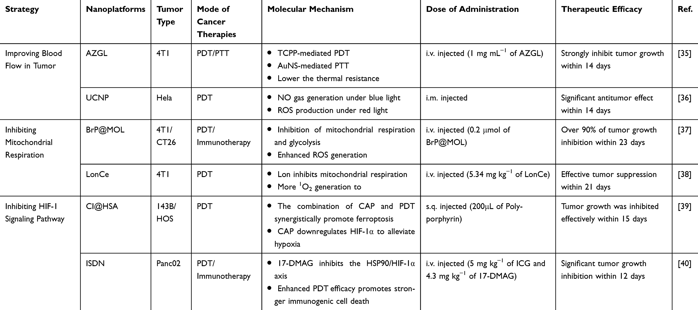 |
Table 1 Representative Paradigms for Improving Blood Flow in Tumor, Inhibiting Mitochondrial Respiration and Inhibiting HIF-1 Signaling Pathway to Relieve Tumor Hypoxia |
Improving Blood Flow in Tumor
Compared to normal vasculature, tumor blood vessels exhibit distinct structural abnormalities, including dilation, tortuosity, and disarray.41–43 These aberrant vessels, along with dysfunctional lymphatic vessels, exacerbate tumor hypoxia, further promoting cancer progression, metastasis, and drug resistance.44–46 Therefore, improving tumor blood flow represents a promising strategy to alleviate hypoxia for enhancing PDT treatment.
Mild hyperthermia (40–42°C) can locally raise tumor temperature, thereby enhancing blood flow and perfusion to lighten tumor hypoxia.47,48 Coincidentally, photothermal therapy (PTT), a revolutionary phototherapy technique, absorbs specific light wavelengths and converts them into thermal energy, inducing tumor cell death through cellular protein denaturation and membrane disruption.20,48–52 Hence, the synergistic therapy of PTT and PDT not only reduces tumor hypoxia by boosting blood flow efficiently but also helps to precisely eliminate malignant tumors.20
Li et al constructed pH-responsive gold nanosystem (AuNS@ZrTCPP-GA@LP) for enhanced therapy of breast cancer by improving blood perfusion, which consisted of gold nanostars (AuNS), Zr4+, tetrakis(4-carboxyphenyl)porphyrin (TCPP), and gambogic acid (GA) (Figure 1A).35 After NIR light irradiation, the mild temperature increase caused by AuNS boosted blood flow to alleviate tumor hypoxia, and thereby efficiently enhancing the therapeutic efficacy of TCPP-mediated PDT.
 |
Figure 1 (A) Schematic illustration of the mechanism of AZGL killed tumor cells by synergy therapy of PTT and PDT. Reproduced with permission.35 (B) Schematic illustration of self-released NO nanotractor to generate ROS under 980 and 808nm light laser. Reproduced with permission.36 Copyright 2024, John Wiley and Sons. |
Furthermore, several agents can also normalize abnormal tumor vasculature to improve blood perfusion to ease tumor hypoxia, including antiangiogenic drugs (eg, sunitinib,53 dexamethasone54 and lenvatinib55) and therapeutic gases (eg, NO56,57). Recently, Zhu et al fabricated an NIR-excitable nitric oxide (NO) nanotractor to improve intratumoral penetration and overcome hypoxic resistance (Figure 1B). 36 SiO2 shell loaded with NO, fuel molecules (RBS), and PDT photosensitizers (ZnPc). UCNP functioned as a light transducer under 980 nm light irradiation, converting NIR light to visible light, which excites the RBS and releases NO. NO not only strengthens intratumoral penetration but also suppresses cellular respiration to reduce hypoxia.
Improving blood flow to increase O2 levels is a potential strategy to enhance nanotechnology-based PDT treatment. However, the precise targeting and penetration of nanodrugs into hypoxic solid tumors remains a major challenge in oncology drug delivery.
Inhibiting Mitochondrial Respiration
Delivering exogenous and generating O2 are typical strategies to ease hypoxia. However, due to the increased tumor proliferation and lack of endogenous H2O2, the above methods can only temporarily improve hypoxia.33,58 The primary functions of O2 consumption include tumor cell respiration and proliferation. Recently, several studies have shown that utilizing the O2 consumption inhibitors to inhibit mitochondrial respiration can reduce O2 consumption rate.59–63
Lin et al constructed a novel 2D nano-photosensitizer (BrP@MOL) that could significantly reduce endogenous O2 consumption by tumor cells to enhance PDT efficacy (Figure 2A). 37 The nano-photosensitizer was fabricated by 3-Bromopyruvate (BrP) and metal-organic layer (MOL). By suppressing both mitochondrial respiration and glycolysis, BrP@MOL induced tumor oxygenation and reduced lactate production. This, in turn, effectively enhanced PDT efficacy and activated the immune response. The significant inhibition of mitochondrial respiration by BrP@MOL improved the antitumor effects of PDT, thereby preventing lung metastasis in breast cancer.
 |
Figure 2 (A) Schematic representation of the designed nano-photosensitizer (BrP@MOL) for enhancing PDT by inhibiting mitochondrial respiration. Reproduced with permission.37 Copyright 2024, Wiley-VCH GmbH. (B) Schematic illustration of the mechanism of LonCe for enhancing PDT by inhibiting mitochondrial respiration. Reproduced with permission.38 Copyright 2022, 2022 Wiley-VCH GmbH. |
For instance, Cheng et al created a carrier-free O2-economizer (LonCe) that exhibited reduced systemic toxicity and immunogenicity in vivo (Figure 2B).38 The antitumor drug Lonidamine (Lon) and the photosensitizer chlorin e6 (Ce6) were used to build the nanoparticle through intermolecular interactions. When LonCe entered tumor cells, a large number of toxic 1O2 was produced to kill tumor cells under irradiation. Subsequently, the releasing Lon could interrupt cell respiration by inhibiting mitochondria complex II s to reduce O2 consumption for enhanced PDT therapy.
The inhibition of mitochondrial respiration can reduce O2 consumption at the source, thus enhancing PDT antitumor efficacy. Nevertheless, the oxygen-sparing effect of this approach is temporally limited (only during active intervention) and is quantitatively insufficient to meet the therapeutic requirements of PDT.
Inhibiting HIF-1 Signaling Pathway
Hypoxia-inducible factor 1α (HIF-1α) is the key regulator of hypoxia.64 HIF-1α can be rapidly degraded under normoxia, whereas under hypoxic conditions, HIF-1α is activated in tumor cells, driving tumor metastasis and progression in a HIF-dependent hypoxia manner.65–67 Therefore, inhibiting HIF-1 signaling pathway to alleviate tumor hypoxia displays a potential strategy for enhancing PDT efficacy.68 Several studies have shown that some natural or artificial agents can selectively inhibit HIF-1α, such as acriflavine, topotecan and capsaicin.39,69 Lv et al created a multifunctional nanomedicine (CAP@IR780@HSA) for increasing PDT efficacy, which was consisted of capsaicin (CAP), photosensitizer IR780 and serum albumin (HSA) (Figure 3A).39 Under NIR irradiation, PSs IR780 could generate ROS to eliminate tumor. Moreover, CAP not only could induce ferroptosis but also suppress the HIF-1α to mitigate tumor hypoxia, thereby mitigating tumor hypoxia and enhancing the antitumor efficiency of PDT.
 |
Figure 3 (A) The preparation of Cl@ HSA NPs and its mechanism for enhanced PDT efficacy. Reproduced with permission.39 Copyright 2024, John Wiley and Sons. (B) The synthesis procedure preparation process of ISDN and its mechanism for improved PDT. Reproduced with permission.40 Copyright 2024, John Wiley and Sons. |
The most representative molecular chaperone is the heat shock protein (HSP).70 Several studies have shown that HSP90 can bind to HIF-1, thereby enhancing its stability.58,71–73 Therefore, to successfully combat hypoxia, it is crucial to focus on HIF itself, as well as the associated proteins. For instance, Deng et al fabricated a self-assembled nanoparticle (ISDN) to downregulate HIF-1α, which contained indocyanine green (ICG) and 17-dimethylaminoethylamino-17demethoxygeldanamycin (17-DMAG).40 Owing to the hydrophobic interactions between ICG and 17-DMAG, ICG could generate more ROS for the treatment of pancreatic cancer. In addition, 17-DMAG was capable of restraining HSP90/HIF-1α axis to ease tumor hypoxia, thereby improving PDT efficacy (Figure 3B).
HIF-independent hypoxia response is another signaling pathway that tumor cells sense to hypoxia. When HIF is restrained, tumor cells survive in a number of signaling pathways: they sense hypoxia directly and adapt rapidly to changing conditions.74–77 Lately, studies on HIF-independent hypoxia have centered chiefly on domains such as ferroptosis, while it has scarcely been investigated in relation to PDT.78,79 In brief, HIF‑dependent and HIF‑independent hypoxia together form the “hypoxic adaptation network” of tumors. Therefore, the design of novel PDT nanomedicines should aim to simultaneously target both HIF‑dependent pathways and HIF‑independent mechanisms, thereby more comprehensively overcoming tumor hypoxia and improving therapeutic outcomes.
Circumventing Intratumoral Hypoxia
Currently, O2-independent therapeutic strategies represent a promising approach to conquer tumor hypoxia by generating cytotoxic ROS without extra O2 to eliminate the tumor cells.19,80–82 Therefore, strategies to circumvent hypoxia may be effective solutions for cancer treatment. Hypoxia circumvention strategies mainly include (I) Type-I PDT, (II) generating O2-unrelated free radicals, and (III) fractional laser PDT (Table 2).
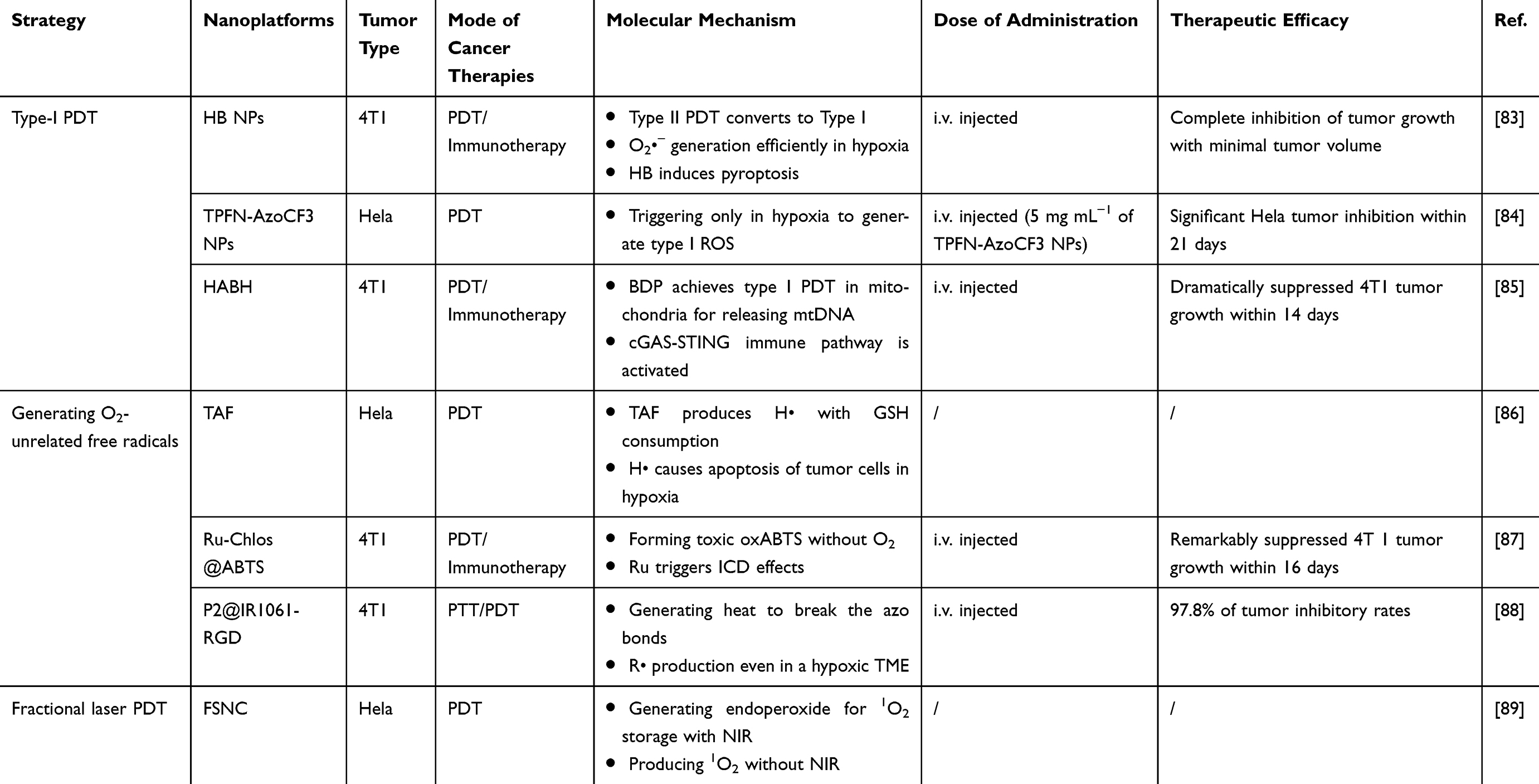 |
Table 2 Representative Paradigms of Type-I PDT, O2-Unrelated Free Radicals Generation and Fractional Laser PDT to Circumvent Hypoxia |
Type-I PDT
The aforementioned strategies to overcome hypoxia are mostly focused on increasing O2 concentration via various methods. However, traditional Type-II PDT has limited therapeutic efficacy because of inherent tumor hypoxia and constant consumption of oxygen.90,91 Conversely, Type-I PDT has attracted a lot of interest in recent years and has shown great superiority in hypoxic tumors, as it is far less or no dependent on O2.92,93
The primary significance of the Type I mechanism is its relative oxygen independence, since the initial reaction does not strictly require the involvement of O2 (Figure 4A).93–96 In addition, Type-I PDT can utilize superoxide dismutase (SOD) to drive the disproportionation reaction (Equation (1)), Haber-Weiss reaction (Equation (2)) or Fenton reaction (Equation (3)) to create O2, which further decreases the dependence on O2.96–99
 |
Figure 4 (A) Illustration showing how the Type-I and Type-II PDTs work. Reproduced with permission.96 Copyright 2021, Wiley-VCH. (B) Schematic illustration of the tumor inhibition of HB NPs for efficient Type I PDT.83 Reproduced with permission. Copyright 2025, Wiley-VCH GmbH. (C) Schematic illustration of antitumor mechanism of TPFN-AzoCF3 NPs for overcoming hypoxia. Reproduced with permission.84 Copyright 2023, Wiley-VCH GmbH. (D) Schematic depiction of HABH nanoagonist for mitochondrion-targeted Type I PDT. Reproduced with permission.85 Copyright 2025, Wiley-VCH GmbH. |


Recently, Huang et al constructed an applicable pyroptosis nanoinducer (HB NPs) composed of uniform carbon nanodots (HNCDs) and bovine serum albumin (BSA) (Figure 4B).83 HNCDs could convert the Type II PDT process to Type I to disregard hypoxia. The result of the experiment proved that HB NPs could generate excessive O2•− to kill tumor cells even under an extremely hypoxic environment (2% O2).
Liu’s group synthesized on-demand switchable nanoparticles (TPFN-AzoCF3 NPs) to achieve precision treatment and reduce side effects (Figure 4C).84 TPFN-AzoCF3 NPs were composed of hypoxia-normoxia cycling-responsive group (AzoCF3), Type I PS (TPFN), and carrier liposomes (DSPE-PEG-2000). In hypoxic tumors, TPFN-AzoCF3 NPs could be activated to produce ROS to suppress tumor growth, whereas in normal tissues, they remained completely non-toxic, which not only reduces potential side effects but also overcomes tumor hypoxia.
Dong et al developed a mitochondrion-targeted nanoagonist (HABH) composed of Type I photosensitizer (BDP) and Au nanoparticles for achieving light-controlled PDT therapy (Figure 4D).85 HABH nanoagonist could effectively target the tumor site, while the released BDP triggered Type I PDT under laser irradiation to generate vast •OH to kill tumor cells. Therefore, HABH nanoagonist showed excellent phototoxicity under normoxic conditions owing to its high potential to surmount the hypoxic constraints of traditional PDT.
Type-I PDT nanotechnology-based methods can inhibit tumors, while neglecting tumor hypoxia to a certain extent. However, stable quantitative methods for evaluating the generation of Type-I ROS are lacking, leading to difficulties in developing novel Type-I PDT agents.93
Generating O2-Unrelated Free Radicals
The strategy for generating O2-unrelated free radicals refers to the generation of other cellular toxic free radicals (eg, H•, •Cl, and R•) to inhibit tumor cells without relying on O2. Beyond Type-I PDT, this approach circumvents the limitations of tumor hypoxia.
Feng’s group constructed a light-responsive organic photosensitizer (TAF) for enhanced PDT (Figure 5A).86 In this work, TAF could efficiently generate vast H• because of its photocatalytic activity. H• exhibited dual phototherapeutic properties, enabling both photooxidative and reductive therapy. It could react with O2 to facilitate ROS generation in the normoxic environment while triggering biological reductions in hypoxic environments for tumor cells apoptosis, which had the potential to conquer hypoxia in the future.
 |
Figure 5 (A) Schematic illustration of the mechanism of H• photogeneration and it’s function in normoxia and hypoxia environment. Reproduced with permission.86 Copyright 2022, Wiley- VCH. (B) Schematic illustration of the mechanism of Ru-Chlos nanoreactors for efficient photoimmunotherapy. Reproduced with permission.87 Copyright 2024, Wiley-VCH GmbH. (C) The synthesis of P2@IR1061-RGD nanosystem and the generation of R• under 1064 nm laser to enhance PDT. Reproduced with permission.88 Copyright 2021, John Wiley and Sons. (D) Schematic illustration of the mechanism of generation and storage for fractional PDT. Reproduced with permission.89 Copyright 2019, American Chemical Society. |
Recently, Sun et al designed a chlorosome-mimetic nanoreactor (Ru-Chlos@ABTS) consisting of ruthenium (Ru), chlorosomes, and the prodrug ABTS for efficient cancer photoimmunotherapy (Figure 5B).87 Upon red light irradiation, Ru-Chlos could catalyze ABTS to produce cytotoxic free radicals (oxABTS) to achieve phototherapy without consuming any O2 and H2O2. Ru-Chlos@ABTS showed significant tumor inhibition and elimination in vivo, indicating that these biomimetic chlorosomes possessed outstanding O2-independent advantages.
Carbon radicals are not only highly reactive but also can be produced without O2.100 Liu et al fabricated a highly tumor targeting nanoplatform (P2@IR1061-RGD), loading with azo-containing polymer (P2), photothermal dye IR1061 and arginine-glycine-aspartic acid tripeptide (RGD).88 Under 1064 nm NIR II laser irradiation, P2@IR1061-RGD was able to degrade and produce photothermal effects to accelerate cytotoxic carbon radical (R•) generation, which disrupted RNA transcription for tumor cells death accurately (Figure 5C).
Generating O2-unrelated free radicals may not only ignore tumor hypoxia but may also display prominent therapeutic efficacy; however, the low heat transfer efficiency and limited availability of photothermal agents restrict their treatment.
Fractional Laser PDT
Fractional laser PDT possesses the ability to continuously release 1O2 compared to traditional PDT and can largely circumvent hypoxia to a certain extent compared to Type I PDT.
For instance, Song et al developed a functional silica nanocarrier (FSNC) composed of protoporphyrin IX derivative, 2-Pyridone derivative, cyanine derivative, and nano-silica carriers for fractional PDT (Figure 5D).89 After entering tumor cells, protoporphyrin IX derivative produced 1O2 to kill tumor cells and formed an endoperoxide to enable 1O2 storage upon light irradiation. The stored 1O2 could then be released in the absence of light, enabling highly continuous PDT treatment.
Harnessing Hypoxia for Enhanced PDT
Overcoming hypoxia is challenging due to the complex TME and disrupted vasculature. Although existing strategies have shown partial success, PDT consumes oxygen continuously to aggravate hypoxic tumors; thus, it is important to develop more effective strategies to enhance PDT efficacy. Recently, some studies have explored the use of hypoxia for PDT in hypoxic tumors. Hypoxia utilization strategies include (I) hypoxia-activated prodrugs, (II) hypoxia-responsive nanoplatforms, and (III) hypoxic cell sensitizers (Table 3).
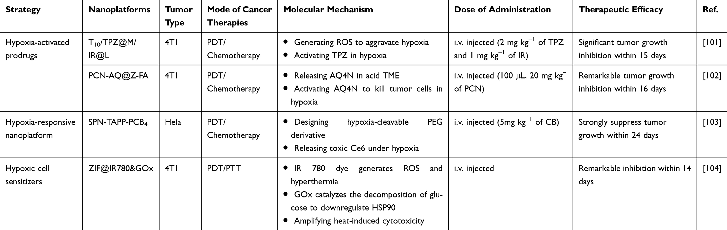 |
Table 3 Representative Paradigms of Hypoxia-Activated Prodrugs, Hypoxia-Responsive Nanoplatform and Hypoxic Cell Sensitizers to Utilize Hypoxia |
Hypoxia-Activated Prodrugs
There is a significant difference in oxygen content between normal and tumor tissues, which can be exploited for tumor-targeted therapy. Hypoxia-activated prodrugs (HAPs) exhibit high cytotoxicity in hypoxic solid tumors but show completely low cytotoxicity in normal tissues.105 Recently, hypoxia-activated prodrugs, such as banoxantrone, tirazamine (TPZ), and AQ4N, have become a hot topic of research.106–108
Huang et al developed a multifunctional lipid-coated nanomedicine (T10/TPZ@M/IR@L) to achieve synergetic therapy of PDT and hypoxia activated chemotherapy (Figure 6A).101 Under light irradiation, the released photosensitizer IR could produce ROS to further aggravate tumor hypoxia. Meanwhile, TPZ was activated by hypoxia and then possessed more cytotoxicity, leading to enhanced tumor-cell killing.
 |
Figure 6 (A) Schematic illustration of the T10/TPZ@M/IR@L mechanism for synergy therapy of PDT and hypoxia activated chemotherapy. Reproduced with permission.101 Copyright 2025, American Chemical Society. (B) Schematic illustration of mechanism of PCN-AQ@Z-FA via a combination of PDT and hypoxia-activated chemotherapy. Reproduced with permission.102 (C) Schematic illustration showing how the nanomedicine triggered hypoxia-responsive for PDT and chemotherapy treatment. Reproduced with permission.103 Copyright 2023, Wiley-VCH GmbH. (D) The preparation and schematic depiction of ZIF@IR780&GOx to suppress tumor for PDT. Reproduced with permission.104 Copyright 2024, American Chemical Society. |
For instance, Chu et al designed a novel hypoxia-activated nano-prodrug (PCN-AQ@Z-FA) to achieve synergy therapy of chemotherapy and PDT (Figure 6B).102 In this system, a zeolitic imidazolate frameworks (ZIF-8) and AQ4N were encapsulated in the mesopores of metal-organic framework (MOFs). The PCN core, as a photosensitizer, could generate ROS to induce DNA damage. In acid TME, ZIF-8 could decompose and release AQ4N in a controlled manner. Once AQ4N was activated to cytotoxic AQ4, it precisely would inhibit the tumor for chemotherapy.
Hypoxia-Responsive Nanoplatform
Except hypoxia-activated prodrugs, nanoplatforms involving hypoxia-sensitive structures can similarly increase the antitumor effect of nanomedicine. Hypoxia-responsive nanoplatforms are engineered with structures like nitroimidazole, azobenzene, or N-oxide that, upon reaching TME, undergo rapid cleavage to release the encapsulated antitumor drugs.103,109–112
Yi et al fabricated a hypoxia-responsive tetrameric supramolecular polypeptide nanomedicine (SPN-TAPP-PCB4) to achieve the combination therapy of PDT and chemotherapy.103 Upon NIR light irradiation, the 5,10,5,20-tetrakis(4-aminophenyl)-porphine (TAPP) central would generate 1O2 (Figure 6C). In acid TME condition, SPN-TAPP-PCB4 was rapidly dissociated. Under hypoxic TME, the disassembly of azobenzene could lead to chlorambucil (CB) release to kill tumor cells, thus achieving a precise and strong antitumor effect.
Although hypoxia-activated prodrugs and hypoxia-responsive nanoplatforms can exploit the hypoxic microenvironment to overcome tumor hypoxia for enhanced PDT antitumor efficacy, their therapeutic impact on deep-seated tumors remains unsatisfactory. Furthermore, the complexity and heterogeneity of the TME, along with non-uniform hypoxia levels, pose challenges in ensuring the consistent and effective activation of nanodrugs.
Hypoxic Cell Sensitizers
Hypoxic cell sensitizers may enhance lethality by increasing the sensitivity of tumor cells. Yan et al created an innovative nanoparticle (ZIF@IR780&GOx) loaded with IR780 dye and GOx, which led to the synergistic therapy of PDT and PTT (Figure 6D).104 IR780 dye not only promoted ROS generation but also improved hyperthermia efficacy and accelerated tumor cell death. Meanwhile, GOx could downregulate the expression of heat shock protein 90 (HSP90), thereby sensitizing hypoxic cells.
Conclusion
PDT has emerged as a highly promising non-invasive antitumor modality with distinct advantages over conventional treatments, including high specificity, minimal systemic toxicity, and reduced risk of drug resistance. However, its therapeutic efficacy is severely constrained by tumor hypoxia, which is a core characteristic of TME. Although conventional approaches such as oxygen delivery and in situ oxygen generation provide certain benefits, their passive and uncontrolled nature restricts their therapeutic potential. Against this backdrop, this review emphasizes innovative nanotechnology-based strategies designed to proactively address tumor hypoxia, moving beyond the limitations of traditional oxygen replenishment techniques. Specifically, we explore three principal, mutually complementary avenues: (1) remodeling the hypoxic tumor microenvironment (eg, through increased blood flow, inhibition of mitochondrial respiration, and suppression of the HIF-1α signaling pathway); (2) circumventing intratumoral hypoxia (eg, via Type-I PDT, oxygen-independent free radical generation, and fractional laser PDT); and (3) harnessing hypoxia as a trigger to enhance PDT efficacy (eg, with hypoxia-activated prodrugs, hypoxia-responsive nanoplatforms, and hypoxic cell sensitizers).
Although nanotechnology-driven strategies have significantly alleviated tumor hypoxia and enhanced PDT efficacy, several critical challenges that constrain their clinical translation remain unresolved. The first and foremost aspect is the long-term biosafety of nanomaterials. Given the complex composition of many nanoplatforms, their inherent toxicity and potential off-target side effects demand systematic and rigorous investigation prior to the initiation of clinical trials, particularly to clarify their long-term biological behaviors (eg, in vivo metabolism, tissue accumulation, and clearance). Thus, more and more scientists have acknowledged the critical importance of biosafety. To this end, they have established degradable green nanoparticle synthesis routes and developed a comprehensive risk assessment framework covering the entire product life cycle.113–116 Second, the scalable manufacturing of complex nanoplatforms presents a major hurdle. Addressing this issue requires the integration of advanced nanofabrication technologies, optimized processing protocols, and robust quality control systems, all of which are essential to ensure the consistent production of multifunctional nanostructures that meet clinical standards at an industrial scale.117 Finally, there is interpatient heterogeneity of tumor hypoxia. This variability often results in suboptimal treatment outcomes and may contribute to tumor recurrence and metastasis. To address this challenge, comprehensive and long-term investigations into the effects of hypoxic heterogeneity on treatment outcomes are imperative. Additionally, the formulation of personalized approaches that customize nanotechnology-based PDT according to individual hypoxic characteristics warrants further in-depth exploration.118,119
In summary, this review challenges conventional passive oxygen supplementation strategies by proposing a beyond oxygenation framework that involves remodeling, circumventing and harnessing hypoxia. Based on this, we take a forward-looking perspective on the active hypoxia conquest by nanotechnology, outlining actionable pathways towards PDT enhancement for next-generation cancer treatment. Furthermore, it is widely recognized that addressing the aforementioned critical challenges will serve as a pivotal breakthrough. Such progress holds great promise for enabling precise hypoxia-resistant PDT against hypoxic tumors, thereby advancing the clinical utility of this noninvasive therapeutic modality.
Acknowledgments
The graphical abstract was created with BioGDP.com.120
Funding
This work was supported by the Health Research Project of the Hunan Provincial Health Commission (Grant No. W20243039), Hunan Provincial Natural Science Foundation of China (Grant No. 2025JJ81090), and NSFC of China (Grant No. 82503028).
Disclosure
The authors declare no competing interests in this work.
References
1. Siegel RL, Miller KD, Wagle NS, Jemal A. Cancer statistics, 2023. CA Cancer J Clin. 2023;73(1):17–18. doi:10.3322/caac.21763
2. Weller M, Remon J, Rieken S, et al. Central nervous system metastases in advanced non-small cell lung cancer: a review of the therapeutic landscape. Cancer Treat Rev. 2024;130:102807. doi:10.1016/j.ctrv.2024.102807
3. Kagawa Y, Smith JJ, Fokas E, et al. Future direction of total neoadjuvant therapy for locally advanced rectal cancer. Nat Rev Gastroenterol Hepatol. 2024;21(6):444–455. doi:10.1038/s41575-024-00900-9
4. Van Der Wilk BJ, Eyck BM, Wijnhoven BPL, et al. Neoadjuvant chemoradiotherapy followed by active surveillance versus standard surgery for oesophageal cancer (SANO trial): a multicentre, stepped-wedge, cluster-randomised, non-inferiority, Phase 3 trial. Lancet Oncol. 2025;26(4):425–436. doi:10.1016/S1470-2045(25)00027-0
5. Leong T, Smithers BM, Michael M, et al. Preoperative chemoradiotherapy for resectable gastric cancer. N Engl J Med. 2024;391(19):1810–1821. doi:10.1056/NEJMoa2405195
6. Tsai CJ, Yang JT, Shaverdian N, et al. Standard-of-care systemic therapy with or without stereotactic body radiotherapy in patients with oligoprogressive breast cancer or non-small-cell lung cancer (consolidative use of radiotherapy to block [CURB] oligoprogression): an open-label, randomised, controlled, Phase 2 study. Lancet. 2024;403(10422):171–182. doi:10.1016/S0140-6736(23)01857-3
7. Van Gorp T, Cibula D, Lv W, et al. ENGOT-en11/GOG-3053/KEYNOTE-B21: a randomised, double-blind, Phase III study of pembrolizumab or placebo plus adjuvant chemotherapy with or without radiotherapy in patients with newly diagnosed, high-risk endometrial cancer. Ann Oncol. 2024;35(11):968–980. doi:10.1016/j.annonc.2024.08.2242
8. Giannakeas V, Lim DW, Narod SA. Bilateral mastectomy and breast cancer mortality. JAMA Oncol. 2024;10(9):1228. doi:10.1001/jamaoncol.2024.2212
9. Park HS, Rimner A, Amini A, et al. Appropriate use criteria (AUC) for the management of non-small cell lung cancer in a central/ultra-central location: guidelines from the American Radium Society. J Thorac Oncol. 2024;19(12):1640–1653. doi:10.1016/j.jtho.2024.09.1386
10. Adams SC, Nambiar AK, Bressler EM, et al. Immunotherapies for locally aggressive cancers. Adv Drug Delivery Rev. 2024;210:115331. doi:10.1016/j.addr.2024.115331
11. Wang K, Tepper JE. Radiation therapy‐associated toxicity: etiology, management, and prevention. Ca Cancer J Clin. 2021;71(5):437–454. doi:10.3322/caac.21689
12. Liu YQ, Wang XL, He DH, Cheng YX. Protection against chemotherapy- and radiotherapy-induced side effects: a review based on the mechanisms and therapeutic opportunities of phytochemicals. Phytomedicine. 2021;80:153402. doi:10.1016/j.phymed.2020.153402
13. De Visser KE, Joyce JA. The evolving tumor microenvironment: from cancer initiation to metastatic outgrowth. Cancer Cell. 2023;41(3):374–403. doi:10.1016/j.ccell.2023.02.016
14. Zhang C, Hu X, Jin L, et al. Strategic design of conquering hypoxia in tumor for advanced photodynamic therapy. Adv Healthcare Mat. 2023;12(24):2300530. doi:10.1002/adhm.202300530
15. Liu X, Lu Y, Li X, Luo L, You J. Nanoplatform-enhanced photodynamic therapy for the induction of immunogenic cell death. J Control Release. 2024;365:1058–1073. doi:10.1016/j.jconrel.2023.11.058
16. Zhao W, Wang L, Zhang M, et al. Photodynamic therapy for cancer: mechanisms, photosensitizers, nanocarriers, and clinical studies. MedComm. 2024;5(7):e603. doi:10.1002/mco2.603
17. Kolarikova M, Hosikova B, Dilenko H, et al. Photodynamic therapy: innovative approaches for antibacterial and anticancer treatments. Med Res Rev. 2023;43(4):717–774. doi:10.1002/med.21935
18. Wang Y, Staudinger JN, Mindt TL, Gasser G. Theranostics with photodynamic therapy for personalized medicine: to see and to treat. Theranostics. 2023;13(15):5501–5544. doi:10.7150/thno.87363
19. Jiang J, Lv X, Cheng H, et al. Type I photodynamic antimicrobial therapy: principles, progress, and future perspectives. Acta Biomater. 2024;177:1–19. doi:10.1016/j.actbio.2024.02.005
20. Overchuk M, Weersink RA, Wilson BC, Zheng G. Photodynamic and photothermal therapies: synergy opportunities for nanomedicine. ACS Nano. 2023;17(9):7979–8003. doi:10.1021/acsnano.3c00891
21. Liao C, Liu X, Zhang C, Zhang Q. Tumor hypoxia: from basic knowledge to therapeutic implications. Semin Cancer Biol. 2023;88:172–186. doi:10.1016/j.semcancer.2022.12.011
22. Beckers C, Pruschy M, Vetrugno I. Tumor hypoxia and radiotherapy: a major driver of resistance even for novel radiotherapy modalities. Semin Cancer Biol. 2024;98:19–30. doi:10.1016/j.semcancer.2023.11.006
23. Telarovic I, Wenger RH, Pruschy M. Interfering with tumor hypoxia for radiotherapy optimization. J Exp Clin Cancer Res. 2021;40(1):197. doi:10.1186/s13046-021-02000-x
24. Xu XX. Research progress on tumor hypoxia-associative nanomedicine. J Control Release. 2022. doi:10.1016/j.jconrel.2022.09.003
25. Zhang H, Cao K, Xiang J, Zhang M, Zhu M, Xi Q. Hypoxia induces immunosuppression, metastasis and drug resistance in pancreatic cancers. Cancer Lett. 2023;571:216345. doi:10.1016/j.canlet.2023.216345
26. Shang T, Jia Z, Li J, et al. Unraveling the triad of hypoxia, cancer cell stemness, and drug resistance. J Hematol Oncol. 2025;18(1):32. doi:10.1186/s13045-025-01684-4
27. Rey-Keim S, Schito L. Origins and molecular effects of hypoxia in cancer. Semin Cancer Biol. 2024;106-107:166–178. doi:10.1016/j.semcancer.2024.10.001
28. Li B, Niu H, Zhao X, et al. Targeted anti-cancer therapy: co-delivery of VEGF siRNA and phenethyl isothiocyanate (PEITC) via cRGD-modified lipid nanoparticles for enhanced anti-angiogenic efficacy. Asian J Pharm Sci. 2024;19(2):100891. doi:10.1016/j.ajps.2024.100891
29. Yao X, Lu S, Feng C, et al. Tumor oxygenation nanoliposome synergistic hypoxia-inducible-factor-1 inhibitor enhanced iodine-125 seed brachytherapy for esophageal cancer. Biomaterials. 2022;289:121801. doi:10.1016/j.biomaterials.2022.121801
30. Harris B, Saleem S, Cook N, Searle E. Targeting hypoxia in solid and haematological malignancies. J Exp Clin Cancer Res. 2022;41(1):318. doi:10.1186/s13046-022-02522-y
31. Giordo R, Wehbe Z, Paliogiannis P, Eid AH, Mangoni AA, Pintus G. Nano-targeting vascular remodeling in cancer: recent developments and future directions. Semi Cancer Biol. 2022;86:784–804. doi:10.1016/j.semcancer.2022.03.001
32. Lai C, Luo B, Shen J, Shao J. Biomedical engineered nanomaterials to alleviate tumor hypoxia for enhanced photodynamic therapy. Pharmacol Res. 2022;186:106551. doi:10.1016/j.phrs.2022.106551
33. Wan Y, Fu L, Li C, Lin J, Huang P. Conquering the hypoxia limitation for photodynamic therapy. Adv Mater. 2021;33(48):2103978. doi:10.1002/adma.202103978
34. Yang F, Chen F, Li S, et al. Emerging tumor hypoxia-relieving nanotherapeutics for boosting photodynamic therapy. Chem Eng J. 2025;516:163962. doi:10.1016/j.cej.2025.163962
35. Li RT, Zhu YD, Li WY, et al. Synergistic photothermal-photodynamic-chemotherapy toward breast cancer based on a liposome-coated core–shell AuNS@NMOFs nanocomposite encapsulated with gambogic acid. J Nanobiotechnol. 2022;20(1):212. doi:10.1186/s12951-022-01427-4
36. Wang J, Zhang Y, Chen H, et al. Motor‐cargo structured nanotractors for augmented NIR phototherapy via gas‐boosted tumor penetration and respiration‐impaired mitochondrial dysfunction. Adv Healthcare Mat. 2024;13(30):2402063. doi:10.1002/adhm.202402063
37. Lin G, Tillman L, Luo T, et al. Nanoscale metal‐organic layer reprograms cellular metabolism to enhance photodynamic therapy and antitumor immunity. Angew Chem Int Ed. 2024;63(37):e202410241. doi:10.1002/anie.202410241
38. Huang J, Zhao L, Zhou X, et al. Carrier free O2 ‐economizer for photodynamic therapy against hypoxic tumor by inhibiting cell respiration. Small. 2022;18(15):2107467. doi:10.1002/smll.202107467
39. Wang Y, Zhou X, Yao L, et al. Capsaicin enhanced the efficacy of photodynamic therapy against osteosarcoma via a pro‐death strategy by inducing ferroptosis and alleviating hypoxia. Small. 2024;20(26):2306916. doi:10.1002/smll.202306916
40. Yang J, Ren B, Yin X, et al. Expanded ROS generation and hypoxia reversal: excipient‐free self‐assembled nanotheranostics for enhanced cancer photodynamic immunotherapy. Adv Mater. 2024;36(30):2402720. doi:10.1002/adma.202402720
41. Liu ZL, Chen HH, Zheng LL, Sun LP, Shi L. Angiogenic signaling pathways and anti-angiogenic therapy for cancer. Sig Transduct Target Ther. 2023;8(1):198. doi:10.1038/s41392-023-01460-1
42. Lanitis E, Irving M, Coukos G. Tumour-associated vasculature in T cell homing and immunity: opportunities for cancer therapy. Nat Rev Immunol. 2025;25(11):831–846. doi:10.1038/s41577-025-01187-w
43. Castaneda M, Den Hollander P, Kuburich NA, Rosen JM, Mani SA. Mechanisms of cancer metastasis. Semi Cancer Biol. 2022;87:17–31. doi:10.1016/j.semcancer.2022.10.006
44. Wei X, Chen Y, Jiang X, et al. Mechanisms of vasculogenic mimicry in hypoxic tumor microenvironments. Mol Cancer. 2021;20(1):7. doi:10.1186/s12943-020-01288-1
45. Dudley AC, Griffioen AW. Pathological angiogenesis: mechanisms and therapeutic strategies. Angiogenesis. 2023;26(3):313–347. doi:10.1007/s10456-023-09876-7
46. Mpekris F, Panagi M, Charalambous A, Voutouri C, Stylianopoulos T. Modulating cancer mechanopathology to restore vascular function and enhance immunotherapy. Cell Rep Med. 2024;5(7):101626. doi:10.1016/j.xcrm.2024.101626
47. González-Rodríguez L, Pérez-Davila S, López-álvarez M, Chiussi S, Serra J, González P. Review article laser-induced hyperthermia on graphene oxide composites. J Nanobiotechnol. 2023;21(1):196. doi:10.1186/s12951-023-01956-6
48. Bravo M, Fortuni B, Mulvaney P, et al. Nanoparticle-mediated thermal cancer therapies: strategies to improve clinical translatability. J Control Release. 2024;372:751–777. doi:10.1016/j.jconrel.2024.06.055
49. Xiong Y, Rao Y, Hu J, Luo Z, Chen C. Nanoparticle‐based photothermal therapy for breast cancer noninvasive treatment. Adv Mater. 2025;37(31):2305140. doi:10.1002/adma.202305140
50. Li N, Wang Y, Li Y, Zhang C, Fang G. recent advances in photothermal therapy at near‐infrared‐II based on 2D MXenes. Small. 2024;20(6):2305645. doi:10.1002/smll.202305645
51. Liu K, Yao Y, Xue S, et al. Recent advances of tumor microenvironment-responsive nanomedicines-energized combined phototherapy of cancers. Pharmaceutics. 2023;15(10):2480. doi:10.3390/pharmaceutics15102480
52. Alamdari SG, Amini M, Jalilzadeh N, et al. Recent advances in nanoparticle-based photothermal therapy for breast cancer. J Control Release. 2022;349:269–303. doi:10.1016/j.jconrel.2022.06.050
53. Tullemans BME, Fernández DI, Veninga A, et al. Tyrosine kinase inhibitor sunitinib delays platelet-induced coagulation: additive effects of aspirin. Thromb Haemost. 2022;122(01):092–104. doi:10.1055/s-0041-1730312
54. Li X, Jiang C, Jia X, et al. Dual “unlocking” strategy to overcome inefficient nanomedicine delivery and tumor hypoxia for enhanced photodynamic‐immunotherapy. Adv Healthcare Mat. 2023;12(6):2202467. doi:10.1002/adhm.202202467
55. Huang JX, Yang R, Long H, Kong J, Shao GQ, Xiong F. Dual-drug loaded chondroitin sulfate embolization beads enhance TACE therapy for HCC by integrating embolization, chemotherapy, and anti-angiogenesis. Mater Today Bio. 2025;30:101419. doi:10.1016/j.mtbio.2024.101419
56. Yang C, Mu G, Zhang Y, et al. Supramolecular nitric oxide depot for hypoxic tumor vessel normalization and radiosensitization. Adv Mater. 2022;34(37):2202625. doi:10.1002/adma.202202625
57. Wang W, Gao Y, Xu J, et al. A NRF2 regulated and the immunosuppressive microenvironment reversed nanoplatform for cholangiocarcinoma photodynamic‐gas therapy. Adv Sci. 2024;11(14):2307143. doi:10.1002/advs.202307143
58. Chen Z, Han F, Du Y, Shi H, Zhou W. Hypoxic microenvironment in cancer: molecular mechanisms and therapeutic interventions. Sig Transduct Target Ther. 2023;8(1):70. doi:10.1038/s41392-023-01332-8
59. Bai X, Lin Y, Gong L, et al. Nanoparticles that target the mitochondria of tumor cells to restore oxygen supply for photodynamic therapy: design and preclinical validation against breast cancer. J Control Release. 2023;362:356–370. doi:10.1016/j.jconrel.2023.07.064
60. Yang F, Li S, Ji Q, et al. Modular prodrug‐engineered oxygen nano‐tank with outstanding nanoassembly performance, high oxygen loading, and closed‐loop tumor hypoxia relief. Adv Sci. 2024;11(34):2405583. doi:10.1002/advs.202405583
61. Zhang Z, Zhao Q, Xu Q, et al. A mitochondria-interfering nanocomplex cooperates with photodynamic therapy to boost antitumor immunity. Biomaterials. 2025;317:123094. doi:10.1016/j.biomaterials.2025.123094
62. Chen X, Kadier M, Shi M, et al. Targeting melatonin to mitochondria mitigates castration‐resistant prostate cancer by inducing pyroptosis. Small. 2025;21(22):2408996. doi:10.1002/smll.202408996
63. Yang J, Zhang C, Chen X, et al. Ultra-efficient radio-immunotherapy for reprogramming the hypoxic and immunosuppressive tumor microenvironment with durable innate immune memory. Biomaterials. 2023;302:122303. doi:10.1016/j.biomaterials.2023.122303
64. Ceranski AK, Carreño-Gonzalez MJ, Ehlers AC, Colombo MV, Cidre-Aranaz F, Grünewald TGP. Hypoxia and HIFs in Ewing sarcoma: new perspectives on a multi-facetted relationship. Mol Cancer. 2023;22(1):49. doi:10.1186/s12943-023-01750-w
65. Bhattacharya R, Brown JS, Gatenby RA, Ibrahim-Hashim A. A gene for all seasons: the evolutionary consequences of HIF-1 in carcinogenesis, tumor growth and metastasis. Semi Cancer Biol. 2024;102-103:17–24. doi:10.1016/j.semcancer.2024.06.003
66. Dai W, Guo R, Na X, et al. Hypoxia and the endometrium: an indispensable role for HIF-1α as therapeutic strategies. Redox Biol. 2024;73:103205. doi:10.1016/j.redox.2024.103205
67. Yuan X, Ruan W, Bobrow B, Carmeliet P, Eltzschig HK. Targeting hypoxia-inducible factors: therapeutic opportunities and challenges. Nat Rev Drug Discovery. 2024;23(3):175–200. doi:10.1038/s41573-023-00848-6
68. Cowman SJ, Koh MY. Revisiting the HIF switch in the tumor and its immune microenvironment. Trends Cancer. 2022;8(1):28–42. doi:10.1016/j.trecan.2021.10.004
69. Tedeschi BBB, Henrique T, Vila APS, et al. Evaluation of hypoxia-inducible factor-1 and 2 alpha inhibitory compounds in the oral cavity and pharyngeal cancer. Biomed Pharmacother. 2025;186:118024. doi:10.1016/j.biopha.2025.118024
70. Zuo WF, Pang Q, Zhu X, et al. Heat shock proteins as hallmarks of cancer: insights from molecular mechanisms to therapeutic strategies. J Hematol Oncol. 2024;17(1):81. doi:10.1186/s13045-024-01601-1
71. Zhang J, Yao M, Xia S, Zeng F, Liu Q. Systematic and comprehensive insights into HIF-1 stabilization under normoxic conditions: implications for cellular adaptation and therapeutic strategies in cancer. Cell Mol Biol Lett. 2025;30(1):2. doi:10.1186/s11658-024-00682-7
72. Wang P, Zhang XP, Liu F, Wang W. Progressive deactivation of hydroxylases controls hypoxia-inducible factor-1α-coordinated cellular adaptation to graded hypoxia. Research. 2025;8:0651. doi:10.34133/research.0651
73. Hu Y, Li J, Chen H, et al. Autophagy related 5 promotes mitochondrial fission and inflammation via HSP90‐HIF‐1 α ‐mediated glycolysis in kidney fibrosis. Adv Sci. 2025;12(17):2414673. doi:10.1002/advs.202414673
74. Schito L, Rey-Keim S. Hypoxia signaling and metastatic progression. Semin Cancer Biol. 2023;97:42–49. doi:10.1016/j.semcancer.2023.11.001
75. Vozdek R, Long Y, Ma DK. The receptor tyrosine kinase HIR-1 coordinates HIF-independent responses to hypoxia and extracellular matrix injury. Sci Signaling. 2018;11(550):eaat0138. doi:10.1126/scisignal.aat0138
76. Kim SH, Wang D, Park YY, et al. HIG2 promotes colorectal cancer progression via hypoxia-dependent and independent pathways. Cancer Lett. 2013;341(2):159–165. doi:10.1016/j.canlet.2013.07.028
77. Lee DC, Sohn HA, Park ZY, et al. A lactate-induced response to hypoxia. Cell. 2015;161(3):595–609. doi:10.1016/j.cell.2015.03.011
78. Dickson AS, Pauzaite T, Arnaiz E, et al. A HIF independent oxygen-sensitive pathway for controlling cholesterol synthesis. Nat Commun. 2023;14(1):4816. doi:10.1038/s41467-023-40541-1
79. Minikes AM, Liu P, Wang H, et al. HIF-independent oxygen sensing via KDM6A regulates ferroptosis. Mol Cell. 2025;85(15):2973–2987.e6. doi:10.1016/j.molcel.2025.07.001
80. Zhao M, Zhang Y, Miao J, et al. An activatable phototheranostic probe for anti‐hypoxic type I photodynamic‐ and immuno‐therapy of cancer. Adv Mater. 2024;36(4):2305243. doi:10.1002/adma.202305243
81. Yin Y, Ge X, Ouyang J, Na N. Tumor-activated in situ synthesis of single-atom catalysts for O2-independent photodynamic therapy based on water-splitting. Nat Commun. 2024;15(1):2954. doi:10.1038/s41467-024-46987-1
82. Yuan M, Liang S, Yang L, et al. Rational design of platinum–bismuth sulfide Schottky heterostructure for sonocatalysis‐mediated hydrogen therapy. Adv Mater. 2023;35(10):2209589. doi:10.1002/adma.202209589
83. Li W, Li H, Jiang G, et al. Creating single atomic coordination for hypoxia‐resistant pyroptosis nano‐inducer to boost anti‐tumor immunotherapy. Adv Mater. 2025;37(9):2414697. doi:10.1002/adma.202414697
84. Tian J, Li B, Zhang F, et al. Activatable type I photosensitizer with quenched photosensitization pre and post photodynamic therapy. Angew Chem Int Ed. 2023;62(46):e202307288. doi:10.1002/anie.202307288
85. Xu Y, An D, Zhang T, et al. Mitochondrion‐targeted type I photodynamic therapy for agonist independent cGAS‐STING activation. Adv Mater. 2025;37(14):2418894. doi:10.1002/adma.202418894
86. Peng J, Du K, Sun J, et al. Photocatalytic generation of hydrogen radical (H⋅) with GSH for photodynamic therapy. Angew Chem Int Ed. 2023;62(9):e202214991. doi:10.1002/anie.202214991
87. Chen F, Huang H, Zhang F, et al. Biomimetic chlorosomes: oxygen‐independent photocatalytic nanoreactors for efficient combination photoimmunotherapy. Adv Mater. 2024.
88. Liu X, Yang Y, Ling M, et al. Near‐infrared II light‐triggered robust carbon radical generation for combined photothermal and thermodynamic therapy of hypoxic tumors. Adv Funct Mater. 2021;31(24):2101709. doi:10.1002/adfm.202101709
89. Jiao L, Zhang X, Cui J, Peng X, Song F. Three-in-one functional silica nanocarrier with singlet oxygen generation, storage/release, and self-monitoring for enhanced fractional photodynamic therapy. ACS Appl Mater Interfaces. 2019;11(29):25750–25757. doi:10.1021/acsami.9b08371
90. Wei X, Song M, Jiang G, et al. Progress in advanced nanotherapeutics for enhanced photodynamic immunotherapy of tumor. Theranostics. 2022;12(12):5272–5298. doi:10.7150/thno.73566
91. Yu L, Liu Z, Xu W, et al. Towards overcoming obstacles of type II photodynamic therapy: endogenous production of light, photosensitizer, and oxygen. Acta Pharmaceutica Sinica B. 2024;14(3):1111–1131. doi:10.1016/j.apsb.2023.11.007
92. Ma N, Wang J, Tang H, et al. The current advances in design strategy (indirect strategy and direct strategy) for type‐I photosensitizers. Adv Sci. 2025;12(7):2413365. doi:10.1002/advs.202413365
93. Teng K, Niu L, Li J, Zhang D, Yang Q. Rational design of type‐I photodynamic agents. Angew Chem Int Ed. 2025;64(30):e202509416. doi:10.1002/anie.202509416
94. Xiong T, Chen Y, Li M, Chen X, Peng X. Recent progress of molecular design in organic type I photosensitizers. Small. 2025;21(22):2501911. doi:10.1002/smll.202501911
95. Austin E, Wang JY, Ozog DM, Zeitouni N, Lim HW, Jagdeo J. Photodynamic therapy: overview and mechanism of action. J Am Acad Dermatol. 2025;S0190962225003214. doi:10.1016/j.jaad.2025.02.037
96. Chen D, Xu Q, Wang W, Shao J, Huang W, Dong X. Type I photosensitizers revitalizing photodynamic oncotherapy. Small. 2021;17(31):2006742. doi:10.1002/smll.202006742
97. Luo X, Jiao Q, Pei S, et al. A photoactivated self‐assembled nanoreactor for inducing cascade‐amplified oxidative stress toward type I photodynamic therapy in hypoxic tumors. Adv Healthcare Mat. 2024;13(30):2401787. doi:10.1002/adhm.202401787
98. Zhu Y, Ding C, Fang W, et al. Metal-polyphenol self-assembled nanodots for NIR-II fluorescence imaging-guided chemodynamic/photodynamic therapy-amplified ferroptosis. Acta Biomater. 2024;185:361–370. doi:10.1016/j.actbio.2024.07.017
99. Wang Y, Wang L, Li T, Ouyang M, Xiong H, Zhou D. Bimetallic nanoparticles as cascade sensitizing amplifiers for low-dose and robust cancer radio-immunotherapy. Acta Pharmaceutica Sinica B. 2024;14(4):1787–1800. doi:10.1016/j.apsb.2023.11.028
100. Kuang S, Sun L, Zhang X, et al. A mitochondrion‐localized two‐photon photosensitizer generating carbon radicals against hypoxic tumors. Angew Chem Int Ed. 2020;59(46):20697–20703. doi:10.1002/anie.202009888
101. Jin M, Wu H, Jin W, et al. Transferrin protein Corona-targeted codelivery of tirapazamine and IR820 facilitates efficient PDT-induced hypoxic chemotherapy on 4T1 breast cancer. ACS Appl Mater Interfaces. 2025;17(1):1892–1910. doi:10.1021/acsami.4c15045
102. Liu B, Duan H, Sun L, Liu Z, Sun Z, Chu H. Core–shell structured metal–organic frameworks for pH-triggered combination photodynamic/chemotherapy-based cancer treatment. Biomater Res. 2025;29:0138. doi:10.34133/bmr.0138
103. Ding Y, Yu W, Shen R, et al. Hypoxia‐responsive tetrameric supramolecular polypeptide nanoprodrugs for combination therapy. Adv Healthcare Mater. 2024;13(6):2303308. doi:10.1002/adhm.202303308
104. Qi G, Chen K, Guan W, et al. One-pot synthesis of a pH-sensitive MOF integrated with glucose oxidase for amplified tumor photodynamic/photothermal therapy. ACS Appl Mater Interfaces. 2024;16(37):49083–49091. doi:10.1021/acsami.4c10006
105. Zhang TQ, Lv QY, Jin WL. The cellular-centered view of hypoxia tumor microenvironment: molecular mechanisms and therapeutic interventions. Biochimica et Biophysica Acta. 2024;1879(5):189137. doi:10.1016/j.bbcan.2024.189137
106. Sun T, Li J, Zhou Y, et al. Metal‐organic framework‐mediated synergistic hypoxia‐activated chemo‐immunotherapy induced by high intensity focused ultrasound for enhanced cancer theranostics. Small. 2024;20(18):2306338. doi:10.1002/smll.202306338
107. Gao X, Cheng H, Teng M, et al. Optimizing interventional therapy: a homogeneous lipiodol formulation of Tirapazamine and Sorafenib responsive to post-embolization microenvironment. J Control Release. 2025;379:879–889. doi:10.1016/j.jconrel.2025.01.074
108. Xiang H, Li X, Wei H, et al. Biomimetic engineering of hybrid radiosensitizers to boost radiotherapy against cancer metastasis. Adv Mater. 2025.
109. Luo J, Shang Y, Zhao N, et al. Hypoxia-responsive micelles deprive cofactor of stearoyl-CoA desaturase-1 and sensitize ferroptotic ovarian cancer therapy. Biomaterials. 2025;314:122820. doi:10.1016/j.biomaterials.2024.122820
110. Filipczak N, Joshi U, Attia SA, et al. Hypoxia-sensitive drug delivery to tumors. J Control Release. 2022;341:431–442. doi:10.1016/j.jconrel.2021.11.034
111. Qu S, Ji Y, Fan L, et al. Light‐enhanced hypoxia‐responsive gene editing for hypoxia‐resistant photodynamic and immunotherapy. Adv Healthcare Mater. 2024;13(7):2302615. doi:10.1002/adhm.202302615
112. Zhang L, Sun J, Huang W, Zhang S, Deng X, Gao W. Hypoxia-triggered bioreduction of poly(N -oxide)–drug conjugates enhances tumor penetration and antitumor efficacy. J Am Chem Soc. 2023;145(3):1707–1713. doi:10.1021/jacs.2c10188
113. Tagaras N, Song H, Sahar S, Tong W, Mao Z, Buerki‐Thurnherr T. Safety landscape of therapeutic nanozymes and future research directions. Adv Sci. 2024;11(46):2407816. doi:10.1002/advs.202407816
114. Ma X, Tian Y, Yang R, et al. Nanotechnology in healthcare, and its safety and environmental risks. J Nanobiotechnol. 2024;22(1):715. doi:10.1186/s12951-024-02901-x
115. Zhao YY, Lu L, Jeong H, et al. Enhancing biosafety in photodynamic therapy: progress and perspectives. Chem Soc Rev. 2025:
116. Huang C, Xin X, Hao X, et al. Construction of a whole-brain panorama for glioma vasculature reveals tumor heterogeneity and blood-brain barrier disruption. Sci Adv. 2025;11(30):eadw8330.
117. Kong X, Xie X, Wu J, et al. Combating cancer immunotherapy resistance: a nano‐medicine perspective. Cancer Commun. 2025;45(7):813–840. doi:10.1002/cac2.70025
118. Tang J, Zhang J, Li Y, et al. Interpretable radiomics model predicts nanomedicine tumor accumulation using routine medical imaging. Adv Mater. 2025;37(12):2416696. doi:10.1002/adma.202416696
119. Wang Y, Xiao Z, Wang Z, et al. Multi-omics approaches to decipher the interactions of nanoparticles and biological systems. Nat Rev Bioeng. 2024;3(4):333–348. doi:10.1038/s44222-024-00264-4
120. Jiang S, Li H, Zhang L, et al. Generic diagramming platform (GDP): a comprehensive database of high-quality biomedical graphics. Nucleic Acids Res. 2025;53(D1):D1670–D1676. doi:10.1093/nar/gkae973
 © 2026 The Author(s). This work is published and licensed by Dove Medical Press Limited. The
full terms of this license are available at https://www.dovepress.com/terms
and incorporate the Creative Commons Attribution
- Non Commercial (unported, 4.0) License.
By accessing the work you hereby accept the Terms. Non-commercial uses of the work are permitted
without any further permission from Dove Medical Press Limited, provided the work is properly
attributed. For permission for commercial use of this work, please see paragraphs 4.2 and 5 of our Terms.
© 2026 The Author(s). This work is published and licensed by Dove Medical Press Limited. The
full terms of this license are available at https://www.dovepress.com/terms
and incorporate the Creative Commons Attribution
- Non Commercial (unported, 4.0) License.
By accessing the work you hereby accept the Terms. Non-commercial uses of the work are permitted
without any further permission from Dove Medical Press Limited, provided the work is properly
attributed. For permission for commercial use of this work, please see paragraphs 4.2 and 5 of our Terms.
Recommended articles

Recent Progress of Metal-Organic Framework-Based Photodynamic Therapy for Cancer Treatment
Ye Y, Zhao Y, Sun Y, Cao J
International Journal of Nanomedicine 2022, 17:2367-2395
Published Date: 23 May 2022
Porous SiO2-Based Reactor with Self-Supply of O2 and H2O2 for Synergistic Photo-Thermal/Photodynamic Therapy
Li Z, Guo L, Lin L, Wang T, Jiang Y, Song J, Feng J, Huang J, Li H, Bai Z, Liu W, Zhang J
International Journal of Nanomedicine 2023, 18:3623-3639
Published Date: 3 July 2023
Photodynamic Therapy for Oral Squamous Cell Carcinoma: Current Status, Challenges, and Prospects

Zhang W, Chen S, Bai Z, Gan M, Chen M, Zhang Y, Liu S, Liu D
International Journal of Nanomedicine 2024, 19:10699-10710
Published Date: 22 October 2024

Improved Hypoxic Microenvironment By Nanoformulation For Effective T Cell Therapy In Mice Model

Feng X, Zhu H, Shen J, Wang Y, Liu S, Chen X, Ke Y, Zhang D, Yu L, Liu B, Liu Q, Wang H, Chu Y
International Journal of Nanomedicine 2025, 20:10073-10087
Published Date: 20 August 2025
From Barrier to Gateway: Nanomaterials Reshaping the Tumor Microenvironment for Therapy
Xie M, Sun W, Hu D, Liu L, Han C, Liu W, Liu L, Bao X, Zhang W, Hao X, Zhou Y
International Journal of Nanomedicine 2026, 21:573490
Published Date: 10 March 2026