Back to Journals » International Journal of Nanomedicine » Volume 20
Improved Hypoxic Microenvironment By Nanoformulation For Effective T Cell Therapy In Mice Model
Authors Feng X, Zhu H, Shen J, Wang Y, Liu S ![]() , Chen X, Ke Y, Zhang D, Yu L, Liu B, Liu Q, Wang H, Chu Y
, Chen X, Ke Y, Zhang D, Yu L, Liu B, Liu Q, Wang H, Chu Y
Received 13 February 2025
Accepted for publication 2 August 2025
Published 20 August 2025 Volume 2025:20 Pages 10073—10087
DOI https://doi.org/10.2147/IJN.S522504
Checked for plagiarism Yes
Review by Single anonymous peer review
Peer reviewer comments 2
Editor who approved publication: Professor Farooq A. Shiekh
Xiaoyu Feng,1,* Hao Zhu,2,* Jingwen Shen,1 Yan Wang,1 Shutong Liu,1 Xinjie Chen,1 Yaohua Ke,1 Dinghu Zhang,3 Lixia Yu,1 Baorui Liu,1 Qin Liu,1 Hao Wang,4 Yanhong Chu1
1Department of Oncology, Nanjing Drum Tower Hospital, Affiliated Hospital of Nanjing University Medical School, Nanjing, Jiangsu Province, People’s Republic of China; 2Department of Gastroenterology, Nanjing Drum Tower Hospital, The Affiliated Hospital of Nanjing University Medical School, Nanjing, Jiangsu, People’s Republic of China; 3Department of Interventional Radiology, Zhejiang Cancer Hospital, Hangzhou, Zhejiang, People’s Republic of China; 4Department of Surgery, Nanjing Drum Tower Hospital, The Affiliated Hospital of Nanjing University Medical School, Nanjing, Jiangsu, People’s Republic of China
*These authors contributed equally to this work
Correspondence: Yanhong Chu, Department of Oncology, Nanjing Drum Tower Hospital, Affiliated Hospital of Nanjing University Medical School, Nanjing, Jiangsu Province, People’s Republic of China, Email [email protected] Hao Wang, Department of Surgery, Nanjing Drum Tower Hospital, The Affiliated Hospital of Nanjing University Medical School, Nanjing, Jiangsu, People’s Republic of China, Email [email protected]
Introduction: Adoptive cell therapy (ACT) has emerged as a powerful strategy for eliciting tumor regression. However, its efficacy in solid tumors remains limited, primarily due to the immunosuppressive tumor microenvironment (TME). We developed a tumor microenvironment-responsive mesoporous silica nanosphere (MSN) formulation co-loaded with the immunostimulant imiquimod (R837), zinc peroxide (ZnO2), and manganese peroxide (MnO2) to alleviate hypoxia and enhance dendritic cell (DC)-mediated antitumor immunity.
Methods: The immunostimulatory efficacy of our nanoparticles was evaluated in vitro using DC activation assays and in vivo in an H22 murine hepatocellular carcinoma model. Flow cytometry was employed to assess immune cell populations in tumors and lymph nodes, while immunofluorescence microscopy was used to analyze tumor hypoxia and T cell infiltration.
Results: The oxygen-generating MSN formulation effectively alleviated intratumoral hypoxia, promoted DC maturation (CD80+CD86+), and facilitated effector CD8+ T cell infiltration into tumors. In vivo, co-administration of the nanoformulation with ACT led to enhanced tumor suppression and systemic antitumor immune responses without evident toxicity to major organs.
Conclusion: This oxygen-producing immunomodulatory nanoplatform remodels the immunosuppressive TME and significantly enhances the efficacy of ACT in solid tumors, offering a promising strategy for overcoming current barriers in T cell-based immunotherapy.
Plain Language Summary: Adoptive T cell therapy is a promising treatment that uses a person’s own immune cells to fight cancer. However, it does not work well in solid tumors because the tumor environment is often low in oxygen and blocks immune responses. To solve this problem, our research team developed tiny particles, called MSN-R837-MnO2/ZnO2 nanoparticles, that are specially designed to help immune cells work better in tumors. These nanoparticles respond to the acidic conditions found in solid tumors. Once they arrive at the tumor site, they do several important things:They release oxygen to improve the low-oxygen conditions (called hypoxia) inside the tumor.They activate immune cells called dendritic cells (DCs), which help trigger stronger immune attacks.
They support the activity of adoptive T cells, which are the key cancer-fighting cells used in this therapy.In tests on mice with liver tumors, we found that these nanoparticles reduced tumor growth, improved oxygen levels in the tumor, and helped immune cells enter and stay active inside the tumor. Mice treated with our approach lived longer and had fewer side effects. This study shows that carefully designed nanoparticles can create a more supportive environment for immune cells in solid tumors. Our work could help make adoptive T cell therapy more effective and safer for people with difficult-to-treat cancers in the future.
Keywords: adoptive T cell therapy, hypoxia, nanoparticles, tumor microenvironment
Graphical Abstract:

Introduction
Adoptive T cell (ATC) therapy, T cells isolated from tumor patients or donors, expanded or modified in vitro, and then infused into patients, has brought great changes to tumor immunotherapy.1 Various cell types are used in this therapy, including cytokine-induced killer (CIK) cells, which has shown significant promise in cancer treatment.2 CIK cells are a subset of T cells, a type of white blood cell involved in immune responses. They are generated in the laboratory from peripheral blood mononuclear cells (PBMCs) by stimulating them with cytokines, particularly interleukin-2 (IL-2).3 CIK cells kill cancer cells through multiple mechanisms, including direct cytotoxicity and the release of cytokines that amplify the immune response. This versatility makes them potentially effective against various types of tumors.4–6 However, the efficacy of adoptive cell transfer (ACT) in solid tumors is significantly limited. Many solid tumors lack tumor-specific antigens (TSAs) and often create a physical barrier composed of abundant tumor-associated fibroblasts (CAFs) and abnormal blood vessels.7 Additionally, the immunosuppressive microenvironment of solid tumors—characterized by low pH, hypoxia, high permeability, and the presence of immunosuppressive cells and cytokines—further hampers the effectiveness of ACT.8 As a Result, ACT struggles to infiltrate and survive in the tumor microenvironment (TME), limiting its ability to recognize and kill tumor cells.9
Approaches to modulate the immunosuppressive TME have led to considerable enhancement of ACT immunotherapy.10 Nanoparticles have significantly contributed to drug therapy due to their tumor-targeting capabilities and high drug-loading capacity.11 By the high permeability of the solid TME or surface modification, nanoparticles can accumulate at the tumor site and release their drug payload, altering the tumor microenvironment. Alili et al constructed cerium oxide nanoparticles (CNP, nanoceria) encapsulated with redox-active polymers to inhibit tumor metastasis by suppressing the formation of alpha-smooth muscle actin positive myofibroblastic cells.12 In addition, Min Gao et al used artificial red blood cell nanoparticles (artificial blood perfluorides encapsulated in biocompatible polymers and wrapped with red blood cell membranes) to deliver oxygen to the tumor site to improve the hypoxic state and enhance the tumor-killing effect of radiotherapy.13 Therefore, some optimally designed nanoparticles, such as nanoclusters, nanochaperone and mesoporous silica, may help overcome the application limitation of ACT in solid tumors. Mesoporous silica materials offer several advantages due to their unique structural and chemical properties like high surface area, tunable pore size, large pore volume, uniform pore structure, chemical stability, biocompatibility, easy functionalization, high selectivity and other such properties, making them effective for drug delivery.14
Immune modulators can be delivered to the TME using nanoparticles to mitigate the prevailing immunosuppressive conditions. Examples of these modulators include Toll-like receptor (TLR) agonists and specific metal ions, among other agents.15 R837 (Imiquimod), a compound that activates TLR7, enhances the migration and antigen-presenting capabilities of dendritic cells (DCs) within tumors by inducing the expression of co-stimulatory molecules (CD80, CD86) on the DC surface. This stimulation facilitates the capacity of DCs to eliminate tumors by continuously activating T cells.16,17 Moreover, metal ions such as manganese (Mn2+) and zinc (Zn2+) modulate the cyclic GMP–AMP synthase (cGAS)–stimulator of interferon genes (STING) pathway, which further enhances the antigen-presenting function of DCs.18
To enhance the efficacy of CIK cells in solid tumors, we not only improved the activation procedure of T cells in vitro, but also designed a novel nanoparticle based on mesoporous silicon, incorporating immune adjuvant R837, MnO2 and ZnO2. The nanoparticles effectively improve hypoxia and promote the normalization of blood vessels in the TME, while stimulating DCs, further activating adoptive T cells. Compared with adoptive T cells alone, the combination therapy significantly improved tumor inhibition and prolonged overall survival rates.
Materials and Methods
The In-Vivo Studies Using Mice Model
ICR mice aged 6–8 weeks were purchased from Changzhou Cavens Laboratory Animal Co. Ltd (Changzhou, China). All mice were kept in the Specific Pathogen Free Laboratory Animal Center of Nanjing Drum Tower Hospital (Nanjing, China), affiliated to Nanjing University Medical School . All animal experimental protocols were approved by the Laboratory Animal Care and Use Committee of Nanjing Drum Tower Hospital Affiliated to Nanjing University Medical School. All animal experiments were conducted in accordance with the Guidelines for the Ethical Review of Laboratory Animal Welfare (GB/T 35892‐2018). When H22 hepatocellular carcinoma reached a maximum volume of 100 mm3, mice were randomized to receive six treatments: NS (control), ATC (1×10⁶ cells), MSN-MnO2/ZnO2(200 µg), MSN-MnO2/ZnO2(200 µg) + ATC (1×10⁶ cells), MSN-R837-MnO2/ZnO2(200 µg), MSN-R837-MnO2/ZnO2(200 µg) + ATC (1×10⁶ cells). The nanoparticles (MSN-MnO2/ZnO2 or MSN-R837-MnO2/ZnO2) were administered in a volume of 200 µL, while ATC cells were injected in a volume of 100 µL. Adoptive T-cell intravenous infusion was performed 4–6 hours after the nanoparticle intravenous injection, and the treatment was repeated one week later. Tumor size (1/2 × length × width 2) was monitored with digital calipers, and body weights were recorded every two or three days. One week after the last treatment, mice in each group were randomly selected and sacrificed. Lymph nodes and tumors were removed for flow cytometry. Heart, lung, liver, kidney, and spleen were collected for histological analysis.
The In-Vitro Studies Using H22 Cell Line
Hepatocellular carcinoma cells (H22) were purchased from the Cell Bank of the Chinese Academy of Sciences (Shanghai, China).
Synthesis of MSN@R837-Mn/ZnO2
All reagents were used as received without further purification. Ethylene glycol, manganese acetate dihydrate, zinc acetate dihydrate, sodium borohydride, and hydrogen peroxide (H2O2) were purchased from Sinopharm Chemical Reagent Co., Ltd. (China). Cetyltrimethylammonium bromide (CTAB), tetraethyl orthosilicate (TEOS), 3-aminopropyltriethoxysilane (APTES), succinic anhydride, trimethylamine, 1-ethyl-3-(3-dimethylaminopropyl)carbodiimide hydrochloride (EDC), and COOH-PEG-COOH (MW = 5000) were obtained from Sigma-Aldrich (USA). The synthesis of MSN@R837-Mn/ZnO2 began with the preparation of radially-oriented mesoporous silica nanoparticles (MSNs) using a surfactant-assembly sol-gel process in a Stöber solution with CTAB, TEOS, ammonia, and ethanol. Then, 300 mg of the obtained MSNs were redispersed in 30 mL of toluene, refluxed for 30 min, 300 μL of APTES was added, and the mixture reacted for 10 h to form amine-functionalized MSNs (MSN-NH2). MSN-NH2 were washed with ethanol. The surface-bound amine moieties of MSN-NH2 were converted into carboxyl groups by dispersing it in 20 mL of DMSO along with 100 mg of succinic anhydride and 100 mL of triethylamine and stirring at 50 °C for 48 h. Next, 3 mmol of zinc acetate dihydrate and 0.06 mmol of manganese acetate dehydrate were dissolved in 80 mL of ethanol and refluxed, then 20 mL of an ethanolic solution of 7.5 mmol of sodium borohydride was added followed by 5 mL of 30% H2O2, the solution was stirred for 15 min under reflux and left to reach room temperature. The slurry was diluted with 200 mL of boiling water, filtered by vacuum, washed with 50 mL of hot ethanol, and the wet Mn/ZnO2 product was dispersed in 80 mL of N,N-dimethylformamide. 200 μL of 3-aminopropyltriethoxysilane was added and the mixture stirred at 120 °C for 10 min, and the amine-functionalized Mn/ZnO2 precipitate was isolated by centrifugation and washed with DMF. For drug loading, 100 mg of carboxyl group-functionalized MSNs were dispersed in 10 mL of a 2 mg/mL drug solution in DMSO and stirred overnight, and the MSN@R837 was separated by centrifugation. To encapsulate the drug, amine-functionalized Mn/ZnO2 was used with EDC chemistry by adding 5 mg of EDC and 5 mL of 10 mg/mL Mn/ZnO2 to the redispersed MSN@R837 in water and vortexing for 5 min at room temperature. After obtaining MSN@R837-Mn/ZnO2, COOH-PEG-COOH was attached to the surface using EDC chemistry to increase its blood circulation time.
Acquisition of Cell Membrane
H22 murine hepatocarcinoma cells were isolated from ascitic fluid of untreated H22 tumor-bearing mice. The cellular suspension was first treated with erythrocyte lysis buffer to eliminate contaminating red blood cells, followed by centrifugation to collect purified H22 cells. Subsequently, six cycles of rapid freeze-thaw treatment were systematically performed, alternating between 37 °C water bath incubation and liquid nitrogen immersion for 10 minutes per cycle. The lysate was then subjected to differential centrifugation to separate membrane fragments from cytoplasmic contents. Finally, the obtained membrane fraction was stabilized through lyophilization (freeze-drying) to maintain bioactivity and structural integrity.
Activation of Naïve ATC in Vitro
Spleens from ICR mice were ground through a 70-μm filter, and red blood cells were removed by incubation with erythrocyte lysis buffer for 5 min at 4°C. Then, on the 0th day, the splenic cells were centrifuged, washed, and resuspended in the medium containing 0.5% fetal bovine serum, rmGM-CSF and rmIL-4. Then on the first day, an appropriate concentration of cell membrane solution was added, and then R848 and LPS were added. On the second day, T cells are cultured in medium containing 10% fetal bovine serum, rmIL-2 and rmIL-7 until day 10. To test the release of gamma interferon, re-obtain untreated spleen cells on day 19, repeat the experimental steps on day 0 and 1, and mix with T cells on day 10 and culture overnight. The supernatant was assayed with the BD™ CBA Mouse Inflammation Kit, obtained from BD Biosciences (USA).
Immunofluorescence
Mouse tissues were embedded in optimal cutting temperature compound (O.C.T). (Thermos Fisher Scientific, USA) and snap-sectioned (4 μm) on a cryostat NX70 cryostat (Thermos, Shandon, USA). After fixation in 95% ethanol, the sections were incubated in 3% bovine serum albumin, stained with specific antibodies and secondary antibodies, and photographed using a confocal microscope for comparison. Sections that were not stained in time were stored at −80 °C and rewarmed to room temperature before staining. The following antibodies were purchased from Beyotime (China) and used for tissue staining: DAPI, FITC-CD3 (2 μg/mL), CD-31 (2 μg/mL), HypoxyprobeTM-1 Green Kit.
Flow Cytometry
Single-cell suspension: lymph nodes were ground, filtered and suspended in PBS (1×10⁶ cells / mL) for use; tumors were cut into small pieces and treated with collagenase type IV (1 mg/mL, Sigma, USA) Incubate for 1–2 hours at 37 °C, filter and suspend in PBS (1×10⁶ cells / mL). All of the tested antigens are expressed on the cell membrane. In this case, samples were stained with specific antibodies for 30 min at 4 °C in the dark, then washed and filtered prior to analysis. The following monoclonal antibodies (mAbs) were purchased from BioLegend (USA) for flow cytometry: CD11c-FITC (5 μg/mL), CD80-APC (2 μg/mL), CD86-PE (2 μg/mL), CD3-FITC (5 μg/mL), CD8-PE-Cy5.5 (2 μg/mL), CD62L-APC (2 μg/mL).
Statistical Analysis
Statistical analysis was performed using GraphPad Prism 8.0.2 statistical software (San Diego, CA, USA). One-way ANOVA (Tukey’s multiple comparison test) or two-way ANOVA (Tukey’s HSD multiple Comparison post hoc test) was performed for multiple groups. For survival studies, log-rank (Mantel–Cox) tests were used. All statistical analyses and statistical graphs were done by GraphPad Prism 9.0 software. Data are presented as mean ± standard error of mean (mean ± SEM). P<0.05 indicates statistical significance.
Results
Characteristics of Activated Naïve ATC in Vitro
Splenic T cells were acquired and incubated in vitro with tumor cell membranes (Figure 1a). To determine that T cells were successfully activated, we measured the amount of IFN-γ released in vitro. At a cell membrane concentration of 2 mg/mL, the average of IFN-γ released by T cells was 48.81 pg/mL, approximately twice that of the NS group (Figure 1b). Hence, 2 mg/mL was considered the optimal membrane concentration and was used to activate naïve ATC.
 |
Figure 1 Characterization and in vitro DC/T cell-activation of adoptive T cells and nanoparticles. (a) Schematic of adoptive T cells generation. The cytokines rmGM-CSF, rmIL-7 and rmIL-2 are highlighted in red to emphasize their use at the indicated stages of the in-vitro culture that generates adoptive T cells. (b) Quantification of INF-γ released by T cells after activation by H22 cell membranes at different concentrations in vitro. P-values were determined by one-way ANOVA with Tukey’s multiple comparisons test. ***p < 0.0001. (c) Transmission electron microscopy (TEM) images of nanoparticles. The left image shows MSN, and the right image shows MSN-R837-MnO2/ZnO2 (The scale bar is 200 nm). (d) Zeta potential (n = 3) measurements of MSN, MSN-MnO2/ZnO2 and MSN-R837-MnO2/ZnO2. Error bars represent mean ± SEM. (e) Size of MSN-R837- MnO2/ZnO2 nanoparticles. (f) Cell viability of mDCs after incubation with different concentrations of MSN-R837-MnO2/ZnO2 (0, 10, 20, 25, 50, 70, 100 μg/mL) for 48 hours. Error bars represent mean ± SEM, **p =0.0087 (0 vs 70), 0.0022 (0 vs 100). |
Synthesis and Characterization of Nanoparticles
We used mesoporous silica (MSN) to load R837 and blocked it with zinc peroxide to prepare nanoparticles. Transmission electron microscopy images showed that, compared with the blank MSN, the structure of MSN loaded with R837 and MnO2/ZnO2 (MSN-R837-MnO2/ZnO2) remained largely unaffected, and both types of nanoparticles were stable at pH 7.5 (Figure 1c), The average zeta potentials of MSNs, MSN-MnO2/ZnO2 and MSN-R837- MnO2/ZnO2 were −39.29 mV, −28.33 mV, and −29.70mV, respectively (Figure 1d). Measurements indicated that MSN-R837-ZnO2 nanoparticles are typical spherical particles with a diameter of about 70–80 nm (Figure 1e).
In Vitro DC Activation and ATC Activation by Nanoparticles
As a recognized immune adjuvant, R837 can promote DC maturation and present antigens to T cells. MnO2 and ZnO2 also play a role in promoting DC maturation and maintaining the antitumor activity of adoptive T cells. CD107/LAMP-1 mobilization is a measure of the cytotoxic potential of killer cells, indicated by the expression of CD107a on the surface of T cells following activation when lytic particles interact with target cells and fuse with the plasma membrane.19 We tested the stimulatory effects of nanoparticles on bone marrow-derived DCs (BMDCs) and adoptive T cells. First, using the Cell Counting Kit-8, we determined that the optimal concentration of nanoparticles that did not affect cell proliferation was 60 μg/mL (Figure 1f).
The immature BMDCs and spleen T cells from ICR mice were incubated with NS, R837, MSN, MSN-MnO2/ZnO2, MSN-R837-MnO2/ZnO2, and LPS for 48 hours. Flow cytometry showed that the nanoparticles had strong stimulatory effects on immature BMDCs and T cells (Figure 2a and b). Compared with the NS group, the maturity of nanoparticle-stimulated BMDCs increased by an average of 15.4%, more than double, and was not statistically different from the positive reference LPS group, which is likely due to the high efficacy of LPS in inducing BMDC maturation (Figure 2c). After co-incubating for 48 hours, MSN-R837-MnO2/ZnO2 induced about 10% CD107a+ ATC, a 9-fold increase compared to the NS group. MSN-R837-MnO2/ZnO2 greatly stimulated the cytotoxic degranulation response of ATC (Figure 2d).
 |
Figure 2 In vitro activation of DCs and T cells by MSN-R837-MnO2/ZnO2 nanoparticles. (a) Representative flow cytometry images showing the expression of DC maturation markers (CD11c, CD80, and CD86) on nanoparticle-stimulated DCs, compared with the normal saline (NS) and R837-treated groups. (b) Representative flow cytometry images illustrating the activation of T cells (CD3+CD8+CD107a+) following treatment with nanoparticles, R837, or LPS. (c) Bar graph showing the proportion of mature DCs (CD11c+CD80+CD86+) after incubation of immature DCs with NS, R837, LPS, or nanoparticles (MSN, MSN-MnO2/ZnO2, MSN-R837-MnO2/ZnO2) for 48 hours. (d) Bar graph showing the proportion of activated T cells (CD3+CD8+CD107a+) after incubation of spleen T cells with the same treatments as described above for 48 hours. Error bars represent mean ± SEM, *** p<0.0001. |
Anti‑tumor Efficacy in Vivo
To evaluate the antitumor effect of nanoparticles combined with ATC intravenous infusion in vivo, a subcutaneous H22 mouse liver cancer model was established in 6-8-week-old female ICR mice. Five to six days after subcutaneously injecting 2×10⁶ H22 tumor cells into the right lower abdomen of the mice, when the tumor volume reached about 100mm3, 200 µg of either MSN-R837-MnO2/ZnO2 or MSN-MnO2/ZnO2 were uniformly dispersed in 200 µL of normal saline (NS). The mice were then randomly divided into six groups for intravenous drug and cell infusion treatments: NS, T cells, MSN-MnO2/ZnO2, MSN-MnO2/ZnO2 + T cells, MSN-R837-MnO2/ZnO2, MSN-R837-MnO2/ZnO2 + T cells. Adoptive T-cell intravenous infusion was performed 4–6 hours after the nanoparticle intravenous injection, and the treatment was repeated one week later (Figure 3a).
 |
Figure 3 Combination therapy based on MSN-R837-MnO2/ZnO2 nanoparticles and adoptive T cells. (a) Schematic illustration of the administration route and treatment schedule for nanoparticles combined with adoptive T cells. 2×10⁶ H22 cells in 100 μL PBS were subcutaneously injected into the right lower quadrant of each mouse to establish the H22 tumor-bearing mouse model. (b) Mean tumor growth curves of ICR mice and (c) individual tumor growth curves in different treatment groups (n = 6) with different treatments (n = 6) for H22 tumors. Except for the mice in the NS group, each mouse was injected with 200μg of different nanoparticles. Tumor size was measured every 2–3 days from the first day, until the mean diameter in the NS group reached 15 mm. Error bars represent mean ± SEM. P-values were calculated using two-way ANOVA with Tukey’s post-test and correction. ns represents p>0.05, *** p<0.0001. (d) Photographs of representative tumors from different groups, taken one week after the last treatment. (e) Survival curves of ICR mice in different groups (n = 6). P-values were calculated by curve comparison with log-rank (Mantal-Cox) test. **p = 0.0022, ***p < 0.001. |
Apparently, the tumors in the NS group grew rapidly, while tumor growth in the other groups, especially the MSN-R837-MnO2/ZnO2 + ATC group, was significantly inhibited (Figure 3b). Tumor growth per mouse in all groups showed that in the MSN-R837-MnO2/ZnO2 + ATC group, all six mice exhibited slower tumor growth, and five mice achieved complete remission 30 days after tumor inoculation, with three mice showing complete tumor disappearance (Figure 3c). Representative images of mice in each group are shown in Figure 3d. In the 60-day survival observation, the MSN-R837-MnO2/ZnO2 + ATC group showed a significant difference compared to the other groups. The survival rate in the MSN-R837-MnO2/ZnO2 + ATC group was 100%, which was significantly better than the groups using MSN-R837- MnO2/ZnO2 or ATC alone, showing excellent combined antitumor efficacy (Figure 3e).
Enhancement of Oxygen Levels and Facilitation of T Cell Infiltration Within the Microenvironment
To verify the effect of nanoparticles on TME hypoxia, we set up control, MSN, and MSN-R837- MnO2/ZnO2 groups for comparison. When the tumor volume reached 100 mm3, hypoxic substrate was injected intravenously into the mice, and the mice were sacrificed 1 hour later, and the tumor tissues were taken to make frozen sections, and stained with hypoxic green, fluorescent probe and DAPI. The green fluorescence (hypoxia area) of the MSN-R837-MnO2/ZnO2 group is significantly lower than those of the control and MSN groups, which shows that MSN-R837-MnO2/ZnO2 can effectively improve the hypoxia of the TME (Figure 4a and b). In addition, we also performed fluorescence coloration of T cell infiltration (CD3+T cells) in each group after combined adoptive T cell infusion. The results showed that the amount of T cell infiltration in the combined MSN-R837- MnO2/ZnO2 infusion group was much more than the other two groups, indicating that the nanoparticles improved hypoxia and effectively promoted the infiltration of T cells in the tumor site (Figure 4c and d).
 |
Figure 4 Improvement of hypoxia and promotion of T cell infiltration by nanoparticles in TME. Twenty-four hours after the infusion of adoptive T cells combined with nanoparticles, the mice were sacrificed, and the tissue frozen sections were stained. Representative fluorescence imaging (a) and quantification (b) of hypoxia. Hypoxia: FITC; nucleus: DAPI. The scale bar is 200 μm. Representative fluorescence imaging (c) and quantification (d) of CD3+ T cells in tumor sites. T cell (CD3+): FITC; nucleus: DAPI. The scale bar is 200 μm. P-values were determined by two-way ANOVA with Tukey’s multiple comparisons test. **p < 0.01, ***p < 0.001. |
Systemic and Local Immune Responses
To assess the in vivo immune response induced by nanoparticles combined with adoptive T cell infusion, we replicated the above experiment and euthanized all mice on day 23, to collect spleens and tumors for analysis using flow cytometry and tissue immunofluorescence staining. Tumor-infiltrating lymphocytes (CD3+CD8+) play an important role in anti-tumor immune responses. In the TME, the proportion of TILs (CD3+CD8+) significantly increased in groups combining adoptive T cell infusion with nanoparticle treatment (MSN-MnO2/ZnO2 + ATC, MSN-R837-MnO2/ZnO2 + ATC). Immunofluorescence staining revealed significantly higher infiltration of CD3+ T cells in the MSN-R837-MnO2/ZnO2 + ATC treatment group compared to the other groups (Figure 5a). In the TME, the proportion of DCs (CD11c+CD80+CD86+) significantly increased in the groups receiving nanoparticle reinfusion (MSN-MnO2/ZnO2, MSN-MnO2/ZnO2 + ATC, MSN-R837-MnO2/ZnO2, MSN-R837-MnO2/ZnO2 + ATC), as depicted in Figure 5b. Specifically, the groups of MSN-R837-ZnO2 and MSN-R837-ZnO2 + ATC showed DC proportions exceeding 10%, more than three times higher than the NS group. These findings collectively demonstrate that combination therapy effectively activates the immune response within tumor tissue, thereby inhibiting tumor growth. Furthermore, statistical analysis confirmed that the immune activation observed in the combination group (MSN-R837-MnO2/ZnO2 + T cell) was significantly greater than that observed in the T cell group alone, supporting the enhanced therapeutic effect of the combinatorial strategy. However, DCs in lymph node sites showed no significant differences among groups (Figure 5c). To further verify the function of reactivated T cells after nanoparticles combined with adoptive T cells, we evaluated the tumor-killing ability of spleen lymphocytes. As shown in the Figure 5d, when the effector-target ratio (spleen lymphocytes: tumor cells) was 25:1, the highest ratio of dead tumor cells was observed after incubation with spleen lymphocytes from the nanoparticle combined adoptive T cell infusion group.
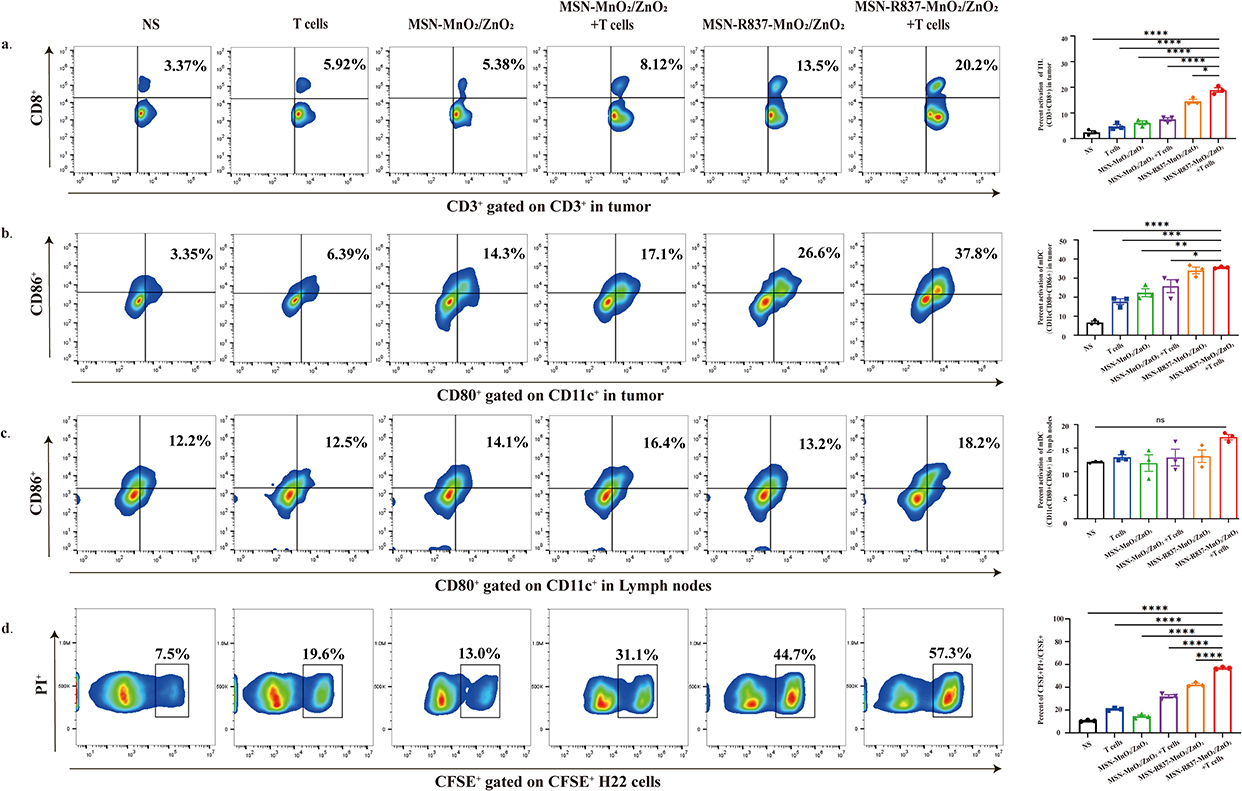 |
Figure 5 Immune responses induced by MSN-R837-MnO2/ZnO2 nanoparticles and adoptive T cells in vivo. One week after the last treatment, cells were obtained from lymph nodes, spleens, and tumors for flow cytometry analysis. (a) Representative flow cytometry images of TILs and proportions of tumor infiltrating lymphocytes in tumors. (b) Representative flow cytometry images of mature DCs in tumor and proportions of mature DCs in tumor. (c) Representative flow cytometry images of mature DCs in lymph nodes and proportions of mature DCs in lymph nodes. (d) Lymphocytes form spleens were incubated with CFSE labeled H22 cells at effector-to-target ratio (E: (T) of 25:1. PI was added 8 hours after incubation, and the percentage of dead tumor cells (CFSE+PI+/CFSE+) was analyzed by flow cytometry. Representative flow cytometry images and proportions of dead tumor cells in different group. P-values were determined by two-way ANOVA with Tukey’s multiple comparisons test. ns represented p > 0.05, *p < 0.05, **p < 0.01, ***p < 0.001, ****p < 0.0001. |
Security
Biosafety is paramount in nanoparticle applications. We observed that Mice in all groups maintained steady weight gain during treatment with nanoparticles and adoptive T cells for H22 liver cancer (Figure 6a). Hematoxylin and eosin (H&E) staining of major organs collected on day 7 after treatment showed no obvious damage across all groups (Figure 6b). Therefore, nanoparticles combined with adoptive T cells demonstrate good biosafety.
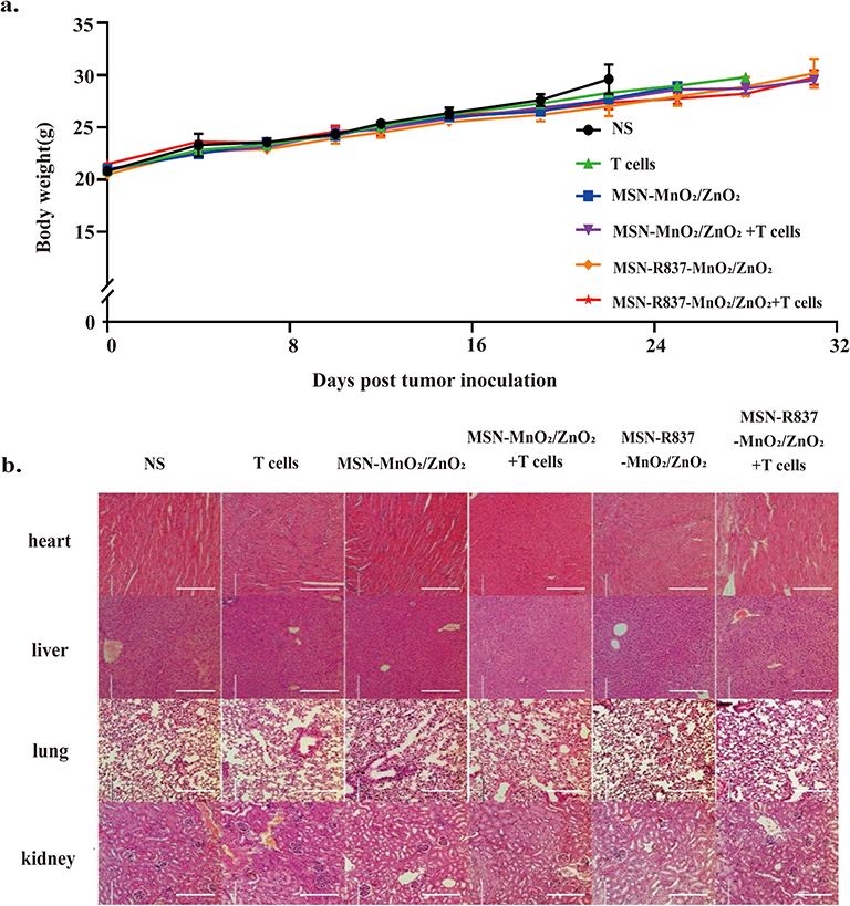 |
Figure 6 Safety evaluation. (a) Body weight of mice in different groups (n = 6). (b) Representative H&E staining of heart, liver, spleen, lung, and kidney in H22 mouse model on one week after different treatments in antitumor effect evaluation experiment. The scale bar is 200 μm. |
Discussion
In recent years, despite significant research attention, adoptive T cell therapy has faced slow progress in treating solid tumors.20 In response to this challenge, scientists have implemented various strategies, such as enhancing antigen recognition by adoptive T cells, improving their infiltration into tumors, and overcoming microenvironment immunosuppression.21
In this project, we improve the culture of CIK cells by incorporating tumor cell membranes, alongside essential cytokines. Traditionally, PBMCs are cultured in a specialized medium that contains supplements like fetal bovine serum (FBS), IL-2, and other growth factors for expansion and activation.22 Our innovation involves the addition of tumor cell membranes to the activating process, alongside essential cytokines, which can enhance the anti-tumor immune activity of T cells compared to the addition of cytokines alone. The increased release of IFN-γ in vitro proves the successful activation of CIK cells (Figure 2b). Interestingly, we observed that excessive concentrations of tumor cell membrane led to a decrease in IFN-γ secretion. This paradoxical outcome is consistent with previous immunological findings that excessive antigen exposure in the absence of appropriate co-stimulatory signals or adjuvants can induce immune suppression rather than activation. It has been reported that high antigenic loads may lead to T cell anergy or exhaustion, resulting in diminished effector cytokine production including IFN-γ. Therefore, we conducted a dose-response screening to determine the optimal concentration of tumor cell membranes to elicit a robust immune response without triggering suppression.
We developed nanoparticles named MSN-R837-MnO2/ZnO2. These nanoparticles served a dual purpose: MnO2/ZnO2 released oxygen directly by reacting with protons in the weakly acidic microenvironment, thereby alleviating hypoxia (Figure 4a); meanwhile, R837 promoted the maturation of DCs (Figure 2a–c). This innovative approach represents a promising adjunct for improving T cell therapy.
Hypoxia, a critical feature of the solid TME, exerts significant inhibitory effects on the survival and function of T cells, playing pivotal roles in tumor initiation, progression, metastasis and drug resistance.23 Enhancing oxygen levels in the TME holds great promise for boosting anti-tumor immune responses.24 In recent years, scientists have explored various approaches, such as hyperbaric oxygen therapy to enhance oxygen delivery efficiency directly,25 fluorocarbons to simulate the delivery of oxygen from blood to tumors,26 biological enzymes to catalyze peroxides to produce oxygen,27 and metal peroxides to generate oxygen in weakly acidic microenvironments.28 These methods have shown promising efficacy. The MnO2/ZnO2 loaded in our nanoparticles has been demonstrated to alleviate hypoxia in the microenvironment, improving the T cell living conditions and enhancing the anti-tumor efficacy of adoptive T cells (Figure 4a). Our nanoparticles are not only easier to produce, but also exhibit greater stability. However, there is a tendency of silica nanoparticles to aggregate in physiological environments, which can compromise the precision of silica nanoparticles in targeting applications and impact their stability.
Tumor-infiltrating DCs play a central role in the initiation and maintenance of T cell anti-tumor immune responses.29 However, the TME interferes with the maturation of DCs in ways such as regulating antigen expression and releasing cytokines, thereby inhibiting the activation of T cells.30 In recent years, the induced maturation of tumor-infiltrating DCs cells has become a research hotspot in tumor immunotherapy.31 By actively or passively targeting DC inducers to tumor sites, or acting on related signaling pathways, the antigen presentation efficiency of DCs is improved and a powerful anti-tumor immune response is induced.32 In this study, R837, a common DC maturation inducer, was delivered to the tumor site through a nanodrug delivery system, and the characteristics of the weak acid response of the nanoparticles were used to release them, thereby activating DCs (Figure 2a–b) and achieving the purpose of continuous activation of adoptive T cells. In recent years, many other R837-loaded nanoparticles designed in other research also have good anti-tumor effects like the application of R837-loaded PMBEOx-COOH nanoparticles wrapped by cancer cell membranes which significantly improves the anti-cancer efficacy of anti-PDL1 antibody.33 Based on the ability to activate immunity, R837 could synergize with T-cell therapy by priming the immune system for a more robust, sustained response against the tumor in this study. Similarly, R837 has great promise as an immune adjuvant in the clinical application of cell therapy in tumors. What’s more, the impact of nanoparticles on other immunosuppressive factors in the tumor microenvironment, such as immunosuppressive cells and related cytokines, requires further investigation.
The study demonstrates that combination therapy significantly enhances anti-tumor effects compared to single-drug treatments.34 This improvement is evident in several key areas, including reduced tumor volume and increased survival time of mice (Figure 3b–e). The enhanced efficacy of the combination therapy may be attributed to its multifaceted approach: it not only alleviates tumor hypoxia but also stimulates the anti-tumor immune response and provides CIK cells with anti-tumor activity directly. These combined effects are crucial for improving the survival and function of immune cells within the tumor environment. Moreover, the combination therapy offers a distinct advantage in terms of treatment safety. The dosage required to achieve comparable anti-tumor effects is considerably lower than that of single-drug therapies. This reduction in dosage helps mitigate risks such as neurotoxicity from accumulated metal ions35 and the potential for transplant rejection and infection associated with adoptive cell therapy.36 Thus, the combination approach not only enhances therapeutic efficacy but also improves safety, making it a promising option for cancer treatment.
Despite the encouraging results, the magnitude of the therapeutic improvement observed was modest (approximately a 15.4% increase in anti-tumor efficacy relative to the non-treated control group). While this enhancement was statistically significant, further studies are required to determine its biological relevance and reproducibility. In particular, it will be important to confirm that the immune changes (eg increased DC activation and T cell infiltration) induced by the nanoparticle translate into substantially improved long-term tumor control and not just minor transient effects. Additionally, the current study was conducted in a murine tumor model; thus, clinical translatability remains to be established. Future investigations should evaluate the MSN-R837-MnO2/ZnO2 strategy in more clinically relevant models, such as humanized mouse models or patient-derived xenografts, to better mimic human tumor immunology. These models would allow assessment of the therapy’s efficacy against human tumor antigens and its interactions with human immune cells. Moreover, extended safety studies are warranted to ensure that the combination treatment does not induce unexpected toxicities when scaled up. Addressing these limitations will help determine whether the demonstrated synergy can be harnessed for meaningful clinical benefit and guide the refinement of nanoparticle-augmented adoptive T cell therapies for eventual translation into cancer patients.
Conclusion
In this study, we successfully constructed a multi-functional nanoparticle MSN-R837-MnO2/ZnO2 based on a mesoporous silica nano-drug delivery system. R837 was loaded into nanoparticles to recruit and activate DCs, while Mn2+ and Zn2+ synergized to produce a substantial number of mature DCs presenting tumor antigens. This combination effectively enhanced the cytotoxicity of adoptive T cells, laying a solid foundation for killing tumor cells. In the H22 mouse liver cancer model, MSN-R837-MnO2/ZnO2 greatly enhanced the inhibitory effect of adoptive T cells on tumor growth. And we also verified its effect on the improvement of hypoxia at the tumor site and the effect of promoting CD3+ T cell infiltration. These findings provide a viable strategy for improving the efficacy of clinical adoptive T cells.
Author Contributions
All authors made a significant contribution to the work reported, whether that is in the conception, study design, execution, acquisition of data, analysis, and interpretation, or in all these areas; took part in drafting, revising or critically reviewing the article; gave final approval of the version to be published; have agreed on the journal to which the article has been submitted; and agree to be accountable for all aspects of the work.
Ethics
All animal experimental protocols were approved by the Laboratory Animal Care and Use Committee of Nanjing Drum Tower Hospital Affiliated to Nanjing University Medical School. All animal experiments were conducted in accordance with the Guidelines for the Ethical Review of Laboratory Animal Welfare (GB/T 35892‐2018).
Funding
This work was supported by the National Natural Science Foundation of China [grant numbers 81972309, 81930080, 82202999], the Fund for Distinguished Young Scholars of Jiangsu Province [grant number BK20230001], Jiangsu Provincial Medical Key Discipline, ZDXK202233, the Nanjing Medical Key Laboratory of Oncology, the Nanjing Jiangbei New Area Key Research and Development Program and Provincial Natural Science Foundation of Zhejiang (LQ23H180003). We thank all members of the Clinical Cancer Institute of Nanjing University for their discussion and suggestions.
Disclosure
The authors declare that they have no known competing financial interests or personal relationships that could influence the work reported in this paper.
References
1. Wang Z, Cao YJ. Adoptive cell therapy targeting neoantigens: a frontier for cancer research. Front Immunol. 2020;11:176. doi:10.3389/fimmu.2020.00176
2. Chen D, Sha H, Hu T, et al. Cytokine-induced killer cells as a feasible adoptive immunotherapy for the treatment of lung cancer. Cell Death Dis. 2018;9:366. doi:10.1038/s41419-018-0404-5
3. Li CMY, Li R, Drew P, et al. Clinical application of cytokine-induced killer (CIK) cell therapy in colorectal cancer: current strategies and future challenges. Cancer Treat Rev. 2024;122:102665. doi:10.1016/j.ctrv.2023.102665
4. Mata-Molanes JJ, González MS, Jiménez BV, Martínez Navarro EM, Masllorens AB. Cancer immunotherapy with cytokine-induced killer cells. Targeted Oncol. 2017;12:289–299. doi:10.1007/s11523-017-0489-2
5. Meng Y, Yu Z, Wu Y, et al. Cell-based immunotherapy with cytokine-induced killer (CIK) cells: from preparation and testing to clinical application. Hum Vaccines Immunother. 2017;13:1–9. doi:10.1080/21645515.2017.1285987
6. Mareschi K, Adamini A, Castiglia S, et al. Cytokine-Induced Killer (CIK) cells, in vitro expanded under Good Manufacturing Process (GMP) conditions, remain stable over time after cryopreservation. Pharmaceuticals. 2020;13.
7. Albarrán V, Román MS, Pozas J, et al. Adoptive T cell therapy for solid tumors: current landscape and future challenges. Front Immunol. 2024;15:1352805. doi:10.3389/fimmu.2024.1352805
8. Dzobo K, Senthebane DA, Dandara C. The tumor microenvironment in tumorigenesis and therapy resistance revisited. Cancers. 2023;16:15. doi:10.3390/cancers16010015
9. Spiga M, Martini E, Maffia MC, et al. Harnessing the tumor microenvironment to boost adoptive T cell therapy with engineered lymphocytes for solid tumors. Semin Immunopathol. 2024;46:8. doi:10.1007/s00281-024-01011-y
10. Morotti M, Albukhari A, Alsaadi A, et al. Promises and challenges of adoptive T-cell therapies for solid tumours. Br J Cancer. 2021;124:1759–1776. doi:10.1038/s41416-021-01353-6
11. Sun L, Liu H, Ye Y, et al. Smart nanoparticles for cancer therapy. Signal Trans Targeted Ther. 2023;8:418. doi:10.1038/s41392-023-01642-x
12. Alili L, Sack M, Karakoti AS, et al. Combined cytotoxic and anti-invasive properties of redox-active nanoparticles in tumor-stroma interactions. Biomaterials. 2011;32:2918–2929. doi:10.1016/j.biomaterials.2010.12.056
13. Gao M, Liang C, Song X, et al. Erythrocyte-membrane-enveloped perfluorocarbon as nanoscale artificial red blood cells to relieve tumor hypoxia and enhance cancer radiotherapy. Adv Mater. 2017;29.
14. Escriche-Navarro B, Escudero A, Lucena-Sánchez E, Sancenón F, García-Fernández A, Martínez-Máñez R. Mesoporous silica materials as an emerging tool for cancer immunotherapy. Adv Sci. 2022;
15. Lee J, Kim D, Le QV, Oh YK. Nanotherapeutics for immune network modulation in tumor microenvironments. Semi Cancer Biol. 2022;86:1066–1087. doi:10.1016/j.semcancer.2021.11.005
16. Melero I, Gaudernack G, Gerritsen W, et al. Therapeutic vaccines for cancer: an overview of clinical trials. Nat Rev Clin Oncol. 2014;11:509–524. doi:10.1038/nrclinonc.2014.111
17. Wang Y, Zhang S, Li H, et al. Small-molecule modulators of toll-like receptors. Acc Chem Res. 2020;53:1046–1055. doi:10.1021/acs.accounts.9b00631
18. Rozenberg JM, Kamynina M, Sorokin M, et al. The role of the metabolism of zinc and manganese ions in human cancerogenesis. Biomedicines. 2022;11:10. doi:10.3390/biomedicines11010010
19. McElroy DS, Badstibner AM, D’Orazio SE. Use of the CD107 mobilization assay reveals that cytotoxic T lymphocytes with novel MHC-Ib restriction are activated during Listeria monocytogenes infection. J Immunol Methods. 2007;328:45–52. doi:10.1016/j.jim.2007.08.005
20. Baulu E, Gardet C, Chuvin N, Depil S. TCR-engineered T cell therapy in solid tumors: state of the art and perspectives. Sci Adv. 2023;9:eadf3700. doi:10.1126/sciadv.adf3700
21. Mardiana S, Solomon BJ, Darcy PK, Beavis PA. Supercharging adoptive T cell therapy to overcome solid tumor-induced immunosuppression. Sci Trans Med. 2019;11.
22. Cappuzzello E, Sommaggio R, Zanovello P, Rosato A. Cytokines for the induction of antitumor effectors: the paradigm of Cytokine-Induced Killer (CIK) cells. Cytokine Growth Factor Rev. 2017;36:99–105. doi:10.1016/j.cytogfr.2017.06.003
23. Muz B, de la Puente P, Azab F, Azab AK. The role of hypoxia in cancer progression, angiogenesis, metastasis, and resistance to therapy. Hypoxia. 2015;3:83–92. doi:10.2147/HP.S93413
24. Zhang J, Tang K, Fang R, et al. Nanotechnological strategies to increase the oxygen content of the tumor. Front Pharmacol. 2023;14:1140362. doi:10.3389/fphar.2023.1140362
25. Moen I, Stuhr LE. Hyperbaric oxygen therapy and cancer--a review. Targeted Oncol. 2012;7:233–242. doi:10.1007/s11523-012-0233-x
26. Krafft MP. Alleviating tumor hypoxia with perfluorocarbon-based oxygen carriers. Curr Opin Pharmacol. 2020;53:117–125. doi:10.1016/j.coph.2020.08.010
27. Ding M, Zhang Y, Li J, Pu K. Bioenzyme-based nanomedicines for enhanced cancer therapy. Nano Convergence. 2022;9:7. doi:10.1186/s40580-022-00297-8
28. He J, Fu LH, Qi C, Lin J, Huang P. Metal peroxides for cancer treatment. Bioact Mater. 2021;6:2698–2710. doi:10.1016/j.bioactmat.2021.01.026
29. Katopodi T, Petanidis S, Charalampidis C, et al. Tumor-infiltrating dendritic cells: decisive roles in cancer immunosurveillance, immunoediting, and tumor t cell tolerance. Cells. 2022;(1):11. doi:10.3390/cells12010011
30. Ma Y, Shurin GV, Peiyuan Z, Shurin MR. Dendritic cells in the cancer microenvironment. J Cancer. 2013;4:36–44. doi:10.7150/jca.5046
31. Zhang Y, Li Y, Xu Z, et al. PPS-TLR7/8 agonist nanoparticles equip robust anticancer immunity by selectively prolonged activation of dendritic cells. Biomaterials. 2025;316:123032. doi:10.1016/j.biomaterials.2024.123032
32. Nava S, Lisini D, Frigerio S, Bersano A. Dendritic cells and cancer immunotherapy: the adjuvant effect. Int J Mol Sci. 2021;23:22. doi:10.3390/ijms23010022
33. Li S, Dong S, Wu J, et al. Surgically derived cancer cell membrane-coated R837-Loaded Poly(2-Oxazoline) nanoparticles for prostate cancer immunotherapy. ACS Appl Mater Interfaces. 2023;15:7878–7886. doi:10.1021/acsami.2c22363
34. Zhao Z, Wang D, Li Y. Versatile biomimetic nanomedicine for treating cancer and inflammation disease. Med Rev. 2023;3:123–151. doi:10.1515/mr-2022-0046
35. Ijomone OM, Ifenatuoha CW, Aluko OM, Ijomone OK, Aschner M. The aging brain: impact of heavy metal neurotoxicity. Crit. Rev. Toxicol. 2020;50:801–814. doi:10.1080/10408444.2020.1838441
36. Amini L, Kaeda J, Fritsche E, Roemhild A, Kaiser D, Reinke P. Clinical adoptive regulatory T cell therapy: state of the art, challenges, and prospective. Front Cell Develop Biol. 2022;10:1081644. doi:10.3389/fcell.2022.1081644
 © 2025 The Author(s). This work is published and licensed by Dove Medical Press Limited. The
full terms of this license are available at https://www.dovepress.com/terms
and incorporate the Creative Commons Attribution
- Non Commercial (unported, 4.0) License.
By accessing the work you hereby accept the Terms. Non-commercial uses of the work are permitted
without any further permission from Dove Medical Press Limited, provided the work is properly
attributed. For permission for commercial use of this work, please see paragraphs 4.2 and 5 of our Terms.
© 2025 The Author(s). This work is published and licensed by Dove Medical Press Limited. The
full terms of this license are available at https://www.dovepress.com/terms
and incorporate the Creative Commons Attribution
- Non Commercial (unported, 4.0) License.
By accessing the work you hereby accept the Terms. Non-commercial uses of the work are permitted
without any further permission from Dove Medical Press Limited, provided the work is properly
attributed. For permission for commercial use of this work, please see paragraphs 4.2 and 5 of our Terms.
Recommended articles
GOx-Functionalized Platelet Membranes-Camouflaging Nanoreactors for Enhanced Multimodal Tumor Treatment
Du Y, Wang S, Luan J, Zhang M, Chen B, Shen Y
International Journal of Nanomedicine 2022, 17:2979-2993
Published Date: 7 July 2022
Oxygenation: Nanotechnological Strategies for Conquering Tumor Hypoxia in Photodynamic Therapy
Liang J, Lai X, Mei Y, Liu X, Wen S, Zhou Y, Liu F
International Journal of Nanomedicine 2026, 21:569340
Published Date: 10 February 2026
Nanoparticles Targeting the Tumor Microenvironment for the Treatment of Osteosarcoma: Recent Progress and Perspectives
Liang G, Wang W, Li C, Zhong B, Zhao L, Zhang Z, Liu J
International Journal of Nanomedicine 2026, 21:579152
Published Date: 16 February 2026
From Barrier to Gateway: Nanomaterials Reshaping the Tumor Microenvironment for Therapy
Xie M, Sun W, Hu D, Liu L, Han C, Liu W, Liu L, Bao X, Zhang W, Hao X, Zhou Y
International Journal of Nanomedicine 2026, 21:573490
Published Date: 10 March 2026