
Back to Journals » Neuropsychiatric Disease and Treatment » Volume 22
Determinants of Post-Stroke Cognitive Impairment After Acute Ischemic Stroke: A Systematic Review and Meta-Analysis

Received 14 March 2026
Accepted for publication 27 June 2026
Published 9 July 2026 Volume 2026:22 608599
DOI https://doi.org/10.2147/NDT.S608599
Checked for plagiarism Yes
Review by Single anonymous peer review
Peer reviewer comments 2
Editor who approved publication: Professor Taro Kishi
Fangjia Shen,1 Xijuan Guo2– 4
1School of Nursing, Sun Yat-Sen University, Guangzhou, Guangdong, People’s Republic of China; 2Department of Cardiology, Qilu Hospital of Shandong University, Jinan, Shandong, People’s Republic of China; 3Nursing Department, Qilu Hospital of Shandong University, Jinan, Shandong, People’s Republic of China; 4Nursing Theory and Practice Innovation Research Center, Shandong University, Jinan, Shandong, People’s Republic of China
Correspondence: Xijuan Guo, Email [email protected]
Background: Acute ischemic stroke (AIS) is frequently complicated by post-stroke cognitive impairment (PSCI), which significantly affects long-term prognosis and quality of life. However, the evidence regarding its risk factors remains inconsistent.
Methods: A systematic search was conducted in PubMed, Embase, Cochrane Library, and Web of Science up to December 30, 2025. Observational studies evaluating risk factors for PSCI after AIS were included. Pooled odds ratios (ORs) with 95% confidence intervals (CIs) were calculated using fixed- or random-effects models. Heterogeneity, sensitivity analyses, and publication bias were assessed.
Results: Fifteen studies involving 30,022 participants were included. Age > 75 years (OR = 1.07, 95% CI: 1.04– 1.10), female sex (OR = 1.72, 95% CI: 1.41– 2.10), diabetes (OR = 1.76, 95% CI: 1.29– 2.40), elevated C-reactive protein (CRP; OR = 1.36, 95% CI: 1.14– 1.62), and lower educational level (OR = 1.24, 95% CI: 1.12– 1.38) were significantly associated with an increased risk of PSCI after AIS. Substantial heterogeneity was observed for some predictors, particularly age (I2 = 89.9%) and educational level (I2 = 89.6%).
Conclusion: Several factors, including advanced age, female sex, diabetes, elevated CRP, and lower educational level, were associated with an increased risk of PSCI after AIS. These findings may support risk stratification and early monitoring strategies. However, the substantial heterogeneity observed for some predictors should be considered when interpreting the pooled estimates. Further prospective studies are needed to confirm these associations and clarify causal relationships.
Keywords: acute ischemic stroke, post-stroke cognitive impairment, risk factors, meta-analysis
Background
Acute ischemic stroke (AIS) is a major global cause of death and disability, imposing substantial physical, psychological, and economic burdens on patients and their families.1 According to statistics from the World Health Organization (WHO), an estimated 15 million people experience stroke annually, with ischemic stroke accounting for approximately 80% of all stroke cases.2 Although advances in medical care have improved outcomes and survival after AIS,3,4 many patients continue to experience post-stroke cognitive impairment (PSCI) beyond the acute phase. PSCI generally refers to decline in one or more cognitive domains after stroke, including memory, attention, executive function, language, and visuospatial ability.5–7 It substantially impairs quality of life and increases long-term caregiving and socioeconomic burdens.8–10
PSCI after AIS is a complex and multidimensional syndrome. Its development may be driven by overlapping mechanisms, including vascular burden, ischemic brain injury, neurodegenerative changes, inflammatory responses, reduced premorbid cognitive reserve, and brain network dysfunction.11–16 Non-biological factors, such as social support, psychological status, and physical activity, may also influence cognitive recovery or deterioration after stroke.16 Thus, PSCI likely reflects interactions among demographic, clinical, biochemical, imaging-related, and psychosocial factors.
Although observational studies17,18 have explored potential risk factors for PSCI following AIS, their findings remain inconsistent because of limited sample sizes, heterogeneous study designs, variable cognitive assessment tools and PSCI definitions, different follow-up durations, and insufficient adjustment for confounders. Previous systematic reviews and meta-analyses19–21 have provided valuable evidence by summarizing potential risk factors for PSCI after stroke. Nevertheless, their applicability to AIS-specific clinical practice remains limited. First, most previous reviews focused on the prevalence and general risk factors of PSCI across all stroke types, while AIS patients were less frequently considered as a distinct clinical population.19–21 Second, most analyses evaluated only a limited range of risk factors, such as sociodemographic and disease-related factors, and therefore could not provide a comprehensive profile integrating demographic, clinical, biochemical, and imaging-related predictors.19,20 Third, some reviews lacked comprehensive subgroup or sensitivity analyses to explore heterogeneity or did not incorporate evidence published in the past three years.21 Thus, the overall profile of factors associated with PSCI after AIS remains insufficiently defined.
To address these gaps, the present meta-analysis specifically focused on patients with AIS and excluded hemorrhagic stroke, previous or chronic ischemic stroke, and other non-AIS populations to reduce clinical heterogeneity. We systematically evaluated a broad range of demographic, clinical, biochemical, and imaging-related predictors and updated the literature search to December 30, 2025. In addition, we performed subgroup analyses, heterogeneity assessments, and sensitivity analyses for key clinical variables to explore potential sources of inconsistency. By integrating updated evidence with a more focused population and broader predictor framework, this study provides a comprehensive synthesis of key factors associated with PSCI after AIS, thereby informing early identification, individualized monitoring, and future research.
Methods
This systematic review and meta-analysis was conducted in accordance with the Preferred Reporting Items for Systematic Reviews and Meta-Analyses (PRISMA) statement. The protocol was prospectively registered in PROSPERO (CRD420261280566).
Literature Retrieval
A systematic literature search was conducted in PubMed, Embase, the Cochrane Library, and Web of Science from database inception to December 30, 2025. The search strategy included terms related to AIS, PSCI, and risk factors. The full electronic search strategy is presented in Supplementary materials.
Inclusion and Exclusion Criteria
Inclusion criteria: Studies were eligible if they included adults (≥18 years) with AIS diagnosed according to internationally accepted criteria and evaluated cognitive function. Eligible studies reported the association between potential risk factors and PSCI. PSCI was mainly defined based on standardized cognitive assessment tools. Observational studies (cohort or case–control) that provided sufficient data for statistical analysis were included. The control group comprised patients with AIS without PSCI or with only mild cognitive decline.
Exclusion criteria: Studies including patients without a confirmed diagnosis of AIS, such as those with hemorrhagic stroke or brain tumors, were excluded. Participants aged <18 years and studies that did not provide data on PSCI assessments will also be excluded. Studies that do not clearly record and analyze factors associated with PSCI following AIS, or those that fail to reasonably assess exposure factors, will not be included. Additionally, studies classified as case reports, reviews, low-quality papers, or articles not peer-reviewed will be excluded.
Study Selection
Two reviewers (FJS and XJG) independently screened the retrieved records using EndNote 21 software. Titles and abstracts were first examined to exclude studies that obviously failed to satisfy the eligibility criteria. The remaining full-text articles were then assessed to determine whether they met the final eligibility requirements according to the preset criteria. Any disagreements in the screening process were solved via discussion between the two reviewers; if no consensus was achieved, a third reviewer (YZS) was involved to reach a final decision.
Data Extraction
Two reviewers (FJS and XJG) independently extracted relevant data from the included articles using a standardized Excel spreadsheet. The extracted data consisted of basic study characteristics (first author, publication year, country, and study design), demographic and clinical features of participants (sample size, number of patients with PSCI, sex distribution, and mean age), statistical methods used in the analysis (multivariable regression or univariate analysis), and the criteria used for diagnosing PSCI. Any inconsistencies identified were resolved through discussion between the two reviewers (FJS and XJG). If consensus could not be reached, a third reviewer (YZS) was consulted to guarantee the accuracy and reliability of the data.
Quality Evaluation
The Newcastle-Ottawa Scale (NOS)22 was used to assess case-control and cohort studies. This tool evaluates potential biases across three key domains: study selection, comparability, and outcome assessment.
Statistical Analysis
All statistical analyses were conducted using Stata 17.0 software. Effect estimates were pooled as odds ratios (ORs) with 95% confidence intervals (CIs). A fixed-effects model was applied if I2 < 50%, indicating low to moderate heterogeneity. Otherwise, a random-effects model was used to provide a more conservative estimate. When substantial heterogeneity was detected, sensitivity analyses were performed to assess the robustness of the pooled estimates. Publication bias was evaluated visually using funnel plots and statistically using Egger’s test. Statistical significance was determined based on the pooled ORs and their corresponding 95% CIs.
Results
Literature Screening Results
A total of 4765 records were initially identified from PubMed (n=591), Embase (n=1632), Cochrane Library (n=228), and Web of Science (n=2314). After removing 1055 duplicates, the remaining 3710 articles underwent title and abstract screening, leading to the exclusion of 3689 records. Following a rigorous full-text review of the remaining papers, an additional 6 studies were excluded. Finally, 15 articles23–37 met the inclusion criteria for the final analysis. The detailed selection process is presented in the PRISMA flow diagram (Figure 1).
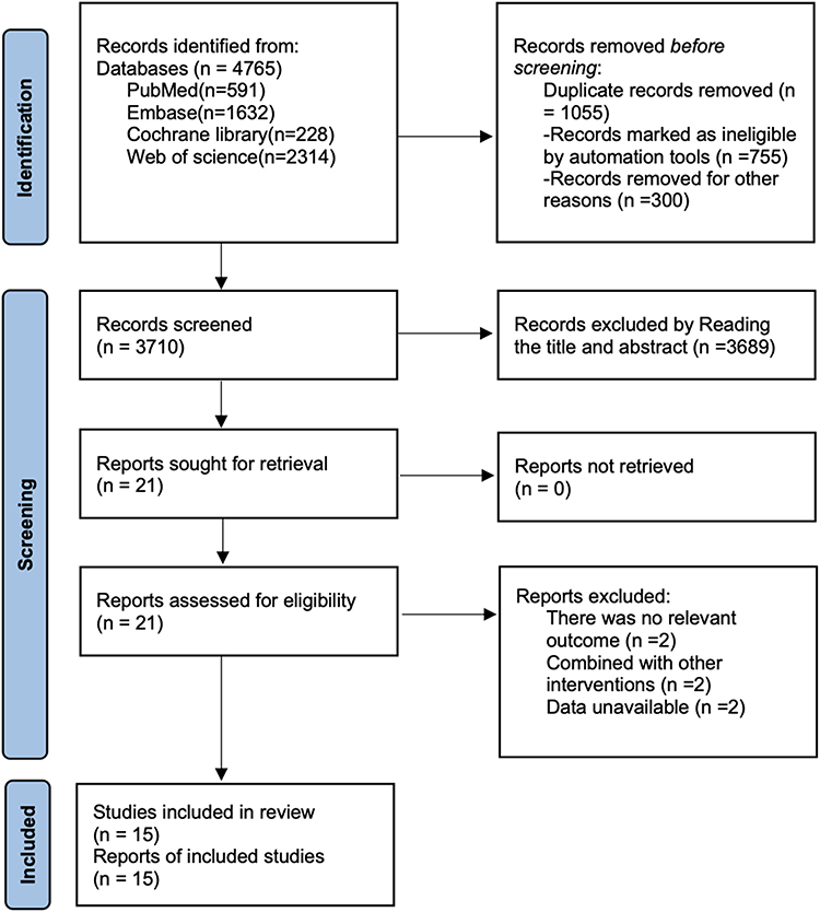 |
Figure 1 Flow diagram of the literature search and study selection process. The diagram illustrates the identification, screening, eligibility, and inclusion of studies according to the PRISMA guidelines. |
Basic Characteristics of Literature
This study included 15 articles involving 30,022 participants, 1023–32 of which were from China, and the others were from Bulgaria,34 Singapore,35 Poland,33 South Korea,36 and the UK.37 One article23 used Cox regression, and the rest used logistic regression (Table 1).
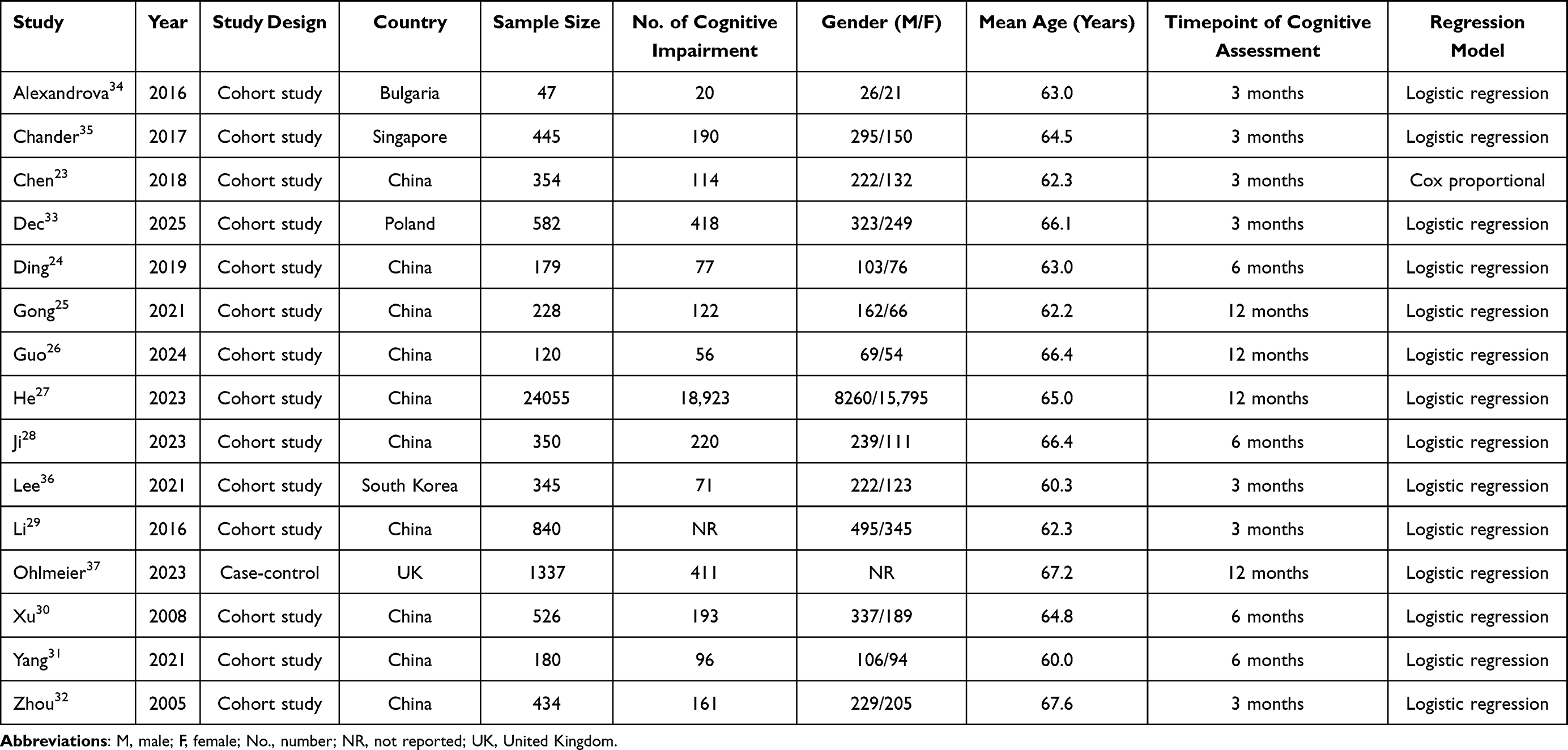 |
Table 1 Table of Basic Characteristics |
Quality Assessment Results
This study used the NOS score, and the results (Table 2) showed that 2 articles30,32 scored 7 points, 5 articles24–27,33 scored 8 points, and 8 articles23,28,29,31,34–37 scored 9 points. These results indicate that all included articles were of high quality.
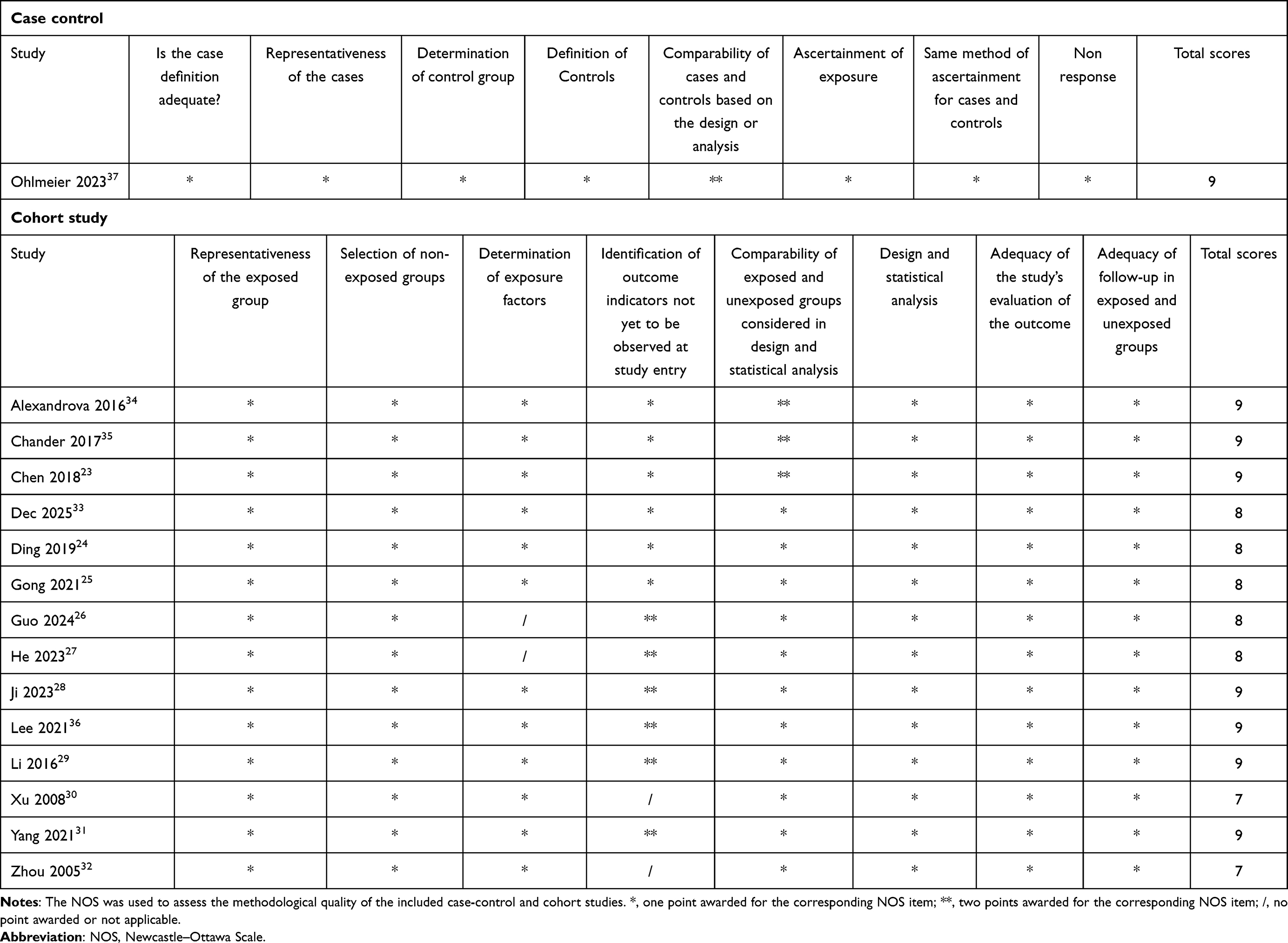 |
Table 2 NOS Scores |
Meta-Analysis Results
A total of 5 potential risk factors for PSCI in patients with AIS were identified across the included studies: age >75 years, female sex, history of diabetes, elevated C-reactive protein (CRP), and lower educational level.
14 studies evaluated age >75 years. Substantial heterogeneity was observed (I2 = 89.9%, P = 0.001), and therefore a random-effects model was applied. The pooled analysis (Figure 2) demonstrated that age >75 years was significantly related to an increased risk of PSCI in AIS patients (OR = 1.07, 95% CI: 1.04–1.10), with stable results in sensitivity analyses (Supplementary Figure S1).
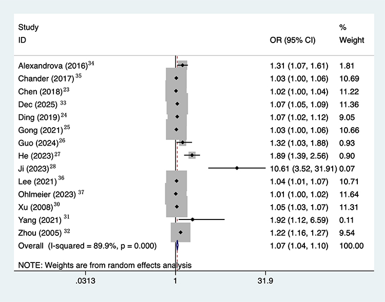 |
Figure 2 Forest plot of the association between age >75 years and cognitive impairment after acute ischemic stroke. Squares represent individual study estimates (ORs) with 95% confidence intervals (CIs), and the diamond indicates the pooled effect. The vertical line represents no association (OR = 1). A random-effects model was used. |
6 studies assessed the relation between female sex and PSCI. No significant heterogeneity was detected (I2 = 0%, P = 0.551). Therefore, a fixed-effects model was used. Female sex was significantly (Figure 3) related to a higher risk of PSCI (OR = 1.72, 95% CI: 1.41–2.10).
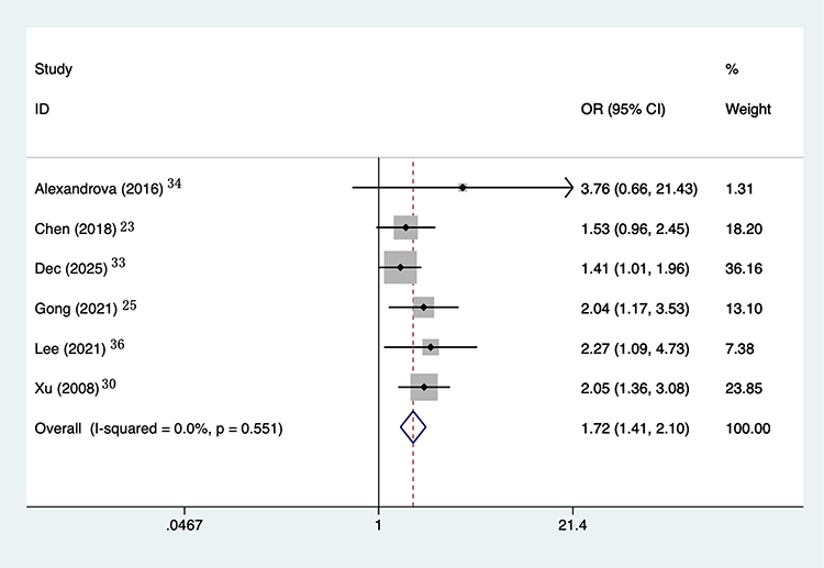 |
Figure 3 Forest plot of the association between female sex and cognitive impairment after acute ischemic stroke. Squares represent individual study estimates (ORs) with 95% CIs, and the diamond indicates the pooled effect. The vertical line represents no association (OR = 1). A fixed-effects model was used. |
9 studies reported the association between diabetes and PSCI. Moderate heterogeneity was observed (I2 = 69.6%, P = 0.001), and a random-effects model was applied. The pooled results (Figure 4) revealed that a history of diabetes increased the risk of PSCI (OR = 1.76, 95% CI: 1.29–2.40), with stable results in sensitivity analyses (Supplementary Figure S2).
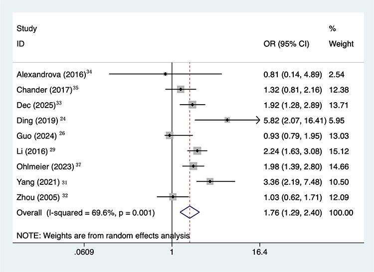 |
Figure 4 Forest plot of the association between history of diabetes and cognitive impairment after acute ischemic stroke. Squares represent individual study estimates (ORs) with 95% CIs, and the diamond indicates the pooled effect. The vertical line represents no association (OR = 1). A random-effects model was applied. |
4 studies investigated elevated CRP levels. Moderate heterogeneity was present (I2 = 51.6%, P = 0.102), and the pooled (Figure 5) analysis using a random-effects model demonstrated a significant association between high CRP and PSCI (OR = 1.36, 95% CI: 1.14–1.62), with stable results in sensitivity analyses (Supplementary Figure S3).
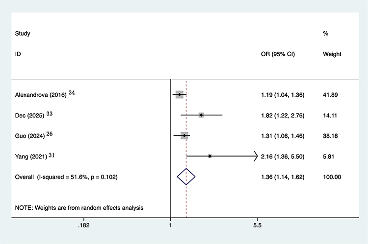 |
Figure 5 Forest plot of the association between elevated C-reactive protein and cognitive impairment after acute ischemic stroke. Squares represent individual study estimates (ORs) with 95% CIs, and the diamond indicates the pooled effect. The vertical line represents no association (OR = 1). A random-effects model was used. |
4 studies examined educational level. Considerable heterogeneity was detected (I2 = 89.6%, P = 0.001), and a random-effects model was applied. Lower educational level (Figure 6) was significantly related to an increased risk of PSCI (OR = 1.24, 95% CI: 1.12–1.38), with stable results in sensitivity analyses (Supplementary Figure S4).
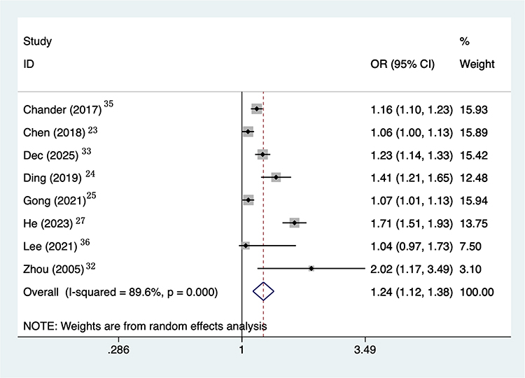 |
Figure 6 Forest plot of the association between lower educational level and cognitive impairment after acute ischemic stroke. Squares represent individual study estimates (ORs) with 95% CIs, and the diamond indicates the pooled effect. The vertical line represents no association (OR = 1). A random-effects model was used. |
Subgroup Analysis
Subgroup analyses according to study design and geographic region are summarized in Table 3. For age >75 years, significant associations were observed in both cohort (OR = 1.08, 95% CI: 1.04–1.12) and case–control studies (OR = 1.01, 95% CI: 1.00–1.02). The association appeared stronger in Chinese populations (OR = 1.14, 95% CI: 1.06–1.23) relative to non-Chinese populations (OR = 1.04, 95% CI: 1.01–1.06). Female sex remained a significant risk factor in both Chinese (OR = 2.04, 95% CI: 1.17–3.53) and non-Chinese studies (OR = 1.56, 95% CI: 1.21–2.00). Similar patterns were observed for diabetes and educational level across study designs and regions, whereas elevated CRP showed comparable but less precise estimates between subgroups.
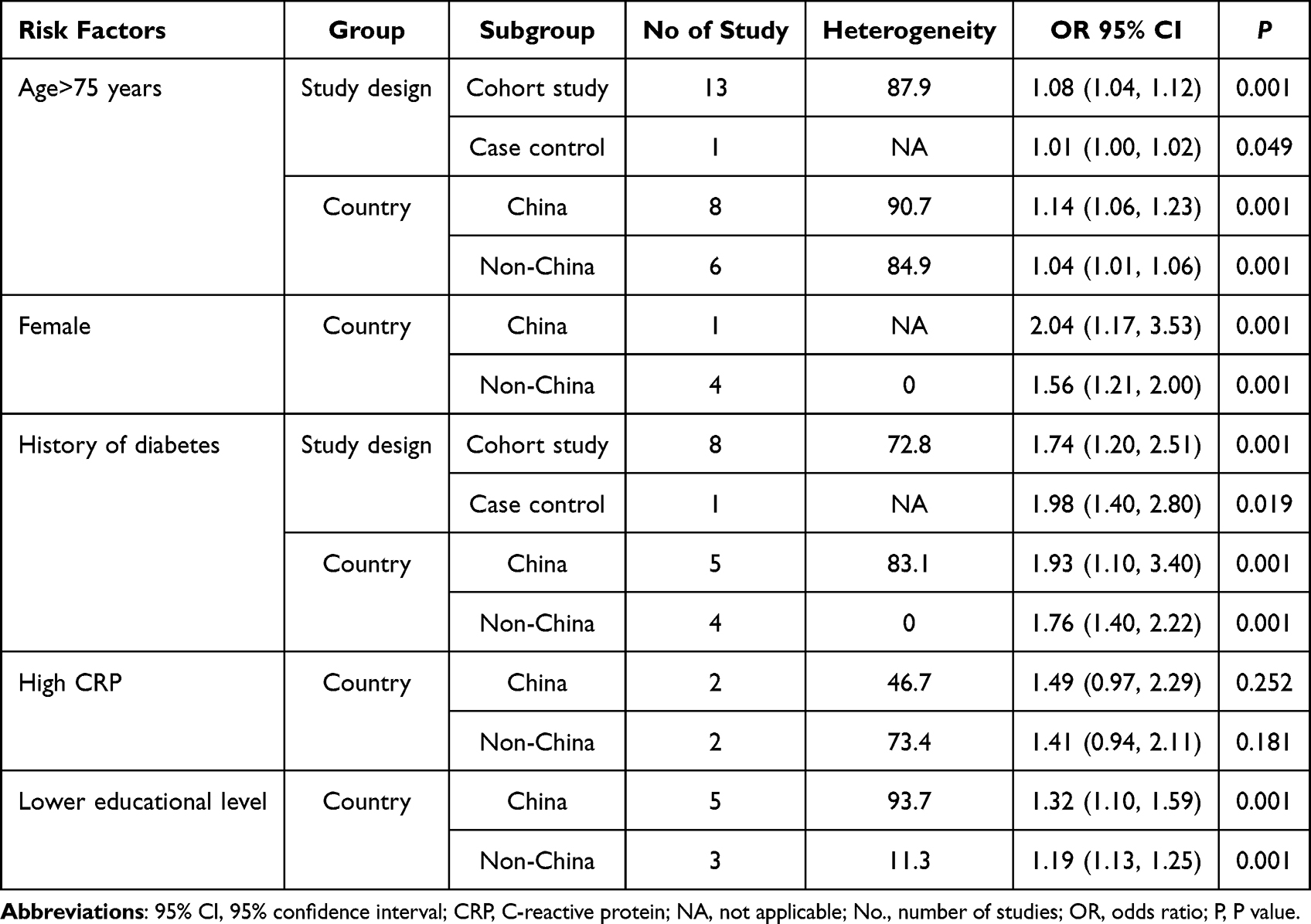 |
Table 3 Subgroup Analysis Results |
Publication Bias
Publication bias was assessed using funnel plots (Supplementary Materials Figures S5-S9) and Egger’s test. Visual inspection suggested asymmetry for age >75 years and CRP. Egger’s test confirmed potential publication bias for age >75 years (P = 0.005) and CRP (P = 0.048). After applying the trim-and-fill method (Supplementary Materials Figures S10–S11), the adjusted estimates remained largely unchanged, indicating that the overall findings were robust. No other bias was found in female sex (P = 0.098), diabetes (P = 0.993), or educational level (P = 0.155).
Meta Regression
Since the heterogeneity for age > 75 and low educational level was close to 90%, we conducted a meta-regression analysis based on year, country, study design, diagnosis of PSCI, and regression model (Supplementary Material Table S1). For age > 75, the diagnosis of PSCI (P = 0.001) was found to be a potential source of heterogeneity. However, for low educational level, the result was P > 0.05, indicating no obvious source of heterogeneity.
Discussion
This systematic review and meta-analysis assessed the risk factors for PSCI in patients with AIS, revealing several significant associated factors, including age >75 years, female sex, history of diabetes, high CRP levels, and low educational level. These risk factors shed light on their roles in PSCI from different biological and sociological perspectives. Although there was significant heterogeneity among the studies and some results were subject to publication bias, the findings were generally robust in sensitivity analyses and meta-regression analysis.
Main Findings
Age is an important factor influencing PSCI. Our meta-analysis results show that AIS patients aged >75 years have a 1.07 times higher risk of PSCI than patients in other age groups. This result is consistent with many previous studies,38,39 indicating that as people age, the elasticity of cerebral blood vessels weakens, the vessel walls harden, and cerebral blood flow decreases. Impaired cerebral vascular function exacerbates the consequences of stroke, leading to insufficient blood supply to brain tissue, neuronal loss, reduced synaptic connections, and white matter lesions in the brain, all of which are neurodegenerative changes.40 After a stroke, these degenerative changes may further exacerbate brain damage, resulting in significant cognitive decline.41 However, the heterogeneity for age >75 years in this study was high (I2 =89.9%), suggesting that there may be differences among studies in terms of study population selection, stroke severity, cognitive assessment tools, and timing of cognitive assessment. Some studies reported weaker or non-significant associations after adjusting for pre-stroke cognitive status or stroke severity, which may reflect differences in baseline cognitive reserve, and comorbidity burden. Sensitivity analyses supported the robustness of the pooled association, but the effect size may still vary across populations and measurement methods and should be interpreted cautiously.
This study found that female patients have a significantly higher risk of PSCI than male patients, with an OR of 1.72. This result is consistent with existing literature,42,43 indicating that gender differences play a role in PSCI following stroke. Although women have a relatively lower risk of stroke, they are more vulnerable to PSCI after stroke. Biologically, hormonal levels in women (particularly estrogen) may play a crucial role in neuroprotection.44 Estrogen is believed to have neuroprotective effects and may play a positive role in the recovery process following a stroke.45 As women age, particularly after menopause, declining hormonal levels may be associated with a higher risk of cognitive decline. Additionally, women are generally more prone to mental symptoms such as depression, which themselves may be closely associated with PSCI.46 Therefore, future research should focus more on gender-specific biological mechanisms and how to tailor personalized treatment to the unique needs of female patients. The gender analysis in this study showed low heterogeneity (I2 = 0%), indicating that the conclusion regarding gender as a risk factor is consistent across studies, with women consistently identified as an important risk factor for PSCI. This finding underscores the importance of gender in the assessment and management of PSCI, and future research could further explore early warning signs and intervention strategies for female stroke patients.
A history of diabetes is a significant risk factor for PSCI in stroke patients. The results of this study indicate that patients with a history of diabetes have a 1.76-fold increased risk of developing PSCI. The association between diabetes and PSCI has been well-established in numerous studies. Diabetes patients experience long-term effects of hyperglycemia and metabolic abnormalities on cerebral vasculature and the nervous system, potentially leading to more pronounced PSCI.47 Diabetes influences cognitive function through multiple mechanisms. Hyperglycemia may exacerbate vascular endothelial damage, leading to impaired cerebral blood flow; simultaneously, chronic inflammation and oxidative stress induced by diabetes can damage brain tissue, resulting in neurodegenerative changes.48 Additionally, diabetes is often accompanied by metabolic abnormalities such as hypertension and hypercholesterolemia, and the combined effects of these factors make stroke patients with diabetes more prone to PSCI.49 Despite the high heterogeneity for diabetes in this study (I2=69.6%), which may stem from differences in population selection, glycemic control level, diabetes duration, cognitive assessment tools, and timing of assessment, sensitivity analyses supported the robustness of the pooled association, but the effect size may still vary across populations and measurement methods and should be interpreted cautiously. Overall, the findings support an association between diabetes and PSCI. Future research should further explore the protective effects of diabetes treatment strategies on cognitive function and implement early cognitive screening and intervention in diabetic patients.
CRP is a common inflammatory marker. In this study, high CRP levels were found to be significantly associated with PSCI in AIS patients, with an OR of 1.36. This finding aligns with the hypothesis that systemic inflammation is related to PSCI.50 High CRP levels are often closely associated with chronic inflammation and vascular damage, and these factors may be linked to more severe brain damage after stroke. Inflammatory responses are thought to be involved in the pathogenesis of stroke.51 The systemic inflammatory response following stroke may be associated with secondary damage to brain tissue, which could contribute to poorer cognitive outcomes. Specifically, elevated CRP levels post-stroke may reflect persistent neuroinflammation, which has been reported in relation to neuronal damage, synaptic dysfunction, and impaired cognitive function.52 Although the heterogeneity of CRP across different studies was moderate (I2 = 51.6%), sensitivity analysis indicated that the pooled association was robust. However, the observed heterogeneity may stem from differences in CRP measurement timing, laboratory methods, cutoff values for defining elevated CRP, and population characteristics. The timing of CRP measurement relative to stroke onset may critically affect its predictive value for PSCI, as CRP levels typically peak within the first week post-stroke and gradually decline thereafter. Future studies should standardize CRP measurement protocols, including timing and assay methods, to better evaluate its potential as a biomarker for PSCI. Investigating the combined predictive value of CRP with other inflammatory markers and exploring the dynamic changes of inflammatory profiles throughout the post-stroke period may provide deeper insights into the inflammatory mechanisms underlying PSCI.
Lower educational level was identified as a risk factor for PSCI in four studies, with an OR of 1.24. Educational level is often regarded as an important indicator of cognitive reserve, and low educational level may imply reduced cognitive reserve, making the brain more susceptible to PSCI.53 The cognitive reserve theory posits that individuals with higher educational level possess greater brain functional resilience, enabling them to better cope with neural damage.54 However, the relationship between low educational level and PSCI is complex and likely multifactorial. While educational level serves as a proxy for cognitive reserve, it may also capture broader socioeconomic disparities, differences in healthcare access, and literacy-related influences on cognitive test performance.19,55,56 For example, individuals with lower education may have fewer opportunities for prevention and timely management of vascular risk factors, which could increase the likelihood of cerebrovascular injury and subsequent PSCI. In addition, limited literacy and reduced familiarity with test materials may affect how individuals understand and complete cognitive screening tasks, potentially contributing to lower measured performance that is not solely attributable to underlying neuropathology.57 Furthermore, low educational level is closely linked to unhealthy behaviors such as smoking, alcohol consumption, and physical inactivity, which may further increase the risk of PSCI. Although substantial heterogeneity was observed among low-education groups in the meta-analysis (I2 = 89.6%), this may stem from differences in educational system classifications, measurement methods, study population selection, cognitive assessment tools, and timing of cognitive assessment. Sensitivity analyses suggested that the pooled estimate was robust, but the effect size may still vary across populations and educational contexts and should be interpreted cautiously. Overall, the findings support an association between low education and PSCI, though the magnitude may differ depending on how education is defined and measured across different cultural and healthcare settings.
Furthermore, the potential influence of age and educational background on cognitive test performance should be acknowledged. Both factors are known to affect scores on commonly used assessments such as MoCA, which may partially inflate the observed associations with PSCI. Older individuals tend to perform more poorly on cognitive tests irrespective of stroke status, and lower educational level is closely linked to reduced cognitive reserve and baseline test scores.58,59 Although our subgroup analyses confirmed that age >75 years and lower educational level were consistently associated with increased risk across study designs and regions, these associations may reflect not only true vulnerability but also differential test sensitivity. Future studies using education-adjusted scoring norms, culturally adapted tools, or longitudinal cognitive trajectories may help better disentangle measurement bias from genuine risk effects.
The subgroup analyses further strengthened the robustness of our findings by demonstrating that several key factors associated with PSCI remained significant across different study designs and populations. Advanced age, particularly those over 75 years, consistently increased the risk of PSCI, with a stronger effect observed in Chinese cohorts, suggesting possible demographic differences. However, it may also be partly influenced by healthcare system related factors and differences in cognitive assessment procedures, educational distributions, and rehabilitation access across settings. These factors could affect both the measurement of PSCI and the observed associations. Female sex and a history of diabetes also showed stable associations across regions, underscoring their generalizable impact on post-stroke cognition. Although elevated CRP displayed a similar direction of effect in all subgroups, the wide confidence intervals indicate that inflammatory biomarkers require further investigation. In addition, the consistent influence of lower educational level across populations highlights the importance of cognitive reserve in mitigating PSCI. Overall, these subgroup findings confirm that the identified risk factors are not restricted to specific study characteristics or geographic settings, reinforcing their clinical relevance.
Many primary studies used heterogeneous cognitive assessment tools, such as NINDS-VCI criteria, or institution-specific neuropsychological batteries, resulting in considerable variability in reported incidence and associated predictors. In our updated evidence synthesis, we systematically examined all available MoCA-based studies published to date; however, the number of eligible studies providing extractable risk-factor data remained limited, preventing a dedicated MoCA-specific meta-analysis. This underscores an important gap in current research: while MoCA has been validated as an effective screening tool for VCI, there is still a lack of large, methodologically consistent studies using MoCA to evaluate risk factors. Our study therefore highlights not only the existing evidence but also the need for future standardized research employing unified, validated cognitive assessments such as MoCA to improve comparability and strengthen prognostic inferences.
Strengths and Limitations
A key strength of this investigation is the systematic synthesis of data from diverse primary studies, providing comprehensive evidence to support the factors associated with PSCI in patients with AIS. This meta-analysis overcomes the issue of limited sample sizes in individual articles, enhancing the reliability and generalizability of the conclusions. The study employed methods such as sensitivity analysis, meta-regression, and publication bias assessment to ensure the stability and accuracy of the analysis results, thereby minimizing the potential impact of bias on the conclusions. Additionally, this study explored multiple related risk factors, including age, gender, history of diabetes, CRP levels, and educational level, providing specific quantitative data that offers valuable references for clinical diagnosis and intervention. The heterogeneity analysis in this study also provides valuable insights into the differences in results across different contexts, further enhancing the scientific rigor and clinical applicability of this research.
However, several limitations remain. First, there was evidence of heterogeneity in this study, particularly when analyzing age >75 years and lower educational level as risk factors, which may reflect differences in study design, sample selection, and assessment criteria. Although these differences were controlled for using a random-effects model and sensitivity analysis, the presence of heterogeneity may still affect the stability of the results. Second, while multiple risk factors were associated with PSCI, these associations do not equate to causal relationships. Future longitudinal studies may help further validate whether these factors directly lead to PSCI. Third, a limitation is publication bias; although the Trim and Fill method was used for correction, there may still be missing unpublished data, affecting the generalizability of the study conclusions. Fourth, the assessment criteria for PSCI vary across studies, which may lead to inconsistent results and further impact the external validity of the study conclusions. Fifth, although studies were included from several countries, the evidence base remains heavily weighted toward Chinese cohorts. This may limit the external validity of our findings, as differences in healthcare system organization, rehabilitation access, cognitive assessment procedures, and the distribution of educational level across settings could affect both the measurement of PSCI and the magnitude of the observed associations. Finally, most of the included studies did not use standardized tools to assess patients’ prestroke cognitive status. Therefore, we could not fully distinguish new-onset PSCI from pre-existing cognitive vulnerability. This limitation may bias the effect estimates for certain risk factors.
Clinical Significance
The clinical significance of this study lies in providing scientific evidence for the prevention and intervention of PSCI in patients with AIS. Specifically, clinicians can reduce the incidence of PSCI by targeting and intervening in these risk factors. For example, for older patients, especially those over 75 years of age, doctors can strengthen assessments of their cognitive function and combine cognitive training and psychological intervention to identify and address signs of PSCI at an early stage. Additionally, female patients may require closer monitoring of their neuroprotective mechanisms post-stroke due to physiological factors such as hormonal changes and can benefit from a combination of medication and lifestyle improvements to slow the progression of PSCI. Patients with a history of diabetes should also prioritize glycemic control and vascular health interventions, as diabetes-related damage to cerebral blood vessels may accelerate PSCI. Patients with elevated CRP levels, especially in cases of strong inflammatory responses, should initiate anti-inflammatory treatment early to reduce brain inflammation and improve cognitive outcomes. Patients with lower educational levels may face greater challenges in cognitive recovery post-stroke, so education and cognitive training should be incorporated into intervention strategies to enhance patients’ brain functional reserves and strengthen the brain’s recovery capacity.
Future Research Directions
Future research should focus on establishing more standardized diagnostic criteria and follow-up procedures for PSCI after AIS to reduce heterogeneity across studies. High-quality prospective cohort studies with larger sample sizes are needed to verify the causal relationships between identified risk factors, including age, sex, diabetes, inflammation, and educational level, and post-stroke cognitive outcomes. Additionally, incorporating neuroimaging biomarkers, genetic indicators, and comprehensive inflammatory profiles may help uncover underlying biological mechanisms. Future studies should also evaluate whether early targeted interventions, including glucose control, anti-inflammatory therapies, rehabilitation programs, and cognitive training, could effectively mitigate the development or progression of PSCI in high-risk AIS patients. In addition, multicenter studies with more geographically balanced cohorts are needed to strengthen the external validity of PSCI risk estimates across different healthcare systems and populations. Finally, prospective cohort studies should routinely use standardized tools to assess patients’ prestroke cognitive status and, whenever possible, obtain objective baseline cognitive measures from pre-stroke health records. Such a design would help disentangle new-onset cognitive decline from pre-existing vulnerability.
Conclusion
This meta-analysis identified several factors associated with PSCI following AIS, including advanced age, female sex, diabetes, elevated CRP, and lower educational level. These factors may be interrelated, collectively reflecting broader vulnerability to PSCI. However, because the included studies were observational in nature, these associations should not be interpreted as evidence of causality. Moreover, the clinical significance of factors with modest effect sizes, particularly age, should be interpreted cautiously and considered within a multifactorial risk assessment framework. Although our findings may help inform risk stratification and early clinical monitoring, they should be interpreted in the context of between-study heterogeneity, variability in cognitive assessment tools and PSCI definitions, and potential publication bias. These limitations may affect the robustness and generalizability of the pooled estimates. Future well-designed prospective longitudinal studies using standardized cognitive assessment methods and harmonized PSCI definitions are necessary to validate these associations, clarify their mechanisms, and determine their clinical utility in predicting PSCI.
Data Sharing Statement
All data used for the analyses are available upon reasonable request from the first author. The data sets supporting the conclusions of this article are included within the article and its additional files.
Author Contributions
All authors made a significant contribution to the work reported, whether that is in the conception, study design, execution, acquisition of data, analysis and interpretation, or in all these areas; took part in drafting, revising or critically reviewing the article; gave final approval of the version to be published; have agreed on the journal to which the article has been submitted; and agree to be accountable for all aspects of the work.
Disclosure
The authors report no conflicts of interest in this work.
References
1. Sharma M, Srivastava A. In acute ischemic stroke with AF, early DOAC initiation was noninferior to delayed initiation for ischemic and hemorrhagic outcomes at 90 d. Ann Intern Med. 2025;178(2):Jc16. doi:10.7326/annals-24-03879-jc
2. Ho JP, Powers WJ. Contemporary management of acute ischemic stroke. Annu Rev Med. 2025;76(1):417–16. doi:10.1146/annurev-med-050823-094312
3. Fladt J, Benali F, Jaroenngarmsamer T, et al. Impact of brain frailty on clinical presentation and neurologic recovery in acute ischemic stroke patients undergoing thrombectomy. Neurology. 2025;104(10):e213619. doi:10.1212/wnl.0000000000213619
4. Kabangu JK, Bhargav AG, Graham D, Hernandez A, Eden SV. Hospital patient demographics and administration of intravenous thrombolysis in acute ischemic stroke. JAMA Network Open. 2025;8(2):e2462271. doi:10.1001/jamanetworkopen.2024.62271
5. Sharma R, Lee K. Advances in treatments for acute ischemic stroke. BMJ. 2025; 389:e076161. doi:10.1136/bmj-2023-076161
6. Zhu ZK, Jiang YY, Yang X, et al. Clinical characteristics, in-hospital management, and outcomes among patients hospitalized for acute ischemic stroke in rural versus urban hospitals in China: a nationwide hospital-based study. Int J Stroke. 2025;20(1):53–63. doi:10.1177/17474930241286709
7. Blauenfeldt RA, Waller JL, Drasbek KR, et al. Effect of remote ischemic conditioning and red blood cells biomarkers on outcomes in patients with acute stroke. J Am Heart Assoc. 2025;14(10):e040787. doi:10.1161/jaha.124.040787
8. Kim TJ, Lee JS, Oh MS, et al. Stroke measures analysis of prognostic testing-mortality nomogram predicts long-term mortality after ischemic stroke. Int J Stroke. 2025;20(1):75–84. doi:10.1177/17474930241278808
9. El-Hakim Y, Mani KK, Pickle KA, et al. Peripheral, but not central, IGF-1 treatment attenuates stroke-induced cognitive impairment in middle-aged female Sprague Dawley rats: the gut as a therapeutic target. Brain Behav Immun. 2024;122:150–166. doi:10.1016/j.bbi.2024.08.008
10. Sandvig HV, Aam S, Alme KN, et al. Neopterin, kynurenine metabolites, and indexes related to vitamin B6 are associated with post-stroke cognitive impairment: the Nor-COAST study. Brain Behav Immun. 2024;118:167–177. doi:10.1016/j.bbi.2024.02.030
11. J-H S, Tan L, J-T Y. Post-stroke cognitive impairment: epidemiology, mechanisms and management. Ann Translat Med. 2014;2 8:80. doi:10.3978/j.issn.2305-5839.2014.08.05
12. Rost N, Brodtmann A, Pase M, et al. Post-stroke cognitive impairment and dementia. Circul Res. 2022;130(8):1252–1271. doi:10.1161/circresaha.122.319951
13. Johansen MC, von Rennenberg R, Nolte CH, Jensen M, Bustamante A, Katan M. Role of cardiac biomarkers in stroke and cognitive impairment. Stroke. 2024;55(9):2376–2384. doi:10.1161/strokeaha.123.044143
14. Levine DA, Whitney RT, Ye W, et al. Associations between stroke type, ischemic stroke subtypes, and poststroke cognitive trajectories. Stroke. 2025;56(4):898–907. doi:10.1161/strokeaha.124.047640
15. Mele F, Cova I, Nicotra A, et al. Prestroke cognitive impairment: frequency and association with premorbid neuropsychiatric, functional, and neuroimaging features. Stroke. 2024;55(7):1869–1876. doi:10.1161/strokeaha.123.045344
16. Hayes A, Kasner SE, Favilla CG, et al. Not so transient?: a narrative review on cognitive impairment after transient ischemic attack. Stroke. 2024;55(10):2558–2566. doi:10.1161/strokeaha.124.046821
17. Ihle-Hansen H, Thommessen B, Fagerland MW, et al. Multifactorial vascular risk factor intervention to prevent cognitive impairment after stroke and TIA: a 12-month randomized controlled trial. Int J Stroke. 2014;9(7):932–938. doi:10.1111/j.1747-4949.2012.00928.x
18. Mazzucco S, Li L, Tuna MA, Pendlebury ST, Wharton R, Rothwell PM. Hemodynamic correlates of transient cognitive impairment after transient ischemic attack and minor stroke: a transcranial Doppler study. Int J Stroke. 2016;11(9):978–986. doi:10.1177/1747493016661565
19. Filler J, Georgakis M, Dichgans M. Risk factors for cognitive impairment and dementia after stroke: a systematic review and meta-analysis. Lancet Healthy Longev. 2023. doi:10.1016/s2666-7568(23)00217-9
20. Wang J, Cheng G, Yang Y, Cheng W. Risk factors of poststroke cognitive impairment: a meta-analysis. Vasc Endovasc Surg. 2024;60:389–396. doi:10.1177/15385744241259700
21. Yuxia YY, Wang X, Yanan Huang, Yifang Yang. Prevalence and risk factors of poststroke cognitive impairment: a systematic review and meta-analysis. Public Health Nurs. 2024;42. doi:10.1111/phn.13503
22. Stang A. Critical evaluation of the Newcastle-Ottawa scale for the assessment of the quality of nonrandomized studies in meta-analyses. Eur J Epidemiol. 2010;25(9):603–605. doi:10.1007/s10654-010-9491-z
23. Chen HJ, Liu YT, Huang GQ, Zhu J, Feng WQ, He JC. Association between vitamin D status and cognitive impairment in acute ischemic stroke patients: a prospective cohort study. Clin Interventions Aging. 2018;13:2503–2509. doi:10.2147/cia.S187142
24. Ding MY, Xu Y, Wang YZ, et al. Predictors of cognitive impairment after stroke: a prospective stroke cohort study. J Alzheimers Dis. 2019;71(4):1139–1151. doi:10.3233/jad-190382
25. Gong L, Wang HC, Zhu XF, et al. Nomogram to predict cognitive dysfunction after a minor ischemic stroke in hospitalized-population. Front Aging Neurosci. 2021:13637363. doi:10.3389/fnagi.2021.637363.
26. Guo J, Huang N, Wang Y, Wu Y, Ye Y, Zhu G. Assessment of cognitive dysfunction and its influencing factors after acute ischemic stroke. Int J Neurosci. 2024;1–8. doi:10.1080/00207454.2024.2350671
27. He A, Wang Z, Wu X, et al. Incidence of post-stroke cognitive impairment in patients with first-ever ischemic stroke: a multicenter cross-sectional study in China. Lancet Reg Health West Pac. 2023;33:100687. doi:10.1016/j.lanwpc.2023.100687
28. Ji Y, Wang X, Wu H, et al. Incidence and risk factors of post-stroke cognitive impairment in convalescent elderly patients with first-episode acute ischemic stroke. Asian J Psychiatr. 2023;84:103583. doi:10.1016/j.ajp.2023.103583
29. Li P, Quan W, Lu D, et al. Association between metabolic syndrome and cognitive impairment after acute ischemic stroke: a cross-sectional study in a Chinese population. PLoS One. 2016;11(12):e0167327. doi:10.1371/journal.pone.0167327
30. Xu Q, Lin Y, Geng JL, Li HW, Chen Y, Li YS. The prevalence and risk factors for cognitive impairment following ischemic stroke. Zhonghua Nei Ke Za Zhi. 2008;47(12):981–984.
31. Yang G, Li C, Wang W, et al. Risk factors for cognitive impairment in patients with first-time ischemic stroke. Am J Transl Res. 2021;13(3):1884–1889.
32. Zhou DH, Wang JY, Li J, Deng J, Gao C, Chen M. Frequency and risk factors of vascular cognitive impairment three months after ischemic stroke in China: the Chongqing stroke study. Neuroepidemiology. 2005;24(1–2):87–95. doi:10.1159/000081055
33. Dec-ćwiek M, Wrona P, Homa T, Słowik J, Bodzioch A, Słowik A. Incidence and factors associated with cognitive impairment 90 days after first ever ischemic stroke. Int J Geriatr Psychiatry. 2025;40(5):e70087. doi:10.1002/gps.70087
34. Alexandrova ML, Danovska MP. Cognitive impairment one year after ischemic stroke: predictorsand dynamics of significant determinants. Turk J Med Sci. 2016;46(5):1366–1373. doi:10.3906/sag-1403-29
35. Chander RJ, Lim L, Handa S, et al. Atrial fibrillation is independently associated with cognitive impairment after ischemic stroke. J Alzheimers Dis. 2017;60(3):867–875. doi:10.3233/jad-170313
36. Lee M, Lim JS, Kim CH, et al. High neutrophil-lymphocyte ratio predicts post-stroke cognitive impairment in acute ischemic stroke patients. Front Neurol. 2021:12693318. doi:10.3389/fneur.2021.693318.
37. Ohlmeier L, Nannoni S, Pallucca C, Brown RB, Loubiere L, Markus HS. Prevalence of, and risk factors for, cognitive impairment in lacunar stroke. Int J Stroke. 2023;18(1):62–69. doi:10.1177/17474930211064965
38. Zhou S, Chen J, Cheng L, et al. Age-dependent association between elevated homocysteine and cognitive impairment in a post-stroke population: a prospective study. Front Nutr. 2021;8:691837. doi:10.3389/fnut.2021.691837
39. Zhu Y, Zhao Y, Lu Y, et al. The association between plasma soluble triggering receptor expressed on myeloid cells 2 and cognitive impairment after acute ischemic stroke. J Affect Disord. 2022;299:287–293. doi:10.1016/j.jad.2021.12.011
40. Weaver NA, Kancheva AK, Lim JS, et al. Post-stroke cognitive impairment on the Mini-Mental State Examination primarily relates to left middle cerebral artery infarcts. Int J Stroke. 2021;16(8):981–989. doi:10.1177/1747493020984552
41. Liu Z, Yang W, Chen J, Wang Q. Circulating HMGB1 in acute ischemic stroke and its association with post-stroke cognitive impairment. PeerJ. 2024;12:e17309. doi:10.7717/peerj.17309
42. Li R, Weng H, Pan Y, et al. Relationship between homocysteine levels and post-stroke cognitive impairment in female and male population: from a prospective multicenter study. J Transl Int Med. 2021;9(4):264–272. doi:10.2478/jtim-2021-0035
43. Arboix A, Milian M, Oliveres M, García-Eroles L, Massons J. Impact of female gender on prognosis in type 2 diabetic patients with ischemic stroke. Eur Neurol. 2006;56(1):6–12. doi:10.1159/000094249
44. Blanco-Rojas L, Arboix A, Canovas D, Grau-Olivares M, Oliva Morera JC, Parra O. Cognitive profile in patients with a first-ever lacunar infarct with and without silent lacunes: a comparative study. BMC Neurol. 2013;13:203. doi:10.1186/1471-2377-13-203
45. Ma ZY, Wu YY, Cui HY, Yao GY, Bian H. Factors influencing post-stroke cognitive impairment in patients with type 2 diabetes mellitus. Clin Interv Aging. 2022;17:653–664. doi:10.2147/cia.S355242
46. Wei M, Zhu X, Yang X, Shang J, Tong Q, Han Q. Development and validation of a novel model to predict post-stroke cognitive impairment within 6 months after acute ischemic stroke. Front Neurol. 2024;15:1451786. doi:10.3389/fneur.2024.1451786
47. Li W, Valenzuela JP, Ward R, et al. Post-stroke neovascularization and functional outcomes differ in diabetes depending on severity of injury and sex: potential link to hemorrhagic transformation. Exp Neurol. 2019;311:106–114. doi:10.1016/j.expneurol.2018.09.013
48. Wu Y, Tan M, Gao Y, et al. Complement proteins in serum astrocyte-derived exosomes are associated with poststroke cognitive impairment in type 2 diabetes mellitus patients. J Alzheimers Dis. 2024;99(1):291–305. doi:10.3233/jad-231235
49. Gu YQ, Zhou X, Yao LH, Wang Q, Zhou CN, Liu ZD. Relationship between serum neutrophil gelatinase-associated lipocalin levels and cognitive impairment, anxiety, and depressive symptoms in acute ischemic stroke. World J Psychiatry. 2024;14(10):1467–1473. doi:10.5498/wjp.v14.i10.1467
50. Zhang H, Wang L, Yang Y, et al. DL-3-n-butylphthalide (NBP) alleviates poststroke cognitive impairment (PSCI) by suppressing neuroinflammation and oxidative stress. Front Pharmacol. 2022;13:987293. doi:10.3389/fphar.2022.987293
51. Cheng Y, Zhu H, Liu C, et al. Systemic immune-inflammation index upon admission correlates to post-stroke cognitive impairment in patients with acute ischemic stroke. Aging. 2024;16(10):8810–8821. doi:10.18632/aging.205839
52. Bao Y, Wang L, Du C, Ji Y, Dai Y, Jiang W. Association between systemic immune inflammation index and cognitive impairment after acute ischemic stroke. Brain Sci. 2023;13(3). doi:10.3390/brainsci13030464
53. Takahashi Y, Saito S, Yamamoto Y, et al. Visually-rated medial temporal lobe atrophy with lower educational history as a quick indicator of amnestic cognitive impairment after stroke. J Alzheimers Dis. 2019;67(2):621–629. doi:10.3233/jad-180976
54. Nunes I, Nunes MVS. The influence of cognitive reserve in the protection of the cognitive status after an acquired brain injury: a systematic review. J Clin Exp Neuropsychol. 2021;43:839–860. doi:10.1080/13803395.2021.2014788
55. Rundek T, Tolea M, Ariko T, Fagerli E, Camargo C. Vascular Cognitive Impairment (VCI). Neurotherapeutics. 2021;19:68–88. doi:10.1007/s13311-021-01170-y
56. Dotson V, Kitner-Triolo M, Evans M, Zonderman A. Effects of race and socioeconomic status on the relative influence of education and literacy on cognitive functioning. J Int Neuropsycholog Soc. 2009;15:580–589. doi:10.1017/s1355617709090821
57. Wang W, Dong FM, Shao K, et al. The Montreal cognitive assessment and mini-mental state examination visuoexecutive subtests in acute ischemic stroke patients and their correlations with demographic and clinical factors. Acta Neurol Belg. 2021;121(6):1707–1714. doi:10.1007/s13760-020-01479-z
58. Battaglia S, Garofalo S, Di Pellegrino G. Context-dependent extinction of threat memories: influences of healthy aging. Sci Rep. 2018;8(1):12592. doi:10.1038/s41598-018-31000-9
59. Di Fazio C, Scaliti E, Stanziano M, et al. Exploring cortical excitability modulation to promote cognitive resilience in aging: an rTMS study protocol. Study Protocol. Front Human Neurosci. 2026. doi:10.3389/fnhum.2026.1743236
 © 2026 The Author(s). This work is published and licensed by Dove Medical Press Limited. The
full terms of this license are available at https://www.dovepress.com/terms
and incorporate the Creative Commons Attribution
- Non Commercial (unported, 4.0) License.
By accessing the work you hereby accept the Terms. Non-commercial uses of the work are permitted
without any further permission from Dove Medical Press Limited, provided the work is properly
attributed. For permission for commercial use of this work, please see paragraphs 4.2 and 5 of our Terms.
© 2026 The Author(s). This work is published and licensed by Dove Medical Press Limited. The
full terms of this license are available at https://www.dovepress.com/terms
and incorporate the Creative Commons Attribution
- Non Commercial (unported, 4.0) License.
By accessing the work you hereby accept the Terms. Non-commercial uses of the work are permitted
without any further permission from Dove Medical Press Limited, provided the work is properly
attributed. For permission for commercial use of this work, please see paragraphs 4.2 and 5 of our Terms.
Recommended articles

Cognitive Dysfunction and Its Risk Factors in Patients Undergoing Maintenance Hemodialysis
Tian X, Xia X, Yu H, Chen H, Jiang A, Xin L
Neuropsychiatric Disease and Treatment 2022, 18:2759-2769
Published Date: 24 November 2022
Plasma Lipoprotein-Associated Phospholipase A2 Affects Cognitive Impairment in Patients with Cerebral Microbleeds
Liu L, Zhang X, Jiang N, Liu Y, Wang Q, Jiang G, Li X, Zhao L, Zhai Q
Neuropsychiatric Disease and Treatment 2023, 19:635-646
Published Date: 22 March 2023
Risk Factors and Diagnostic Model Construction of Chronic Pain with Cognitive Impairment
Zhang C, Su Y, Zeng X, Zhu X, Gao R, Liu W, Du R, Chen C, Liu J
Journal of Pain Research 2024, 17:4331-4342
Published Date: 17 December 2024
Leisure Activity Interventions on Cognition in Mild Cognitive Impairment Patients: A Meta-Analysis
He X, Liu C, Li Z, Cai X
Neuropsychiatric Disease and Treatment 2025, 21:1671-1687
Published Date: 13 August 2025
New Findings on Risk Factors for Postherpetic Neuralgia From 2014 to 2024: A Systematic Review and Meta-Analysis
Liang B, Wan Y, Su X, Pan X, Zhang Z, Guo X, Li M
Journal of Pain Research 2025, 18:5631-5643
Published Date: 29 October 2025