Back to Journals » International Journal of Nanomedicine » Volume 20
Current Advances in Nanocarriers for Cancer Therapy
Authors Zeinali R ![]() , Zaeifi D
, Zaeifi D ![]() , Zolfaghari-Moghaddam SY, Paul MK, Biazar E
, Zolfaghari-Moghaddam SY, Paul MK, Biazar E ![]()
Received 18 June 2025
Accepted for publication 23 September 2025
Published 7 October 2025 Volume 2025:20 Pages 12217—12262
DOI https://doi.org/10.2147/IJN.S548088
Checked for plagiarism Yes
Review by Single anonymous peer review
Peer reviewer comments 2
Editor who approved publication: Prof. Dr. Anderson Oliveira Lobo
Reza Zeinali,1 Davood Zaeifi,2 S. Yasaman Zolfaghari-Moghaddam,3 Manash Kumar Paul,4,5 Esmaeil Biazar6
1Group of Molecular and Industrial Biotechnology, Department of Chemical Engineering, Universität Politècnica de Catalunya, Terrassa, Spain; 2Department of Medical Biotechnology and Nanotechnology, Faculty of Medicine, Mashhad University of Medical Sciences, Mashhad, Iran; 3Department of Biomedical Engineering, S.R.C., Islamic Azad University, Tehran, Iran; 4Department of Radiation Biology and Toxicology, Manipal School of Life Sciences, Manipal Academy of Higher Education, Manipal, 576104, India; 5Department of Pulmonary and Critical Care Medicine, David Geffen School of Medicine, University of California Los Angeles (UCLA), Los Angeles, CA, USA; 6Biomaterials Group, Department of Biomedical Engineering, To.C., Islamic Azad University, Tonekabon, Iran
Correspondence: Esmaeil Biazar, Biomaterials Group, Department of Biomedical Engineering, To.C., Islamic Azad University, Tonekabon, Iran, Email [email protected] [email protected] Manash Kumar Paul, Department of Pulmonary and Critical Care Medicine, David Geffen School of Medicine, University of California Los Angeles (UCLA), Los Angeles, CA, USA, Email [email protected]
Abstract: Nanocarriers have shown optimal therapeutic outcomes through their great potential for encapsulating and effectively delivering bioactive compounds into target cells by navigating a series of intracellular barriers. In this review, drug resistance and cell barriers impeding the drug delivery process have been discussed. Besides, the efficiency of nanocarriers, along with recent advances and novel strategies to overcome drug resistance, increase cell internalization, promote intracellular trafficking, target subcellular locations, and control drug release, has been reviewed. Different types of nanocarriers from the viewpoint of cancer treatment have been introduced, and their prospect as drug delivery vehicles for cancer therapy have been visualized. Hence, this review may contribute to developing nanocarriers for effective and precise drug delivery to a wide range of cells and intracellular targets.
Keywords: cancer, drug resistance, drug delivery system, nanotechnology, nanocarriers
Introduction
Cancer is a genetic disorder marked by the unregulated proliferation and spread of aberrant cells, which may be triggered by external elements such as tobacco, pathogens, poor nutrition, chemical agents, radiation, and internal factors. Most cancers arise from DNA alterations in proto-oncogenes or tumor-suppressor genes.1 Cancer remains the second most common cause of death, highlighting the urgent need for ongoing research and development of innovative therapies and treatment strategies.2 The mainstay of cancer therapy revolves around chemotherapy, surgery, radiation therapy, and their combinations.3 However, radiation causes acute radiation damage, including skin irritation and adverse effects on adjacent healthy tissues, and tissue-specific stem cells and tumor removal could prove fatal.2 Chemotherapeutics may eliminate rapidly proliferating malignant cells; they may damage normal, fast-dividing cells, including those in the bone marrow, gastrointestinal tract, and hair follicles. The harmful effects of chemotherapy include direct toxicity and indirect toxicity, mediated by liver metabolites, systemic immune suppression, decreased oxygen delivery, and enhanced inflammation.4 Consequently, creative options must be used to provide safer and more targeted cancer therapeutics.4,5 Nanotechnology-based drug delivery systems (DDS) can optimize the delivery process by enhancing drug solubility, blood circulation, and tumor site accumulation, while reducing therapeutic interactions and facilitating controlled release.6 The application of nanotechnology in cancer treatment has shown significant progress in terms of negating the adverse effects like neurotoxicity and tissue damage.7 Nanoparticles (NPs) (particles less than 100 nm in size) have made a profound impact on DDS by shielding drugs from degradation, delivering them precisely to the intended targets, and regulating their release.8 There has been a significant improvement in NP delivery strategy, including efficacy, stability, safety, and the pharmacokinetic profile of the NPs used for chemotherapeutic purposes.9 Cancer-targeting nanocarriers (NCs) are an emerging platform that can be tailored in terms of tumor pathophysiology to enhance therapeutic properties, tumor permeability, and drug retention time. Similarly, using NPs for therapeutic encapsulation makes it possible to reduce the chemotherapy-associated adverse side effects.10 Such nanosystems can increase the therapeutic index and tumor tissue concentrations, enhancing the efficacy of current regimens. Benefits include overcoming solubility and stability challenges, protecting drugs from degradation, enhancing drug distribution and targeting, facilitating sustained drug release, enabling multiple drug delivery, and reducing drug resistance.11 Nanoscale drug delivery systems (NDDS) offer several advantages in targeting cancer, like targeted drug delivery, multifunctional targeting, and tumor selectivity. These systems can provide a controlled and sustained drug release condition, perform multiple functions, and target tumor lesions either passively or actively.9,12 NDDS can extend the drug’s half-life and improve its uptake in the tumor, based on the size and surface properties of NPs. Nanotechnology has transformed drug delivery by enabling medications to enter previously inaccessible target areas of the body, such as transversing the blood-brain barrier (BBB) for neurological disorders/cancers.2 The NPs can release drugs in response to stimuli like light, pH, temperature, electromagnetic waves, magnetism, and ultrasound. Based on the physicochemical properties of drugs, customized nanocarriers can be designed to avoid unwanted clearance by macrophages and the kidneys.7 On the contrary, new technologies, like theranostic materials, combine therapeutic agents with imaging capabilities to monitor real-time treatment responses.13 Additionally, the development of personalized nanocarriers tailored to the molecular profiles of individual tumors enhances targeting and therapeutic efficacy, paving the way for more effective and individualized cancer treatment strategies.14
Many nano-therapeutic drugs have been commercialized or entered into clinical trials in recent years. For example, in 2010, the first clinical trial delivered small interfering RNA (siRNA) to patients with solid cancers using a targeted NP-based system.2 Herein, different types of nanocarriers and details on their composition, preparation method, and functionalization, as well as loaded drugs/active agents, have been reviewed, and the validation of each system in specific cancer-related applications has been highlighted.
Understanding Drug Resistance in Cancer Therapy
Drug resistance is a substantial obstacle to effective cancer treatment, accounting for approximately 90% of chemotherapy-associated failures.15,16 Multi-drug resistance (MDR) is a condition in which cancer cells develop resistance to various anticancer drugs, thereby diminishing the efficacy of the treatment. Higher doses of chemotherapy agents have been used to tackle MDR, but this approach frequently results in increased toxicity and may potentially damage healthy organs and tissues.17,18 This resistance can be classified into intrinsic resistance (present before treatment) and acquired resistance (developed during therapy) and is mediated by key genetic alterations and associated pathways. Oncogenes like KRAS and BRAF activate the MAPK/ERK signaling pathway, promoting cell survival, while HER2 overexpression activates the PI3K/AKT/mTOR pathway, contributing to resistance against therapies like trastuzumab.19,20 Tumor suppressor genes such as TP53 and BRCA1/BRCA2 are critical; mutations in TP53 disrupt apoptotic pathways, and alterations in BRCA genes impair DNA repair, leading to increased genomic instability and resistance to agents like cisplatin. Drug metabolism and efflux mechanisms, such as the overexpression of permeability-glycoprotein (P-gp), reduce the effectiveness of chemotherapeutic agents. Epigenetic modifications, including aberrant DNA methylation by DNA methyltransferases (DNMTs), can silence tumor suppressor genes, while histone modifications may impact drug sensitivity.19,20 Microsatellite instability (MSI) from DNA mismatch repair defects increases mutation rates and affects immunotherapy responses. Gene rearrangements like the BCR-ABL fusion gene activate tyrosine kinase signaling, contributing to resistance in chronic myeloid leukemia (CML). Cancer stem cells (CSCs) exhibit enhanced DNA repair and high drug efflux pump expression, making them resistant to conventional therapies. Alternative pathway activation, such as the MET signaling pathway, allows cancer cells to evade targeted therapies.19,20 There are a number of potential causes of MDR in cancer cells which revealed in Figure 1,21 including overexpression of drug efflux systems (Figures 1A), deregulated apoptosis process, and alterations in cell signaling pathways (Figure 1B–D), DNA repair mechanisms (Figures 1E and F), tumor heterogeneity, genetic and epigenetic modifications in cells (Figure 1I–L).17,21 Generally, the cooperation of these mechanisms leads to drug resistance.22 One example of MDR is seen in patients with metastatic non-small cell lung cancer (NSCLC) who acquire resistance to epidermal growth factor receptor (EGFR) inhibitors.23 Insufficient drug delivery to deep tumors and continuous exposure to sub-lethal doses of cytotoxic drugs are factors that promote drug resistance.17 Also, molecularly-targeted therapies, like tyrosine kinase inhibitors and antibodies, can cause drug resistance. The resistance is imparted by the growth of resistant cancer cell populations, shifts in target expression, and adaptation of immune evasion mechanisms. These therapies may contribute to the selective proliferation of CSCs. CSCs are a small subset of cells within tumors capable of self-renewing, developing new tumors, and resisting traditional cancer treatments. CSCs are crucial in driving cancer progression, heterogeneity development, and relapse. Addressing drug resistance and adequately eradicating CSCs in tumors is crucial to enhancing treatment effectiveness and increasing survival rates.22
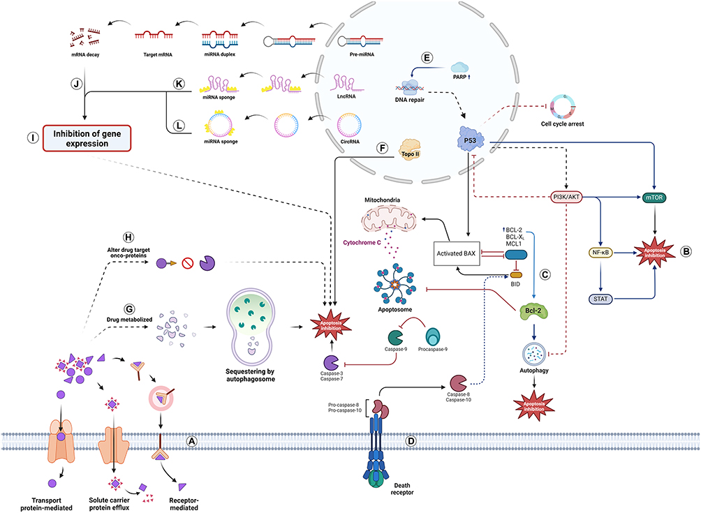 |
Figure 1 An overview of drug resistance mechanisms in cancer cells. (A) Anti-cancer Drug Efflux: Cancer cells depend on various transport mechanisms, including solute carrier (SLC) protein, receptor-mediated, and transport protein-mediated efflux, to expel anticancer drugs, thereby reducing their intracellular concentrations. (B–D) Apoptosis blocking pathways: Reveal the crosstalk between intrinsic and extrinsic through the signaling pathways. In IIA these pathways are involved in regulating apoptosis and their activation promoting apoptosis inhibition; In IIB, the loss-of-function of p53 and inhibition of pro-apoptotic signals, such as increased BCL-2 levels, prevents the formation of the apoptosome, a critical complex for initiating apoptosis; In IIC, the death receptor pathway can trigger apoptosis through receptor-mediated mechanisms by activation of caspases while dysfunction promoting survival signals. (E) DNA Repair Pathway Activation: Increased levels of poly (ADP-ribose) polymerase (PARP) enhance DNA repair mechanisms, allowing cancer cells to survive DNA damage caused by therapies. (F) Decreased Topoisomerase II Activity: Reduced activity of Topoisomerase II contributes to the resistance against drugs that target DNA replication and repair. (G) Drug Metabolism: Anticancer drugs are metabolized within the cell, forming metabolites that autophagosomes can sequester. (H) Alteration of Drug Targets: Oncogenic proteins may be modified, diminishing the effectiveness of targeted therapies. (I-L) Inhibition of Gene Expression: The presence of sponges on microRNA (miRNA) (VA) leads to the degradation of target mRNA; in circular RNAs (circRNAs) and long non-coding RNAs (lncRNAs) resulting in translational repression and leads to inhibiting the expression of pro-apoptotic genes and promoting survival pathways. |
Mechanisms of Drug Resistance
Drug Efflux Pumps
MDR in tumor cells is primarily caused by the increased expression of drug efflux pumps, specifically adenosine triphosphate (ATP)-binding cassette (ABC) proteins (Figure 1A).18 These proteins, classified into various subfamilies (ABCA, ABCB, ABCC, ABCD, ABCE, ABCF, and ABCG), are crucial in pumping out chemotherapeutic drugs from the cell, reducing their therapeutic concentration. One of the well-known efflux pumps is P-gp, encoded by the MDR1 gene. P-gp is highly expressed in cancer cells and is associated with low therapeutic efficacy, especially in colorectal cancer (CRC). Its overexpression leads to the expulsion of drugs like paclitaxel (PTX) and doxorubicin (DOX), resulting in drug resistance.21,24 Advanced DDS, known as nanomedicines, have been developed to overcome MDR. These systems utilize NPs, nanotubes, micelles, liposomes, and metal nanomaterials to bypass P-gp-mediated drug efflux. NPs can achieve drug release under specific conditions such as pH, hypoxia, and reducibility, allowing drug accumulation in tumors through passive targeting effect, ie, enhanced permeability and retention (EPR) effect or active targeting, ie, ligand-receptor binding (Figure 1A). They can also utilize the endosome-lysosome pathway to transport drugs into cells instead of relying on passive diffusion. Certain substances or groups, such as clathrin, can attach to glycoproteins or lipoproteins on the cell membrane, causing the membrane to curve and form early endosomes. These endosomes can then encapsulate NPs or other cargo and transport them inside the cell. This mechanism, clathrin-mediated endocytosis (CME), has been extensively utilized in reversing MDR.18 Solute carrier (SLC) transporters (Figure 1A) also play a crucial role in cancer drug resistance by regulating the cellular uptake of anticancer drugs and essential nutrients. The expression levels of SLC transporters in tumors and host tissues significantly impact drug efficacy, side effects, and interactions. Downregulation of SLC transporters in cancer cells can lead to ineffective therapy.25 This resistance mechanism has been observed in various malignancies, including multiple myeloma.26 In multiple myeloma, both ABC and SLC transporters contribute to resistance against novel therapies like proteasome inhibitors and immunomodulatory drugs.26 To target the problem of drug efflux pumps, novel efflux pump inhibitors can be screened using small-molecule/natural product/repurposed drug libraries, screening for anti-adaptive molecules, designing peptide-based inhibitors, and applying CRISPR Cas to fine-tune their functions. Among the other interesting approaches are prodrug design, photosensitizers, and photodynamic therapy (PDT) to damage drug efflux pumps, as well as artificial intelligence (AI)-based structural modification of potential drug candidates.
Inhibition of Apoptosis Pathways
Most anticancer treatments aim to induce apoptosis, or programmed cell death, in tumor cells.27 Cell death is primarily regulated by necrosis, apoptosis, and autophagy.28 Apoptosis occurs through two pathways: the intrinsic pathway is controlled by mitochondria and involves B-cell leukemia/lymphoma 2 (BCL-2) family members, caspase-9, and protein kinase B (or Akt) (Figure 1B and C). The extrinsic pathway is initiated by death receptors on the cell surface (Figure 1D). Both of these pathways converge through caspase-3 and 7 activation, resulting in apoptosis. However, there are additional interactions between the pathways. In various cancers, the overexpression of anti-apoptotic proteins, viz, BCL-2 and Akt, and the increased activity of downstream transcription modulators like NF-κB and STAT are observed, making them promising drug targets. Resistance to chemotherapeutics can be associated with the up-regulation of anti-apoptotic (eg, BCL-2, AKT) and down-regulation of pro-apoptotic genes (eg, Bax, Bcl-xL) in tumor cells. An imbalance between anti-apoptotic (such as BCL-2, Mcl-1, and Bcl-xL) and pro-apoptotic proteins (such as Bax, Puma, Noxa, Bak, Bil, and Bid) has been linked to drug resistance in cancer.24,28 Overexpression of BCL-2 can reduce tumor cell death and lead to treatment resistance. Studies have shown that excessive BCL-2 expression can contribute to MDR in lymphoma cells, colon cancer, and gastric cancer. This upregulation of BCL-2 can diminish the apoptosis triggered by chemotherapeutic drugs and decrease the cells’ susceptibility to these drugs. BCL-2 overexpression has also been shown to increase cellular resistance to drugs like cisplatin (CP), docetaxel (DTX), and methotrexate (MTX). Mutations in apoptotic functions can mediate MDR in tumor cells, making them resistant to many chemotherapeutics that induce apoptosis. These mutations also play a crucial role in carcinogenesis and cancer development. For example, the wild-type P53 gene regulates normal cell growth, triggers programmed cell death, and prevents abnormal cell growth (Figure 1E and F).16 However, the mutant P53 gene can render cellular DNA damage irreparable, compromising the link between DNA damage and apoptosis induction.16,28
Reduced Drug Uptake
Drug Inactivation
The other drug resistance mechanism may be the drug’s lack of activation or inactivation due to alterations in enzymatic conditions associated with cancer (Figure 1G).29 In vivo, drug activation involves complex interactions between proteins and substances, potentially forming complexes or undergoing partial degradation. Many anticancer medications require metabolic activation for clinical efficacy, yet diminished activation may lead to tumor cell resistance, highlighting the importance of careful drug activation.30 An illustrative instance is the alteration and reduction of phosphorylation occurrences in converting Cytarabine (AraC) to AraC-triphosphate, a substance employed in treating acute myelogenous leukemia (AML).24 AraC does not affect the cancer cells, but its phosphorylated form is lethal in many cancers. Down-regulation or mutations that induce phosphorylation reduce the AraC activity, resulting in drug resistance. Additional examples of drug activations and inactivation mechanisms include the cytochrome P450 (CYP) system, glutathione S-transferase (GST) superfamily, and UDP-glycosyltransferase (UGT) superfamily.31 Synthetic lethality, generation of resistant prodrugs, new nanocarriers, and dual inhibitors can be interesting ways to target drug inactivation.
Alteration of Drug Targets
Targeted therapy utilizes medications that effectively damage cancer cells and specifically target crucial elements like genes, proteins, or the environment of cancer-specific tissues, which impedes cancer growth and enhances overall survival rates. However, the cancer cells can become resistant by modifying these pharmacological targets via genetic mutations or shifts in epigenetic gene expression (Figure 1H).32 An example of resistance caused by modifications to the therapeutic target is the identification and use of estrogen-related receptor inhibitors in breast cancer treatment. Patients with estrogen receptor (ER)-positive breast tumors are often prescribed tamoxifen (TAM), which functions by competing with estrogen for the ligand-binding domain of the ER. However, extended exposure to TAM usually leads to drug resistance. The resistance mechanisms can differ among cases, but two prevalent mechanisms include mutations in the ER gene and reduced expression of the ER.30
Role of Non-Coding RNAs
MicroRNAs (miRNAs) are short non-coding RNA (~21-24 nucleotides) molecules that regulate gene expression (Figure 1I).28,33 They are too small to code for proteins but are involved in controlling the expression of protein-coding genes, including those involved in cancer and drug resistance.28 Altered expression of miRNAs in cancer cells can lead to the development of MDR by misregulating the expression of genes essential for MDR.16,33 Research has shown that miRNAs can amplify tumor-promoting genes or genes related to apoptosis, cellular proliferation, and the cell cycle. Depending on its target tissue, the same miRNA can enhance or hinder chemotherapy resistance. For example, in breast cancer, increased expression of miRNA-21 leads to a decrease in the expression of PTEN protein, impacting the effectiveness of DOX in tumor targeting. Conversely, when PTEN is overexpressed, the expression of miRNA-21 is suppressed, reducing the resistance of breast cancer cells to DOX.24 Recent research has focused on the 3’ half of the miRNA’s involvement in target selectivity and gene regulation, suggesting that nucleotides outside the seed can influence miRNA function and existence, and can be investigated further. Moreover, recent advances in machine learning (ML) can help decipher drug-miRNA interaction, discovery of miRNA therapies, miRNA-miRNA pairings, generating miRNA disease-association models, miRNA treatment prediction, and determining synergy (Figure 1J). Long non-coding RNAs (lncRNAs) (Figure 1K) regulate drug resistance by influencing ATP-binding transporter expression, DNA damage response, epithelial-mesenchymal transition (EMT), apoptosis, and cancer stem cell formation.34 They are involved in resistance to multiple cancer drugs, including platinum drugs, TAM, trastuzumab, 5-fluorouracil (5-FU), PTX, and androgen deprivation therapy across the top five prevalent cancers.35 Along with other non-coding RNAs like microRNAs and circular RNAs, lncRNAs modulate the expression of specific target genes and m6A modification involved in drug resistance mechanisms by regulating RNA stability, localization, and translation.36 Circular RNAs (circRNAs) primarily function as miRNA sponges, regulating gene expression and signaling pathways in drug resistance (Figure 1L).37 They also regulate drug efflux, apoptosis, autophagy, DNA damage repair, immune cells, cytokines, metabolism, and tumor microenvironment (TME) interactions.37 They contribute to chemotherapeutic resistance in non-small cell lung and breast cancer by affecting ATP-binding cassette transporters, inhibiting apoptosis, and promoting autophagy.37,38
Influence of Tumor Microenvironment (TME)
Cancer cells can manipulate their surrounding area by releasing signaling networks that benefit their growth and survival. This interaction with the TME allows cancer cells to adapt and overcome stressful conditions, leading to cancer progression, metastasis, and drug resistance.10 The TME consists of various stromal cells, including tumor-associated macrophages,27 as well as abnormal physiological and biochemical characteristics such as low pH, hypoxia, and high levels of intracellular glutathione (GSH).27 Hypoxia, a condition of low oxygen levels, is commonly experienced by tumors due to uncontrolled cell growth and inadequate blood supply.16 The transcription factor HIF-1α plays a crucial role in hypoxia, as its overexpression has been observed in many human cancers.16 HIF-1α is rapidly degraded under normal oxygen levels, but under hypoxia, it escapes degradation and forms a functional heterodimer with the HIF-1β subunit. This heterodimer enters the nucleus and activates genes related to vascularization, metastasis, and drug resistance. HIF-1α also contributes to chemotherapy resistance in cancer cells.33,39 Nanocarrier-based strategies have been explored to target cancer hypoxia and improve the therapy, including the encapsulation and delivery of hypoxia-activated prodrugs (HAPs) to hypoxic cancers (Figure 2).22 To combat MDR cases, researchers are developing advanced nanocarriers that can bypass efflux pumps like P-gp. These nanoparticles can be engineered to release drugs in response to the acidic environment of tumors, which enhances drug accumulation in resistant cancer cells.40 For example, nanocarriers that encapsulate siRNA targeting P-gp inhibit its expression, thereby increasing the concentration of chemotherapeutics in resistant cells. Furthermore, the development of dual-action nanocarriers that respond to both pH and temperature changes allows for targeted delivery to resistant tumors.41 Combination therapy formulations that deliver both chemotherapeutics and agents inhibiting resistance pathways, such as paclitaxel alongside a P-gp inhibitor, represent promising strategies to overcome MDR in cancer treatment.42
 |
Figure 2 Schematic illustration showing how nanocarriers overcome the cellular barriers for drug delivery. Nanocarriers could deliver drugs into subcellular locations by passing a cascade of barriers, including interface barrier, endocytic pathway barrier, drug resistance, subcellular targeting barrier and controlled drug release. |
Targeting Mechanism of Nanocarriers
Engineering drug and gene delivery systems that are able to target affected cells with no influence on healthy cells is a significant challenge in cancer treatment. Understanding the biology of tumors and the interaction of nanocarriers with cancer cells is crucial to tackling therapeutic challenges and developing effective and efficient nanocarrier systems.43 Targeting strategies to deliver drugs and active agents to the desired organs and tissues include passive, active, inverse, ligand-mediated, physical, dual, double, and combination targeting (Figure 3).44
 |
Figure 3 Different strategies of drug targeting. (A) Passive, (B) Active, (C) Inverse, (D) Ligand-mediated, (E) Physical, F) Dual, (G) Double, (H) Combination. |
Passive Targeting
Passive targeting is a drug delivery system that targets the drug to the systemic circulation (Figure 3A).45 Nanocarrier-based cancer therapies primarily consist of first-generation nanomedicines that rely on passive targeting. The first-generation nanocarriers regulate biodistribution and pharmacokinetics by modulating the EPR effect, thereby enhancing nanomedicine accumulation in tumor cells.5 While the smaller particles have higher penetrability without leaking into normal vessels, the larger particles are more susceptible to being eliminated by the immune system.2 Solid tumors have inherent abnormalities of tumor vasculature, which can be effectively exploited for targeted delivery of anticancer NP agents. However, large-sized drugs cannot penetrate tight endothelial junctions of normal blood vessels, resulting in a long plasma half-life, which builds up in tumor tissues due to their abnormal vascular nature.44 EPR effect-based passive targeting is inefficient in controlling cytotoxic drug side effects and can negatively affect drug delivery due to cancer heterogeneity and stroma.5
Active Targeting
This strategy includes drug targeting by identifying a particular set of cells and attaching a drug delivery system to their receptors (Figure 3B).45 Active targeting methods incorporate ligands, such as antibodies or small organic molecules, which facilitate the uptake by the targeted cells. These ligands can target surface molecules expressed in the diseased cells, proteins, sugars, lipids in the organs, and molecules in the diseased cells’ microenvironment or the physicochemical environment.46 Ligands on the surface of NPs are selected to target the molecules overexpressed on the surface of cancer cells, allowing them to distinguish between healthy and targeted cells. The interaction between NPs’ ligands and cancer cells’ receptors triggers receptor-mediated endocytosis, enabling the successful release of therapeutic drugs. This active targeting approach is particularly suitable for delivering macromolecular drugs like proteins and siRNAs.2 This way, direct tumors or the mildly acidic TME, the TME’s vascularization, or the tumor nucleus can be targeted.
Inverse Targeting
Inverse targeting of drugs occurs when the reticuloendothelial system (RES) prevents colloidal carrier absorption, suppressing its regular function, which is achieved by pre-injecting blank carriers or macromolecules such as dextran sulfate, facilitating RES saturation and suppressing defense mechanisms, making it a practical method for targeting drugs to the non-RES body organs (Figure 3C).44
Ligand-Mediated Targeting
Ligands are affixed to carrier surfaces, guiding the carrier to designated locations. Colloidal carrier systems may be functionalized with biologically relevant molecular ligands like antibodies, polypeptides, oligosaccharides, viral proteins, and fusogenic residues. These ligands facilitate recognition and specificity, allowing targeted drug delivery (Figure 3D).47
Physical Targeting
This approach employs environmental changes like pH, temperature, light intensity, ionic strength, electric field, and specific stimuli like glucose concentration to locate the drug carrier, making it optimal for tumor targeting and cytosolic delivery (Figure 3E).12
Dual and Multiple Targeting Approaches
The dual targeting mechanism enhances the therapeutic effect by synchronizing carrier molecules with antiviral drugs, eg, enhancing the therapeutic effect when entrapped in a carrier molecule with antiviral activity (Figure 3F).45
The double targeting strategy combines the temporal and spatial methodologies to target specific organs, tissues, cells, or subcellular compartments with a controlled drug release rate to the target site (Figure 3G).12
Combination Targeting
These systems, which include carriers, polymers, and molecularly specific homing devices, provide a direct targeting approach (Figure 3H). 48 Recent studies have explored hybrid targeting strategies that combine passive and active targeting mechanisms to enhance drug delivery to tumors. Nanoparticles can be engineered to exploit the enhanced EPR effect, which allows them to accumulate in tumor tissues due to leaky vasculature.49 Additionally, these nanocarriers can be functionalized with ligands that specifically bind to overexpressed receptors on cancer cells, such as HER2 or folate receptors, enhancing tumor cell uptake.50 Magnetic nanoparticles that can be guided to tumor sites using external magnetic fields further enhance targeting precision, reducing systemic exposure and improving therapeutic outcomes.51,52
Impact of Nanocarrier Physicochemical Properties
Understanding the physicochemical properties of NPs is crucial for improving their concentration and functionality in tumor tissues.53 While advancements have been made in drug nanocarriers, few have been effectively implemented in clinical studies. The properties of these nanocarriers influence their in vivo biological barriers, which can affect the therapeutic index of the cargo and the desired outcome.54 Comprehension of the physicochemical peculiarities of nanocarriers is essential since their size, shape, surface charges, and chemistry affect drug distribution, behavior, and delivery efficiency.54
Shape
The shape of nanocarriers is an important property affecting their behavior in vivo. Different shapes, such as rods or spheres, can impact processes like uptake by macrophages, circulation in the blood, distribution in tissues, and targeting of diseases.54 Factors like the curvature structure of NPs and the time needed for cell membrane wrapping can influence the interactions between nanocarriers and biological systems. For example, rod-shaped NPs have a larger contact area with cell membrane receptors but longer internalization than spherical NPs.53 Gold nanorods have lower uptake efficiency than spherical particles, which decreases as the aspect ratio increases.55 Anisotropic nanocarriers with a non-uniform shape have distinct interactions with cells and drugs. Their increased surface area allows for better drug encapsulation and delivery through localized degradation. Additionally, their larger surface area facilitates binding to specific cells and enhances targeting.56
Size
The size of nanocarriers is a crucial physicochemical characteristic affecting their in vivo behavior since it directly influences the available surface area for interaction with biological environments. Plasma proteins attach to nanocarriers, creating a protein corona, which gives them a unique “biological identity” and determines their interactions with the phagocytes.54 While nanocarriers of less than 5 nm in diameter can be rapidly cleared from the blood, those of more than 15 μm can be accumulated in the liver, spleen, and bone marrow. The optimal size for DDS is currently considered 100–200 nm, as this size allows the EPR effect in tumors and avoids filtration in the spleen.57
Surface Chemistry
Surface chemistry design plays a critical role in the development of nanomaterials for biomedical applications. Research studies have shown that the surface chemistry of polymer-based nanomaterials significantly affects their interactions with cells.46 Surface modification of NPs ensures their stability in physiological environments. However, the reactive nature of NP’s surface can also influence biological responses.58 Surface functionalization of NPs prevents their aggregation and minimizes the non-specific cellular absorption. Additionally, surface chemical modification can reduce the potential hazards of NPs and establish a safe-by-design approach for their future use. Overall, surface chemistry design is essential for optimizing the performance and safety of nanomaterials in biomedical applications.53
Surface Charge
The surface charge of NPs is critical to their interaction with biological systems. Their surface charge influences cellular uptake, while positively charged nanocarriers show better internalization and higher efficiency than negatively charged ones.46 However, the positive surface charge also rapidly removes NDDSs from the bloodstream. The interaction between negatively charged albumin and positively charged NPs is the main factor in removing cationic nanocarriers.59 Surface charge also affects the fate of nanocarriers in vivo by influencing the biodistribution, opsonization process, and plasma protein adsorption. High surface charge densities lead to an acceleration of blood clearance and capture by the RES, while neutral charges promote prolonged blood circulation and reduced clearance by the RES.54
Types of Nanocarriers in Cancer Therapy
Nanocarriers are colloidal particles smaller than 500 nm that are used to encapsulate drugs and biological materials. They can be made from various materials, including (i) organic NPs, such as solid lipid NPs, polymeric micelles (PMs), liposomes, nanoemulsions (NEs), polymeric NPs, and exosomes, (ii) inorganic NPs, such as carbon-based nanomaterials, metals and metal oxides, quantum dots (QDs), and (iii) hybrids (Figure 4). Stimuli-responsive nanocarriers are the third generation of controlled-release DDS, allowing precise drug release within target cancer cells. Their benefits include improving drug pharmacokinetics and biodistribution, enhancing solubility and permeability, minimizing therapeutic doses, and reducing toxicities. At the same time, modifying the properties, such as composition, size, shape, and surface properties of nanocarriers, can protect drugs from premature degradation to overcome MDR in cancer cells. Cancer cell-targeted drug delivery strategies often involve nanocarriers with cancer cell-specific ligands on their surface and mechanisms that cause the release of drugs within the cytoplasm of cancer cells.60 The development of third-generation nanocarriers, including stimuli-responsive and hybrid systems, marks a significant advancement in cancer therapy. These carriers can be designed to release their drug payloads in response to specific triggers such as temperature or light, allowing for controlled drug release at the tumor site.61,62 For instance, light-activated nanocarriers can release DOX upon exposure to specific wavelengths of light, while biodegradable polymeric nanoparticles made from materials like poly(lactic-co-glycolic acid) (PLGA) degrade into non-toxic byproducts, enhancing safety and reducing long-term toxicity.63 Additionally, temperature-sensitive nanocarriers that release drugs in response to hyperthermia enable localized treatment of tumors, providing a targeted approach to cancer therapy.
 |
Figure 4 Classification of nanocarriers for cancer therapy. |
Organic Nanocarriers
Lipid-Based Nanocarriers (LBNCs)
LBNCs, a class of nanomedicines with FDA approval, offer benefits like easy formulation, self-assembly, biocompatibility, high bioavailability, large payload capacity, and adjustable physicochemical features. They are classified as liposomes, nano-emulsions, and solid lipids.64
Innovations in lipid-based nanocarriers, particularly polyethylene glycol (PEG)ylated liposomes, have improved their circulation time and stability in the bloodstream, enhancing their effectiveness in cancer treatment.65 Enhanced formulations of liposomes, such as Doxil, encapsulate doxorubicin for breast cancer treatment, reducing immunogenicity and improving pharmacokinetics. New developments also involve combination therapy liposomes that co-encapsulate multiple drugs, allowing for synergistic effects against cancer cells.66 Furthermore, lipid nanoparticles specifically designed for the delivery of messenger RNA (mRNA) vaccines or therapies have shown promise in improving stability and cellular uptake, representing a significant advancement in the field of cancer immunotherapy.67
Liposomes
Liposomes, spherical structures made up of phospholipids, are widely used in drug delivery, particularly in cancer treatment. They have several advantages, including compatibility with both hydrophilic and hydrophobic drugs, low toxicity, extended-release potential, and the ability to carry large drug payloads and protect them from the external environment. Because they are biocompatible, biodegradable, and non-immunogenic, liposomes are suitable choices for DDS. However, liposome has limitations, including possible carrier toxicity and challenges in large-scale production and stability.68–70 One of the most successful applications of liposomes is the delivery of chemotherapeutic drugs/agents for cancer treatment. There are several approved and commercially available liposomal formulations for cancer therapy, such as Vyxeos®, Onivyde®, Marqibo®, DepoCyt®, and Doxil®. In the year 2024, the FDA approved the use of liposomal irinotecan as a first-line therapy for pancreatic adenocarcinoma. These liposomal formulations have shown promising results in improving drug delivery and reducing toxicity.69 Akbari et al developed nanoliposomes containing hydrophobic hydroxyurea (HU) for breast cancer cell therapy. The HU-loaded nanoliposomes were 174 nm in size, not significantly larger than blank ones, and showed a rapid release over 5 hours, followed by a sustained release for up to 36 hours. This release pattern was attributed to adsorbed HU on the nanoliposome surface, liposome erosion, and HU diffusion mechanisms. The nanoliposomes also demonstrated high cellular internalization and uptake in breast cancer cells, indicating their affinity for the cells, which led to an increased accumulation of HU.71 To improve the properties of liposomes, researchers have attached PEG to the liposomes and developed PEGylated liposomes,72 which can prolong the blood circulation and protect the drug from clearance by the mononuclear phagocyte system (MPS).73 PEGylated liposomes modified with PEG2000 have enhanced blood retention.74 Some ionizable cationic liposomes have a surface charge of neutrality under physiological conditions after being loaded with nucleic acids.75,76 Altering the composition of liposome head groups, lipid tails, and cholesterol quantity can also affect cellular uptake and tumor penetration in specific cancer types like glioblastoma and triple-negative breast cancer (TNBC).77 Liposomes have emerged as a promising tool in gastric cancer therapy, mainly combined with SATB1 siRNA, CD44 antibodies, and DNA complexes. These formulations facilitate enhanced drug accumulation in tumor-bearing mice, demonstrating superior targeting precision and effective silencing of SATB1 gene expression in CD44+ gastric CSCs. This approach significantly improves the therapeutic efficacy for this challenging cancer type.78 Additionally, Myocet®, a non-PEG-DOX liposome, has been utilized in temozolomide-based chemotherapy for high-grade glioma, establishing the maximum tolerable dose for patients aged 28 to 65 and diagnosed with gliomas who received nanoliposomal irinotecan.79 Furthermore, the combination of PEG-DOX liposomes with lapatinib is the most effective treatment strategy for individuals with HER2-positive breast cancer. In xenograft models, liposomal formulations loaded with DOX and Apache have been reported to elevate DOX levels in tumor tissues, enhancing therapeutic outcomes.80 Liposome nanoparticles, due to their unique structure, have been used in cancer therapy through gene therapy.81 They were applied in gene therapy through encapsulation of siRNA molecules and regulating the responsible genes in the multiplication of cancerous cells through gene modulation and silencing.82 In addition, liposomal nanoparticles helped destroy and kill cancer cells by delivering plasmid DNA- encoding tumor suppressors to tumor sites. Liposomes have also been used to deliver various diagnostic agents, such as64 Cu,14,83 Cisotopes,84 QDs,85 gadolinium (Gd)-based contrast agents,86 etc. Researchers hope that liposomes will soon be used as a simultaneous carrier of therapeutic drugs and diagnostic agents for cancer patients in clinical trials. Clinical trials have shown that liposomal formulations, especially liposomal doxorubicin, effectively treat various cancers like breast, ovarian, and lung cancer. Research highlights its cardiac safety and effectiveness as a standalone treatment and in combination with other therapies.87 Recent advancements include thermosensitive liposomes that release drugs in response to temperature changes, enhancing targeting and reducing side effects.88 In pediatric oncology, the use of liposomes is limited, but optimizing their design could improve drug delivery for children with cancer.89 Innovative approaches, such as dual pH-responsive liposomes, are being explored to release drugs specifically in tumor environments, while combining liposomes with immunotherapy shows potential for enhanced anti-tumor effects.90 The use of liposomal DNA delivery and DNA barcoding can be an efficient method to predict treatment responses.
Solid Lipid Nanoparticles
Solid lipid NPs are physiological fluids (room and body temperature) used to administer colloidal drugs, ranging from 50 to 1000 nm.91 These particles are contained within a drug-encapsulating matrix made of glycerides/fatty acids complex combinations.92 Nanotechnology can enhance patient prognoses. A steroid prepared by cold dilution techniques for chitosan-coated lipid NPs and filled with curcumin (CUR) could inhibit the growth of PANC-1 cells.93 PTX and 5-FU-charged lipid nanocarriers (such as Intaxel®) were used in managing male patients with LivC, shielding the medication from enzymatic deterioration by its plasma buildup and performance.94 Sorafenib (SOR) drug and superparamagnetic iron oxide NPs have also been co-loaded in SLNs for the dual treatment of HepG2 cells.95
Nanoemulsions
NEs are colloidal mixtures of two non-miscible liquids that exhibit thermodynamic instability. They are used as carriers in cancer treatment to deliver drugs to the targeted site. The drug is enclosed in the core of the NEs, which enhances its bioavailability and reduces its undesired action on other cells or tissues. NEs have droplets with diameters ranging from 10 to 200 nm and a protective coating of emulsifier molecules,96 which have several advantages in drug delivery. They have a higher drug solubilization capacity, better thermodynamic stability, long shelf life, rapid onset of action, and reduced inter-subject variability.97 In addition, NEs are non-toxic and non-irritant and permit easy application to the skin and mucous membranes.98 They can be formulated as foams, creams, liquids, and sprays, and if their formulation contains biocompatible surfactants, they can also be orally administered.99 NEs are particularly effective in delivering drugs through the skin, and due to their small size, they can penetrate the rough skin surface and enhance the penetration of active ingredients.98 Several NE delivery systems have been marketed in the past decade for various applications, including ophthalmic treatments for dry eye syndrome and oral immunosuppressive agents.100 In a study by Miranda et al, a NEs containing a novel fucoside derivative of Lapachol was prepared and characterized for intravenous treatment. The NEs with a size of around 190 nm and a zeta potential close to −20 mV showed excellent drug encapsulation efficiency (nearly 100%). The NEs also demonstrated sustained drug release, with approximately 50% of the drug remaining after 24 hours.101 The NE oil-in-water formulation, a toxoid drug associated with omega-3 fatty acids, has demonstrated a 12-fold reduction in the toxoid IC50 of PPT2 cells, resulting in a greater reduction in tumor volume in tumor-bearing mice than Abraxane™.102 Catechin-extract NE has also shown anticancer properties in PC-3 cells.103 DTX-NE and NE lipophilic diffuse methane show enhanced antitumor activity in A-549 cells. Nebulized DTX NE (MRC-5 cells) has succeeded more than normal cells, creating a standard framework for co-encapsulating gemcitabine (GC) and PTX with a ligand targeting the glucose receptor and revealing the potent synergism.104 Clinical studies such as Phase III of PTX intratemporal liposome injection have also shown successful results for lung cancer.105
Nanoscale lipid-based theranostic systems, particularly lipid-based nanoparticles (LNPs), are prospective carriers for cancer theranostic agents to cure different types of tumors. LNPs can be tailored with different targeting moieties and possess the potential to bypass different physiological barriers and improve the pharmacokinetics. Zhang et al106 developed a fluorinated nanoemulsion that significantly improved fluorescence imaging signals and revealed effective diagnosis of specific tumors, facilitating photodynamic therapy. Similarly, Hou et al developed a porphyrin shell-nanoemulsion that stabilized the oil core, resulting in a monodisperse nanostructure for imaging and phototherapy.107 Liang et al developed a theranostic nano platform using smaller (<20 nm) iron oxide loaded with porphyrin-grafted lipid nanoparticles (Fe3O4@PGLNPs), which demonstrated significant photodynamic effects against HT-29 cancer cells in vitro.108 The negligible toxicity, multifunctional capabilities, and flexibility in functionalization of LNPs position them as a vital tool in cancer theranostics, enabling effective treatment strategies across various tumor types.109
Polymer-Based Nanocarriers
Polymeric NPs have gained significant attention in biomaterials and drug delivery due to their unique properties and versatility. These NPs can be synthesized using synthetic and natural polymers, allowing customization and optimization for various biomedical applications.5,110,111 One of the key advantages of using polymeric NPs as drug carriers is their ability to deliver therapeutic agents directly to the target site, such as inflammation regions or the central nervous system, which is particularly important when dealing with potentially dangerous drugs that need to cross the BBB.110 Polymeric NPs can have a core-shell structure, with a hydrophilic shell and a hydrophobic core, effectively protecting the drug from degradation.112,113 They offer a controlled-release potential, allowing for prolonged drug delivery, and can also be manipulated to have different release kinetics, ensuring the drug is released at the desired rate and duration. This is beneficial, especially in the case of chemotherapeutic agents, where site-specific targeting and side effect reduction are crucial. Their distinct features, including drug solubility, stability, and selective accumulation, render them adaptable platforms for delivering therapeutic agents. In addition to drug delivery, polymeric NPs can be used in biomedical applications such as biosensors and catalysis. They can be designed to have different chemical compositions, charges, and physical structures, allowing for customization based on specific requirements.46 One commonly used polymeric NP is PLGA, known for its biocompatibility, biodegradability, and EPR effect.114 PLGA NPs have been extensively studied and utilized as drug carriers.115 For example, Zumaya et al developed a multifunctional PLGA-based nanocarrier system for co-delivering colchicine and purpurin 18 to human cancer cells. They compared the release of colchicine from PEGylated and non-PEGylated PLGA NPs, and the latter nanocarriers exhibited a slower and more sustained release profile.116 Wu-Cheng et al investigated the use of a nitric oxide (NO) conjugate of albumindinitrosyl iron unit, [(NO)2Fe(m-SCH2CH2OH)2Fe(NO)2] (DNIC-1), for oral delivery to activate hippocampal neurogenesis in chronic neuropathy, demonstrating effective mucosal absorption and sustained NO release without toxic byproducts. On the flip side, DNIU [Fe(NO)2]’s binding affinity to the cysteine residue (thiol group) in serum albumin oxidizes DNIC, which leads to the release of NO without toxic peroxynitrite formation.117 Yafang Qin et al, in 2022, developed luteinizing hormone-releasing hormone (LHRH) peptide-conjugated tumor targeting ruthenium (II) complex (Ru1-LHRH) that selectively targets the mitochondria, induces apoptosis by inducing reactive oxygen species (ROS) and activating the Caspase3/7 pathway, while imaging property reveals theranostic capabilities.118 Costagliola di Polidoro et al developed a hyaluronic acid conjugated Angiopep-2 (Thera-ANGcHANPs) for glioma targeting, which enhances imaging and selectively delivers therapeutic agents across the BBB, exhibits significant cytotoxic effects.119 Yan et al introduced a novel synthetic nanocomplex (NC) to overcome MDR cancer, contained synthetic redox-responsive polyethyleneimine (PEI) polymer (disulfide linked PSP), tetrahedral DNAs (TDNs) loaded with DOX drug (PSP/TDNs@DOX) that enhances drug delivery by creating pores-due to the difference in charge- in the cell membrane and facilitate the internalization of DOX while bypassing endocytosis pathways. PSP/TDNs@DOX NC revealed higher therapeutic efficacy in xenograft drug-resistant tumor mouse models, including human breast cancer (MCF-7/R) and ovarian cancer (SKOV3/R), due to its respond to GSH and DNase I to cleave and release DOX.120 Cai et al developed a theranostic system based on a small gadolinium chelate with PTX, linked via a cathepsin B-responsive linker, and covalently attaching a fluorescent probe, pheophorbide, to a branched glycopolymer. Their study demonstrated that this multifunctional system, derived from the glycopolymer prodrug in 4T1 xenograft mice, exhibited improved pharmacokinetics, enhanced tumor accumulation, angiogenesis inhibition, excellent biocompatibility, and significantly reduced gadolinium ion retention post-injection. The system also showed prolonged circulation time, a tumor inhibition rate exceeding 90%, and brighter MRI contrast intensity.121 The Incorporation of targeting ligands, such as antibodies, aptamers, and peptides, enhanced the specificity of these systems. For example, Zhong Yuan Chen et al reported aptamer-dendrimer-functionalized magnetic nano-octahedrons that consist of PAMAM dendrimer, zinc metal doped with superparamagnetic iron oxide, hydrophobic anti-cancer drug DOX, and HSP70/HSP90 siRNAs surface modified by PEG (ZIPPApt: DOX/siHSPs), which combines hyperthermia and as a chemotherapy theranostic system effectively enhanced apoptosis in 4T1 cancer cells.122 Additionally, Hosseini et al recently developed a theranostic dendrimer system to target CRC by attaching the cetuximab monoclonal antibody to PAMAM G4 and labeling it with Lu-177 via DTPA-CHX chelator on the mouse tumor and SW480 cancer cell line by expressing EGFR. Single-photon emission computed tomography (SPECT) images showed that the system caused apoptosis those cells that expressing epidermal growth factor.123
Micelles
Polymeric micelles (PMs) are nanosized molecules with a core-shell structure formed by the self-association of amphiphilic block copolymers in an aqueous solvent.110 They have gained attention as potential nanocarriers for the controlled delivery of hydrophobic anticancer drugs, which can enhance solubility, extend blood circulation time, improve cellular uptake, and passively target tumor cells through EPR.115 The advantages of PMs in drug delivery include biocompatibility, low toxicity, core-shell arrangement, micellar association, morphology, size, and stability.110 Their size (10–100 nm) is critical for the passive targeting of solid tumors, especially poorly vascularized ones. The outer surface of micelles is composed of components that do not react to tissue or blood, allowing them to remain in the blood for a long time without being recognized by phagocytic cells or specific proteins while protecting drug inactivation through the biological environment effect. This longevity is a significant feature of micelles as drug carriers.124 The core-shell structure of PMs consists of amphiphilic polymers, with the hydrophobic part carrying the hydrophobic drugs and the hydrophilic corona imparting hydrophilicity.125 Parameters such as cellular internalization, circulation, renal clearance, and the strength of the interface between the hydrophobic core and external aqueous environment are affected by the stability of micelles. The critical micelle temperature and the critical micelle concentration (CMC) determine the spontaneous assembly of micelles and play a key role in micelle formation. While the CMC micelle is formed at lower concentrations, the nanostructure can be dissolved, leading to a premature drug release.2 Lamey et al found that co-loading dasatinib (DAS) and celecoxib (CXB) into sodium caseinate micelles improved in vivo anti-breast cancer efficacy with reduced drug toxicity. The micelles showed sustained release of both drugs, with a faster CXB release than DAS due to their hydrophobic nature. The drugs’ encapsulation efficiency and compatibility with the micellar core influenced the release rate. The dual drug-loaded micelles showed minimal hemolytic rate, indicating low toxicity and also superior tumor growth inhibition in tumor-bearing mice, attributed to better cellular internalization by tumor cells.126 Another example is the development of a polymeric amphiphilic ionophilic micelle by Lu et al. This micelle had an ultralow CMC and exhibited excellent stability under serum dilution conditions. It enhanced the accumulation of DOX at the tumor site, eradicating melanoma tumors in mice and effectively reversing tumor growth.127 Genexol-PM is a micellar formulation loaded with PTX that is approved for cancer therapy. It is used for the treatment of NSCLC, metastatic breast cancer, and ovarian cancer.128 Having developed various novel amphiphilic block copolymers, micelle-based nanosystems have emerged as potential nano-drug delivery vehicles for insoluble drugs.129 Yun Zhu et al developed PMs containing Chlorin e6 (Ce6) using chitosan and hydrophobic polymethylacrylamide derivatives in another study, which were loaded with chemotherapy agent (DOX) and created Ce6-CSPD/DOX as a theranostic system. The system, which induces apoptosis in cancer cells while restoring fluorescence and photoactive properties.130 Inoue et al further revealed that combining TGF-β and chloroquine with nano-eruptions in a murine transplant model of GFP-labeled pancreatic tumor cells enhances the drug delivery efficiency and improves the diffusion of PMs through the tumor stroma by increasing tumor vessel diameter (chloroquine) and dynamic valve formation (TGF-b).131 Additionally, a study involving PEG-PLL conjugated PMs revealed significant accumulation at tumor sites and improved T1 imaging. The distinctive characteristics of polymeric micelles, including high loading capacity and structural flexibility, spot them as promising candidates for theranostic applications, with numerous nanomedicines currently in clinical trials targeting various cancers, particularly for delivering concisely soluble chemotherapeutic agents.132
Dendrimers
Dendrimers are a class of polymers extensively studied for their potential applications in nanomedicine,114 particularly in drug delivery.133 These artificial polymers have unique structural characteristics, such as high branching and well-defined, monodisperse structures.134 They can be synthesized at desired sizes and molecular weights, allowing precise control over their properties. One of the critical advantages of dendrimers is their ability to act as carriers for drugs. Due to their unique physicochemical properties and biodegradable backbones, dendrimers can effectively encapsulate drugs and deliver them to specific tissues or cells.135 The size of dendrimers, typically 2–10 nm in diameter,136 allows for efficient targeting of cancer cells or injured tissues. Additionally, dendrimers can be functionalized with multiple functional groups on their outer surface, further enhancing their ability to deliver drugs to specific targets.110 The structure of dendrimers also plays a crucial role in their drug delivery capabilities. The highly branched nature of dendrimers allows for incorporating multiple functional groups, which can be used to load drugs into the dendrimer core or conjugate targeting ligands onto the surface.134 This versatility in structure and functionalization enables dendrimers to effectively encapsulate drugs and enhance their solubility, stability, and release profile.7 For instance, Rai et al encapsulated chrysin in fucose-conjugated poly(amidoamine) (PAMAM) dendrimers and studied its anticancer activity against human lung cancer cells. The dissolution studies showed that the dendrimer formulation significantly improved the solubility of chrysin compared to the free drug. In both acidic and neutral media, the dendrimer formulation exhibited faster dissolution rates and higher drug release percentages than the free drug suspension, which can be crucial for their therapeutic efficacy.137 Furthermore, dendrimers have shown promise in overcoming the limitations of oral drug administration. The ability of dendrimers to penetrate intestinal membranes allows for improved drug absorption and bioavailability, which is particularly important as the permeability barriers in the gastrointestinal tract often limit the effectiveness of oral drug delivery. By controlling the properties such as size, structure, and solubility, researchers can optimize the dendrimers’ ability to overcome these barriers and enhance drug delivery.2 Due to unique properties, such as well-defined and monodisperse structures, dendrimers are also attractive choices for other biomedical applications, including gene delivery, imaging, and tissue engineering.138 By modifying the surface functional groups of dendrimers (eg, conjugating targeting ligands onto the surface endings) and loading drugs into their core cavities through hydrogen bonds, chemical linkages or hydrophobic interactions, researchers can tailor their properties to suit specific applications and enhance their performance.134
Polymersomes
Polymersomes are nano-sized hollow spherical structures that self-assemble from synthetic polymer/polypeptide amphiphiles, widely used in drug delivery applications. There are two main methods to obtain polymersomes, ie, solvent conversion and organic solvent-free.139 In the solvent conversion method, a block copolymer is dissolved in an organic solvent. Then, an aqueous phase is introduced, allowing self-assembly to occur. Finally, the organic solvent is removed by dialysis, resulting in the formation of polymersomes. In the organic solvent-free method, the organic solvent is removed by rotary evaporation, and then the system is hydrated to form polymersomes.140 Polymersomes have a similar morphology and structure to liposomes but are more stable due to their mechanical properties and tunable functions. They consist of a hydrophilic core and a hydrophobic bilayer, allowing the loading of cargoes with different characteristics.141 One of the critical advantages of polymersomes as delivery systems is their ability to be functionalized for accurate and targeted release at the tumor site.142 This can be achieved by adding targeted ligands such as antibodies, proteins, and carbohydrates, improving their biological distribution. Optimizing the chemical structure of the block copolymer in response to the tumor’s internal and external conditions, such as pH, oxidative stress, hypoxia, and enzymes, is also a practical approach to vesicle delivery.143,144 Designing polymersomes that are able to respond to these conditions as exogenous-responsive DDS allows for a controlled drug release with reduced side effects, as well as an improved therapeutic efficacy.143 Zhu et al evolved miRNA-190-Cy7 and Dox co-encapsulated nanoparticles, m-PPDCNPs, revealed no bio-toxicity, high loading efficiency, and precise tumor targeting. The use of membrane-coated PLGA-b-PEG DC-chol nanoparticles (m-PPDCNPs) successfully co-loaded DOX and miR-190, with DOX’s encapsulation efficiency reaching 46% and miR-190-Cy7’s 88%. Utilizing homologous targeting properties, m-PPDCNPs facilitated effective uptake in CRC cells and enhanced tumor tissue accumulation. The release of miR-190 inhibited VEGF production, thereby reducing tumor angiogenesis. While, miR-190 increased the sensitivity of HCT116 colon cancer cell to DOX, promotes apoptosis and enables combination therapy. Incorporating biomedical imaging, m-PPDCNPs have significant advantages in drug-delivery monitoring, tumor imaging, and vascular therapy. With abilities in drug delivery and monitoring and tumor imaging, m-PPDCNPs represent significant potential for CRC treatment and broader therapeutic applications.145
Protein Nanoparticles
Protein-based NPs can transport many molecules, including genetic materials, anticancer medicines, polypeptide hormones, growth factors, DNA, RNA, and more.146 Protein NPs offer unique advantages compared to other NP-based therapeutics, such as biocompatibility, easy surface modifiability, biodegradability, size controllability, and high loading capacity.147–149 They exhibit notable stability and long half-life, overcoming enzymatic degradation and renal clearance, allowing enhanced drug retention and accumulation in tumor tissues. Protein NPs can bind drugs through covalent conjugation and electrostatic /hydrophobic interactions, making them versatile carriers for therapeutic compounds.150 Various techniques can be used to prepare protein NPs, including emulsion/solvent extraction, complex coacervation, electrospray, and self-assembly.147 Many proteins can serve as the building blocks for protein-based NPs, such as albumin, collagen, ferritin, gelatin, casein, legumin, gliadin, elastin, whey proteins, silk proteins, soy proteins, and lectins.151,152 Albumin, in particular, has gained significant attention as a multifunctional nanocarrier for drug delivery153 because it encompasses both hydrophobic and hydrophilic domains and an abundance of charged amino acids.154 It is non-toxic, non-immunogenic, cost-effective, and can be produced in a wide size range (20–300 nm).155 Albumin-bound (nab™)-PTX (nab-PTX; Abraxane®) is an example of an albumin-based NP used for the treatment of various cancers such as NSCLC, metastatic breast, ovarian, and pancreatic cancer.155 Chen et al generated intelligent multicomponent albumin-coated MnO2 NPs using a one-step bio-mineralization technique that exhibits pH and H2O2 responsiveness, allowing for photodynamic and chemotherapeutic approaches.156 Environmentally friendly methods have also been employed for the production of albumin NPs, which can encapsulate several drugs and break the BBB without cross-linking agents.157 Yu et al developed gemcitabine andpheophorbide-a (P@) loaded human serum albumin (HSA)(P@-Gem-HSA) for treating lymphatic pancreatic ductal adenocarcinoma (PDAC) metastases, using human serum albumin (HAS) as a natural transporter (for superparamagnetic iron oxide, organic/inorganic oxides, IR780, IR825, and Ce6)158 for effective theranostics.159
Inorganic Nanocarriers
Metal-Based Nanocarriers
Due to their unique potential and physicochemical properties, metal NPs have attracted researchers’ attention, specifically in drug delivery and cancer diagnosis and treatment.160 These NPs can be synthesized with a controllable size and shape, a stealth ligand-coated surface able to evade the body’s immune system and circulate longer in the blood.161 Metal NPs, such as Ag, Au, Pd, Ti, Zn, and Cu NPs, exhibit improved optical properties and can be functionalized to attach targeting agents and active biomolecules using covalent bonds or electrostatic interactions such as hydrogen bonds.162 They also have a large surface-to-volume ratio, which allows for chemical modification, enhances cellular uptake, protects drugs within the biological medium, and improves bioavailability.163
Metal NPs can be used as carriers and contrast agents for active or passive tumor cell targeting. In passive targeting, the NPs can accumulate in tumor sites through permeability and retention due to the poor lymphatic drainage and defective vasculature of tumors. Functionalizing the surface of NPs with hydrophilic moieties, such as PEG, can improve drug solubility, prevent macrophage uptake, and shield enzymatic degradation during in vivo studies.164 Metal NPs can directly induce DNA damage and eliminate tumor cells, making them a promising tool for cancer treatment. By selectively targeting cancer cells and inducing DNA damage, metal NPs can trigger apoptosis or necrosis, release tumor antigens, and activate immune cells. They can also synergistically activate multiple pathways to trigger cell death mechanisms.165 However, preparing a stable colloidal suspension of metal NPs remains challenging due to metal-metal aggregation caused by their high surface energy. Researchers have used stabilizers like polyvinylpyrrolidone and polyacrylic acid to form a surface layer on metal NPs to reduce particle accumulation and improve the stability of the colloidal mixture.164 In addition, as a safe-by-design strategy to attenuate the intrinsic toxicity of some metal/metal oxide NPs, a superficial layer can be deposited on their surface, and these NPs have been capped by biocompatible natural compounds with a core-shell structure.166
Gold Nanocarriers (AuNCs)
Gold nanoparticles are emerging as promising agents for cancer therapy and are being investigated as drug carriers, photothermal agents, contrast agents, and radiosensitizers.167,168 Additionally, they can easily attach to biomolecules through Au-S bonds, further increasing their utility in biomedical contexts.169 Metal-based nanoparticles, particularly gold and silver nanoparticles, are being explored for their unique optical properties that can be harnessed for effective photothermal therapy. Gold nanoparticles, for example, can absorb near-infrared light to generate heat, leading to localized tumor destruction while minimizing damage to surrounding healthy tissues. Recent advancements in nanoparticle engineering have significantly enhanced cancer treatment by enabling targeted drug delivery to cancer cells while protecting healthy tissues. Stimulus-responsive nanoparticles facilitate regulated drug release, ensuring precise action against tumors.170 Gold nanoparticles are being used in gene therapy and photothermal treatment (PTT) to transfer nucleic acids into tumor cells, a potential frontier in molecular medicine for cancer treatment.112 This method eliminates cancer cells while minimizing harm to healthy tissues, enhancing the effectiveness of treatment. DOX is a highly effective chemotherapy drug but has significant toxicity to organs like the kidneys, liver, and heart.171 Lee et al examined the in vivo and in vitro anticancer activity of DOX-loaded DNA/Au NPs for ovarian cancer treatment. They evaluated the release of DOX from the DOX-DNA-Au NPs in different pH buffers. An initial burst release was observed within 5 hours, with a higher release in the acidic pH 5.6 compared to pH 7.4. The release of DOX continued to increase over 48 hours, reaching approximately 14.6% in the pH 7.5 buffer, which indicates the high stability of DOX-DNA-Au NPs and the possibility of pH-dependent cancer-targeting drug delivery. The cellular internalization of the NPs was investigated in ovarian cancer cell lines, demonstrating a high cellular permeability and successfully reaching the nuclei of the cancer cells. The DOX concentration increased with the treatment time with drug-loaded NPs, while the free DOX concentration remained constant. The in vivo ovarian anticancer activity in the mice model showed that the NPs significantly reduced tumor size compared to the control group and free DOX, and the tumor growth inhibition rate was about 2.5 times higher than that of free DOX. Furthermore, no accumulation of NPs in other organs was observed, indicating their successful distribution on tumor sites, inducing minimal side effects.172 Tam et al’s study reveals the potential of gold nanoparticles/PLA polymer nanospheres as a theranostic platform. Gold nanoparticles over 50 nm have significant potential in the near-infrared radiation (NIR) region for efficient clearance, while the 4 nm size, which was loaded into a polymeric nanocluster, leads to efficient renal clearance.173 There is an opportunity to investigate different gold nanoparticle shapes for organ-specific distribution, and generating luminescent gold nanoparticles (L-AuNPs) with long life is an opportunity for biomedical applications.
Silver Nanocarriers (AgNCs)
Silver nanoparticles not only deliver drugs but also possess antimicrobial properties, reducing the risk of infections in cancer patients. Additionally, magnetic nanoparticles can be directed to tumor sites using external magnetic fields, enhancing targeting precision and improving the overall effectiveness of cancer therapies.174,175 Gene carriers (Ag NPs) with a high affinity for binding genes provide a promising gene delivery system for treating various diseases, including cancer.161 Erdemir et al studied the effects of selenic acid (SA) and pyruvic acid (PA)-loaded silver nanocarriers on the viability of CRC cells. The researchers generated Ag NPs ranging from 2.32 to 5.61 nm in size, Ag-SA NPs from 3.61 to 13.54 nm, and Ag-PA NPs from 78.82 to 295.3 nm, and a gradual increase in the cumulative release of SA and PA from NPs was observed over time. The anticancer properties evaluations on the CRC cell line showed that Ag-SA NPs inhibited cancer cell viability, while Ag-PA NPs were ineffective. Additionally, PA was not cytotoxic and increased the CRC cell proliferation, but SA increased the cytotoxicity of Ag NPs by 5.3 times and demonstrated visible signs of apoptotic death and necrotic death in the treated cell groups.176
Platinum Nanocarriers (PtNCs)
Platinum-based anticancer drugs (eg, CP) have been widely used as a standard chemotherapeutic cancer treatment for many years. However, some of these drugs, such as carboplatin, oxaliplatin, and nedaplatin, often come with severe systemic toxicity and can cause side effects.177 Regarding this drawback, researchers have employed platinum-based NPs because they have the potential to accumulate at tumor sites and be absorbed by tumor cells, which makes them more effective and reduces the side effects.178 New prodrugs based on platinum (IV) have also been developed to minimize off-target interactions and side effects on healthy cells.179 These prodrugs are activated inside cancer cells, releasing cytotoxic platinum (II) drugs.180 Hydrogels containing platinum NPs have also been explored for tumor treatment. For example, an injectable and degradable photo-thermal hydrogel encapsulated in a platinum NP dendrimer led to complete tumor regression. Another study developed a biodegradable hydrogel for co-delivery of antitumor agents, which demonstrated superior efficacy and minimized systemic side effects.181,182 Recently, a two-layer fibrin-based multicomponent gel was presented as a local drug delivery system that could effectively suppress residual tumor cells after tumor resection and prevent recurrence and metastases.183
Molybdenum Nanocarriers (MoNCs)
Molybdenum disulfide (MoS2) is a promising functional material due to its tunable structure and exceptional physicochemical properties. Due to its capacity for easy surface functionalization and high drug adsorption, MoS2 is considered an ideal material for loading drugs. MoS2 nanoflower structures have better encapsulation efficiency, making them ideal for chemotherapeutic drug loading. The intense light absorption of MoS2 makes it a suitable carrier for photo-thermal and photodynamic therapeutic agents, and its different morphologies have shown promising results in controlled and targeted drug delivery, minimizing side effects and increasing therapeutic efficacy. While prior reviews mainly focused on the optical/thermal characteristics of photodynamic/photo-thermal therapy, the remarkable catalytic properties in cancer treatments are frequently disregarded.184 For intense, Soltani et al utilized exfoliated molybdenum disulfide nano-sheets modified with N-isopropyl acrylamide/methyl methacrylate and glutamine conjugation, achieving a maximum DOX adsorption efficiency.185
Metal/Metalloid Oxide Nanocarriers
Metal oxide nanoparticles, such as ZnO,186 SiO2,187 and TiO2 nanoparticles, have gained attention in drug delivery due to their unique properties. Metal oxide nanoparticles can be quickly loaded with drugs, functionalized with target agents, and localized to diseased tissue. They have high stability and a tunable shape and can be engineered to the desired size.188 They also have a negative surface charge, allowing further functionalization with different molecules.189
Zinc Oxide Nanoparticles (ZnO NPs)
ZnO nanoparticles, in particular, have been extensively studied for drug delivery and cancer diagnosis and treatment. Their small particle size allows for easy absorption by the human body, is cost-effective, non-toxic, and has a high drug-loading capacity.190 They can also be programmed for controlled drug release and targeted delivery. The ability to synthesize ZnO nanoparticles into hollow nanotube-type structures makes them suitable for prolonged drug-release applications.191 Gomaa et al conducted a study on a combined anti-tumor and anti-inflammatory approach using zinc oxide nanoparticles loaded with DOX and folic acid (FA). The drug-loaded nanoparticles showed a spherical structure with a size of 19–23 nm. In vitro assays showed that the treatment of Ehrlich ascites carcinoma (EAC) tumor cells with ZnO nanoparticles loaded with DOX and/or FA accelerated the growth inhibition of tumor cells in a dose-dependent manner compared to the cells treated with DOX.192
Silica Nanoparticles (SiO2 NPs)
Silicon dioxide (SiO2) -also known as silica- is an oxide of silicon that is recognized as a metalloid. Mesoporous silica nanoparticles, with their large pore sizes, diverse functionality, and biocompatibility, are ideal for creating synergistic nanoplatforms, serving as drug carriers and therapeutic agents in complementary chemotherapy.193 Silica nanoparticles are being utilized in early cancer treatment through enhanced drug delivery and imaging techniques, encapsulated with various chemotherapeutic agents.194 Application of these nanoparticles in imaging techniques like fluorescence and magnetic resonance imaging, providing high image resolution and real-time visualization of anatomical structures in the body, while also protecting drugs from degradation by enzymes, enhances solubility with the ability of controlled releases.195 A recent study has focused on a specific formulation of mesoporous silica nanoparticles coated with polydopamine (PDA), containing umbelliprenin with anticancer properties. The nanocomplex exhibited cytotoxicity against MCF-7 carcinoma cells and induced programmed cell death while showing low cytotoxicity to normal cells, indicating its safety. The study also demonstrated the induction of apoptosis in MCF-7 cells through the up-regulation of specific genes and fluorescent staining. The promising results suggest that further preclinical studies should be conducted to evaluate the potential use of this formulation in cancer treatment.196
Titanium Oxide Nanoparticles (TiO2 NPs)
Biocompatible TiO2 nanoparticles are being explored for tumor targeting and delivery of anticancer drugs like DOX, PTX, and platinum-based drugs (eg, CP) to the tumor site.197 Kim et al developed an ultrasound-driven system containing TiO2 nanoparticles to deliver DOX to tumor cells.198 Faria et al designed a novel system based on ZnS-doped TiO2 nanotubes to release 5-FU in physiologic conditions, which showed promising potential as an anticancer drug delivery system.199 Another example is the successful loading of DOX on TiO2 nanotubes through adsorption forces to develop an efficient pH-controlled release system for antitumor drug delivery.200 Studies showed that biocompatible coatings can inhibit the release of titanium ions and reduce the toxic effects of TiO2, thereby improving the performance of nanoparticles for drug release.199,200 For example, Pulit-Prociak et al modified TiO2 nanoparticles with GSH to release tadalafil, revealing an 82% decrease in active substance release compared to non-modified nanoparticles. The modified nanoparticles also enhanced Chinese hamster ovary cell (CHO) proliferation by over 60% and had a less cytotoxic effect by 37%. The materials showed satisfactory purity and surface morphology. The results of in vitro studies demonstrated that the modified TiO2 nanoparticles have a great potential for being applied as a drug carrier.201
Iron Oxide-Based NPs
Iron oxide nanoparticles hold significant promise as tools for cancer diagnosis and drug delivery. In modern theragnostics, they are used in hyperthermia therapy, which involves raising the temperature of cancer cells to induce cell death.202 Physicians utilize targeted delivery of iron oxide nanoparticles to tumor cells, enhancing the effectiveness of magnetic hyperthermia in destroying these cells. Additionally, iron nanoparticles are employed in regenerative medicine203 for magnetic cell labeling and tracking, allowing for the monitoring of cancerous cells via magnetic resonance imaging (MRI).204
Carbon-Based Nanocarriers
Research on nanocarbons, such as carbon nanotubes (CNTs), graphene, fullerene, nanodiamonds, and carbon nanoparticles, has been booming in recent years, particularly for developing delivery vehicles for imaging agents and drugs.205,206 These carbon-based nanomaterials have unique properties suitable for biological applications, including a high surface-to-volume ratio, thermal conductivity, rigid structural properties, and excellent biocompatibility. Accordingly, they can be used to deliver water-insoluble drugs, antigens, antibodies, and nucleic acids to cancerous cells. Another advantage is their ability to incorporate targeting and aromatic drugs via hydrophobic interactions or supramolecular π-π stacking, making them better suited to drug delivery platforms with enhanced loading capacity and sustained release ability.207 Carbon-based nanomaterials can be functionalized either covalently or non-covalently to enhance their biocompatibility, and can be combined with other diagnostic and therapeutic components. Furthermore, their tunable pore structure allows for reasonable control over drug release.205 Graphene, in particular, is an attractive candidate for drug delivery due to its large surface area, unique mechanical features, and easy functionalization.207 Potassium-containing graphene oxide nanocarriers were employed by Tiwari et al for dual delivery of gefitinib and camptothecin (CPT) anticancer drugs, and a better release result was reported for dual delivery compared to single delivery of each drug.208 CNTs also have exceptional chemical and physical characteristics that make them useful for drug delivery. Specifically, the large surface area of CNTs increases the interaction with biological material, and their high aspect ratio (width to height) provides excellent cell penetration. Besides, their conductivity facilitates the integration of muscular and nervous tissues.209 CNTs can be easily functionalized to target specific cancer cell surface receptors. Unlike traditional drug delivery approaches, CNTs do not require solvents, which reduces the damage to healthy cells caused by the drug.210 Yang et al showed that the functionalized single-walled CNTs (SWCNTs) as DOX carriers effectively treated MCF-7 cells, which suggests SWCNT-PEG-PEI reveals significant antitumor impact and drug delivery ability compared to CNT-COOH and CNT-PEG. Fluorescence-based studies and flow cytometry analyses indicate that SWCNT-PEG-PEI is more easily internalized, enhancing apoptosis and inducing tumor cell death due to its improved dispersibility and stronger affinity for cancer cells.211 In a study by Carneiro et al, a novel targeting drug delivery system was developed using multi-walled carbon nanotubes (MWCNTs), incorporating digoxin and FA. The release study of digoxin and FA under biological and slightly acidified pH conditions indicated a constant and non-pH-dependent release for FA, while a significant increase in digoxin release upon a pH change was observed.212 Carbon nanoparticles can also be used as biological agent carriers to enhance the therapeutic index at a tumor site. Alternatively, they can be combined with external stimuli, such as light, to induce irreversible physical damaging effects on cells. In a study,213 an intelligent theranostic platform was designed from an NIR-responsive core-shell hybrid nanocomposite using hyaluronic acid-PLA and hydrophobic carbon dots. This composite structure targets cancer cells that overexpress CD44 receptors and enables NIR-triggered chemo-phototherapy for tumors. Nicosia et al developed carbon nanodots modified with biotin and loaded with a high concentration of irinotecan for controlled release, imaging of MDA-MB231 and MCF-7 cancer cells, and their destruction through photothermal and chemotherapeutic methods. The results demonstrated that the developed system is a safe and promising theranostic complex for IG-PTT in breast cancer.214 Mirrahimi et al designed a structure of graphene oxide linked to superparamagnetic iron oxides and AuNPs, and the phase change agent, 1-tetradecanol, for drug release. The synergistic effect of NIR absorption of graphene oxide, X-ray attenuation of AuNPs, MRI contrast of iron oxides, and thermosensitive properties made this platform very suitable for controlled drug release.215 To overcome lysosomal degradation of RNAi, Wang et al introduced an amphiphilic fullerene derivative (C60-Dex-NH2) for siRNA delivery to cancer cells.216 The results showed that the synthesized structure can cause lysosomal trapping and membrane disruption by producing controllable reactive oxygen species under visible light irradiation. In another study, CNT-loaded ginsenoside (a component of puffed ginseng with anti-cancer activities) showed promise as a potential therapeutic strategy for TNBC immunotherapy.217
Quantum Dot-Based Nanocarriers
QDs are semiconductor nanocrystals with unique optical, thermal, and electrical characteristics.218 They are made of a few atoms and have been widely used in nanoparticulate DDS.219 QDs have sharp fluorescence and high photo-stability, which makes them ideal for in vitro and in vivo studies of particle trafficking.5 One of the main applications of QDs in cancer nanomedicine is targeted drug delivery. They can be modified with target ligands, such as antibodies or peptides, to enhance drug targeting and bioavailability in specific areas. QDs also improve drug stability, prolong in vivo circulation time, and strengthen drug distribution and metabolism.209,219 Amraee et al developed a delivery system based on functionalized graphene QDs loaded with TAM anticancer drugs. Functionalizing with FA and methoxy PEG led to a better binding to folate-positive breast cancer cells and a longer lifetime, respectively. They reported a sustainable and continuous increase in drug release over time, such that 89% of the whole drug was released after 42 hours, being effective against MCF-7 breast cancer cells.220 QDs are being utilized not only for their imaging capabilities but also as an innovative DDS. Their tunable optical properties enable precise tracking of drug distribution in vivo, which is critical for assessing treatment responses. Recent advancements include the development of QDs that can release drugs in response to specific stimuli, such as light or ultrasound, thus enhancing their therapeutic potential while minimizing side effects.221,222 Additionally, bioconjugated QDs linked to targeting ligands improve specificity for cancer cells, enabling more effective targeted drug delivery and imaging.223,224
Exosomes and Their Role in Targeted Therapy
Exosomes are extracellular vesicles produced by cells that act as cell-to-cell short/long-range interaction regulators. They carry various molecules such as nucleic acids, proteins, lipids, and metabolites. These vesicles have unique characteristics that efficiently deliver therapeutic payloads to target cells.225,226 They have low toxicity and immunogenicity and have been used to deliver therapeutic payloads such as siRNAs, immunological modulators, and chemotherapeutic medicines. They have shown promise in improving immunotherapy and suppressing immune function in cancer treatment. Exosomes have also demonstrated remarkable anticancer activity with reduced toxicity levels.227 Besides their therapeutic applications, exosomes also hold promise for disease diagnosis and prognosis prediction. Due to their high concentration of tumor-derived components, such as DNA, RNA, proteins, and lipids, exosomes can serve as potential biomarkers for tumor detection.227,228 Researchers have developed methods to modify exosomes for imaging, monitoring, and diagnostics, making them valuable tools in biomarker research.229 Furthermore, exosomes have been used to profile miRNA expression, which can provide insights into extracellular vesicle-based biomarkers, miRNA function, and liquid biopsy.230 Specific tumor-type-specific extracellular vesicles and particle proteins have been discovered, suggesting their potential use in diagnosing tumors of unknown sources.231 Biosensors capable of detecting specific exosome miRNAs have also been developed for cancer diagnosis.232 Yong et al developed a biocompatible drug carrier for targeted chemotherapy using exosome-biomimetic porous silicon nanoparticles (PSiNPs). After incubating tumor cells with DOX-loaded PSiNPs, exosome-sheathed porous silicon nanoparticles (E-PSiNPs) were generated by tumor cells and showed enhanced tumor accumulation and penetration into deep tumor parenchyma (Figure 5). Their significant cellular uptake and cytotoxicity in bulk cancer cells and CSCs led to a significant anticancer activity as well as CSC reduction in tumor models.233
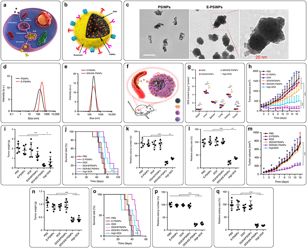 |
Figure 5 Schematic illustration of E-PSiNPs as drug carriers for targeted cancer chemotherapy. (a) Schematic illustration of the preparation of DOX@E-PSiNPs. After incubation, DOX@PSiNPs are endocytosed into cancer cells and localized in multivesicular bodies (MVBs) and autophagosomes. After MVBs or amphisomes fuse with the cell membrane, DOX@E-PSiNPs are exocytosed into extracellular space. (b) Schematics showing how DOX@E-PSiNPs efficiently target tumor cells after intravenous injection into tumor-bearing mice. (c) TEM images of PSiNPs and E-PSiNPs. Scale bar: 200 nm, (d and e) Hydrodynamic diameter of PsiNPs, E-PSiNPs, and DOX@E-PSiNPs by DLS analysis. (f) Schematic illustration of the efficient accumulation of DOX@E-PSiNPs in tumor tissues. (g) DOX content in tumor tissues and major organs of H22 tumor-bearing mice at 24 h after intravenous injection of DOX, DOX@PSiNPs or DOX@E-PSiNPs at DOX dosage of 0.5 mg kg−1, or high dosage of DOX at 4 mg kg−1. (h and l) Anticancer activity of DOX@E-PSiNPs in H22 tumor-bearing mice (h) Tumor volume; (i) Tumor weight; (j) Survival rate %; (k) Relative colony number and (l) The size of tumor spheroids). (m–q) Anticancer activity of DOX@E-PSiNPs in orthotopic 4T1 tumor-bearing mice. (m) Tumor volume; (n) Tumor weight; (o) Survival rate %; (p) Relative colony number and (q) Size of tumor spheroids). p-values were considered significant at * p < 0.05, ** p < 0.01, and *** p < 0.001.233 |
Molecularly Imprinted Nanoparticles
Molecularly imprinted polymers (MIPs) are classified as synthetic materials capable of binding to target molecules through their specific recognition sites.234 These materials have high affinity and selectivity, making them useful in various applications. In nanotechnology, MIPs can be found in different forms, such as spherical NPs, nanorods, nanofibers, and nanofilms, while those smaller than 200 nm have gained attention in cancer treatment. Nano-MIPs have a high surface area to volume ratio, allowing for efficient binding of target molecules. They also have fast binding kinetics, excellent dispersion and processability, and can be easily functionalized and modified. Additionally, nano-MIPs are compatible with various medical devices and materials used in vivo.234,235 Scientists have developed several synthetic approaches for nano-MIPs preparation, including emulsion polymerization, precipitation polymerization, solid phase imprinting, and surface imprinting.236 Strategies like nano-precipitation imprinting, monomolecular imprinting, and self-assembly imprinting have also been developed but are less used nonetheless.234,236 Li et al synthesized water-compatible hollow MIPs with hydrophilic polymer brushes on the surface, allowing for the delivery of drugs in aqueous environments.237 Lu et al developed biodegradable silicon nanoparticles loaded with a model protein and modified with a sialic acid imprinting layer. These nano-MIPs targeted tumor cells, overexpressing sialic acid, and released the loaded protein to induce apoptosis.238 Nano-MIPs have also been used in immunotherapy, and Gu et al developed anti-Programmed death-1 (PD-1) nano-MIPs that could block the PD-1/Programmed death ligand-1 (PD-L1) interaction, activating T-cell functions and reversing chemotherapy resistance in tumor cells.239
Hybrid Nanocarriers
Hybrid nano-vehicles, formed by combining two or more different NPs, offer numerous advantages, such as improved therapeutic efficacy, targeted delivery, reduced toxicity, and triggered release. The development of hybrid nanocarriers has revolutionized the drug delivery system by harnessing the combined effect of different components.240 The current trend in the development of hybrid formulations allows for the synergy of beneficial properties from various systems, such as organic/inorganic, polymer/inorganic, lipid/inorganic, and lipid/polymer. This approach enables the design of nanocarriers with enhanced functionalities and tailored release profiles.240–245 Hybrid nanoparticles are pharmacokinetically similar to drugs or small molecules and have shown great promise in cancer therapy through their efficient accumulation/retention at tumor sites, high penetration ability, and ability to carry/release chemotherapeutics in response to internal/external triggers.246 One example of hybrid nanocarriers is the lipid-polymer hybrid (LPH) system, which combines the benefits of lipid-based carriers, such as better loading capacity and biomimetic nature, with the architectural benefits of polymeric carriers, acquiring desirable release profiles and targeting capabilities.247 In a study by Rajana et al, FA-decorated palbociclib-loaded lipid/polymer hybrid NPs were fabricated using a quality-by-design approach. These hybrid NPs exhibited a uniform distribution with sustained drug release, and cellular uptake studies confirmed the uptake of nanoparticles by breast cancer cell lines.248 In another study by Korganbayev et al, copper oxide nanoparticles were encapsulated in polymer nanospheres composed of PLGA and PDA/PEG. This theranostic nanosystem induced a photothermal response in the NIR range (808 nm) and increased temperature in tumors, making it a suitable construct for photothermal chemotherapies combined with diagnostic imaging for cancer treatment.249 Liu et al synthesized a pH-responsive biocompatible hybrid polymer of Fe-Gallic acid hollow nanospheres with a combination of bovine serum albumin and DOX. This combination effectively responded to the TME in vitro and in vivo, releasing DOX and Fe (III) ions, which consumed GSH in cancer cells, generating Fe (II) for the Fenton reaction, which ultimately led to the generation of hydroxyl radicals for chemodynamic therapy. In addition, it effectively converted NIR light into heat as a therapeutic agent and enhanced the quality of MRI imaging.250 Gao et al designed a mesoporous Fe-MIL-53-NH2 nanomaterial as a carrier for the anticancer drug 5-FU and 5-carboxyfluorescein as a fluorescence imaging agent, as well as the targeting agent (FA). This transgenic metal-organic framework (MOF) showed excellent biocompatibility, high tumor uptake, significant anticancer effects, and good fluorescence and MRI imaging performance.251 In another study, a surface modification by covalent bonding of FA and 5-carboxyfluorescein was performed on 5-FU-loaded UIO-66-NH2 nanoparticles (20–200 nm). These theranostic MOFs showed improved fluorescence imaging and enhanced antitumor efficacy due to sustained drug release.252 Cherkasov et al synthesized a core-shell structure consisting of magnetic nanostructures carrying DOX and daunorubicin, and a polymeric outer surface linked with an antibody. The biocompatible nanoMOFs effectively carried therapeutic drugs at optimal concentrations and gradually released them near target cells.253 Ni et al synthesized nanoscale hafnium-porphyrin-Fe MOFs and, with low-dose X-ray irradiation, they not only destroyed primary tumors but also suppressed distant tumors by activating systemic antitumor immunity.254 To enhance radiotherapy and reduce the risk of postoperative recurrence, Chen et al synthesized and investigated FA-modified hafnium-based manganoporphyrin nanoMOFs.255
Smart Nanocarriers
Smart NPs are a promising option for cancer therapy because they can be triggered by specific stimuli and deliver drugs to targeted sites. Unlike conventional NPs, smart NPs can aggregate at the desired location and release their payload, resulting in a more precise and controlled treatment. This targeted drug delivery reduces the toxicity to healthy tissue and improves the effectiveness of cancer treatments.256–258 Stimuli-responsive NDDSs are used to deliver therapeutic drugs to tumor sites without affecting surrounding areas. The advantages of such systems include reduced side effects, improved pharmacokinetics and pharmacodynamics, and controlled drug release.259 They benefit from the EPR effect, which allows the nanocarriers to accumulate in the tumor.260 Smart nanocarriers should possess specific characteristics such as not being eliminated by the body’s immune system, accumulating only at the target site, releasing the cargo at the right concentration upon stimulation, and having the ability to co-deliver different substances such as chemotherapeutics, genetic materials, and imaging agents.261 Stimuli-responsive NDDSs, by altering their physicochemical characteristics, intelligently respond to specific stimuli, either endogenous (ie, generated in the TME, such as pH, redox potential, overexpressed proteins, and enzymes) or exogenous (ie, applied from outside, such as light, ultrasound, magnetic, or thermal), which results in higher drug release through the EPR effect.259
Stimuli-Responsive Mechanisms
Endogenous Stimuli
Hypoxia in tumor cells produces reducing molecules like reductase and GSH,262 which are crucial for maintaining intracellular redox balance.259 The GSH level in the TME of tumor mesenchymal stem cells is around 0.5×10⁻³ M, indicating more extraordinary reduction conditions and hypoxia than normal or healthy cells.263 This difference triggers the controlled release of redox-sensitive NDDSs.262 Bu et al evaluated a redox-responsive alginate–ibuprofen derivative-based drug delivery system. They found that the release of DOX from the system was slow in a neutral pH environment but significantly increased in the presence of GSH, a reducing agent commonly found in tumor cells, which indicates the system can be triggered to release the drug in a tumor-specific environment.264 Gupta et al studied redox-responsive NPs loaded with DOX and CUR. They observed that the release of both drugs was enhanced in the presence of GSH, leading to higher cumulative release rates and demonstrating the potential for dual drug delivery in CRC.265 Liu et al developed self-assembled micelles for DOX delivery and showed that the cells could take up the micelles, and the drug could be released rapidly at the target site, enhancing the antitumor efficacy. This system was able to track the drug location and release it precisely at the tumor site.266 The metabolic pathway of tumor cells differs from that of normal cells, leading to glycolysis and lactic acid production. However, abnormal blood vessel distribution and lymphatic reflux in tumor sites prevent timely lactic acid removal, resulting in acid accumulation and an acidic TME.267 The pH of tumor tissue (mostly ranging from 6.5 to 7.2), which is lower than that of normal tissues, can act as a trigger for selective anticancer drug release.268 The acidic conditions can lead to the protonation of functional groups, such as carboxylic acids.269 This protonation increases the solubility and swelling of these polymers, which destabilizes the interactions between the drug and the polymer, allowing the encapsulated drugs to be released more easily.270 Moreover, the acidic environment can also promote the hydrolysis of ester bonds within these polymeric carriers, further aiding in drug release. This mechanism enables a more targeted delivery of therapeutics directly to the tumor site, which helps minimize systemic exposure and reduces the risk of side effects.271 Feng et al developed mesoporous silica nanocomposites for the delivery of bicalutamide. They found that the drug release rate could be regulated by altering the pH of the environment. This pH-responsive system showed promise as a carrier for anticancer drugs.272 Enzyme-responsive DDS can also differentiate target sites from healthy tissues by utilizing active enzymes in the tissue.273 Enzyme-sensitive carriers take advantage of the overexpression of certain enzymes in the TME to achieve targeted drug release. Candidate enzymes that act like molecular scissors, cleaving peptide linkers that hold the drugs within their carriers. When these linkers encounter higher concentrations of these enzymes, they undergo a process called hydrolysis, breaking down the peptide bonds.274,275 As a result, the therapeutic agents are released in a controlled manner, allowing for targeted delivery right at the tumor site.276 This approach not only enhances the effectiveness of the treatment but also helps reduce exposure to healthy tissues, minimizing potential side effects. However, variability in enzyme expression across different tumors and within tumor regions can lead to unpredictable drug release rates, complicating treatment strategies.277 Additionally, similar enzymes in healthy tissues raise concerns about off-target effects, resulting in unintended drug release and potential side effects.278 Proteases are particularly interested in creating such novel systems due to their over-expression in infectious diseases like cancer and inflammation.279 Since enzymes can catalyze chemical reactions under mild conditions, delivery approaches based on enzymatic-responsive nanomaterials and able to provide bio-specificity and selectivity can fulfil many therapeutic requirements. For instance, active tumor-targeting NPs integrated with site-specific enzyme-triggered moieties could enhance drug release at the tumor site, reduce uptake by non-targeted tissue, and improve targeting, permeability and drug accumulation time, which ultimately led to a better antitumor effect.262 Hsu et al investigated targeting drug delivery carriers for breast cancer using GSH-sensitive nanoparticles. They found that the release of DOX from the nanoparticles was triggered by the presence of GSH, leading to cytotoxic activity against breast cancer cells.280
Exogenous Stimuli
Light-mediated therapy uses extrinsic activation to prevent nonspecific drug release, enabling on-demand therapies and diagnosis in specific locations. Nanotechnology advances have enabled nanomaterials, including metal-organic frameworks, for light-responsive targeted DDS.281 Quazi et al designed a light-triggered delivery system containing AuNPs by entrapping a cancer-specific peptide drug (ie, Buforin IIb) within the DNA networks of a nanoscale DNA hydrogel. Irradiation of a 660 nm laser stimulated the flocculated AuNPs, which led to the denaturation of DNA hydrogel nanocomplexes and the release of the peptide drugs. Their results suggested this light-activated system’s efficacy in the delivery of anticancer peptide drugs specifically to cancer cells, without cytotoxicity or harmful effects on standard cell lines.282 Ultrasound waves have attracted researchers’ attention to be used as external stimuli in target-controlled drug release systems, due to their safety, non-invasiveness, proper spatiotemporal control, and tissue penetration.279 In this regard, important parameters such as sustained drug encapsulation, release performance in response to ultrasound waves, and release monitorability should be taken into account while designing such nanocarriers for therapeutic purposes.283 In a study by Wang et al, nanobubbles carrying androgen receptor siRNA were investigated for their anti-tumor effects on androgen-independent prostate cancer. In vitro studies showed that when combined with ultrasonic irradiation, the nanobubbles significantly inhibited the growth of cancer cells and suppressed androgen receptor mRNA and protein expression. Contrast-enhanced ultrasound also demonstrated that the nanobubbles had stronger signals than the control in the tumor area.284 Another study by Wang et al focused on ultrasound-responsive DDS for hepatocellular carcinoma. Ultrasound-induced structural destabilization accelerated the release rate of DOX payloads, leading to potent cytotoxic efficacy in affected cells.285 Thermosensitive nanocarriers such as micelles, hydrogels, liposomes, and dendrimers are another stimuli-responsive delivery system with beneficial features, including high solubility, stability, longevity, and responsiveness to local changes.273 Temperature plays a crucial role in cellular metabolism, and exposing pathological sites like tumor tissues to elevated temperatures induces hyperthermia. Heating tumor tissue increases pore size externally, blood flow, and perfusion, which enhances nanocarrier extravasation.283 Some researchers have studied the potential of liposome-micelle-hybrid carriers to enhance the co-formulation and delivery of PTX and 5-FU. It was reported that the drug release was more pronounced at higher temperatures, and the release profile also varied depending on the carrier type. The hybrid nanocarrier also increased the permeability and transport of PTX across cell monolayers and enhanced cytotoxicity against cancer cells.286 An effective system for dual delivery of DOX and carboplatin drugs to breast cancer cells, through a pH/temperature-sensitive core-shell nanoformulation has been developed. It was reported that the nanoformulation could effectively release the drugs in the acidic conditions and showed significant cellular uptake in the breast cancer cells.287 An external magnetic field can be applied as an exogenous stimulus to control magnetic nanoparticles and enhance the function of anticancer drugs like DOX by enhancing reactive oxygen species production. Magnetic NPs are nanostructures ranging from 1–100 nm, usually with a central magnetic core and a surface functional coat.288 They have been proposed for drug delivery since 1970, but their use in treating malignant tumors has recently gained attention.289 The magnetic targeting approach allows for targeted drug release in tumor sites with reduced side effects, as well as monitorability of magnetic NPs using magnetic resonance imaging.3,290 Iron oxide has been widely used as the central core of NPs due to their high magnetic susceptibility, low coercivity, Curie temperature, toxicity, and production costs.291 In order to treat glioblastoma, Norouzi et al developed DOX nanocarriers based on magnetic iron oxide NPs, stabilized with trimethoxysilylpropyl-ethylenediamine triacetic acid (TMSEDTA). The NPs demonstrated a burst release of 42% within 3 hours, with the remaining DOX gradually released over 4 days. The release was accelerated at pH 4.5, which is beneficial for targeting the glioma. The drug-loaded NPs could bypass the efflux system and showed enhanced cellular uptake and drug accumulation in tumor cells through the magnetic field in comparison with DOX alone.292 In another study, magnetic-luminescent composite nanoparticles with a magnetic core of CoFe2O4 were designed for co-delivery of the anticancer drugs MTX and DOX in breast cancer cells. The nanocomposite carriers with sizes below 50 nm showed high loading efficiency for both drugs and released 100% of MTX and 97% of DOX payloads in a simulated cancerous tissue environment after 84 hours. The cells treated with the drug-loaded nanoparticles showed an increase in apoptotic cells, indicating an effective cancer cell targeting.293 Hybrid nanoparticles, like PLGA hybrid nanoparticles, can also control the drug release using a remote radiofrequency field.273,281 Table 1 shows the classification of nanocarriers with anticancer agents along with their effective parameters, while Table 2 provides a comparative overview of the advantages and disadvantages of nanocarriers in cancer therapy.
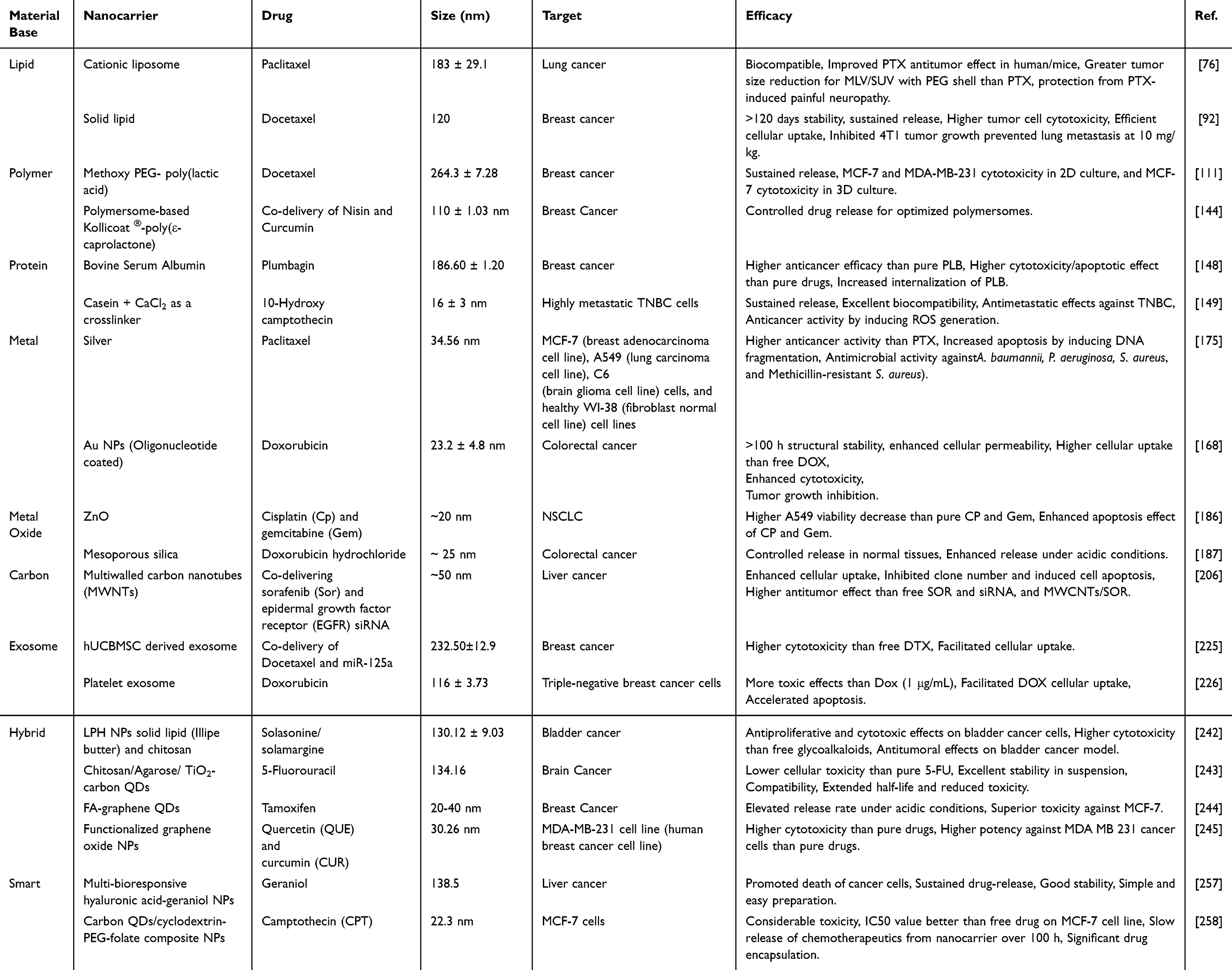 |
Table 1 Classification of Nanocarriers and Effective Parameters for Cancer Therapy |
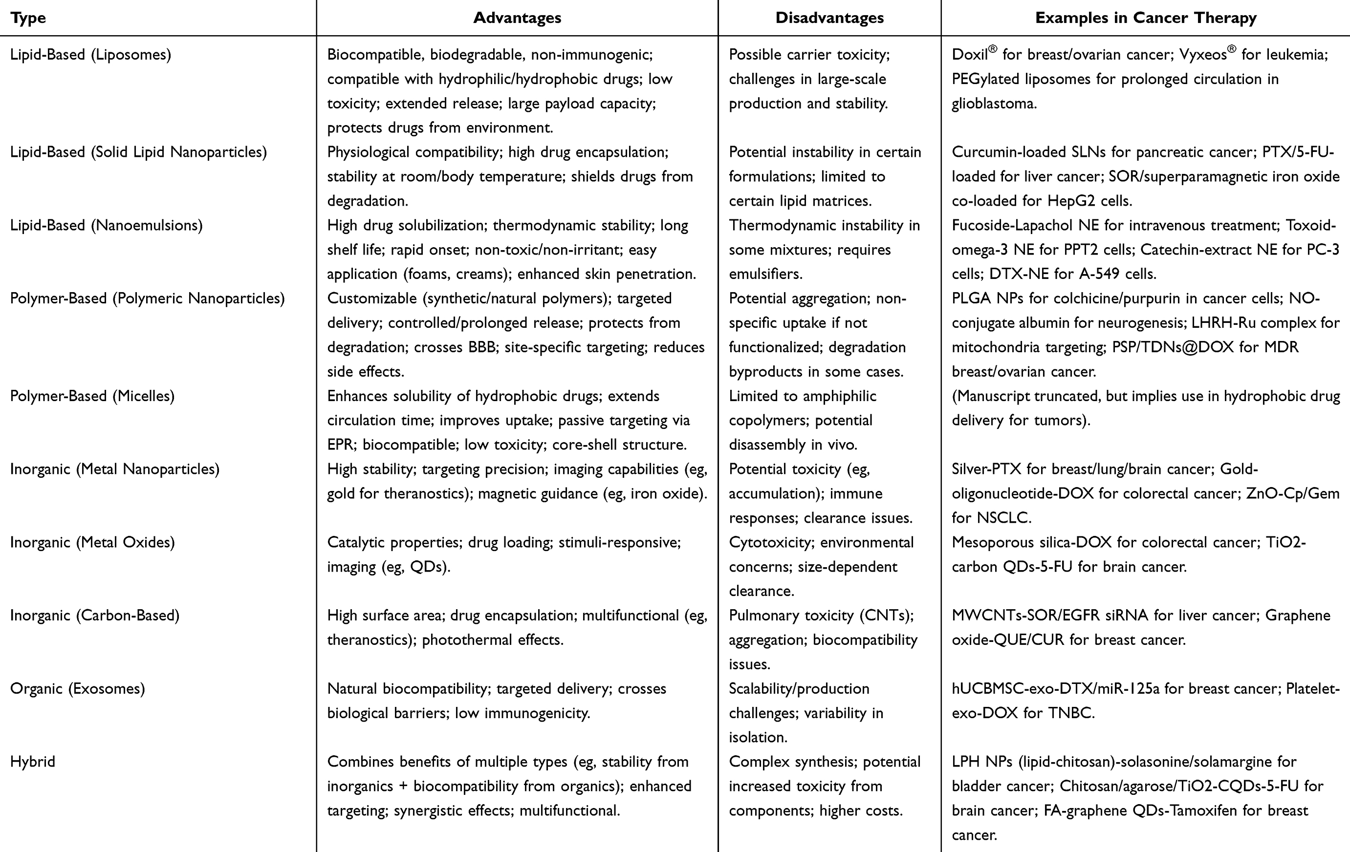 |
Table 2 Comparative Advantages and Disadvantages of Nanocarrier Types for Cancer Therapy |
Integrating Immunotherapy with Nanocarrier Applications
Immunotherapy has emerged as a promising strategy for treating cancer due to its specificity in boosting the body’s natural defense mechanism against cancer cells.294 However, the conventional delivery of immunotherapeutic agents often leads to side effects and reduced patient compliance. Additionally, the tumor’s immunosuppressive microenvironment limits the efficacy of immunotherapy. Nanotechnology offers a potential solution to these challenges and has the potential to revolutionize cancer immunotherapy. Nanoparticles have shown the ability to enhance the effectiveness of immunotherapeutic agents by targeting immunocytes and reprogramming the TME.112 This approach improves the treatment outcomes of immunotherapy by enhancing the delivery of immunotherapeutic agents directly to the tumor site, minimizing systemic toxicity.295 This treatment destroys tumor cells and prevents their recurrence by triggering a significant immune reaction against the tumor.296 There are three essential factors for effective cancer immunotherapy: effective transfer of cancer antigens to immune cells (especially antigen-presenting cells), induction of an anti-cancer immune response, and modulation of the immune-suppressive TME. Nanotechnology can be used for each of these aspects and has the potential to induce anti-cancer immune responses effectively.297 By altering the biophysical features of NPs and incorporating functional ligands, they can target specific cell types such as tumor cells, dendritic cells, T cells, or TME.298 Nanoparticles can also incorporate multiple immunotherapeutic agents for cancer targeting or immune cell delivery. Co-delivery of immunotherapeutic agents within a single cell has been shown to enhance the immune response and optimize antigen processing and presentation.299 The TME significantly influences drug penetration and efficacy, correlating with treatment resistance and diminished response rates. The modulation of the TME has garnered interest from researchers focused on cancer immunotherapy. Nanoparticles can extend retention duration and facilitate targeted distribution, hence minimizing toxicity. They can also change the immunosuppressive environment in the TME by targeting its significant components and transforming it into an immunosupportive state, thus promoting the efficacy of cancer immunotherapy. CAR-T therapy of acute lymphoblastic leukemia has shown limited efficacy in melanoma and non-small-cell lung cancer, with reported adverse events. Nanocarriers are increasingly being employed to enhance the delivery of immunotherapeutic agents, such as checkpoint inhibitors and cancer vaccines. By encapsulating these agents within nanoparticles, researchers can improve their stability and bioavailability while ensuring targeted delivery to tumor sites. For instance, nanoparticles that encapsulate immune checkpoint inhibitors enhance their stability and bioavailability, allowing for more effective targeting of tumors. Additionally, the use of nanoparticles to deliver cancer vaccines can stimulate a robust immune response, improving vaccine efficacy, for example, Meng et al highlighted that biomimetic nanoparticles can enhance the immunogenicity of dendritic cell (DC) vaccines, allowing for better antigen presentation and accumulation in lymph nodes.300 Formulations that co-deliver immunoadjuvants alongside therapeutic agents further enhance the overall immune response against tumors, representing a promising strategy in cancer immunotherapy.301 Nanoparticles also have the potential to overcome the limitations of current immunotherapies and improve treatment outcomes for various types of cancer.302 Miao et al’s study uses liposome-protamine-DNA nanoparticles to treat pancreatic cancer by encapsulating plasmids encoding PD-L1 and CXCL12 traps, effectively tuning immunosuppressive TME and facilitating T-cell infiltration.303
Nanoparticle formulation significantly improves the local delivery of immunoadjuvants to peripheral lymphoid organs while limiting systemic distribution, enhancing their immunological activity, and reducing systemic immunotoxicity.304 Additionally, nanoparticle-based delivery of antibodies effectively targets and activates antigen-presenting cells (APCs) and T cells, demonstrating clinical benefits in various tumors, including melanoma, non-small cell lung cancer, renal cancer, and Hodgkin’s lymphoma.305 Traditional nanomedicine faces challenges in tumor infiltration due to systemic and local barriers that limit the efficacy of antitumor treatments. In contrast, nanoparticles can be directed to immune cells in lymphoid organs or other immune sites, stimulating systemic antitumor immunity and bypassing significant barriers associated with direct tumor targeting. Immune cells can penetrate natural barriers, such as tumor vasculature and the BBB, which nanoparticles typically cannot access. This allows for potentially more effective therapeutic action through immune modulation.306 Thus, nanocarrier-based immunotherapy offers a novel delivery approach that targets immune cells while also directly modulating tumors, leading to enhanced antitumor activity through multifaceted therapeutic mechanisms.
Challenges in Nanocarrier Development and Application
The field of nanomedicine, particularly in cancer treatment, is rapidly evolving, with approximately 2000 nano-formulations currently undergoing clinical trials. Despite this promising landscape, the journey from development to market is fraught with challenges, including high production costs, technological complexities, and failures in clinical translation.307 As a result, only a limited number of the numerous developed nanomedicines have successfully reached commercialization. The intricate nature of these formulations means that even minor alterations in their production can significantly impact both their therapeutic efficacy and side effect profiles.308 Furthermore, while nanomedicines can effectively target tumors through the EPR effect, they also tend to accumulate in other organs with fenestrated endothelium, such as the spleen and liver, leading to unique cytotoxic effects influenced by their physicochemical properties.309 Research in nanotoxicology has highlighted the specific side effects of nanomedicines at various biological levels.310 Common adverse effects often stem from immunological reactions due to the accumulation of nanoparticles in the spleen and liver, their foreign nature, and the generation of ROS.311 To address these toxicity concerns, strategies such as modifying nanoparticle surfaces with targeting agents that bind to receptors overexpressed on cancer cells or targeting motifs of both cell-specific and tissue-specific are being explored in personalized medicine.312 For example, albumin-based nanocarriers can leverage the EPR effect while also targeting glycoprotein gp60 receptors, enhancing their therapeutic potential.313 Despite these advancements, significant hurdles remain for the clinical translation of nanomedicines. The complex synthesis and modification processes complicate the consistent and affordable manufacture of clinical-grade products. Moreover, treatment regimens must be personalized to optimize efficacy and minimize toxicity, which adds another layer of complexity. The safety of exogenously produced nanocarriers is also a concern, as the biohazards associated with nanoparticle formulations are not fully understood, particularly regarding chronic toxicity from repeated use. While the toxicology of certain elements, such as heavy metals, is well understood in bulk, nano-manipulation can alter their properties, potentially leading to biological toxicity. The toxicity of nanoparticles is influenced by various factors, including size, shape, composition, and surface modification, necessitating detailed investigations for each new nanoparticle. This requires case-by-case assessments for potential toxicity, which is labor-intensive and must be conducted across diverse patient populations. Therefore, identifying biomarkers linked to clinical responses and selecting suitable patients for treatment are essential to minimize health risks and maximize therapeutic benefits.
Beyond the general toxicological concerns, the biocompatibility and long-term safety profiles of nanocarriers are paramount for clinical translation. Different classes of nanocarriers elicit distinct biological responses depending on their composition, surface chemistry, and degradation pathways.314,315 For instance, lipid-based nanoparticles, while generally biocompatible, can trigger complement activation and acute hypersensitivity reactions in some patients, particularly those with pre-existing immune sensitivities.314 Polymeric nanoparticles, such as those made from PLGA, may induce oxidative stress and inflammatory responses in immune cells like macrophages, mainly when stabilizers or surfactants are used in their formulation.315 Inorganic nanoparticles, including gold, silica, and iron oxide variants, pose risks of bioaccumulation and organ-specific toxicity, with surface modifications playing a critical role in mitigating these effects.311,316,317 Moreover, long-term exposure to specific nanomaterials may lead to unforeseen chronic toxicity, such as fibrosis or autoimmune reactions, which are not fully captured in short-term preclinical models. Immune responses, ranging from opsonization and rapid clearance by the mononuclear phagocyte system to more specific immunogenic reactions, also vary significantly across nanocarrier types and must be carefully evaluated to avoid adverse outcomes in clinical settings.310,318
Even beyond toxicity, nanocarriers face significant physiological barriers that limit their efficacy. Rapid clearance from circulation limits their therapeutic window, while prolonged circulation can lead to off-target accumulation.319 A critical obstacle is the BBB, a selective membrane that restricts the entry of nanocarriers and their payloads into the central nervous system.320 Upon reaching the tumor site, nanocarriers encounter the TME, characterized by hypoxia, acidity, and high interstitial fluid pressure, all of which can negatively impact nanocarrier performance. Additionally, cellular membranes limit entry into tumor cells, and intracellular barriers like endosomal entrapment impede cargo delivery to specific subcellular compartments. To overcome these, nanocarriers can be engineered with specific materials that respond to the intracellular environment.22 For instance, Feng et al321 developed iR@ZIF-8 nanocomposites using a zeolitic imidazolate framework (ZIF) that degrades in acidic endosomes, successfully delivering miRNA into the cytoplasm. Looking even further ahead, next-generation, swarm-strategy, nanocarriers aim to navigate these physiological hurdles actively. Such systems would require effective propulsion mechanisms (eg, magnetic or ultrasonic), precise control systems for targeting, and a stable power source for sustained operation within the body, representing a significant shift from passive delivery to active navigation.
In the near term, bio-inspired strategies provide a compelling approach to overcoming these barriers. Coating nanoparticles with cell membranes can enhance evasion, targeting, and uptake. Hu et al322 created cancer cell membrane-coated nanoparticles (CCNPs) to evade immune recognition and enhance tumor targeting by mimicking cancer cell surface antigens, thereby facilitating homotypic targeting and improving cellular uptake. Similarly, Harris et al showed that coating nanoparticles with red blood cell membranes extended circulation time and reduced macrophage clearance, leading to greater accumulation in tumors.323 In another study, Luk et al developed leukocyte membrane-coated nanocarriers, leveraging the homing ability of immune cells to target inflamed or cancerous tissues for improved delivery within the tumor microenvironment.324 These strategies enhance biocompatibility, reduce immunogenicity, and improve delivery within the TME.
Translation is also hindered by manufacturing and economic challenges. Scaling up production requires overcoming hurdles in scalability, reproducibility, and navigating stringent regulatory approval processes. The high costs of research, development, manufacturing, and regulatory compliance further hinder widespread clinical adoption, particularly in cancer therapies.325,326 Comprehensive assessments using advanced in vitro models and longitudinal in vivo studies are essential to understand these interactions and fully design safer nanotherapeutic platforms.
Iron oxide nanoparticles (IONPs) face significant challenges in their application for imaging and therapeutic delivery, primarily due to their inability to accumulate effectively at tumor sites.327 Non-targeted nanoparticles often fail to reach sufficient concentrations in tumors, limiting their utility in clinical settings.328 To address this issue, researchers propose the development of highly sensitive imaging probes that can conjugate large amounts of therapeutic agents, specifically targeting tumors. However, the size of antibodies relative to the surface area of IONPs presents a challenge in effectively linking them, which can impede the nanoparticles’ penetration into target tissues and diminish their imaging and therapeutic efficacy.329 Additionally, concerns regarding the safety and potential toxicity of IONPs, particularly in living organisms, further complicate their application.330 Strategies to overcome these limitations include surface modifications to enhance stability and biocompatibility, as well as the creation of targeted delivery systems to improve specificity and minimize off-target effects. Ultimately, optimizing the formation of IONPs to control their size, shape, and surface properties is crucial for enhancing their application efficiency in medical application.331 Gold nanocarriers, as one of the nanomaterials used in the field of cancer therapy, face significant challenges regarding biocompatibility and toxicity, influenced by variability in nanoparticle preparation that affects size, shape, and surface chemistry. Inadequate functionalization can lead to adverse biological interactions.317 Surface modification with biocompatible materials (polymers or proteins) has been shown to increase stability, reduce nonspecific interactions, and improve overall biocompatibility.332 Assessing the toxicity of silica nanoparticles presents significant challenges, particularly regarding their higher doses and cumulative effects. While silica is generally considered safe, its nanoparticle form exhibits distinct biological behaviors, necessitating thorough biocompatibility evaluations.316 The manufacturing process allows for precise control over particle size and shape, yet variations in physicochemical properties can influence biological interactions and cellular uptake. Therefore, managing these factors is crucial to ensure consistent and predictable therapeutic outcomes. Pulit-Prociak showed that modifying titanium nanoparticles could inhibit the release of titanium ions and decrease the toxicity of TiO2 nanoparticles. By coating the nanoparticles with GSH and galactose, they demonstrated that the toxicity of the nanoparticles was reduced and therefore could be useful for drug delivery.201,333 The integration of CNTs into patient care necessitates a comprehensive understanding of their biocompatibility and potential toxicity. Due to their fibrous structure, CNTs pose safety risks, including bioaccumulation that can lead to oxidative stress, inflammation, and cellular damage.334 Therefore, evaluating their biocompatibility is crucial. To mitigate adverse effects, researchers employ surface modifications and functionalization techniques, alongside investigating the metabolism and excretion pathways of CNTs to ensure their safe application in theragnostics.335 Evidence indicates that CNTs can adversely affect various organs, particularly the nervous system, lungs, and blood vessels, with implications for gene expression related to anxiety and depression.336 Specific types, such as SWNTs, have been linked to increased inflammation and neurotoxicity, underscoring the importance of size in their biological impact. Despite these concerns, CNTs have demonstrated efficacy in animal models for tumor suppression and drug delivery (PTX), highlighting their therapeutic potential.336 Understanding the pharmacokinetics of CNTs and their long-term biological effects is essential for developing the safety data required for clinical translation. While CNTs present promising applications in medicine, addressing their biocompatibility and toxicity challenges remains a priority to ensure safe and effective patient care.337 In vivo studies on animals have been pivotal in evaluating the toxicity of QDs through acute and repeated-dose assessments (subacute, subchronic, or chronic), shedding light on their adsorption, distribution, metabolism, and excretion (ADME) as well as interactions with the immune system. However, researchers emphasize the need to minimize such testing due to ethical considerations.338 While polymer coatings have been shown to mitigate toxicity, concerns remain regarding the long-term effects and potential systemic accumulation of QDs.339 Additionally, the inherent rigidity of QDs poses challenges in drug conjugation, necessitating further investigation into optimal functional groups for effective binding.340,341 Spherical liposomes used in nanotherapeutics have limitations such as instability and the potential for active agents to leak before reaching their target.342 To enhance stability, researchers are optimizing lipid compositions, using stabilizing agents like sodium benzoate, and improving encapsulation technologies.343 Polymeric nanoparticles offer several advantages, including biodegradability, biocompatibility, and the ability to carry both hydrophobic and hydrophilic drugs while allowing for controlled drug release. However, they also have limitations, such as self-aggregation, the use of toxic organic solvents in some preparation methods, and high synthesis costs.344 Common solvents like dichloromethane and chloroform used in the solvent evaporation method can produce toxic nanospheres.345 The toxicity of polymeric nanoparticles is significantly influenced by quantum-size effects, which are associated with oxidative stress, cytotoxicity, and genotoxicity. Notably, the use of slightly cytotoxic CH-polymer as a stabilizer markedly increased the cytotoxicity of PLGA nanoparticles in THP-1 macrophages, highlighting the critical role of nanoparticles stabilizers in determining the local toxicity of PLGA-based nanomedicine.318 The size of nanocarriers is a key concern when it comes to PAMAM dendrimers. Dendrimers of generation 5 or lower can be effectively eliminated through the kidneys, while those of generation 6 and higher rely more on liver clearance.346,347 Dendrimers sized between 4–10 nm can interact with cellular components and overcome the endocytosis barrier. However, higher-generation PAMAM dendrimers are often avoided due to their high costs and significant toxicity.346 Cationic dendrimers have a strong binding capacity with nuclei and anionic compounds, aiding in cell internalization.348 However, they face challenges like nonspecific binding to plasma proteins and rapid elimination by the reticuloendothelial system.349 Additionally, their interaction with negatively charged cell membranes can destabilize biological membranes, leading to cell lysis and higher toxicity, especially at elevated doses.350 For example, a study by Pryor et al found that cationic PAMAM generation 6 was significantly more toxic to embryonic zebrafish than both neutral and anionic PAMAM of the same generation at the same concentration.351
The development of theranostic agents as a novel method in cancer treatment presents several challenges, primarily due to the complexity of tumor biology, which results in variability in receptor expression among patients and within tumors. This variability complicates the selection of appropriate agents.352,353 Additionally, creating new theranostic agents necessitates rigorous testing for safety and efficacy, which is often lengthy and costly.352,354 Regulatory barriers further impede the transition from research to clinical application.352,355 Key considerations for clinical use include the biocompatibility and stability of these agents, particularly nanoparticles, which must effectively deliver therapeutics without harming healthy tissues.356 Researchers are investigating multifunctional theranostic platforms that integrate imaging and therapy, showing promising outcomes.357 Ongoing clinical trials are crucial for overcoming these challenges and validating the effectiveness of theranostics, which have the potential to enhance personalized cancer therapies and improve patient outcomes in precision medicine.352,355,358
Future Perspectives in Nanocarrier Development
The landscape of nanocarrier technology is evolving rapidly, bringing forth exciting innovations that promise to improve cancer therapies significantly. Immune-cell-derived extracellular vehicles (EVs) carry immune-associated molecules and can be re-appropriated to target cancers. CD8 T cell-derived EVs carry anti-tumor cargo and can cause localized inflammation, causing further immune infiltration, which can be exploited for cancer targeting. Engineering the immune cells to generate copious amounts of EV PD-1 to mediate a PD-1-PD-L1 interaction can work like immunotherapeutics and be an alternative to checkpoint inhibitors. One of the most transformative directions in this field is integrating personalized medicine into the design of nanocarriers. This approach aims to tailor treatments to individual patients by considering their unique genetic makeup, biomarkers, and other personal health factors. For instance, researchers are exploring the use of patient-derived exosomes as nanocarriers to deliver targeted therapeutic agents specifically designed for individual tumors.304,359 This level of customization enhances treatment effectiveness and minimizes unwanted side effects, as seen in studies where exosomes have been used to deliver chemotherapeutics directly to triple-negative breast cancer cells, resulting in reduced systemic toxicity.360 As these innovations unfold, we can anticipate several exciting clinical applications. One promising area is targeted gene therapy, where nanoparticles can deliver nucleic acids—such as siRNA or mRNA—directly to specific cancer cells. This targeted delivery can potentially correct genetic defects or silence oncogenes effectively. For instance, lipid nanoparticles have shown success in delivering mRNA vaccines for cancer immunotherapy,361 such as the BNT162b2 vaccine developed for COVID-19,362 which has implications for treating tumors by eliciting a robust immune response against melanoma cells. Furthermore, the role of nanocarriers in immunotherapy is expected to expand, particularly in enhancing the delivery of immune-modulating agents directly to the tumor microenvironment. By designing nanoparticles that co-deliver tumor antigens and adjuvants, researchers can stimulate a robust immune response, thereby improving the effectiveness of cancer vaccines like Sipuleucel-T for prostate cancer.363 Another promising avenue is the advancement of combination therapies made possible by nanocarriers. By enabling the simultaneous delivery of multiple therapeutic agents—such as traditional chemotherapeutics, immunotherapeutics, and gene therapies—nanocarriers can enhance treatment efficacy and help overcome the challenges posed by drug resistance. A notable example is the use of lipid-polymer hybrid nanoparticles that effectively deliver both paclitaxel and immune checkpoint inhibitors like anti-PD-1 antibodies. Recent studies have demonstrated that this combination leads to improved tumor regression in preclinical models of non-small cell lung cancer, showcasing the potential for synergistic effects that result in better therapeutic outcomes.364,365 Moreover, the development of smart nanocarriers that respond to specific stimuli—such as changes in pH, temperature, or enzymatic activity—holds great potential for enhancing the precision of drug delivery. These innovative carriers can be programmed to release their therapeutic payloads only when they encounter specific conditions within the tumor microenvironment. For example, pH-sensitive nanocarriers have been designed to release their contents in the acidic environment typical of tumors, significantly improving the therapeutic index of drugs like doxorubicin in breast cancer treatment.366,367 Additionally, temperature-sensitive nanocarriers are being investigated for their ability to release drugs in response to the elevated temperatures often found in tumors, further enhancing localized treatment efficacy in models of prostate cancer.366 However, the journey from laboratory to clinic is not without its challenges, particularly regarding regulatory considerations. The unique properties of nanocarriers necessitate a comprehensive understanding of their pharmacokinetics, biodistribution, and long-term safety profiles. Regulatory agencies, such as the FDA and EMA, are increasingly focused on the characterization of nanomaterials, requiring detailed data on their interactions with biological systems. This includes evaluating potential toxicity, immunogenicity, and environmental impact.368 Consequently, researchers must prioritize the development of standardized protocols to assess the safety and efficacy of nanocarriers, facilitating smoother regulatory approval processes.368
The development of nanosystems capable of delivering treatment and diagnostic imaging in a unified system (theranostic) enhances efficacy and reduces tumor resistance and microenvironmental challenges. The use of nanocarriers, especially polymeric-based NPs, gained more attention due to their various advantages, such as versatility, biocompatibility, biodegradability, drug release control, on-site delivery, and the presence of various natural and synthetic forms, such as polymer conjugate complexes, dendrimers, polymeric micelles, and polymeric nanospheres for intelligent theranostic systems. Diverse polymeric NPs can facilitate targeted gene and drug delivery, along with various imaging contrast enhancement agents, radiotherapy, PDT, and PTT. Research on polymers for disease treatment and diagnosis is growing, but theranostic nanosystems are still in their early stages. As an interdisciplinary field, progress is expected to accelerate, providing new therapeutic strategies in the future. To navigate the complexities of nanocarrier technology and ensure these advancements translate into clinical practice, continued collaboration among researchers, clinicians, and regulatory bodies will be essential. As we move forward, addressing challenges such as tumor heterogeneity, optimizing nanocarrier design, and ensuring patient safety will be critical. By integrating advanced materials, innovative design strategies, and robust clinical validation, we can pave the way for the next generation of nanocarrier-based therapies, ultimately improving patient outcomes in the ongoing battle against cancer. As we look to the future, it is crucial to continue research investment and conduct rigorous clinical trials to address the challenges that remain in this field. This includes refining nanocarrier designs, ensuring their safety and efficacy, and navigating the complex regulatory landscape. Successful integration of these technologies into everyday clinical practice will depend on strong collaboration among researchers, clinicians, and regulatory bodies to establish standardized protocols and guidelines.
Conclusion
Innovative approaches such as the use of nanocarriers, by significantly enhancing DDS, not only improve drug efficacy but also minimize the side effects often associated with traditional therapies. The unique properties of nanocarriers allow them to address some of the most difficult challenges in cancer treatment, such as drug resistance and systemic toxicity. With the ability to deliver drugs directly to the tumor site, nanocarriers enable more personalized treatment plans tailored to the specific needs of patients, ultimately leading to better clinical outcomes. By deepening our understanding of how these systems work and exploring their diverse applications, as well as utilizing combination approaches such as theranostics therapy, we can pave the way for more effective, personalized, and safer cancer treatments. These technologies have the potential to significantly improve the lives of patients facing the challenges of this complex disease and offer hope for a brighter future in cancer care.
Abbreviations
5-FU, Fluorouracil; ABC, ATP-binding cassette; ADME, Adsorption, distribution, metabolism, and excretion; AI, Artificial intelligence; Akt, Protein kinase B; AML, Acute myelogenous leukemia; AraC, Cytarabine; ATP, Adenosine triphosphate; BBB, Blood-brain barrier; BCL-2, B-cell leukemia/lymphoma 2; CXB, Celecoxib; circRNA, Circular RNA; Ce6, Chlorin e6; CMC, Critical micelle concentration; CME, Clathrin-mediated endocytosis; CML, Chronic myeloid leukemia; CNTs, Carbon nanotubes; CP, Cisplatin; CPT, Camptothecin; CRC, Colorectal cancer; CSCs, Cancer stem cells; CXB, Celecoxib; CYP, Cytochrome p450; DAS, Dasatinib; DDS, Drug delivery systems; DNMTs, DNA methyltransferases; DOX, Doxorubicin; DTX, Docetaxel; EAC, Ehrlich Ascites Carcinoma; EGFR, Epidermal growth factor receptor; EMT, Epithelial-mesenchymal transition; EPR, Enhanced permeability and retention; E-PSiNPs, Exosome-sheathed porous silicon nanoparticles; ER, Estrogen receptor; EVs, Extracellular vesicles; FA, Folic acid; GC, Gemcitabine; GSH, Glutathione; GST, Glutathione S-transferase; HAPs, Hypoxia-activated prodrugs; HAS, Human serum albumin; HU, Hydroxyurea; IONPs, Iron oxide nanoparticles; LBNCs, Lipid-based nanocarriers; LHRH, Luteinizing hormone-releasing hormone; lncRNA, Long non-coding RNA; LNPs, Lipid-based nanoparticles; LPH, Lipid-polymer hybrid; L-AuNPs, Luminescent gold nanoparticles; MDR, Multi-drug resistance; MIPs, Molecularly imprinted polymers; miRNA, MicroRNA; ML, Machine learning; MOF, Metal-organic framework; MPS, Mononuclear phagocyte system; mRNA, Messenger RNA; MSI, Microsatellite instability; MTX, Methotrexate; MWCNTs, Multi-walled carbon nanotubes; NC, Nanocomplex; NCs, Nanocarriers; NDDS, Nanoscale drug delivery systems; NEs, Nanoemulsions; NF-κB, Nuclear factor kappa B; NIR, Near-infrared radiation; NO, Nitric oxide; NPs, Nanoparticles; NSCLC, Non-small cell lung cancer; PA, Pyruvic acid; PAMAM, Poly(amidoamine); PARP, Poly(ADP-ribose) polymerase; PDA, polydopamine; PDAC, Pancreatic ductal adenocarcinoma; PDT, Photodynamic therapy; PEG, Polyethylene glycol; PEI, Polyethylenimine; P-gp, Permeability-glycoprotein; PLGA, poly(lactic-co-glycolic acid); PMs, Polymeric micelles; PSiNPs, Porous silicon nanoparticles; PTT, Photothermal treatment; PTX, Paclitaxel; QDs, Quantum dots; RES, Reticuloendothelial system; ROS, Reactive oxygen species; SA, Selenic acid; siRNA, Small interfering RNA; SLC, Solute carrier; SLNPs, Solid lipid nanoparticles; SOR, Sorafenib; SPECT, Single-photon emission computed tomography; SWCNTs, Single-walled CNTs; TAM, Tamoxifen; TME, Tumor microenvironment; TMSEDTA, Trimethoxysilylpropyl-ethylenediamine triacetic acid; TMZ, Temozolomide; TNBC, Triple-negative breast cancer; UGT, UDP-glycosyltransferase.
Author Contributions
All authors made a significant contribution to the work reported, whether that is in the conception, study design, execution, acquisition of data, analysis and interpretation, or in all these areas; took part in drafting, revising or critically reviewing the article; gave final approval of the version to be published; have agreed on the journal to which the article has been submitted; and agree to be accountable for all aspects of the work.
Funding
This research received no external funding.
Disclosure
The authors declare no competing interests.
References
1. Fernandese Silva E, FdS F, Lettnin AP, et al. C-Phycocyanin: cellular targets, mechanisms of action and multi drug resistance in cancer. Pharmacol Rep. 2018;70:75–80. doi:10.1016/j.pharep.2017.07.018
2. Henn JG, Aguirre TAS, Nugent M, Moura DJ. Cancer nanomedicine: recent developments in drug delivery systems and strategies to overcome eventual barriers to achieve a better outcome. J Drug Delivery Sci Technol. 2024;91:105254. doi:10.1016/j.jddst.2023.105254
3. Cheng Z, Li M, Dey R, Chen Y. Nanomaterials for cancer therapy: current progress and perspectives. J hematol oncol. 2021;14:1–27.
4. Rashidi N, Davidson M, Apostolopoulos V, Nurgali K. Nanoparticles in cancer diagnosis and treatment: progress, challenges, and opportunities. J Drug Delivery Sci Technol. 2024;95:105599. doi:10.1016/j.jddst.2024.105599
5. Sell M, Lopes AR, Escudeiro M, et al. Application of nanoparticles in cancer treatment: a concise review. Nanomaterials. 2023;13(21):2887. doi:10.3390/nano13212887
6. Pandita D, Poonia N, Chaudhary G, Jain GK, Lather V, Khar RK. pH-sensitive polymeric nanocarriers for enhanced intracellular drug delivery. In: Smart Polymeric Nano-Constructs in Drug Delivery. Elsevier; 2023:65–107.
7. Ashique S, Faiyazuddin M, Afzal O, et al. Advanced nanoparticles, the hallmark of targeted drug delivery for osteosarcoma-an updated review. J Drug Delivery Sci Technol. 2023;87:104753. doi:10.1016/j.jddst.2023.104753
8. Ai J, Biazar E, Jafarpour M, et al. Nanotoxicology and nanoparticle safety in biomedical designs. Int J Nanomed;2011. 1117–1127. doi:10.2147/IJN.S16603
9. Elumalai K, Srinivasan S, Shanmugam A. Review of the efficacy of nanoparticle-based drug delivery systems for cancer treatment. Biomed Technol. 2024;5:109–122. doi:10.1016/j.bmt.2023.09.001
10. Haider M, Elsherbeny A, Pittalà V, et al. Nanomedicine strategies for management of drug resistance in lung cancer. Int J Mol Sci. 2022;23(3):1853. doi:10.3390/ijms23031853
11. Qu H, Chen H, Cheng W, et al. A supramolecular assembly strategy for hydrophilic drug delivery towards synergistic cancer treatment. Acta Biomater. 2023;164:407–421. doi:10.1016/j.actbio.2023.04.026
12. Ahmad W, Khan T, Basit I, Imran J. A comprehensive review on targeted drug delivery system. Asian J Pharmaceutical Res. 2022;12(4):335–340. doi:10.52711/2231-5691.2022.00053
13. Song Y, Zou J, Castellanos EA, et al. Theranostics-a sure cure for cancer after 100 years? Theranostics. 2024;14(6):2464. doi:10.7150/thno.96675
14. Al-Thani AN, Jan AG, Abbas M, Geetha M, Sadasivuni KK. Nanoparticles in cancer theragnostic and drug delivery: a comprehensive review. Life Sci. 2024;352:122899. doi:10.1016/j.lfs.2024.122899
15. Davodabadi F, Sajjadi SF, Sarhadi M, et al. Cancer chemotherapy resistance: mechanisms and recent breakthrough in targeted drug delivery. Eur J Pharmacol. 2023;958:176013. doi:10.1016/j.ejphar.2023.176013
16. Duan C, Yu M, Xu J, Li B-Y, Zhao Y, Kankala RK. Overcoming Cancer Multi-drug Resistance (MDR): reasons, mechanisms, nanotherapeutic solutions, and challenges. Biomed. Pharmacother. 2023;162:114643. doi:10.1016/j.biopha.2023.114643
17. Pote MS, Gacche RN. ATP-binding cassette efflux transporters and MDR in cancer. Drug Discovery Today. 2023;28(5):103537. doi:10.1016/j.drudis.2023.103537
18. Wang C, Li F, Zhang T, Yu M, Sun Y. Recent advances in anti-multidrug resistance for nano-drug delivery system. Drug Delivery. 2022;29(1):1684–1697. doi:10.1080/10717544.2022.2079771
19. Kurmi BD, Patel P, Paliwal R, Paliwal SR. Molecular approaches for targeted drug delivery towards cancer: a concise review with respect to nanotechnology. J Drug Delivery Sci Technol. 2020;57:101682.
20. Shaik R, Malik MS, Basavaraju S, et al. Cellular and molecular aspects of drug resistance in cancers. DARU J Pharma Sci. 2025;33(1):1–25.
21. Bharathiraja P, Yadav P, Sajid A, Ambudkar SV, Prasad NR. Natural medicinal compounds target signal transduction pathways to overcome ABC drug efflux transporter-mediated multidrug resistance in cancer. Drug Resist Updates. 2023;71:101004. doi:10.1016/j.drup.2023.101004
22. Liu J, Cabral H, Mi P. Nanocarriers address intracellular barriers for efficient drug delivery, overcoming drug resistance, subcellular targeting and controlled release. Adv Drug Delivery Rev. 2024;115239. doi:10.1016/j.addr.2024.115239
23. Wang M, Chen W, Chen J, et al. Abnormal saccharides affecting cancer multi-drug resistance (MDR) and the reversal strategies. Eur J Med Chem. 2021;220:113487. doi:10.1016/j.ejmech.2021.113487
24. Mir SA, Hamid L, Bader GN, et al. Role of nanotechnology in overcoming the multidrug resistance in cancer therapy: a review. Molecules. 2022;27(19):6608. doi:10.3390/molecules27196608
25. Puris E, Fricker G, Gynther M. The role of solute carrier transporters in efficient anticancer drug delivery and therapy. Pharmaceutics. 2023;15(2):364. doi:10.3390/pharmaceutics15020364
26. Mynott RL, Wallington-Beddoe CT. Drug and solute transporters in mediating resistance to novel therapeutics in multiple myeloma. ACS Pharmacol Transl Sci. 2021;4(3):1050–1065. doi:10.1021/acsptsci.1c00074
27. Sun X, Zhao P, Lin J, Chen K, Shen J. Recent advances in access to overcome cancer drug resistance by nanocarrier drug delivery system. Cancer Drug Resistance. 2023;6(2):390. doi:10.20517/cdr.2023.16
28. Mansoori B, Mohammadi A, Davudian S, Shirjang S, Baradaran B. The different mechanisms of cancer drug resistance: a brief review. Adv Pharm Bull. 2017;7(3):339. doi:10.15171/apb.2017.041
29. Haider T, Pandey V, Banjare N, Gupta PN, Soni V. Drug resistance in cancer: mechanisms and tackling strategies. Pharmacol Rep. 2020;72(5):1125–1151. doi:10.1007/s43440-020-00138-7
30. Kadhum WR, Majeed AA, Saleh RO, et al. Overcoming drug resistance with specific nano scales to targeted therapy: focused on metastatic cancers. Pathol Res Pract. 2024;255:155137. doi:10.1016/j.prp.2024.155137
31. Housman G, Byler S, Heerboth S, et al. Drug resistance in cancer: an overview. Cancers. 2014;6(3):1769–1792. doi:10.3390/cancers6031769
32. Ortíz R, Quiñonero F, García-Pinel B, et al. Nanomedicine to overcome multidrug resistance mechanisms in colon and pancreatic cancer: recent progress. Cancers. 2021;13(9):2058. doi:10.3390/cancers13092058
33. Mowla M, Hashemi A. Functional roles of exosomal miRNAs in multi-drug resistance in cancer chemotherapeutics. Exp Mol Pathol. 2021;118:104592. doi:10.1016/j.yexmp.2020.104592
34. Ying Z, Wenjing S, Jing B, Songbin F, Kexian D. Advances in long non-coding RNA regulating drug resistance of cancer. Gene. 2023;887:147726. doi:10.1016/j.gene.2023.147726
35. Shi Y, Adu-Amankwaah J, Zhao Q, et al. Long non-coding RNAs in drug resistance across the top five cancers: update on their roles and mechanisms. Heliyon. 2024;2024:1.
36. Zhou X, Ao X, Jia Z, et al. Non-coding RNA in cancer drug resistance: underlying mechanisms and clinical applications. Front Oncol. 2022;12:951864. doi:10.3389/fonc.2022.951864
37. Yan T, Tian X, Liu F, et al. The emerging role of circular RNAs in drug resistance of non-small cell lung cancer. Front Oncol. 2022;12:1003230. doi:10.3389/fonc.2022.1003230
38. Misir S, Yaman SO, Petrović N, Sumer C, Hepokur C, Aliyazicioglu Y. circRNAs in drug resistance of breast cancer. Oncol Res. 2022;30(4):157. doi:10.32604/or.2022.027547
39. Emran TB, Shahriar A, Mahmud AR, et al. Multidrug resistance in cancer: understanding molecular mechanisms, immunoprevention and therapeutic approaches. Front Oncol. 2022;12:891652.
40. Gong S, Shang M, Li D, Cai Y, Jin J, Yang Z. Functionalized ZIF‐90 for gene‐mediated chemotherapy to ameliorate multidrug resistance in breast cancer therapy. ChemNanoMat. 2022;8(2):e202100389. doi:10.1002/cnma.202100389
41. Deng F, Sistonen J, Neuvonen M, Niemi M. Inhibition of efflux transporters by poly ADP‐ribose polymerase inhibitors. Basic Clin Physiol Pharmacol. 2023;133(4):428–436. doi:10.1111/bcpt.13928
42. Zhang H, You J, Liu W, Chen D, Zhang S, Wang X. The efficacy and safety of bevacizumab combined with FOLFOX regimen in the treatment of advanced colorectal cancer: a systematic review and meta-analysis. Medicine. 2021;100(30):e26714. doi:10.1097/MD.0000000000026714
43. Ashique S, Garg A, Farid A, Hussain A, Kumar P, Taghizadeh‐Hesary F. Nanodelivery systems: an efficient and target‐specific approach for drug‐resistant cancers. Cancer Med. 2023;12(18):18797–18825. doi:10.1002/cam4.6502
44. Dunuweera SP, Rajapakse RMSI, Rajapakshe RBSD, et al. Review on targeted drug delivery carriers used in nanobiomedical applications. Curr Nanosci. 2019;15(4):382–397. doi:10.2174/1573413714666181106114247
45. Prabahar K, Alanazi Z, Qushawy M. Targeted drug delivery system: advantages, carriers and strategies. Indian J Pharm Educ. 2021;55:346–353. doi:10.5530/ijper.55.2.72
46. Navya P, Kaphle A, Srinivas S, Bhargava SK, Rotello VM, Daima HK. Current trends and challenges in cancer management and therapy using designer nanomaterials. Nano Convergence. 2019;6(1):23. doi:10.1186/s40580-019-0193-2
47. Yan S, Na J, Liu X, Wu P. Different targeting ligands-mediated drug delivery systems for tumor therapy. Pharmaceutics. 2024;16(2):248. doi:10.3390/pharmaceutics16020248
48. Jin H, Wang L, Bernards R. Rational combinations of targeted cancer therapies: background, advances and challenges. Nat Rev Drug Discov. 2023;22(3):213–234. doi:10.1038/s41573-022-00615-z
49. Ejigah V, Owoseni O, Bataille-Backer P, Ogundipe OD, Fisusi FA, Adesina SK. Approaches to improve macromolecule and nanoparticle accumulation in the tumor microenvironment by the enhanced permeability and retention effect. Polymers. 2022;14(13):2601. doi:10.3390/polym14132601
50. Chen H-A, Lu Y-J, Dash BS, Chao Y-K, Chen J-P. Hyaluronic acid-modified cisplatin-encapsulated poly (lactic-co-glycolic acid) magnetic nanoparticles for dual-targeted NIR-responsive chemo-photothermal combination cancer therapy. Pharmaceutics. 2023;15(1):290. doi:10.3390/pharmaceutics15010290
51. Chouhan RS, Horvat M, Ahmed J, Alhokbany N, Alshehri SM, Gandhi S. Magnetic nanoparticles—A multifunctional potential agent for diagnosis and therapy. Cancers. 2021;13(9):2213. doi:10.3390/cancers13092213
52. Huber CM, Pavan TZ, Ullmann I, et al. A review on ultrasound-based methods to image the distribution of magnetic nanoparticles in biomedical applications. Ultrasound Med Biol. 2024;2024:1.
53. Yagublu V, Karimova A, Hajibabazadeh J, et al. Overview of physicochemical properties of nanoparticles as drug carriers for targeted cancer therapy. J Funct Biomat. 2022;13(4):196. doi:10.3390/jfb13040196
54. Zhao Z, Ukidve A, Krishnan V, Mitragotri S. Effect of physicochemical and surface properties on in vivo fate of drug nanocarriers. Adv Drug Delivery Rev. 2019;143:3–21. doi:10.1016/j.addr.2019.01.002
55. Yue J, Feliciano TJ, Li W, Lee A, Odom TW. Gold nanoparticle size and shape effects on cellular uptake and intracellular distribution of siRNA nanoconstructs. Bioconjugate Chem. 2017;28(6):1791–1800. doi:10.1021/acs.bioconjchem.7b00252
56. Lagarrigue P, Moncalvo F, Cellesi F. Non-spherical polymeric nanocarriers for therapeutics: the effect of shape on biological systems and drug delivery properties. Pharmaceutics. 2022;15(1):32. doi:10.3390/pharmaceutics15010032
57. Soltani M, Souri M, Moradi Kashkooli F. Effects of hypoxia and nanocarrier size on pH-responsive nano-delivery system to solid tumors. Sci Rep. 2021;11(1):19350. doi:10.1038/s41598-021-98638-w
58. Zaky MF, Youssef YL, Megahed MA. Impact of surface design and coating on the efficacy of nano-carriers as drug delivery systems: a review. ERU Res J. 2023;2(3):415–446. doi:10.21608/erurj.2023.219322.1045
59. Zhang P, Chen D, Li L, Sun K. Charge reversal nano-systems for tumor therapy. J Nanobiotechnol. 2022;20:1–27. doi:10.1186/s12951-021-01184-w
60. Peer D, Karp JM, Hong S, Farokhzad OC, Margalit R, Langer R. Nanocarriers as an emerging platform for cancer therapy. Nano-Enabled Med Appl. 2020;2020:61–91.
61. Liu J, Sun B, Li W, et al. Wireless sequential dual light delivery for programmed PDT in vivo. Light Sci Appl. 2024;13(1):113. doi:10.1038/s41377-024-01437-x
62. Liu ZS, Wen J, Huang CY, et al. Nanomedicines based on responsive nanocarriers for cancer therapy. Adv Ther. 2024;7(2):2300223. doi:10.1002/adtp.202300223
63. Wang Y, Yu W, Niu C, et al. A NIR light-activated PLGA microsphere for controlled release of mono-or dual-drug. Polym Test. 2022;116:107762. doi:10.1016/j.polymertesting.2022.107762
64. Waheed I, Ali A, Tabassum H, Khatoon N, Lai W-F, Zhou X. Lipid-based nanoparticles as drug delivery carriers for cancer therapy. Front Oncol. 2024;14:1.
65. Zhao Y-Q, Li L-J, Zhou E-F, et al. Lipid-based nanocarrier systems for drug delivery: advances and applications. Pharmaceutical Fronts. 2022;4(02):e43–e60. doi:10.1055/s-0042-1751036
66. Priya S, Desai VM, Singhvi G. Surface modification of lipid-based nanocarriers: a potential approach to enhance targeted drug delivery. ACS omega. 2022;8(1):74–86. doi:10.1021/acsomega.2c05976
67. Ramadan E, Ahmed A, Naguib YW. Advances in mRNA LNP-based cancer vaccines: mechanisms, formulation aspects, challenges, and future directions. J Personalized Med. 2024;14(11):1092. doi:10.3390/jpm14111092
68. Hamad I, Harb AA, Bustanji Y. Liposome-based drug delivery systems in cancer research: an analysis of global landscape efforts and achievements. Pharmaceutics. 2024;16(3):400. doi:10.3390/pharmaceutics16030400
69. Kumbham S, Ajjarapu S, Ghosh B, Biswas S. Current trends in the development of liposomes for chemotherapeutic drug delivery. J Drug Delivery Sci Technol. 2023;104854. doi:10.1016/j.jddst.2023.104854
70. Gatto MS, Johnson MP, Najahi-Missaoui W. Targeted liposomal drug delivery: overview of the current applications and challenges. Life. 2024;14(6):672. doi:10.3390/life14060672
71. Akbari A, Akbarzadeh A, Rafiee Tehrani M, Cohan RA, Mozaffari A, Memarzadeh M. Preparation and evaluation of a liposome drug delivery system in cancer treatment in vitro. J Nanostruct. 2020;10(1):140–147.
72. Maeki M, Uno S, Niwa A, Okada Y, Tokeshi M. Microfluidic technologies and devices for lipid nanoparticle-based RNA delivery. J Control Release. 2022;344:80–96. doi:10.1016/j.jconrel.2022.02.017
73. Ibrahim M, Ramadan E, Elsadek NE, et al. Polyethylene glycol (PEG): the nature, immunogenicity, and role in the hypersensitivity of PEGylated products. J Control Release. 2022;351:215–230. doi:10.1016/j.jconrel.2022.09.031
74. Deng C, Zhang Q, Jia M, et al. Tumors and their microenvironment dual‐targeting chemotherapy with local immune adjuvant therapy for effective antitumor immunity against breast cancer. Adv Sci. 2019;6(6):1801868. doi:10.1002/advs.201801868
75. Jörgensen AM, Wibel R, Bernkop‐Schnürch A. Biodegradable cationic and ionizable cationic lipids: a roadmap for safer pharmaceutical excipients. Small. 2023;19(17):2206968. doi:10.1002/smll.202206968
76. Jiménez-López J, Bravo-Caparrós I, Cabeza L, et al. Paclitaxel antitumor effect improvement in lung cancer and prevention of the painful neuropathy using large pegylated cationic liposomes. Biomed. Pharmacother. 2021;133:111059. doi:10.1016/j.biopha.2020.111059
77. Liu S, Liu J, Li H, et al. An optimized ionizable cationic lipid for brain tumor-targeted siRNA delivery and glioblastoma immunotherapy. Biomaterials. 2022;287:121645. doi:10.1016/j.biomaterials.2022.121645
78. Ali A, Kouvari M, Riaz S, et al. Potential of allium sativum in blood pressure control involves signaling pathways: a narrative review. Food Front. 2023;4(4):1666–1680. doi:10.1002/fft2.289
79. Milano G, Innocenti F, Minami H. Liposomal irinotecan (Onivyde): exemplifying the benefits of nanotherapeutic drugs. Cancer Sci. 2022;113(7):2224–2231. doi:10.1111/cas.15377
80. Sartaj A, Baboota S, Ali J. Exploring the therapeutic potential of nanostructured lipid carrier approaches to tackling the inherent lacuna of chemotherapeutics and herbal drugs against breast cancer. J Drug Delivery Sci Technol. 2021;63:102451. doi:10.1016/j.jddst.2021.102451
81. Garanina AS, Vishnevskiy DA, Chernysheva AA, et al. Neutrophil as a carrier for cancer nanotherapeutics: a comparative study of liposome, PLGA, and magnetic nanoparticles delivery to tumors. Pharmaceuticals. 2023;16(11):1564. doi:10.3390/ph16111564
82. Yu S, Chen L, Xu H, et al. Application of nanomaterials in diagnosis and treatment of glioblastoma. Front Chem. 2022;10:1063152. doi:10.3389/fchem.2022.1063152
83. Biankin AV, Waddell N, Kassahn KS, et al. Pancreatic cancer genomes reveal aberrations in axon guidance pathway genes. Nature. 2012;491(7424):399–405. doi:10.1038/nature11547
84. Al-Jamal WT, Al-Jamal KT, Tian B, Cakebread A, Halket JM, Kostarelos K. Tumor targeting of functionalized quantum dot− liposome hybrids by intravenous administration. Mol Pharmaceut. 2009;6(2):520–530. doi:10.1021/mp800187d
85. Wang Y, Tang M. Review of in vitro toxicological research of quantum dot and potentially involved mechanisms. Sci Total Environ. 2018;625:940–962. doi:10.1016/j.scitotenv.2017.12.334
86. Lamichhane N, Udayakumar TS, D’Souza WD, et al. Liposomes: clinical applications and potential for image-guided drug delivery. Molecules. 2018;23(2):288. doi:10.3390/molecules23020288
87. Aloss K, Hamar P. Recent preclinical and clinical progress in liposomal doxorubicin. Pharmaceutics. 2023;15(3):893. doi:10.3390/pharmaceutics15030893
88. Regenold M, Kaneko K, Wang X, et al. Triggered release from thermosensitive liposomes improves tumor targeting of vinorelbine. J Control Release. 2023;354:19–33. doi:10.1016/j.jconrel.2022.12.010
89. Feng Z, Gu Y, Yuan M, Xiao R, Fei Z. Clinical trials of liposomes in children’s anticancer therapy: a comprehensive analysis of trials registered on ClinicalTrials. gov. Int J Nanomed. 2022;Volume 17:1843–1850. doi:10.2147/IJN.S359666
90. Zhai L, Luo C, Gao H, Du S, Shi J, Wang F. A dual pH-responsive DOX-encapsulated liposome combined with glucose administration enhanced therapeutic efficacy of chemotherapy for cancer. Int J Nanomed. 2021;Volume 16:3185–3199. doi:10.2147/IJN.S303874
91. Kesharwani R, Jaiswal P, Patel DK, Yadav PK. Lipid-based drug delivery system (LBDDS): an emerging paradigm to enhance oral bioavailability of poorly soluble drugs. Biomed Mat Devices. 2023;1(2):648–663. doi:10.1007/s44174-022-00041-0
92. da Rocha MCO, da Silva PB, Radicchi MA, et al. Docetaxel-loaded solid lipid nanoparticles prevent tumor growth and lung metastasis of 4T1 murine mammary carcinoma cells. J Nanobiotechnol. 2020;18:1–20. doi:10.1186/s12951-020-00604-7
93. Jacob S, Nair AB, Shah J, et al. Lipid nanoparticles as a promising drug delivery carrier for topical ocular therapy—an overview on recent advances. Pharmaceutics. 2022;14(3):533. doi:10.3390/pharmaceutics14030533
94. Xu Z, Wang Q, Zhong H, et al. Carrier strategies boost the application of CRISPR/Cas system in gene therapy. Wiley Online Lib. 2022;2022:20210081.
95. Singh S, Tyagi S, Tiwari S. Nanomaterials for pharmaceutical applications. Emerg Trends Nanotechnol. 2021;2021:221–265.
96. Kumar V, Garg V, Dureja H. Nanoemulsion for delivery of anticancer drugs. Cancer Adv. 2022;5:e22016.
97. Fatima A, Nasim N, Haider MF, et al. A comprehensive review on nanocarriers as a targeted delivery system for the treatment of breast cancer. Intelligent Pharm. 2024;2024:1.
98. Bhatt P, Madhav S. A detailed review on nanoemulsion drug delivery system. Int J Pharm Sci Res. 2011;2(10):2482.
99. Duarte J, Sharma A, Sharifi E, et al. Topical delivery of nanoemulsions for skin cancer treatment. Appl Mater Today. 2023;35:102001. doi:10.1016/j.apmt.2023.102001
100. Nishitani Yukuyama M, Tomiko Myiake Kato E, Lobenberg R, Araci Bou-Chacra N. Challenges and future prospects of nanoemulsion as a drug delivery system. Curr Pharm Des. 2017;23(3):495–508. doi:10.2174/1381612822666161027111957
101. Miranda SEM, de Alcantara Lemos J, Ottoni FM, et al. Preclinical evaluation of L-fucoside from lapachol-loaded nanoemulsion as a strategy to breast cancer treatment. Biomed. Pharmacother. 2024;170:116054. doi:10.1016/j.biopha.2023.116054
102. Sheoran S, Arora S, Samsonraj R, Govindaiah P, Vuree S. Lipid-based nanoparticles for treatment of cancer. Heliyon. 2022;8(5):e09403. doi:10.1016/j.heliyon.2022.e09403
103. Zeng S, Chen Y, Zhou F, et al. Recent advances and prospects for lipid-based nanoparticles as drug carriers in the treatment of human retinal diseases. Adv Drug Delivery Rev. 2023;199:114965. doi:10.1016/j.addr.2023.114965
104. Antoniou AI, Giofrè S, Seneci P, Passarella D, Pellegrino S. Stimulus-responsive liposomes for biomedical applications. Drug Discovery Today. 2021;26(8):1794–1824. doi:10.1016/j.drudis.2021.05.010
105. Liang P, Ballou B, Lv X, et al. Monotherapy and combination therapy using anti‐angiogenic nanoagents to fight cancer. Adv Mater. 2021;33(15):2005155. doi:10.1002/adma.202005155
106. Zhang H, Yu Q, Li Y, et al. Fluorinated cryptophane-A and porphyrin-based theranostics for multimodal imaging-guided photodynamic therapy. Chem Commun. 2020;56(25):3617–3620. doi:10.1039/D0CC00694G
107. Hou W, Lou JW, Bu J, et al. A nanoemulsion with a porphyrin shell for cancer theranostics. Angew Chem. 2019;131(42):15116–15120. doi:10.1002/ange.201908664
108. Liang X, Chen M, Bhattarai P, Hameed S, Tang Y, Dai Z. Complementing cancer photodynamic therapy with ferroptosis through iron oxide loaded porphyrin-grafted lipid nanoparticles. ACS nano. 2021;15(12):20164–20180. doi:10.1021/acsnano.1c08108
109. Mendes M, Sousa JJ, Pais A, Vitorino C. Targeted theranostic nanoparticles for brain tumor treatment. Pharmaceutics. 2018;10(4):181. doi:10.3390/pharmaceutics10040181
110. Yadav HK, Almokdad AA, Sumia I, Debe MS. Polymer-based nanomaterials for drug-delivery carriers. Nanocarriers for drug delivery. Elsevier. 2019;2019:531–556.
111. Miraj S, Saeed H, Iqtedar M, et al. Docetaxel-loaded methoxy poly (ethylene glycol)-poly (L-lactic Acid) nanoparticles for breast cancer: synthesis, characterization, method validation, and cytotoxicity. Pharmaceuticals. 2023;16(11):1600. doi:10.3390/ph16111600
112. Huang X, He T, Liang X, et al. Advances and applications of nanoparticles in cancer therapy. MedComm–Oncol. 2024;3(1):e67.
113. Amiryaghoubi N, Fathi M, Barar J, Omidian H, Omidi Y. Advanced nanoscale drug delivery systems for bone cancer therapy. Biochimica et Biophysica Acta. 2023;1869(6):166739. doi:10.1016/j.bbadis.2023.166739
114. Yao Y, Zhou Y, Liu L, et al. Nanoparticle-based drug delivery in cancer therapy and its role in overcoming drug resistance. Front Mol Biosci. 2020;7:193. doi:10.3389/fmolb.2020.00193
115. Haider M, Zaki KZ, El Hamshary MR, Hussain Z, Orive G, Ibrahim HO. Polymeric nanocarriers: a promising tool for early diagnosis and efficient treatment of colorectal cancer. J Adv Res. 2022;39:237–255. doi:10.1016/j.jare.2021.11.008
116. Zumaya ALV, Pavlíčková VS, Rimpelová S, et al. PLGA-based nanocarriers for combined delivery of colchicine and purpurin 18 in cancer therapy: multimodal approach employing cancer cell spheroids. Int J Pharm. 2024;657:124170. doi:10.1016/j.ijpharm.2024.124170
117. Wu C-R, Huang Y-D, Hong Y-H, et al. Endogenous conjugation of biomimetic dinitrosyl iron complex with protein vehicles for oral delivery of nitric oxide to brain and activation of hippocampal neurogenesis. Jacs Au. 2021;1(7):998–1013. doi:10.1021/jacsau.1c00160
118. Qin Y, Tang X, Chen J, et al. An LHRH peptide-conjugated ruthenium (II) complex as tumor-targeted theranostic anticancer agent. Inorg Chem Commun. 2022;136:109166. doi:10.1016/j.inoche.2021.109166
119. Di Polidoro A C, Zambito G, Haeck J, et al. Theranostic design of angiopep-2 conjugated hyaluronic acid nanoparticles (Thera-ANG-cHANPs) for dual targeting and boosted imaging of glioma cells. Cancers. 2021;13(3):503. doi:10.3390/cancers13030503
120. Yan J, Zhang N, Zhang Z, et al. Redox-responsive polyethyleneimine/tetrahedron DNA/doxorubicin nanocomplexes for deep cell/tissue penetration to overcome multidrug resistance. J Control Release. 2021;329:36–49. doi:10.1016/j.jconrel.2020.11.050
121. Cai H, Xiang Y, Zeng Y, et al. Cathepsin B-responsive and gadolinium-labeled branched glycopolymer-PTX conjugate-derived nanotheranostics for cancer treatment. Acta Pharmaceutica Sinica B. 2021;11(2):544–559. doi:10.1016/j.apsb.2020.07.023
122. Chen Z, Peng Y, Li Y, et al. Aptamer-dendrimer functionalized magnetic nano-octahedrons: theranostic drug/gene delivery platform for near-infrared/magnetic resonance imaging-guided magnetochemotherapy. ACS nano. 2021;15(10):16683–16696. doi:10.1021/acsnano.1c06667
123. Hosseini S, Mohammadnejad J, Salamat S, Zadeh ZB, Tanhaei M, Ramakrishna S. Theranostic polymeric nanoparticles as a new approach in cancer therapy and diagnosis: a review. Mater Today Chem. 2023;29:101400. doi:10.1016/j.mtchem.2023.101400
124. Fei Z, Yoosefian M. Design and development of polymeric micelles as nanocarriers for anti-cancer Ribociclib drug. J Mol Liq. 2021;329:115574. doi:10.1016/j.molliq.2021.115574
125. Ch S, Patel TK, Biswas S, Ghosh B. Micelles-based drug delivery for pancreatic cancer. In: Recent Advances in Nanocarriers for Pancreatic Cancer Therapy. Elsevier. 2024;115–144.
126. Lamey CA, Moussa N, Helmy MW, Haroun M, Sabra SA. Simultaneous encapsulation of dasatinib and celecoxib into caseinate micelles towards improved in vivo anti-breast cancer efficacy with reduced drug toxicity. J Drug Delivery Sci Technol. 2023;87:104807. doi:10.1016/j.jddst.2023.104807
127. Lu Y, Yue Z, Xie J, et al. Micelles with ultralow critical micelle concentration as carriers for drug delivery. Nat Biomed Eng. 2018;2(5):318–325. doi:10.1038/s41551-018-0234-x
128. Keam B, Lee KW, Lee SH, et al. A Phase II study of genexol‐PM and cisplatin as induction chemotherapy in locally advanced head and neck squamous cell carcinoma. oncologist. 2019;24(6):751–e231. doi:10.1634/theoncologist.2019-0070
129. Zhou Q, Zhang L, Wu H. Nanomaterials for cancer therapies. Nanotechnol Rev. 2017;6(5):473–496.
130. Zhu Y, Yu F, Tan Y, et al. Guiding appropriate timing of laser irradiation by polymeric micelles for maximizing chemo-photodynamic therapy. Int J Nanomed. 2020;Volume 15:6531–6543. doi:10.2147/IJN.S256477
131. Inoue Y, Matsumoto Y, Toh K, et al. Manipulating dynamic tumor vessel permeability to enhance polymeric micelle accumulation. J Control Release. 2021;329:63–75. doi:10.1016/j.jconrel.2020.11.063
132. Shiraishi K, Kawano K, Maitani Y, Yokoyama M. Polyion complex micelle MRI contrast agents from poly (ethylene glycol)-b-poly (L-lysine) block copolymers having Gd-DOTA; preparations and their control of T1-relaxivities and blood circulation characteristics. J Control Release. 2010;148(2):160–167. doi:10.1016/j.jconrel.2010.08.018
133. Mahjubin-Tehran M, Khandan-Nasab N, Kesharwani P, Sahebkar A. Dendrimer-encapsulated curcumin: illuminating pathways to cancer treatment excellence. Eur Polym J. 2024;113358. doi:10.1016/j.eurpolymj.2024.113358
134. Rai DB, Medicherla K, Pooja D, Kulhari H. Dendrimer-mediated delivery of anticancer drugs for colon cancer treatment. Pharmaceutics. 2023;15(3):801. doi:10.3390/pharmaceutics15030801
135. Crintea A, Motofelea AC, Șovrea AS, et al. Dendrimers: advancements and potential applications in cancer diagnosis and treatment—an overview. Pharmaceutics. 2023;15(5):1406. doi:10.3390/pharmaceutics15051406
136. Anitha P, Bhargavi J, Sravani G, Aruna B, Ramkanth S. Recent progress of dendrimers in drug delivery for cancer therapy. Int J Appl Pharm. 2018;10(5):34–42. doi:10.22159/ijap.2018v10i5.27075
137. Rai DB, Solanki R, Patel S, Pooja D, Kulhari H. Designing of fucosylated dendrimers as a biocompatible carrier for the targeted delivery of chrysin to human lung cancer cells. Next Mat. 2024;5:100257. doi:10.1016/j.nxmate.2024.100257
138. Yan X, Sun Y, Yang Y. Dendrimer Applications for Cancer Therapies. IOP Publishing. 2021;012205.
139. Jaskiewicz K, Larsen A, Schaeffel D, et al. Incorporation of nanoparticles into polymersomes: size and concentration effects. ACS nano. 2012;6(8):7254–7262. doi:10.1021/nn302367m
140. He Y, Guo S, Zhang Y, Liu Y, Ju H. Near-infrared photo-controlled permeability of a biomimetic polymersome with sustained drug release and efficient tumor therapy. ACS Appl Mater Interfaces. 2021;13(13):14951–14963. doi:10.1021/acsami.1c00842
141. Popescu M-T, Tsitsilianis C. Controlled delivery of functionalized gold nanoparticles by pH-sensitive polymersomes. ACS Macro Lett. 2013;2(3):222–225. doi:10.1021/mz300637c
142. Scheerstra J, Wauters A, Tel J, Abdelmohsen L, Van Hest J. Polymersomes as a potential platform for cancer immunotherapy. Mater Today Adv. 2022;13:100203. doi:10.1016/j.mtadv.2021.100203
143. Tsai M-F, Lo Y-L, Soorni Y, et al. Near-infrared light-triggered drug release from ultraviolet-and redox-responsive polymersome encapsulated with core–shell upconversion nanoparticles for cancer therapy. ACS Appl Bio Mater. 2021;4(4):3264–3275. doi:10.1021/acsabm.0c01621
144. Salehi S, Boddohi S, Ghiass MA, Behmanesh M. Microfluidic preparation and optimization of (Kollicoat® IR-b-PCL) polymersome for co-delivery of Nisin-Curcumin in breast cancer application. Int J Pharm. 2024;124371. doi:10.1016/j.ijpharm.2024.124371
145. Zhu S, Li Z, Zheng D, et al. A cancer cell membrane coated, doxorubicin and microRNA co-encapsulated nanoplatform for colorectal cancer theranostics. Mol Ther Oncol. 2023;28:182–196. doi:10.1016/j.omto.2022.12.002
146. Jun H, Jang E, Kim H, et al. TRAIL & EGFR affibody dual-display on a protein nanoparticle synergistically suppresses tumor growth. J Control Release. 2022;349:367–378. doi:10.1016/j.jconrel.2022.07.004
147. Hong S, Choi DW, Kim HN, Park CG, Lee W, Park HH. Protein-based nanoparticles as drug delivery systems. Pharmaceutics. 2020;12(7):604. doi:10.3390/pharmaceutics12070604
148. Solanki R, Saini M, Mochi J, Pappachan A, Patel S. Synthesis, characterization, in-silico and in-vitro anticancer studies of Plumbagin encapsulated albumin nanoparticles for breast cancer treatment. J Drug Delivery Sci Technol. 2023;84:104501. doi:10.1016/j.jddst.2023.104501
149. Khatun S, Pebam M, Putta CL, Rengan AK. Camptothecin loaded casein nanosystem for tuning the therapeutic efficacy against highly metastatic triple-negative breast cancer cells. Biomater Sci. 2023;11(7):2518–2530. doi:10.1039/D2BM01814D
150. Xiao W, Gao H. The impact of protein corona on the behavior and targeting capability of nanoparticle-based delivery system. Int J Pharm. 2018;552(1–2):328–339. doi:10.1016/j.ijpharm.2018.10.011
151. Miserez A, Yu J, Mohammadi P. Protein-based biological materials: molecular design and artificial production. Chem Rev. 2023;123(5):2049–2111. doi:10.1021/acs.chemrev.2c00621
152. Wang Z, Dai Y, Wang Z, et al. Metal ion assisted interface re-engineering of a ferritin nanocage for enhanced biofunctions and cancer therapy. Nanoscale. 2018;10(3):1135–1144. doi:10.1039/C7NR08188J
153. Cortes J, Saura C. Nanoparticle albumin-bound (nab™)-paclitaxel: improving efficacy and tolerability by targeted drug delivery in metastatic breast cancer. Eur J Cancer Supplements. 2010;8(1):1–10. doi:10.1016/S1359-6349(10)70002-1
154. Tian L, Shao M, Gong Y, et al. Albumin-binding lipid-aptamer conjugates for cancer immunoimaging and immunotherapy. Sci China Chem. 2022;65(3):574–583. doi:10.1007/s11426-021-1168-4
155. Wang D, Meng L, Fei Z, et al. Multi-layered tumor-targeting photothermal-doxorubicin releasing nanotubes eradicate tumors in vivo with negligible systemic toxicity. Nanoscale. 2018;10(18):8536–8546. doi:10.1039/C8NR00663F
156. Chen Q, Feng L, Liu J, et al. Intelligent albumin-MnO2 nanoparticles as pH-/H2 O2-responsive dissociable nanocarriers to modulate tumor hypoxia for effective combination therapy. Adv Mat. 2016;28(33):7129–7136. doi:10.1002/adma.201601902
157. Lin T, Zhao P, Jiang Y, et al. Blood–brain-barrier-penetrating albumin nanoparticles for biomimetic drug delivery via albumin-binding protein pathways for antiglioma therapy. ACS nano. 2016;10(11):9999–10012. doi:10.1021/acsnano.6b04268
158. Gou Y, Miao D, Zhou M, Wang L, Zhou H, Su G. Bio-inspired protein-based nanoformulations for cancer theranostics. Front Pharmacol. 2018;9:421. doi:10.3389/fphar.2018.00421
159. Yu X, Zhu W, Di Y, et al. Triple-functional albumin-based nanoparticles for combined chemotherapy and photodynamic therapy of pancreatic cancer with lymphatic metastases. Int J Nanomed;2017. 6771–6785. doi:10.2147/IJN.S131295
160. Paresishvili T, Kakabadze Z. Challenges and opportunities associated with drug delivery for the treatment of solid tumors. Oncol Rev. 2023;17:10577. doi:10.3389/or.2023.10577
161. Sharma AR, Lee Y-H, Bat-Ulzii A, Bhattacharya M, Chakraborty C, Lee -S-S. Recent advances of metal-based nanoparticles in nucleic acid delivery for therapeutic applications. J Nanobiotechnol. 2022;20(1):501. doi:10.1186/s12951-022-01650-z
162. Chandrakala V, Aruna V, Angajala G. Review on metal nanoparticles as nanocarriers: current challenges and perspectives in drug delivery systems. Emergent Mat. 2022;5(6):1593–1615. doi:10.1007/s42247-021-00335-x
163. Desai N, Momin M, Khan T, Gharat S, Ningthoujam RS, Omri A. Metallic nanoparticles as drug delivery system for the treatment of cancer. Expert Opin Drug Delivery. 2021;18(9):1261–1290. doi:10.1080/17425247.2021.1912008
164. Khursheed R, Dua K, Vishwas S, et al. Biomedical applications of metallic nanoparticles in cancer: current status and future perspectives. Biomed Pharmacother. 2022;150:112951. doi:10.1016/j.biopha.2022.112951
165. Chen Q, Fang C, Xia F, Wang Q, Li F, Ling D. Metal nanoparticles for cancer therapy: precision targeting of DNA damage. Acta Pharmaceutica Sinica B. 2023;2023:1.
166. Zeinali R, Tzanov T. Injectable Smart Nanocomposite Hydrogels for Biomedical Applications. Injectable Smart Hydrogels Biomed Appl. 2024;17:215.
167. Hossain A, Rayhan MT, Mobarak MH, et al. Advances and significances of gold nanoparticles in cancer treatment: a comprehensive review. Results Chem. 2024;8:101559. doi:10.1016/j.rechem.2024.101559
168. Lee C-S, Kim H, Yu J, et al. Doxorubicin-loaded oligonucleotide conjugated gold nanoparticles: a promising in vivo drug delivery system for colorectal cancer therapy. Eur J Med Chem. 2017;142:416–423. doi:10.1016/j.ejmech.2017.08.063
169. Rosero WAA, Barbezan AB, de Souza CD, Rostelato MECM. Review of advances in coating and functionalization of gold nanoparticles: from theory to biomedical application. Pharmaceutics. 2024;16(2):255. doi:10.3390/pharmaceutics16020255
170. Pawar V, Maske P, Khan A, et al. Responsive nanostructure for targeted drug delivery. J Nanotheranos. 2023;4(1):55–85. doi:10.3390/jnt4010004
171. Fu C-P, Cai X-Y, Chen S-L, et al. Hyaluronic acid-based nanocarriers for anticancer drug delivery. Polymers. 2023;15(10):2317. doi:10.3390/polym15102317
172. Lee C-S, Kim TW, Oh DE, et al. In vivo and in vitro anticancer activity of doxorubicin-loaded DNA-AuNP nanocarrier for the ovarian cancer treatment. Cancers. 2020;12(3):634. doi:10.3390/cancers12030634
173. Tam JM, Tam JO, Murthy A, et al. Controlled assembly of biodegradable plasmonic nanoclusters for near-infrared imaging and therapeutic applications. ACS nano. 2010;4(4):2178–2184. doi:10.1021/nn9015746
174. Vahabirad M, Daei S, Abbasalipourkabir R, Ziamajidi N. Anticancer action of silver nanoparticles in SKBR3 breast cancer cells through promotion of oxidative stress and apoptosis. Biomed Res Int. 2024;2024(1):7145339. doi:10.1155/2024/7145339
175. Tunç T, Hepokur C, Kariper A. Synthesis and characterization of paclitaxel‐loaded silver nanoparticles: evaluation of cytotoxic effects and antimicrobial activity. Bioinorg Chem Appl. 2024;2024(1):9916187. doi:10.1155/2024/9916187
176. Erdemir G, Danişman-Kalindemirtaş F, Kariper IA, Kuruca DS, Özerkan D. Comparison of selenic acid and pyruvic acid-loaded silver nanocarriers impact on colorectal cancer viability. J Fluoresc. 2024;34(3):1025–1037. doi:10.1007/s10895-023-03339-4
177. Kang X, Xiao -H-H, Song H-Q, Jing X-B, Yan L-S, Qi R-G. Advances in drug delivery system for platinum agents based combination therapy. Cancer Biol Med. 2015;12(4):362.
178. Cheng Q, Liu Y. Multifunctional platinum‐based nanoparticles for biomedical applications. Wiley Interdiscip Rev Nanomed Nanobiotechnol. 2017;9(2):e1410.
179. Gibson D. Multi-action Pt (IV) anticancer agents; do we understand how they work? J Inorganic Biochem. 2019;191:77–84. doi:10.1016/j.jinorgbio.2018.11.008
180. Ferrari B, Roda E, Priori EC, et al. A new platinum-based prodrug candidate for chemotherapy and its synergistic effect with hadrontherapy: novel strategy to treat glioblastoma. Front Neurosci. 2021;15:589906. doi:10.3389/fnins.2021.589906
181. Li L, Wang C, Huang Q, Xiao J, Zhang Q, Cheng Y. A degradable hydrogel formed by dendrimer-encapsulated platinum nanoparticles and oxidized dextran for repeated photothermal cancer therapy. J Mat Chem B. 2018;6(16):2474–2480. doi:10.1039/C8TB00091C
182. Shi K, Xue B, Jia Y, et al. Sustained co-delivery of gemcitabine and cis-platinum via biodegradable thermo-sensitive hydrogel for synergistic combination therapy of pancreatic cancer. Nano Res. 2019;12:1389–1399. doi:10.1007/s12274-019-2342-7
183. Kakabadze MZ, Paresishvili T, Mardaleishvili K, et al. Local drug delivery system for the treatment of tongue squamous cell carcinoma in rats. Oncol Lett. 2022;23(1):1–8. doi:10.3892/ol.2021.13119
184. Ghosh S, Lai J-Y. An insight into the dual role of MoS2-based nanocarriers in anticancer drug delivery and therapy. Acta Biomater. 2024;179:36–60. doi:10.1016/j.actbio.2024.03.019
185. Soltani ER, Panahi HA, Moniri E, et al. Construction of a pH/Temperature dual-responsive drug delivery platform based on exfoliated MoS2 nanosheets for effective delivery of doxorubicin: parametric optimization via central composite design. Mater Chem Phys. 2023;295:127159. doi:10.1016/j.matchemphys.2022.127159
186. Hu C, Du W. Zinc oxide nanoparticles (ZnO NPs) combined with cisplatin and gemcitabine inhibits tumor activity of NSCLC cells. Aging. 2020;12(24):25767. doi:10.18632/aging.104187
187. Hasan M, Pathan MKM, Kabir MF. Functionalized mesoporous silica nanoparticles as potential drug delivery vehicle against colorectal cancer. J Med Health Stud. 2024;5(3):56–62. doi:10.32996/jmhs.2024.5.3.7
188. Shabatina TI, Vernaya OI, Shimanovskiy NL, Melnikov MY. Metal and metal oxides nanoparticles and nanosystems in anticancer and antiviral theragnostic agents. Pharmaceutics. 2023;15(4):1181. doi:10.3390/pharmaceutics15041181
189. Mondal A, Paul P, Banerjee S. Applications of metal oxide nanoparticles in cancer therapy. In: Advances in Nanotechnology-Based Drug Delivery Systems. Elsevier. 2022;471–516.
190. García MC, Torres J, Córdoba AVD, Longhi M, Uberman PM. Drug delivery using metal oxide nanoparticles. In: Metal Oxides for Biomedical and Biosensor Applications. Elsevier. 2022;35–83.
191. Anjum S, Hashim M, Malik SA, et al. Recent advances in zinc oxide nanoparticles (ZnO NPs) for cancer diagnosis, target drug delivery, and treatment. Cancers. 2021;13(18):4570. doi:10.3390/cancers13184570
192. Gomaa S, Nassef M, Tabl G, Zaki S, Abdel-Ghany A. Doxorubicin and folic acid-loaded zinc oxide nanoparticles-based combined anti-tumor and anti-inflammatory approach for enhanced anti-cancer therapy. BMC Cancer. 2024;24(1):34. doi:10.1186/s12885-023-11714-4
193. Gao Y, Gao D, Shen J, Wang Q. A review of mesoporous silica nanoparticle delivery systems in chemo-based combination cancer therapies. Front Chem. 2020;8:598722. doi:10.3389/fchem.2020.598722
194. Nady DS, Hassan A, Amin MU, Bakowsky U, Fahmy SA. Recent innovations of mesoporous silica nanoparticles combined with photodynamic therapy for improving cancer treatment. Pharmaceutics. 2023;16(1):14. doi:10.3390/pharmaceutics16010014
195. Cheng X, Xie Q, Sun Y. Advances in nanomaterial-based targeted drug delivery systems. Front Bioeng Biotechnol. 2023;11:1177151. doi:10.3389/fbioe.2023.1177151
196. Edalatian Tavakoli S, Motavalizadehkakhky A, Homayouni Tabrizi M, Mehrzad J, Zhiani R. Study of the anti-cancer activity of a mesoporous silica nanoparticle surface coated with polydopamine loaded with umbelliprenin. Sci Rep. 2024;14(1):11450. doi:10.1038/s41598-024-62409-0
197. Rezaei M, Khoshgard K, Mohammadi S. Recent applications of titanium dioxide nanoparticles as cancer theranostic agents. Int J Basic Sci Med. 2022;7(4):147–156. doi:10.34172/ijbsm.2022.26
198. Kim S, Im S, Park E-Y, et al. Drug-loaded titanium dioxide nanoparticle coated with tumor targeting polymer as a sonodynamic chemotherapeutic agent for anti-cancer therapy. Nanomed Nanotechnol Biol Med. 2020;24:102110. doi:10.1016/j.nano.2019.102110
199. Faria HAM, de Queiroz AAA. A novel drug delivery of 5-fluorouracil device based on TiO2/ZnS nanotubes. Mater Sci Eng C. 2015;56:260–268.
200. Wang Y, Yuan L, Yao C, Fang J, Wu M. Cytotoxicity evaluation of pH-controlled antitumor drug release system of titanium dioxide nanotubes. J nanosci nanotechnol. 2015;15(6):4143–4148. doi:10.1166/jnn.2015.9792
201. Pulit-Prociak J, Długosz O, Staroń A, Radomski P, Domagała D, Banach M. In vitro studies of titanium dioxide nanoparticles modified with glutathione as a potential drug delivery system. Nanotechnol Rev. 2023;12(1):20230126. doi:10.1515/ntrev-2023-0126
202. Salimi M, Mosca S, Gardner B, Palombo F, Matousek P, Stone N. Nanoparticle-mediated photothermal therapy limitation in clinical applications regarding pain management. Nanomaterials. 2022;12(6):922. doi:10.3390/nano12060922
203. Chen Y, Hou S. Application of magnetic nanoparticles in cell therapy. Stem Cell Res Ther. 2022;13(1):135. doi:10.1186/s13287-022-02808-0
204. Mehta KJ. Iron oxide nanoparticles in mesenchymal stem cell detection and therapy. Stem Cell Rev Rep. 2022;18(7):2234–2261. doi:10.1007/s12015-022-10343-x
205. Thomas DT, Baby A, Raman V, Balakrishnan SP. Carbon‐based nanomaterials for cancer treatment and diagnosis: a review. ChemistrySelect. 2022;7(36):e202202455. doi:10.1002/slct.202202455
206. Wen Z, Feng Y, Hu Y, et al. Multiwalled carbon nanotubes co-delivering sorafenib and epidermal growth factor receptor siRNA enhanced tumor-suppressing effect on liver cancer. Aging. 2021;13(2):1872. doi:10.18632/aging.103905
207. Sajjadi M, Nasrollahzadeh M, Jaleh B, Soufi GJ, Iravani S. Carbon-based nanomaterials for targeted cancer nanotherapy: recent trends and future prospects. J Drug Targeting. 2021;29(7):716–741. doi:10.1080/1061186X.2021.1886301
208. Tiwari H, Karki N, Tewari C, et al. Dual drug loaded potassium-contained graphene oxide as a nanocarrier in cocktailed drug delivery for the treatment of human breast cancer. Current Drug Delivery. 2023;20(7):943–950. doi:10.2174/1567201819666220524152558
209. Khizar S, Alrushaid N, Khan FA, et al. Nanocarriers based novel and effective drug delivery system. Int J Pharm. 2023;632:122570. doi:10.1016/j.ijpharm.2022.122570
210. Gupta TK, Budarapu PR, Chappidi SR, et al. Advances in carbon based nanomaterials for bio-medical applications. Curr Med Chem. 2019;26(38):6851–6877. doi:10.2174/0929867326666181126113605
211. Yang S, Wang Z, Ping Y, et al. PEG/PEI-functionalized single-walled carbon nanotubes as delivery carriers for doxorubicin: synthesis, characterization, and in vitro evaluation. Beilstein J Nanotechnol. 2020;11(1):1728–1741. doi:10.3762/bjnano.11.155
212. Carneiro PG, Pereira DG, da Silva BMO, et al. Multifunctional modified carbon nanotubes as potential anti-tumor drug delivery. Surf Interfaces. 2023;41:103211. doi:10.1016/j.surfin.2023.103211
213. Mauro N, Utzeri MA, Drago SE, et al. Hyaluronic acid dressing of hydrophobic carbon nanodots: a self-assembling strategy of hybrid nanocomposites with theranostic potential. Carbohydr Polym. 2021;267:118213. doi:10.1016/j.carbpol.2021.118213
214. Nicosia A, Cavallaro G, Costa S, et al. Carbon nanodots for on demand chemophotothermal therapy combination to elicit necroptosis: overcoming apoptosis resistance in breast cancer cell lines. Cancers. 2020;12(11):3114. doi:10.3390/cancers12113114
215. Mirrahimi M, Alamzadeh Z, Beik J, et al. A 2D nanotheranostic platform based on graphene oxide and phase-change materials for bimodal CT/MR imaging, NIR-activated drug release, and synergistic thermo-chemotherapy. Nanotheranostics. 2022;6(4):350. doi:10.7150/ntno.64790
216. Wang J, Xie L, Wang T, et al. Visible light-switched cytosol release of siRNA by amphiphilic fullerene derivative to enhance RNAi efficacy in vitro and in vivo. Acta Biomater. 2017;59:158–169. doi:10.1016/j.actbio.2017.05.031
217. Luo X, Wang H, Ji D. Carbon nanotubes (CNT)-loaded ginsenosides Rb3 suppresses the PD-1/PD-L1 pathway in triple-negative breast cancer. Aging. 2021;13(13):17177. doi:10.18632/aging.203131
218. Soumya K, More N, Choppadandi M, Aishwarya D, Singh G, Kapusetti G. A comprehensive review on carbon quantum dots as an effective photosensitizer and drug delivery system for cancer treatment. Biomed Technol. 2023;4:11–20. doi:10.1016/j.bmt.2023.01.005
219. Hamidu A, Pitt WG, Husseini GA. Recent breakthroughs in using quantum dots for cancer imaging and drug delivery purposes. Nanomaterials. 2023;13(18):2566. doi:10.3390/nano13182566
220. Amraee N, Majd A, Torbati MB, Shaabanzadeh M. Folic acid-decorated and PEGylated graphene quantum dots as efficient tamoxifen delivery system against breast cancer cells: in vitro studies. 2023.
221. Rana A, Adhikary M, Singh PK, Das BC, Bhatnagar S. “Smart” drug delivery: a window to future of translational medicine. Front Chem. 2023;10:1095598. doi:10.3389/fchem.2022.1095598
222. Srinivasan MK, Premnath BJ, Parimelazhagan R, Namasivayam N. Synthesis, characterization, and evaluation of the anticancer properties of pH‐responsive carvacrol‐zinc oxide quantum dots on breast cancer cell line (MDA‐MB‐231). Cell Biochem Funct. 2024;42(4):e4062. doi:10.1002/cbf.4062
223. Baig MS, Suryawanshi RM, Zehravi M, et al. Surface decorated quantum dots: synthesis, properties and role in herbal therapy. Front Cell Develop Biol. 2023;11:1139671. doi:10.3389/fcell.2023.1139671
224. Biswas MC, Islam MT, Nandy PK, Hossain MM. Graphene quantum dots (GQDs) for bioimaging and drug delivery applications: a review. ACS Mater Lett. 2021;3(6):889–911. doi:10.1021/acsmaterialslett.0c00550
225. Basak M, Sahoo B, Chaudhary DK, et al. Human umbilical cord blood-mesenchymal stem cell derived exosomes as an efficient nanocarrier for Docetaxel and miR-125a: formulation optimization and anti-metastatic behaviour. Life Sci. 2023;322:121621. doi:10.1016/j.lfs.2023.121621
226. Uslu D, Abas BI, Demirbolat GM, Cevik O. Effect of platelet exosomes loaded with doxorubicin as a targeted therapy on triple-negative breast cancer cells. Molecular Diversity. 2024;28(2):449–460. doi:10.1007/s11030-022-10591-6
227. Tang Q, Lu B, He J, et al. Exosomes-loaded thermosensitive hydrogels for corneal epithelium and stroma regeneration. Biomaterials. 2022;280:121320. doi:10.1016/j.biomaterials.2021.121320
228. Yu W, Hurley J, Roberts D, et al. Exosome-based liquid biopsies in cancer: opportunities and challenges. Ann Oncol. 2021;32(4):466–477. doi:10.1016/j.annonc.2021.01.074
229. Liu T, Xiao B, Xiang F, et al. Ultrasmall copper-based nanoparticles for reactive oxygen species scavenging and alleviation of inflammation related diseases. Nat Commun. 2020;11(1):2788. doi:10.1038/s41467-020-16544-7
230. Kamerkar S, Leng C, Burenkova O, et al. Exosome-mediated genetic reprogramming of tumor-associated macrophages by exoASO-STAT6 leads to potent monotherapy antitumor activity. Sci Adv. 2022;8(7):eabj7002. doi:10.1126/sciadv.abj7002
231. Hoshino A, Kim HS, Bojmar L, et al. Extracellular vesicle and particle biomarkers define multiple human cancers. Cell. 2020;182(4):1044–1061.e18. doi:10.1016/j.cell.2020.07.009
232. Pang Y, Wang C, Lu L, Wang C, Sun Z, Xiao R. Dual-SERS biosensor for one-step detection of microRNAs in exosome and residual plasma of blood samples for diagnosing pancreatic cancer. Biosens Bioelectron. 2019;130:204–213. doi:10.1016/j.bios.2019.01.039
233. Yong T, Zhang X, Bie N, et al. Tumor exosome-based nanoparticles are efficient drug carriers for chemotherapy. Nat Commun. 2019;10(1):3838. doi:10.1038/s41467-019-11718-4
234. Zhang H. Molecularly imprinted nanoparticles for biomedical applications. Adv Mater. 2020;32(3):1806328. doi:10.1002/adma.201806328
235. Xu S, Wang L, Liu Z. Molecularly imprinted polymer nanoparticles: an emerging versatile platform for cancer therapy. Angew Chem Int Ed. 2021;60(8):3858–3869. doi:10.1002/anie.202005309
236. Dabrowski M, Lach P, Cieplak M, Kutner W. Nanostructured molecularly imprinted polymers for protein chemosensing. Biosens Bioelectron. 2018;102:17–26. doi:10.1016/j.bios.2017.10.045
237. Li C, Ma Y, Niu H, Zhang H. Hydrophilic hollow molecularly imprinted polymer microparticles with photo-and thermoresponsive template binding and release properties in aqueous media. ACS Appl Mater Interfaces. 2015;7(49):27340–27350. doi:10.1021/acsami.5b08868
238. Lu H, Xu S, Guo Z, Zhao M, Liu Z. Redox-responsive molecularly imprinted nanoparticles for targeted intracellular delivery of protein toward cancer therapy. ACS nano. 2021;15(11):18214–18225. doi:10.1021/acsnano.1c07166
239. Gu Z, Xu S, Guo Z, Liu Z. Rational development of molecularly imprinted nanoparticles for blocking PD-1/PD-L1 axis. Chem Sci. 2022;13(36):10897–10903. doi:10.1039/D2SC03412C
240. Kashapov R, Ibragimova A, Pavlov R, et al. Nanocarriers for biomedicine: from lipid formulations to inorganic and hybrid nanoparticles. Int J Mol Sci. 2021;22(13):7055. doi:10.3390/ijms22137055
241. Rout SR, Gowtham K, Sheikh A, Parvez S, Dandela R, Kesharwani P. Recent advances and future prospective of hybrid drug delivery systems. Hybrid Nanomat Drug Delivery. 2022;2022:357–374.
242. Carvalho IPS, Silva LB, Do Amaral RLF, et al. Evaluation of in vivo and in vitro efficacy of solasonine/solamargine-loaded lipid-polymer hybrid nanoparticles against bladder cancer. Int J Pharm. 2024;661:124411. doi:10.1016/j.ijpharm.2024.124411
243. Masnavi M, Abdouss M, Pourmadadi M, Rahdar A, Fathi-Karkan S, Pandey S. Synthesis of nanocarrier based on chitosan/agarose biopolymers containing carbon quantum dot doped titanium dioxide for targeted delivery of 5-fluorouracil for brain cancer treatment. BioNanoScience. 2024;2024:1–11.
244. Ghanbari N, Fam AM, Salehi Z, Khodadadi AA, Safarian S. Synergistic effects of graphene quantum dots nanocarriers and folic acid targeting agent on enhanced killing of breast cancer cells by tamoxifen. Can J Chem Eng. 2024;102(9):3029–3038. doi:10.1002/cjce.25267
245. Matiyani M, Rana A, Karki N, Garwal K, Pal M, Sahoo NG. Development of multi-functionalized graphene oxide based nanocarrier for the delivery of poorly water soluble anticancer drugs. J Drug Delivery Sci Technol. 2023;83:104412. doi:10.1016/j.jddst.2023.104412
246. Thorat ND, Townley HE, Patil RM, Tofail SA, Bauer J. Comprehensive approach of hybrid nanoplatforms in drug delivery and theranostics to combat cancer. Drug Discovery Today. 2020;25(7):1245–1252. doi:10.1016/j.drudis.2020.04.018
247. Date T, Nimbalkar V, Kamat J, Mittal A, Mahato RI, Chitkara D. Lipid-polymer hybrid nanocarriers for delivering cancer therapeutics. J Control Release. 2018;271:60–73. doi:10.1016/j.jconrel.2017.12.016
248. Rajana N, Chary PS, Bhavana V, et al. Targeted delivery and apoptosis induction of CDK-4/6 inhibitor loaded folic acid decorated lipid-polymer hybrid nanoparticles in breast cancer cells. Int J Pharm. 2024;651:123787. doi:10.1016/j.ijpharm.2024.123787
249. Korganbayev S, Asadi S, Maor I, et al. Measurement of enhanced photothermal effects of CuO-encapsulated polymeric nanospheres. IEEE. 2021;2021:1–5.
250. Liu C, Li C, Jiang S, Zhang C, Tian Y. pH-responsive hollow Fe–gallic acid coordination polymer for multimodal synergistic-therapy and MRI of cancer. Nanoscale Adv. 2022;4(1):173–181. doi:10.1039/D1NA00721A
251. Gao X, Zhai M, Guan W, Liu J, Liu Z, Damirin A. Controllable synthesis of a smart multifunctional nanoscale metal–organic framework for magnetic resonance/optical imaging and targeted drug delivery. ACS Appl Mater Interfaces. 2017;9(4):3455–3462. doi:10.1021/acsami.6b14795
252. Gao X, Cui R, Ji G, Liu Z. Size and surface controllable metal–organic frameworks (MOFs) for fluorescence imaging and cancer therapy. Nanoscale. 2018;10(13):6205–6211. doi:10.1039/C7NR08892B
253. Cherkasov VR, Mochalova EN, Babenyshev AV, Rozenberg JM, Sokolov IL, Nikitin MP. Antibody-directed metal-organic framework nanoparticles for targeted drug delivery. Acta Biomater. 2020;103:223–236. doi:10.1016/j.actbio.2019.12.012
254. Ni K, Lan G, Song Y, Hao Z, Lin W. Biomimetic nanoscale metal–organic framework harnesses hypoxia for effective cancer radiotherapy and immunotherapy. Chem Sci. 2020;11(29):7641–7653. doi:10.1039/D0SC01949F
255. Chen Y, Zhong H, Wang J, et al. Catalase-like metal–organic framework nanoparticles to enhance radiotherapy in hypoxic cancer and prevent cancer recurrence. Chem Sci. 2019;10(22):5773–5778. doi:10.1039/C9SC00747D
256. Sun L, Liu H, Ye Y, et al. Smart nanoparticles for cancer therapy. Signal Transduction Targeted Ther. 2023;8(1):418. doi:10.1038/s41392-023-01642-x
257. Duan S, Xia Y, Tian X, et al. A multi-bioresponsive self-assembled nano drug delivery system based on hyaluronic acid and geraniol against liver cancer. Carbohydr Polym. 2023;310:120695. doi:10.1016/j.carbpol.2023.120695
258. Balan A, Kennedy MMR, Manikantan V, et al. Dysprosium-doped carbon quantum dot nanocarrier: in vitro anticancer activity. Bull Mater Sci. 2024;47(1):28. doi:10.1007/s12034-023-03109-9
259. Mirhadi E, Mashreghi M, Maleki MF, et al. Redox-sensitive nanoscale drug delivery systems for cancer treatment. Int J Pharm. 2020;589:119882. doi:10.1016/j.ijpharm.2020.119882
260. Kandula S, Singh PK, Kaur GA, Tiwari A. Trends in smart drug delivery systems for targeting cancer cells. Mat Sci Eng. 2023;297:116816. doi:10.1016/j.mseb.2023.116816
261. Hossen S, Hossain MK, Basher M, Rahman M, Mia M, Uddin MJ. Smart nanocarrier-based drug delivery systems for cancer therapy and toxicity studies: a review. J Adv Res. 2019;15:1–18. doi:10.1016/j.jare.2018.06.005
262. Li J, Cao Y, Zhang X, An M, Liu Y. The application of nano-drug delivery system with sequential drug release strategies in cancer therapy. Am J Clin Oncol. 2023;46(10):459–473. doi:10.1097/COC.0000000000001030
263. Gaddimath S, Payamalle S, Channabasavana Hundi Puttaningaiah KP, Hur J. Recent advances in pH and redox responsive polymer nanocomposites for cancer therapy. J Compos Sci. 2024;8(1):28. doi:10.3390/jcs8010028
264. Bu Y, Wang H, Wu T, et al. Development and evaluation of redox-responsive alginate–SS–ibuprofen derivative based anti-tumor target drug delivery system for the controlled release of doxorubicin. J Drug Delivery Sci Technol. 2024;91:105208. doi:10.1016/j.jddst.2023.105208
265. Gupta A, Dhiman A, Sood A, et al. Redox responsive poly (allylamine)/eudragit S-100 nanoparticles for dual drug delivery in colorectal cancer. Biomat Adv. 2022;143:213184. doi:10.1016/j.bioadv.2022.213184
266. Liu Z, Bu Y, Feng M, Chen X, Yan H, Lin Q. Synthesis and characterization of the redox-responsive amphiphilic fluorescent alginate derivatives as the multi-functional hydrophobic anticancer drug delivery system with visualization and sustained release. Mater Today Commun. 2024;38:108384. doi:10.1016/j.mtcomm.2024.108384
267. Lu S, Zhang C, Wang J, Zhao L, Li G. Research progress in nano-drug delivery systems based on the characteristics of the liver cancer microenvironment. Biomed. Pharmacother. 2024;170:116059. doi:10.1016/j.biopha.2023.116059
268. Liu Y, Wang W, Yang J, Zhou C, Sun J. pH-sensitive polymeric micelles triggered drug release for extracellular and intracellular drug targeting delivery. Asian J Pharm Sci. 2013;8(3):159–167. doi:10.1016/j.ajps.2013.07.021
269. Nasimova IR, Rudyak VY, Doroganov AP, Kharitonova EP, Kozhunova EY. Microstructured macromaterials based on IPN microgels. Polymers. 2021;13(7):1078. doi:10.3390/polym13071078
270. Wójcik-Pastuszka D, Barczyszyn K, Musiał W. The influence of the hydrophobic polymeric coating on 5-ASA release from the bipolymeric milibeads with amidated pectin. Materials. 2021;14(14):3924. doi:10.3390/ma14143924
271. Dai F, Chen F, Zhang J, et al. Folate-modified pH and ROS dual-responsive polymeric nanocarriers for targeted anticancer drug delivery. ACS Appl Nano Mater. 2024;7(7):7289–7299. doi:10.1021/acsanm.4c00021
272. Feng D, Wang J, Gao P, et al. Synergic fabrication of folic acid-targeted hyperbranched polymer (HBP)-modified pH-response drug delivery system for the treatment of breast cancer. Process Biochem. 2023;130:191–202. doi:10.1016/j.procbio.2023.03.020
273. Abasian P, Shakibi S, Maniati MS, Khalili S, Nouri Khorasani S. Targeted delivery, drug release strategies, and toxicity study of polymeric drug nanocarriers. Polym Adv Technol. 2021;32(3):931–944. doi:10.1002/pat.5168
274. Palminteri M, Dhakar NK, Ferraresi A, et al. Cyclodextrin nanosponge for the GSH-mediated delivery of Resveratrol in human cancer cells. Nanotheranostics. 2021;5(2):197. doi:10.7150/ntno.53888
275. Xu H, Ling J, Xu H, Zhao X, X-k O, Song X. In vitro antitumor properties of fucoidan-coated, doxorubicin-loaded, mesoporous polydopamine nanoparticles. Molecules. 2022;27(23):8455. doi:10.3390/molecules27238455
276. García-Martínez E, Lino-Silva LS, Romo-Pérez A, et al. Expression of Hexokinase-2 (HK2), Glutaminase-1 (GLS1) Y Fatty Acid Synthase (FASN) in gastric cancer and their prognostic significance. SSRN Electron J. 2022. doi:10.2139/ssrn.4050407
277. Göktuna S. Characterization of differential expression patterns of the extracellular purinergic enzymes in colorectal cancer. Trakya Univ J Nat Sci. 2022;23(2):177–184. doi:10.23902/trkjnat.1139062
278. Liu Q, Huang Y, Li L, Li Z, Li M. Endogenous enzyme‐operated spherical nucleic acids for cell‐selective protein capture and localization regulation. Angew Chem. 2023;135(18):e202214958. doi:10.1002/ange.202214958
279. Raza A, Rasheed T, Nabeel F, Hayat U, Bilal M, Iqbal HM. Endogenous and exogenous stimuli-responsive drug delivery systems for programmed site-specific release. Molecules. 2019;24(6):1117. doi:10.3390/molecules24061117
280. Hsu W-H, Ku C-L, Lai Y-R, Wang -S-S-S, Chou S-H, Lin T-H. Developing targeted drug delivery carriers for breast cancer using glutathione-sensitive doxorubicin-coupled glycated bovine serum albumin nanoparticles. Int J Biol Macromol. 2023;249:126114. doi:10.1016/j.ijbiomac.2023.126114
281. Gautam S, Lakhanpal I, Sonowal L, Goyal N. Recent advances in targeted drug delivery using metal-organic frameworks: toxicity and release kinetics. Next Nanotechnol. 2023;3:100027. doi:10.1016/j.nxnano.2023.100027
282. Quazi MZ, Park N. DNA hydrogel-based nanocomplexes with cancer-targeted delivery and light-triggered peptide drug release for cancer-specific therapeutics. Biomacromolecules. 2023;24(5):2127–2137. doi:10.1021/acs.biomac.3c00021
283. Kashkooli FM, Soltani M, Souri M. Controlled anti-cancer drug release through advanced nano-drug delivery systems: static and dynamic targeting strategies. J Control Release. 2020;327:316–349. doi:10.1016/j.jconrel.2020.08.012
284. Wang L, Zhang M, Tan K, et al. Preparation of nanobubbles carrying androgen receptor siRNA and their inhibitory effects on androgen-independent prostate cancer when combined with ultrasonic irradiation. PLoS One. 2014;9(5):e96586. doi:10.1371/journal.pone.0096586
285. Wang J, Xia Y, Liu H, et al. Poly (lactobionamidoethyl methacrylate)-based amphiphiles with ultrasound-labile components in manufacture of drug delivery nanoparticulates for augmented cytotoxic efficacy to hepatocellular carcinoma. J Colloid Interface Sci. 2019;551:1–9. doi:10.1016/j.jcis.2019.05.008
286. Hassan MM, Romana B, Mao G, et al. Liposome-Micelle-Hybrid (LMH) carriers for controlled co-delivery of 5-FU and paclitaxel as chemotherapeutics. Pharmaceutics. 2023;15(7):1886. doi:10.3390/pharmaceutics15071886
287. Dashti N, Akbari V, Varshosaz J, Soleimanbeigi M, Rostami M. Co-delivery of carboplatin and doxorubicin using ZIF-8 coated chitosan-poly (N-isopropyl acrylamide) nanoparticles through a dual pH/thermo responsive strategy to breast cancer cells. Int J Biol Macromol. 2024;269:131971. doi:10.1016/j.ijbiomac.2024.131971
288. Edis Z, Wang J, Waqas MK, Ijaz M, Ijaz M. Nanocarriers-mediated drug delivery systems for anticancer agents: an overview and perspectives. Int J Nanomed. 2021;Volume 16:1313–1330. doi:10.2147/IJN.S289443
289. Mody VV, Cox A, Shah S, Singh A, Bevins W, Parihar H. Magnetic nanoparticle drug delivery systems for targeting tumor. Appl Nanosci. 2014;4:385–392. doi:10.1007/s13204-013-0216-y
290. Veselov VV, Nosyrev AE, Jicsinszky L, Alyautdin RN, Cravotto G. Targeted delivery methods for anticancer drugs. Cancers. 2022;14(3):622. doi:10.3390/cancers14030622
291. Sachdeva V, Monga A, Vashisht R, Singh D, Singh A, Bedi N. Iron oxide nanoparticles: the precise strategy for targeted delivery of genes, oligonucleotides and peptides in cancer therapy. J Drug Delivery Sci Technol. 2022;74:103585. doi:10.1016/j.jddst.2022.103585
292. Norouzi M, Yathindranath V, Thliveris JA, Kopec BM, Siahaan TJ, Miller DW. Doxorubicin-loaded iron oxide nanoparticles for glioblastoma therapy: a combinational approach for enhanced delivery of nanoparticles. Sci Rep. 2020;10(1):11292. doi:10.1038/s41598-020-68017-y
293. Ghazimoradi M, Tarlani A, Alemi A, Hamishehkar H, Ghorbani M. pH-responsive, magnetic-luminescent core/shell carriers for co-delivery of anticancer drugs (MTX & DOX) for breast cancer treatment. J Alloys Compounds. 2023;936:168257. doi:10.1016/j.jallcom.2022.168257
294. Velpurisiva P, Gad A, Piel B, Jadia R, Rai P. Nanoparticle design strategies for effective cancer immunotherapy. J Biomed. 2017;2(2):64. doi:10.7150/jbm.18877
295. Debele TA, Yeh C-F, Su W-P. Cancer immunotherapy and application of nanoparticles in cancers immunotherapy as the delivery of immunotherapeutic agents and as the immunomodulators. Cancers. 2020;12(12):3773. doi:10.3390/cancers12123773
296. Verma J, Warsame C, Seenivasagam RK, Katiyar NK, Aleem E, Goel S. Nanoparticle-mediated cancer cell therapy: basic science to clinical applications. Cancer Metastasis Rev. 2023;42(3):601–627. doi:10.1007/s10555-023-10086-2
297. Park W, Heo Y-J, Han DK. New opportunities for nanoparticles in cancer immunotherapy. Biomater Res. 2018;22(1):24. doi:10.1186/s40824-018-0133-y
298. Jia Y, Omri A, Krishnan L, McCluskie MJ. Potential applications of nanoparticles in cancer immunotherapy. Hum Vaccines Immunother. 2017;13(1):63–74. doi:10.1080/21645515.2016.1245251
299. Lim S, Park J, Shim MK, et al. Recent advances and challenges of repurposing nanoparticle-based drug delivery systems to enhance cancer immunotherapy. Theranostics. 2019;9(25):7906. doi:10.7150/thno.38425
300. Meng L, Teng Z, Yang S, et al. Biomimetic nanoparticles for DC vaccination: a versatile approach to boost cancer immunotherapy. Nanoscale. 2023;15(14):6432–6455. doi:10.1039/D2NR07071E
301. Wu Q-J, Lv W-L. Cancer vaccines designed based the nanoparticle and tumor cells for the treatment of tumors: a perspective. IET Nanobiotechnol. 2024;2024(1):5593879. doi:10.1049/2024/5593879
302. Yang M, Li J, Gu P, Fan X. The application of nanoparticles in cancer immunotherapy: targeting tumor microenvironment. Bioact Mater. 2021;6(7):1973–1987. doi:10.1016/j.bioactmat.2020.12.010
303. Miao L, Li J, Liu Q, et al. Transient and local expression of chemokine and immune checkpoint traps to treat pancreatic cancer. ACS nano. 2017;11(9):8690–8706. doi:10.1021/acsnano.7b01786
304. Rana I, Oh J, Baig J, Moon JH, Son S, Nam J. Nanocarriers for cancer nano-immunotherapy. Drug Delivery Transl Res. 2023;13(7):1936–1954. doi:10.1007/s13346-022-01241-3
305. Topalian SL, Drake CG, Pardoll DM. Immune checkpoint blockade: a common denominator approach to cancer therapy. Cancer Cell. 2015;27(4):450–461. doi:10.1016/j.ccell.2015.03.001
306. Barua S, Mitragotri S. Challenges associated with penetration of nanoparticles across cell and tissue barriers: a review of current status and future prospects. Nano Today. 2014;9(2):223–243. doi:10.1016/j.nantod.2014.04.008
307. 2021) H-CGAohwcgaoA.
308. Luan X, Yuan H, Song Y, et al. Reappraisal of anticancer nanomedicine design criteria in three types of preclinical cancer models for better clinical translation. Biomaterials. 2021;275:120910. doi:10.1016/j.biomaterials.2021.120910
309. Zein R, Sharrouf W, Selting K. Physical properties of nanoparticles that result in improved cancer targeting. J Oncol. 2020;2020(1):5194780. doi:10.1155/2020/5194780
310. Wolfram J, Zhu M, Yang Y, et al. Safety of nanoparticles in medicine. Current Drug Targets. 2015;16(14):1671–1681. doi:10.2174/1389450115666140804124808
311. Halamoda-Kenzaoui B, Bremer-Hoffmann S. Main trends of immune effects triggered by nanomedicines in preclinical studies. Int J Nanomed. 2018;5419–5431. doi:10.2147/IJN.S168808
312. Villaverde G, Baeza A. Targeting strategies for improving the efficacy of nanomedicine in oncology. Beilstein J Nanotechnol. 2019;10(1):168–181. doi:10.3762/bjnano.10.16
313. Yin Q, Tang L, Cai K, et al. Albumin as a “Trojan Horse” for polymeric nanoconjugate transendothelial transport across tumor vasculatures for improved cancer targeting. Biomater Sci. 2018;6(5):1189–1200. doi:10.1039/C8BM00149A
314. Kubiatowicz LJ, Mohapatra A, Krishnan N, Fang RH, Zhang L. mRNA Nanomedicine: Design and Recent Applications. Wiley Online Library; 2022:20210217.
315. Liu Y, Zhang J, Guo Y, et al. Drug‐grafted DNA as a novel chemogene for targeted combinatorial cancer therapy. Wiley Online Lib. 2022;2022:20210172.
316. Janjua TI, Cao Y, Kleitz F, Linden M, Yu C, Popat A. Silica nanoparticles: a review of their safety and current strategies to overcome biological barriers. Adv Drug Delivery Rev. 2023;203:115115. doi:10.1016/j.addr.2023.115115
317. Kus-Liśkiewicz M, Fickers P, Ben Tahar I. Biocompatibility and cytotoxicity of gold nanoparticles: recent advances in methodologies and regulations. Int J Mol Sci. 2021;22(20):10952. doi:10.3390/ijms222010952
318. Nicolete R, Dos Santos DF, Faccioli LH. The uptake of PLGA micro or nanoparticles by macrophages provokes distinct in vitro inflammatory response. Int Immunopharmacol. 2011;11(10):1557–1563. doi:10.1016/j.intimp.2011.05.014
319. Brüßeler MM, Zam A, Moreno-Zafra VM, et al. Polyinosinic/polycytidylic lipid nanoparticles enhance immune cell infiltration and improve survival in the glioblastoma mouse model. Mol Pharmaceut. 2024;21(12):6339–6352. doi:10.1021/acs.molpharmaceut.4c00875
320. Wang C, Liu M, Wang Z, Li S, Deng Y, He N. Point-of-care diagnostics for infectious diseases: from methods to devices. Nano Today. 2021;37:101092. doi:10.1016/j.nantod.2021.101092
321. Feng H, Li Z, Xie W, et al. Delivery of therapeutic miRNAs using nanoscale zeolitic imidazolate framework for accelerating vascularized bone regeneration. Chem Eng J. 2022;430:132867. doi:10.1016/j.cej.2021.132867
322. C-MJ H, Fang RH, Wang K-C, et al. Nanoparticle biointerfacing by platelet membrane cloaking. Nature. 2015;526(7571):118–121. doi:10.1038/nature15373
323. Jain S, Hirst D, O’Sullivan J. Gold nanoparticles as novel agents for cancer therapy. Brit j radiol. 2012;85(1010):101–113. doi:10.1259/bjr/59448833
324. Luk BT, Zhang L. Cell membrane-camouflaged nanoparticles for drug delivery. J Control Release. 2015;220:600–607. doi:10.1016/j.jconrel.2015.07.019
325. Lee DS, Rojas OL, Gommerman JL. B cell depletion therapies in autoimmune disease: advances and mechanistic insights. Nat Rev Drug Discov. 2021;20(3):179–199. doi:10.1038/s41573-020-00092-2
326. Yasa IC, Ceylan H, Bozuyuk U, Wild A-M, Sitti M. Elucidating the interaction dynamics between microswimmer body and immune system for medical microrobots. Sci Rob. 2020;5(43):eaaz3867. doi:10.1126/scirobotics.aaz3867
327. Tripathi D, Hajra K, Maity D. Recent advancement of bio-inspired nanoparticles in cancer theragnostic. J Nanotheranos. 2023;4(3):299–322. doi:10.3390/jnt4030014
328. Qiao R, Fu C, Forgham H, et al. Magnetic iron oxide nanoparticles for brain imaging and drug delivery. Adv Drug Delivery Rev. 2023;197:114822. doi:10.1016/j.addr.2023.114822
329. Abo‐zeid Y, Williams GR. The potential anti‐infective applications of metal oxide nanoparticles: a systematic review. Wiley Interdisciplinary Rev. 2020;12(2):e1592.
330. Chen Y, Hou S. Recent progress in the effect of magnetic iron oxide nanoparticles on cells and extracellular vesicles. Cell Death Discovery. 2023;9(1):195. doi:10.1038/s41420-023-01490-2
331. Li J, Wang Q, Xia G, et al. Recent advances in targeted drug delivery strategy for enhancing oncotherapy. Pharmaceutics. 2023;15(9):2233. doi:10.3390/pharmaceutics15092233
332. Anik MI, Mahmud N, Al Masud A, Hasan M. Gold nanoparticles (GNPs) in biomedical and clinical applications: a review. Nano Select. 2022;3(4):792–828. doi:10.1002/nano.202100255
333. Pulit-Prociak J, Długosz O, Staroń A, et al. In vitro and in vivo studies of titanium dioxide nanoparticles with galactose coating as a prospective drug carrier. ACS omega. 2024;9(34):36220–36231. doi:10.1021/acsomega.4c02232
334. Florek E, Witkowska M, Szukalska M, et al. Oxidative stress in long-term exposure to multi-walled carbon nanotubes in male rats. Antioxidants. 2023;12(2):464. doi:10.3390/antiox12020464
335. Zhao J, Ruan J, Lv G, et al. Cell membrane-based biomimetic nanosystems for advanced drug delivery in cancer therapy: a comprehensive review. Colloids Surf B. 2022;215:112503. doi:10.1016/j.colsurfb.2022.112503
336. Farshad O, Heidari R, Zamiri MJ, et al. Spermatotoxic effects of single-walled and multi-walled carbon nanotubes on male mice. Front Vet Sci. 2020;7:591558. doi:10.3389/fvets.2020.591558
337. Sonowal L, Gautam S. Advancements and challenges in carbon nanotube-based drug delivery systems. Nano-Struct Nano-Objects. 2024;38:101117. doi:10.1016/j.nanoso.2024.101117
338. Sairam AB, Sanmugam A, Pushparaj A, et al. Toxicity of polymeric nanodrugs as drug carriers. ACS Chem Health Saf. 2023;30(5):236–250. doi:10.1021/acs.chas.3c00008
339. Das S, Mondal S, Ghosh D. Carbon quantum dots in bioimaging and biomedicines. Front Bioeng Biotechnol. 2024;11:1333752. doi:10.3389/fbioe.2023.1333752
340. Le N, Kim K. Current advances in the biomedical applications of quantum dots: promises and challenges. Int J Mol Sci. 2023;24(16):12682. doi:10.3390/ijms241612682
341. Yu W, Chamkouri H, Chen L. Recent Advancement on quantum dot-coupled heterojunction structures in catalysis: a review. Chemosphere. 2024;357:141944. doi:10.1016/j.chemosphere.2024.141944
342. Chiang C-L, Cheng M-H, Lin C-H. From nanoparticles to cancer nanomedicine: old problems with new solutions. Nanomaterials. 2021;11(7):1727. doi:10.3390/nano11071727
343. Nsairat H, Khater D, Sayed U, Odeh F, Al Bawab A, Alshaer W. Liposomes: structure, composition, types, and clinical applications. Heliyon. 2022;8(5):e09394. doi:10.1016/j.heliyon.2022.e09394
344. Ferrari R, Sponchioni M, Morbidelli M, Moscatelli D. Polymer nanoparticles for the intravenous delivery of anticancer drugs: the checkpoints on the road from the synthesis to clinical translation. Nanoscale. 2018;10(48):22701–22719. doi:10.1039/C8NR05933K
345. Zielińska A, Carreiró F, Oliveira AM, et al. Polymeric nanoparticles: production, characterization, toxicology and ecotoxicology. Molecules. 2020;25(16):3731. doi:10.3390/molecules25163731
346. Surekha B, Kommana NS, Dubey SK, Kumar AP, Shukla R, Kesharwani P. PAMAM dendrimer as a talented multifunctional biomimetic nanocarrier for cancer diagnosis and therapy. Colloids Surf B. 2021;204:111837. doi:10.1016/j.colsurfb.2021.111837
347. Yang H. Targeted nanosystems: advances in targeted dendrimers for cancer therapy. Nanomed Nanotechnol Biol Med. 2016;12(2):309–316. doi:10.1016/j.nano.2015.11.012
348. Kesharwani P, Banerjee S, Gupta U, et al. PAMAM dendrimers as promising nanocarriers for RNAi therapeutics. Mater Today. 2015;18(10):565–572. doi:10.1016/j.mattod.2015.06.003
349. Kannan R, Nance E, Kannan S, Tomalia DA. Emerging concepts in dendrimer‐based nanomedicine: from design principles to clinical applications. J Internal Med. 2014;276(6):579–617. doi:10.1111/joim.12280
350. RVd A, SdS S, Igne Ferreira E, Giarolla J. New advances in general biomedical applications of PAMAM dendrimers. Molecules. 2018;23(11):2849. doi:10.3390/molecules23112849
351. Pryor JB, Harper BJ, Harper SL. Comparative toxicological assessment of PAMAM and thiophosphoryl dendrimers using embryonic zebrafish. Int J Nanomed. 2014;1947–1956. doi:10.2147/IJN.S60220
352. Jeyamogan S, Khan NA, Siddiqui R. Application and importance of theranostics in the diagnosis and treatment of cancer. Archiv Med Res. 2021;52(2):131–142. doi:10.1016/j.arcmed.2020.10.016
353. Sreedevi B, Kishore N, Ghosh S, Kaur G. Radiotheranostics in oncology. Int J Trends OncoSci. 2024;2024:19–26.
354. Te Beek ET, Burggraaf J, Teunissen JJ, Vriens D. Clinical pharmacology of radiotheranostics in oncology. Clin Pharmacol Ther. 2023;113(2):260–274. doi:10.1002/cpt.2598
355. Morgan KA, Rudd SE, Noor A, Donnelly PS. Theranostic nuclear medicine with Gallium-68, Lutetium-177, Copper-64/67, Actinium-225, and Lead-212/203 radionuclides: focus review. Chem Rev. 2023;123(20):12004–12035. doi:10.1021/acs.chemrev.3c00456
356. Kalita H, Patowary M. Biocompatible polymer nano-constructs: a potent platform for cancer theranostics. Technol Cancer Res Treat. 2023;22:15330338231160391. doi:10.1177/15330338231160391
357. Kawasaki R, Kondo K, Miura R, et al. Theranostic agent combining fullerene nanocrystals and gold nanoparticles for photoacoustic imaging and photothermal therapy. Int J Mol Sci. 2022;23(9):4686. doi:10.3390/ijms23094686
358. Liu C, Wang X, Zhu H, et al. Multifunctional theranostic probe based on the Pim-1 kinase inhibitor with the function of tracking pH fluctuations during treatment. Anal Chem. 2023;95(31):11732–11740. doi:10.1021/acs.analchem.3c01818
359. Lu Q, Kou D, Lou S, et al. Nanoparticles in tumor microenvironment remodeling and cancer immunotherapy. J hematol oncol. 2024;17(1):16.
360. Raguraman R, Bhavsar D, Kim D, et al. Tumor-targeted exosomes for delivery of anticancer drugs. Cancer Lett. 2023;558:216093. doi:10.1016/j.canlet.2023.216093
361. Baker A, Lorch J, VanderWeele D, Zhang B. Smart nanocarriers for the targeted delivery of therapeutic nucleic acid for cancer immunotherapy. Pharmaceutics. 2023;15(6):1743. doi:10.3390/pharmaceutics15061743
362. Thomas SJ, Perez JL, Lockhart SP, et al. Efficacy and safety of the BNT162b2 mRNA COVID-19 vaccine in participants with a history of cancer: subgroup analysis of a global Phase 3 randomized clinical trial. Vaccine. 2022;40(10):1483–1492. doi:10.1016/j.vaccine.2021.12.046
363. Lasek W, Ł Z. Therapeutic metastatic prostate cancer vaccines: lessons learnt from urologic oncology. Central Eur J Urol. 2021;74(3):300. doi:10.5173/ceju.2021.0094
364. Choi B, Kim D-H. Multifunctional nanocarriers‐mediated synergistic combination of immune checkpoint inhibitor cancer immunotherapy and interventional oncology therapy. Adv Nanobiomed Res. 2021;1(10):2100010. doi:10.1002/anbr.202100010
365. Martínez-Relimpio AM, Benito M, Pérez-Izquierdo E, Teijón C, Olmo RM, Blanco MD. Paclitaxel-loaded folate-targeted albumin-alginate nanoparticles crosslinked with ethylenediamine. Synthesis and in vitro characterization. Polymers. 2021;13(13):2083. doi:10.3390/polym13132083
366. Bayram NN, Ulu GT, Topuzoğulları M, Baran Y, Işoğlu S D. HER2‐targeted, degradable core cross‐linked micelles for specific and dual pH‐sensitive DOX release. Macromol biosci. 2022;22(1):2100375. doi:10.1002/mabi.202100375
367. Zhang M-R, Fang -L-L, Guo Y, et al. Advancements in stimulus-responsive co-delivery nanocarriers for enhanced cancer immunotherapy. Int J Nanomed. 2024;Volume 19:3387–3404. doi:10.2147/IJN.S454004
368. Guo D, Ji X, Luo J. Rational nanocarrier design towards clinical translation of cancer nanotherapy. Biomed Mater. 2021;16(3):032005.
 © 2025 The Author(s). This work is published and licensed by Dove Medical Press Limited. The
full terms of this license are available at https://www.dovepress.com/terms
and incorporate the Creative Commons Attribution
- Non Commercial (unported, 4.0) License.
By accessing the work you hereby accept the Terms. Non-commercial uses of the work are permitted
without any further permission from Dove Medical Press Limited, provided the work is properly
attributed. For permission for commercial use of this work, please see paragraphs 4.2 and 5 of our Terms.
© 2025 The Author(s). This work is published and licensed by Dove Medical Press Limited. The
full terms of this license are available at https://www.dovepress.com/terms
and incorporate the Creative Commons Attribution
- Non Commercial (unported, 4.0) License.
By accessing the work you hereby accept the Terms. Non-commercial uses of the work are permitted
without any further permission from Dove Medical Press Limited, provided the work is properly
attributed. For permission for commercial use of this work, please see paragraphs 4.2 and 5 of our Terms.
Recommended articles
Nanotechnology: A Promising Approach for Cancer Diagnosis, Therapeutics and Theragnosis
Dessale M, Mengistu G, Mengist HM
International Journal of Nanomedicine 2022, 17:3735-3749
Published Date: 26 August 2022
Prevalence and Predictors of Multi-Drug Resistant Organisms Among Ambulatory Cancer Patients with Urinary Tract Infections
AbuSara A, Tayyeb N, Matalka L, Almomani B, Abaza H, Nazer L
Infection and Drug Resistance 2023, 16:747-753
Published Date: 3 February 2023
Recent Research Advances in Nano-Based Drug Delivery Systems for Local Anesthetics
Ma H, Pan Z, Lai B, Zan C, Liu H
Drug Design, Development and Therapy 2023, 17:2639-2655
Published Date: 30 August 2023
Design of pH-Responsive Nanomaterials Based on the Tumor Microenvironment

Liu Y, Si L, Jiang Y, Jiang S, Zhang X, Li S, Chen J, Hu J
International Journal of Nanomedicine 2025, 20:705-721
Published Date: 18 January 2025
Key Nano-Strategies for Cisplatin Resistance in Advancing Anti-Cancer Therapy
Lei SY, Qin H, Liu S, Sheng J
International Journal of Nanomedicine 2025, 20:13255-13292
Published Date: 3 November 2025