
Back to Journals » Journal of Pain Research » Volume 19
CT-Guided Dorsal Root Ganglion Pulsed Radiofrequency Alone, with Ozone, or with Platelet-Rich Plasma for Postherpetic Neuralgia: A Randomized Controlled Trial

Received 24 March 2026
Accepted for publication 7 July 2026
Published 21 July 2026 Volume 2026:19 605629
DOI https://doi.org/10.2147/JPR.S605629
Checked for plagiarism Yes
Review by Single anonymous peer review
Peer reviewer comments 5
Editor who approved publication: Professor King Hei Stanley Lam
Kai Cao, Jie Wu, JiaLei Zhang
Department of Pain Treatment, Changzhi People’s Hospital Affiliated to Changzhi Medical College, Changzhi, People’s Republic of China
Correspondence: Jie Wu, Department of Pain Treatment, Changzhi People’s Hospital Affiliated to Changzhi Medical College, 502 Changxing Middle Road, Luzhou District, Changzhi, Shanxi, 046000, People’s Republic of China, Email [email protected]
Background: Direct randomized evidence comparing dorsal root ganglion pulsed radiofrequency (PRF) alone, PRF plus ozone, and PRF plus platelet-rich plasma (PRP) for postherpetic neuralgia (PHN) remains limited.
Objective: To compare the 90-day efficacy and safety of computed tomography-guided dorsal root ganglion PRF alone, PRF plus ozone, and PRF plus PRP in patients with PHN.
Methods: This single-center randomized controlled trial was conducted at a pain-treatment center in Changzhi, China, from July to December 2025. Adults aged 50– 70 years with single-segment PHN lasting > 3 months were assigned 1:1:1 to PRF alone as the active control, PRF plus ozone, or PRF plus PRP. Outcomes were assessed at baseline and 30, 60, and 90 days. Outcomes included 10-cm Visual Analog Scale (VAS), Pittsburgh Sleep Quality Index (PSQI), 12-Item Short Form Health Survey (SF-12), and neutrophil-to-lymphocyte ratio (NLR). Longitudinal outcomes were assessed using mixed-effects models with Holm–Bonferroni-adjusted pairwise comparisons.
Results: The complete-case/per-protocol population included 122 participants: 38 PRF alone, 43 PRF plus ozone, and 41 PRF plus PRP. Baseline characteristics were comparable. At 90 days, PRF plus PRP showed greater improvement in VAS than PRF alone and PRF plus ozone (mean differences, − 2.49 [95% CI, − 2.88 to − 2.10] and − 1.43 [− 1.80 to − 1.05]), PSQI (− 5.72 [− 7.09 to − 4.36] and − 4.77 [− 6.09 to − 3.44]), SF-12 (20.91 [17.44 to 24.38] and 17.72 [14.35 to 21.08]), and NLR (− 0.89 [− 1.30 to − 0.47] and − 1.00 [− 1.40 to − 0.59]); all adjusted P values were < 0.001. No PRP-related complications were observed.
Conclusion: PRF plus PRP was associated with greater 90-day multidimensional improvement than PRF alone or PRF plus ozone. Given the single-center design, these results should be regarded as preliminary and require validation in larger, well-designed multicenter randomized trials.
Keywords: postherpetic neuralgia, platelet-rich plasma, pulsed radiofrequency, ozone, neuropathic pain, sleep quality, quality of life
Introduction
After primary infection, varicella-zoster virus (VZV), a double-stranded DNA alphaherpesvirus, can persist in a latent state within sensory ganglia. Clinically, VZV causes varicella during primary infection and herpes zoster after viral reactivation later in life. The virus then reaches dorsal root ganglia, cranial nerve ganglia, and other sensory ganglia through retrograde axonal transport and remains latent. Reactivation of latent VZV, particularly in older adults or individuals with impaired cell-mediated immunity, may induce ganglionitis, sensory neuronal injury, demyelination, peripheral and central sensitization, and persistent neuropathic pain.1–3
Herpes zoster can be understood as a clinical continuum from viral reactivation to acute neural injury and, in some patients, chronic neuropathic pain. The prodromal stage may include fatigue, headache, low-grade fever, and abnormal dermatomal sensations such as itching, burning, tingling, or prickling. During the acute phase, patients typically develop unilateral dermatomal vesicles accompanied by pain, which may disrupt sleep, daily functioning, and overall well-being. When dermatomal neuropathic pain persists after rash resolution, the condition is referred to as postherpetic neuralgia (PHN). According to commonly used clinical definitions and the International Association for the Study of Pain classification of chronic neuropathic pain, PHN is generally defined as pain persisting for at least 90 days, or more than 3 months, after herpes zoster rash onset.2–7
PHN represents a common and clinically burdensome chronic sequela of herpes zoster. Approximately 10%–18% of patients with herpes zoster develop PHN, with the risk increasing substantially with age and immune vulnerability.2,4 Clinically, PHN may manifest as burning, stabbing, or electric shock-like pain, allodynia, hyperalgesia, numbness, or unpleasant paresthesia. Its impact extends beyond pain intensity alone, because persistent neuropathic pain can impair sleep, emotional status, daily functioning, and health-related quality of life, particularly in older patients.4,7,8 Therefore, clinically meaningful treatment for PHN should address pain relief, sleep disturbance, functional impairment, quality of life, systemic inflammatory status, and safety.
Management of PHN is usually stepwise, individualized, and multimodal, because established PHN is difficult to reverse and no definitive disease-modifying therapy is currently available. Current strategies include vaccination, early antiviral treatment during acute herpes zoster, systemic analgesic therapy, topical treatment, nerve blocks, neuromodulation, and minimally invasive interventional procedures.9 First-line medical treatment usually relies on neuropathic pain medications, such as gabapentinoids, tricyclic antidepressants, serotonin–norepinephrine reuptake inhibitors, topical lidocaine or capsaicin formulations, and, when appropriate, opioids or tramadol. However, medication may be limited by incomplete analgesia, delayed onset, poor tolerability, drug-related adverse effects, and insufficient improvement in sleep and quality of life.9–12 For patients with refractory PHN or inadequate response to conservative pharmacological treatment, image-guided neuromodulatory or adjunctive interventional strategies may provide additional clinical value, but the comparative effectiveness and safety of different adjunctive approaches remain insufficiently defined.12–15
Pulsed radiofrequency (PRF) is a minimally invasive neuromodulation technique that may modulate ectopic discharges and abnormal pain transmission without producing destructive thermal lesions. The dorsal root ganglion (DRG) is a rational target in PHN because VZV latency, reactivation-related sensory neuronal injury, and persistent nociceptive transmission are closely associated with sensory ganglion pathology. Previous clinical studies and evidence syntheses have suggested that DRG-targeted PRF may relieve pain and improve functional outcomes in patients with refractory PHN or zoster-associated pain.13–16
Ozone therapy has also been used as an adjunctive intervention for herpes zoster-related pain and PHN. Its potential effects may include anti-inflammatory activity, oxidative preconditioning, improved local oxygenation and microcirculation, reduction of nerve-root edema, and modulation of pain-related inflammatory mediators. Previous studies have reported that ozone injection, particularly when combined with PRF, may improve pain outcomes in selected patients with zoster-associated pain or PHN.17–20 Nevertheless, ozone therapy is concentration-dependent and may have a relatively narrow therapeutic window; excessive oxidative stimulation may theoretically increase local irritation, inflammatory aggravation, or neural toxicity.21,22
Platelet-rich plasma (PRP) is prepared from autologous blood and delivers a concentrated platelet fraction containing growth factors, cytokines, and other biologically active mediators. PRP may exert anti-inflammatory, anti-apoptotic, angiogenic, neurotrophic, and tissue-repair effects through bioactive mediators such as platelet-derived growth factor, vascular endothelial growth factor, epidermal growth factor, and insulin-like growth factor-1.23–27 These properties suggest that PRP may improve the local microenvironment around injured or sensitized neural structures by attenuating neuroinflammation, promoting Schwann cell activation, supporting axonal repair and remyelination, and enhancing neurovascular recovery. PRP has been explored in peripheral nerve injury and peripheral neuropathic pain conditions, but its clinical application in PHN remains insufficiently studied.28,29
Several evidence gaps therefore remain. First, most previous studies have evaluated PRF, ozone therapy, or PRP separately, and direct randomized comparisons among PRF alone, PRF plus ozone, and PRF plus PRP are limited.12–15,19,20,30 Second, evidence for PRP in PHN is less mature than evidence for PRF and ozone-based interventions, and part of its biological rationale is extrapolated from studies of peripheral nerve injury or other peripheral neuropathic pain conditions.28–30 Third, many previous studies have focused mainly on pain intensity, whereas PHN also affects sleep quality, daily functioning, health-related quality of life, inflammatory status, and safety.2,4,7,8,10 These limitations reduce the ability to determine whether a combined interventional strategy provides clinically meaningful benefit beyond analgesia alone.
The rationale for the three-group design was based on both clinical practice and biological plausibility. PRF alone was selected as the active control group because DRG-targeted PRF is a clinically relevant minimally invasive intervention for refractory PHN and provided the common neuromodulatory backbone for all comparisons. PRF plus ozone was included to evaluate whether an anti-inflammatory and microcirculatory adjunct could provide additional benefit beyond PRF alone. PRF plus PRP was included to evaluate whether an autologous biological adjunct with potential growth factor-mediated anti-inflammatory, neurotrophic, angiogenic, and tissue-repair effects could produce more sustained improvement. By maintaining the same anatomical target and PRF parameters across all groups, this design allowed comparison of PRF alone with two biologically distinct adjunctive strategies while reducing procedural differences unrelated to the assigned adjunctive intervention.
Guided by this rationale, this single-center randomized study assessed 90-day treatment response and safety among patients with refractory PHN receiving CT-guided DRG PRF alone, PRF plus ozone, or PRF plus PRP. The primary hypothesis was that PRF plus PRP would be associated with greater and more sustained improvement in pain intensity than PRF alone or PRF plus ozone. The secondary hypotheses were that PRF plus PRP would also be associated with greater improvements in sleep quality and health-related quality of life. The neutrophil-to-lymphocyte ratio was included as an exploratory inflammatory outcome to evaluate whether systemic inflammatory changes paralleled clinical improvement.31,32 Safety was assessed by the incidence, type, severity, and management of adverse events during follow-up. By testing these hypotheses, this study aimed to provide clinically relevant evidence for researchers designing future trials, clinicians selecting adjunctive interventional strategies, and patients seeking sustained symptom relief and functional improvement.
Methods
Study Design, Setting, and Ethics
This prospective randomized controlled trial was performed at a single pain-treatment center in Changzhi People’s Hospital Affiliated to Changzhi Medical College, Changzhi, China. Participants were recruited between July 2025 and December 2025, and each participant was followed for 90 days after treatment. Trial reporting followed the CONSORT framework for randomized studies. A CONSORT participant flow diagram is provided as Figure 1, and the completed CONSORT checklist is provided as Supplementary File 1.
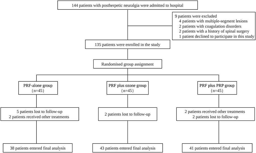 |
Figure 1 CONSORT participant flow diagram. The diagram summarizes patient screening, randomization, follow-up, and final complete-case/per-protocol analysis. Of 144 patients assessed for eligibility, 135 were randomized equally to PRF alone, PRF plus ozone, or PRF plus PRP. Overall, 122 participants were included in the final repeated-measures analysis. Abbreviations: PRF, pulsed radiofrequency; PRP, platelet-rich plasma. |
The study protocol was reviewed and approved by the Medical Ethics Committee of Changzhi People’s Hospital Affiliated to Changzhi Medical College, Changzhi, China (approval number: 2025K035). Trial registration was completed in the Chinese Clinical Trial Registry under the identifier ChiCTR2500104555. The trial was carried out under the ethical principles outlined in the Declaration of Helsinki, with written consent provided by every participant before study entry.
Participants and Eligibility Criteria
Patients with postherpetic neuralgia (PHN) were screened in the Department of Pain Treatment. PHN was diagnosed according to the International Association for the Study of Pain criteria and was defined as dermatomal neuropathic pain persisting for more than 3 months after herpes zoster rash onset. According to the International Classification of Diseases, 11th Revision, PHN is classified as 1E91.5. In the International Classification of Diseases, 10th Revision, Clinical Modification, PHN with other postherpetic nervous system involvement is coded as B02.29. Because this trial enrolled participants with single-segment thoracic, lumbar, or dorsal PHN and did not include trigeminal PHN, B02.29 was considered the applicable ICD-10-CM classification for the study population.
The diagnosis of PHN and eligibility for trial participation were confirmed before randomization by two licensed pain physicians with formal training in pain medicine and experience in neuropathic pain management and computed tomography (CT)-guided interventional procedures. Diagnosis was based on a documented history of herpes zoster, dermatomal pain distribution, persistence of neuropathic pain for more than 3 months after rash onset, compatible sensory symptoms and signs, and exclusion of other pain conditions that could interfere with outcome assessment. Diagnostic uncertainty was resolved by consensus before enrollment.
Eligible participants were men or women aged 50–70 years who had single-segment thoracic, lumbar, or dorsal PHN; pain persisting for longer than 3 months after the initial herpes zoster rash; inadequate pain relief after conservative pharmacological treatment including pregabalin; stable general condition without severe organ dysfunction; ability to complete the VAS, PSQI, and SF-12 assessments; willingness to complete the 90-day follow-up; had not participated in another clinical trial during the study period; and provided written informed consent.
Participants were excluded if they had any condition that could affect treatment response, outcome assessment, or procedural safety, including coagulation disorders or anticoagulant/antiplatelet therapy that could not be safely interrupted; platelet disorders, severe thrombocytopenia, or other hematological conditions affecting platelet-rich plasma (PRP) preparation; active systemic or local infection, fever, autoimmune disease, or immunosuppressive therapy; severe allergy or contraindication to ozone or PRP injection; previous similar interventional treatment for PHN; other chronic pain or neurological disorders, such as diabetic peripheral neuropathy, trigeminal neuralgia, non-zoster radiculopathy, peripheral nerve entrapment, spinal cord disease, or malignancy-related pain; severe psychiatric disorder, unrelated severe sleep disorder, or cognitive impairment; uncontrolled diabetes mellitus, severe cardiopulmonary disease, hepatic or renal insufficiency, malignancy, or any life-threatening condition; spinal deformity or previous spinal surgery affecting CT localization; pregnancy or lactation; refusal to participate; or inability to complete follow-up.
Baseline demographic and clinical characteristics were recorded before randomization, including age, sex, country of residence, body mass index, smoking status, alcohol consumption, hypertension, diabetes mellitus, coronary artery disease, pulmonary disease, and baseline outcome values. Comorbidities were defined according to documented medical history, admission records, current medication use, and relevant clinical or laboratory findings. Patients with stable comorbidities were eligible if their general condition permitted trial participation and follow-up completion, whereas those with severe, uncontrolled, or life-threatening comorbidities were excluded.
Randomization and Masking
Following eligibility confirmation, participants were allocated in equal proportions (1:1:1) to receive PRF alone, PRF plus ozone, or PRF plus PRP. Explicit intervention names were used throughout the manuscript instead of letter-based group labels. The PRF-alone group served as the active control group because dorsal root ganglion-targeted PRF was the common neuromodulatory intervention and withholding active interventional care from patients with refractory PHN was not considered ethically appropriate. The PRF plus ozone group was included to assess the additional effect of ozone as an anti-inflammatory and microcirculatory adjunct, and the PRF plus PRP group was included to assess the additional effect of PRP as an autologous biological adjunct with potential anti-inflammatory, angiogenic, neurotrophic, and tissue-repair effects.
The random allocation sequence was generated before enrollment by an independent statistician who was not involved in recruitment, intervention delivery, outcome assessment, follow-up, or data analysis. Randomization was computer generated, using blocks of three participants to preserve balance across the three treatment arms. No stratification was applied.
Allocation concealment was implemented using sequentially numbered, opaque, sealed envelopes prepared by the independent statistician and kept by a study coordinator independent of recruitment, intervention delivery, outcome assessment, and statistical analysis. For each participant, allocation was revealed only after eligibility confirmation, written informed consent, and completion of baseline assessment.
Because the three interventions differed procedurally, blinding of participants and treating physicians was not feasible. Participants in the PRF-alone group received PRF only, those in the PRF plus ozone group received ozone injection after PRF, and those in the PRF plus PRP group underwent autologous blood collection, PRP preparation, and PRP injection. Group assignments were masked from the outcome assessors and statistician to reduce potential assessment and analysis bias. Outcome assessors were not involved in randomization, envelope opening, intervention delivery, PRP preparation, ozone administration, or routine clinical management. The final dataset was coded using anonymized group labels, and group allocation was not revealed to the statistician until the database had been locked and the primary analyses had been completed.
Interventions
All procedures were performed in a dedicated CT-guided procedure room under strict aseptic conditions by experienced pain physicians. The target segment was determined according to the painful dermatome, previous herpes zoster distribution, sensory findings, and preprocedural imaging review. Standard monitoring, including electrocardiography, pulse oximetry, and noninvasive blood pressure monitoring, was applied throughout the procedure.
After the participant was positioned prone, an initial CT scan was obtained to localize the involved intervertebral foramen and determine an appropriate needle pathway. After routine skin preparation, sterile draping, and local anesthetic infiltration, a 22-gauge radiofrequency cannula with a 10-mm active tip was advanced under CT guidance to the posterior region of the superior third of the target intervertebral foramen. Needle position was adjusted using repeated CT scans to avoid vascular, pleural, visceral, or neural injury.
After the needle tip was positioned near the dorsal root ganglion, negative aspiration for blood, air, and cerebrospinal fluid was confirmed. Contrast medium was then injected to confirm appropriate perineural distribution and exclude intravascular or unintended spread. The radiofrequency electrode was inserted through the cannula. Sensory stimulation was performed at 50 Hz and 0.5 V, and motor stimulation at 2 Hz and 0.5 V. Correct localization was confirmed when paresthesia corresponding to the original painful dermatome or an appropriate segmental motor response was elicited at a stimulation threshold of <0.5 mA.
The same PRF protocol was applied in all groups: 2-Hz pulsed stimulation, 20-ms pulse duration, a maximum tip temperature of 42°C, and a treatment time of 300 s. After the assigned procedure, the needle was removed, hemostasis was achieved by local compression, and the puncture area was dressed under sterile conditions. All participants were monitored for 30 minutes after the procedure.
In the PRF-alone group, no additional injectate was administered after PRF. In the PRF plus ozone group, after PRF and confirmation of negative aspiration, 3 mL per segment of medical ozone at 30 μg/mL was slowly injected into the target dorsal root ganglion region under CT guidance. Repeat CT scanning was performed to confirm local diffusion and exclude unintended spread.
In the PRF plus PRP group, PRP was prepared immediately before injection under sterile conditions. Briefly, 20 mL of peripheral venous blood was collected from the cubital vein into sterile tubes containing 3 mL of sodium citrate anticoagulant. The sample was processed at room temperature, 18°C–26°C, with a centrifuge rotor radius of 10 cm. A two-step centrifugation protocol was used: 1,500 rpm for 10 minutes, approximately 252 × g, followed by 3,500 rpm for 8 minutes, approximately 1,370 × g. The platelet-rich fraction was resuspended to obtain approximately 3 mL of autologous PRP. The final platelet concentration was maintained within 500–1300 × 10^9/L, corresponding to approximately 4–5-fold enrichment compared with baseline peripheral blood, and residual red blood cell concentration was ≤0.01 × 10^6/mL. No exogenous activator was used; activation was expected to occur in situ after contact with local collagen and tissue factors. After PRF and confirmation of correct needle position and negative aspiration, approximately 3 mL of freshly prepared PRP was slowly injected perineurally around the dorsal root ganglion under CT guidance.
All participants received standardized conventional therapy and routine clinical care during the study period according to institutional practice for PHN. Conventional therapy mainly consisted of pregabalin-based neuropathic pain management when clinically indicated, supportive care, patient education, and adverse-event monitoring. The same principles were applied across all three groups to ensure ethical care and reduce confounding from unequal background treatment. Additional non-study interventional procedures or major treatment changes during follow-up were recorded; participants who received other non-study treatments were not included in the final complete-case efficacy analysis.
To enhance reproducibility without publishing patient-level procedural images, the intervention protocol was described in detail in the manuscript and summarized in Supplementary Table S1. Real-time procedural images were not included because intraoperative CT images were obtained for clinical localization rather than standardized publication, and separate patient consent for publication of procedural imaging was not obtained.
Outcomes and Follow-up
Outcome measures were selected to capture the multidimensional burden of PHN, including pain intensity, sleep quality, health-related quality of life, systemic inflammatory status, and safety. Assessments were performed at baseline and at 30, 60, and 90 days after treatment by outcome assessors blinded to treatment allocation. The day of the intervention was defined as day 0.
The primary endpoint was pain intensity, assessed on a 10-cm VAS anchored at 0 for no pain and 10 for the most severe imaginable pain. The VAS was considered appropriate for repeated pain assessment because it is responsive and psychometrically established, and its Chinese version has demonstrated acceptable reliability and convergent validity.33
Secondary outcomes included sleep quality assessed using the PSQI and health-related quality of life assessed using the SF-12. The PSQI evaluates sleep quality over the preceding month and yields a total score from 0 to 21, with lower scores indicating better sleep quality. The Chinese version of the PSQI has demonstrated acceptable internal consistency, stable factor structure, and good screening performance.34 The SF-12 assesses physical and mental health status over the preceding 4 weeks and was transformed to a 0–100 scale, with higher scores indicating better health-related quality of life. The Chinese version of the SF-12 has demonstrated satisfactory reliability and validity.35 The neutrophil-to-lymphocyte ratio (NLR) was included as an exploratory inflammatory outcome. NLR was calculated from routine peripheral blood tests by dividing the absolute neutrophil count by the absolute lymphocyte count. It was used as an accessible laboratory-derived indicator of systemic inflammatory status.36
Safety outcomes included the incidence, type, severity, management, and group distribution of adverse events from the start of the procedure to the end of the 90-day follow-up period. Immediate safety assessment was performed during the procedure and within the 30-minute post-procedural observation period. Delayed adverse events were assessed at 30, 60, and 90 days by reviewing procedural records, post-procedural notes, follow-up records, and patient-reported symptoms. Serious adverse events were defined as death, life-threatening events, hospitalization or prolonged hospitalization, persistent or significant disability, or events requiring urgent medical intervention. A summary of the reliability and validity of the outcome measures is provided in Supplementary Table S2.
Sample Size
The sample size was calculated based on the primary efficacy outcome, the VAS score. Because the trial aimed to compare three interventional strategies, the calculation was based on detecting a clinically meaningful between-group difference in VAS during follow-up. According to recommendations for chronic pain clinical trials, a change of approximately 1–2 points on a 0–10 pain scale may represent clinically relevant improvement.37 Therefore, a between-group difference of 1.2 points was considered clinically meaningful.
The required sample size for comparing two independent group means was estimated using the following formula:38

where n is the required number of participants per group, σ is the estimated standard deviation, δ is the expected between-group difference, Zα/2 is the standard normal deviate for a two-sided significance level, and Zβ is the standard normal deviate for statistical power. With a two-sided α of 0.05, power of 80%, expected between-group difference of 1.2 points, and estimated standard deviation of 1.8, the calculated sample size was 35.28 participants per group. Therefore, at least 36 participants were required in each group. Considering an anticipated dropout rate of approximately 10%, at least 40 participants per group were required. To ensure adequate analyzable data, 45 participants were randomized to each group, for a total randomized sample size of 135 participants.
Statistical Analysis
All analyses were prespecified according to the randomized three-group design, outcome type, and repeated-measures structure. Distributional assumptions for continuous data were examined using the Shapiro–Wilk test, histogram and Q–Q plot inspection, and residual diagnostics when required. Continuous variables that satisfied normality assumptions were summarized with means and standard deviations; differences among groups were evaluated by one-way ANOVA after Levene’s test confirmed homogeneity of variance. Skewed continuous data and ordinal measures were described using medians and interquartile ranges, and between-group differences were evaluated with the Kruskal–Wallis test. Categorical data were presented as counts and percentages, with group comparisons performed using Pearson’s chi-square test, the continuity-corrected chi-square test, or Fisher’s exact test, depending on expected cell counts. Baseline homogeneity was assessed before longitudinal outcome interpretation by comparing demographic and clinical variables among the three treatment groups, including age, sex, country of residence, body mass index, smoking status, alcohol consumption, comorbidities, and baseline outcome values. Country of residence was recorded descriptively but was not included in inferential testing because all participants resided in China. Because no statistically significant or clinically meaningful baseline imbalance was identified, the primary longitudinal models did not include additional covariate adjustment.
Because VAS, PSQI, SF-12, and NLR were repeatedly measured within the same participants at baseline and at 30, 60, and 90 days after treatment, linear mixed-effects models were used as the primary analytical method. For each repeated outcome, treatment group, time, and the group-by-time interaction were included as fixed effects, and participant identifier was included as a random intercept. The group-by-time interaction was the primary inferential term used to determine whether longitudinal changes differed among the three groups. Model assumptions were evaluated by inspecting residual distributions and fitted-value plots.
For outcomes showing a significant group-by-time interaction, model-derived between-group contrasts were examined at each scheduled follow-up visit. The prespecified contrasts compared PRF plus ozone with PRF alone, PRF plus PRP with PRF alone, and PRF plus PRP with PRF plus ozone. Estimated mean differences were presented with lower-to-upper 95% confidence intervals, and multiplicity was controlled using the Holm–Bonferroni procedure within each outcome and time point.
Hedges’ g was used to quantify the magnitude of between-group differences; effect sizes were classified as small, moderate, or large when they approached 0.2, 0.5, or 0.8, respectively. Minimal clinically important difference (MCID) benchmarks were considered where available. For VAS, a between-group difference of at least 1.0 point on the 0–10 scale was considered minimally clinically important, and a difference of approximately 2.0 points was interpreted as a more substantial clinically meaningful improvement. For PSQI, a 4.4-point reduction was used as the reference MCID. For SF-12, because no PHN-specific MCID for the transformed SF-12 total score has been established, a conservative 3-point difference was used as a supportive benchmark. Because no validated MCID has been established for NLR in PHN, NLR was interpreted as an exploratory inflammatory outcome using the estimated mean difference, 95% confidence interval, adjusted P value, and Hedges’ g.
VAS scores were expressed in centimeters on a 0–10 cm scale and summarized as medians with interquartile ranges because pain intensity scores may have ordinal or non-normally distributed characteristics. Cross-sectional descriptive comparisons across treatment groups were conducted at each assessment visit using the Kruskal–Wallis test; however, longitudinal treatment effects were interpreted primarily from the mixed-effects models.
Safety outcomes were summarized by treatment group as n (%). Because adverse events were limited and several categories had small expected cell counts, safety data were analyzed primarily using descriptive statistics. Fisher’s exact test or chi-square-based methods were used only when appropriate. Serious adverse events were reported separately according to prespecified clinical criteria.
The primary efficacy analysis was restricted to the complete-case/per-protocol population, comprising participants who had been randomized, underwent their allocated treatment, and had outcome data available at baseline and at each scheduled follow-up visit. All randomized participants were included in the participant-flow summary according to their assigned group. A formal full intention-to-treat analysis was not performed because complete post-randomization outcome data were unavailable for participants who were lost to follow-up or received other non-study treatments, which prevented reliable attribution of subsequent outcomes to the assigned intervention. No item-level missing values were present for VAS, PSQI, SF-12, or NLR among participants included in the final analysis, and no statistical imputation was performed. Post-randomization exclusions were documented by treatment group and reason in the CONSORT flow diagram and Results section.
A two-sided testing framework was applied, and adjusted P < 0.05 was taken to indicate statistical significance. P values are reported consistently with an uppercase P; exact values are given to three decimal places when ≥0.001 and as P < 0.001 when below this threshold. All analyses were performed using SPSS Statistics 27.0.
Results
Participant Flow and Analysis Population
From July to December 2025, 144 patients with postherpetic neuralgia underwent eligibility screening in the pain treatment department of Changzhi People’s Hospital Affiliated to Changzhi Medical College. Nine patients were excluded before randomization: four had multiple-segment lesions, two had coagulation disorders, two had a history of spinal surgery affecting computed tomography localization, and one declined to participate. Accordingly, 135 participants entered random allocation, with 45 assigned to each of the three treatment arms.
During follow-up, 13 randomized participants were not included in the final complete-case efficacy analysis because of loss to follow-up or receipt of other non-study treatments. The final analysis included 38 participants in the PRF-alone group after seven post-randomization exclusions, comprising five follow-up non-completions and two cases of non-study treatment. The PRF plus ozone group contributed 43 participants after two follow-up non-completions. The PRF plus PRP group contributed 41 participants after four post-randomization exclusions, including two follow-up non-completions and two cases of non-study treatment. Overall, 122 participants were included in the repeated-measures analysis. No item-level missing values were identified for VAS, PSQI, SF-12, or NLR among these 122 participants. Because 13 randomized participants had incomplete post-randomization outcome data, the final efficacy analysis was based on the complete-case/per-protocol population rather than a formal full intention-to-treat population. Participant flow is summarized in Figure 1, with post-randomization exclusions presented according to treatment arm and reason.
All 135 randomized participants completed the allocated intervention, yielding a treatment compliance rate of 100.0% in each group. Overall, 122 of 135 randomized participants completed all scheduled baseline, 30-day, 60-day, and 90-day assessments, corresponding to a follow-up adherence rate of 90.4%. The adherence rates were 84.4% in the PRF-alone group, 95.6% in the PRF plus ozone group, and 91.1% in the PRF plus PRP group. The overall dropout rate due to loss to follow-up was 6.7% (9/135), and the overall post-randomization non-completion rate due to loss to follow-up or receipt of other non-study treatment was 9.6% (13/135).
Baseline Characteristics and PRP Quality Control
Baseline demographic, clinical, and potential confounding characteristics are summarized in Table 1. No significant between-group differences were observed in age, sex, body mass index, smoking status, alcohol consumption, hypertension, diabetes mellitus, coronary artery disease, pulmonary disease, or baseline pain intensity. All participants included in the complete-case analysis resided in China and were treated in the same single-center clinical setting; therefore, country of residence was constant across groups and was not included in inferential testing. These findings indicated acceptable baseline homogeneity before longitudinal outcome analysis.
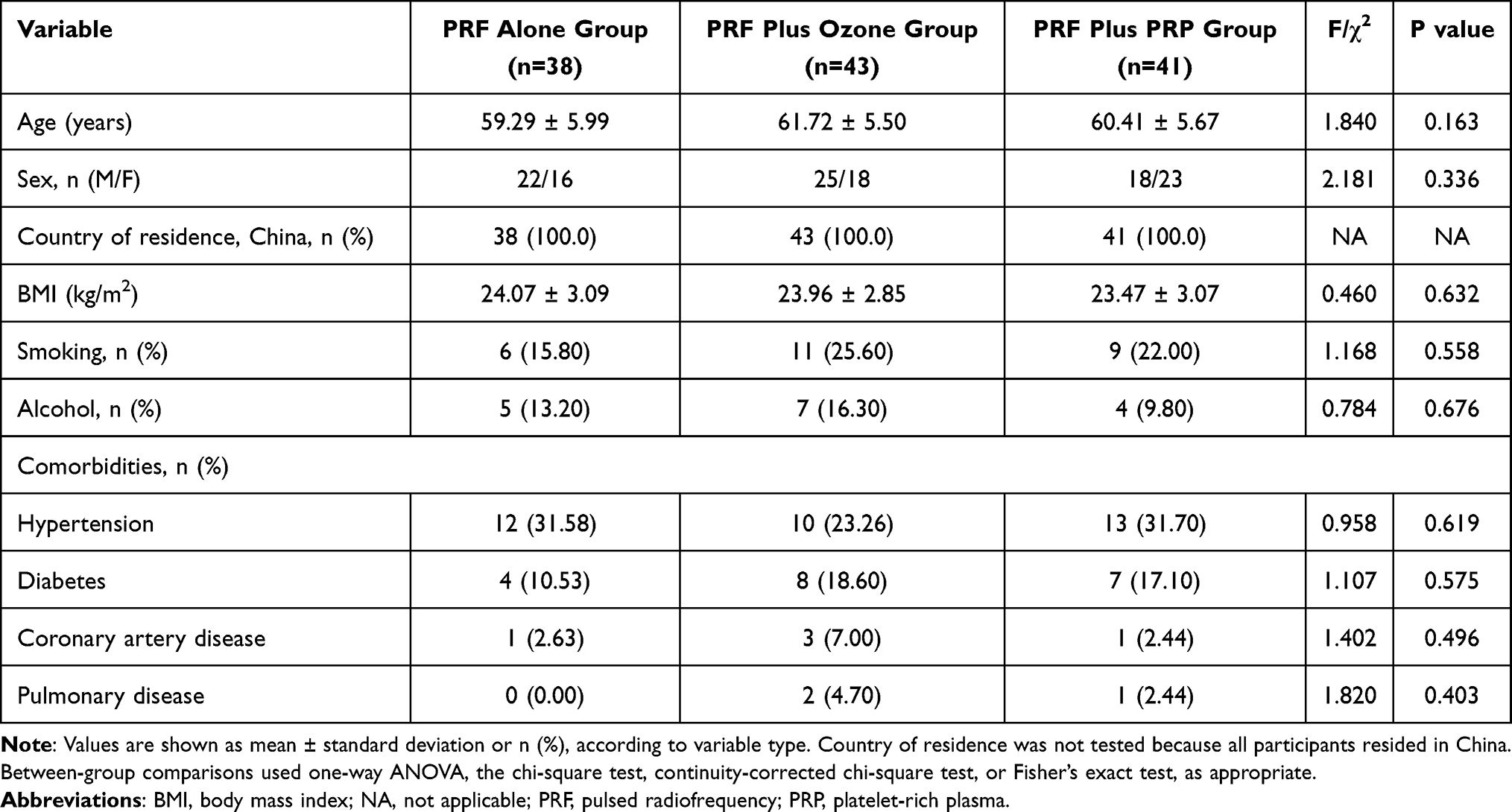 |
Table 1 Baseline Demographic, Clinical, and Potential Confounding Characteristics of the Study Population |
Medication exposure, pain-related characteristics, age, and adherence were also considered when interpreting the treatment effects. Conventional therapy was provided according to the same institutional principles across the three groups, and additional non-study treatments during follow-up were recorded as protocol deviations. All included participants had single-segment thoracic, lumbar, or dorsal postherpetic neuralgia lasting more than 3 months, and baseline VAS scores were comparable among groups. These findings suggested that the main measured confounding factors were sufficiently addressed in the trial design and analysis.
Fresh autologous PRP was successfully prepared for all participants assigned to the PRF plus PRP group. The final PRP volume used for injection was approximately 3 mL per participant. The platelet concentration achieved in the final PRP preparation was 500–1300 × 10^9/L, corresponding to approximately 4–5-fold enrichment compared with baseline peripheral blood. The residual red blood cell concentration was ≤0.01 × 10^6/mL. No exogenous activator was used during PRP preparation.
Longitudinal Outcomes
Pain intensity was assessed using the 10-cm VAS and expressed in centimeters on a 0–10 cm scale. Descriptive VAS distributions are presented as medians with interquartile ranges in Table 2. VAS scores were comparable among the three groups at baseline and at 30 days after treatment. Differences among the three groups became clearer at days 60 and 90, when the PRF plus PRP group demonstrated the most favorable VAS profile relative to the PRF-alone and PRF plus ozone groups. The PRF plus ozone group also showed lower VAS scores than the PRF-alone group at 60 and 90 days. Because VAS values were summarized as medians with interquartile ranges and many participants reported similar integer or near-integer values on the 0–10 cm scale, some descriptive values appeared numerically clustered. Therefore, the primary inference for treatment effects was based on the longitudinal mixed-effects model rather than on descriptive medians alone.
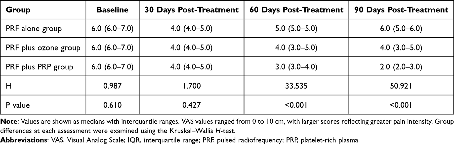 |
Table 2 Visual Analog Scale Scores Among the Three Treatment Groups Before and After Treatment |
Linear mixed-effects models showed significant group-by-time interactions for all repeated outcome variables, indicating that the longitudinal trajectories of VAS, PSQI, SF-12, and NLR differed significantly among the three treatment groups. Significant interactions were observed for VAS (Wald χ2 = 195.818, df = 6, P < 0.001), PSQI (Wald χ2 = 114.602, df = 6, P < 0.001), SF-12 (Wald χ2 = 145.376, df = 6, P < 0.001), and NLR (Wald χ2 = 23.948, df = 6, P < 0.001) (Table 3).
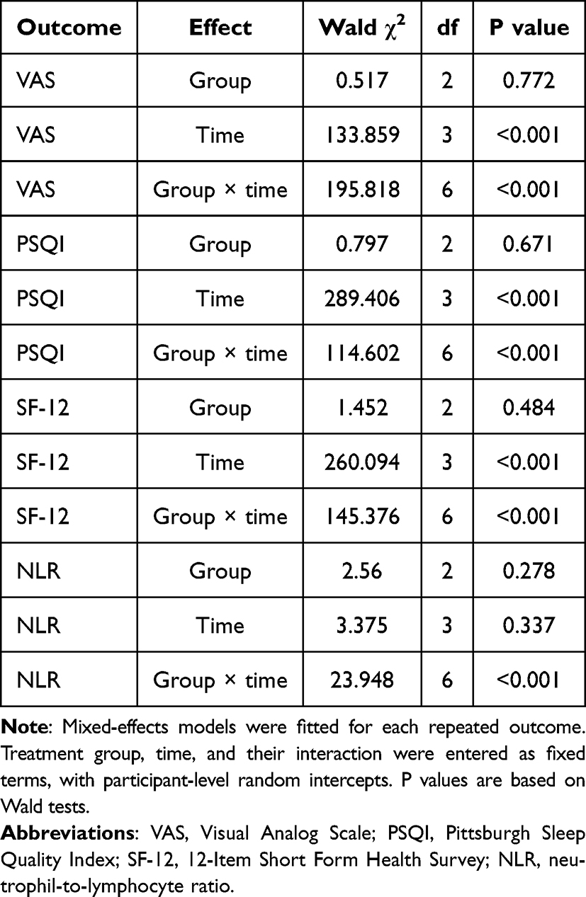 |
Table 3 Linear Mixed-Effects Model Analysis of Repeated Outcomes |
Table 4 summarizes the model-derived pairwise contrasts, including estimated mean differences, 95% confidence intervals from lower to upper limits, Holm-adjusted P values, and Hedges’ g effect sizes. At 60 days after treatment, PRF plus PRP was associated with greater improvement than PRF alone in VAS, PSQI, and SF-12, and greater improvement than PRF plus ozone in VAS, PSQI, and SF-12. Between-group differences in NLR at 60 days were not statistically significant after adjustment.
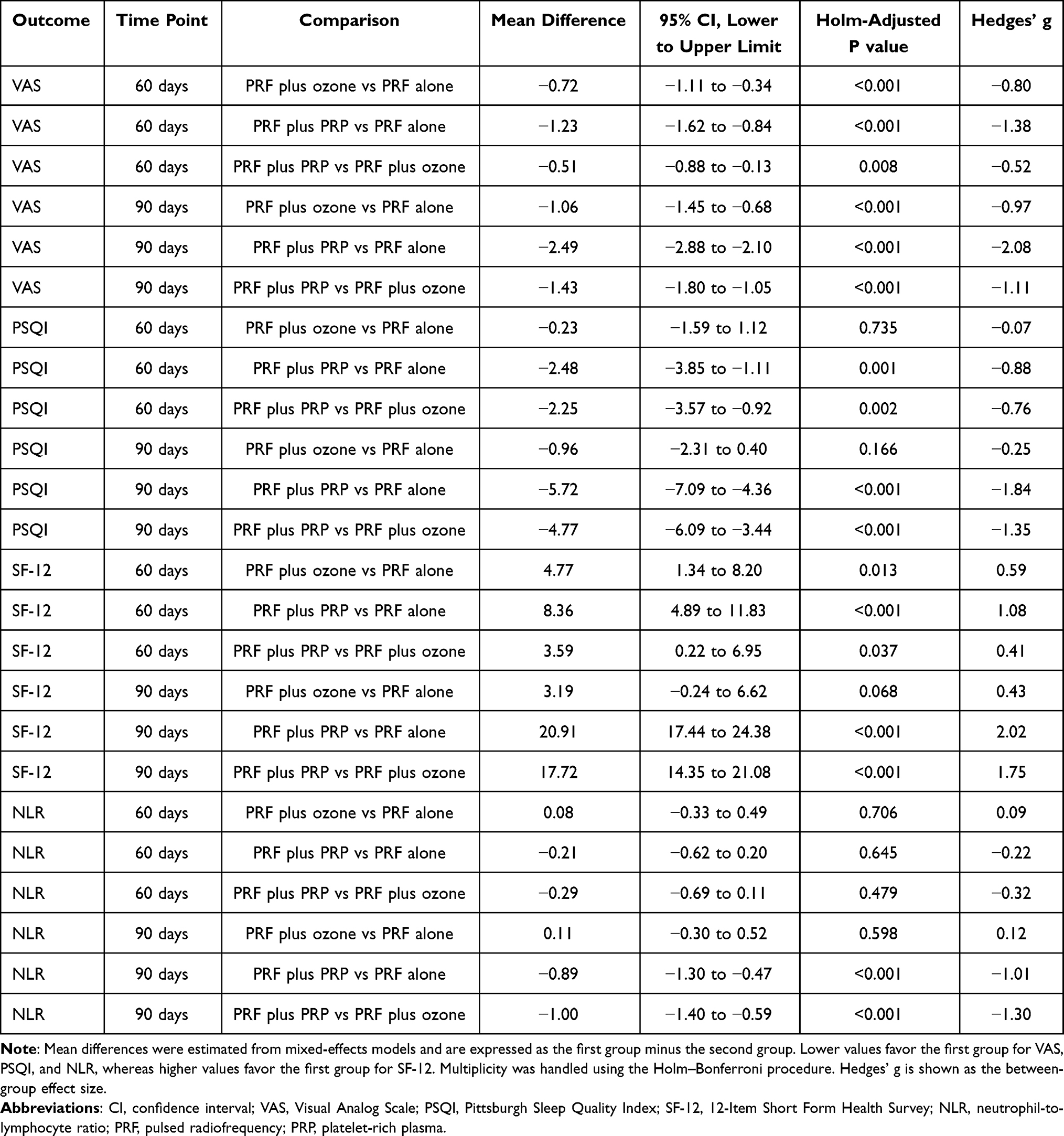 |
Table 4 Model-Based Pairwise Comparisons with P values and 95% Confidence Intervals for Repeated Outcome Variables at 60 and 90 days After Treatment |
At 90 days after treatment, PRF plus PRP showed greater improvement than both PRF alone and PRF plus ozone across all repeated outcome variables. Compared with PRF alone, PRF plus PRP was associated with lower VAS scores (mean difference, −2.49; 95% CI, −2.88 to −2.10; adjusted P < 0.001), lower PSQI scores (mean difference, −5.72; 95% CI, −7.09 to −4.36; adjusted P < 0.001), higher SF-12 scores (mean difference, 20.91; 95% CI, 17.44 to 24.38; adjusted P < 0.001), and lower NLR values (mean difference, −0.89; 95% CI, −1.30 to −0.47; adjusted P < 0.001). Compared with PRF plus ozone, PRF plus PRP was associated with lower VAS scores (mean difference, −1.43; 95% CI, −1.80 to −1.05; adjusted P < 0.001), lower PSQI scores (mean difference, −4.77; 95% CI, −6.09 to −3.44; adjusted P < 0.001), higher SF-12 scores (mean difference, 17.72; 95% CI, 14.35 to 21.08; adjusted P < 0.001), and lower NLR values (mean difference, −1.00; 95% CI, −1.40 to −0.59; adjusted P < 0.001).
In contrast, several comparisons involving the PRF plus ozone group showed small effect sizes or 95% confidence intervals crossing zero, particularly for PSQI, SF-12, and NLR. PRF plus ozone did not show a statistically significant advantage over PRF alone for PSQI at 60 or 90 days, SF-12 at 90 days, or NLR at 60 or 90 days. These findings suggest that the adjunctive benefit of ozone beyond PRF alone was less consistent for secondary and exploratory outcomes than the benefit observed with PRF plus PRP.
Clinical Relevance and Graphical Presentation
The practical significance of treatment effects was evaluated using prespecified MCID benchmarks and Hedges’ g effect sizes.37,39,40 At 90 days, the VAS difference between the PRF plus PRP group and the PRF-alone group was −2.49 points, exceeding both the 1.0-point MCID threshold and the 2.0-point benchmark for a more substantial clinically meaningful improvement. The VAS difference between the PRF plus PRP group and the PRF plus ozone group was −1.43 points, which also exceeded the MCID threshold. Both comparisons showed large effect sizes.
For sleep quality, the 90-day PSQI differences between the PRF plus PRP group and the PRF-alone group and between the PRF plus PRP group and the PRF plus ozone group were −5.72 and −4.77 points, respectively. Both exceeded the reference PSQI MCID of 4.4 points and showed large effect sizes.39 For health-related quality of life, the corresponding SF-12 differences were 20.91 and 17.72 points, respectively, both exceeding the conservative 3-point benchmark used for clinically meaningful SF-12 change, with large effect sizes.40 For NLR, no validated MCID threshold has been established in patients with PHN; therefore, NLR was interpreted as an exploratory inflammatory outcome. At 90 days, PRF plus PRP was associated with lower NLR than PRF alone and PRF plus ozone, with large effect sizes. A summary of MCID benchmarks and effect-size interpretation for the 90-day endpoint is provided in Table 5.
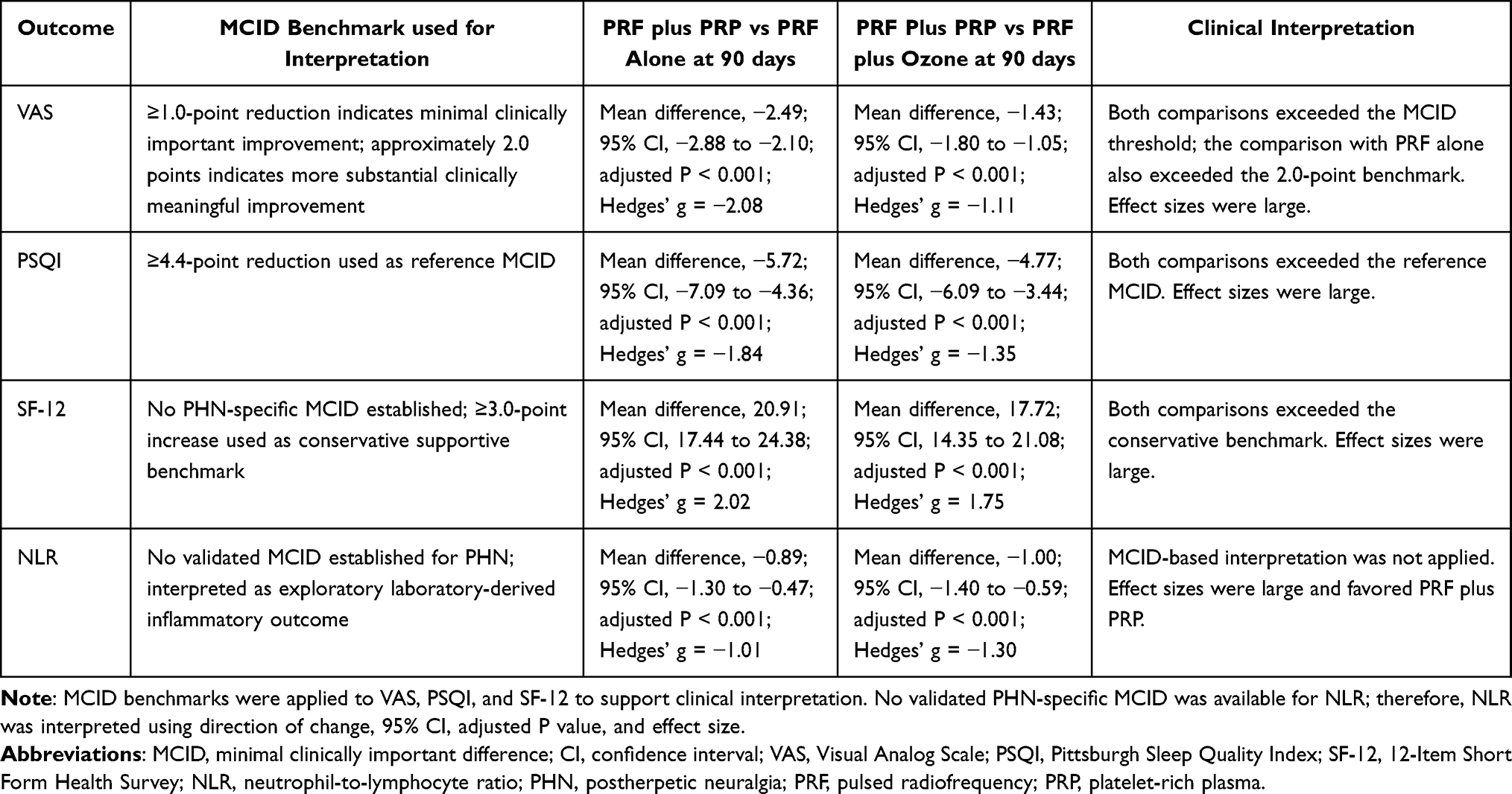 |
Table 5 Clinical Interpretation of MCID and Effect Size for Repeated Outcome Variables at 90 days After Treatment |
The longitudinal patterns of PSQI, SF-12, and NLR are displayed in Figures 2–4. Baseline is shown as day 0, and subsequent assessments are shown at 30, 60, and 90 days after treatment. Error bars indicate standard deviation. Detailed model-based estimates, 95% CIs, adjusted P values, and effect sizes are provided in Table 4; therefore, statistical annotations were not added to the figure panels. Overall, the PRF plus PRP group showed the most favorable trajectories for sleep quality, health-related quality of life, and NLR during follow-up.
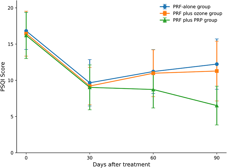 |
Figure 2 Longitudinal changes in sleep quality among the three treatment groups. PSQI scores were assessed at baseline and at 30, 60, and 90 days after treatment. Baseline is shown as day 0. Data are displayed as group means with standard deviation error bars. Lower PSQI scores indicate better sleep quality. Model-based pairwise comparisons are reported in Table 4. Abbreviations: PRF, pulsed radiofrequency; PRP, platelet-rich plasma; PSQI, Pittsburgh Sleep Quality Index. |
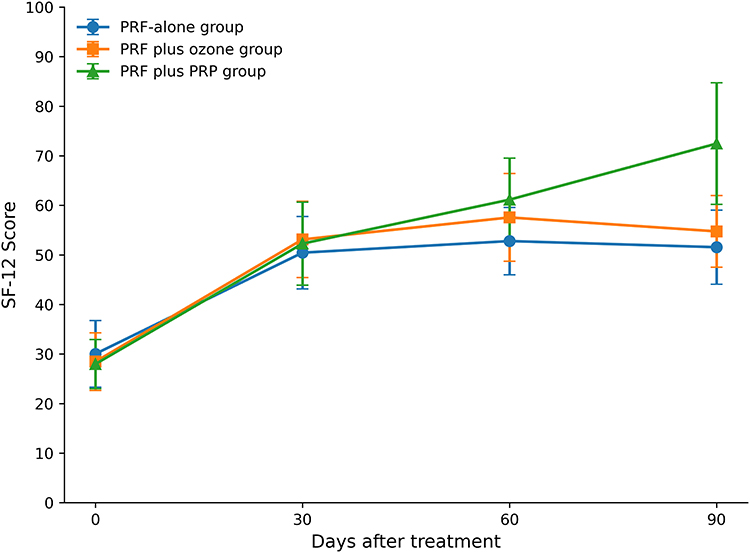 |
Figure 3 Longitudinal changes in health-related quality of life among the three treatment groups. SF-12 scores were assessed at baseline and at 30, 60, and 90 days after treatment. Baseline is shown as day 0. Data are displayed as group means with standard deviation error bars. Higher SF-12 scores indicate better health-related quality of life. Model-based pairwise comparisons are reported in Table 4. Abbreviations: PRF, pulsed radiofrequency; PRP, platelet-rich plasma; SF-12, 12-Item Short Form Health Survey. |
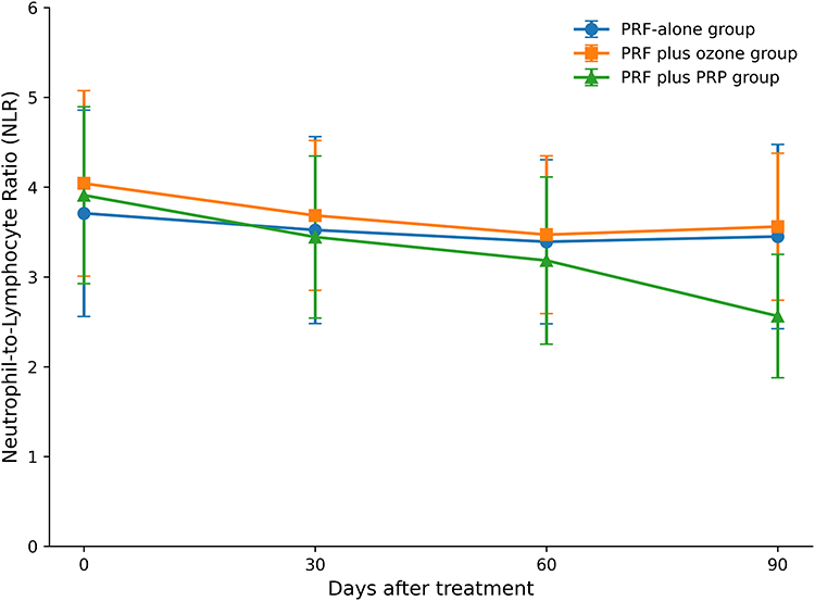 |
Figure 4 Longitudinal changes in systemic inflammatory status among the three treatment groups. NLR values were assessed at baseline and at 30, 60, and 90 days after treatment. Baseline is shown as day 0. Data are displayed as group means with standard deviation error bars. Lower NLR values indicate lower systemic inflammatory status. Model-based pairwise comparisons are reported in Table 4. Abbreviations: PRF, pulsed radiofrequency; PRP, platelet-rich plasma; NLR, neutrophil-to-lymphocyte ratio. |
Adverse Events
Detailed adverse events and their distribution by treatment group are shown in Table 6. During the 90-day follow-up period, 13 adverse events were documented among the 122 participants included in the complete-case safety summary, corresponding to an overall adverse-event rate of 10.7%. Adverse events occurred in eight participants in the PRF-alone group, four participants in the PRF plus ozone group, and one participant in the PRF plus PRP group. Most adverse events were mild to moderate, procedure-related, and managed conservatively.
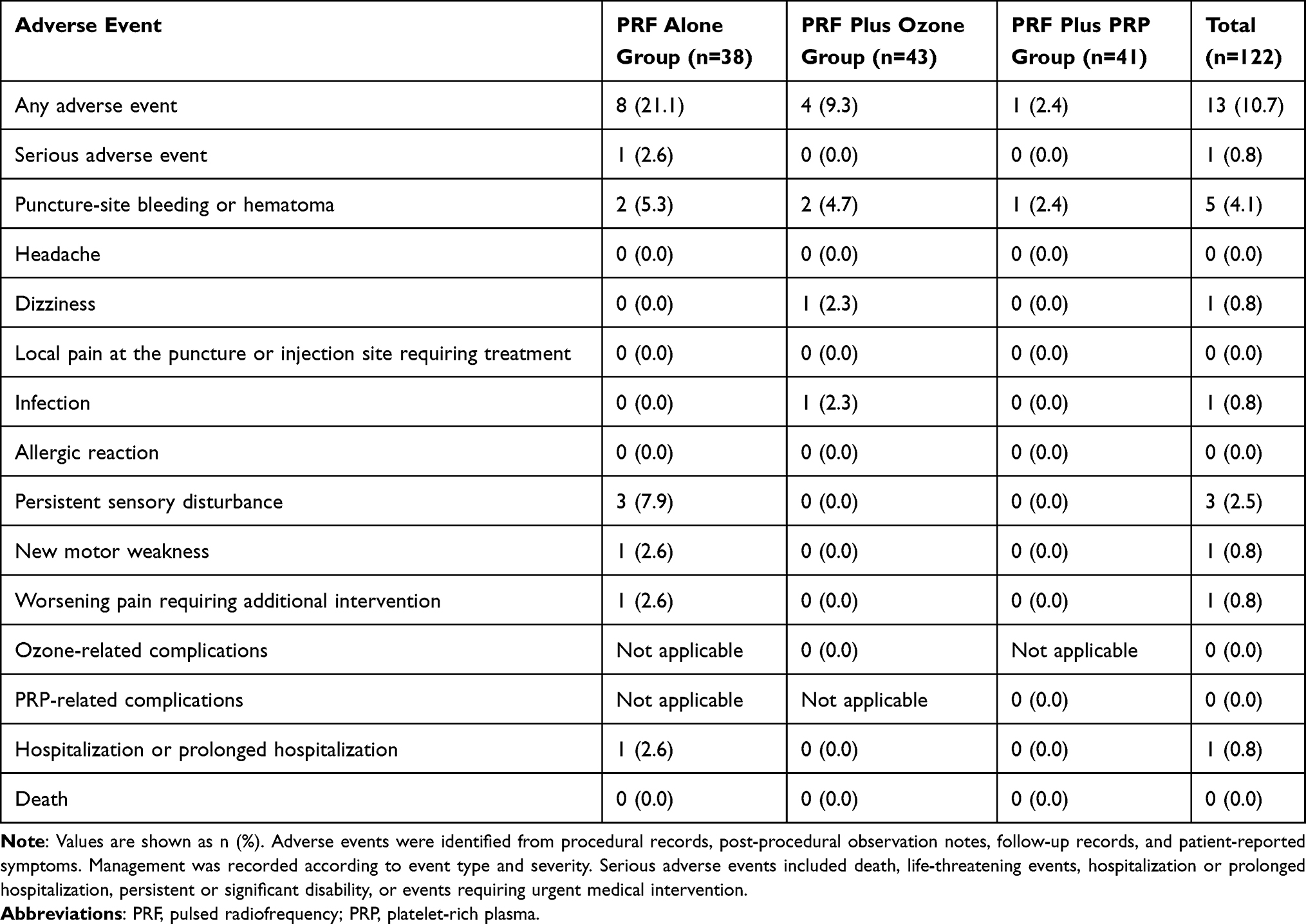 |
Table 6 Adverse Events During the 90-Day Follow-Up Period |
The most frequently recorded adverse event was puncture-site bleeding or hematoma, which occurred in five participants and was managed with local compression, sterile dressing, puncture-site observation, and routine monitoring. One participant in the PRF plus ozone group experienced dizziness after the procedure and recovered after rest, vital-sign monitoring, and observation. One participant in the PRF plus ozone group developed infection during follow-up and was managed with local wound care, dressing changes, and anti-infective treatment according to clinical judgment. No deep tissue infection, systemic infection, or sepsis was recorded.
Neurological adverse events were mainly observed in the PRF-alone group. Persistent sensory disturbance was recorded in three participants and was managed with observation, neurological assessment, reassurance, and adjustment of conventional neuropathic pain management when clinically needed. One participant developed new motor weakness, and one participant experienced worsening pain requiring additional intervention. One serious adverse event, defined as hospitalization or prolonged hospitalization, occurred in the PRF-alone group. No deaths, allergic reactions, ozone-related complications, PRP-related complications, or local pain at the puncture or injection site requiring treatment were recorded during the study period. Overall, no PRP-related safety signal was observed during the 90-day follow-up period.
Discussion
The present randomized controlled trial was designed to compare CT-guided dorsal root ganglion PRF alone, PRF plus ozone, and PRF plus PRP in patients with refractory PHN, with a 90-day evaluation of pain intensity, sleep quality, health-related quality of life, exploratory systemic inflammatory status, and safety. The main finding was that PRF plus PRP was associated with the most consistent improvement across VAS, PSQI, SF-12, and NLR at 90 days. These findings support the primary hypothesis that adding PRP to DRG-targeted PRF may provide more sustained clinical benefit than PRF alone or PRF plus ozone. However, the results should be interpreted as evidence for the comparative adjunctive value of these interventions when added to standardized conventional management, rather than as evidence that interventional therapy should replace conventional pharmacological care. In the present trial, conventional therapy was provided using the same principles across all three groups because withholding standard analgesic care from patients with chronic neuropathic pain would not have been ethically appropriate.9–12
The clinical relevance of the observed treatment effects should be interpreted beyond statistical significance alone. At 90 days, the between-group improvement in VAS with PRF plus PRP exceeded the prespecified MCID threshold compared with both PRF alone and PRF plus ozone, indicating that the additional analgesic benefit was likely meaningful from a clinical perspective. The PSQI differences also exceeded the reference MCID for sleep-quality improvement, suggesting that the benefit of PRF plus PRP extended beyond analgesia to sleep restoration. Similarly, the SF-12 differences exceeded the conservative benchmark used for clinically meaningful improvement in health-related quality of life.37,39,40 Although NLR does not have an established MCID in PHN and should therefore be interpreted as an exploratory biomarker, the larger effect sizes observed at 90 days suggest a more pronounced reduction in systemic inflammatory status in the PRF plus PRP group. These findings indicate that the observed benefits were not only statistically significant but also clinically interpretable across multiple patient-centered outcomes.
The interpretation of small effect sizes and wide or zero-crossing confidence intervals is also important. In the present study, small effect sizes or confidence intervals including zero were mainly observed in comparisons involving PRF plus ozone for PSQI, SF-12, and NLR, as well as in NLR comparisons at 60 days. These findings suggest that the effect of PRF plus ozone beyond PRF alone was uncertain or clinically modest for some secondary and exploratory outcomes. A small effect size indicates that the magnitude of the between-group difference may be limited even when the direction of change appears favorable, whereas a wide confidence interval indicates reduced precision and may reflect individual response variability, limited sample size, or insufficient statistical information for that specific comparison. When the confidence interval crosses zero, the data are compatible with both a small benefit and no meaningful effect. Therefore, these findings should not be interpreted as robust evidence of superiority. Instead, they suggest that ozone may provide some adjunctive analgesic benefit, but its effects on sleep quality, health-related quality of life, and systemic inflammatory status were less consistent than those observed with PRF plus PRP.
The present findings are generally consistent with previous studies supporting DRG-targeted PRF as a clinically relevant neuromodulatory intervention for herpes zoster-related neuralgia and PHN. The dorsal root ganglion is closely involved in VZV-related sensory neuronal injury, ectopic discharge, and persistent nociceptive transmission, making it a rational therapeutic target in PHN.7,13–16 In previous nerve-related pain studies, PRF has commonly been performed under ultrasound, CT, or fluoroscopic guidance, with sensory and motor stimulation used to confirm target localization before pulsed current delivery. The temperature is usually maintained below the neurodestructive range, commonly around 42°C, which allows neuromodulation while reducing the risk of permanent neural injury.14 In the present study, CT guidance was used to improve anatomical visualization of the intervertebral foramen and perineural target area, which may help reduce puncture-related risk. Nevertheless, PRF remains an invasive procedure, and potential adverse events include transient puncture-site pain, numbness, hypoesthesia, dysesthesia, local soreness, bleeding or hematoma, infection, and rare nerve irritation or motor weakness.15
The findings related to ozone therapy should be interpreted more cautiously. Ozone has been used in pain medicine because of its potential anti-inflammatory, analgesic, oxidative preconditioning, immunomodulatory, oxygenation-related, and microcirculatory effects.18–20,41 Previous studies have reported that ozone injection, particularly when combined with PRF, may improve pain outcomes in selected patients with zoster-associated pain or PHN.17,19,20 In the present study, PRF plus ozone showed some analgesic advantage over PRF alone at later follow-up, which is compatible with its proposed anti-inflammatory and microcirculatory effects. However, this benefit was less consistent for sleep quality, health-related quality of life, and NLR. Moreover, ozone therapy is concentration-dependent and may have a relatively narrow therapeutic window; excessive oxidative stimulation may theoretically contribute to local irritation, inflammatory aggravation, or neural toxicity.21,22 These considerations may partly explain why PRF plus ozone produced more variable and less definitive benefits than PRF plus PRP.
The more sustained improvement observed in the PRF plus PRP group may be explained by the combined effects of DRG neuromodulation and autologous biological repair support. PRP is a patient-derived blood preparation that delivers concentrated platelets together with growth factors, cytokines, and other bioactive molecules. Through platelet-derived mediators, including platelet-derived growth factor, vascular endothelial growth factor, epidermal growth factor, and insulin-like growth factor-1, PRP may support anti-inflammatory, anti-apoptotic, angiogenic, neurotrophic, and reparative processes.24–27 PRP may also influence the perineural microenvironment by supporting Schwann cell activation, axonal repair, remyelination, and neurovascular recovery.25,28,29 Similar PRP-based approaches have been explored in peripheral neuropathic pain and peripheral nerve-related conditions, with reported improvements in pain, sensory symptoms, functional outcomes, and selected nerve-recovery indicators.28,29 A recent retrospective analysis of PHN also found that combining CT-guided DRG PRF with PRP injection was associated with improvements in pain intensity, sleep quality, and patient-reported functional well-being.30 The present randomized comparison extends these observations by directly comparing PRF plus PRP with both PRF alone and PRF plus ozone within the same study framework.
Several mechanisms may contribute to the differences among the three interventions. PRF may primarily modulate abnormal DRG-mediated nociceptive transmission and reduce ectopic neural activity.13–16 Ozone may provide anti-inflammatory and microcirculatory modulation, but its biological effects may depend on concentration, exposure, and local tissue response.18,19,21,22 PRP may provide a more prolonged biological effect through local release of growth factors and cytokines, potentially improving the injured perineural microenvironment and supporting neurovascular repair.24–26,28,29 Therefore, the superior 90-day outcomes in the PRF plus PRP group may reflect a combined neuromodulatory and biological-repair effect rather than simple immediate analgesia. However, this mechanistic interpretation remains indirect because local growth factor release, cytokine profiles, Schwann cell activity, nerve regeneration markers, and imaging or electrophysiological nerve-repair indicators were not directly measured.
NLR was included to provide an accessible exploratory inflammatory perspective on treatment response. PHN is increasingly recognized as a condition involving both neuropathic injury and immune-inflammatory dysregulation after VZV reactivation.42,43 NLR, a readily available blood-based inflammatory marker, integrates information on neutrophil-related inflammatory activity and lymphocyte-associated immune status, and has been used to characterize systemic inflammatory burden in various diseases.44–46 In the present study, the significantly lower NLR observed in the PRF plus PRP group at 90 days was directionally consistent with greater improvements in VAS, PSQI, and SF-12 scores. Nevertheless, NLR should be interpreted as an exploratory and indirect biomarker rather than a disease-specific indicator of PHN. Future studies should combine NLR with more specific inflammatory cytokines, neuroimmune markers, or local tissue biomarkers to better clarify the biological mechanisms underlying treatment response.
The clinical implications of this study can be considered from the perspectives of researchers, clinicians, and patients. For researchers, this trial addresses an important evidence gap by directly comparing CT-guided DRG PRF alone, PRF plus ozone, and PRF plus PRP in patients with PHN, while incorporating pain intensity, sleep quality, health-related quality of life, systemic inflammatory status, effect-size estimation, MCID-based interpretation, and adverse-event reporting. For clinicians, the findings suggest that adding PRP to DRG-targeted PRF may be a clinically reasonable adjunctive option in appropriately selected patients with refractory PHN, particularly when sustained symptom control and functional improvement are treatment priorities. For patients, the potential value of this combined approach lies in its focus on daily-life outcomes, including pain relief, sleep restoration, quality of life, and functional recovery. Nevertheless, PRF plus PRP should be regarded as a promising adjunctive strategy rather than an established standard treatment, and clinical decisions should be individualized according to disease severity, comorbidities, procedural risks, cost, availability of PRP preparation, and patient preferences.
Safety reporting is essential for interventional pain studies. In the present study, adverse events were recorded in 13 of 122 participants during the 90-day follow-up period. Most events were non-serious and included puncture-site bleeding or hematoma, dizziness, infection, persistent sensory disturbance, worsening pain requiring additional intervention, and new motor weakness. The overall adverse-event rate was numerically lower in the PRF plus PRP group than in the PRF-alone and PRF plus ozone groups. Most adverse events were procedure-related and clinically manageable with conservative measures, including local compression and observation for puncture-site bleeding or hematoma, symptomatic observation for dizziness, local wound care and anti-infective treatment for infection, and neurological reassessment with symptomatic management for sensory or motor symptoms. Only one serious adverse event, hospitalization or prolonged hospitalization, was documented and occurred in the PRF-alone group. No deaths, allergic reactions, ozone-related complications, or PRP-related complications were recorded. These findings suggest that CT-guided PRF combined with PRP was generally well tolerated in the present study. However, given the limited sample size and 90-day follow-up period, larger multicenter studies with longer follow-up are required to further confirm the comparative safety profile of these interventions.
This study has several strengths. First, it used a prospective randomized controlled design with concealed allocation, blinded outcome assessment, and blinded statistical analysis. Second, the three intervention groups were explicitly named and compared using standardized CT-guided procedures, and all groups received conventional therapy according to the same principles. Third, outcomes were assessed across several clinically relevant domains, including pain burden, sleep disturbance, health-related functioning, systemic inflammation, and adverse events. Fourth, the statistical analysis incorporated linear mixed-effects models, adjustment for multiple comparisons, 95% confidence intervals, effect sizes, and MCID-based interpretation, which strengthened both statistical and clinical interpretation.
Although these findings are clinically encouraging, they should be interpreted in light of several methodological constraints, which are discussed in detail below. In particular, the single-center design, modest sample size, 90-day follow-up, complete-case/per-protocol analysis, and absence of direct mechanistic biomarkers limit the generalizability and biological interpretation of the results.
Limitations
This study has several limitations. First, this was a single-center randomized controlled trial with a relatively limited sample size, which may restrict the generalizability of the findings and reduce the precision of effect estimates, particularly for secondary outcomes, exploratory inflammatory markers, and adverse events. The follow-up duration was limited to 90 days; therefore, the long-term durability of treatment effects and delayed adverse events could not be fully evaluated. Although PRF plus PRP was associated with greater 90-day improvements than PRF alone or PRF plus ozone, the study was not powered to establish definitive clinical superiority or to support immediate changes in standard treatment recommendations. Several secondary and exploratory comparisons also showed small effect sizes or relatively wide confidence intervals, indicating uncertainty and limited precision for some outcomes. Second, complete blinding was not feasible because the three interventions differed procedurally. Participants in the PRF-alone group received PRF only, whereas those in the PRF plus ozone and PRF plus PRP groups received additional injection procedures, and the PRF plus PRP group also required autologous blood collection and PRP preparation. This may have introduced performance bias. To reduce this risk, allocation concealment was maintained until enrollment and baseline assessment were completed, and both outcome assessors and the statistician remained blinded to treatment allocation. Third, the primary efficacy analysis was based on a complete-case/per-protocol population rather than a formal full intention-to-treat population. Complete post-randomization outcome data were unavailable for participants who were lost to follow-up or received other non-study treatments, which may have introduced attrition bias. Future trials should incorporate strategies to minimize missing data and enable prespecified intention-to-treat analyses. Fourth, residual confounding should be considered. Although conventional therapy was provided according to the same principles across all groups, detailed dose-level changes in background analgesic medication and rescue medication use were not analyzed as time-varying covariates. Pain-related heterogeneity was reduced by restricting eligibility to single-segment thoracic, lumbar, or dorsal PHN lasting more than 3 months and by excluding other chronic pain or neurological disorders; however, detailed pain phenotypes, including allodynia, hyperalgesia, numbness, and paresthesia, were not quantitatively stratified. Differences in follow-up adherence or protocol deviation may also have influenced complete-case estimates. Fifth, the mechanistic interpretation remains indirect. The study did not directly measure local growth factor release, cytokine profiles, Schwann cell activity, nerve regeneration markers, electrophysiological recovery, or imaging-based nerve repair indicators. NLR was included only as an exploratory systemic inflammatory marker and should not be interpreted as disease-specific mechanistic evidence. Finally, practical implementation and cost-effectiveness were not formally evaluated. PRF plus PRP requires CT-guided procedural expertise, sterile PRP preparation, centrifugation, platelet counting, and quality control, which may limit its generalizability to centers without similar technical resources. Future multicenter randomized trials should enroll larger cohorts and extend follow-up, while incorporating standardized adverse-event grading, detailed medication tracking, prespecified intention-to-treat analyses, mechanistic biomarker evaluation, and economic assessment to better define the durability, safety profile, biological basis, and value of PRF plus PRP in PHN.
Conclusion
In this single-center randomized controlled trial, the complete-case/per-protocol analysis showed that computed tomography-guided dorsal root ganglion pulsed radiofrequency combined with platelet-rich plasma was associated with greater 90-day improvements in pain intensity, sleep quality, health-related quality of life, and exploratory systemic inflammatory status than pulsed radiofrequency alone or pulsed radiofrequency combined with ozone in patients with postherpetic neuralgia. These findings were supported by the prespecified statistical approach, including linear mixed-effects models for repeated outcomes, Holm–Bonferroni-adjusted post hoc pairwise comparisons, 95% confidence intervals, Hedges’ g effect sizes, and minimal clinically important difference-based interpretation where applicable. Platelet-rich plasma was not associated with recorded treatment-related complications during follow-up; however, this safety observation remains limited by the single-center setting and the complete-case/per-protocol analytical approach. Several methodological constraints should be acknowledged: the study was performed in a single clinical center, the sample size was modest, follow-up was limited to 90 days, and a formal full intention-to-treat analysis was not conducted because complete post-randomization outcome data were unavailable for participants who were lost to follow-up or received other non-study treatments. Therefore, the present results suggest that pulsed radiofrequency combined with platelet-rich plasma may be a promising adjunctive interventional strategy for postherpetic neuralgia, but they do not establish definitive superiority or justify immediate replacement of standard pharmacological management. Larger multicenter randomized controlled trials with longer follow-up, prespecified intention-to-treat analysis, standardized safety assessment, and health-economic evaluation are needed to confirm these findings.
Abbreviations
BMI, body mass index; CI, confidence interval; CONSORT, Consolidated Standards of Reporting Trials; CT, computed tomography; DRG, dorsal root ganglion; ICD, International Classification of Diseases; MCID, minimal clinically important difference; NLR, neutrophil-to-lymphocyte ratio; PHN, postherpetic neuralgia; PRF, pulsed radiofrequency; PRP, platelet-rich plasma; PSQI, Pittsburgh Sleep Quality Index; SF-12, 12-Item Short Form Health Survey; VAS, Visual Analog Scale; VZV, varicella-zoster virus.
Data Sharing Statement
De-identified individual participant data underlying the results reported in this article, including baseline characteristics and VAS, PSQI, SF-12, and NLR outcome data, will be available from the corresponding author upon reasonable request after publication for a period of 5 years. Requests should be directed to Jie Wu, Department of Pain Treatment, Changzhi People’s Hospital Affiliated to Changzhi Medical College, 502 Changxing Middle Road, Luzhou District, Changzhi, Shanxi Province 046000, China; Email: [email protected]. De-identified data will be made available for academic, non-commercial research after approval of a justified request and signing of a data-sharing agreement designed to safeguard participant confidentiality.
Ethics Approval and Informed Consent
The protocol received ethics approval from the Medical Ethics Committee of Changzhi People’s Hospital Affiliated with Changzhi Medical College, Changzhi, China (approval No. 2025K035). All procedures followed the ethical principles of the Declaration of Helsinki, and written informed consent was obtained from every participant before study entry.
Trial Registration
Trial registration was completed in the Chinese Clinical Trial Registry with the identifier ChiCTR2500104555. The registered protocol examined CT-guided dorsal root ganglion PRF as a standalone intervention or in combination with ozone or platelet-rich plasma for the treatment of postherpetic neuralgia.
Acknowledgments
The authors sincerely thank the patients for their contribution to this clinical study.
Author Contributions
All authors made a significant contribution to the work reported, whether that is in the conception, study design, execution, acquisition of data, analysis and interpretation, or in all these areas; took part in drafting, revising or critically reviewing the article; gave final approval of the version to be published; have agreed on the journal to which the article has been submitted; and agree to be accountable for all aspects of the work.
Funding
No external funding was obtained for this study, manuscript preparation, or publication.
Disclosure
The authors declare that they have no commercial, financial, or non-financial conflicts of interest that could be construed as influencing the conduct, interpretation, or publication of this study.
References
1. Han D-G. Strategic latency with temporal mutualism: a coevolutionary model of the host–varicella-zoster virus relationship. Front Immunol. 2025;16:1682707. doi:10.3389/fimmu.2025.1682707
2. Adriaansen EJM, Jacobs JG, Vernooij LM, et al. Herpes zoster and post herpetic neuralgia. Pain Pract. 2025;25(1):e13423. doi:10.1111/papr.13423
3. Hasan S, Khan NI, Nakeb AA, Tarranum F. Herpes zoster with oro-facial involvement: report of a case and detailed review of literature. Indian J Dent. 2012;3(2):94–22. doi:10.1016/j.ijd.2012.03.011
4. Saguil A, Kane S, Mercado M, Lauters R. Herpes zoster and postherpetic neuralgia: prevention and management. Am Fam Physician. 2017;96(10):656–663.
5. Scholz J, Finnerup NB, Attal N, et al. The IASP classification of chronic pain for ICD-11: chronic neuropathic pain. Pain. 2019;160(1):53–59. doi:10.1097/j.pain.0000000000001365
6. Ji NN, Xia M. Recent advancements of postherpetic neuralgia. Chin J Pain Med. 2024;30(7):485–493. doi:10.3969/j.issn.1006-9852.2024.07.002
7. Liu Q, Han J, Zhang X. Peripheral and central pathogenesis of postherpetic neuralgia. Skin Res Technol. 2024;30(8):e13867. doi:10.1111/srt.13867
8. Shen Y, Lin P. Association between frailty and postherpetic neuralgia in the older adult with herpes zoster. Front Public Health. 2025;13:1511898. doi:10.3389/fpubh.2025.1511898
9. Lim DZJ, Tey HL, Salada BMA, et al. Herpes zoster and post-herpetic neuralgia: diagnosis, treatment, and vaccination strategies. Pathogens. 2024;13(7):596. doi:10.3390/pathogens13070596
10. Yu SY, Wan Y, Wan Q, et al. Chinese expert consensus on the diagnosis and treatment of postherpetic neuralgia. Chin J Pain Med. 2016;22(3):161–167. doi:10.3969/j.issn.1006-9852.2016.03.001
11. Kaye AD, Armistead G, Amedio LS, et al. Evolving treatment strategies for neuropathic pain: a narrative review. Medicina. 2025;61(6):1063. doi:10.3390/medicina61061063
12. Wang Y, Shen Y, Guo H, et al. Non-oral pharmacological interventions in the management of herpes zoster-related pain: a review of current research. Front Pain Res. 2024;5:1485113. doi:10.3389/fpain.2024.1485113
13. Wen B, Wang Y, Zhang C, Xu W, Fu Z. Efficacy of different interventions for the treatment of postherpetic neuralgia: a Bayesian network meta-analysis. J Int Med Res. 2020;48(12):300060520977416. doi:10.1177/0300060520977416
14. Li Y, Wang J, Chen Y, Qiu F, Sun T, Zhao X. Comparative long-term efficacy of short-term spinal cord stimulation versus bipolar pulsed radiofrequency for refractory postherpetic neuralgia: a 24 month prospective study. Eur J Med Res. 2025;30(1):272. doi:10.1186/s40001-025-02560-0
15. Chen J, Deng J, Wang R, Ma L. The efficacy of CT-guided pulsed radiofrequency of the dorsal root ganglion combined with methylene blue sympathetic injection on herpes-zoster neuralgia: a retrospective study. J Pain Res. 2025;18:3551–3564. doi:10.2147/JPR.S516413
16. Wan C-F, Liu Y, Dong D-S, et al. Bipolar high-voltage, long-duration pulsed radiofrequency improves pain relief in postherpetic neuralgia. Pain Physician. 2016;19(5):E721–E728.
17. Wang J-L, Li H-L, Liu X-B, et al. Efficacy and safety of ozone injection into the intervertebral foramen for treating patients with chronic, intractable postherpetic neuralgia: a one-year follow-up study. Front Neurol. 2025;16:1602689. doi:10.3389/fneur.2025.1602689
18. de Sire A, Marotta N, Ferrillo M, et al. Oxygen-ozone therapy for reducing pro-inflammatory cytokines serum levels in musculoskeletal and temporomandibular disorders: a comprehensive review. Int J Mol Sci. 2022;23(5):2528. doi:10.3390/ijms23052528
19. Wang X, Yu J, Han CF, et al. The effect of CT-guided pulsed radiofrequency combined with ozone injection on zoster-associated pain: a retrospective study. J Pain Res. 2023;16:1321–1332. doi:10.2147/JPR.S398578
20. Bocci V, Borrelli E, Zanardi I, Travagli V. The usefulness of ozone treatment in spinal pain. Drug Des Devel Ther. 2015;9:2677–2685. doi:10.2147/DDDT.S74518
21. Zhang X, Wang SJ, Wan SC, Li X, Chen G. Ozone: complicated effects in central nervous system diseases. Med Gas Res. 2024;15(1):44–57. doi:10.4103/mgr.MEDGASRES-D-24-00005
22. Viebahn-Haensler R, León Fernández OS. Ozone in medicine: the low-dose ozone concept and its basic biochemical mechanisms of action in chronic inflammatory diseases. Int J Mol Sci. 2021;22(15):7890. doi:10.3390/ijms22157890
23. Fang J, Wang X, Jiang W, et al. Platelet-rich plasma therapy in the treatment of diseases associated with orthopedic injuries. Tissue Eng Part B Rev. 2020;26(6):571–585. doi:10.1089/ten.TEB.2019.0292
24. Kia C, Baldino J, Bell R, Ramji A, Uyeki C, Mazzocca A. Platelet-rich plasma: review of current literature on its use for tendon and ligament pathology. Curr Rev Musculoskelet Med. 2018;11(4):566–572. doi:10.1007/s12178-018-9515-y
25. Shang K, Liu Y, Qadeer A. Platelet-rich plasma in peripheral nerve injury repair: a comprehensive review of mechanisms, clinical applications, and therapeutic potential. Exp Biol Med. 2025;250:10746. doi:10.3389/ebm.2025.10746
26. Xu J, Chen X, Zhang H, et al. Platelet-rich plasma relieves inflammation and pain by regulating M1/M2 macrophage polarization in knee osteoarthritis rats. Sci Rep. 2025;15(1):12805. doi:10.1038/s41598-025-97501-6
27. Sriram S, Hasan S, Alqarni A, et al. Efficacy of platelet-rich plasma therapy in oral lichen planus: a systematic review. Medicina. 2023;59(4):746. doi:10.3390/medicina59040746
28. Kennedy PJ, Nag S, Baria M, Rice E, Moore AM. A systematic review of the clinical application of platelet-rich plasma for peripheral neuropathic pain. J Orthop Re. 2026;5(2):100725. doi:10.1016/j.jorep.2025.100725
29. Jiang J, Xing F, Luo R, Liu M. Effectiveness of platelet-rich plasma for patients with carpal tunnel syndrome: a systematic review and meta-analysis of current evidence in randomized controlled trials. Front Pharmacol. 2022;13:834213. doi:10.3389/fphar.2022.834213
30. Wang Z, Chen J, Guo X, Lin Y, Ge W, Chu Y. Therapeutic effect of computed tomography-guided dorsal root ganglion pulsed radiofrequency regulation combined with platelet-rich plasma injection on postherpetic neuralgia: a retrospective study. Folia Neuropathol. 2024;62(3):270–276. doi:10.5114/fn.2024.136436
31. Islam MM, Satici MO, Eroglu SE. Unraveling the clinical significance and prognostic value of the neutrophil-to-lymphocyte ratio, platelet-to-lymphocyte ratio, systemic immune-inflammation index, systemic inflammation response index, and delta neutrophil index: an extensive literature review. Turk J Emerg Med. 2024;24(1):8–19. doi:10.4103/tjem.tjem_198_23
32. Heshmat-Ghahdarijani K, Sarmadi V, Heidari A, Falahati Marvasti A, Neshat S, Raeisi S. The neutrophil-to-lymphocyte ratio as a new prognostic factor in cancers: a narrative review. Front Oncol. 2023;13:1228076. doi:10.3389/fonc.2023.1228076
33. Wu S, Ma C, Mai M, Li G. Translation and validation study of Chinese versions of the neck disability index and the neck pain and disability scale. Spine. 2010;35(16):1575–1579. doi:10.1097/BRS.0b013e3181c6ea1b
34. Lu YY, Li Y, Xia P, Zhang GQ, Wu DR. Analysis on reliability and validity of the Pittsburgh Sleep Quality Index. Chongqing Med. 2014;43(3):260–263. doi:10.3969/j.issn.1671-8348.2014.03.002
35. Xiao HM, Kuang HR. Reliability and validity analysis of the SF-12 scale in assessing the quality of life among Chinese seniors. Chin J Gerontol. 2014;34(4):1018–1020. doi:10.3969/j.issn.1005-9202.2014.04.073
36. Ma Y, Zhu D, Shi N, et al. Diagnostic value of neutrophil-lymphocyte ratio and platelet-lymphocyte ratio in patients with severe COVID-19. 2022;4(10):195–198. DOI:10.46234/ccdcw2022.047
37. Dworkin RH, Turk DC, Wyrwich KW, et al. Interpreting the clinical importance of treatment outcomes in chronic pain clinical trials: IMMPACT recommendations. J Pain. 2008;9(2):105–121. doi:10.1016/j.jpain.2007.09.005
38. Julious SA. Sample sizes for clinical trials with normal data. Stat Med. 2004;23(12):1921–1986. doi:10.1002/sim.1783
39. Longo UG, Berton A, De Salvatore S, et al. Minimal clinically important difference and patient acceptable symptom state for the Pittsburgh Sleep Quality Index in patients who underwent rotator cuff tear repair. Int J Environ Res Public Health. 2021;18(16):8666. doi:10.3390/ijerph18168666
40. Clement ND, Weir D, Holland J, Gerrand C, Deehan DJ. Meaningful changes in the Short Form 12 physical and mental summary scores after total knee arthroplasty. Knee. 2019;26(4):861–868. doi:10.1016/j.knee.2019.04.018
41. Chansoria M, Saluja SK, Verma A. Ozone in pain medicine: modern-day Asclepius. Indian J Pain. 2025;39(1):4. doi:10.4103/ijpn.ijpn_41_24
42. Niemeyer CS, Harlander-Locke M, Bubak AN, Rzasa-Lynn R, Birlea M. Trigeminal postherpetic neuralgia: from pathophysiology to treatment. Curr Pain Headache Rep. 2024;28(4):295–306. doi:10.1007/s11916-023-01209-z
43. Gu J, Yuan Y, Wang J, Liu H, Zhang Z, Yan Y. Serum inflammatory cytokine levels in herpes zoster patients and their association with postherpetic neuralgia: a prospective study. Med Sci Monit. 2023;29:e941878. doi:10.12659/MSM.941878
44. Cupp MA, Cariolou M, Tzoulaki I, Aune D, Evangelou E, Berlanga-Taylor AJ. Neutrophil-to-lymphocyte ratio and cancer prognosis: an umbrella review of systematic reviews and meta-analyses of observational studies. BMC Med. 2020;18(1):360. doi:10.1186/s12916-020-01817-1
45. García-Escobar A, Vera-Vera S, Tébar-Márquez D, et al. Neutrophil-to-lymphocyte ratio, an inflammatory biomarker and prognostic marker in heart failure, cardiovascular disease and chronic inflammatory diseases: new insights for a potential predictor of anti-cytokine therapy responsiveness. Microvasc Res. 2023;150:104598. doi:10.1016/j.mvr.2023.104598
46. Buonacera A, Stancanelli B, Colaci M, Malatino L. Neutrophil-to-lymphocyte ratio: an emerging marker of the relationships between the immune system and diseases. Int J Mol Sci. 2022;23(7):3636. doi:10.3390/ijms23073636
 © 2026 The Author(s). This work is published and licensed by Dove Medical Press Limited. The
full terms of this license are available at https://www.dovepress.com/terms
and incorporate the Creative Commons Attribution
- Non Commercial (unported, 4.0) License.
By accessing the work you hereby accept the Terms. Non-commercial uses of the work are permitted
without any further permission from Dove Medical Press Limited, provided the work is properly
attributed. For permission for commercial use of this work, please see paragraphs 4.2 and 5 of our Terms.
© 2026 The Author(s). This work is published and licensed by Dove Medical Press Limited. The
full terms of this license are available at https://www.dovepress.com/terms
and incorporate the Creative Commons Attribution
- Non Commercial (unported, 4.0) License.
By accessing the work you hereby accept the Terms. Non-commercial uses of the work are permitted
without any further permission from Dove Medical Press Limited, provided the work is properly
attributed. For permission for commercial use of this work, please see paragraphs 4.2 and 5 of our Terms.
Recommended articles

The Burden of Metastatic Cancer–Induced Bone Pain: A Narrative Review
Colosia A, Njue A, Bajwa Z, Dragon E, Robinson RL, Sheffield KM, Thakkar S, Richiemer SH
Journal of Pain Research 2022, 15:3399-3412
Published Date: 25 October 2022
The Efficacy of a Preoperative Occupational Therapy Educational Session for Saudi Patients Undergoing a Lower Extremity Joint Replacement
Al-Heizan MO, Shoman A, Tawffeq A, Banamah A, Balkhair F, Filimban S, Alsinan W, Batouk O, Turkistani T
Journal of Multidisciplinary Healthcare 2023, 16:31-38
Published Date: 10 January 2023
A Systematic Review and Meta-Analysis of the Effects of Rehabilitation Using Digital Healthcare on Musculoskeletal Pain and Quality of Life
Jang S, Lee B, Lee E, Kim J, Lee JI, Lim JY, Hwang JH, Jang S
Journal of Pain Research 2023, 16:1877-1894
Published Date: 31 May 2023
Efficacy and Safety of Pulsed Radiofrequency Combined with Platelet-Rich Plasma in the Treatment of Postherpetic Neuralgia: A Systematic Review and Meta-Analysis
Lu Z, Zhao C, Ni H, Yao M
Journal of Pain Research 2025, 18:7213-7224
Published Date: 30 December 2025
Comparative Efficacy of Pulsed Radiofrequency-Based and Related Interventions for Postherpetic Neuralgia: A Network Meta-Analysis of Randomized Controlled Trials
Li L, Yang X, Chen S, Xuan C, Zhou M, Zhang Y, Wang X
Journal of Pain Research 2026, 19:607566
Published Date: 29 June 2026