
Back to Journals » Cancer Management and Research » Volume 18

Clinicopathological and Genomic Analysis of SMARCA4-Deficient Non-Small Cell Lung Cancer: A Retrospective Cohort Study
Authors Chen D  , Zhang H, Wang D , Ye W , Zhao H, Zhang C, Wu S, Shi Q
Received 24 September 2025
Accepted for publication 10 February 2026
Published 17 February 2026 Volume 2026:18 565394
DOI https://doi.org/10.2147/CMAR.S565394
Checked for plagiarism Yes
Review by Single anonymous peer review
Peer reviewer comments 3
Editor who approved publication: Professor Seema Singh
Dongmei Chen,1,* Heng Zhang,1,2,* Diming Wang,1 Wei Ye,1,3 Hongfei Zhao,1 Chi Zhang,1 Sihan Wu,1 Qingming Shi1
1Department of Oncology, Anhui Chest Hospital, Hefei, Anhui, 230022, People’s Republic of China; 2Department of Thoracic Surgery II, Anhui Chest Hospital, Hefei, Anhui, 230022, People’s Republic of China; 3Department of Pathology, Anhui Chest Hospital, Hefei, Anhui, 230022, People’s Republic of China
*These authors contributed equally to this work
Correspondence: Qingming Shi, Email [email protected]
Background: SMARCA4-deficient non-small cell lung cancer (SMARCA4-dNSCLC) is a rare, aggressive subtype with limited treatment options. This study analyzed clinical, pathological, molecular, and prognostic features to improve recognition and identify survival determinants.
Methods: This retrospective cohort study included 47 patients with pathologically confirmed SMARCA4-dNSCLC diagnosed at Anhui Chest Hospital between July 2022 and January 2024. SMARCA4 deficiency was defined as loss of BRG1 expression by immunohistochemistry. Clinical data, imaging findings, histopathology, molecular profiles, treatment modalities, and follow-up outcomes were reviewed. Overall survival (OS) was analyzed using Kaplan-Meier curves and Cox proportional hazards regression models to determine independent prognostic factors.
Results: The cohort was predominantly older male smokers (median age 66; 87% male) with advanced disease (47% with distant metastasis at diagnosis). Imaging typically showed large, necrotic masses with ill-defined borders. Adenocarcinoma was the most common subtype (60%). Immunohistochemistry revealed BRG1 loss (91%), frequent TTF-1 negativity, and high Ki-67 expression. Common genetic alterations included TP53, KRAS, and STK11 mutations, while EGFR mutations were rare. Median overall survival was not reached in the treated group (median follow-up: 12.3 months; IQR: 8.5– 15.1 months), compared with 3 months in the untreated group (median follow-up: 4.2 months; IQR: 2.8– 5.6 months). Advanced TNM stage, distant metastasis, and absence of treatment were independent adverse prognostic factors (p< 0.05).
Conclusion: SMARCA4-dNSCLC represents a distinct clinicopathologic entity with poor outcomes. Given the aggressive nature and poor prognosis of untreated SMARCA4-dNSCLC, timely diagnosis and multimodal treatment are essential to improving survival. Further prospective studies are needed to optimize management strategies.
Keywords: SMARCA4-deficient lung cancer, BRG1 loss, immunohistochemistry, prognosis, treatment outcome, molecular profiling
Introduction
SMARCA4-dNSCLC is an emerging, biologically distinct, and highly aggressive subtype of lung cancer characterized by the inactivation of the SMARCA4 gene,1–3 which encodes the BRG1 protein, a core catalytic subunit of the SWI/SNF chromatin remodeling complex.4–6 This subtype is increasingly recognized for its unique clinical and pathological features, most notably its predilection for older male patients with a history of heavy smoking.7
The loss of BRG1 protein expression, as determined by immunohistochemistry, is a hallmark diagnostic feature of SMARCA4-dNSCLC.8,9 Histologically, these tumors frequently display poorly differentiated or undifferentiated morphology, often with rhabdoid or solid features, making diagnosis challenging, especially in small biopsy specimens.10–12 Moreover, these tumors typically lack common targetable driver mutations such as EGFR and ALK, limiting therapeutic options. As a result, conventional treatments including surgery, chemotherapy, and radiotherapy have shown limited efficacy in this population. Clinical outcomes remain dismal, with reported median OS as short as six months in many studies. SMARCA4 encodes the BRG1 protein, a core catalytic subunit of the SWI/SNF chromatin remodeling complex that regulates gene transcription through ATP-dependent chromatin accessibility. Loss of BRG1 disrupts the regulation of key pathways involved in cell cycle control, DNA repair, and differentiation, driving malignant transformation and enhancing tumor aggressiveness.13 Additionally, SMARCA4 deficiency is associated with increased genomic instability and altered tumor immune microenvironment, which may influence responses to chemotherapy and immunotherapy.14 These biological features further underscore the uniqueness of SMARCA4-dNSCLC and the need for targeted research.
Notably, SMARCA4-deficient NSCLC is closely related to SMARCA4-deficient undifferentiated tumor (SMARCA4-UT), both characterized by loss of BRG1 expression. However, SMARCA4-dNSCLC retains epithelial differentiation markers (eg, cytokeratin positivity) and NSCLC histologic features (eg, adenocarcinoma), while SMARCA4-UT lacks lineage-specific markers and exhibits undifferentiated morphology.15 Clinically, SMARCA4-UT is more aggressive with shorter median survival, whereas SMARCA4-dNSCLC may have relatively better responses to conventional NSCLC therapies, reflecting a spectrum of SWI/SNF-deficient thoracic malignancies.16
Due to its rarity (accounting for 1–3% of NSCLC cases), overlapping morphology with other aggressive subtypes, and lack of routine BRG1 immunohistochemical screening, SMARCA4-dNSCLC is frequently underrecognized in clinical practice.17,18 Key knowledge gaps include: (1) unclear correlation between molecular alterations (eg, co-mutations in TP53, STK11) and treatment responses; (2) lack of standardized diagnostic algorithms for early detection; (3) limited real-world data on prognostic factors specific to this subtype; and (4) absence of targeted therapies for patients with no actionable driver mutations.17,19 There is a paucity of large-scale data describing its clinical presentation, imaging and pathological characteristics, molecular profiles, and treatment responses in real-world settings. Consequently, the prognostic factors affecting survival outcomes in this subgroup remain poorly defined.
Given the rarity of SMARCA4-dNSCLC and unclear prognostic factors, we hypothesized that this subtype exhibits distinct clinicopathological/molecular features associated with survival, and that active multimodal treatment could improve outcomes. Therefore, this study aimed to comprehensively analyze the clinical characteristics, pathological features, imaging findings, molecular alterations, treatment strategies, and prognostic factors in a cohort of 47 patients diagnosed with SMARCA4-dNSCLC at our institution. By better characterizing this rare entity, we hope to enhance its clinical recognition and provide evidence to inform risk stratification and therapeutic decision-making.
Materials and Methods
Study Design and Patients
This was a retrospective cohort study approved by the Medical Ethics Committee of Anhui Chest Hospital (Ethics Approval No. KJ2024-059). A total of 47 patients diagnosed with SMARCA4-dNSCLC at Anhui Chest Hospital between July 2022 and January 2024 were included. SMARCA4 deficiency was defined as complete loss of BRG1 protein expression confirmed by IHC. For the 4 patients with partial BRG1 expression (9%), further molecular testing (Sanger sequencing) confirmed SMARCA4 gene mutations (frameshift or nonsense mutations) leading to functional inactivation, thus meeting the diagnostic criteria for SMARCA4 deficiency. Inclusion criteria were: (1) pathological diagnosis of SMARCA4-dNSCLC, (2) availability of complete clinical data, imaging studies, and follow-up survival information. Patients with incomplete essential information (eg, lack of confirmatory IHC or missing follow-up) were excluded.
Data Collection
Clinical and pathological data were retrieved from medical records and pathology databases. Imaging reports were reviewed by two board-certified radiologists. Follow-up data were obtained through outpatient visits and/or telephone interviews. The following variables were collected.
Clinical data: Age, sex, smoking history (including pack-years when available), alcohol use, family history of lung cancer, history of tuberculosis, comorbidities (especially emphysema/COPD), presenting symptoms, and performance status (ECOG score if documented).
Pathological data: Diagnostic approach (surgical resection, bronchoscopy biopsy, or CT-guided percutaneous lung biopsy), histological subtype classified according to the 2021 WHO Classification of Thoracic Tumors, TNM staging based on the 8th edition of the AJCC staging system, and maximum tumor diameter.20 IHC results included BRG1 (SMARCA4), cytokeratin subtypes (CK, CK7), TTF-1, Napsin A, p40, CDX-2, ALK, PD-L1 tumor proportion score (TPS categorized as <1%, 1–49%, ≥50%), and Ki-67 labeling index (categorized as <10%, 10–50%, >50%). For patients undergoing surgery, pathological features such as vascular, pleural, bronchial, and neural invasion were recorded.
Imaging features: A total of 43 patients had complete chest CT data. The following features were evaluated: lesion location, morphology (mass, nodule, thick-walled cavity, subpleural consolidation), tumor margins (clear vs indistinct), size, internal components (eg, necrosis, calcification, air-fluid level), and enhancement characteristics (where available). Associated pulmonary conditions such as emphysema, chronic bronchitis, bullae, and interstitial lung disease were noted, as well as mediastinal or hilar lymphadenopathy, pleural or pericardial effusion, adrenal abnormalities, and metastatic spread. Vascular and airway involvement (eg, vessel encasement, bronchial truncation) were also assessed.
Molecular profiling: Next-generation sequencing (NGS) was performed in 24 patients. The NGS panels employed, though not identical across all patients (including both commercial and in-hospital platforms), consistently covered the key oncogenic driver genes analyzed in this study, including EGFR, ALK, ROS1, KRAS, TP53, STK11, PIK3CA, and MET. All testing was performed in certified clinical laboratories using standard procedures.
Treatment information: Treatment modalities included surgery, chemotherapy (platinum-based regimens such as cisplatin, carboplatin, or nedaplatin), immunotherapy (immune checkpoint inhibitors, agent details when available), targeted therapy (EGFR-TKI or MET inhibitors), radiotherapy (intent and site), and supportive care (Supportive care primarily included symptomatic management (eg, analgesia for pain relief), nutritional support, and adverse event monitoring for patients receiving anti-tumor therapy). For untreated patients, reasons such as economic burden, patient refusal, or advanced disease were documented.
Follow-up and survival: Follow-up was conducted via clinic visits or telephone interviews. The last follow-up date was July 28, 2024. OS was defined as the time from pathological diagnosis to death from any cause or to the last follow-up date for surviving patients.
Ethics Statement
This study was approved by the Ethics Committee of Anhui Provincial Chest Hospital (Approval Number: KJ2024-059). The study was conducted in accordance with the principles of the Declaration of Helsinki and relevant national laws and regulations.
Statistical Analysis
All statistical analyses were performed using SPSS version 17.0. Continuous variables were expressed as mean ± standard deviation (SD) or median with interquartile range (IQR), and categorical variables as counts and percentages. Kaplan-Meier survival curves were generated to estimate OS, and differences between groups were compared using the Log rank test. Univariate Cox proportional hazards regression analysis was used to identify potential prognostic factors, including age, sex, smoking history, TNM stage, distant metastasis, treatment status, PD-L1 expression, Ki-67 index, and others. Variables with a P value < 0.10 in univariate analysis or considered clinically relevant were included in multivariate Cox regression models to identify independent predictors of survival. Hazard ratios (HR) and 95% confidence intervals (CI) were calculated. A two-sided p< 0.05 was considered statistically significant. Patients with missing key data (eg, IHC markers) were excluded from survival analysis. The proportional hazards assumption for Cox regression analysis was verified using Schoenfeld residual tests and log-log survival plots; no significant violations were observed (P > 0.05).
Results
Patient Cohort and Baseline Characteristics
This retrospective analysis of 47 SMARCA4-dNSCLC patients reveals distinctive demographic and clinical patterns. The cohort showed a marked male predominance (87.2%) with a median age of 66 years (range: 46–81 years). Smoking history was prevalent (63.8%) and exclusively observed in male patients, as was alcohol consumption (31.9%), suggesting potential gender-specific risk factors. Tumor staging at diagnosis demonstrated the aggressive nature of this malignancy. While T-stage distribution varied (T1: 23.4%, T2: 21.3%, T3: 38.3%, T4: 17.0%), nearly half the patients (46.8%) presented with Stage IV disease. The mean tumor diameter was 48.42 ± 25.02 mm (range: 11–132 mm), with metastases commonly affecting multiple sites including lung, liver, brain, and bone. Anatomically, tumors favored the upper lobes, with the right upper lobe being the most frequent location (29.8%), followed by the left upper lobe (19.1%). Notably, 14.9% of cases exhibited multifocal or bilateral disease, indicating the cancer’s invasive potential. Emphysema accompanied over half the cases (55.3%), reinforcing the smoking connection, while family history of lung cancer (6.4%) and tuberculosis history (10.6%) were less common. Most patients (63.8%) presented with respiratory complaints including chest tightness, dyspnea, cough, and hemoptysis. However, the asymptomatic discovery of cancer in 21.3% of patients during routine examinations highlights a significant challenge for early detection.
These findings characterize SMARCA4-dNSCLC as predominantly affecting older male smokers, often diagnosed at advanced stages, and exhibiting diverse clinical manifestations. This profile underscores the importance of thorough screening in high-risk populations and suggests potential pathways for targeted research and intervention. The detailed clinical and demographic characteristics of the cohort are summarized in Table 1.
 |
Table 1 General Characteristics of Patients Included in the Study |
Pathology and Immunohistochemical Features
The pathological analysis of the tumor samples provided critical insights into the histological characteristics of SMARCA4-dNSCLC in this cohort. The histological types of NSCLC observed were diverse, with adenocarcinoma being the most frequent (60%, 28/47 cases), including various subtypes such as poorly differentiated adenocarcinoma, infiltrative adenocarcinoma, and adenocarcinoma with other carcinoma components. Squamous cell carcinoma was identified in 3 cases (6%), while poorly differentiated carcinoma and undifferentiated carcinoma each accounted for 6 cases (13%) and 3 cases, respectively. Other less frequent histological types included large cell carcinoma, carcinoma, and mucinous epidermoid carcinoma. This histological heterogeneity highlights the morphological diversity of SMARCA4-dNSCLC, which can present diagnostic challenges, although the predominance of adenocarcinoma aligns with previous reports.
Immunohistochemical analysis revealed several key features. Cytokeratin (CK) expression was positive in the majority of cases (98%, 46/47), confirming the epithelial nature of these tumors. A subset of cases (64%, 30/47) showed CK7 positivity. In contrast, BRG-1 expression was completely negative in 43 patients (91%), and 4 patients (9%) showed partial expression with confirmed SMARCA4 gene mutations (functional inactivation); all 47 patients met the diagnostic criteria for SMARCA4 deficiency. Other markers, including TTF-1 and Napsin A, showed variable expression, with TTF-1 positive in 17 cases and negative in 29 cases, and Napsin A positive in 12 cases and negative in 21 cases. P40 was positive in 7 cases and negative in 37 cases. The variable expression of TTF-1 and Napsin A suggests that SMARCA4-dNSCLCs can exhibit a range of differentiation patterns, complicating accurate classification. PD-L1 expression, evaluated in 39 cases, showed varying levels<1% in 17 cases, 1–49% in 20 cases, and ≥50% in 4 cases. This suggests that some SMARCA4-dNSCLCs may be potentially responsive to immunotherapy, although the varying PD-L1 levels necessitate careful patient selection. The Ki-67 proliferation index was high in most cases, with 64% (25/39) exhibiting>50% expression, indicative of a high proliferative rate and consistent with the aggressive nature of this cancer. These results are summarized in Table 2.
 |
Table 2 Pathological and Immunohistochemical Results |
Microscopic examination of the tumor tissue revealed distinct morphological features. As shown in Figure 1A, the tumor cells displayed a loss of BRG1 nuclear expression, a hallmark of SMARCA4-dNSCLC. These cells were also positive for CK, as shown in Figure 1B, confirming their epithelial origin. Histologically, the tumor exhibited coagulative necrosis with loosely cohesive, atypical cells arranged in sheets and nests. These cells were characterized by a high nuclear-cytoplasmic ratio, vesicular chromatin, prominent nucleoli, and eosinophilic cytoplasm, demonstrating significant cellular atypia (Figure 1C and D). These features are indicative of a highly malignant tumor with aggressive growth potential.
 |
Figure 1 Representative Histopathological and Immunohistochemical Features of SMARCA4-dNSCLC. (A) Immunohistochemical staining demonstrates the loss of BRG1 nuclear expression in tumor cells (brown), with positive control in inflammatory cells (scale bar: 100 μm). (B) Immunohistochemical staining shows CK positivity in tumor cells (brown) (scale bar: 100 μm). (C) Hematoxylin and eosin (H&E) staining reveals coagulative necrosis and loosely cohesive, atypical tumor cells with high nuclear-cytoplasmic ratio, vesicular chromatin, and prominent nucleoli (scale bar: 100 μm). (D) Higher magnification H&E staining highlights the cellular atypia and necrosis in more detail (scale bar: 60 μm). |
Characteristic Imaging Features
Chest CT scans from 43 patients with SMARCA4-dNSCLC revealed distinctive imaging patterns reflecting the disease’s aggressive nature. Primary tumors frequently presented as large, mass-like soft tissue opacities with a predilection for the right upper lobe. These masses typically displayed ill-defined, spiculated margins with heterogeneous density and occasional internal calcifications. Many tumors exerted significant mass effect on surrounding structures, including bronchial compression, while adjacent lung parenchyma often showed increased density. Figure 2A illustrates these characteristic features, highlighting the tumor’s invasive appearance.
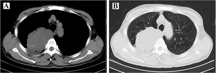 |
Figure 2 Representative chest CT images of SMARCA4-dNSCLC. (A) Axial chest CT (lung window) showing a large soft tissue mass in the right upper lobe with speculated margins and internal calcifications. (B) Axial chest CT (mediastinal window) showing displacement of the right upper lobe bronchus and patchy ground-glass opacities in both lungs. |
Beyond the primary lesion, multifocal disease was common, manifesting as satellite nodules in other lobes. Peribronchial and peripheral ground-glass opacities frequently accompanied the primary mass, suggesting inflammatory changes or infectious processes. Notably, signs of underlying COPD, including increased lung lucency and thin-walled bullae, were prevalent, consistent with the high incidence of smoking history in this cohort. Figure 2B demonstrates both the mass effect on bronchial structures and concurrent COPD changes.
Regional and distant spread was evident in many cases. Mediastinal lymphadenopathy appeared frequently, indicating lymphatic metastasis. Pleural involvement, characterized by small effusions, suggested either direct extension or inflammatory response. While not uniformly documented, adrenal gland evaluation remained important given this cancer’s metastatic potential. Incidental findings included thyroid low-density lesions requiring further investigation and benign-appearing cystic lesions in the liver and kidneys. Collectively, these imaging features (large, invasive primary tumors with irregular margins, multifocal pulmonary disease, and regional spread) create a radiographic profile consistent with the known aggressive biological behavior of SMARCA4-dNSCLC. The key chest CT findings are detailed in Table 3.
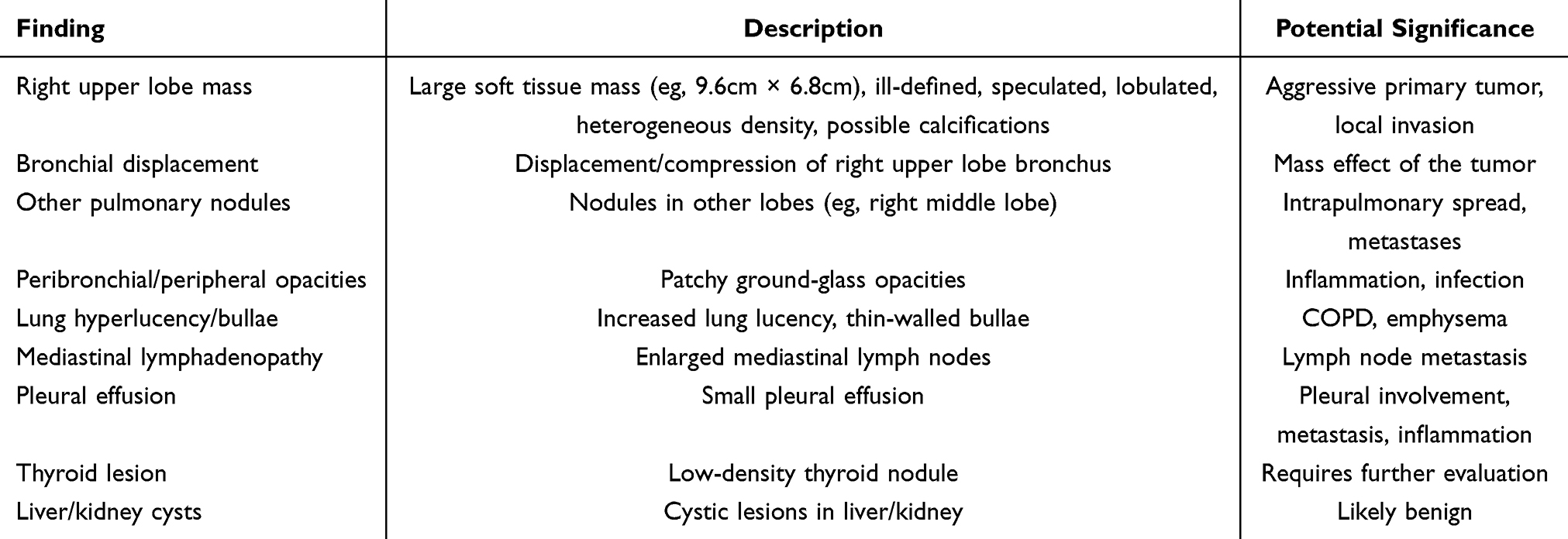 |
Table 3 Key Chest CT Findings in SMARCA4-dNSCLC |
Molecular Characteristics and Co-Mutations
To gain insights into the molecular underpinnings of SMARCA4-dNSCLC, we conducted next-generation sequencing (NGS) on a subset of 24 patients from the total cohort of 47. The selection criterion for these patients was based on the availability of adequate tissue samples and the clinical decision to perform molecular profiling to explore potential therapeutic targets. This molecular analysis aimed to uncover key molecular features and co-mutations that might influence disease progression and shape treatment strategies.
The NGS analysis revealed a notable absence of frequently targetable driver genes such as EGFR and ALK in the subset of SMARCA4-dNSCLC patients analyzed. A mere 4% of patients exhibited EGFR mutations, and no cases of ALK rearrangements were identified, suggesting a potential resistance to targeted therapies that are effective against these mutations. This information is visually represented in Figure 3A, which displays the mutation frequency of various molecular alterations identified by NGS, highlighting the rarity of EGFR and ALK mutations.
 |
Figure 3 Molecular profiling data. (A) Bar chart showing the distribution of molecular alterations identified in the 24 patients. (B) Pie chart representing the proportion of patients with co-mutations in TP53, KRAS, and STK11. |
Furthermore, the analysis identified common co-mutations in genes known to be associated with lung cancer progression. Specific variants included TP53 R273H (2 cases), KRAS G12C (1 case), STK11 L148P (1 case), and PIK3CA E545K (1 case), co-occurring mutations were limited to 1 patient with KRAS G12C + STK11 L148P. These co-mutations are recognized for enhancing tumor aggressiveness and could potentially influence the tumor’s response to various treatments. The significance of these co-mutations is underscored by Figure 3B, that illustrates the proportion of patients with co-mutations in TP53, KRAS, and STK11, emphasizing their role in the molecular landscape of SMARCA4-dNSCLC.
Notably, the co-occurrence of TP53, KRAS, and STK11 mutations in this cohort may further enhance tumor aggressiveness by disrupting DNA damage repair and metabolic pathways, which is consistent with previous reports that SMARCA4 deficiency synergizes with these mutations to promote malignant progression.21,22 The rarity of EGFR/ALK mutations also explains the limited efficacy of conventional targeted therapies, highlighting the need for novel strategies targeting chromatin remodeling defects or co-mutated pathways.
In addition to these findings, the molecular analysis detected other alterations that could influence disease behavior. Mutations in KEAP1 and PIK3CA were identified in a small subset of patients, indicating alternative pathways for tumor growth and resistance to therapy. These results suggest that while SMARCA4-dNSCLC may be less responsive to standard targeted therapies, the identified molecular alterations could guide the development of novel therapeutic strategies tailored to the specific molecular profile of the disease.
Treatment Patterns
Understanding the treatment patterns is crucial for evaluating the efficacy of various therapeutic approaches and identifying potential areas for improvement. We analyzed the treatment strategies employed for the 47 patients with SMARCA4-dNSCLC, focusing on the distribution across different disease stages and the types of treatments administered (Summarized in Table 4).
 |
Table 4 Treatment Modalities by Stage |
The treatment strategies varied significantly depending on the stage of the disease at diagnosis. For patients diagnosed with early-stage disease (Stages I and II), surgical resection was the primary treatment approach, often combined with adjuvant chemotherapy to reduce the risk of recurrence. Specifically, six patients with Stage I disease underwent surgery, three of whom also received chemotherapy. Among the six patients with Stage II disease, two underwent surgery alone, while the others received a combination of surgery and chemotherapy. For patients with more advanced disease (Stages III and IV), the treatment landscape was more complex. Thirteen patients with Stage III disease received a mix of treatments, including chemotherapy (with or without targeted therapy), immunotherapy (specific agents: sintilimab, atezolizumab, camrelizumab), and in some cases, a combination of these modalities. The selection of immune checkpoint inhibitors was based on clinical practice guidelines and patient performance status. Notably, one Stage III patient received Traditional Chinese Medicine as palliative supportive care, with the clinical rationale being poor performance status (ECOG PS score = 4) and refusal of chemotherapy/radiotherapy to alleviate symptoms.
A preliminary survival advantage was observed in the subgroup of patients receiving ICIs compared with chemotherapy-only patients in our cohort. Notably, recent studies have highlighted the intrinsic resistance of SMARCA4-deficient NSCLC to immune checkpoint inhibitors, which is closely associated with co-occurring mutations in STK11 and KEAP1.23,24 These mutations typically induce an “immune-cold” tumor microenvironment characterized by reduced infiltration of cytotoxic T lymphocytes and downregulated PD-L1 expression, thereby impairing the therapeutic efficacy of ICIs. In our cohort, the low frequency of STK11/KEAP1 co-mutations may partially explain the observed preliminary survival benefit, though validation in larger cohorts is warranted.
The most challenging treatment decisions arose for the 22 patients with Stage IV disease, who were presented with distant metastases. The treatments for these patients were diverse, ranging from systemic chemotherapy and immunotherapy to targeted therapy for those with actionable mutations. Notably, nine patients did not receive any treatment due to personal reasons, economic constraints, or rapid disease progression, highlighting the need for more accessible and tolerable treatment options. Among the 9 untreated patients, 7 (77.8%) had ECOG PS score ≥3 (poor performance status) and 8 (88.9%) were at Stage IV with multiple distant metastases (higher disease burden), which may have contributed to the survival difference between treated and untreated groups.
A significant observation from this analysis was the heterogeneity in treatment approaches, even within the same disease stage. This variability underscores the complexity of managing SMARCA4-dNSCLC and the importance of personalized treatment plans. Additionally, the relatively high number of patients who did not receive treatment (9 out of 47) points to potential barriers in accessing care or the lack of suitable treatment options.
Survival Analysis and Prognostic Factors
The follow-up cutoff date of this study was July 28, 2024. The median follow-up time for the entire cohort (47 patients) was 10.5 months (interquartile range [IQR]: 6.8–14.2 months). Among them, the median follow-up time was 12.3 months (IQR: 8.5–15.1 months) in the treated group (n=38), 4.2 months (IQR: 2.8–5.6 months) in the untreated group (n=9), 13.1 months (IQR: 9.2–16.5 months) in the subgroup receiving immunotherapy (n=17), and 9.8 months (IQR: 6.1–13.3 months) in the subgroup not receiving immunotherapy (n=30). Survival analysis results showed significant differences in OS between the treated group (n=38) and untreated group (n=9). Kaplan-Meier survival curve analysis (Figure 4A) demonstrated that the median OS was not reached in the treated group, with a survival rate of 66% (25/38) and mortality rate of 34% (13/38), whereas the median OS in the untreated group was only 3 months (95% CI: 2.72–6.62 months). The 6-month and 12-month survival rates for the entire cohort were 62% (95% CI: 47–75%) and 32% (95% CI: 20–47%), respectively.
 |
Figure 4 Overall survival analysis in patients with SMARCA4-dNSCLC. (A) Kaplan-Meier curves comparing overall survival between treated (n = 38) and untreated (n = 9) patients. (B) Kaplan-Meier curves comparing overall survival between patients who received immunotherapy (n = 17) and those who did not (n = 30). Statistical analysis was performed using the log-rank test, differences were statistically significant (p < 0.05). |
Overall, 26 patients (54.2%) survived and 21 patients (43.8%) died, including 2 patients who survived less than one month. The mean survival time for the entire cohort was 14.257 months (standard error 1.424, 95% CI: 11.466–17.047), with the median survival time not yet reached. The 6-month, 12-month, and 18-month survival rates were 62% (29/47), 32% (15/47), and 11% (5/47), respectively. Further subgroup analysis (Figure 4B) revealed that patients who received immunotherapy (n=17) had significantly higher overall survival rates compared to those who did not receive immunotherapy (n=30) (p<0.05), suggesting a potential survival benefit associated with immunotherapy in this cohort.
To investigate factors affecting survival time in SMARCA4-dNSCLC patients, Cox regression analysis was performed on 39 patients with complete data (Table 5). Univariate analysis showed that tumor TNM stage, treatment status, and distant metastasis were significantly associated with patient survival time (p<0.05). Multivariate regression analysis further confirmed that advanced TNM stage (HR=5.147, 95% CI: 1.766–15.002, p=0.003), lack of treatment (HR=0.227, 95% CI: 0.090–0.572, p=0.002), and distant metastasis (HR=0.194, 95% CI: 0.067–0.566, p=0.003) were independent risk factors for shortened survival time in patients with SMARCA4-dNSCLC.
 |
Table 5 Cox Regression Analysis of Factors Influencing Survival Time in SMARCA4-dNSCLC Patients |
These results suggest that the prognosis of patients with SMARCA4-dNSCLC is closely related to tumor stage, treatment status, and metastatic condition. For these patients, early diagnosis and active treatment are crucial, especially for those with advanced diseases, for whom comprehensive treatment strategies should be considered to improve prognosis.
Discussion
SMARCA4-dNSCLC represents a rare and aggressive subtype of non-small cell lung cancer (NSCLC) characterized by loss of BRG1 protein expression due to SMARCA4 gene alterations. In line with previous studies, our cohort confirmed its strong association with male sex, heavy smoking history, and advanced age, with a median age of 66 years and 87% of patients being male. These findings support the notion that SMARCA4-deficient tumors are tightly linked to tobacco-related carcinogenesis.15,25
Consistent with its aggressive biological behavior, more than 60% of patients in this cohort were diagnosed at advanced TNM stages (Stage III–IV), and nearly half (47%) exhibited distant metastases at diagnosis. Common metastatic sites included the adrenal glands, pleura, and distant lymph nodes, reflecting the early and widespread dissemination pattern of this tumor type. Moreover, the frequent co-existence of underlying emphysema and chronic bronchitis, observed in over half of our cases, further underscores the etiological role of smoking and chronic lung damage in SMARCA4-deficient tumorigenesis.
Pathologically, adenocarcinoma was the predominant histologic subtype (60%), followed by poorly differentiated and undifferentiated carcinomas. Immunohistochemically, SMARCA4-deficient tumors displayed a characteristic profile with nearly universal BRG1 loss (91%), diffuse cytokeratin (CK) positivity, and a high Ki-67 proliferative index, with over 50% of cases exhibiting Ki-67 >50%. Interestingly, only 37% of cases expressed TTF-1, reinforcing earlier reports that TTF-1 negativity is a diagnostic clue for SMARCA4 deficiency. PD-L1 expression was observed in more than half of evaluable cases, with 56% showing ≥1% expression and 10% ≥50%, potentially indicating a role for immune checkpoint inhibition in selected patients.
Radiologically, SMARCA4-deficient tumors frequently manifested as large, ill-defined masses with central necrosis, bronchial truncation, and adjacent structure invasion, reflecting their aggressive and infiltrative growth pattern. Nearly 60% of patients exhibited lymph node metastases on imaging, and over 85% had patchy high-density lesions or nodules in other lung lobes. These imaging features, while not pathognomonic, may raise suspicion for SMARCA4 deficiency in the appropriate clinical context.
Molecular profiling revealed a low prevalence of actionable driver mutations, with only one patient harboring an EGFR mutation. However, co-mutations involving TP53, KRAS, STK11, and PIK3CA were present in a minority of cases, aligning with previous genomic studies that have shown SMARCA4-dNSCLC to be enriched for tumor suppressor gene alterations rather than oncogenic drivers.26,27 These findings emphasize the need for broader molecular screening to identify rare but potentially targetable alterations.
Despite the lack of standardized treatment strategies, our study suggests that therapeutic intervention, including surgery, chemotherapy, immunotherapy, and combination regimens, may confer survival benefits. The median OS in treated patients was not reached, while those without any treatment had a dismal median OS of only 3 months. Multivariate Cox analysis confirmed that advanced stage, distant metastasis, and absence of treatment were independent predictors of poor prognosis, underscoring the importance of early diagnosis and active management.
Several limitations of our study should be acknowledged. First, the single-center, retrospective design and relatively small sample size may limit the statistical power and generalizability of our findings. Second, the heterogeneity in treatment regimens and NGS testing platforms reflects real-world practice but may introduce confounding factors. In particular, the observed association between immunotherapy and survival is based on a small subgroup and requires validation in larger, prospective studies. Moreover, a potential limitation of this study is the confounding bias between treated and untreated groups, as untreated patients had poorer performance status and higher disease burden; future studies with propensity score matching may help reduce this bias.
In conclusion, SMARCA4-deficient NSCLC is a clinicopathologically and molecularly distinct entity associated with poor differentiation, advanced stage, high proliferative index, and limited treatment responsiveness. Consistent with our molecular profiling, this subtype exhibits a low prevalence of actionable driver mutations (eg, EGFR/ALK) but frequent co-mutations in tumor suppressor genes (TP53, KRAS, STK11), distinguishing it from common NSCLC subtypes. Notably, subgroup analysis suggests a potential survival benefit of immunotherapy in selected patients, particularly those with non-negligible PD-L1 expression or absence of STK11/KEAP1 co-mutations. Our findings also highlight the need for strengthened routine BRG1 immunohistochemical (IHC) screening in high-risk populations (eg, older male smokers with advanced NSCLC) to improve early recognition of this rare subtype. Overall, our study underscores the importance of heightened clinical awareness, early diagnostic workup (including IHC for BRG1), and consideration of individualized therapeutic approaches. Further multicenter prospective studies and molecularly guided trials are warranted to develop effective treatment strategies and improve outcomes in this challenging patient population.
Further multicenter prospective studies and molecularly guided trials are warranted to develop effective treatment strategies and improve outcomes in this challenging patient population. Building on our findings, future translational research should prioritize several key directions. These include evaluating novel therapeutic agents that target epigenetic vulnerabilities or specific co-mutations such as KRAS or STK11 in SMARCA4-dNSCLC, identifying predictive biomarkers beyond PD-L1, particularly tumor mutational burden and features of the immune microenvironment, to optimize patient selection for immunotherapy, and developing non-invasive diagnostic or monitoring tools that exploit the distinctive imaging or molecular signatures characteristic of this aggressive subtype.
Data Sharing Statement
The data that support the findings of this study are available from the corresponding author upon reasonable request. Data will be shared in a de-identified format to protect patient privacy. Requests for data access should be directed to Qingming Shi (email: [email protected]). Data requestors will need to sign a data access agreement to ensure the confidentiality and appropriate use of the data.
Acknowledgments
We acknowledge the contributions of all the medical staff and researchers at Anhui Chest Hospital for their support in data collection and patient care. Special thanks to the Department of Pathology and the Department of Radiology at Anhui Chest Hospital for their assistance in pathology and imaging analysis.
Author Contributions
All authors made a significant contribution to the work reported, whether that is in the conception, study design, execution, acquisition of data, analysis and interpretation, or in all these areas; took part in drafting, revising or critically reviewing the article; gave final approval of the version to be published; have agreed on the journal to which the article has been submitted; and agree to be accountable for all aspects of the work.
Disclosure
The authors report no conflicts of interest in this work.
References
1. Hong Q, Cai L, Cai L. Clinicopathological characteristics and treatment outcomes of patients with advanced SMARCA4-deficient non-small cell lung cancer. J Thorac Oncol. 2024;19(10):S271–13. doi:10.1016/j.jtho.2024.09.488
2. Grant C, Nagasaka M. Neoadjuvant EGFR-TKI therapy in non-small cell lung cancer. Cancer Treat Rev. 2024;126:102724. doi:10.1016/j.ctrv.2024.102724
3. Plaugher DR, Childress AR, Gosser CM, et al. Therapeutic potential of tumor-infiltrating lymphocytes in non-small cell lung cancer. Cancer Lett. 2024;605:217281. doi:10.1016/j.canlet.2024.217281
4. Jones CA, Wang J, Evans JR, et al. Super-enhancer dysregulation in rhabdoid tumor cells is regulated by the SWI/SNF ATPase BRG1. Cancers. 2024;16(5):916. doi:10.3390/cancers16050916
5. Xiao M, Cui X, Xu C, et al. Deep-targeted gene sequencing reveals ARID1A mutation as an important driver of glioblastoma. CNS Neurosci Ther. 2024;30(4):e14698. doi:10.1111/cns.14698
6. Engl W, Kunstar-Thomas A, Chen S, Ng WS, Sielaff H, Zhao ZW. Single-molecule imaging of SWI/SNF chromatin remodelers reveals bromodomain-mediated and cancer-mutants-specific landscape of multi-modal DNA-binding dynamics. Nat Commun. 2024;15(1):7646. doi:10.1038/s41467-024-52040-y
7. Shi M, Pang L, Zhou H, et al. Rare SMARCA4-deficient thoracic tumor: insights into molecular characterization and optimal therapeutics methods. Lung Cancer. 2024;192:107818. doi:10.1016/j.lungcan.2024.107818
8. Clarke BA, Witkowski L, Ton Nu TN, et al. Loss of SMARCA4 (BRG1) protein expression as determined by immunohistochemistry in small‐cell carcinoma of the ovary, hypercalcaemic type distinguishes these tumours from their mimics. Histopathology. 2016;69(5):727–738. doi:10.1111/his.12988
9. Bilodeau S, Vallette-Kasic S, Gauthier Y, et al. Role of Brg1 and HDAC2 in GR trans-repression of the pituitary POMC gene and misexpression in Cushing disease. Genes Dev. 2006;20(20):2871–2886. doi:10.1101/gad.1444606
10. Pelosi G. The new taxonomy of lung adenocarcinoma stemming from a multidisciplinary integrated approach: novel pathology concepts and perspectives. J Thorac Oncol. 2011;6(2):241–243. doi:10.1097/JTO.0b013e31820bfcba
11. Zhou P, Fu Y, Tang Y, Jiang L, Wang W. Thoracic SMARCA4-deficient undifferentiated tumor: a clinicopathological and prognostic analysis of 35 cases and immunotherapy efficacy. Lung Cancer. 2024;189:107471. doi:10.1016/j.lungcan.2024.107471
12. Lin DI, Allen JM, Hecht JL, et al. SMARCA4 inactivation defines a subset of undifferentiated uterine sarcomas with rhabdoid and small cell features and germline mutation association. Mod Pathol. 2019;32(11):1675–1687. doi:10.1038/s41379-019-0303-z
13. Schoenfeld AJ, Bandlamudi C, Lavery JA, et al. The genomic landscape of SMARCA4 alterations and associations with outcomes in patients with lung cancer. Clin Cancer Res. 2020;26(21):5701–5708. doi:10.1158/1078-0432.CCR-20-1825
14. Khalil A, Collins MP, Quintás‐Cardama A. Prognostic implications ofSMARCA4,ARID1A, and other BAF mutations in non-small cell lung cancer. Cancer Med. 2025;14(24):e71442. doi:10.1002/cam4.71442
15. Longo V, Catino A, Montrone M, et al. Treatment of thoracic SMARCA4-deficient undifferentiated tumors: where we are and where we will go. Int J Mol Sci. 2024;25(6):3237. doi:10.3390/ijms25063237
16. Nguyen VT, Tessema M, Weissman BE. The SWI/SNF complex: a frequently mutated chromatin remodeling complex in cancer. In: Epigenetics in Oncology. Springer; 2023:211–244.
17. Armon S, Hofman P, Ilié M. Perspectives and issues in the assessment of SMARCA4 deficiency in the management of lung cancer patients. Cells. 2021;10(8):1920. doi:10.3390/cells10081920
18. Liang X, Gao X, Wang F, et al. Clinical characteristics and prognostic analysis of SMARCA4 -deficient non-small cell lung cancer. Cancer Med. 2023;12(13):14171–14182. doi:10.1002/cam4.6083
19. Wang A, Jin Y, Cao Z, Lu L, Li Z. Clinicopathological characteristics and treatment outcomes of advanced SMARCA4-deficient thoracic tumors. Cancer Med. 2024;13(1):e6809. doi:10.1002/cam4.6809
20. Yang L, Wang S, Zhou Y, et al. Evaluation of the 7th and 8th editions of the AJCC/UICC TNM staging systems for lung cancer in a large North American cohort. Oncotarget. 2017;8(40):66784. doi:10.18632/oncotarget.18158
21. Wang G, Zhou G, Han W, Jiang H. The role of SMARCA4 in lung cancer. Sci Rep. 2025;15(1):28605. doi:10.1038/s41598-025-13913-4
22. Dai W, Li T, Li Y, et al. Concurrent EGFR mutation and SMARCA4 deficiency in non-small cell lung cancer: a case report and literature review. Medicine. 2024;103(41):e40081. doi:10.1097/MD.0000000000040081
23. Paredes R, Borea R, Drago F, Russo A, Nigita G, Rolfo C. Genetic drivers of tumor microenvironment and immunotherapy resistance in non-small cell lung cancer: the role of KEAP1, SMARCA4, and PTEN mutations. J ImmunoTherapy Cancer. 2025;13(8):e012288. doi:10.1136/jitc-2025-012288
24. Han Y, Wang J, Pang B, et al. Immune checkpoint inhibitors have limited efficacy in SMARCA4-deficient non-small cell lung cancer. Transl Lung Cancer Res. 2025;14(11):5000–5016. doi:10.21037/tlcr-2025-921
25. Rekhtman N, Montecalvo J, Chang JC, et al. SMARCA4-deficient thoracic sarcomatoid tumors represent primarily smoking-related undifferentiated carcinomas rather than primary thoracic sarcomas. J Thorac Oncol. 2020;15(2):231–247. doi:10.1016/j.jtho.2019.10.023
26. Tian Y, Xu L, Li X, Li H, Zhao M. SMARCA4: current status and future perspectives in non-small-cell lung cancer. Cancer Lett. 2023;554:216022. doi:10.1016/j.canlet.2022.216022
27. Foggetti G, Li C, Cai H, et al. Genetic determinants of EGFR-driven lung cancer growth and therapeutic response in vivo. Cancer Discov. 2021;11(7):1736–1753. doi:10.1158/2159-8290.CD-20-1385
 © 2026 The Author(s). This work is published and licensed by Dove Medical Press Limited. The
full terms of this license are available at https://www.dovepress.com/terms
and incorporate the Creative Commons Attribution
- Non Commercial (unported, 4.0) License.
By accessing the work you hereby accept the Terms. Non-commercial uses of the work are permitted
without any further permission from Dove Medical Press Limited, provided the work is properly
attributed. For permission for commercial use of this work, please see paragraphs 4.2 and 5 of our Terms.
© 2026 The Author(s). This work is published and licensed by Dove Medical Press Limited. The
full terms of this license are available at https://www.dovepress.com/terms
and incorporate the Creative Commons Attribution
- Non Commercial (unported, 4.0) License.
By accessing the work you hereby accept the Terms. Non-commercial uses of the work are permitted
without any further permission from Dove Medical Press Limited, provided the work is properly
attributed. For permission for commercial use of this work, please see paragraphs 4.2 and 5 of our Terms.
Recommended articles

High Expression of DEPDC1B Predicts Poor Prognosis in Lung Adenocarcinoma

Li P, Chen X, Zhou S, Xia X, Wang E, Han R, Zeng D, Fei G, Wang R
Journal of Inflammation Research 2022, 15:4171-4184
Published Date: 23 July 2022
The Prognostic Role of HuR Varies Between Different Subtypes of Breast Cancer Patients: Data Mining and Retrospective Analysis
Liao Y, Liao Y, Li J, Li Y, Fan Y
Breast Cancer: Targets and Therapy 2023, 15:135-146
Published Date: 11 February 2023
Comprehensive Analysis of the Prognostic Significance of ZHX Family Members in Esophageal Carcinoma
You Y, Li W, Ma Y, Gao L, Li T, Zhang X, Luo X
International Journal of General Medicine 2025, 18:7127-7141
Published Date: 27 November 2025
Establishment of Predictive Models for Second Hepatoma Recurrence
Su M, Zhong W, Chen Z, Zhang G
Cancer Management and Research 2026, 18:566522
Published Date: 24 March 2026