
Back to Journals » Journal of Inflammation Research » Volume 18
Bromodomain and Extraterminal Protein Inhibition: A Novel Therapeutic Strategy in Arthritis
Authors Sheng W  , Zhao J, Yu F, Wang D, Zeng H, Liu P
Received 17 April 2025
Accepted for publication 22 September 2025
Published 30 September 2025 Volume 2025:18 Pages 13503—13517
DOI https://doi.org/10.2147/JIR.S535057
Checked for plagiarism Yes
Review by Single anonymous peer review
Peer reviewer comments 3
Editor who approved publication: Professor Yan Chen
Weibei Sheng,1 Jin Zhao,1 Fei Yu,2 Deli Wang,1 Hui Zeng,3,* Peng Liu1,*
1Department of Bone & Joint Surgery, Shenzhen Key Laboratory of Orthopaedic Diseases and Biomaterials Research, Peking University Shenzhen Hospital, Shenzhen, 518036, People’s Republic of China; 2Department of Spine Surgery, Shenzhen Second People’s Hospital, The First Affiliated Hospital of Shenzhen University, Shenzhen, 518035, People’s Republic of China; 3Department of Orthopedic Trauma, Shenzhen Second People’s Hospital, The First Affiliated Hospital of Shenzhen University, Shenzhen, People’s Republic of China
*These authors contributed equally to this work
Correspondence: Hui Zeng, Email [email protected] Peng Liu, Email [email protected]
Abstract: Arthritis is an inflammatory condition that affects the joints and surrounding tissues, triggered by factors such as inflammation, infection, degeneration, and trauma. The major forms of arthritis include osteoarthritis (OA), rheumatoid arthritis (RA), and gouty arthritis (GA). Its pathogenesis primarily involves synovial inflammation, cartilage degradation, and subchondral bone remodeling, with pro-inflammatory cytokines, collagenases, and other mediators playing central roles in disease onset and progression. The bromodomain and extraterminal (BET) protein family-a subclass of the larger bromodomain protein superfamily-comprises BRD2, BRD3, BRD4, and BRDT. The regulatory functions of BET proteins in inflammation highlight their considerable potential for mitigating arthritis-related pathology. This review provides a comprehensive overview of recent research on the role of BET proteins in OA, RA, and GA, aiming to deepen our understanding of the protective mechanisms of BET inhibitors, underscore their potential as therapeutic targets, and emphasize their relevance in the development of novel treatment strategies.
Keywords: arthritis, osteoarthritis, rheumatoid arthritis, gouty arthritis, bromodomain and extraterminal protein
Introduction
Arthritis is an inflammatory disease affecting the joints and surrounding tissues, triggered by factors such as inflammation, infection, degeneration, and trauma.1–3 Based on its etiology, pathogenesis, and clinical presentation, arthritis can be classified into osteoarthritis (OA), rheumatoid arthritis (RA), gouty arthritis (GA), and other subtypes.1–3 OA is a chronic degenerative joint disease that primarily affects the cartilage and surrounding tissues, commonly seen in the elderly.1 Current treatments for OA are largely limited to symptom management, including the use of nonsteroidal anti-inflammatory drugs (NSAIDs), intra-articular corticosteroid injections, and, in advanced cases, joint replacement surgery. These approaches do not effectively halt disease progression or reverse cartilage damage, highlighting the need for disease-modifying therapies. RA is a chronic, systemic autoimmune disease characterized by inflammation in multiple joints.2 Current treatments, including disease-modifying antirheumatic drugs (DMARDs), biologics, and corticosteroids, primarily aim to control inflammation and slow disease progression; however, they often fail to achieve complete remission, can cause significant side effects, and may be insufficient to prevent long-term joint damage in all patients. GA is an inflammatory joint disease caused by the deposition of urate crystals within the joints.3,4 Current clinical treatments, including urate-lowering therapies, nonsteroidal anti-inflammatory drugs (NSAIDs), colchicine, and corticosteroids, primarily aim to control acute flares and reduce uric acid levels. However, these approaches often fail to fully prevent recurrent attacks or halt long-term joint damage, highlighting the need for more effective and disease-modifying strategies. Although the etiology and pathogenesis of these conditions differ, all three types of arthritis involve inflammation and damage to the joints and surrounding tissues, with inflammatory mediators playing a crucial role in disease progression.1–3 Additionally, interactions among chondrocytes, synovial fibroblasts, macrophages, and other cells are central to the progression of arthritis.
Chondrocytes, subjected to mechanical stress and inflammatory stimuli, secrete matrix metalloproteinases (MMPs) and pro-inflammatory cytokines, including Interleukin-1 (IL-1) and Tumor Necrosis Factor-alpha (TNF-α), which contribute to the degradation of the cartilage matrix in OA.5 Meanwhile, synovial fibroblasts and macrophages within the synovium proliferate and become activated, producing significant amounts of inflammatory mediators such as prostaglandin E2 (PGE2) and nitric oxide (NO).6,7 These mediators exacerbate joint inflammation and tissue destruction. Additionally, the dorsal root ganglion is involved in pain perception and transmission in OA.8 In rheumatoid arthritis (RA), synovial fibroblasts undergo abnormal proliferation and activation, exhibiting aggressive invasiveness. These cells secrete large quantities of MMPs and inflammatory cytokines, such as Interleukin 6 (IL-6), Interleukin 8 (IL-8), and TNF-α, leading to the degradation of joint cartilage and bone.9 Immune cells, including macrophages, monocytes, T cells, and B cells, drive the inflammatory response within the synovium, further contributing to tissue destruction.10,11 In gouty arthritis (GA), macrophages are key effector cells.12 They recognize and engulf monosodium urate (MSU) crystals deposited in the joints. The MSU crystals trigger excessive activation of the NLRP3 inflammasome within macrophages, leading to caspase-1 activation and the subsequent secretion of large amounts of pro-inflammatory cytokines, particularly IL-1β.13 This cascade significantly exacerbates joint inflammation. The etiology and pathogenesis of arthritis are complex and multifactorial, with increasing evidence pointing to a significant role of epigenetic regulation in its development.14,15
The bromodomain (BRD) is a highly conserved protein domain that specifically recognizes and binds to acetylated lysine residues on histones. It is composed of approximately 110 amino acids, arranged into four antiparallel α-helices and two hydrophobic loops.16 BRD proteins are categorized into eight families, with the bromodomain and extra-terminal domain (BET) protein family being the most extensively studied.17 This family includes BRD2, BRD3, BRD4, and BRDT, which regulate gene transcription by recognizing acetylated lysine residues. Members of the BET family play crucial roles in the regulation of inflammatory gene expression, cell cycle progression, and DNA damage repair.18 Given that inflammation is a key pathological feature of arthritis, BET proteins are likely critical in its progression.
BET inhibitors, particularly those targeting BRD4, have shown promise in suppressing inflammatory responses in chondrocytes by inhibiting the Nuclear Factor kappa B (NF-κB) signaling pathway.19 Additionally, these inhibitors can activate the Nuclear Factor Erythroid 2-Related Factor 2 (NRF2) signaling pathway, reducing oxidative stress and apoptosis in chondrocytes.20 BET inhibitors also downregulate the expression of pro-inflammatory cytokines in inflammatory macrophages and alleviate joint pain by modulating dorsal root ganglion function.21 In RA, BET inhibitors suppress disease progression by inhibiting inflammatory responses in RA synovial fibroblasts (RASFs) and reducing their proliferation, migration, and invasion.22,23 They also modulate receptor expression on the surface of monocytes, further inhibiting RA progression. In gouty arthritis (GA), BET inhibitors reduce joint inflammation by preventing macrophage pyroptosis.13
This review highlights recent advancements in BET inhibitor research within the contexts of OA, RA, and GA, offering new perspectives for the development of novel therapeutics to mitigate these diseases.
Structure and Function of BET Proteins
The BET protein family is a subclass of the larger bromodomain protein superfamily. The human genome encodes over 40 different proteins containing more than 60 types of bromodomains.24 Based on sequence homology, the bromodomain protein family is divided into eight subfamilies, with the BET family representing a distinct group. As shown in Figure 1, the BET family includes BRD2, BRD3, BRD4 (which has subtypes A, B, and C), and BRDT. BET proteins are characterized by two conserved bromodomains (BD1 and BD2) at the N-terminus and an extraterminal (ET) domain at the C-terminus.
 |
Figure 1 Domain Architecture of BET proteins. Three isoforms of BRD4 are indicated. Isoform B of BRD4 has a unique C terminus, which interacts with condensing II complexes.25 The CTD of BRD4 and BRDT is referred to as a C-terminal motif (CTM). Abbreviations: ET, extraterminal domain; CTD, C-terminal domain. |
The primary functions of the BET proteins are centered on gene transcription regulation, chromatin structure modulation, and the maintenance of cellular functions.24 BET proteins activate gene transcription by binding to acetylated histones and recruiting the transcription initiation complex to promoter regions, thereby facilitating the initiation of RNA polymerase II.16 These proteins play critical roles in regulating inflammation and immune responses.26–28 For instance, BRD4 enhances the expression of inflammation-related genes such as TNF-α and IL-6 by binding to acetylated histones, a process that is crucial in various inflammatory diseases, including OA and RA.28–30
Moreover, BET proteins influence immune cell functions, including the proliferation, differentiation, and cytokine secretion of T cells, B cells, and macrophages.31–33 By binding to acetylated histones, BET proteins also contribute to chromatin relaxation and remodeling, thereby modulating gene accessibility. Additionally, BET proteins, particularly BRD4, are involved in cell cycle regulation, especially during the G1/S phase transition.34 BRD4 ensures proper cell cycle progression by regulating the expression of key cyclins. These diverse functions position BET proteins as crucial regulatory factors in many diseases, providing a strong scientific foundation for their targeting in therapeutic strategies.
The Role of BET Inhibitors in Osteoarthritis
Osteoarthritis (OA) is the most common joint disease, primarily characterized by cartilage damage and affecting the entire joint structure.1,35,36 Pathological changes in OA include cartilage destruction, subchondral bone sclerosis, and synovial inflammation, among others.37 Current treatments for OA are mainly conservative, focusing on pain relief through medication.38,39 However, no drugs are currently available that can slow the progression of the disease, highlighting the need for further research. In recent years, there has been growing interest in the role of BET proteins in OA. BET inhibitors, especially BRD4 inhibitors, have demonstrated potential in protecting OA chondrocytes, reducing the expression of inflammatory cytokines in activated macrophages, and alleviating joint pain by modulating the function of the dorsal root ganglia (DRG). These findings suggest that BET inhibitors, particularly BRD4 inhibitors, could represent a promising therapeutic option for OA. The regulatory functions of BRD4 inhibitors in OA are summarized in Figure 2 and Table 1.
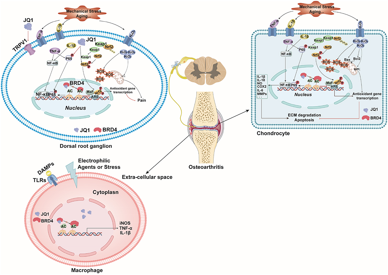 |
Figure 2 The regulatory mechanism of the BRD4 inhibitor JQ1 on chondrocytes, macrophages, and dorsal root ganglia (DRG) in osteoarthritis. Abbreviations: TNF-α, Tumor Necrosis Factor-alpha; IL-1β, Interleukin-1 beta; H2O2, Hydrogen Peroxide; P-P65, Phosphorylated Nuclear Factor Kappa B P65 Subunit; NRF2, Nuclear Factor Erythroid 2–Related Factor 2; Keap1, Kelch-like ECH-Associated Protein 1; AC, acetylated chromatin; MaF, Musculoaponeurotic Fibrosarcoma Oncogene Homolog; ARE, Antioxidant Response Element; NF-κB, Nuclear Factor Kappa B; IL-18, Interleukin-18; NO, Nitric Oxide; COX2, Cyclooxygenase-2; IL-6, Interleukin-6; MMPs, Matrix Metalloproteinases; ECM, Extracellular Matrix; TRPV1, Transient Receptor Potential Vanilloid 1; DAMPs, Damage-Associated Molecular Patterns; TLRs, Toll-Like Receptors; iNOS, Inducible Nitric Oxide Synthase. |
 |
Table 1 Advances in Targeting BET Proteins for the Treatment of Osteoarthritis |
Protecting Chondrocytes
Inhibiting Inflammatory Responses in Chondrocytes
Chondrocytes, the only cell type found in articular cartilage, are responsible for maintaining the balance of the cartilage matrix.43 In OA, this balance between matrix synthesis and degradation is disrupted, leading chondrocytes to adopt an inflammatory phenotype.44–47 Under inflammatory stimuli, chondrocytes secrete significant amounts of pro-inflammatory factors, such as TNF-α, IL-1β, and IL-6. These factors not only directly damage cartilage but also further activate chondrocytes and synovial cells through autocrine and paracrine mechanisms, creating a positive feedback loop of inflammation.48 Therefore, inhibiting the expression of these inflammatory factors is crucial for mitigating inflammatory responses and protecting chondrocytes.
The BRD4 inhibitor JQ1 (200 nM, 400 nM) effectively suppresses the expression of inflammatory factors IL-6 and TNF-α in SW1353 chondrocytes treated with 10 ng/mL IL-1β.19 JQ1 also inhibits the mRNA expression of inflammatory mediators, such as inducible nitric oxide synthase (iNOS) and cyclooxygenase-2 (COX2), in chondrocytes treated with IL-1β, IL-6, or TNFα.41,49 Additionally, JQ1 reduces the mRNA expression of pro-inflammatory factors (IL-1β, IL-6) following joint damage. Mechanistically, BRD4 inhibitors modulate the expression of high mobility group box protein 1 (HMGB1) and NF-κB, thereby alleviating inflammatory responses. HMGB1, a ubiquitous chromatin component widely expressed in immune and other cells, activates the NF-κB signaling pathway, which is involved in the inflammatory responses of various diseases. The NF-κB signaling pathway is notably upregulated in OA. Thus, JQ1 effectively inhibits inflammation and slows OA progression by targeting HMGB1 and the NF-κB pathway.
The BRD2-specific inhibitor BBC0403 (20 μM) significantly suppresses the expression of inflammation-related factors, including COX2, prostaglandin E2 (PGE2), and IL-6, in mouse primary chondrocytes treated with 1 ng/mL IL-1β.40 The anti-inflammatory effects of the BRD2 inhibitor are also mediated through the inhibition of the NF-κB signaling pathway.
Inhibiting Oxidative Stress in Chondrocytes
Oxidative stress plays a critical role in the pathogenesis of OA.50 It is defined as an imbalance between reactive oxygen species (ROS) and the antioxidant defense system, leading to excessive ROS accumulation, which in turn causes cellular damage and tissue destruction.51–53 In OA, oxidative stress not only disrupts chondrocyte function and degrades the cartilage matrix but also affects other joint tissues, such as the synovium and bone, thereby accelerating disease progression.54 Consequently, therapeutic strategies targeting oxidative stress hold promise for developing new treatment options for OA patients, potentially alleviating symptoms and slowing disease progression.
The BRD4 inhibitor JQ1 and BRD4 silencing (si-BRD4) have been shown to significantly reduce ROS and malondialdehyde (MDA) levels in rat chondrocytes treated with hydrogen peroxide (H2O2).20 Additionally, JQ1 and si-BRD4 may enhance the expression of antioxidant enzymes such as superoxide dismutase (SOD), catalase (CAT), and glutathione peroxidase (GPx), indicating that BRD4 inhibitors can effectively lower oxidative stress in chondrocytes exposed to H2O2. Similarly, the BRD2 inhibitor BBC0403 has been found to reduce oxidative stress in a Destabilization of Medial Meniscus (DMM) mouse model. Treatment with 10 μg/kg BBC0403 for 10 weeks in DMM mice decreases the levels of oxidative stress markers, such as 8-hydroxy-2′-deoxyguanosine (8-OHdG), in the joints.40,55 These findings suggest that BRD2 and BRD4 inhibitors may help mitigate OA progression by targeting and reducing oxidative stress in chondrocytes.
Reducing Chondrocyte Apoptosis
In OA, factors such as mechanical stress, inflammatory cytokines, and oxidative stress can induce chondrocyte apoptosis, which accelerates cartilage degradation.56,57 As the number of chondrocytes decreases, the synthesis of the cartilage matrix diminishes, impairing the tissue’s repair capacity and leading to progressive thinning and degeneration. Additionally, apoptotic chondrocytes release degradative enzymes and inflammatory mediators, such as MMPs and A disintegrin and metalloproteinase with thrombospondin motifs (ADAMTS), which degrade type II collagen and proteoglycans in the cartilage matrix, further damaging the cartilage.58,59 Moreover, apoptotic chondrocytes can activate synovial and immune cells by releasing fragmented organelles and DNA, triggering local inflammatory responses. This inflammation not only increases the release of inflammatory cytokines but also induces further chondrocyte apoptosis, creating a vicious cycle that accelerates OA progression.56 Therefore, reducing chondrocyte apoptosis is crucial for mitigating OA progression.
BRD4 inhibitors have been shown to significantly reduce chondrocyte apoptosis induced by H2O2, thereby protecting chondrocytes.20 At the molecular level, BRD4 inhibitors increase the expression of the anti-apoptotic protein B-cell lymphoma 2 (Bcl-2) in H2O2-treated chondrocytes while inhibiting the expression of pro-apoptotic proteins Bcl-2-associated X protein (Bax) and cysteine-aspartic protease 3 (Caspase-3).20
Protecting the Cartilage Matrix
The cartilage matrix is the primary component of articular cartilage, consisting of collagen, proteoglycans, glycoproteins, and water. It provides structural support and functional elasticity to the cartilage.60 In OA, an imbalance between the degradation and remodeling of the cartilage matrix leads to cartilage degeneration and subsequent loss of joint function.61
BRD4 inhibitors have been shown to effectively mitigate cartilage matrix damage induced by oxidative stress, such as that caused by H2O2. These inhibitors enhance the expression of matrix synthesis-related factors, including Collagen Type II Alpha 1 Chain (Col2A1) and aggrecan, while reducing the expression of matrix degradation-related factors, such as MMP13 and ADAMTS5. Additionally, the BRD4 inhibitor JQ1 can decrease cartilage degradation in explant cultures and reduce the expression of MMPs in damaged joints in vivo.41 These findings suggest that BRD4 inhibitors may protect against cartilage matrix damage associated with oxidative stress, inflammation, or joint injury.
Similarly, the BRD2 inhibitor BBC0403 has demonstrated protective effects against cartilage matrix degradation induced by IL-1β. When mouse cartilage explants were treated with BBC0403 (5, 10, or 20 μM) and IL-1β (1 ng/mL) for 72 hours, followed by Alcian blue staining, BBC0403 significantly reduced IL-1β-induced cartilage matrix damage, with the most pronounced effect observed at 20 μM. BBC0403 also decreased the expression of MMP3 and MMP13 in IL-1β-treated chondrocytes, thereby protecting the cartilage matrix.
The protective effects of BBC0403 have been further validated in vivo. In a DMM mouse model, intra-articular injection of 10 μg/kg BBC0403 for 10 weeks significantly reduced the expression of MMP3 and MMP13 in chondrocytes and effectively protected joint cartilage.40 Mechanistically, BBC0403 inhibits the upregulation of MMPs and the activation of the p38 signaling pathway, which is involved in type II collagen degradation. A deeper understanding of the protective mechanisms of BRD inhibitors on the cartilage matrix will provide valuable insights into how these inhibitors alleviate OA.
Inhibiting M1 Macrophages
M1 macrophages, also known as classically activated macrophages, are typically considered pro-inflammatory cells that play a significant role in the pathology of OA.6,62,63 These macrophages contribute to cartilage matrix degradation by secreting pro-inflammatory cytokines and degradative enzymes. In OA, M1 macrophages exacerbate synovial inflammation through various mechanisms, further deteriorating the joint environment. They can induce chondrocyte apoptosis by releasing nitric oxide (NO), ROS, and other pro-inflammatory factors.64 Additionally, M1 macrophages contribute to joint damage by influencing bone remodeling processes.65 Therefore, targeting M1 macrophages presents a potential therapeutic strategy to alleviate OA symptoms and slow disease progression.
BRD4 is highly expressed in iNOS-positive M1 macrophages within the synovium of both human and mouse OA models.42 A research team led by Huan Tian Zhang at Jinan University employed Lyz2-cre conditional knockout to delete BRD4 specifically in the myeloid lineage. In BRD4 flox/flox mice, this deletion resulted in a significant reduction in M1 macrophage accumulation and synovial inflammation in an anterior cruciate ligament transection (ACLT) mouse model.42
Relieving Joint Pain
Pain is a central symptom of OA, arising from various factors such as cartilage damage, synovial inflammation, osseous lesions, and soft tissue abnormalities.66,67 Among these factors, the DRG plays a pivotal role in OA-related pain.68 Located outside the spinal cord, the DRG houses the cell bodies of sensory neurons responsible for transmitting peripheral pain signals to the central nervous system.69 In OA, DRG neurons are not only involved in pain signal transmission but also in the perception and maintenance of pain.70 Given the critical role of the DRG in OA pain, it has emerged as a promising therapeutic target.
BRD4 inhibitors have demonstrated the ability to alleviate joint pain through multiple mechanisms. The BRD4 inhibitor JQ1, for instance, reduces pain perception in OA mice by inhibiting Transient Receptor Potential Ankyrin 1 (TRPV1) in the DRG.42 TRPV1, an ion channel discovered by David Julius in 2021, is known to play a significant role in pain perception, particularly in response to stimuli such as ACLT surgery.71,72 Furthermore, BRD4 inhibition suppresses NF-κB and NF-κB-mediated inflammatory cytokines in both the spinal cord and DRG in rats with monosodium iodoacetate (MIA)-induced OA pain.21
Oxidative stress, marked by increased ROS accumulation and impaired antioxidant activity, is also critical in the development of OA pain.73,74 BRD4 inhibition mitigates oxidative stress and promotes NRF2-dependent antioxidant gene expression in both the spinal cord and DRG in models of MIA-induced OA pain, thereby alleviating joint pain associated with OA.21
The Role of BET Inhibitors in Rheumatoid Arthritis
Rheumatoid arthritis (RA) is a chronic, systemic autoimmune disease characterized by symmetrical inflammation of multiple joints.75,76 Its hallmark features include inflammation and hyperplasia of the synovial membrane, which result in joint pain, swelling, stiffness, and functional impairment.75 Recent research has highlighted the significance of BET proteins in RA.29,77,78 The BET protein family is increasingly recognized for its crucial roles in regulating gene expression and inflammatory responses.28 BET inhibitors, especially BRD4 inhibitors, have shown potential in modulating RA through several mechanisms. They inhibit the expression of inflammatory factors and matrix-degrading enzymes,23 suppress Fcγ receptor expression in monocytes,77 and impede the proliferation, migration, and invasion of RA synovial fibroblasts (RASFs).22,23 Figure 3 and Table 2 summarize the regulatory effects of BRD4 inhibitors on RASFs and monocytes.
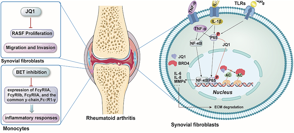 |
Figure 3 The regulatory mechanism of the BRD4 inhibitor JQ1 on synovial fibroblasts and monocytes in rheumatoid arthritis. Abbreviations: DAMPs, Damage-Associated Molecular Patterns; TLRs, Toll-Like Receptors; IL-1β, Interleukin-1 Beta; TNF-α, Tumor Necrosis Factor Alpha; P-P65, Phosphorylated Nuclear Factor Kappa B P65 Subunit; AC, acetylated chromatin; IL-6, Interleukin-6; IL-8, Interleukin-8; MMPs, Matrix Metalloproteinases; ECM, Extracellular Matrix; RASF, Rheumatoid Arthritis Synovial Fibroblasts; BET, Bromodomain and Extra-Terminal Domain; FcγR, Fc Gamma Receptor. |
 |
Table 2 Advances in Targeting BET Proteins for the Treatment of Rheumatoid Arthritis |
Regulating the Function of Rheumatoid Arthritis Synovial Fibroblasts
Inhibition of the Expression of Inflammatory Factors and Matrix Degrading Enzymes
Inflammatory factors and matrix-degrading enzymes play crucial roles in the pathogenesis of RA.80 These factors collaboratively drive disease progression, leading to chronic joint inflammation and damage.
Inflammatory factors are small proteins secreted by immune cells, synovial fibroblasts, and other cells that regulate immune responses and inflammation.11,81,82 Key inflammatory factors in RA include TNF-α, IL-1, IL-6, interleukin-17 (IL-17), and so on.83 TNF-α is a critical factor in RA, promoting synovial fibroblast proliferation, stimulating the production of additional inflammatory factors such as IL-1β and IL-6, and increasing MMPs secretion. This cascade ultimately leads to cartilage and bone destruction.84 IL-1 contributes to RA by promoting synovial cell proliferation and inflammatory responses, enhancing the expression of matrix-degrading enzymes, and further stimulating the release of inflammatory factors.85 IL-6 is a multifunctional cytokine that promotes inflammation, synovial cell proliferation, and bone resorption.86 IL-17 amplifies the effects of other inflammatory factors like TNF-α and IL-1, exacerbating inflammation and tissue destruction.87
Matrix-degrading enzymes, which are secreted by synovial fibroblasts, chondrocytes, and immune cells, disrupt the structural integrity of joint tissues.88,89 Matrix metalloproteinases (MMPs), zinc-dependent enzymes, degrade extracellular matrix (ECM) components such as collagen and elastin.90 In RA, elevated levels of MMP1, MMP3, and MMP13 lead to cartilage matrix degradation and joint damage.91 Additionally, the ADAMTS family, particularly ADAMTS4 and ADAMTS5, degrades proteoglycans in the cartilage matrix, causing structural damage.92 Therefore, targeting both inflammatory factors and matrix-degrading enzymes is essential for mitigating RA progression.
BRD4, a BET bromodomain coactivator protein, modulates inflammation via the NF-κB signaling pathway.93 The small-molecule inhibitor JQ1, which targets BRD4, has demonstrated anti-inflammatory effects.94 BRD4 siRNA significantly reduces the expression of pro-inflammatory factors, including IL-1β, IL-6, IL-17, and IL-18, in TNF-α-stimulated synovial fibroblasts (SFs).23 In vitro RA models, created by stimulating SFs with TNF-α, IL-1β, and toll-like receptor (TLR) ligands (Pam3, pIC, and lipopolysaccharide (LPS)), and then treating with BET inhibitors such as I-BET151 or siRNA targeting BRD2, BRD3, and BRD4, show that BET inhibition significantly reduces inflammatory mediator expression in RA synovial fibroblasts (RASFs).29 Another study confirms that BET pan-inhibitors, including JQ1, shBRD2, or shBRD4, significantly inhibit the expression of pro-inflammatory factors like IL-1β, IL-6, and IL-8 in TNF-α-treated SFs.22 Furthermore, BET pan-inhibitors, such as JQ1, shBRD2, or shBRD4, significantly reduce the expression of matrix-degrading enzymes like MMP1, MMP3, and MMP13 in RASFs.22
Mechanistically, BET inhibitors suppress inflammation in the synovium by inhibiting IκB kinase-dependent NF-κB activation {Xiao, 2016 #179}.
Inhibition of RASFs’ Proliferation
In RA, SFs undergo abnormal proliferation and activation, a condition referred to as “synovial hyperplasia”. Proliferated SFs release elevated levels of inflammatory factors, chemokines, and MMPs. These substances collectively exacerbate the destruction of articular cartilage and bone tissue.9,95 Thus, inhibiting the abnormal proliferation of RASFs is essential for managing RA.96,97 BET pan-inhibitors, such as JQ1, shBRD2, and shBRD4, effectively suppress the proliferation of RASFs, thereby slowing RA progression.22 However, the mechanism by which it inhibits the proliferation of RASFs needs further investigation.
Inhibition of RASFs’ Migration and Invasion
The migration and invasion of SFs are critical processes in the pathology of RA.98 In RA, SFs exhibit abnormal migratory behavior, allowing them to extend from the synovium into the surrounding articular cartilage and bone tissues.98 Furthermore, RASFs demonstrate significant invasive capabilities, enabling them to penetrate the cartilage and bone matrices.98 They release MMPs, cathepsins, and other degradative enzymes, which contribute to the breakdown of joint cartilage and bone matrix.99 In addition to causing direct tissue destruction, the migration and invasion of SFs sustain local inflammatory responses, thereby exacerbating disease progression.95,99 Targeting these migratory and invasive capabilities may offer a novel therapeutic approach to prevent joint damage. BRD4 siRNA can inhibit the migration and invasion of RASFs, positioning it as a potential therapeutic target for RA. However, the specific mechanisms underlying this effect require further investigation.23
Inhibition of Monocytes’ Fcγ Receptors
Fcγ receptors (FcγRs) are critical in the pathogenesis of RA.100 These receptors are present on various immune cells and bind to the Fc region of IgG antibodies within immune complexes. This binding mediates several immune responses, including phagocytosis, antibody-dependent cellular cytotoxicity, and amplification of inflammatory reactions.100 In RA, FcγRs significantly contribute to inflammatory responses and tissue damage. Activation of FcγRs, such as FcγRI (CD64), FcγRIIA (CD32a), FcγRIIC (CD32c), and FcγRIIIA (CD16a), involves binding to IgG and triggering downstream signaling pathways that lead to immune cell activation, phagocytosis, oxidative bursts, and the secretion of inflammatory cytokines.101–103 Thus, inhibiting activating FcγRs can reduce immune complex-mediated inflammation and alleviate RA symptoms.
FcγRs on myeloid cells bind immunoglobulin G (IgG) immune complexes, which induces an inflammatory phenotype, resulting in tissue damage and exacerbation of the inflammatory response.104 The BRD4 inhibitor PLX51107 has been shown to significantly downregulate the expression of FcγRIIA, FcγRIIB, FcγRIIIA, and the common γ-chain (FcϵR1-γ) in monocytes from both healthy donors and RA patients.77 Treatment with PLX51107 also attenuates signaling events downstream of FcγR activation. In a collagen-induced arthritis (CIA) mouse model, PLX51107 treatment reduced FcγR expression and significantly decreased footpad swelling.77
Regulate the Architectural Integrity of Bone
JQ1 markedly inhibited osteoclast differentiation and function, underscoring the pivotal role of BRD4 in osteoclastogenesis and its potential as a target for therapeutic intervention in RA-induced bone destruction.79
Suppressing the Progression of Arthritis in Mice
In RA research, mouse models are critical for studying the disease’s pathogenesis, pathophysiology, and for evaluating new therapeutic agents. Among these models, CIA is extensively used due to its close similarity to human RA. This model replicates key pathological features of RA, including joint inflammation, synovial hyperplasia, and cartilage and bone erosion.105 The therapeutic effects of the BRD inhibitor JQ1 have been demonstrated in the CIA mouse model. JQ1 significantly reduces arthritis scores and paw swelling in CIA mice.22,23 Additionally, it lowers serum levels of anti-CII IgG, anti-IgG1, anti-IgG2a, IL-6, and TNFα,22 and effectively mitigates cartilage and bone destruction.23
The Role of BET Inhibitors in Gouty Arthritis
Gouty arthritis (GA) is an inflammatory disease characterized by the deposition of urate crystals in the joints, leading to severe pain and inflammation.106 Anti-inflammatory strategies may serve as preventive approaches to limit long-term joint destruction. BRD4 inhibitors influence the progression of GA by modulating inflammatory responses and immune cell activation.107 The mechanisms by which BRD4 inhibitors affect monosodium urate (MSU) crystal-induced macrophages are illustrated in Figure 4 and Table 3.
 |
Figure 4 The regulatory mechanism of the BRD4 inhibitor JQ1 on macrophages in gouty arthritis. Abbreviations: MSU, Monosodium Urate; AC, acetylated chromatin; MDM2, Mouse Double Minute 2; PPARγ, Peroxisome Proliferator-Activated Receptor Gamma; NF-κB, Nuclear Factor-Kappa B; NLRP3, Nod-Like Receptor Protein 3; ASC, Apoptosis-Associated Speck-Like Protein Containing a CARD; Caspase-1, Cysteine-Dependent Aspartate-Directed Protease 1; IL-1β, Interleukin-1 Beta. |
 |
Table 3 Advances in Targeting BET Proteins for the Treatment of Gouty Arthritis |
Inhibition of MSU-Induced Macrophage Pyroptosis
MSU crystals can trigger excessive activation of the NLRP3 inflammasome.109 The assembly of the NLRP3 inflammasome involves the interaction between Nod-Like Receptor Protein 3 (NLRP3) and apoptosis-associated speck-like protein (ASC), as well as the cleavage of pro-caspase-1, a pattern recognition receptor.110 This activation leads to the secretion of the inflammatory cytokine IL-1β by activating Caspase-1, which promotes the maturation of pro-IL-1β.111,112
In acute GA induced by MSU crystals, elevated articular BRD4 expression is associated with joint lesions and pyroptosis.108 Compound 68 was shown to inhibit MSU-induced pyroptosis in THP-1 cells via the BRD4/NF-κB/NLRP3 signaling pathway.108 Similarly, the protective effects of BRD4 inhibition—achieved either through JQ-1 treatment or siRNA-mediated knockdown—in MSU-induced models appear to be mediated by the BRD4/NF-κB/NLRP3/Gasdermin D (GSDMD) axis.13 Research led by Hongbin Qiu at Jiamusi University has further elucidated that BRD4 enhances inflammation and pyroptosis in GA through Murine Double Minute 2 (MDM2)-mediated degradation of Peroxisome Proliferator-Activated Receptor Gamma (PPARγ).107 The regulatory effects of BET inhibitors on macrophages in GA models require further investigation. Additionally, research should also be expanded to explore the effects of BET inhibitors on other cell types involved in GA, including synovial fibroblasts, chondrocytes, osteocytes, and T cells.
Conclusion and Prospects
The BET protein family regulates the transcription of inflammation-related genes by recognizing acetylated lysine residues. Inhibitors targeting BET proteins, such as BRD4 and BRD2, have demonstrated substantial therapeutic potential across various forms of arthritis, including OA, RA, and GA. In OA, BET inhibitors suppress inflammatory responses and oxidative stress in chondrocytes, thereby reducing apoptosis and preserving the cartilage matrix. They also decrease inflammatory macrophage infiltration, mitigate synovial inflammation, and alleviate arthritis-associated pain by activating the NRF2 signaling pathway in the DRG while inhibiting TRPV1 and NF-κB signaling. In RA, these inhibitors primarily suppress the expression of inflammatory cytokines and matrix-degrading enzymes in synovial fibroblasts, inhibit the proliferation, migration, and invasion of RASFs, and reduce Fcγ receptor expression on monocytes. Moreover, they attenuate RANKL-induced osteoclast activation, thereby preventing bone erosion. In GA, BET inhibitors inhibit MSU-induced macrophage pyroptosis, limiting inflammation and joint damage. This cell-type-specific variability underscores the need for personalized strategies in clinical applications. Approaches such as targeted drug delivery systems or patient stratification based on cellular or molecular profiles could help mitigate heterogeneous treatment responses, enhance therapeutic efficacy, and minimize potential adverse effects. Incorporating these strategies into future research will strengthen the translational potential of BET inhibitors across diverse patient populations and arthritis subtypes.
While the manuscript primarily discusses BET inhibitors as standalone therapies, investigating their potential in combination with conventional treatments is highly valuable. For instance, combining BET inhibitors with DMARDs in RA, urate-lowering agents in GA, or viscosupplements in OA could enhance therapeutic efficacy, lower the required doses, and potentially reduce adverse effects. Such combination strategies represent a promising direction for future research and may facilitate the development of more effective, personalized treatment approaches for arthritis patients.
While BET inhibitors have demonstrated promising results in clinical research for cancer and immune-related diseases, clinical trials have also highlighted safety concerns and adverse effects. Common toxicities include hematological effects, such as thrombocytopenia, anemia, and neutropenia, as well as non-hematological effects, including nausea and diarrhea. In addition, BET inhibitors may trigger off-target consequences, such as activation of resistance mechanisms, reactivation of latent viral genomes, and potential cardiovascular complications.
Therefore, translating preclinical findings into effective clinical therapies for chronic arthritic conditions remains highly challenging due to the complexity of drug design, the need for optimization of clinical trial protocols, and the requirement for comprehensive evaluation of long-term safety and efficacy. Future research should focus on delineating cell-type-specific mechanisms, determining optimal dosing regimens, developing more selective BET inhibitors, identifying reliable and validated biomarkers, and advancing precision medicine strategies to improve patient stratification and therapeutic outcomes. Addressing these aspects will not only facilitate the clinical translation of BET inhibitors but also contribute to the development of safer and more effective therapeutic approaches for arthritis.
Author Contributions
All authors made a significant contribution to the work reported, whether that is in the conception, study design, execution, acquisition of data, analysis and interpretation, or in all these areas; took part in drafting, revising or critically reviewing the article; gave final approval of the version to be published; have agreed on the journal to which the article has been submitted; and agree to be accountable for all aspects of the work.
Funding
This research was continuously funded by Shenzhen “San-Ming” Project of Medicine (No.SZSM202211038) and Shenzhen Key Laboratory of Orthopaedic Diseases and Biomaterials Research (No. ZDSYS20220606100602005).
Disclosure
The authors report no conflicts of interest in this work.
References
1. Hunter DJ, Bierma-Zeinstra S. Osteoarthritis. Lancet. 2019;393(10182):1745–1759. doi:10.1016/S0140-6736(19)30417-9
2. Smolen JS, Aletaha D, McInnes IB. Rheumatoid arthritis. Lancet. 2016;388(10055):2023–2038. doi:10.1016/S0140-6736(16)30173-8
3. Dalbeth N, Gosling AL, Gaffo A, Abhishek A. Gout. Lancet. 2021;397(10287):1843–1855. doi:10.1016/S0140-6736(21)00569-9
4. Schlesinger N, Thiele RG. The pathogenesis of bone erosions in gouty arthritis. Ann Rheumatic Dis. 2010;69(11):1907–1912. doi:10.1136/ard.2010.128454
5. Segarra-Queralt M, Crump K, Pascuet-Fontanet A, Gantenbein B, Noailly J. The interplay between biochemical mediators and mechanotransduction in chondrocytes: unravelling the differential responses in primary knee osteoarthritis. Phys Life Rev. 2024;48:205–221. doi:10.1016/j.plrev.2024.02.003
6. Zhang H, Cai D, Bai X. Macrophages regulate the progression of osteoarthritis. Osteoarthr Cartil. 2020;28(5):555–561.
7. Sellam J, Berenbaum F. The role of synovitis in pathophysiology and clinical symptoms of osteoarthritis. Nat Rev Rheumatol. 2010;6(11):625–635. doi:10.1038/nrrheum.2010.159
8. Koda K, Hyakkoku K, Ogawa K, et al. Sensitization of TRPV1 by protein kinase C in rats with mono-iodoacetate-induced joint pain. Osteoarthr Cartil. 2016;24(7):1254–1262. doi:10.1016/j.joca.2016.02.010
9. Nygaard G, Firestein GS. Restoring synovial homeostasis in rheumatoid arthritis by targeting fibroblast-like synoviocytes. Nat Rev Rheumatol. 2020;16(6):316–333. doi:10.1038/s41584-020-0413-5
10. Weyand CM, Goronzy JJ. The immunology of rheumatoid arthritis. Nat Immunol. 2021;22(1):10–18. doi:10.1038/s41590-020-00816-x
11. Udalova IA, Mantovani A, Feldmann M. Macrophage heterogeneity in the context of rheumatoid arthritis. Nat Rev Rheumatol. 2016;12(8):472–485. doi:10.1038/nrrheum.2016.91
12. Liu L, Zhu L, Liu M, et al. Recent insights into the role of macrophages in acute gout. Front Immunol. 2022;13:955806. doi:10.3389/fimmu.2022.955806
13. Hao K, Jiang W, Zhou M, et al. Targeting BRD4 prevents acute gouty arthritis by regulating pyroptosis. Int J Bio Sci. 2020;16(16):3163–3173. doi:10.7150/ijbs.46153
14. Klein K, Gay S. Epigenetic modifications in rheumatoid arthritis, a review. Curr Opin Pharmacol. 2013;13(3):420–425. doi:10.1016/j.coph.2013.01.007
15. Im GI, Choi YJ. Epigenetics in osteoarthritis and its implication for future therapeutics. Expert opin biol ther. 2013;13(5):713–721. doi:10.1517/14712598.2013.764410
16. Filippakopoulos P, Knapp S. Targeting bromodomains: epigenetic readers of lysine acetylation. Nat Rev Drug Discov. 2014;13(5):337–356. doi:10.1038/nrd4286
17. Tang P, Zhang J, Liu J, Chiang CM, Ouyang L. Targeting bromodomain and extraterminal proteins for drug discovery: from current progress to technological development. J Med Chem. 2021;64(5):2419–2435. doi:10.1021/acs.jmedchem.0c01487
18. Shi J, Vakoc CR. The mechanisms behind the therapeutic activity of BET bromodomain inhibition. Mol Cell. 2014;54(5):728–736. doi:10.1016/j.molcel.2014.05.016
19. Jiang Y, Zhu L, Zhang T, et al. BRD4 has dual effects on the HMGB1 and NF-κB signalling pathways and is a potential therapeutic target for osteoarthritis. Biochim Biophys Acta Mol Basis Dis. 2017;1863(12):3001–3015. doi:10.1016/j.bbadis.2017.08.009
20. An QD, Li YY, Zhang HX, et al. Inhibition of bromodomain-containing protein 4 ameliorates oxidative stress-mediated apoptosis and cartilage matrix degeneration through activation of NF-E2-related factor 2-heme oxygenase-1 signaling in rat chondrocytes. J Cell Biochem. 2018;119(9):7719–7728. doi:10.1002/jcb.27122
21. Sun J, Wang XH, Song FH, et al. Inhibition of Brd4 alleviates osteoarthritis pain via suppression of neuroinflammation and activation of Nrf2-mediated antioxidant signalling. Br J Pharmacol. 2023;180(24):3194–3214. doi:10.1111/bph.16195
22. Xiao Y, Liang L, Huang M, et al. Bromodomain and extra-terminal domain bromodomain inhibition prevents synovial inflammation via blocking IκB kinase-dependent NF-κB activation in rheumatoid fibroblast-like synoviocytes. Rheumatology. 2016;55(1):173–184. doi:10.1093/rheumatology/kev312
23. Zhang QG, Qian J, Zhu YC. Targeting bromodomain-containing protein 4 (BRD4) benefits rheumatoid arthritis. Immunol Lett. 2015;166(2):103–108. doi:10.1016/j.imlet.2015.05.016
24. Filippakopoulos P, Qi J, Picaud S, et al. Selective inhibition of BET bromodomains. Nature. 2010;468(7327):1067–1073. doi:10.1038/nature09504
25. Floyd SR, Pacold ME, Huang Q, et al. The bromodomain protein Brd4 insulates chromatin from DNA damage signalling. Nature. 2013;498(7453):246–250. doi:10.1038/nature12147
26. Gilan O, Rioja I, Knezevic K, et al. Selective targeting of BD1 and BD2 of the BET proteins in cancer and immunoinflammation. Science. 2020;368(6489):387–394. doi:10.1126/science.aaz8455
27. Wang CY, Filippakopoulos P. Beating the odds: bETs in disease. Trends Biochem Sci. 2015;40(8):468–479. doi:10.1016/j.tibs.2015.06.002
28. Belkina AC, Denis GV. BET domain co-regulators in obesity, inflammation and cancer. Nat Rev Cancer. 2012;12(7):465–477. doi:10.1038/nrc3256
29. Klein K, Kabala PA, Grabiec AM, et al. The bromodomain protein inhibitor I-BET151 suppresses expression of inflammatory genes and matrix degrading enzymes in rheumatoid arthritis synovial fibroblasts. Ann Rheumatic Dis. 2016;75(2):422–429. doi:10.1136/annrheumdis-2014-205809
30. Lee H, Nam J, Jang H, et al. Novel molecule BBC0901 inhibits BRD4 and acts as a catabolic regulator in the pathogenesis of osteoarthritis. Biomed Pharmacother. 2023;166:115426. doi:10.1016/j.biopha.2023.115426
31. Kohnken R, Wen J, Mundy-Bosse B, et al. Diminished microRNA-29b level is associated with BRD4-mediated activation of oncogenes in cutaneous T-cell lymphoma. Blood. 2018;131(7):771–781. doi:10.1182/blood-2017-09-805663
32. Schmitt A, Grimm M, Kreienkamp N, et al. BRD4 inhibition sensitizes diffuse large B-cell lymphoma cells to ferroptosis. Blood. 2023;142(13):1143–1155. doi:10.1182/blood.2022019274
33. Yang T, Hu Y, Miao J, et al. A BRD4 PROTAC nanodrug for glioma therapy via the intervention of tumor cells proliferation, apoptosis and M2 macrophages polarization. Acta Pharmaceutica Sinica B. 2022;12(6):2658–2671. doi:10.1016/j.apsb.2022.02.009
34. Shi M, Li Y, Pan W, et al. A BRD4-targeting photothermal agent for controlled protein degradation. Angew Chem. 2024;63(29):e202403258. doi:10.1002/anie.202403258
35. Safiri S, Kolahi AA, Smith E, et al. Global, regional and national burden of osteoarthritis 1990–2017: a systematic analysis of the Global Burden of Disease Study 2017. Ann Rheumatic Dis. 2020;79(6):819–828. doi:10.1136/annrheumdis-2019-216515
36. Mao X, Yan B, Chen H, Lai P, Ma J. BRG1 mediates protective ability of spermidine to ameliorate osteoarthritic cartilage by Nrf2/KEAP1 and STAT3 signaling pathway. Int immunopharmacol. 2023;122:110593. doi:10.1016/j.intimp.2023.110593
37. Katz JN, Arant KR, Loeser RF. Diagnosis and treatment of hip and knee osteoarthritis: a review. JAMA. 2021;325(6):568–578. doi:10.1001/jama.2020.22171
38. Latourte A, Kloppenburg M, Richette P. Emerging pharmaceutical therapies for osteoarthritis. Nat Rev Rheumatol. 2020;16(12):673–688. doi:10.1038/s41584-020-00518-6
39. Cho Y, Jeong S, Kim H, et al. Disease-modifying therapeutic strategies in osteoarthritis: current status and future directions. Exp Mol Med. 2021;53(11):1689–1696. doi:10.1038/s12276-021-00710-y
40. Lee H, Nam J, Jang H, et al. BRD2-specific inhibitor, BBC0403, inhibits the progression of osteoarthritis pathogenesis in osteoarthritis-induced C57BL/6 male mice. Br J Pharmacol. 2024;181(15):2528–2544. doi:10.1111/bph.16359
41. Fukui T, Yik JHN, Doyran B, et al. Bromodomain-containing-protein-4 and cyclin-dependent-kinase-9 inhibitors interact synergistically in vitro and combined treatment reduces post-traumatic osteoarthritis severity in mice. Osteoarthr Cartil. 2021;29(1):68–77. doi:10.1016/j.joca.2020.07.012
42. Xu YD, Liang XC, Li ZP, et al. Apoptotic body-inspired nanotherapeutics efficiently attenuate osteoarthritis by targeting BRD4-regulated synovial macrophage polarization. Biomaterials. 2024;306:122483. doi:10.1016/j.biomaterials.2024.122483
43. Fujii Y, Liu L, Yagasaki L, Inotsume M, Chiba T, Asahara H. Cartilage homeostasis and osteoarthritis. Int J Mol Sci. 2022;23(11):6316. doi:10.3390/ijms23116316
44. Wu X, Liyanage C, Plan M, et al. Dysregulated energy metabolism impairs chondrocyte function in osteoarthritis. Osteoarthr Cartil. 2023;31(5):613–626. doi:10.1016/j.joca.2022.11.004
45. Horváth E, Sólyom Á, Székely J, Nagy EE, Popoviciu H. Inflammatory and metabolic signaling interfaces of the hypertrophic and senescent chondrocyte phenotypes associated with osteoarthritis. Int J Mol Sci. 2023;24(22):16468. doi:10.3390/ijms242216468
46. Arra M, Swarnkar G, Alippe Y, Mbalaviele G, Abu-Amer Y. IκB-ζ signaling promotes chondrocyte inflammatory phenotype, senescence, and erosive joint pathology. Bone Res. 2022;10(1):12. doi:10.1038/s41413-021-00183-9
47. Housmans BAC, van den Akker GGH, Neefjes M, et al. Direct comparison of non-osteoarthritic and osteoarthritic synovial fluid-induced intracellular chondrocyte signaling and phenotype changes. Osteoarthr Cartil. 2023;31(1):60–71. doi:10.1016/j.joca.2022.09.004
48. Arra M, Abu-Amer Y. Cross-talk of inflammation and chondrocyte intracellular metabolism in osteoarthritis. Osteoarthr Cartil. 2023;31(8):1012–1021. doi:10.1016/j.joca.2023.04.003
49. Zhou D, Tian JM, Li Z, Huang J. Cbx4 SUMOylates BRD4 to regulate the expression of inflammatory cytokines in post-traumatic osteoarthritis. Exp Mol Med. 2024;56(10):2184–2201. doi:10.1038/s12276-024-01315-x
50. Riegger J, Schoppa A, Ruths L, Haffner-Luntzer M, Ignatius A. Oxidative stress as a key modulator of cell fate decision in osteoarthritis and osteoporosis: a narrative review. Cell Mol Biol Lett. 2023;28(1):76.
51. Altindag O, Erel O, Aksoy N, Selek S, Celik H, Karaoglanoglu M. Increased oxidative stress and its relation with collagen metabolism in knee osteoarthritis. Rheumatol Int. 2007;27(4):339–344. doi:10.1007/s00296-006-0247-8
52. Liu L, Luo P, Yang M, Wang J, Hou W, Xu P. The role of oxidative stress in the development of knee osteoarthritis: a comprehensive research review. Front Mol Biosci. 2022;9:1001212. doi:10.3389/fmolb.2022.1001212
53. Bolduc JA, Collins JA, Loeser RF. Reactive oxygen species, aging and articular cartilage homeostasis. Free Radic Biol Med. 2019;132:73–82. doi:10.1016/j.freeradbiomed.2018.08.038
54. Lepetsos P, Papavassiliou AG. ROS/oxidative stress signaling in osteoarthritis. Biochim Biophys Acta. 2016;1862(4):576–591. doi:10.1016/j.bbadis.2016.01.003
55. Guo Z, Liu X, Liu Y, Wu G, Lu X. Constructing a novel 8-hydroxy-2’-deoxyguanosine electrochemical sensor and application in evaluating the oxidative damages of DNA and guanine. Biosens Bioelectron. 2016;86:671–676. doi:10.1016/j.bios.2016.07.033
56. Hwang HS, Kim HA. Chondrocyte Apoptosis in the Pathogenesis of Osteoarthritis. Int J Mol Sci. 2015;16(11):26035–26054. doi:10.3390/ijms161125943
57. Salucci S, Falcieri E, Battistelli M. Chondrocyte death involvement in osteoarthritis. Cell Tissue Res. 2022;389(2):159–170. doi:10.1007/s00441-022-03639-4
58. Loeser RF, Collins JA, Diekman BO. Ageing and the pathogenesis of osteoarthritis. Nat Rev Rheumatol. 2016;12(7):412–420. doi:10.1038/nrrheum.2016.65
59. Mehana EE, Khafaga AF, El-Blehi SS. The role of matrix metalloproteinases in osteoarthritis pathogenesis: an updated review. Life Sci. 2019;234:116786. doi:10.1016/j.lfs.2019.116786
60. Sophia Fox AJ, Bedi A, Rodeo SA. The basic science of articular cartilage: structure, composition, and function. Sports Health. 2009;1(6):461–468. doi:10.1177/1941738109350438
61. Rahmati M, Nalesso G, Mobasheri A, Mozafari M. Aging and osteoarthritis: central role of the extracellular matrix. Ageing Res Rev. 2017;40:20–30.
62. Zhang H, Lin C, Zeng C, et al. Synovial macrophage M1 polarisation exacerbates experimental osteoarthritis partially through R-spondin-2. Ann Rheumatic Dis. 2018;77(10):1524–1534. doi:10.1136/annrheumdis-2018-213450
63. Wu CL, Harasymowicz NS, Klimak MA, Collins KH, Guilak F. The role of macrophages in osteoarthritis and cartilage repair. Osteoarthr Cartil. 2020;28(5):544–554. doi:10.1016/j.joca.2019.12.007
64. Xue C, Tian J, Cui Z, et al. Reactive oxygen species (ROS)-mediated M1 macrophage-dependent nanomedicine remodels inflammatory microenvironment for osteoarthritis recession. Bioact Mater. 2024;33:545–561.
65. Wang S, Xiao L, Prasadam I, Crawford R, Zhou Y, Xiao Y. Inflammatory macrophages interrupt osteocyte maturation and mineralization via regulating the Notch signaling pathway. Mol Med. 2022;28(1):102. doi:10.1186/s10020-022-00530-4
66. Dieppe PA, Lohmander LS. Pathogenesis and management of pain in osteoarthritis. Lancet. 2005;365(9463):965–973. doi:10.1016/S0140-6736(05)71086-2
67. Conaghan PG, Cook AD, Hamilton JA, Tak PP. Therapeutic options for targeting inflammatory osteoarthritis pain. Nat Rev Rheumatol. 2019;15(6):355–363. doi:10.1038/s41584-019-0221-y
68. Yu G, Segel I, Zhang Z, Hogan QH, Pan B. Dorsal root ganglion stimulation alleviates pain-related behaviors in rats with nerve injury and osteoarthritis. Anesthesiology. 2020;133(2):408–425. doi:10.1097/ALN.0000000000003348
69. Abd-Elsayed A, Vardhan S, Aggarwal A, Vardhan M, Diwan SA. Mechanisms of action of dorsal root ganglion stimulation. Int J Mol Sci. 2024;25(7):3591. doi:10.3390/ijms25073591
70. Miller RE, Tran PB, Ishihara S, et al. Microarray analyses of the dorsal root ganglia support a role for innate neuro-immune pathways in persistent pain in experimental osteoarthritis. Osteoarthr Cartil. 2020;28(5):581–592. doi:10.1016/j.joca.2020.01.008
71. Zhang K, Julius D, Cheng Y. Structural snapshots of TRPV1 reveal mechanism of polymodal functionality. Cell. 2021;184(20):5138–5150.e12. doi:10.1016/j.cell.2021.08.012
72. Moilanen LJ, Hämäläinen M, Nummenmaa E, et al. Monosodium iodoacetate-induced inflammation and joint pain are reduced in TRPA1 deficient mice--potential role of TRPA1 in osteoarthritis. Osteoarthr Cartil. 2015;23(11):2017–2026. doi:10.1016/j.joca.2015.09.008
73. Sun J, Song FH, Wu JY, et al. Sestrin2 overexpression attenuates osteoarthritis pain via induction of AMPK/PGC-1α-mediated mitochondrial biogenesis and suppression of neuroinflammation. Brain Behav Immun. 2022;102:53–70. doi:10.1016/j.bbi.2022.02.015
74. Yamada EF, Salgueiro AF, Goulart ADS, et al. Evaluation of monosodium iodoacetate dosage to induce knee osteoarthritis: relation with oxidative stress and pain. Int J Rheum Dis. 2019;22(3):399–410. doi:10.1111/1756-185X.13450
75. Di Matteo A, Bathon JM, Emery P. Rheumatoid arthritis. Lancet. 2023;402(10416):2019–2033. doi:10.1016/S0140-6736(23)01525-8
76. Lau CS. Burden of rheumatoid arthritis and forecasted prevalence to 2050. Lancet Rheumatol. 2023;5(10):e567–e568. doi:10.1016/S2665-9913(23)00240-0
77. Shankar D, Merchand-Reyes G, Buteyn NJ, et al. Inhibition of BET proteins regulates Fcγ receptor function and reduces inflammation in rheumatoid arthritis. Int J Mol Sci. 2023;24(8):7623. doi:10.3390/ijms24087623
78. Loh C, Park SH, Lee A, Yuan R, Ivashkiv LB, Kalliolias GD. TNF-induced inflammatory genes escape repression in fibroblast-like synoviocytes: transcriptomic and epigenomic analysis. Ann Rheumatic Dis. 2019;78(9):1205–1214. doi:10.1136/annrheumdis-2018-214783
79. Wang C, Zhang H, Wang X, Ma X, Zhang J, Zhang Y. Targeting BRD4 to attenuate RANKL-induced osteoclast activation and bone erosion in rheumatoid arthritis. Mol Cell Biochem. 2025;480(3):1669–1684. doi:10.1007/s11010-024-05073-2
80. Firestein GS, McInnes IB. Immunopathogenesis of Rheumatoid Arthritis. Immunity. 2017;46(2):183–196. doi:10.1016/j.immuni.2017.02.006
81. Neumann E, Lefèvre S, Zimmermann B, Gay S, Müller-Ladner U. Rheumatoid arthritis progression mediated by activated synovial fibroblasts. Trends Mol Med. 2010;16(10):458–468. doi:10.1016/j.molmed.2010.07.004
82. Chen Z, Bozec A, Ramming A, Schett G. Anti-inflammatory and immune-regulatory cytokines in rheumatoid arthritis. Nat Rev Rheumatol. 2019;15(1):9–17. doi:10.1038/s41584-018-0109-2
83. McGonagle D, Watad A, Savic S. Mechanistic immunological based classification of rheumatoid arthritis. Autoimmunity Rev. 2018;17(11):1115–1123. doi:10.1016/j.autrev.2018.06.001
84. Feldmann M, Maini RN. Anti-TNF alpha therapy of rheumatoid arthritis: what have we learned? Ann Rev Immunol. 2001;19:163–196. doi:10.1146/annurev.immunol.19.1.163
85. Arend WP, Dayer JM. Inhibition of the production and effects of interleukin-1 and tumor necrosis factor alpha in rheumatoid arthritis. Arthritis Rheum. 1995;38(2):151–160. doi:10.1002/art.1780380202
86. Tanaka T, Narazaki M, Kishimoto T. Interleukin (IL-6) Immunotherapy. Cold Spring Harbor Perspect Biol. 2018;10(8):a028456. doi:10.1101/cshperspect.a028456
87. Gaffen SL. The role of interleukin-17 in the pathogenesis of rheumatoid arthritis. Curr Rheumatol Rep. 2009;11(5):365–370. doi:10.1007/s11926-009-0052-y
88. Murphy G, Nagase H. Reappraising metalloproteinases in rheumatoid arthritis and osteoarthritis: destruction or repair? Nature clinical practice. Rheumatology. 2008;4(3):128–135. doi:10.1038/ncprheum0727
89. Murphy G, Lee MH. What are the roles of metalloproteinases in cartilage and bone damage? Ann Rheumatic Dis. 2005;64(Suppl 4):iv44–7. doi:10.1136/ard.2005.042465
90. De Almeida LGN, Thode H, Eslambolchi Y, et al. Matrix Metalloproteinases: from Molecular Mechanisms to Physiology, Pathophysiology, and Pharmacology. Pharmacol Rev. 2022;74(3):712–768. doi:10.1124/pharmrev.121.000349
91. Mohammed FF, Smookler DS, Khokha R. Metalloproteinases, inflammation, and rheumatoid arthritis. Ann Rheumatic Dis. 2003;62(Suppl 2):ii43–7. doi:10.1136/ard.62.suppl_2.ii43
92. Lin EA, Liu CJ. The role of ADAMTSs in arthritis. Protein Cell. 2010;1(1):33–47. doi:10.1007/s13238-010-0002-5
93. Xu Y, Vakoc CR. Brd4 is on the move during inflammation. Trends Cell Biol. 2014;24(11):615–616. doi:10.1016/j.tcb.2014.09.005
94. Meng S, Zhang L, Tang Y, et al. BET inhibitor JQ1 blocks inflammation and bone destruction. J Dent Res. 2014;93(7):657–662. doi:10.1177/0022034514534261
95. Bottini N, Firestein GS. Duality of fibroblast-like synoviocytes in RA: passive responders and imprinted aggressors. Nat Rev Rheumatol. 2013;9(1):24–33. doi:10.1038/nrrheum.2012.190
96. Németh T, Nagy G, Pap T. Synovial fibroblasts as potential drug targets in rheumatoid arthritis, where do we stand and where shall we go? Ann Rheumatic Dis. 2022;81(8):1055–1064. doi:10.1136/annrheumdis-2021-222021
97. Tong Y, Li X, Deng Q, Shi J, Feng Y, Bai L. Advances of the small molecule drugs regulating fibroblast-like synovial proliferation for rheumatoid arthritis. Front Pharmacol. 2023;14:1230293. doi:10.3389/fphar.2023.1230293
98. Lefèvre S, Knedla A, Tennie C, et al. Synovial fibroblasts spread rheumatoid arthritis to unaffected joints. Nature Med. 2009;15(12):1414–1420. doi:10.1038/nm.2050
99. Komatsu N, Takayanagi H. Mechanisms of joint destruction in rheumatoid arthritis - immune cell-fibroblast-bone interactions. Nat Rev Rheumatol. 2022;18(7):415–429. doi:10.1038/s41584-022-00793-5
100. Nimmerjahn F, Ravetch JV. Fcgamma receptors as regulators of immune responses. Nat Rev Immunol. 2008;8(1):34–47. doi:10.1038/nri2206
101. Radstake TR, van Lieshout AW, van Riel PL, van den Berg WB, Adema GJ. Dendritic cells, Fc{gamma} receptors, and Toll-like receptors: potential allies in the battle against rheumatoid arthritis. Ann Rheumatic Dis. 2005;64(11):1532–1538. doi:10.1136/ard.2004.033779
102. Hoepel W, Newling M, Vogelpoel LTC, et al. FcγR-TLR cross-talk enhances TNF production by human monocyte-derived DCs via IRF5-dependent gene transcription and glycolytic reprogramming. Front Immunol. 2019;10:739. doi:10.3389/fimmu.2019.00739
103. Vogelpoel LT, Baeten DL, de Jong EC, den Dunnen J. Control of cytokine production by human fc gamma receptors: implications for pathogen defense and autoimmunity. Front Immunol. 2015;6:79. doi:10.1016/j.arr.2017.07.004
104. Guilliams M, Bruhns P, Saeys Y, Hammad H, Lambrecht BN. The function of Fcγ receptors in dendritic cells and macrophages. Nat Rev Immunol. 2014;14(2):94–108. doi:10.1038/nri3582
105. Brand DD, Latham KA, Rosloniec EF. Collagen-induced arthritis. Nat Protoc. 2007;2(5):1269–1275. doi:10.1038/nprot.2007.173
106. Choi HK, Mount DB, Reginato AM. Pathogenesis of gout. Ann Internal Med. 2005;143(7):499–516. doi:10.7326/0003-4819-143-7-200510040-00009
107. Xu X, Qiu H. BRD4 promotes gouty arthritis through MDM2-mediated PPARγ degradation and pyroptosis. Mol Med. 2024;30(1):67. doi:10.1186/s10020-024-00831-w
108. Jiang F, Hu Q, Zhang Z, et al. Discovery of Benzo[cd]indol-2(1H)-ones and Pyrrolo[4,3,2-de]quinolin-2(1H)-ones as Bromodomain and Extra-Terminal Domain (BET) inhibitors with selectivity for the first bromodomain with potential high efficiency against acute gouty arthritis. J Med Chem. 2019;62(24):11080–11107. doi:10.1021/acs.jmedchem.9b01010
109. Marchetti C, Swartzwelter B, Koenders MI, et al. NLRP3 inflammasome inhibitor OLT1177 suppresses joint inflammation in murine models of acute arthritis. Arthritis Res Therapy. 2018;20(1):169. doi:10.1186/s13075-018-1664-2
110. Guo H, Callaway JB, Ting JP. Inflammasomes: mechanism of action, role in disease, and therapeutics. Nature Med. 2015;21(7):677–687. doi:10.1038/nm.3893
111. Strowig T, Henao-Mejia J, Elinav E, Flavell R. Inflammasomes in health and disease. Nature. 2012;481(7381):278–286. doi:10.1038/nature10759
112. Szabo G, Csak T. Inflammasomes in liver diseases. J Hepatol. 2012;57(3):642–654. doi:10.1016/j.jhep.2012.03.035
 © 2025 The Author(s). This work is published and licensed by Dove Medical Press Limited. The
full terms of this license are available at https://www.dovepress.com/terms
and incorporate the Creative Commons Attribution
- Non Commercial (unported, 4.0) License.
By accessing the work you hereby accept the Terms. Non-commercial uses of the work are permitted
without any further permission from Dove Medical Press Limited, provided the work is properly
attributed. For permission for commercial use of this work, please see paragraphs 4.2 and 5 of our Terms.
© 2025 The Author(s). This work is published and licensed by Dove Medical Press Limited. The
full terms of this license are available at https://www.dovepress.com/terms
and incorporate the Creative Commons Attribution
- Non Commercial (unported, 4.0) License.
By accessing the work you hereby accept the Terms. Non-commercial uses of the work are permitted
without any further permission from Dove Medical Press Limited, provided the work is properly
attributed. For permission for commercial use of this work, please see paragraphs 4.2 and 5 of our Terms.
Recommended articles

Serum Collagen Triple Helix Repeat Containing-1 Levels are Related to Radiological Affection and Disease Activity in Rheumatoid Arthritis
Nassef EM, Elabd HA, Elzomor HM, El Nagger BMMA, Ibrahim AS, Ibrahim AH, Kotb HG, Hassan DA, Abd ElAziz REM, Mohamed EES
Open Access Rheumatology: Research and Reviews 2022, 14:291-299
Published Date: 9 December 2022
The m6A/m1A/m5C-Related Methylation Modification Patterns and Immune Landscapes in Rheumatoid Arthritis and Osteoarthritis Revealed by Microarray and Single-Cell Transcriptome
Zheng H, Aihaiti Y, Cai Y, Yuan Q, Yang M, Li Z, Xu K, Xu P
Journal of Inflammation Research 2023, 16:5001-5025
Published Date: 1 November 2023
The Hippo-YAP Signaling Pathway in Osteoarthritis and Rheumatoid Arthritis
Li M, Zhang FJ, Bai RJ
Journal of Inflammation Research 2024, 17:1105-1120
Published Date: 19 February 2024
Clinical Outcomes of Methotrexate Usage in Postoperative Arthroplasty Patients: An Evidence Based Review
Koppert J, Heikoop D, Lanting BA
Orthopedic Research and Reviews 2025, 17:269-274
Published Date: 26 June 2025
The Protective Activity of Apigenin Against Bone and Cartilage Diseases

Chen J, Liao X, Wu L
Clinical Interventions in Aging 2025, 20:1235-1251
Published Date: 13 August 2025