
Back to Journals » International Medical Case Reports Journal » Volume 19

Bilateral Superior Branch Retinal Artery Occlusions in a Patient on Pembrolizumab
Authors Zhao CS, Mruthyunjaya P, Narala R, DeBoer CM
Received 3 December 2025
Accepted for publication 13 March 2026
Published 23 March 2026 Volume 2026:19 584574
DOI https://doi.org/10.2147/IMCRJ.S584574
Checked for plagiarism Yes
Review by Single anonymous peer review
Peer reviewer comments 3
Editor who approved publication: Prof. Dr. Giuseppe Giannaccare
Cindy S Zhao,1 Prithvi Mruthyunjaya,1– 3 Ramsudha Narala,1,2 Charles MT DeBoer1
1Department of Ophthalmology, Byers Eye Institute at Stanford University, Palo Alto, CA, USA; 2Alan Adler Center for Ophthalmic Oncology, Byers Eye Institute at Stanford University, Palo Alto, CA, USA; 3Department of Radiation Oncology, Stanford Medicine, Palo Alto, CA, USA
Correspondence: Charles MT DeBoer, Department of Ophthalmology, Byers Eye Institute, Stanford University, Palo Alto, CA, USA, Tel +1 650.723.6995, Fax +1 650.498.1528, Email [email protected]
Purpose: To describe a case of bilateral, multiple branch retinal artery occlusions (BRAO) in a woman on pembrolizumab immunotherapy for metastatic uterine cancer.
Methods: Case report of one patient and literature review.
Case Presentation: A female in her mid-40s with obesity, hypertension, type 2 diabetes, and metastatic uterine cancer status post chemotherapy, debulking surgery, and whole-brain radiation presented with bilateral worsening of vision and a symptomatic scotoma in her right eye. Ten months prior to presentation, she was found to have recurrence of her cancer and was started on immunotherapy with pembrolizumab and lenvatinib. Dilated fundus examination showed vascular tortuosity, intraretinal hemorrhages, microaneurysms, and macular edema in both eyes. The macular edema was attributed to diabetic retinopathy and was treated with intravitreal bevacizumab. Although there was improvement of edema on optical coherence tomography, her vision did not significantly improve. Over sixteen months, her vision worsened from 20/40 and 20/50 to 20/200 and 20/150 in the right and left eye, respectively. Fluorescein angiography was performed and revealed multiple, bilateral branch retinal artery occlusions, symmetrically involving the superior arterial vessels in both eyes. Extensive imaging and laboratory work-up was negative. Her BRAOs were attributed to pembrolizumab.
Conclusion: Pembrolizumab can be associated with multiple branch retinal artery occlusions. Fluorescein angiography is a useful tool in distinguishing between etiologies of retinal vascular disease. Oncologic and ophthalmologic providers should be cognizant of this rare cause of vision loss and screen appropriately.
Plain Language Summary: This case reports an unusual ophthalmic complication attributed to an immune checkpoint inhibitor (ICI), a class of medications which are increasingly used to treat refractory solid organ tumors. In this case, our patient had been on an ICI for sixteen months with excellent response of her metastatic uterine cancer. However, her vision progressively worsened in both eyes. Additional ophthalmic testing with fluorescein angiography, a test that highlights the vasculature in the posterior segment of the eye, revealed multiple branch retinal artery occlusions. ICIs are more commonly known to cause autoimmune and inflammatory conditions within the eye, more broadly termed uveitis. They have rarely been reported in association with retinal vascular occlusion, which could potentially be related to vasculitis and occlusion of the retinal arterial lumen. The case presented here shows multiple branch retinal artery occlusions in both eyes, which could not be associated with another ophthalmic condition and ultimately was attributed to her ICI. This report highlights the need for providers to refer appropriately for patients on ICIs with vision loss and for ophthalmologists to consider additional imaging for patients on ICIs with unexplained vision loss.
Keywords: Immune checkpoint inhibitor, immunotherapy, PD-1 inhibitor, PD-1/PD-L1 immunotherapy, retinal artery occlusions
Introduction
Immune checkpoint inhibitors (ICI) have revolutionized the treatment of a growing list of solid organ malignancies by activating the body’s own immune system to target tumor cells. However, this disinhibition of the immune system can cause immune-related adverse events throughout multiple organs.1–3 Pembrolizumab is an ICI that targets programmed cell death protein 1 (PD-1) and can cause ocular inflammation ranging from uveitis to optic neuritis.1–3 Narala et al described a case of giant cell arteritis with retinal arterial occlusion and paracentral acute middle maculopathy secondary to pembrolizumab therapy4 Here we report a case of a young female on pembrolizumab for metastatic uterine cancer who developed multiple, bilateral BRAOs.
Case Presentation
A female in her mid-40s with obesity (body mass index 40), well controlled hypertension off medication, type 2 diabetes mellitus (DM) (glycated hemoglobin (HbA1C) 7.5), and metastatic uterine cancer presented to the ophthalmology clinic with bilateral blurry vision and a scotoma in her right eye. Due to the metastatic uterine cancer, her medical course had been complicated by transient renal failure which resolved with percutaneous nephrostomy and brain metastases, which led to vasogenic cerebral edema, seizures, and a left parietal mass. She had already completed treatment with six cycles of carboplatin, paclitaxel, and bevacizumab chemotherapy, as well as tumor debulking surgery and 30 grays (Gy) of whole-brain radiation. Given cancer recurrence, she was started on pembrolizumab and lenvatinib 10 months prior to presenting to ophthalmology clinic. Her only other medications included metformin and levetiracetam.
Her baseline visual acuity (VA) was 20/30 in the right eye and 20/40 in the left eye. Her intraocular pressures were normal. She had dot blot hemorrhages and cotton wool spots in both eyes consistent with severe non-proliferative diabetic retinopathy and bilateral center-involving diabetic macular edema (DME) with early subretinal fluid on optical coherence tomography (OCT) (Figure 1). She was regularly monitored for non-proliferative diabetic retinopathy and center-involving diabetic macular edema without significant change in vision.
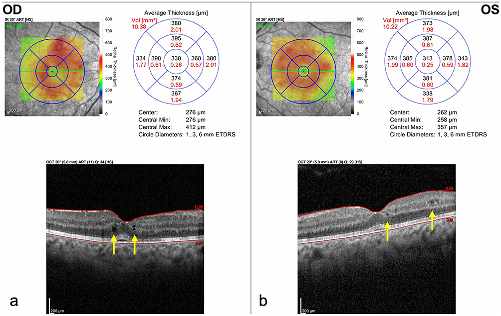 |
Figure 1 Optical Coherence Tomography (OCT) of the (a) right and (b) left eye demonstrating center-involving diabetic macular edema (yellow arrows) with trace subretinal fluid. At this visit, best corrected visual acuity was 20/30 in the right eye and 20/40 in the left eye. |
After 15 months and 20 cycles of treatment with pembrolizumab and lenvatinib, she was noted to have worsening of her VA to 20/80 in both eyes. Fundus exam revealed increase in severity of intraretinal hemorrhages, cotton wool spots and vascular tortuosity. OCT also showed worsening center-involving intraretinal fluid (Figure 2). Intravitreal anti-vascular endothelial growth factor (anti-VEGF) therapy was recommended, but the patient elected to monitor.
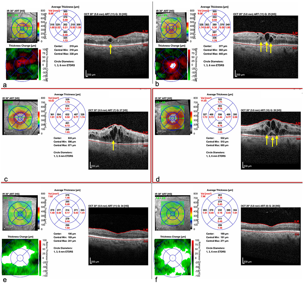 |
Figure 2 Optical Coherence Tomography (OCT) of the (a) right and (b) left eye showing worsening center-involving intraretinal fluid (yellow arrows) following fifteen months and twenty cycles of pembrolizumab and lenvatinib therapy. At this time, visual acuity was 20/80 in both eyes. Subsequently, the right and left eye improved from before intravitreal bevacizumab with visual acuity 20/200 in the (c) right eye and 20/150 in the (d) left eye to 20/80 in the (e) right eye and 20/100 in the (f) left eye. |
One month after the patient deferred intravitreal anti-VEGF therapy, her vision declined further to 20/200 in the right eye and 20/150 in the left eye. At this point, she agreed to bilateral intravitreal injection of bevacizumab for diabetic macular edema, which improved her intraretinal fluid but did not improve vision. Dilated fundus examination showed sclerotic vessels in the superior retina. Fluorescein angiography (FA) revealed multiple, bilateral branch retinal artery occlusions involving the superior retina in both eyes in a relatively symmetric pattern (Figures 3 and 4). At this time, Magnetic Resonance Imaging (MRI) of the brain and neck and echocardiography were negative for stroke, white matter disease, carotid occlusive disease, and sources of cardiac thrombus or emboli. Infectious (Tuberculosis, Syphilis), hypercoagulability, and inflammatory workup (erythrocyte sedimentation rate, C-reactive protein, human leukocyte antigen B27, angiotensin-converting enzyme, lysozyme, antinuclear antibody) were also negative. Her presentation of bilateral, relatively symmetric, BRAOs was attributed to pembrolizumab therapy.
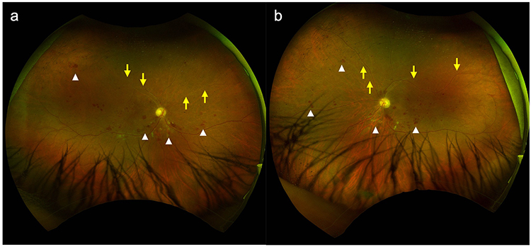 |
Figure 3 Optos Fundus Photos of the (a) right and (b) left eye showing posterior intraretinal hemorrhages and cotton wool spots (white arrowheads), as well as bilateral superior branch retinal artery occlusions and superior vascular sclerosis (yellow arrows). |
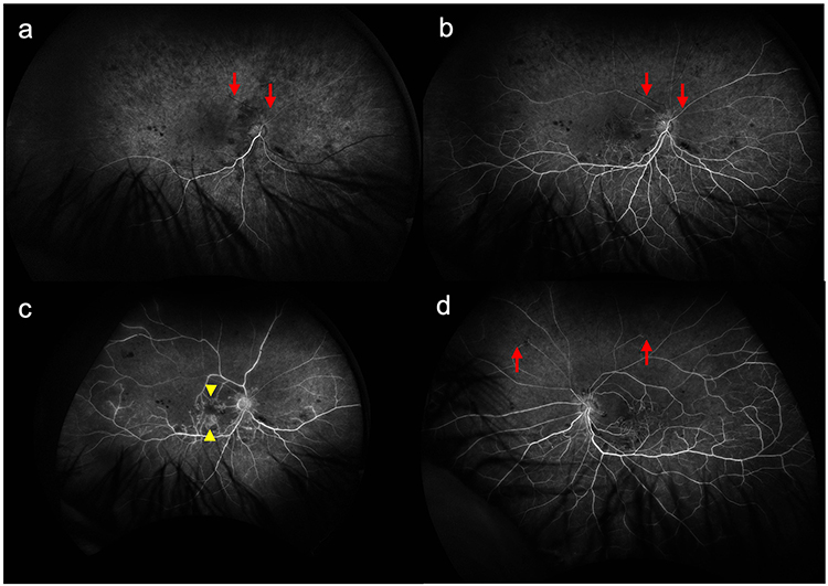 |
Figure 4 Fluorescein angiography (FA) of the right eye in (a) arterial phase (b) arteriovenous phase and (c) late phase and (d) of the left eye in arteriovenous phase demonstrating delayed filling of the superior retinal arterioles and superior filling defect from retinal artery occlusion (red arrows), as well as late central macular capillary leakage (yellow arrowheads). |
Following extensive discussion with the patient and her oncologist, the decision was made to discontinue pembrolizumab and lenvatinib. The patient has been in remission and continues systemic surveillance every three months. Systemic steroids were considered to treat possible inflammation to the retinal vessels causing subsequent vascular occlusion from pembrolizumab, but the patient has deferred. Her macular edema has been managed with intravitreal anti-VEGF injections, and her vision has improved. At her most recent visit, her best corrected visual acuity was 20/60 in the right eye and 20/125 in the left eye.
Discussion
In older patients and those with known cardiovascular risk factors such as smoking, diabetes, hypertension, coronary artery disease, and carotid occlusive disease, retinal arterial occlusions (RAO) are attributed to embolic events. However, when multiple BRAOs present in a younger individual (such as in those under the age of 50), hypercoagulable conditions and inflammatory states from autoimmune or infectious disease should be suspected. In this case, an extensive work-up with laboratory evaluations and neuroimaging was unrevealing. Hence, we attributed her multiple, bilateral BRAOs in both eyes in a relatively symmetric pattern to pembrolizumab, an immune checkpoint inhibitor which is known to cause various ocular adverse events.1–4 However, several other key diagnoses were considered. Radiation retinopathy is rare at treatment doses of 30 Grays but has been reported.5,6 After discussion with the radiation oncology team, radiation retinopathy at such low doses (3 Grays per fraction over 10 fractions) seemed unlikely but cannot be fully excluded. In addition, her ischemia progressed rapidly over the time she was being monitored while on pembrolizumab treatment but 2.5 years after she completed whole-brain radiation treatment. Given that the patient had retinal hemorrhages throughout the fundus in both eyes with retinal vascular occlusions and widespread non-perfusion, occlusive retinal vasculitis was also considered. Vasculitis and uveitis are known side effects of pembrolizumab.1,2,7,8 However, the patient lacked vascular sheathing and other inflammatory evidence on fluorescein angiography and examination such as disc hyperemia, vascular leakage, and vitreous cell. The rate of progression and symmetric pattern in a patient with well-controlled diabetes and hypertension would also be unusual for occlusive retinal vasculitis. Her angiographic findings suggested multiple BRAOs in the superior arterioles, and OCT through the superior macula showed inner retinal thinning consistent with superior BRAOs involving the papillomacular bundle, which may have contributed to the reduction in visual acuity.
Pembrolizumab is a recombinant monoclonal IgG antibody which acts at the programmed cell death protein 1 (PD-1) receptor. Other ICIs which target the PD-1 receptor include nivolumab and cemiplimab.2 Together, the PD-1 inhibitors have received US Food & Drug Administration (FDA) approval for a growing list of metastatic, unresectable, and refractory malignancies.
Under normal circumstances, the PD-1 receptor induces T-cell apoptosis when bound by programmed cell death-ligand 1 (PD-L1). In malignancy, tumor cells evade detection by the host’s immune system by co-opting immune checkpoint systems such as that between PD-1 and PD-L1. ICIs modulate the immune host system to recognize tumor cells; however, these same pathways can non-specifically activate the entire immune system, increasing the risk of autoimmune adverse events.1–3 Systemic immune-related adverse events are known to include pneumonitis, colitis, hepatitis, nephritis, dermatologic side effects (pruritus, rash, vitiligo), and endocrinopathies such as diabetes mellitus, thyroiditis, hypo-pituitarism, and adrenal insufficiency.1
Vision loss and ocular toxicities are rare but known adverse events associated with ICIs. The most reported ocular immune-mediated adverse events are dry eyes (1–24%) and uveitis or vasculitis (1%). Ocular surface toxicities associated with ICIs include conjunctivitis, episcleritis, keratitis, and corneal graft rejection.1 When uveitis occurs in the setting of PD-1 inhibitor treatment, panuveitis (38–46%) and anterior uveitis (30–36%) are more common than intermediate (0–1%) or posterior uveitis (14%).2 Known orbital complications of ICIs include myasthenia gravis, thyroid and thyroid-like ophthalmopathy, and optic neuritis.1,2,9 PD-1 inhibitors have also been associated with cranial nerve III, VI, and VII palsies.1 Typically, ocular adverse events are managed with topical, periocular, and/or systemic immunosuppression.1,2 However, 4% to 39% of patients on PD-1 or PD-L1 inhibitors need to discontinue the drug due to treatment-related adverse events.3
We believe our patient had bilateral, multiple BRAOs in a superior distribution, which were related to her pembrolizumab. Narala et al reported a case of an 86-year-old patient on pembrolizumab for metastatic uveal melanoma who developed pembrolizumab-related giant cell arteritis manifesting as retinal arterial occlusion and paracentral acute middle maculopathy4 Evidence in mouse models and histopathologic specimens suggest that blockade of PD-1/PD-L1 allows unopposed immune stimulation, formation of granulomatous lesions in the walls of affected arteries, microvascular angiogenesis, and intimal hyperplasia.10,11 Hence, Narala et al posited that the breakdown of PD-1/PD-L1 checkpoints in vessel walls could contribute to vasculitis and retinal arterial luminal occlusion4 We postulate a similar mechanism in our patient case. Of note, our patient endorsed occasional tinnitus, but this symptom was attributed to a side effect from her chemotherapy. She is awaiting further audiologic and vestibular evaluation. Magnetic resonance imaging (MRI) did not show white matter lesions. This negative finding is pertinent since Susac’s syndrome classically presents with a triad of BRAOs, sensorineural hearing loss, and encephalopathy and has also been reported as an immune-related adverse event in association with pembrolizumab.12 Moreover, reports suggest that cisplatin or carboplatin and paclitaxel can cause ischemic retinopathy,13–16 and the role of toxicity from her chemotherapy cannot be completely excluded. However, she had completed six cycles of chemotherapy relative to the twenty cycles of pembrolizumab and lenvatinib, and her chemotherapy was completed nearly two years prior to her vision decline. Finally, although the role of lenvatinib could not be excluded, the case reports on lenvatinib have shown retinal vein occlusion,17 but have not reported retinal artery occlusions.
Conclusion
Immune checkpoint inhibitors, which include targeted blockade of the PD-1/PD-L1 checkpoint, allow the host immune system to recognize tumor cells for a growing number of metastatic and refractory malignancies. However, immune-related adverse events can occur, with ocular adverse events including dry eye syndrome, uveitis or vasculitis, conjunctivitis, myasthenia gravis, retinal vasculitis, orbitopathy, and optic neuropathy.1,2 Herein, we report the case of a patient with metastatic uterine cancer on pembrolizumab therapy who presented with vision loss from multiple BRAOs. Patients on pembrolizumab or other ICIs with visual symptoms should be promptly examined by an ophthalmologist, with additional retinal imaging including OCT and FA. Clinical examination or imaging findings suggestive of retinal vascular occlusion, uveitis, or vasculitis should prompt a laboratory and systemic work-up. If no other explanation can be found for the visual symptoms or ocular findings, pembrolizumab as the cause of arterial occlusion should be considered. A discussion should take place between the patient, the ophthalmologist, and the oncologist on treatment options with consideration of local or systemic steroid and/or cessation of the ICI.18
Abbreviations
BRAO, branch retinal artery occlusion; RAO, retinal artery occlusion; ICI, immune checkpoint inhibitor; DM, diabetes mellitus; DME, diabetic macular edema; VA, visual acuity; OCT, Optical Coherence Tomography; FA, fluorescein angiography; DR, diabetic retinopathy; Anti-VEGF, anti-vascular endothelial growth factor; PD-1, programmed cell death protein 1; PD-L1, programmed cell death-ligand 1.
Ethics Approval and Informed Consent
This study adhered to the tenets of the Declaration of Helskinski and the Health Insurance Portability and Accountability Act. Ethical approval was not required by the Stanford Institutional Review Board or by national guidelines since this was a retrospective case report which did not change management of patient care. Written informed consent was obtained from this patient for publication of case details and associated images.
Acknowledgments
The authors would like to thank the many multi-disciplinary providers who provided care for this patient, as well as to the patient for her willingness to contribute to medical education.
Disclosure
P.M. reports grants from Genentech; nonpaid consultant for Castle Biosciences and Aura Biosciences, outside the submitted work. The authors report no conflicts of interest in this work. C.D. is supported by the National Institute of Health (NIH) K08EY036955-01, The Robert Machemer Foundation, The E. Matilda Ziegler Foundation for the Blind, The Alcon Research Institute, and acknowledgement is made to the donors of Macular Degeneration Research, a program of the BrightFocus Foundation, for support of this research (grant M2025004N). All authors were supported via departmental grants including the P30 Vision Research Core Grant, National Eye Institute (NEI) P30-EY026877, and Research to Prevent Blindness, New York and the Alan and Irene Adler Initiative in Ocular Cancer. The authors report no other conflicts of interest in this work.
References
1. Dalvin LA, Shields CL, Orloff M, Sato T, Shields JA. Checkpoint inhibitor immune therapy: systemic indications and ophthalmic side effects. Retina. 2018;38(6):1063. doi:10.1097/IAE.0000000000002181
2. Dow ER, Yung M, Tsui E. Immune checkpoint inhibitor-associated uveitis: review of treatments and outcomes. Ocul Immunol Inflamm. 2021;29(1):203–7. doi:10.1080/09273948.2020.1781902
3. Young L, Finnigan S, Streicher H, et al. Ocular adverse events in PD-1 and PD-L1 inhibitors. J Immunother Cancer. 2021;9(7):e002119. doi:10.1136/jitc-2020-002119
4. Narala R, Reddy SA, Mruthyunjaya P. Giant cell arteritis manifesting as retinal arterial occlusion and paracentral acute middle maculopathy in a patient on pembrolizumab for metastatic uveal melanoma. Am J Ophthalmol Case Rep. 2020;20:100891. doi:10.1016/j.ajoc.2020.100891
5. Chan L, Sneed PK, Horton JC. Damage to the superior retinae after 30 gy whole-brain radiation. Adv Radiat Oncol. 2021;6(4):100706. doi:10.1016/j.adro.2021.100706
6. Sverdlichenko I, Tayeb S, Narmandakh A, Margolin E, Tsang DS, Yan P. Radiation retinopathy after whole-brain radiotherapy in a patient with pineal gland tumor. J Vitreoretin Dis. 2025;24741264251359075. doi:10.1177/24741264251359075
7. Aaberg MT, Aaberg TM. Pembrolizumab administration associated with posterior uveitis. Retin Cases Brief Rep. 2017;11(4):348–351. doi:10.1097/ICB.0000000000000368
8. Kim KW, Kusuhara S, Tachihara M, Mimura C, Matsumiya W, Nakamura M. A case of panuveitis and retinal vasculitis associated with pembrolizumab therapy for metastatic lung cancer. Am J Ophthalmol Case Rep. 2021;22:101072. doi:10.1016/j.ajoc.2021.101072
9. Telfah M, Whittaker TJ, Doolittle C. Vision loss with pembrolizumab treatment: a report of two cases. J Oncol Pharm Pract. 2019;25(6):1540–1546. doi:10.1177/1078155219841683
10. Weyand CM, Berry GJ, Goronzy JJ. The immunoinhibitory PD-1/PD-L1 pathway in inflammatory blood vessel disease. J Leukoc Biol. 2018;103(3):565–575. doi:10.1189/jlb.3MA0717-283
11. Zhang H, Watanabe R, Berry GJ, et al. Immunoinhibitory checkpoint deficiency in medium and large vessel vasculitis. Proc Natl Acad Sci U S A. 2017;114(6):E970–E979. doi:10.1073/pnas.1616848114
12. De Groot M, Compter A, De Langen AJ, Brandsma D. Susac’s syndrome as an immune-related adverse event after pembrolizumab: a case report. J Neurol. 2020;267(1):282–284. doi:10.1007/s00415-019-09587-4
13. Togna GI, Togna AR, Franconi M, Caprino L. Cisplatin triggers platelet activation. Thrombosis Res. 2000;99(5):503–509. doi:10.1016/S0049-3848(00)00294-2
14. Elhusseiny AM, Smiddy WE. Carboplatin- and/or paclitaxel-induced ischemic retinopathy. Can J Ophthalmol. 2020;55(3):e95–e98. doi:10.1016/j.jcjo.2019.09.004
15. Matsuyama K, Ando A, Wada M, Ogata N, Nishimura T. Case of ischemic retinopathy induced by chemotherapy with paclitaxel and carboplatin. Acta Ophthalmol. 2008;86(s243). doi:10.1111/j.1755-3768.2008.639.x
16. Kwan ASL, Sahu A, Palexes G. Retinal ischemia with neovascularization in cisplatin related retinal toxicity. Am J Ophthalmol. 2006;141(1):196–197. doi:10.1016/j.ajo.2005.07.046
17. Foulsham W, Edghill BZ, Julia Canestraro OD, et al. Central retinal vein occlusion in the setting of fibroblast growth factor receptor inhibition. Am J Ophthalmol Case Rep. 2022;27:101657. doi:10.1016/j.ajoc.2022.101657
18. Bello F, Fagni F, Bagni G, et al. Arterial and venous thrombosis in systemic and monogenic vasculitis. Nat Rev Rheumatol. 2025;21(6):355–369. doi:10.1038/s41584-025-01252-7
 © 2026 The Author(s). This work is published and licensed by Dove Medical Press Limited. The
full terms of this license are available at https://www.dovepress.com/terms
and incorporate the Creative Commons Attribution
- Non Commercial (unported, 4.0) License.
By accessing the work you hereby accept the Terms. Non-commercial uses of the work are permitted
without any further permission from Dove Medical Press Limited, provided the work is properly
attributed. For permission for commercial use of this work, please see paragraphs 4.2 and 5 of our Terms.
© 2026 The Author(s). This work is published and licensed by Dove Medical Press Limited. The
full terms of this license are available at https://www.dovepress.com/terms
and incorporate the Creative Commons Attribution
- Non Commercial (unported, 4.0) License.
By accessing the work you hereby accept the Terms. Non-commercial uses of the work are permitted
without any further permission from Dove Medical Press Limited, provided the work is properly
attributed. For permission for commercial use of this work, please see paragraphs 4.2 and 5 of our Terms.
Recommended articles

Comparison of Neoadjuvant Immunotherapy Plus Chemotherapy versus Neoadjuvant Chemoradiotherapy for Patients with Esophageal Squamous Cell Carcinoma: A Propensity Score Matching Study
Zhao J, Hao S, Tian J, Li Y, Han D
Journal of Inflammation Research 2023, 16:3351-3363
Published Date: 8 August 2023
Cemiplimab for the Treatment of Advanced Cutaneous Squamous Cell Carcinoma: Appropriate Patient Selection and Perspectives
Mager L, Gardeen S, Carr DR, Shahwan KT
Clinical, Cosmetic and Investigational Dermatology 2023, 16:2135-2142
Published Date: 9 August 2023
Synergistic Induction of Immunogenic Cell Death by Biomineralized Manganese and Bisphosphonates Enhances Anti-PD-L1 Therapy in Triple-Negative Breast Cancer
Zhang X, Liu N, Wei M, Yang C, Lin Y, Zeng Y, Li Y, Zhou L, Li T, Zheng Q, Yu H, Zhang J, Gu Y, Xia N, Li S
International Journal of Nanomedicine 2025, 20:5001-5016
Published Date: 17 April 2025

Immunotherapy-Based Strategies versus Transcatheter Arterial Chemoembolization and the Impact of Treatment Duration in Hepatocellular Carcinoma with Microvascular Invasion: A Retrospective Multicenter Cohort Study
Chen X, Xing J, Zhang B, Peng W, Huang Z, Zhu T, Liu L, Liu X, Wan X, Mao Y, Chang X, Zhou K, Pan J, Hu D, Tan H, Zhang Y, Guan M, Du S
Journal of Hepatocellular Carcinoma 2025, 12:2379-2391
Published Date: 21 October 2025
Adjuvant Sintilimab with or without Anti-VEGF Therapy After Resection in Hepatocellular Carcinoma with Microvascular Invasion: A Multicenter Retrospective Study
Wang K, Xiang YJ, Yu HM, Cheng YQ, Zheng YT, Shan YF, Cheng SQ
Journal of Hepatocellular Carcinoma 2025, 12:2735-2744
Published Date: 16 December 2025