
Back to Journals » Clinical Ophthalmology » Volume 20
Visual and Anatomic Outcomes of Faricimab in Naïve Neovascular Age-Related Macular Degeneration with Subretinal Hemorrhage: A Multi-Centre Retrospective Analysis
Authors Sivachandran N  , Ji PX , Khan H, Pickel L, Mojumder O, Aziz AA , Khan H, Sulahria H , Gahn GM, Khanani AM
Received 17 December 2025
Accepted for publication 2 April 2026
Published 16 April 2026 Volume 2026:20 589703
DOI https://doi.org/10.2147/OPTH.S589703
Checked for plagiarism Yes
Review by Single anonymous peer review
Peer reviewer comments 2
Editor who approved publication: Dr Yousef Fouad
Nirojini Sivachandran,1– 5 Patrick Xiang Ji,6 Hannah Khan,7 Lauren Pickel,6 Ohidul Mojumder,8 Aamir A Aziz,7 Huma Khan,8 Humza Sulahria,8 Greggory M Gahn,7,8 Arshad M Khanani7,8
1Toronto Retina Institute, Oakville, ON, Canada; 2School of Medicine, Toronto Metropolitan University, Toronto, ON, Canada; 3Department of Chemistry and Biology, Toronto Metropolitan University, Toronto, ON, Canada; 4Institute for Biomedical Engineering, Science and Technology (iBEST) at Toronto Metropolitan University and St. Michael’s Hospital, Toronto, ON, Canada; 5University of Ottawa, Department of Ophthalmology, Ottawa, ON, Canada; 6Temerty Faculty of Medicine, University of Toronto, Toronto, ON, Canada; 7University of Nevada, Reno School of Medicine, Reno, NV, USA; 8Sierra Eye Associates, Reno, Nevada, USA
Correspondence: Nirojini Sivachandran, Toronto Retina Institute, 1425 Cornwall Road, Unit C5, Oakville, ON, L6J7T5, Canada, Tel +1 647 886-5863, Email [email protected]
Purpose: To evaluate functional and anatomical outcomes of faricimab in treatment-naïve patients with neovascular age-related macular degeneration (nAMD) presenting with subretinal hemorrhage (SRH).
Subjects and methods: A multicenter retrospective chart review was completed in retina practices in Canada and the United States from June 2022 to September 2025. Treatment-naïve nAMD patients with SRH receiving faricimab were evaluated. Standardized imaging protocols and treat-and-extend guidelines were employed. Primary outcome was visual acuity (VA) change; secondary outcomes included central subfield thickness (CST), pigment epithelial detachment (PED) height, and qualitative assessment of subretinal hyperreflective material (SHRM) and subretinal fibrosis. Outcomes were analyzed following three consecutive loading doses using Friedman statistics for repeated measures. Paired analysis was performed using Wilcoxon signed rank test. Detailed characterization of SRH included measurements of hemorrhage size in disc diameters (DD), location in terms of subfoveal versus extrafoveal, and duration from symptom onset to treatment initiation. Univariate regression analysis was performed to identify predictors of SRH resolution (effect ratio and 95% CI) and visual acuity improvement (odds ratio and 95% CI).
Results: This study included 63 treatment-naïve patients with nAMD and SRH (mean age was 81.6 ± 8.2 years; 58.2% females; n = 28 from Canada and n = 35 from United States; mean total follow-up duration was 17.5 ± 6.0 months). Significant VA improvement was observed (Friedman statistic: 26.3, p < 0.00001), with 42.6% of patients gaining ≥ 3 lines of vision after loading doses. CST decreased substantially from 391.8 μm to 249.4 μm (p < 0.00001, z = − 5.48) and mean PED height reduced from 309.6 μm to 110.7 μm (p < 0.00001, z = − 3.51). SHRM and subretinal fibrosis were noted in 81.6% and 25.4% of cases, respectively, at baseline. Of this, 70% had resolution of SHRM and 50% had improvement in fibrosis, at last follow up visit. Univariate analysis of predictors of faricimab injection account to achieve SRH resolution in n = 33 patients identified three significant predictors: SHRM-presence at baseline (effect ratio = 0.531, 95% CI 0.399– 0.707, p < 0.0001), combined intraretinal fluid (IRF) and subretinal fluid (SRF) (effect ratio = 1.43, 95% CI 1.09– 1.87, p = 0.011), and non-Caucasian ethnicity (effect ratio = 1.45, 95% CI 1.03– 2.04, p = 0.031). Analysis of predictors of VA improvement > 3 lines did not reveal any clinically meaningful associations.
Conclusion: This study demonstrates faricimab’s efficacy in nAMD with SRH, a challenging phenotype traditionally associated with poor visual prognosis. The dual-pathway inhibition of VEGF-A and Ang-2 achieved rapid hemorrhage clearance and meaningful vision gains, offering clinical hope for this high-risk population.
Keywords: neovascular age-related macular degeneration, subretinal hemorrhage, faricimab, anti-VEGF therapy, anti-Ang-2, dual-inbhition, visual improvement
Introduction
Neovascular age-related macular degeneration (nAMD) affects 10–20% of those living with AMD (200 million people worldwide).1 Choroidal neovascular membranes (CNVM) in nAMD can lead to severe and irreversible vision loss from exudation of intraretinal, subretinal and sub-retinal pigment epithelium (RPE) fluid, macular hemorrhage, fibrovascular scarring or RPE tear or detachment (PED).2,3 The pathogenesis of CNVM is driven by vascular endothelial growth factors (VEGF), a cytokine that promotes angiogenesis and vascular permeability. Targeting VEGF has become the mainstay of treatment for nAMD and has revolutionized patient outcomes.4,5
Subretinal hemorrhage (SRH) is a rare and dreaded complication of nAMD. It carries a poor prognosis as retained blood can lead to photoreceptor damage from iron toxicity, subretinal fibrosis, atrophic scarring and RPE tears.6 Prior treatments involved vitrectomy or pneumatic displacement away from the central retina, with or without tissue-plasminogen activator (tPA).5,6 There is increasing evidence supporting the use of intravitreal anti-VEGF monotherapy to improve visual outcomes compared to the natural history, particularly for hemorrhages less than four-disc diameters (DD).4,6–11 Subretinal hyper-reflective material (SHRM) is commonly seen in eyes with nAMD and often persists after anti-VEGF treatment. It is a vision-affecting morphological feature seen on spectral-domain optical coherence tomography (SD-OCT) as hyper-reflective material in the outer retina.12 Subretinal fibrosis on the other hand affects central vision and is a common natural sequela of macular neovascularization. It can cause damage to the outer retinal, including photoreceptors, RPE, and choriocapillaris.13
In addition to VEGF, angiopoietin-2 (Ang-2) has been shown to play a role in retinal disorders including nAMD. Ang-2 is involved in several key processes of disease pathogenesis by promoting choroidal neovascularization, increasing vascular permeability, and promoting inflammation and fibrosis.14–16 Furthermore, Ang-2 compared to Ang-1 has been shown to localize to highly vascularized regions of many CNVMs from post-mortem specimens.17 Faricimab is the first bispecific monoclonal antibody to inhibit VEGF and Ang-2. Randomized Clinical trials as well as real world studies have shown the efficacy and durability of faricimab in patients with nAMD.18–22 However, its role in patients with SRH, SHRM or subretinal fibrosis is yet to be explored. In this study, we sought to examine the role of dual inhibition of VEGF and Ang-2 in treatment naïve patients with SRH in nAMD.
Methods
This is a retrospective, uncontrolled cohort study of treatment naïve patients who presented with nAMD with SRH between June 2022 to September 2025 from community ophthalmology practices in Canada (Toronto Retina Institute) and the United States (TRUCKEE sub-cohort). A standard protocol of three loading doses of faricimab (faricimab-svoa, 6 mg/0.05 mL) in a single dose vial or pre-filled syringe was followed by a treat and extend regimen guided by disease activity. All hemorrhages were included in this retrospective analysis, and a detailed characterization of each SRH lesion was subsequently performed, which included:
- Duration assessment: time from symptom onset to treatment initiation was recorded based on patient-reported symptom onset and clinical presentation
- Size measurement: the extent of hemorrhage was quantified in disc diameters (DD)
- Location: SRH was categorized as subfoveal or extrafoveal
- Thickness measurement: maximal vertical extent of hemorrhage was measured on SD-OCT (Topcon)
Treatment-naïve nAMD patients who received a minimum of three injections of faricimab were included. As this study aims to establish the baseline efficacy and safety profile of faricimab in SRH without confounding effects from prior anti-VEGF treatments, previous treatment with any anti-VEGF therapy warrants exclusion. Exclusion criteria also included off-label use of faricimab, less than three doses, or incomplete records. Large and tall hemorrhages measuring greater than 4-DD were often treated with pneumatic displacement, tissue plasminogen activator (tPA), or vitrectomy, and thus were excluded from analysis.
Treat and extend protocol was followed for all patients included in the study. The follow-up was the last documented visit with complete assessment data available by September 2025, being at least 12 weeks apart from the baseline visit. Institutional review board approval was obtained from Advarra (Pro00083770), an independent ethics committee (Canada) and exemption received for patients included in the original TRUCKEE protocol (US).23 The study complies with the guidelines for human studies and was conducted in accordance with the World Health Organization Declaration of Helsinki.
The primary endpoint was changes in best corrected visual acuity (BCVA) from baseline to last follow-up. Secondary endpoints measured the efficacy of faricimab for treatment of SRH as quantified by changes in central subfield thickness (CST), presence and height of PED, SHRM, subretinal fibrosis, and resolution of SRH. These endpoint outcomes were analyzed at baseline, following the first and the third injection (loading doses), and at last visit, and they were specifically defined. Snellen visual acuities were converted to their logMAR equivalents, with count fingers being assigned 1.85. Spectral-domain optical coherence tomography (SD-OCT) was used to quantify CST, type of fluid, and presence of SHRM, subretinal fibrosis and PED type and height. Adverse events were recorded during the study period. SD-OCT also determines fluid resolution in terms of intra-retinal fluid (IRF), which is the hypo-reflective space within the neurosensory retina, as well as subretinal fluid (SRF), which is the hypo-reflective space between the RPE and neurosensory retina. Demographic information including age, sex, and ethnicity were collected and summarized using descriptive statistics.
Statistical Analysis
To determine treatment effects across timepoints (baseline, after 1 injection, 3 injections, and last follow up), the Shapiro–Wilk test was first used to assess the normality of the outcomes. Given that our outcomes were not normally distributed, non-parametric analyses were utilized. When a significant effect of time was observed, individual timepoints were compared using the Wilcoxon signed-rank test for non-normally distributed data. Given the lack of a control comparative group, we performed within-subject, pre- vs post-treatment analyses.
To identify baseline characteristic associated with (1) faricimab injection count required to achieve SRH resolution and (2) visual acuity improvement of greater than 3 lines, we performed univariate meta-regression analyses in a subset of n = 33 patients with complete baseline predictor and outcome data. Given the small sample size, multivariate adjusted models were not performed to avoid overfitting and unstable estimates. The dependent variables were: (1) the number of faricimab injections required to achieve complete SRH resolution, and (2) achieving improvement in VA of >3 lines at the last follow up. Independent baseline characteristics analyzed included age, sex (female vs male), ethnicity (non-Caucasian vs Caucasian), hemorrhage size (disc diameters), fluid type (categorized as SRF, IRF, or both vs SRF), baseline VA (in logMAR), CST (µm), and PED (µm), as well as baseline presence of SHRM and fibrosis (present or absent). For SRH resolution analysis, effect ratios with 95% confidence intervals (CI) were calculated. Effect ratios >1.0 indicate increased injection count required for resolution, while effect ratios <1.0 suggest decreased injection count. For VA improvement analysis, odds ratios with 95% CI were calculated. Odds ratios >1.0 indicate higher odds of achieving >lines of VA improvement, whereas odds ratios <1.0 suggest reduced odds. P <0.05 was considered statistical significance.
Results
Sample
A total of 63 patients from multiple centers in the US (n = 35) and Canada (n = 28) with AMD and SRH treated with a minimum of 3 faricimab injections were included (Table 1). All patients received faricimab treatment, and there was no comparative control group treated with standard anti-VEGF agents. Demographic characteristics did not differ between Canadian and US sites. Median age was 81.6 ± 1.1 years (range 51–94 years), and 58.2% of patients were females. Out of n = 46 patients with available data, the mean hemorrhage size was 1.6 ± 0.2DD (range 0.1 to 4 DD). The distribution of hemorrhage sizes was small (<1DD): 21.7% (n = 10), medium (1–2DD): 54.3% (n = 25), and large (2–4DD): 23.9% (n = 11). Out of n = 37 patients with available data for hemorrhage location, 73% (n = 27) were classified as subfoveal, while 27% (n = 10) were extrafoveal. The mean duration from symptom onset to treatment initiation was 30.9 ± 7.3 days (range 1–197 days). Overall, 51.8% of patients had subretinal fluid (SRF) without intraretinal fluid (IRF), 41.1% of patients had a combination of SRF and IRF, 7.1% of patients showed only IRF. Mean follow-up duration was 17.5 ± 1.0 months, with patients receiving an average of 8.8 ± 0.43 injections during this period.
 |
Table 1 Baseline Characteristics and Secondary Outcomes |
Primary Endpoint
Visual acuity significantly improved with faricimab treatment (Friedman statistic 26.3, p < 0.00001, Figure 1A and Supplementary Table 1). Mean visual acuity increased from 0.81 (±0.08) at baseline to 0.70 (±0.07) after one treatment, to 0.63 (±0.07) after three, and to 0.56 (±0.07) after the last injection. After one treatment, approximately 16.7% of study eyes (n = 10, of total n = 60 with available data) had gained ≥3 lines of vision from baseline. This increased to 27.3% (n = 15 of total n = 55 with available data) after three treatments. At last follow up, 42.6% (of total n = 54 with complete data) had gained ≥3 lines of vision from baseline. Visual acuity declined ≥3 lines in 5.6% of cases, while remaining relatively stable in the rest of the cases.
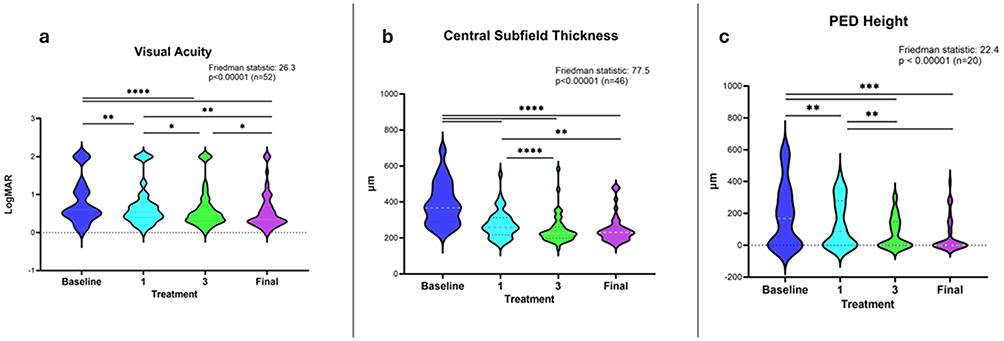 |
Figure 1 (a) Best corrected visual acuity (VA, logMAR units) at baseline, after one faricimab injection, after three injections, and at last follow up. (b) Central subfield thickness (CST) in microns (µm) at baseline, after one faricimab injection, after three injections, and at last follow up. (c) Pigment epithelial detachment (PED) height in microns (µm) at baseline, after one faricimab injection, after three injections, and at last follow up. Distributions represented by violin plot. Repeated measures for all non-parametric data were compared using the Friedman test with post hoc pairwise comparisons using Wilcoxon signed rank test. * p < 0.05, ** p < 0.01, *** p < 0.001, **** p < 0.0001. |
Secondary Endpoints
Treatment with faricimab also reduced CST (p < 0.00001, Friedman statistic 77.5, Figure 1B) from 391.8 ± 17.2 µm at baseline to 274.5 ± 11.1 µm after one injection (p < 0.00001), to 245.4 ± 11.5 µm after three injections (p < 0.0001), and to 249.4 ± 11.5 µm at last follow up (p < 0.00001). CST did not significantly change from third injection to last follow up (mean difference of −3.96 µm, p = 0.16). Finally, PED height was also reduced by faricimab treatment (Friedman statistic 24.2, p = 0.00002, Figure 1C). Baseline PED height reduced from an average of 309.6 ± 32.3 µm to 228.9 ± 27.5 µm after one injection (p = 0.00453) and reduced greatly to 125.7 ± 20.9 µm after three injections (p = 0.00013) and this was sustained at last follow up at 110.7 ± 26.2 µm (no significant reduction between injection 3 vs last follow up, p = 0.27).
On average, only two faricimab injections were required for resolution of SRH (Table 1). Absence or resolution of hemorrhage was determined using a combination of clinical examination and fundus imaging. Complete resolution was the absence of visible hemorrhage on both clinical and imaging tests, whereas partial resolution represented >50% improvement from baseline. The time to resolution was defined as the time it takes from baseline to achieve complete resolution. Resolution of subretinal hyper-reflective material (SRHM) was observed in 70% of cases where initially present (an example from one patient is shown in Figure 2). Subretinal fibrosis improved or resolved in 50% of cases where initially present. At last follow up, patients had received an average of 8.8 ± 0.43 injections and were followed at injection intervals of 9.7 ± 0.38 weeks.
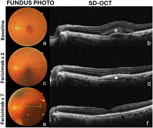 |
Figure 2 An 87-year-old male with nAMD and macular hemorrhage (a) and SHRM noted on OCT in the subretinal space noted by asterisk * (b). After two injections of faricimab (c) resolution of SHRM noted with subretinal fibrosis indicated by arrowhead ▴ (d). After seven injections of faricimab (e) resolution of fibrosis noted (f). |
Univariate regression analysis was performed on a subset of n = 33 patients with complete baseline and outcome data to determine which baseline characteristics were associated with the number of injections required to achieve SRH resolution (2.5 ± 1.1 injections, range: 1–5). From the univariate analysis, three baseline predictors were significantly associated with injection count to SRH resolution (Table 2), and these included SHRM presence, fluid complexity, and non-Caucasian ethnicity. Paradoxically, the presence of SHRM at baseline was associated with 47% fewer injections required for SRH resolution (effect ratio = 0.53, 95% CI 0.40–0.71, p < 0.0001), compared to SHRM-absent eyes. However, only 2 of the 33 patients (6.1%) had absent SHRM at baseline, leading to a severe group imbalance. With fluid type of SRF as a reference point, eyes with IRF alone were associated with fewer injections needed (effect ratio = 0.46, 95% CI 0.39–0.56, p < 0.0001), whereas combined IRF and SRF required significantly more injections to achieve resolution (IRF + SRF: effect Ratio = 1.43, 95% CI 1.09–1.87, p < 0.011), with the mixture fluid type requiring highest injection burden, 43% more than the requirement of SRF at baseline. However, only 1 of the 33 patients (3.0%) presented with isolated IRF at baseline. Non-Caucasian ethnicity was associated with significantly increased injection count (effect ratio = 1.45, 95% CI 1.03–2.04, p = 0.031), requiring approximately 45% more than Caucasian patients. Besides these three variables, there were no other baseline parameters that showed significant univariate associations (Table 2).
 |
Table 2 Univariate Meta-Regression Analysis of Baseline Characteristics as Predictors of Faricimab Injection Count to Achieve SRH Resolution |
Univariate analysis was also performed for baseline predictors of VA improvement of >3 lines using the same patient group with complete baseline data (Table 3). IRF was the only predictor that showed a statistically significant association with VA improvement, however, this represented a highly unstable finding (odds ratio = 2.95 × 10⁻⁸, 95% CI 3.31 × 10⁻⁸–3.62 × 10⁻⁷, p < 0.0001). This is in the context of the aforementioned severe limitation in sample size, with only 1 of 33 patients (3.0%) with isolated IRF. This again must be interpreted with caution. Besides this, there were no other predictors that showed significant associations with VA improvement.
 |
Table 3 Univariate Meta-Regression Analysis of Baseline Characteristics as Predictors of Visual Acuity Improvement of ≥3 Lines at the Last Follow Up |
Safety and Adverse Events
There were no associated adverse events including anterior or posterior segment inflammation, retinal vasculitis, endophthalmitis or vitreous hemorrhage.
Discussion
SRH in nAMD, if not treated promptly, can lead to poor visual outcomes.10,11 Various modalities of treatment exist for SRH including anti-VEGF monotherapy, pneumatic displacement with or without anti-VEGF or tPA, and vitrectomy with subretinal tPA.5–7 Clinical trials including TENAYA and LUCERNE, demonstrating the non-inferiority of faricimab to aflibercept, tend to exclude patients with SRH occupying >50% of lesion size.24,25 This is the first report to highlight the effectiveness of dual inhibition of VEGF-A and Ang-2 on anatomic and functional outcomes in naïve patients presenting with SRH in nAMD.
Acute treatment of SRH from the subretinal space minimizes toxicity to photoreceptors and decreases risk of fibrosis of the macula, a region of high-acuity central vision.6 In our study, patients presented with an average SRH size of 1.6 ± 0.2DD (range 0.1DD to 4DD), with detailed characterization revealing that 71.4% had subfoveal involvement and mean duration from symptom onset to treatment initiation was approximately 30.9 ± 7.3 days. An improvement in hemorrhage was noted with as few as two intravitreal injections of faricimab. The majority of the visual gains occurred within the first three months and were comparable in magnitude to previous studies for anti-VEGF monotherapy.26–29 Approximately 43% of patients gained >3 lines of vision at last follow up. Regarding visual outcomes, existing observational evidence shows that about 40–60% of patients with SMH in nAMD treated with pneumatic displacement and anti-VEGF (with or without tPA) will have improved vision at 3–6 months, with an average gain of around 3 lines.7,30 Interestingly, we did not observe any cases of vitreous hemorrhage, even in those with larger SRH, in contrast to anti-VEGF monotherapy in previous studies.26,27 It is conceivable that dual inhibition of both VEGF-A and Ang-2 leads to less leakage (rapid improvement of fluid as noted in this study) and halts progression of hemorrhage limiting breakthrough vitreous hemorrhage.31 Similarly, there was a significant reduction in CST at all time points examined, from 391.8 µm to 249.4 µm at last follow up. To achieve fluid free status, the median number of faricimab injections was two, highlighting the efficacy of faricimab in its ability to dry the retina as previously established.32 Ang-2 has been shown to cause vascular instability and hyperpermeability. The rapid resolution of fluid and improvement in CST is a reflection on dual inhibition of VEGF-A and Ang-2, yielding retinal integrity and improved visual outcomes as noted above.31
Acute treatment of SRH from the subretinal space minimizes toxicity to photoreceptors and decreases risk of fibrosis of the macula, a region of high-acuity central vision.6 In our study, patients presented with an average SRH size of 1.6 ± 0.2DD (range 0.1DD to 4DD), with detailed characterization revealing that 71.4% had subfoveal involvement and mean duration from symptom onset to treatment initiation was approximately 30.9 ± 7.3 days. An improvement in hemorrhage was noted with as few as two intravitreal injections of faricimab. The majority of the visual gains occurred within the first three months and were comparable in magnitude to previous studies for anti-VEGF monotherapy.26–29 Approximately 43% of patients gained >3 lines of vision at last follow up. Regarding visual outcomes, existing observational evidence shows that about 40–60% of patients with SMH in nAMD treated with pneumatic displacement and anti-VEGF (with or without tPA) will have improved vision at 3–6 months, with an average gain of around 3 lines.7,30 Interestingly, we did not observe any cases of vitreous hemorrhage, even in those with larger SRH, in contrast to anti-VEGF monotherapy in previous studies.26,27 It is conceivable that dual inhibition of both VEGF-A and Ang-2 leads to less leakage (rapid improvement of fluid as noted in this study) and halts progression of hemorrhage limiting breakthrough vitreous hemorrhage.31 Similarly, there was a significant reduction in CST at all time points examined, from 391.8 µm to 249.4 µm at last follow up. To achieve fluid free status, the median number of faricimab injections was two, highlighting the efficacy of faricimab in its ability to dry the retina as previously established.32 Ang-2 has been shown to cause vascular instability and hyperpermeability. The rapid resolution of fluid and improvement in CST is a reflection on dual inhibition of VEGF-A and Ang-2, yielding retinal integrity and improved visual outcomes as noted above.31
SHRM is a finding on OCT that has prognostic value. The exact composition of SHRM is not well defined; it is presumed to be a mixture of lipid, immune cells, hemorrhage and fibrovascular tissue, all of which have similar reflectivity on OCT.13 In our study, we observed resolution of SHRM with faricimab in 70% of cases, which is remarkable compared to resolution rates reported for aflibercept (31.4% to 47.6%)29,33–35 or brolicizumab (54.4%).29 Although these studies are not directly comparable, this difference likely reflects the added benefit of Ang-2 blockade.
This mechanism likely contributes to the notable improvement in fibrosis in this study. At baseline, 25.4% of cases presented with subretinal fibrosis, of which half had improvement or resolution with faricimab treatment (fibrosis as determined on colored fundus photography and SD-OCT). Fibrosis has significant consequences for visual outcomes, where accumulation of the fibrotic material in the subretinal space can cause irreversible damage to the photoreceptors limiting central vision and development of a scotoma.35 Subretinal fibrosis is commonly reported with anti-VEGF treatment in nAMD. For instance, approximately half of all study eyes developed fibrosis by 24 months in the CATT and IVAN trials.36 This is the first study with intravitreal injection inhibiting VEGF-A and Ang-2 to show improvement in subretinal fibrosis. The pathophysiology of fibrosis can be complex, resulting from SRH, SHRM, and large basal CNVMs.13 It is known that Ang-2 increases fibroblast activity through the activation of inflammatory mediators in various tissues including the retina.14 Using two independent mouse models, Linder et al have shown that dual inhibition of Ang-2 and VEGF-A led to sustained prevention of fibrosis.37 To our knowledge, this is the first clinical evidence to suggest that Ang-2 blockade can minimize or improve subretinal fibrosis.
Our univariate analysis revealed a counterintuitive and paradoxical association, where the presence of SHRM at baseline was associated with faster SRH resolution, requiring approximately 47% fewer injections compared to SHRM-absent patients. This contradicts current knowledge that SHRM represents a poorer prognosis.38 However, this finding must be interpreted clinically with extreme caution, given a critical limitation with only 2 of 33 patients (6.1%) having absent SHRM at baseline. This severe group imbalance likely led to substantial statistical instability, as the effect estimates were strongly influenced by these two outliers, and this may represent an artifact as opposed to true clinical association. The proposed mechanism suggests that faricimab’s dual inhibition may selectively target the pathophysiological processes of SHRM formation, given that Ang-2 is well characterized to increase vascular instability, inflammation and activity of fibroblasts. By simultaneously blocking both angiogenesis and vascular instability, faricimab may be more efficacious in addressing the underlying pathophysiology of SHRM development/progression, ultimately leading to rapid hemorrhage resolution. However, given the small sample size constraints and large group imbalances, this finding is at best hypothesis-generating and needs to be interpreted with caution; it requires detailed validation in larger prospective trials with SHRM characterization and quantification.
In contrast to SHRM results, the association between fluid complexity and injection count represents a more robust and clinically actional predictor. Our analysis has shown that eyes with more complex fluid patterns, such as those with combined IRF+SRF, required significantly more injections to achieve SRH resolution, compared with eyes that had isolated SRF.38,39 This suggests a dose-response relationship, such that IRF, rather than SRF alone, represents the critical pathophysiological driver of continued disease activity requiring intensive treatments. This also provides preliminary evidence that baseline fluid characteristic may generate prognostic information helping to guide management and patient counseling. Eyes with IRF in combination with SRF required significantly more injections for SRH resolution, likely reflecting greater underlying disease activity, more severe vascular permeability, or deeper retinal structural disruption requiring prolonged dual VEGF-A and Ang-2 inhibition.
We note that only one patient presented with isolated IRF, creating statistical instability for this baseline predictor.
An unexpected finding from the univariate analysis was that being non-Caucasian was associated significantly with increased injection count to reach resolution, with a 45% increase. This relationship needs careful interpretation because of the small sample size of non-caucasian patients (n = 6, 18.2%) limiting statistical power and effect estimate. Without multivariate analysis, we cannot determine if ethnic differences represent true biological variation in hemorrhage pathophysiology or anti-VEGF response, if it is confounded by other unmeasured sociodemographical factors. Moreover, the binary classification of this baseline characteristic as either Caucasian or non-Caucasian masks potential diversity within the non-Caucasian group, making it difficult to evaluate effects specific to certain racial or ethnic subgroups.
Other baseline factors did not independently associate with or predict SRH resolution rate or VA improvement. For instance, the lack of hemorrhage size effect is interesting to note, as it has often been considered a primary determinant of prognosis. This may reflect farcimab’s dual-inhibition mechanism achieving consistent hemorrhage resolution regardless of initial extent of lesion. It should be noted that patients with really large hemorrhages requiring other interventions were excluded from this study and may limit the corroboration of the effect of hemorrhage size. However, given our limited sample size for the regression analysis, the power is again limited to detect small to moderate effect sizes for these baseline variables, and lack of statistical significance may not necessarily represent definitive lack of clinical relevance.
Overall, we believe faricimab has added value in patients presenting with SRH in nAMD as it rapidly improves hemorrhage with improvement in retinal fluid in as little as two treatments. It also has added benefit in SHRM resolution and may limit subretinal fibrosis. We acknowledge several limitations in this study. A key limitation is its retrospective design, which can introduce selection bias. Selection criteria were strictly adhered to in order to mitigate this risk. Additionally, a major limitation is the absence of a control group. nAMD with SRH has been well-described in literature to improve with conventional anti-VEGF treatment, thus, a missing control group consisting of only non-dual inhibition therapy would limit the ability to draw comparative conclusions. The lack of a comparative arm with other long-acting anti-VEGF treatments, such as Eylea HD or brolucizumab, represents a significant limitation that may reduce our ability to definitively attribute clinical outcomes to the dual-inhibition advantages of faricimab. The objective, however, of the study was to evaluate the efficacy of faricimab in naïve nAMD patients with SRH, as often, these patients are excluded from clinical trials. The observed improvement across several outcome variables may reflect only the natural course of anti-VEFG treatment in nAMD with SRH, as opposed to a unique dual-inhibition effect of faricimab. Although two recent studies in treatment experienced patients have shown that faricimab may confer less risk of recurrent SRH in nAMD. Kauffman et al completed a retrospective review of all patients who had SRH during anti-vegf treatment and showed that patients treated with faricimab were significantly less likely to experience SRH (1.7%, p = 0.0004) compared to ranibizumab (24.7%), aflibercept (35.4%) or bevacizumab (37.8%).40 Furthermore, switching patients with sub-optimal response with faricimab to aflibercept 8 mg, did not confer visual acuity or treatment extension benefit, it resulted in increased SRH from 8.1% to 21.7% (p = 0.004).41 The statistical analyses across variables thus may overestimate the effect of treatment, such that our claims regarding the effectiveness of faricimab for patients with SRH cannot be fully substantiated.
Additionally, the wide variability in SRH characteristics introduces substantial heterogeneity. While we performed detailed characterization to account for these variables, the heterogeneity limits our ability to optimize treatment approach for specific SRH subtypes. Finally, although the sample size of the present study with 63 patients is comparable to similar studies examining the role of anti-VEGF monotherapy, the analysis is limited by incomplete medical records, necessitating a complete case analysis approach. The decision to exclude patients with prior anti-VEGF therapy, while scientifically applausible to establish baseline efficacy, limits generalizability to the broader nAMD population where many patients have received previous treatments.
Conclusion
In conclusion, faricimab was associated with improved visual and anatomical outcomes in patients with nAMD with SRH. Regression analysis identified SHRM presence and simpler fluid patterns as independent predictors of faster SRH resolution, suggesting these baseline factors may help guide treatment planning and patient counseling. Detailed characterization of hemorrhage characteristics identified important predictors of treatment response, however the lack of a control group limits definitive conclusions about comparative efficacy. Given our uncontrolled, retrospective study design as well as other heterogeneities in study duration and inclusion criteria, the interpretation of these results should be considered from a preliminary, and pilot study perspective. Further large prospective comparative studies with uniform follow-up duration and subgroup analyses are warranted to confirm any specific advantage of dual inhibition. This treatment has the potential to greatly benefit patients with SRH through more rapid resolution of hemorrhage, improving vision and thereby quality of life.
Ethics Approval and Informed Consent
Ethics approval was obtained from Advarra (Pro00083770). As per IRB, informed consent was exempt for this retrospective review. To ensure patient data confidentiality, all information were de-identified prior to analysis and reported only in aggregate form to prevent the identification of any individual participant.
Acknowledgments
The authors would like to thank the TRUCKEE study investigators, Dr Mark R Barakat (Retina Macula Institute of Arizona, Scottsdale, AZ, United States), Dr Jordan M. Graff (Barnet Dulaney Perkins Eye Center, Phoenix, AZ, United States), Dr Nicholas J.S. London (Retina Consultants San Diego, Poway, CA, United States), Dr. Veeral Sheth (University Retina, Chicago, IL, United States), Dr. Michael A. Singer (Medical Center Ophthalmology Associates, San Antonio, TX, United States) and Dr. Jeremy D. Wolfe (Associated Retinal Consultants PC, Royal Oak, MI, United States), with participant recruitment and for facilitating data collection for the TRUCKEE cohort. Their support was instrumental in the successful completion of this study.
Funding
Sentrex (Canada) in support of IRB from a general research grant to NS.
Disclosure
Dr Greggory Gahn reports personal fees from Genentech and Regenxbio, outside the submitted work. Dr Arshad Khanani reports grants, personal fees from Genentech and Roche, outside the submitted work. The authors report no other conflicts of interest in this work.
References
1. Chaudhuri M, Hassan Y, Bakka Vemana PPS, Bellary Pattanashetty MS, Abdin ZU, Siddiqui HF. Age-related macular degeneration: an exponentially emerging imminent threat of visual impairment and irreversible blindness. Cureus. 2023;15(5):e39624. doi:10.7759/cureus.39624
2. Hobbs SD, Tripathy K, Pierce K. Wet age-related macular degeneration (AMD). In: StatPearls. StatPearls Publishing; 2025. Available from. http://www.ncbi.nlm.nih.gov/books/NBK572147/.
3. Wong T, Chakravarthy U, Klein R, et al. The natural history and prognosis of neovascular age-related macular degeneration. Ophthalmology. 2008;115(1):116–126.e1. doi:10.1016/j.ophtha.2007.03.008
4. Schmidt-Erfurth U, Chong V, Loewenstein A, et al. Guidelines for the management of neovascular age-related macular degeneration by the European society of retina specialists (EURETINA). Br J Ophthalmol. 2014;98(9):1144–12. doi:10.1136/bjophthalmol-2014-305702
5. Khanna S, Komati R, Eichenbaum DA, Hariprasad I, Ciulla TA, Hariprasad SM. Current and upcoming anti-VEGF therapies and dosing strategies for the treatment of neovascular AMD: a comparative review. BMJ Open Ophth. 2019;4(1):e000398. doi:10.1136/bmjophth-2019-000398
6. Gabrielle PH, Delyfer MN, Glacet-Bernard A, et al. Surgery, tissue plasminogen activator, antiangiogenic agents, and age-related macular degeneration study. Ophthalmology. 2023;130(9):947–957. doi:10.1016/j.ophtha.2023.04.014
7. Jeong S, Park DG, Sagong M. Management of a submacular hemorrhage secondary to age-related macular degeneration: a comparison of three treatment modalities. JCM. 2020;9(10):3088. doi:10.3390/jcm9103088
8. Shin JY, Lee J, Byeon SH. Anti–vascular endothelial growth factor with or without pneumatic displacement for submacular hemorrhage. Am J Ophthalmol. 2015;159(5):904–914.e1. doi:10.1016/j.ajo.2015.01.024
9. Kitahashi M, Sakurai M, Yokouchi H, et al. Pneumatic displacement with intravitreal bevacizumab for massive submacular hemorrhage due to polypoidal choroidal vasculopathy. OPTH. 2014:485. doi:10.2147/OPTH.S55413
10. Avery RL, Fekrat S, Hawkins BS, Bressler NM. Natural history of subfoveal subretinal hemorrhage in age-related macular degeneration. Retina. 1996;16(3):183–189. doi:10.1097/00006982-199616030-00001
11. Scupola A, Coscas G, Soubrane G, Balestrazzi E. Natural history of macular subretinal hemorrhage in age-related macular degeneration. Ophthalmologica. 1999;213(2):97–102. doi:10.1159/000027400
12. Willoughby AS, Ying GS, Toth CA, et al. Subretinal hyperreflective material in the comparison of age-related macular degeneration treatments trials. Ophthalmology. 2015;122(9):1846–1853.e5. doi:10.1016/j.ophtha.2015.05.042
13. Tenbrock L, Wolf J, Boneva S, et al. Subretinal fibrosis in neovascular age-related macular degeneration: current concepts, therapeutic avenues, and future perspectives. Cell Tissue Res. 2022;387(3):361–375. doi:10.1007/s00441-021-03514-8
14. Joussen AM, Ricci F, Paris LP, Korn C, Quezada-Ruiz C, Zarbin M. Angiopoietin/Tie2 signalling and its role in retinal and choroidal vascular diseases: a review of preclinical data. Eye. 2021;35(5):1305–1316. doi:10.1038/s41433-020-01377-x
15. Khanani AM, Russell MW, Aziz AA, et al. Angiopoietins as potential targets in management of retinal disease. Clin Ophthalmol. 2021;15:3747–3755. doi:10.2147/OPTH.S231801
16. Gahn GM, Khanani AM. New therapies of neovascular AMD beyond Anti-VEGF injections. Vision. 2018;2(1):15. doi:10.3390/vision2010015
17. Otani A, Takagi H, Oh H, Koyama S, Matsumura M, Honda Y. Expressions of angiopoietins and Tie2 in human choroidal neovascular membranes. Invest Ophthalmol Vis Sci. 1999;40(9):1912–1920.
18. Heier JS, Khanani AM, Quezada Ruiz C, et al. Efficacy, durability, and safety of intravitreal faricimab up to every 16 weeks for neovascular age-related macular degeneration (TENAYA and LUCERNE): two randomised, double-masked, Phase 3, non-inferiority trials. Lancet. 2022;399(10326):729–740. doi:10.1016/S0140-6736(22)00010-1
19. Khanani AM, Guymer RH, Basu K, et al. TENAYA and LUCERNE: rationale and design for the Phase 3 clinical trials of faricimab for neovascular age-related macular degeneration. Ophthalmol Sci. 2021;1(4):100076. doi:10.1016/j.xops.2021.100076
20. Khanani AM, Kotecha A, Chang A, et al. TENAYA and LUCERNE: two-year results from the phase 3 neovascular age-related macular degeneration trials of faricimab with treat-and-extend dosing in year 2. Ophthalmology. 2024;131(8):914–926. doi:10.1016/j.ophtha.2024.02.014
21. Penha FM, Masud M, Khanani ZA, et al. Review of real-world evidence of dual inhibition of VEGF-A and ANG-2 with faricimab in NAMD and DME. Int J Retin Vitr. 2024;10(1). doi:10.1186/s40942-024-00525-9
22. Khanani AM, Aziz AA, Khan H, et al. The real-world efficacy and safety of faricimab in neovascular age-related macular degeneration: the TRUCKEE study - 6 month results. Eye. 2023;37(17):3574–3581. doi:10.1038/s41433-023-02553-5
23. Khan H, Aziz AA, Abbey AM, et al. Visual and anatomic outcomes in nAMD patients with subretinal hemorrhage after treatment with faricimab in the TRUCKEE study. Invest Ophthalmol Visual Sci. 2023;64(8):2187.
24. Gale R, Korobelnik JF, Yang Y, Wong TY. Characteristics and predictors of early and delayed responders to ranibizumab treatment in neovascular age-related macular degeneration: a retrospective analysis from the ANCHOR, MARINA, HARBOR, and CATT trials. Ophthalmologica. 2016;236(4):193–200. doi:10.1159/000451065
25. Maruyama-Inoue M, Kitajima Y, Yanagi Y, Inoue T, Kadonosono K. Intravitreal anti-vascular endothelial growth factor monotherapy in age-related macular degeneration with submacular hemorrhage. Sci Rep. 2023;13(1):5688. doi:10.1038/s41598-023-32874-0
26. Kim HS, Cho HJ, Yoo SG, et al. Intravitreal anti-vascular endothelial growth factor monotherapy for large submacular hemorrhage secondary to neovascular age-related macular degeneration. Eye. 2015;29(9):1141–1151. doi:10.1038/eye.2015.131
27. Kim JH, Chang YS, Kim JW, Kim CG, Yoo SJ, Cho HJ. Intravitreal anti-vascular endothelial growth factor for submacular hemorrhage from choroidal neovascularization. Ophthalmology. 2014;121(4):926–935. doi:10.1016/j.ophtha.2013.11.004
28. Shienbaum G, Garcia Filho CAA, Flynn HW, Nunes RP, Smiddy WE, Rosenfeld PJ. Management of submacular hemorrhage secondary to neovascular age-related macular degeneration with anti-vascular endothelial growth factor monotherapy. Am J Ophthalmol. 2013;155(6):1009–1013. doi:10.1016/j.ajo.2013.01.012
29. Sadda S, Sarraf D, Khanani AM, et al. Comparative assessment of subretinal hyper-reflective material in patients treated with brolucizumab versus aflibercept in HAWK and HARRIER. Br J Ophthalmol. 2024;108(6):852–858. doi:10.1136/bjo-2023-323577
30. Kitahashi M, Baba T, Sakurai M, et al. Pneumatic displacement with intravitreal bevacizumab for massive submacular hemorrhage due to polypoidal choroidal vasculopathy. Clin Ophthalmol. 2014;8:485–492. doi:10.2147/OPTH.S55413
31. Chaudhary V, Mar F, Amador MJ, et al. Emerging clinical evidence of a dual role for Ang-2 and VEGF-A blockade with faricimab in retinal diseases. Graefes Arch Clin Exp Ophthalmol. 2025;263(5):1239–1247. doi:10.1007/s00417-024-06695-4
32. Mizuki Y, Inokuchi S, Kuroki T, et al. Two-year real-world outcomes of faricimab for diabetic macular edema in Japan. Clin Ophthalmol. 2025;19:3051–3058. doi:10.2147/OPTH.S547179
33. Bachmeier I, Yu S, Glittenberg C, Maunz A, Fauser S. Model for resolution of subretinal hyperreflective material (SHRM) in neovascular age-related macular degeneration (nAMD) using deep learning (DL) image segmentation. Invest Ophthalmol Visual Sci. 2024;65(9):PB009–PB009.
34. Alex D, Giridhar A, Gopalakrishnan M, Indurkhya S, Madan S. Subretinal hyperreflective material morphology in neovascular age-related macular degeneration: a case control study. Indian J Ophthalmol. 2021;69(7):1862–1866. doi:10.4103/ijo.IJO_3156_20
35. Kawashima Y, Hata M, Oishi A, et al. Association of vascular versus avascular subretinal hyperreflective material with aflibercept response in age-related macular degeneration. Am J Ophthalmol. 2017;181:61–70. doi:10.1016/j.ajo.2017.06.015
36. Evans RN, Reeves BC, Maguire MG, et al. Associations of variation in retinal thickness with visual acuity and anatomic outcomes in eyes with neovascular age-related macular degeneration lesions treated with anti-vascular endothelial growth factor agents. JAMA Ophthalmol. 2020;138(10):1043–1051. doi:10.1001/jamaophthalmol.2020.3001
37. Linder M, Foxton R, Uhles S, et al. Simultaneous Ang-2/VEGF-A inhibition prevents subretinal fibrosis progression in preclinical mouse models of choroidal neovascularization (CNV). Invest Ophthalmol Visual Sci. 2021;62(8):349.
38. Nanji K, Grad J, Hatamnejad A, et al. Baseline OCT biomarkers predicting visual outcomes in neovascular age-related macular degeneration: a meta-analysis. Ophthalmology. 2025;132(11):1241–1252. doi:10.1016/j.ophtha.2025.06.018
39. Fuchs P, Schmidt-Erfurth U, Coulibaly LM, et al. Impact of AI-quantified fluid dynamics on visual outcomes over 5 years in patients with treatment-naïve nAMD from the FRB! registry. Sci Rep. 2025;15(1):32881. doi:10.1038/s41598-025-17417-z
40. Kaufmann GT, Boucher N, Sharma C, Aggarwal N, Starr MR. Submacular hemorrhage rates following anti-vascular endothelial growth factor injections for exudative age-related macular degeneration. Am J Ophthalmol. 2025;270:172–182. doi:10.1016/j.ajo.2024.10.017
41. Momenaei B, Yonekawa Y, Abril P, McCullough R, Abbey AM. Outcomes of intravitreal aflibercept 8 mg in eyes with neovascular age-related macular degeneration previously treated with faricimab. Ophthalmic Surg Lasers Imaging Retina. 2025;56(4):206–211. doi:10.3928/23258160-20241205-01
 © 2026 The Author(s). This work is published and licensed by Dove Medical Press Limited. The
full terms of this license are available at https://www.dovepress.com/terms
and incorporate the Creative Commons Attribution
- Non Commercial (unported, 4.0) License.
By accessing the work you hereby accept the Terms. Non-commercial uses of the work are permitted
without any further permission from Dove Medical Press Limited, provided the work is properly
attributed. For permission for commercial use of this work, please see paragraphs 4.2 and 5 of our Terms.
© 2026 The Author(s). This work is published and licensed by Dove Medical Press Limited. The
full terms of this license are available at https://www.dovepress.com/terms
and incorporate the Creative Commons Attribution
- Non Commercial (unported, 4.0) License.
By accessing the work you hereby accept the Terms. Non-commercial uses of the work are permitted
without any further permission from Dove Medical Press Limited, provided the work is properly
attributed. For permission for commercial use of this work, please see paragraphs 4.2 and 5 of our Terms.
Recommended articles

Intravitreal Faricimab for Aflibercept-Resistant Neovascular Age-Related Macular Degeneration
Rush RB, Rush SW
Clinical Ophthalmology 2022, 16:4041-4046
Published Date: 9 December 2022
Initial Real-World Experience with Faricimab in Treatment-Resistant Neovascular Age-Related Macular Degeneration
Leung EH, Oh DJ, Alderson SE, Bracy J, McLeod M, Perez LI, Bottini A, Chin Yee D, Mukkamala K
Clinical Ophthalmology 2023, 17:1287-1293
Published Date: 5 May 2023
Short-Term Outcomes of 3 Monthly intravitreal Faricimab On Different Subtypes of Neovascular Age-Related Macular Degeneration
Tanaka A, Hata M, Tsuchikawa M, Ueda-Arakawa NUA, Tamura H, Miyata M, Takahashi A, Kido A, Muraoka Y, Miyake M, Ooto S, Tsujikawa A
Clinical Ophthalmology 2024, 18:507-516
Published Date: 20 February 2024
Tailored Anti-VEGF Therapy with New Generation Optimizations (TANGO) Treatment Regimen for Neovascular Age-Related Macular Degeneration: Rationale, Design, and Simulation Study
Subhi Y, Andersen LMM, Hajari JN, Henningsen N, Larsen KI, Schneider M
Clinical Ophthalmology 2025, 19:1435-1441
Published Date: 29 April 2025

Efficacy and Safety of Aflibercept Biosimilars Relative to Reference Aflibercept Therapy for Neovascular Age-Related Macular Degeneration: A Systematic Review and Meta-Analysis
Aljuhani HS, Hubayni RA, Qedair J, Bukhari ZM, Alzahrani A, Bawazir RO, Badghaish OS, Alqahtani F
Clinical Ophthalmology 2025, 19:1911-1918
Published Date: 19 June 2025