
Back to Journals » Journal of Pain Research » Volume 19
Trigeminal Terminal Branch Neuralgias in a Headache Unit: A 15-Year Series
Authors Ros González I  , Recio García A, Sierra Mencía Á, González Osorio Y, García-Azorín D , Guerrero Peral ÁL
Received 29 January 2026
Accepted for publication 8 May 2026
Published 27 May 2026 Volume 2026:19 588731
DOI https://doi.org/10.2147/JPR.S588731
Checked for plagiarism Yes
Review by Single anonymous peer review
Peer reviewer comments 2
Editor who approved publication: Dr Rune Häckert Christensen
Isabel Ros González,1 Andrea Recio García,2 Álvaro Sierra Mencía,2 Yesica González Osorio,2 David García-Azorín,3,4 Ángel Luis Guerrero Peral2,4
1Neurology Department, Hospital Infanta Elena, Madrid, Spain; 2Headache Unit, Neurology Department, Hospital Clínico Universitario de Valladolid, Valladolid, Spain; 3Headache Unit, Neurology Department, Hospital Universitario Río Hortega, Valladolid, Spain; 4Department of Medicine, Dermatology and Toxicology, University of Valladolid, Valladolid, Spain
Correspondence: David García-Azorín, Hospital Universitario Río Hortega, Calle Dulzaina 2, Valladolid, 47014, Spain, Email [email protected]
Introduction: ICHD-3 integrates neuralgias affecting trigeminal terminal branches within broader trigeminal pain categories rather than defining them as distinct entities. In this context, their frequency and clinical characteristics remain poorly characterized. We aimed to describe their frequency and clinical characteristics in a headache unit registry.
Methods: We conducted a retrospective, descriptive cohort study based on a prospective registry initiated in January 2008 at a headache unit. Patients diagnosed between January 2008 and January 2023 who fulfilled ICHD-2 criteria for nasociliary (code 13.5), supraorbital (13.6), or other terminal branch neuralgias (13.7) were included. Demographic and clinical data were collected and analyzed.
Results: Among 8728 patients evaluated during the study period, 108 (1.2%) met inclusion criteria; 71 (65.7%) were women. Mean age at symptom onset was 47.4 ± 18.7 years (range 6– 89), and mean time from onset to diagnosis was 34.4 ± 68.5 months (range 1– 420). The most frequent neuralgias were supraorbital (39.8%) and auriculotemporal (25.9%), followed by supratrochlear (8.3%), infraorbital (7.4%), lacrimal (7.4%), mental (5.6%), nasociliary (4.6%), and infratrochlear (0.9%).
Conclusion: Trigeminal terminal branch neuralgias represent a small but consistent clinical presentation defined by topographic distribution and sensory features. Recognition of these features supports anatomically guided evaluation and peripheral interventions.
Keywords: neuralgia, supraorbital, neuralgia, auriculotemporal, atypical neuralgias, nerve pain, paroxysmal, pain, neuropathic, block, nerve
Introduction
Craniofacial pain syndromes present a diagnostic challenge due to their overlapping etiologies and heterogeneous clinical presentations. From a neurological point of view, craniofacial pain is a prevalent symptom with a broad spectrum of clinical phenotypes.1 The variability in clinical presentations complicates diagnosis, and the lack of individualized recognition in commonly used classifications further hinders accurate identification.2
According to the definition provided by the International Association for the Study of Pain, the term “neuralgia” is characterized by the presence of pain in the innervation territory of a nerve or nerve root.3 Sensory transmission in the facial, head, and neck areas is mediated by the afferent fibers of the trigeminal, facial, glossopharyngeal, and vagus cranial nerves, as well as the first cervical roots through the occipital nerves.4 Pain in these regions may arise from nociceptive mechanisms due to irritation or injury, or from neuropathic processes.5
Craniofacial neuralgia pain varies in its characteristics (lancinating or electric), temporality (continuous or paroxysmal), severity (moderate to severe), the presence or absence of triggers, and response to different types of treatments (oral pharmacological treatment or anesthetic blocks). Diagnosis relies primarily on pain topography, which must be limited to the territory of a specific nerve.6
Although relatively rare, craniofacial neuralgias are associated with significant disability and severe pain.7 Early diagnosis is essential not only for appropriate management but also for detecting secondary causes. While some cases are idiopathic, a subset is related to underlying structural or systemic conditions, warranting systematic evaluation. Structural causes include vascular compression, tumors, and other space-occupying lesions. The workup should therefore include neuroimaging and screening for infectious, metabolic, traumatic, compressive, and infiltrative processes.8
Craniofacial neuralgias are included in Section 13 of the Third Edition of the International Classification of Headache Disorders (ICHD-3), encompassing trigeminal, glossopharyngeal, intermediate nerve, and occipital neuralgias.9 In the previous edition (ICHD-2), neuralgias involving terminal branch of the trigeminal nerve—such as the supraorbital (13.5), infraorbital (13.6), and auriculotemporal nerves (13.7)—were listed as distinct diagnostic entities.10
These categories are not defined as distinct entities in ICHD-3, which adopts a more integrated classification approach.11 In this context, neuralgias affecting terminal trigeminal branches are incorporated into broader diagnostic categories, particularly trigeminal neuralgia (TN), defined as pain confined to one or more trigeminal divisions (ICHD-3, 13.1.1), potentially involving distal branch territories. Depending on the clinical context and etiology, cases may also be classified under other forms of painful trigeminal neuropathy, including post-traumatic or post-herpetic subtypes.9 A comparable approach is adopted in the International Classification of Orofacial Pain (ICOP, 2020), in which these presentations are similarly included within broader categories of trigeminal neuropathic pain (ICOP 4.1.2).12
TN is the most common form of cranial neuralgia, with an estimated prevalence of 0.1–0.2 cases per thousand people and is well characterized in the literature.13 In contrast, neuralgias affecting its terminal branches may present with distinct sensory topographies and therapeutic responses. While these are no longer recognized as separate diagnostic entities, they persist as consistent clinical patterns.14
Given the lack of well-defined epidemiological and clinical profiles, the present study aims to characterize the frequency, demographic traits, and clinical features of terminal trigeminal branch neuralgia in patients evaluated at a tertiary headache unit.
Materials and Methods
Study Design
This was a retrospective, observational, descriptive cohort study based on a prospective registry initiated in January 2008 at the Headache Unit of the Hospital Clínico Universitario de Valladolid. Clinical records and follow-up data were systematically reviewed up to January 2023. In cases of missing or ambiguous information, structured telephone interviews were conducted to complete the data. The study adhered to the STROBE (Strengthening the Reporting of Observational Studies in Epidemiology) guidelines15 and was conducted in accordance with the Declaration of Helsinki.
Setting and Participants
The Headache Unit is part of a tertiary, university-affiliated public hospital serving a reference population of approximately 261,000 inhabitants. Patients diagnosed between January 2008 and January 2023 with neuralgia involving terminal branches of the trigeminal nerve—nasociliary (ICHD-2 13.5), supraorbital (13.6), or other terminal trigeminal branches (13.7)—were eligible for inclusion. Cases involving more than one trigeminal branch were not included, as the study focused on pain confined to a single terminal branch to ensure a well-defined topographic distribution.
Participants were identified through two complementary prospective registries:
- A general database of all patients evaluated in the Headache Unit with diagnoses coded according to the ICHD-2.
- A specific registry for trigeminal terminal branch neuralgia, also coded using ICHD-2 terminology.
Diagnosis Criteria
Inclusion Criteria
Diagnosis of trigeminal terminal branch neuralgia according to ICHD-2 criteria by a headache specialist; the diagnostic criteria are detailed in Figure 1; fluency in Spanish; ability to provide a coherent description of symptoms during the interview; and a pain duration of ≥3 months.
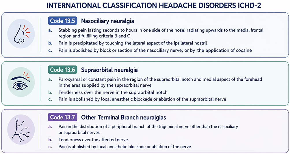 |
Figure 1 Diagnostic criteria in ICDH-2 for nasociliary, supraorbital, and other terminal branches of the trigeminal nerve neuralgias. |
Exclusion Criteria
Major cognitive impairment precluding clinical assessment; active illicit drug use or substance dependence; comorbid neurological or psychiatric conditions that could confound diagnosis; and incomplete clinical records or insufficient data for retrospective confirmation.
All participants provided written informed consent prior to inclusion, in accordance with institutional protocols and the Declaration of Helsinki.
Data Sources and Collection
Data were extracted from electronic medical records, including clinical notes, neurological evaluations, and diagnostic tests. Diagnoses and clinical variables were confirmed retrospectively through detailed chart review. Where data were unclear, patients were contacted by phone. Two neurologists independently reviewed all records to confirm eligibility. Discrepancies were resolved by consensus.
Variables
- Demographic variables: Sex, age, age at symptom onset, age at diagnosis, and time in months from symptom onset to diagnosis.
- Clinical variables: pain location and laterality; baseline pain characteristics (eg, pressing, stabbing, burning, throbbing); temporal pattern (continuous vs. remitting); and potential triggers. For pain paroxysms, frequency, duration, and character were also recorded. Pain intensity was evaluated using a Verbal Analogue Scale (VAS) from 0 to 10, both for baseline pain and paroxysms.
- Sensory findings: hypoesthesia and dysesthesia were evaluated by light touch and pinprick testing and documented when present.
- Etiological Investigation: All patients underwent a standardized work-up including blood tests (erythrocyte sedimentation rate and C-reactive protein) and brain Magnetic Resonance Imaging (MRI) with high-resolution sequences (FIESTA or CISS). In selected cases, Computed Tomography (CT) was also performed. No structural abnormalities, including neurovascular compression or other lesions affecting the trigeminal nerve, were identified despite systematic evaluation.
- Treatment: All patients received a diagnostic and/or therapeutic nerve block, following national consensus guidelines.14
Clinical Evaluation
All patients underwent a comprehensive neurological examination, including:
- Palpation of the trochlear area, nasal bones, and emergence points of the trigeminal terminal branches (supratrochlear, infratrochlear, supraorbital, infraorbital, lacrimal, auriculotemporal).
- Assessment of cervical range of motion and temporomandibular joint function.
- Sensory testing with light touch and pinprick to assess hypoesthesia or dysesthesia in the affected territory.
Study Size
There was no formal sample size estimation, and the analyses were done based on the available sample.
Statistical Methods
Quantitative variables were expressed as mean ± standard deviation (SD) or, for non-normally distributed data or small sample sizes, as median and interquartile range (IQR). Qualitative variables were expressed as frequencies and percentages. Statistical analyses were performed using SPSS software, version 22.
Given the descriptive nature of the study, no inferential statistical analyses or hypothesis testing were performed, and therefore p-values were not calculated.
Ethics Statement
This study is a retrospective, non-interventional analysis of a 15-year clinical registry. All data were irreversibly anonymized prior to analysis, and no patient contact or intervention was performed. According to institutional policy and applicable regulations, studies based exclusively on fully anonymized data and with no impact on patient care do not require formal Ethics Committee review. The study was conducted in accordance with the General Data Protection Regulation (EU) 2016/679, Ley Orgánica 3/2018 de Protección de Datos Personales y garantía de los derechos digitales, and the principles of the Declaration of Helsinki.
Results
During the 15-year study period, 108/8728 (1.2%) patients were diagnosed with terminal branch neuralgia, 71/108 (65.7%) of whom were female. According to ICHD-2 criteria, the types were: nasociliary neuralgia in 5/8728 (0.06%), supraorbital neuralgia in 43/8728 (0.49%), auriculotemporal neuralgia in 28/8728 (0.32%), supratrochlear neuralgia in 9/8728 (0.10%), infraorbital neuralgia in 8/8728 (0.09%), lacrimal neuralgia in 8/8728 (0.09%), mental neuralgia in 6/8728 (0.07%), and infratrochlear neuralgia in 1/8728 (0.01%) (Figure 2).
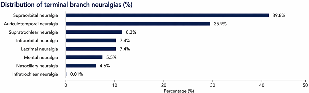 |
Figure 2 Distribution (%) of trigeminal terminal branch neuralgias in our sample. |
The demographic characteristics of the sample are detailed in Table 1. The mean age at inclusion was 50.5 ± 18.5 years, the mean age at symptom onset was 47.4 ± 18.7 years, and the mean duration from symptom onset to diagnosis was 34.4 ± 68.5 months. The neuralgias with the longest intervals from symptom onset to diagnosis were supratrochlear neuralgia (75.7 ± 134.5 months) and auriculotemporal neuralgia (44.6 ± 85.3 months) (Figure 3).
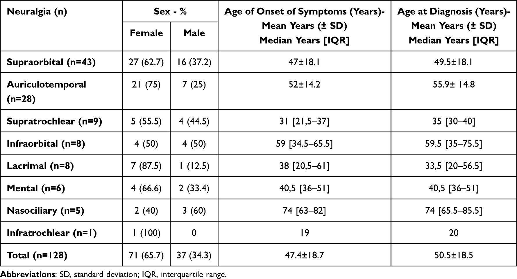 |
Table 1 Demographic Characteristics of Trigeminal Terminal Branch Neuralgias in Our Sample |
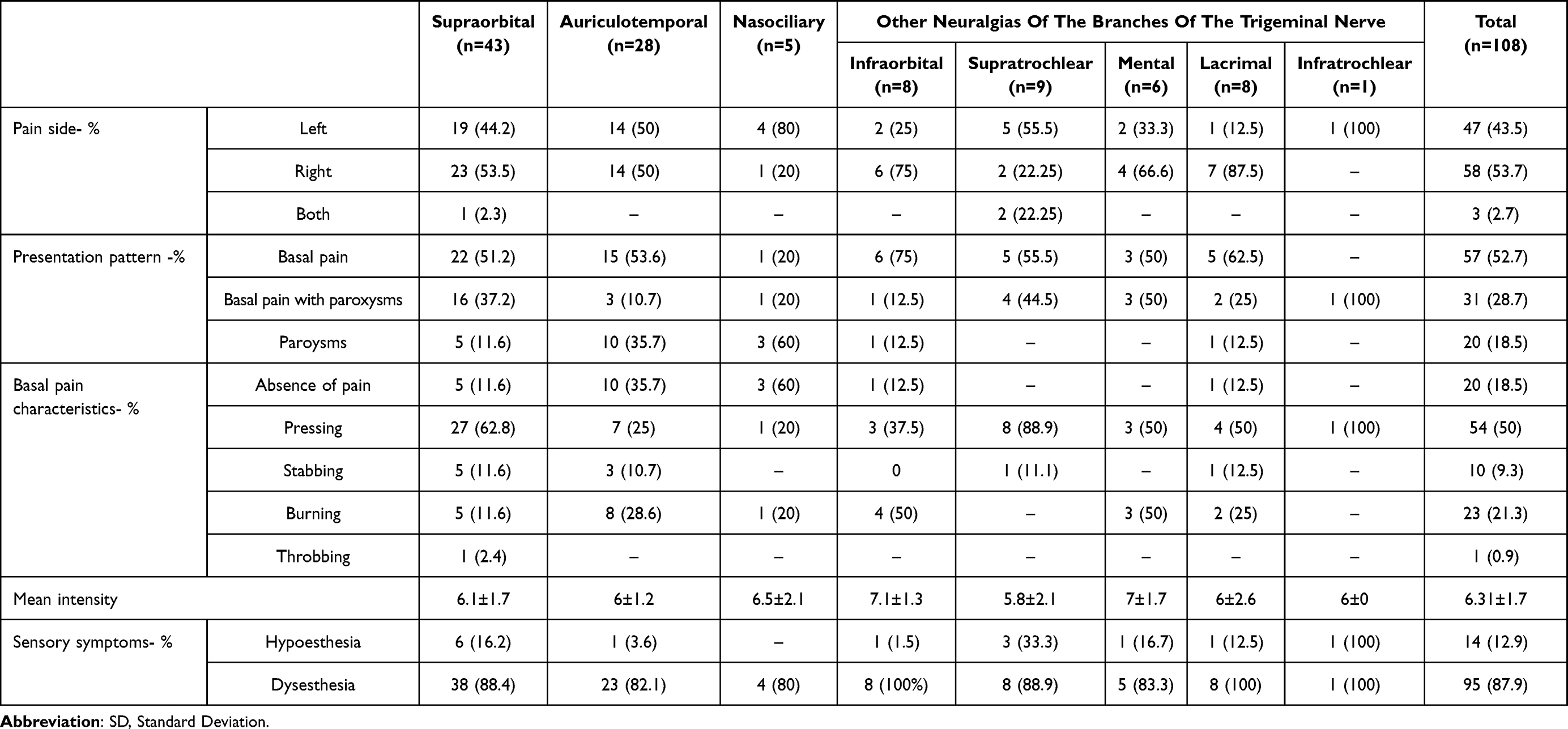 |
Table 2 Quantitative and Qualitative Clinical Characteristics of the Sample’s Neuralgias |
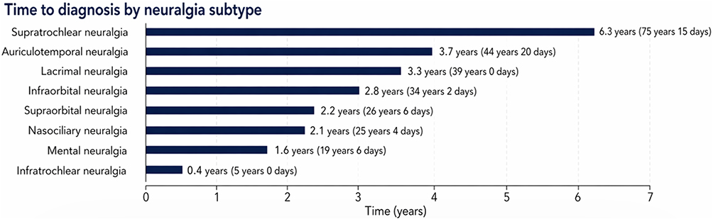 |
Figure 3 Bar graph showing the average time in months from onset to diagnosis of neuralgias of the terminal branches of the trigeminal nerve. |
The clinical characteristics of the neuralgias are detailed in Table 2 and 3. The proportion of patients who reported background pain was 57 (52.7%) and the proportion of patients who described pain with paroxysms was 31 (28.7%). The most frequent pain character was pressing in 54 (50%). The mean intensity of background pain was 6.3 ± 1.7 on the VAS, and the mean intensity of paroxysmal pain was 8.5 ± 0.6 on the VAS. The mean duration of paroxysmal pain was 14 minutes. In 13 (12%) cases, the pain was triggered by trauma (Table 4).
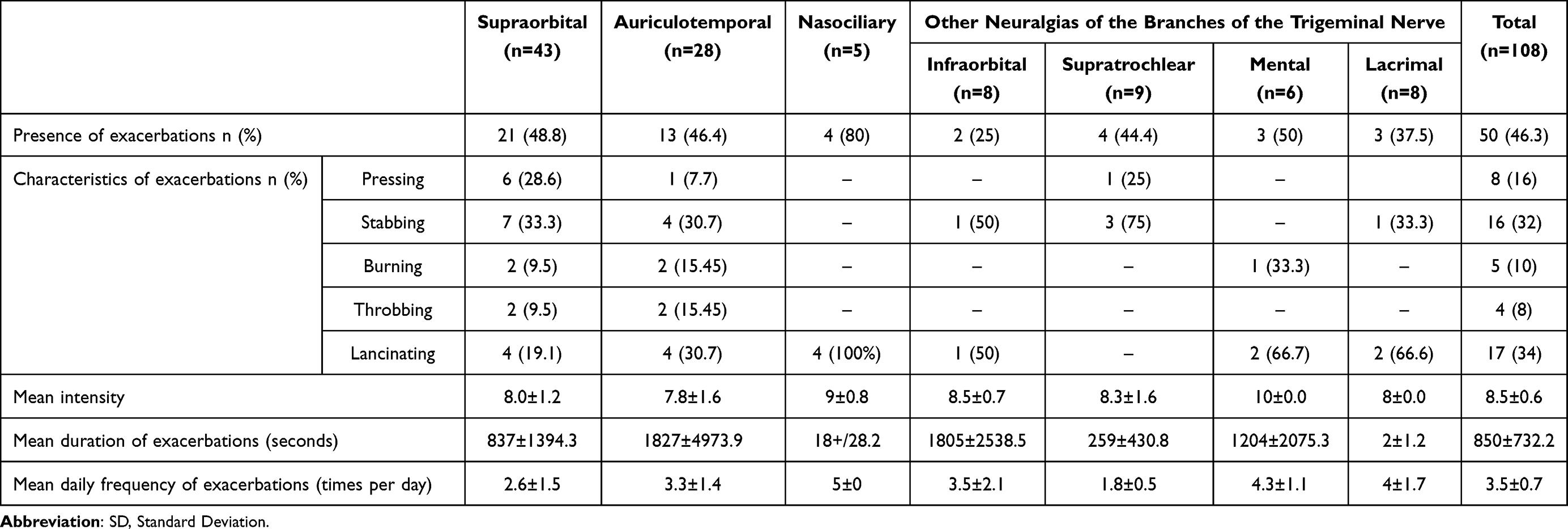 |
Table 3 Quantitative and Qualitative Clinical Characteristics of the Paroxysms of the Sample’s Neuralgias |
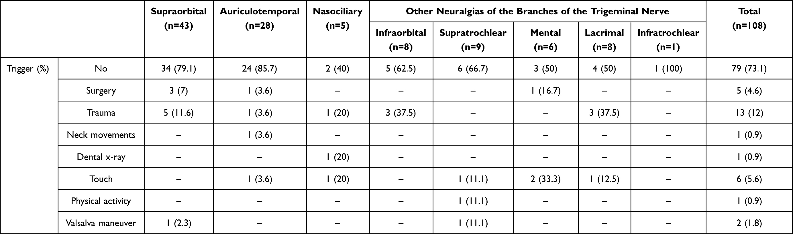 |
Table 4 Reported Clinical Triggers (%) by Terminal Trigeminal Branch in Patients with Terminal Branch Neuralgia |
Sensory disturbances were common. Dysesthesia was reported in 95 patients (87.9%) and hypoesthesia in 14 (12.9%), both confined to the same topographic distribution as the pain.
Discussion
This study evaluates the clinical and demographic characteristics of trigeminal terminal branch neuralgia in a cohort of 108 patients diagnosed using ICHD-2 criteria. The relative prevalence within a large series of >8700 was 1.2%, with the most prevalent conditions being auriculotemporal (28, 0.32%) and supraorbital (43, 0.49%) neuralgias. The most frequently observed pattern consisted of continuous, oppressive, unilateral pain often accompanied by dysesthesia. Additionally, 46.3% of patients experienced pain exacerbations of severe, lancinating quality. A prolonged delay between symptom onset and diagnosis was observed, particularly in less common neuralgias.
The trigeminal nerve, being the largest of the cranial nerves, exhibits a complex anatomy and includes both sensory and motor components. It provides facial innervation through three main branches: the ophthalmic nerve (V1), the maxillary nerve (V2), and the mandibular nerve (V3), which has both sensory and motor functions. V1 gives rise to the frontal, lacrimal, and nasociliary nerves. The frontal nerve divides into the supraorbital and supratrochlear branches, which innervate the forehead and upper eyelid. The nasociliary nerve gives rise to the anterior ethmoidal and infratrochlear nerves, which supply the nasal root and medial canthus. V2 innervates the lower eyelid, cheek, nasal ala, and upper lip via the infraorbital nerve, and the lateral orbital region through the zygomaticofacial and zygomaticotemporal nerves. V3 includes the auriculotemporal and mental nerves, responsible for sensory innervation of the preauricular region, chin, and lower lip.16 (Figure 4).
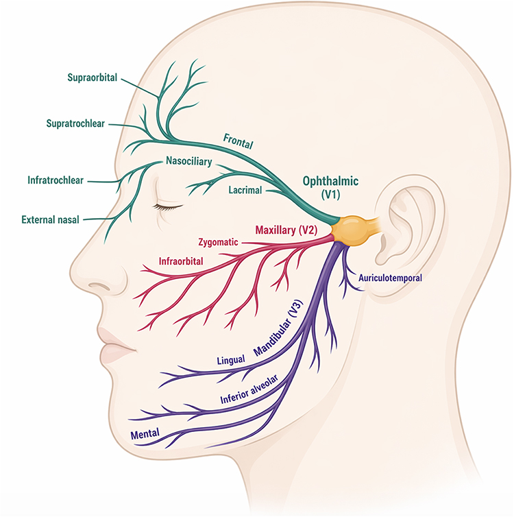 |
Figure 4 Main divisions of V1, V2, and V3 of the trigeminal nerve. Created with BioRender.com (© BioRender.com) under an academic use license. |
Any of these anatomical structures—sensory roots, ganglia, nerve trunks, secondary branches, or terminal branches—may contribute to facial pain, although not all have been fully characterized. Stimulation at different levels of the trigeminal pathway may produce pain localized to the corresponding sensory territory.17 In TN, vascular compression is considered the main underlying mechanism; however, other structural factors have also been implicated, including bony constraints along the trigeminal pathway. Spatial restrictions within the cerebellopontine angle, angulation of the nerve along the petrous ridge, or bony interfaces at the level of Meckel’s cave may contribute to proximal constraints along the trigeminal pathway.18 Within this continuum, the trigeminal porus represents a transitional zone between the cisternal and ganglionic segments where such constraints may occur. Variations in the site and nature of compression may influence clinical presentation, ranging from classical paroxysmal TN to forms with a more continuous pain profile.19
In contrast, distal involvement of the trigeminal pathway, affecting terminal branches, gives rise to pain confined to well-defined peripheral territories. Accordingly, terminal branch neuralgias were described in ICHD-2 as distinct entities based on their topographic distribution.10 In more recent classifications, including ICHD-3 and ICOP, these entities have been incorporated into the broader spectrum of orofacial pain syndromes, despite some differences in clinical presentation.9,12 In clinical practice, this may result in patients being assigned to common diagnostic labels such as TN or Persistent Idiopatic Facial Pain (PIFP; 13.12), even when their features do not fully align with core diagnostic criteria.20 Current classifications support a structured approach to orofacial pain and differential diagnosis, although some limitations remain in atypical presentations.21
Within this framework, non-odontogenic orofacial pain is divided into two main types: attack-like and persistent. Attack-like pain includes cranial neuralgias and orofacial variants of headache disorders. These are further classified as facial migraine attacks confined to the V2 or V3 dermatomes; trigeminal autonomic cephalalgias (TACs), also restricted to the same distribution and associated with conjunctival injection and tearing; and orofacial migraine, a phenotype presenting exclusively in the facial region. Persistent orofacial pain may be classified according to the presence or absence of clinical signs of nerve involvement: neuropathic, when associated with positive and/or negative neurological signs, or non-neuropathic in their absence, including entities such as PIFP, persistent idiopathic dentoalveolar pain, or chronic unilateral facial pain of unclear origin.21
Conditions affecting the head and neck—such as glossopharyngeal neuralgia, migraine, cervicogenic headache, and TACs—can usually be differentiated from TN or PIFP through careful clinical history. Among these, TN and PIFP remain the most challenging to distinguish from each other, particularly in cases with atypical features or overlapping symptomatology.22
Intracranial neuralgias typically present as paroxysmal and intermittent pain.23 TN is characterized by unilateral, electric shock-like pain with pain-free intervals and is occasionally associated with mild hypoesthesia.24 In contrast, pain arising from lesions or dysfunction in the peripheral branches of the trigeminal nerve tends to present with a chronic, continuous pattern.25 In our cohort, the predominant presentation was unilateral, oppressive, ongoing pain frequently accompanied by dysesthesia. This sensory profile differs from that of classical TN and may reflect a peripheral neuropathic mechanism.26 Pain exacerbations also differed between TN and terminal branch neuralgia. Approximately 77% of patients with TN report brief paroxysms lasting less than one hour.27 In contrast, 46.3% of patients with terminal branch neuralgia in our series reported sharp, high-intensity, lancinating exacerbations lasting a mean of 14 minutes, superimposed on baseline pain.
Whereas TN typically involves a single trigeminal division and presents with brief, paroxysmal, electric shock-like attacks separated by pain-free intervals, PIFP is defined as daily facial pain lasting more than two hours per day for over three months, without neurological deficits and not restricted to a peripheral nerve distribution. PIFP is usually described as deep, poorly localized, and characterized by aching, burning, or stabbing sensations.1 Accordingly, the cases in our cohort are not consistent with PIFP, as pain was clearly confined to discrete terminal trigeminal branches.
Within this spectrum, the ICHD-3 recognizes classical TN with concomitant continuous pain (CCP; 13.1.1.1.2), defined as a persistent background pain, typically described as throbbing or burning.28 This entity differs clinically from terminal branch neuralgias: onset may include a brief initial paroxysmal phase, although CCP is not considered a consequence of prolonged paroxysmal pain. Sensory abnormalities are uncommon, and overall response to treatment is typically poor. In contrast to the favorable and anatomically response of terminal branch neuralgias to targeted anaesthetic nerve blocks, CCP is often refractory to both pharmacological and surgical treatments.29 Both central and peripheral mechanisms have been proposed, and recent evidence suggests an association with a narrower foramen ovale, although its clinical relevance remains uncertain. In selected cases, percutaneous procedures and gamma knife radiosurgery may be considered.30
Moreover, terminal branch neuralgia may share common features but often present with distinct clinical profiles depending on the affected branch. In our cohort, infraorbital neuralgia was frequently associated with burning pain, whereas three of five patients with nasociliary neuralgia reported intermittent paroxysms rather than continuous discomfort. Lacrimal neuralgia showed the highest proportion of hypoesthesia, a finding previously reported by Pareja and Cuadrado.31 In auriculotemporal neuralgia, most patients experienced continuous pain with superimposed exacerbations, although up to 30% reported a purely paroxysmal pattern, consistent with previous descriptions.32
Secondary causes of trigeminal terminal branch neuralgias should be thoroughly evaluated before establishing a primary diagnosis. Detailed knowledge of the anatomical course of each nerve is essential to guide this process and to identify specific underlying etiologies. Although uncommon, potentially treatable conditions—such as neoplasms, infections, or inflammatory diseases—must be excluded through appropriate imaging and laboratory investigations, including inflammatory markers. Among the terminal branches, the mental nerve has been most frequently associated with secondary causes, as reported in the literature.33
For instance, infraorbital neuralgia has been reported as an initial manifestation of leptomeningeal metastasis,34 while mental nerve involvement may reflect metastatic spread from primary breast or lung cancer.31 Likewise, in cases of suspected supraorbital or supratrochlear neuralgia, inflammatory conditions such as giant cell arteritis must be considered due to their overlapping clinical presentation.35,36 In the context of auriculotemporal neuralgia, it is essential to exclude dysfunctions of the parotid gland or temporomandibular joint.22
In our cohort, trauma was identified as a predisposing factor in 17.6% of cases, most frequently following ophthalmic procedures. Minor orbital floor fractures, eye surgeries, and facial injuries were associated with subsequent development of infraorbital, lacrimal, and supratrochlear neuralgias.31,37 According to the definitions provided by ICHD-3 and ICOP, these presentations may be categorized as post-traumatic trigeminal neuropathy (PTNP; 13.1.2.3), characterized by persistent facial or oral pain following trigeminal nerve injury.12 PTNP commonly presents with continuous burning pain and sensory abnormalities, reflecting the involvement of both peripheral and central mechanisms.38 Despite advances in understanding, no specific molecular pathway has been conclusively identified.39 The clinical heterogeneity and limited response to standard treatments underscore the need for personalized therapeutic strategies.40
The features observed in our cohort, together with the algorithm used to systematically exclude secondary causes, are consistent with the current diagnostic framework for trigeminal neuropathy—defined as dysfunction of cranial nerve V at any point along its course, from the brainstem nuclei to its distal peripheral branches.41 Within the ICHD-3 framework, once secondary etiologies are excluded, such cases are classified as idiopathic painful trigeminal neuropathy (IPTN; 13.1.2.5).9 While the terminal branch neuralgias described in our study may formally fit within this classification, they appear to represent a distinct clinical entity. The broader category encompasses conditions involving the entire trigeminal pathway—not only the peripheral segments—and often displays different therapeutic responses compared to those observed in our cohort.42
Finally, a key distinguishing characteristic of trigeminal terminal branch neuralgia, compared to other previously described categories, is their response to peripheral nerve blocks. In our series, temporary but complete relief was frequently achieved by targeting the affected distal branch, supporting both diagnostic accuracy and therapeutic efficacy. When pain was confined to a well-defined sensory territory, anatomical precision became clinically relevant. For example, although the infratrochlear nerve is anatomically a branch of the nasociliary nerve, its distinct clinical presentation and the need for a specific anesthetic technique allowed us to differentiate it functionally.14
Peripheral nerve blocks rely on well-defined bony landmarks corresponding to the points of emergence of each terminal branch. The supraorbital, supratrochlear, and infraorbital nerves are targeted at the supraorbital notch, medial supraorbital margin, and infraorbital foramen, respectively. In contrast, other branches such as the lacrimal nerve are approached more superficially along the lateral orbital rim, while the external nasal nerve is accessed at the junction between the nasal bone and the lateral nasal cartilage.14
These anatomical targets are consistent with those used in selected interventional procedures. CT-guided radiofrequency techniques for the infraorbital nerve and ultrasound-guided pulsed radiofrequency of the supraorbital nerve rely on precise localization at their respective bony exit points, while pulsed radiofrequency of the auriculotemporal nerve has been performed following localization in the preauricular region, typically in relation to the superficial temporal artery. Although available series remain limited, the observed response to these second-line targeted treatments is consistent with a peripheral mechanism.43–45
On the other hand, nerve blocks with anesthetics are not generally recommended for TN,25 but they have been used in other facial pain conditions. Their utility has been described in PIFP, particularly when symptoms are anatomically localized. However, there is limited consensus regarding the optimal technique, anatomical landmarks, and procedural standardization.21 This approach may be applicable to selected cases with a clearly defined peripheral distribution, as observed in our cohort, where anatomically targeted nerve blocks were both diagnostically and therapeutically informative. In this context, maintaining an anatomical framework may contribute to diagnostic refinement and support targeted therapeutic approaches in selected cases.
Our study has some limitations. It is a single-center series focused on rare conditions that are seldom encountered in routine clinical practice, which makes it essential to rule out more common causes of facial pain before considering terminal branch neuralgia.25 Additionally, data collection began prior to the publication of ICHD-3 in 2018, so diagnoses were initially based on ICHD-2, the standard at that time. As a result, some terminology may not fully align with current classification. Finally, the absence of a structured long-term follow-up represents a limitation, as it could have provided additional confirmation of diagnostic stability.
However, despite these methodological caveats, we consider that the anatomical approach adopted remains clinically relevant. It facilitates recognition and management in selected cases and helps guide the search for specific secondary causes, particularly when pain follows the distribution of a terminal trigeminal branch.
Conclusion
Trigeminal terminal branch neuralgias represent clinically recognizable syndromes defined by consistent topographic patterns. In this 15-year tertiary headache unit cohort, we provide a detailed clinical characterization of these presentations, adding data to an area that remains insufficiently described.
Although currently grouped within broader categories in ICHD-3, our findings suggest that a topography-based approach may retain clinical value, particularly in refining the differential diagnosis and guiding peripheral interventions. The observed diagnostic delay underscores the need for greater clinical awareness and more accurate recognition, with implications for management.
Abbreviations
CM, chronic migraine; ICHD-3, Third Edition of the International Classification of Headache Disorders; ICHD-2, Second Edition of the International Classification of Headache Disorders; ICOP, International Classification of Orofacial Pain; VAS, verbal analogue scale; TN, trigeminal neuralgia; PIFP, persistent idiopathic facial pain; PTNP, post-traumatic trigeminal neuropathy; TACs, trigeminal autonomic cephalalgias; IPTN, Idiopathic Painful Trigeminal Neuropathy; V1, Ophthalmic Nerve; V2, Maxillary Nerve; V3, Mandibular Nerve; MRI, Magnetic Resonance Imaging; CCP, Concomitant continuous pain; CT, Computed Tomography; SD, Standard Deviation; IQR, Interquartile Range.
Ethical Considerations
There are no ethical conflicts, as this was a purely observational study with no treatment intervention.
Author Contributions
All authors made a significant contribution to the work reported, whether that is in the conception, study design, execution, acquisition of data, analysis and interpretation, or in all these areas; took part in drafting, revising or critically reviewing the article; gave final approval of the version to be published; have agreed on the journal to which the article has been submitted; and agree to be accountable for all aspects of the work.
Funding
This research did not receive any specific grants from public, commercial, or non-profit sectors.
Disclosure
Dr David García-Azorín has a scientific rapporteur contract with World Health Organization; reports grants from Carlos III Institute; personal fees for advisory board and/or speaker from Teva Pharma, Lundbeck, Organon, Abbvie and Pfizer, outside the submitted work. The authors have no other conflicts of interest for this work.
References
1. Benoliel R, Gaul C. Persistent idiopathic facial pain. Cephalalgia. 2017;37(7):680–14. doi:10.1177/0333102417706349
2. Benoliel R, Birman N, Eliav E, Sharav Y. The international classification of headache disorders: accurate diagnosis of orofacial pain? Cephalalgia. 2008;28(7):752–762. doi:10.1111/j.1468-2982.2008.01586.x
3. Scholz J, Finnerup NB, Attal N, et al. The IASP classification of chronic pain for ICD-11: chronic neuropathic pain. Pain. 2019;160(1):53–59. doi:10.1097/j.pain.0000000000001365
4. Gambeta E, Chichorro JG, Zamponi GW. Trigeminal neuralgia: an overview from pathophysiology to pharmacological treatments. Mol Pain. 2020;16:1744806920901890. doi:10.1177/1744806920901890
5. De Simone R, Ranieri A, Bilo L, Chiara F, Bonavita V. Cranial neuralgias: from physiopathology to pharmacological treatment. Neurol Sci. 2008;29(1):69–78. doi:10.1007/s10072-008-0892-7
6. Barral JP, Croibier A. Evaluation and Treatment of Cranial Nerves. En: Manual Therapy for the Cranial Nerves. Elsevier; 2009:37–49. doi:10.1016/B978-0-7020-3100-7.50011-2
7. Fabry A, Nedunchelian M, Stacoffe N, et al. Review of craniofacial pain syndromes involving the greater occipital nerve: relevant anatomy, clinical findings, and interventional management. Neuroradiology. 2024;66(2):161–178. doi:10.1007/s00234-023-03273-z
8. Cruccu G, Di Stefano G, Truini A. Trigeminal neuralgia. N Engl J Med. 2020;383(8):754–762. doi:10.1056/NEJMra1914484
9. Headache Classification Committee of the International Headache Society (IHS). The international classification of headache disorders, 3rd edition. Cephalalgia. 2018;38(1):1–211. 10.1177/0333102417738202.
10. Headache Classification Committee of the International Headache Society. The International Classification of Headache Disorders, 2nd edition. Cephalalgia. 2004;24(1):1–152. 10.1111/j.1468-2982.2003.00824.x.
11. Maarbjerg S, Sørensen MT, Gozalov A, Bendtsen L, Olesen J. Field-testing of the ICHD-3 beta diagnostic criteria for classical trigeminal neuralgia. Cephalalgia. 2015;35(4):291–300. doi:10.1177/0333102414542291
12. Svensson P, Michelotti A, Benoliel R, May A. International classification of orofacial pain, 1st edition (ICOP). Cephalalgia. 2020;40(2):129–221. doi:10.1177/0333102419893823
13. Manzoni GC, Torelli P. Epidemiology of typical and atypical craniofacial neuralgias. Neurol Sci. 2005;26(2):65–67. doi:10.1007/s10072-005-0410-0
14. Santos LS, Cuadrado PML, Guerrero PAL, et al. Consensus recommendations for anaesthetic peripheral nerve block. Neurologia. 2017;32(5):316–330. doi:10.1016/j.nrl.2016.04.017
15. Von Elm E, Altman DG, Egger M, Pocock SJ, Gøtzsche PC, Vandenbroucke JP. STROBE initiative. the strengthening the reporting of observational studies in epidemiology (STROBE) statement: guidelines for reporting observational studies. PLoS Med. 2007;4:e296. doi:10.1016/j.jclinepi.2007.11.008
16. Price S, Daly DT. Neuroanatomy, Cranial Nerve 5 (Trigeminal). In: StatPearls. Treasure Island (FL): StatPearls Publishing. NCBI Bookshelf; 2023.
17. Svensson P, May A. Classification: the key to understanding facial pain. Cephalalgia. 2017;37(7):609–612. doi:10.1177/0333102417706313
18. Brinzeu A, Dumot C, Sindou M. Role of the petrous ridge and angulation of the trigeminal nerve in the pathogenesis of trigeminal neuralgia, with implications for microvascular decompression. Acta Neurochir. 2018;160(5):971–976. doi:10.1007/s00701-018-3468-1
19. Ogut E, Armagan K, Barut C. Reappraisal of the types of trigeminal porus and importance in surgical applications. Surg Radiol Anat. 2021;43(7):1169–1178. doi:10.1007/s00276-020-02651-z
20. May A, Svensson P. One nerve, three divisions, two professions and nearly no crosstalk? Cephalalgia. 2017;37(7):603. doi:10.1177/0333102417704605
21. May A, Benoliel R, Imamura Y, et al. Orofacial pain for clinicians: a review of constant and attack-like facial pain syndromes. Cephalalgia. 2023;43(8):3331024231187160. doi:10.1177/03331024231187160
22. Gerwin R. Chronic facial pain: trigeminal neuralgia, persistent idiopathic facial pain, and myofascial pain syndrome-an evidence-based narrative review and etiological hypothesis. Int J Environ Res Public Health. 2020;17(19):7012. doi:10.3390/ijerph17197012
23. Allam AK, Sharma H, Larkin MB, et al. Trigeminal neuralgia: diagnosis and treatment. Neurolog Clin. 2023; 41(1): 107–121. DOI: 10.1016/j.ncl.2022.09.001.
24. Pareja JA, Cuadrado ML, Caminero AB, Barriga FJ, Barón M, Sánchez Del Río M. Duration of attacks of first division trigeminal neuralgia. Cephalalgia. 2005;25:305–308. doi:10.1111/j.1468-2982.2004.00864.x
25. O’Neill F, Nurmikko T, Sommer C. Other facial neuralgias. Cephalalgia. 2017;37(7):658–669. doi:10.1177/0333102417689995
26. Finnerup NB, Haroutounian S, Kamerman P, et al. Neuropathic pain: an updated grading system for research and clinical practice: an updated grading system for research and clinical practice. Pain. 2016;157(8):1599–1606. doi:10.1097/j.pain.0000000000000492
27. Maarbjerg S, Gozalov A, Olesen J, Bendtsen L. Trigeminal neuralgia—a prospective systematic study of clinical characteristics in 158 patients. Headache. 2014;54(10):1574–1582. doi:10.1111/head.12441
28. Maarbjerg S, Gozalov A, Olesen J, Bendtsen L. Concomitant persistent pain in classical trigeminal neuralgia--evidence for different subtypes. Headache. 2014;54(7):1173–1183. doi:10.1111/head.12384
29. Di Stefano G, Truini A, Cruccu G. Current and innovative pharmacological options to treat typical and atypical trigeminal neuralgia. Drugs. 2018;78(14):1433–1434. doi:10.1007/s40265-018-0964-9
30. Li S, Liao C, Yang X, Zhang W. Association of concomitant continuous pain in trigeminal neuralgia with a narrow foramen ovale. Front Neurol. 2023;14:1277654. doi:10.3389/fneur.2023.1277654
31. Pareja JA, Cuadrado ML. Lacrimal neuralgia: so far, a missing cranial neuralgia. Cephalalgia. 2013;33(14):1198–1202. doi:10.1177/0333102413488000
32. Ruiz M, Porta-Etessam J, Garcia-Ptacek S, de la Cruz C, Cuadrado ML, Guerrero AL. Auriculotemporal neuralgia: eight new cases report. Pain Med. 2016;17(9):1744–1748. doi:10.1093/pm/pnw016
33. Lu S-Y, Huang S-H, Chen Y-H, et al. Numb chin with mandibular pain or masticatory weakness as indicator for systemic malignancy – a case series study. J Formos Med Assoc. 2017;11. 897–906. doi:10.1016/j.jfma.2017.07.002
34. Smith RM, Hassan A, Robertson CE. Numb chin syndrome. Curr Pain Headache Rep. 2015;19(9). doi:10.1007/s11916-015-0515-y
35. Caminero AB, Pareja JA. Supraorbital neuralgia: a clinical study. Cephalalgia. 2001;21(3):216–223. doi:10.1046/j.1468-2982.2001.00190.x
36. Pareja JA, López-Ruiz P, Mayo D, et al. Supratrochlear neuralgia: a prospective case series of 15 patients. Headache. 2017;57(9):1433–1442. doi:10.1111/head.13158
37. Beigi B, Beigi M, Niyadurupola N, Saldana M, El-Hindy, Gupta D. Infraorbital nerve decompression for infraorbital neuralgia/causalgia following blowout orbital fractures: a case series. Craniomaxillofac Trauma Reconstr. 2017;10(1):22–28. doi:10.1055/s-0036-1592095
38. Van Hecke O, Austin SK, Khan RA, et al. Neuropathic pain in the general population: a systematic review of epidemiological studies. Pain. 2014;155(4):654–662. doi:10.1016/j.pain.2013.11.013
39. Park H-J, Ahn J-M, Ryu J-W. Post-traumatic trigeminal neuropathic pain: a narrative review of understanding, management, and prognosis. Biomedicines. 2024;12(9):2058. doi:10.3390/biomedicines12092058
40. Korczeniewska OA, Kohli D, Benoliel R, Baddireddy SM, Eliav E. Pathophysiology of post-traumatic trigeminal neuropathic pain. Biomolecules. 2022;12(12):1753. doi:10.3390/biomedicines12092058
41. Ghislain B, Rabinstein AA, Braksick SA. Etiologies and utility of diagnostic tests in trigeminal neuropathy. Mayo Clin Proc. 2022;97(7):1318–1325. doi:10.1016/j.mayocp.2022.01.006
42. Bendtsen L, Zakrzewska JM, Abbott J, et al. European academy of neurology guideline on trigeminal neuralgia. Eur J Neurol. 2019;26(6):831–849. doi:10.1111/ene.13950
43. Luo F, Lu J, Ji N. Treatment of refractory idiopathic supraorbital neuralgia using percutaneous pulsed radiofrequency. Pain Pract. 2018;18(7):871–878. doi:10.1111/papr.12687
44. Sun Z, Liu L, Liu H, Luo F. Effectiveness and safety of radiofrequency thermocoagulation treatment guided by computed tomography for infraorbital neuralgia following failed conservative treatment: a retrospective study. J Pain Res. 2023;16:1005–1015. doi:10.2147/JPR.S395420
45. Tereshko Y, Belgrado E, Lettieri C, et al. Pulsed radiofrequency for auriculotemporal neuralgia: a case report. Neurol Int. 2024;16(2):349–355. doi:10.3390/neurolint16020025
 © 2026 The Author(s). This work is published and licensed by Dove Medical Press Limited. The
full terms of this license are available at https://www.dovepress.com/terms
and incorporate the Creative Commons Attribution
- Non Commercial (unported, 4.0) License.
By accessing the work you hereby accept the Terms. Non-commercial uses of the work are permitted
without any further permission from Dove Medical Press Limited, provided the work is properly
attributed. For permission for commercial use of this work, please see paragraphs 4.2 and 5 of our Terms.
© 2026 The Author(s). This work is published and licensed by Dove Medical Press Limited. The
full terms of this license are available at https://www.dovepress.com/terms
and incorporate the Creative Commons Attribution
- Non Commercial (unported, 4.0) License.
By accessing the work you hereby accept the Terms. Non-commercial uses of the work are permitted
without any further permission from Dove Medical Press Limited, provided the work is properly
attributed. For permission for commercial use of this work, please see paragraphs 4.2 and 5 of our Terms.
Recommended articles


Head and Neck Cancer-Related Pain: A Descriptive Analysis of the Pain Phenotypes
Khawaja S, Bavarian R, Abdul Rehman S, Hafeez H
Journal of Pain Research 2023, 16:2919-2927
Published Date: 25 August 2023

Update on Treating Painful Diabetic Peripheral Neuropathy: A Review of Current US Guidelines with a Focus on the Most Recently Approved Management Options
Mallick-Searle T, Adler JA
Journal of Pain Research 2024, 17:1005-1028
Published Date: 13 March 2024
Comparison of Pecto-Intercostal Fascial Block Vs Transversus Thoracic Plane Block and Their Pharmacogenomics for Pain Management After Sternotomy in Cardiac Surgery: A Narrative Review
Breaux AM, Von-Koeller-Jones BM, Dufour SI, Drinkard J, Moore P, Griffin P, Shekoohi S, Ahmadzadeh S
Journal of Pain Research 2026, 19:599378
Published Date: 25 May 2026