Back to Journals » Therapeutics and Clinical Risk Management » Volume 22
The Future of Interventional Keratoconus Management: Perspectives on Integrating Epithelium-on Oxygen-Enriched Cross-Linking into Comprehensive Care Pathways
Authors Beckman KA, Syed ZA, Gromacki SJ
Received 8 November 2025
Accepted for publication 21 May 2026
Published 5 June 2026 Volume 2026:22 580025
DOI https://doi.org/10.2147/TCRM.S580025
Checked for plagiarism Yes
Review by Single anonymous peer review
Peer reviewer comments 2
Editor who approved publication: Dr Sandeep Ajoy Saha
Kenneth A Beckman,1,2 Zeba A Syed,3 Susan J Gromacki4
1Comprehensive EyeCare of Central Ohio, Westerville, OH, USA; 2Department of Ophthalmology, The Ohio State University, Columbus, OH, USA; 3Cornea Service, Wills Eye Hospital, Philadelphia, PA, USA; 4First Sight Vision Care, Fulton, MD, USA
Correspondence: Kenneth A Beckman, Comprehensive Eye Care of Central Ohio, 450 Alkyre Run Dr #100, Westerville, OH, 43082, USA, Tel +1-614-890-5692, Fax +1-614-890-5629, Email [email protected]
Abstract: This perspective presents a new conceptual framework for the comprehensive care of patients with keratoconus. Keratoconus is an ectatic degeneration of the cornea associated with progressive vision loss and diminished quality of life. Until recently, treatment consisted of optical correction in early stages and corneal transplantation in later stages, neither of which impacted the progressive nature of the disease. The development of corneal cross-linking—a minimally-invasive procedure that stabilizes the disease process and prevents progressive ectasia and vision loss—has significantly altered the therapeutic approach to keratoconus. Until recently, the procedure required removal of the corneal epithelium, but a newly FDA-approved epithelium-on oxygen-enriched option may simplify the process with a favorable benefit-to-risk balance. The FDA indication for this new epithelium-on treatment option also does not require disease progression, further facilitating earlier intervention. Interventional keratoconus management is a novel paradigm characterized by early diagnosis enhanced by new and emerging technology, and early proactive intervention to prevent vision loss and quality of life. Some technologies like epithelium-on oxygen-enriched cross-linking are FDA-approved, while others such as pharmaceutical treatments and customized cross-linking are in development. A decentralized coordinated effort from all eyecare providers, including optometrists, comprehensive ophthalmologists, and corneal surgeons may be necessary for identification of early cases and timely referral for early interventional cross-linking treatment. This approach facilitates preservation of vision and quality of life for patients with keratoconus; it also may reduce the economic burden of keratoconus for health systems and society by reducing the long-term costs of care as well as lost productivity and disability. Development and implementation of a viable community-based, patient-centered, interventional keratoconus care pathway will likely require expanded awareness and education regarding keratoconus and its treatment, as well as creating recommendations and guidelines for healthcare systems and providers to streamline screening, referral, and treatment processes. Given that evidence is emerging and clinical practices are evolving for this novel technology, the paper represents a modern perspective based on a synthesis of available evidence, rather than a traditional consensus guideline.
Keywords: Keratoconus, cross-linking, epithelium, transepithelial, intervention, screening, detection, diagnosis
Keratoconus causes debilitating vision loss and decreased quality of life in the United States and around the world. The disease is characterized by ectasia with progressive thinning and steepening of the cornea into the namesake conical shape with resulting vision loss due initially to irregular astigmatism and/or scarring, and later to corneal hydrops, with more severe scarring in more advanced cases.1 The condition is typically bilateral and asymmetric and results in irreversible vision loss and decreased quality of life.2 Historically, the management of keratoconus has consisted of optical correction of astigmatism in early stages and corneal transplantation in later stages. While optical correction in early stages can significantly improve visual acuity, it does not address the ongoing morphological changes associated with progression of the disease. More recently, the development of corneal cross-linking has shifted the treatment paradigm: cross-linking is a minimally invasive procedure that can stabilize corneal shape, prevent further ectasia and vision loss, and in some cases, regain lost vision.3,4 From 2016 to 2025, the only FDA-approved procedure involved removing the central 7–9mm of corneal epithelium to enhance riboflavin absorption (epithelium-off corneal cross-linking; Photrexa®, Glaukos Corporation, Aliso Viejo, CA). However, on October 20, 2025, the FDA approved epithelium-on cross-linking (Epioxa®, Glaukos Corporation). Epithelium-on oxygen-enriched corneal cross-linking is an incision-free drug therapy, catalyzed by oxygen and light, which leaves the epithelium intact and is able to halt keratoconus progression with one treatment. The preservation of the corneal epithelium improves the safety profile versus epithelium-off cross-linking, potentially producing a better benefit-to-risk ratio that may enable earlier intervention. In addition, unlike epithelium-off cross-linking, the FDA indication for epithelium-on cross-linking does not require a patient to have disease progression prior to treatment,5 further facilitating earlier intervention. As a newer technique, there are relatively fewer studies supporting epithelium-on treatment, although data are rapidly emerging. For example, recently the pivotal trial evaluating epithelium-on crosslinking reported meeting its primary efficacy endpoint (significant improvement in maximal keratometry versus sham/placebo) with no serious ocular safety issues.6 This study evaluated outcomes at 12 months post-treatment, while longer-term data will be valuable to inform any discussions regarding longer-term outcomes of epithelium-on versus -off crosslinking. There is certainly a solid evidence base supporting the need for early detection and treatment of keratoconus to prevent avoidable vision loss and preserve quality of life. In the present paper, we propose a strategy for implementing early diagnosis and treatment within the existing comprehensive eye care environment. This is a narrative literature review providing a novel perspective on recent developments in keratoconus diagnosis and management, with a focus on cross-linking technologies and evolving care models. Supporting data are drawn from the existing keratoconus literature with emphasis on new developments in diagnosis, treatment, and the broad public health impact of keratoconus. No specific hypotheses are proposed or tested via statistical analysis of data, and the conclusions are derived from interpretation of existing studies and expert opinion.
Early Detection Technologies and Their Integration with Epithelium-on Oxygen-Enriched Cross-Linking
The development and FDA approval of epithelium-on cross-linking, which has been shown in systemic reviews and meta-analyses to provide generally comparable efficacy outcomes with greater patient comfort and faster recovery time,7–13 provides the opportunity for proactive therapy to prevent progression rather than reactive therapy after loss of structural integrity and visual function. The timing of therapy— particularly the role of prophylactic therapy—remains a topic of discussion, as well as the role of individualized risk assessment in guiding care. However, the evidence suggests that waiting for progression before intervening may result in irreversible structural damage and potential vision loss. Even the brief delay while waiting for scheduled cross-linking (3–5 months) can lead to disease progression in substantial proportions (25–40%) of eyes.14,15 Given the favorable safety profile of minimally-invasive epithelium-on crosslinking,6 a careful analysis of risks and benefits as well as cost-effectiveness supports proactive therapy to prevent damage and vision loss as well as the lifetime costs of transplant- and other treatment-related costs and patients’ diminished educational and/or work capacity due to compromised vision.16–18 This is directly analogous to intraocular pressure reduction in ocular hypertension to prevent the development of glaucoma, a strategy to prevent irreversible ocular damage and vision loss that has been validated and is standard of care based on a favorable benefit/risk balance19 and cost-effectiveness at as low as a 2–5% annual risk of conversion to open-angle glaucoma.20
The ability to proactively stabilize the disease process and prevent avoidable vision loss from keratoconus with corneal cross-linking mandates a concerted effort to screen for and treat affected individuals. Screening has been challenging for a variety of reasons, but novel techniques and technologies offer an opportunity to identify those patients who would benefit the most from early treatment. Once keratoconus is diagnosed, the availability of a less invasive epithelium-on treatment option may reduce the barriers to intervention during these early stages.
Many early cases of keratoconus are misdiagnosed as simple refractive error. The effectiveness of population-based screening is limited by the lack of simple, accurate, and cost-effective screening tests. Corneal topography and tomography can successfully identify early cases, but these technologies are not inexpensive or easily portable and therefore they are not well suited for widespread screening efforts.21 Early diagnosis requires a high degree of familiarity with the risk factors and telltale signs consistent with keratoconus, and a commitment to screen for these factors consistently in general eye exams. Risk factors include atopy/allergy, eye rubbing, Down syndrome, and a family history of keratoconus.22–24 Clinicians should also check for keratoconus in any patient who is unable to be refracted to 20/20 visual acuity with spectacles or soft contact lenses, and in any patient being evaluated for refractive surgery. Targeted, opportunistic screening25 may enable optimal case detection.
Rotating Scheimpflug photography and anterior segment optical coherence tomography can effectively identify early cases of keratoconus. In combination—and when data analysis is enhanced using artificial intelligence—detection of early cases is improved.26–29 Biomarker discovery using genomics, transcriptomics, proteomics, epigenomics, and metabolomics may further enhance diagnostic accuracy and facilitate more individualized treatment planning.30 Combining all of these diagnostic modalities to develop predictive models is expected to further enhance screening for early, asymptomatic keratoconus at its most effectively treatable stage before any loss of best-corrected visual acuity (BCVA).31,32
In summary, the development and implementation of effective screening programs has the potential to identify patients most likely to benefit from early treatment with corneal cross-linking. Consequently, availability and access to treatment will become essential, acknowledging that practical challenges such as insurance coverage, access to equipment, and training requirements may constitute real barriers to adoption in some cases in the real world. Indeed, it will be worthwhile – and needed - to develop seamless clinical pathways integrating the efforts of all stakeholders—including optometrists, comprehensive ophthalmologists, and cornea specialists—to provide care from detection though treatment.
Combination Therapy Approaches
Corneal cross-linking has driven a paradigm change in the treatment of keratoconus. However, maximizing disease stabilization and visual acuity often requires a multimodal treatment approach – one that encompasses both structural stabilization (with cross-linking) and functional optimization (with refractive correction). Cross-linking results in structural reorganization of collagen within the corneal stroma, stabilizing the biomechanical properties of the cornea and preventing further ectasia, steepening, and loss of vision. When administered early in the natural history of the disease before significant cone formation or vision changes, cross-linking alone may be sufficient for long-term disease control and preservation of optimal BCVA. Initial cross-linking protocols required epithelial removal (epithelium-off). Long-term studies using the epithelium-off Dresden technique have demonstrated long-term stability and reduced need for corneal transplantation in progressing eyes.33,34 More recent protocols allow for retention of the epithelium (epithelium-on), increasing postoperative comfort and allowing faster visual recovery and return to contact lenses while minimizing the risks of infection and postoperative haze.12 Modifications can be made to the riboflavin solution to enhance epithelial penetration,35 while strategies to promote stromal absorption can include hypotonic solutions and the inclusion of various additives to increase viscosity and corneal surface resident time.35 Likewise, UVA exposure can be modified—from the initial 30-minute exposure to more accelerated protocols—to maximize cross-linking.35 Additionally, oxygen supplementation has been shown to increase epithelium-on cross-linking efficacy.36–38 Customized cross-linking—which individualizes the procedure by focusing on specific regions of corneal weakness to deliver more targeted therapy—may offer even better biomechanical and visual outcomes. This approach is currently undergoing FDA clinical trials and remains in-development.39–41
In eyes that have already developed significant myopia and/or astigmatism that affects BCVA or can no longer be corrected by eyeglasses or contact lenses, additional refractive correction may be warranted in combination with cross-linking. For example, scleral or corneal gas permeable contact lenses can improve acuity in mild-to-moderate cases;42 in eyes with high myopia and/or irregular astigmatism, simultaneous or sequential laser refractive procedures with cross-linking can effectively improve visual function.43 Importantly, however, these optical corrections treat visual dysfunction, and concomitant or sequential cross-linking is still needed to halt the disease process.
Noninvasive pharmaceutical approaches to the maintenance and enhancement of corneal health are in clinical development and remain experimental at present. Topical copper sulfate (IVMED-80) is a required cofactor for lysyl oxidase, an important enzyme in the formation of cross-linking between extracellular collagen and elastin molecules in the cornea.44 In animal studies, topical administration of IVMED-80 twice daily for 7 weeks resulted in increased corneal cross-linking and central keratometric flattening.45 In ex vivo human cornea studies, IVMED-80 resulted in increased lysyl oxidase activity and enhanced corneal stiffness.46 More recently, a human Phase 1/2A trial demonstrated the safety and tolerability of IVMED-80 as well as early efficacy signals including a statistically significant reduction in central keratometry.47
Extracellular vesicles are another promising area of research in the pharmacological management of keratoconus. These are small, membrane-bound packets containing proteins, lipids, and nucleic acids that play a role in intercellular communication as well as the healing response (including inflammation, tissue repair, and homeostasis).48 Exosomes are a type of extracellular vesicle with therapeutic potential as they share some of the regenerative properties of stem-cell therapy.49 Studies comparing exosomes of patients with and without keratoconus showed differences in the expression of tetraspanins, which serve as scaffolding proteins on cell membranes.50 In in vivo and animal models, exosomes from corneal cells have been shown to enhance corneal epithelial repair and wound healing.51,52 Their role in keratoconus requires further research.
To summarize, the treatment paradigm for keratoconus continues to evolve. The development of corneal cross-linking provided disease-modifying therapy that arrests, and in some cases reverses, the structural changes leading to cone formation and vision loss. The procedure is evolving from epithelium-off to epithelium-on techniques, with modifications to the riboflavin formulation to enhance absorption and to the UV exposure protocol and oxygen supplementation to maximize cross-linking, and through customization to target the corneal regions of greatest weakness. Optical correction post-cross-linking may include spectacles, contact lenses, or corneal refractive surgery, and novel pharmacological agents in development may play adjunctive roles in fine-tuning cross-linking noninvasively.
Decentralized Care Models Enabled by Epithelium-on Technology
The advent of corneal cross-linking affords the opportunity for office-based comprehensive interventional keratoconus management. The cross-linking procedure can conveniently be performed in the clinic setting. Post-treatment optical correction—whether via spectacles, contact lenses, or refractive procedures—also may be easily addressed in the outpatient clinic setting. This is in contrast to the need for hospital- or surgical center-based care for corneal transplantation.
Judicious use of telemedicine may help facilitate attentive longitudinal monitoring of keratoconus by a cornea specialist. Given that cross-linking patients often present when they are of school or working age, there is benefit in minimizing time away from school or work to monitor the condition. While not optimal for many applications, telehealth has been shown to be as effective as in-person care for a wide variety of health outcomes and clinical areas.53 Imaging devices can be slit lamp-based or, more integral to telemedicine, smartphone-based.54 Some evidence supports that keratoconus can be monitored remotely. In one study, agreement regarding keratoconus stability and the need for cross-linking between telemedicine and in-office assessments was 94%.55
The implementation of a telemedicine approach to keratoconus monitoring will necessitate a collaborative integration of cornea specialists with community ophthalmologists and optometrists. Assessments necessary for telehealth monitoring of keratoconus include visual acuity, refraction, and topography. These can easily be collected by providers close to a patient’s home and then transmitted to the managing cornea specialist for evaluation and decision-making. Effective and secure means of transmitting protected health information must be established within these networks; lines of communication between providers and patients must be established; and billing and reimbursement policies and procedures must be implemented to ensure fiscal accountability.
The development of decentralized keratoconus telehealth networks may also extend the benefits of remote monitoring to low-resource regions where keratoconus is also common. The global prevalence of keratoconus appears to be highest in the Middle East, Asia, and Africa.1,56 Barriers to eye care in low-resource regions such as in sub-Saharan Africa (SSA) have been well described.57,58 The challenges of eye care access and delivery in SSA are multifactorial, and telehealth implementation has been proposed as a component of the solution to this complex problem.59 Most (76%) primary care providers in SSA have no co-management relationship with an ophthalmologist and more than half do not have a cornea specialist in the area.60 Lessons learned through keratoconus telehealth initiatives in developed regions may be transferrable to low-resource regions and improve outcomes globally.
In brief, the process of keratoconus diagnosis, monitoring, and management lends itself to models of decentralized care involving collaborative efforts of centralized cornea specialists in partnership with community ophthalmologists and optometrists. Developing effective lines of communication coupled with electronic transmission of health data may facilitate a telehealth approach to keratoconus. Such functional models may also be transferable to high-prevalence, low-resource global regions to further reduce keratoconus-related vision loss.
Health Economics of Early Intervention
Keratoconus imposes significant costs to affected patients, to the healthcare system, and to society as a whole. The lifetime economic burden of keratoconus to patients in the United States has been estimated at ~$24,000-$29,000 per patient,61,62 and at $3.8 billion cumulatively to the US.61 These figures, however, were generated from models that predated corneal cross-linking and instead incorporate costs associated with corneal transplantation. Models that incorporate costs of cross-linking have not been reported and are needed, as cross-linking is rising and transplantation is consequently falling around the world.63–68 Corneal cross-linking has been demonstrated to be cost-effective in the long-term management of keratoconus in the US,69,70 Canada,70 United Kingdom,71 Netherlands,72 and Brazil.73
The direct treatment costs of cross-linking are substantially less than those of conventional management including transplantation,69 but these figures do not incorporate indirect costs such as subsequent vision correction or lost productivity, the latter of which is an important economic consideration for patients with keratoconus.74 The lifetime direct medical costs of cross-linking in the US are less than those associated with conventional management transplantation ($30,994 versus $39,671) but when indirect costs such as lost productivity are considered, the net lifetime cost savings of cross-linking over transplantation is $43,759 per patient.69 In the US, when considering all the post-treatment costs, cross-linking becomes cost-effective after two years and cost-saving after four years.
Due to lost productivity, disability associated with keratoconus is an important source of societal costs. In the Collaborative Longitudinal Evaluation of Keratoconus (CLEK) study, 2.1% of participants reported keratoconus-related disability,16 while in another study, the disease-specific disability rate was nearly 8%.75 In addition, 1.4–4.9% of patients reported changing jobs due to the disease and 11.5% reported missing work because of it.16,75 Further, 12.5% reported difficulties performing the activities of daily living and were considered dependent.75 In contrast, corneal cross-linking has been shown to improve the ability to perform daily activities.76
We have sought to describe a value-based care approach to keratoconus. Value-based care is an integrative approach to healthcare focusing on patient-centered care, coordination of care and communication, preventive care, and quality and efficiency to improve patient outcomes and satisfaction, provider performance and satisfaction, and the overall therapeutic experience while also seeking cost-effectiveness.77 We have made the case that early treatment of keratoconus with corneal cross-linking preserves visual function. However, the ultimate goal of keratoconus management is to preserve quality of life. Keratoconus decreases quality of life2 and has adverse effects on mental health and emotional well-being.78 As a counterbalance, cross-linking can improve or stabilize quality of life.76,79–81 Given that vision-related quality of life is less affected in early-stage keratoconus but declines with disease progression, some authors have advocated that “current strategies to perform [cross-linking] only after a progression is diagnosed should be re-evaluated.”82 This view aligns with the FDA indication for epithelium-on corneal cross-linking, which does not require documentation of disease progression prior to treatment. In other words, value-based care prioritizes proactive management of early keratoconus to preserve visual function and quality of life over reactive management in response to such losses. This embryonic interventional keratoconus mindset closely mirrors earlier interventional therapy change occurring in glaucoma to arrest disease progression and improve patient quality of life.83–88
In summary, keratoconus carries significant individual and societal costs, both economic and in terms of well-being, employment, independence, and quality of life. Corneal cross-linking decreases societal costs—both direct and indirect—associated with keratoconus and its treatment and also has a beneficial impact on quality of life. A value-based approach to interventional keratoconus care (Figure 1) warrants early diagnosis and intervention to preserve visual function and quality of life.
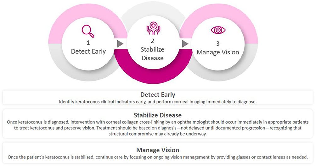 |
Figure 1 Interventional Keratoconus Care Pathway. |
Conclusion: A New Vision for Keratoconus Management
In this perspective based on emerging evidence and evolving clinical practice, we have synthesized evidence supporting a new conceptual framework for the comprehensive care of patients with keratoconus. It is our position that the development and clinical ability of corneal cross-linking to beneficially alter the natural history of keratoconus, thereby preserving both visual function and quality of life, mandates a retooling of ocular healthcare. This reorganization may involve the integration of centralized cornea specialists with community ophthalmologists and optometrists to develop an evidence-based pathway of detection, prevention, and treatment. Collaboration and communication, including the development and utilization of data transfer protocols, has the potential to facilitate care in the communities where patients live. Given that keratoconus often begins to affect young people of school or working age, such patient-centered pathways may minimize both the direct and indirect costs of care, resulting in savings for patients, healthcare systems, and society at large. Successful development and implementation of a community-based, patient-centered, interventional keratoconus care pathway will likely require expanded awareness and education regarding keratoconus and its treatment, as well as the creation of recommendations and guidelines for healthcare systems and providers. Future studies and real-world evidence will be valuable in confirming the long-term effectiveness and cost-efficiency of epithelium-on cross-linking within proposed care models.
Acknowledgments
The authors received no compensation for their work on this manuscript. The authors would like to thank Dr. Dana M. Hornbeak, MD, MPH (Glaukos Corporation) and Dr. Anthony Realini, MD (Hypotony Holdings LLC; funded by Glaukos Corporation) for medical writing and editing support.
Disclosure
Kenneth A. Beckman: Glaukos (consultant and contracted research); Oculus (consultant). Zeba A. Syed: Glaukos (consultant, research funding). Susan J. Gromacki: Bausch & Lomb SVP (consultant, speaker), Glaukos (consultant, speaker), J+J Vision Care (consultant, speaker), Scope (consultant), Tenpoint Therapeutics (consultant). The authors report no other conflicts of interest in this work.
References
1. Santodomingo-Rubido J, Carracedo G, Suzaki A, Villa-Collar C, Vincent SJ, Wolffsohn JS. Keratoconus: an updated review. Cont Lens Anterior Eye. 2022;45(3):101559. doi:10.1016/j.clae.2021.101559
2. Kandel H, Pesudovs K, Watson SL. Measurement of Quality of Life in Keratoconus. Cornea. 2020;39(3):386–9. doi:10.1097/ICO.0000000000002170
3. Naranjo A, Manche EE. A comprehensive review on corneal crosslinking. Taiwan J Ophthalmol. 2024;14(1):44–49. doi:10.4103/tjo.TJO-D-23-00055
4. American Academy of Ophthalmology. Corneal Ectasia Preferred Practice Pattern; 2023.
5. Epioxa HD and Epioxa: highlights of Presecribing Information. Available from: https://www.accessdata.fda.gov/drugsatfda_docs/label/2025/219910s000lbl.pdf.
6. Beckman KA, Parkhurst GD, Lee JH, et al. Randomized, Controlled Study to Evaluate the Safety and Efficacy of Oxygen-Enriched Epithelium-On Corneal Cross-Linking for the Treatment of Keratoconus. Ophthalmol Ther. 2026;15(4):1463–1484. doi:10.1007/s40123-026-01364-7
7. Nughays RO, Bazayd AS, Alshamekh LA, et al. Efficacy and Safety of Epi-On vs Epi-Off Corneal Cross-Linking in Corneal Ectasia: a Systematic Review and Meta-Analysis of Randomized Controlled Trials. Clin Ophthalmol. 2025;19:1531–1541. doi:10.2147/OPTH.S508618
8. D’Oria F, Palazon A, Alio JL. Corneal collagen cross-linking epithelium-on vs. epithelium-off: a systematic review and meta-analysis. Eye Vis. 2021;8(1):34. doi:10.1186/s40662-021-00256-0
9. Ng SM, Ren M, Lindsley KB, Hawkins BS, Kuo IC. Transepithelial versus epithelium-off corneal crosslinking for progressive keratoconus. Cochrane Database Syst Rev. 2021;3(3):CD013512. doi:10.1002/14651858.CD013512.pub2
10. Ng SM, Hawkins BS, Kuo IC. Transepithelial Versus Epithelium-Off Corneal Crosslinking for Progressive Keratoconus: findings From a Cochrane Systematic Review. Am J Ophthalmol. 2021;229:274–287. doi:10.1016/j.ajo.2021.05.009
11. Borchert GA, Kandel H, Watson SL. Epithelium-on versus epithelium-off corneal collagen crosslinking for keratoconus: a systematic review and meta-analysis. Graefes Arch Clin Exp Ophthalmol. 2024;262(6):1683–1692. doi:10.1007/s00417-023-06287-8
12. Jaruseviciene R, Tamuleviciute R, Galgauskas S. Corneal Cross-Linking in Keratoconus: comparative Analysis of Standard, Accelerated and Transepithelial Protocols. J Clin Med. 2026;15(2):490. doi:10.3390/jcm15020490
13. Alajmi TS, Alasmari FF, Alasmari TF, et al. Comparison of epithelium-off versus iontophoresis-assisted transepithelial corneal collagen cross-linking in keratoconus: a systematic review and meta-analysis. Int Ophthalmol. 2026;46(1):107. doi:10.1007/s10792-026-03977-0
14. Goh YW, Gokul A, Yadegarfar ME, et al. Prospective Clinical Study of Keratoconus Progression in Patients Awaiting Corneal Cross-linking. Cornea. 2020;39(10):1256–1260. doi:10.1097/ICO.0000000000002376
15. Romano V, Vinciguerra R, Arbabi EM, et al. Progression of Keratoconus in Patients While Awaiting Corneal Cross-linking: a Prospective Clinical Study. J Refract Surg. 2018;34(3):177–180. doi:10.3928/1081597X-20180104-01
16. Zadnik K, Barr JT, Edrington TB, et al. Baseline findings in the Collaborative Longitudinal Evaluation of Keratoconus (CLEK) Study. Invest Ophthalmol Visual Sci. 1998;39(13):2537–2546.
17. Angelo L, Gokul A, Samuels I, McGhee CN, Ziaei M. Patient evaluated economic impact of keratoconus in New Zealand. Clin Exp Optom. 2024;1–6. doi:10.1080/08164622.2024.2410026
18. Rapuano CJ, Lindstrom RL, Donnenfeld E, et al. Economics of corneal cross-linking for keratoconus treatment. J Med Economics. 2025;28(1):1696–1708. doi:10.1080/13696998.2025.2564576
19. Kass MA, Heuer DK, Higginbotham EJ, et al. The Ocular Hypertension Treatment Study: a randomized trial determines that topical ocular hypotensive medication delays or prevents the onset of primary open-angle glaucoma. Arch Ophthalmol. 2002;120(6):701–713. doi:10.1001/archopht.120.6.701
20. Stewart WC, Stewart JA, Nasser QJ, Mychaskiw MA. Cost-effectiveness of treating ocular hypertension. Ophthalmology. 2008;115(1):94–98. doi:10.1016/j.ophtha.2007.01.040
21. Gideon Abou Said A, Gispets J, Shneor E. Strategies for Early Keratoconus Diagnosis: a Narrative Review of Evaluating Affordable and Effective Detection Techniques. J Clin Med. 2025;14(2):460. doi:10.3390/jcm14020460
22. Ozalp O, Atalay E, Yildirim N. Prevalence and risk factors for keratoconus in a university-based population in Turkey. J Cataract Refract Surg. 2021;47(12):1524–1529. doi:10.1097/j.jcrs.0000000000000669
23. Almusawi LA, Hamied FM. Risk Factors for Development of Keratoconus: a Matched Pair Case-Control Study. Clin Ophthalmol. 2021;15:3473–3479. doi:10.2147/OPTH.S248724
24. Hashemi H, Heydarian S, Hooshmand E, et al. The Prevalence and Risk Factors for Keratoconus: a Systematic Review and Meta-Analysis. Cornea. 2020;39(2):263–270. doi:10.1097/ICO.0000000000002150
25. Al-Amri AM. Prevalence of Keratoconus in a Refractive Surgery Population. J Ophthalmol. 2018;2018:5983530. doi:10.1155/2018/5983530
26. Accardo PA, Pensiero S. Neural network-based system for early keratoconus detection from corneal topography. J Biomed Inform. 2002;35(3):151–159. doi:10.1016/s1532-0464(02)00513-0
27. Muhsin ZJ, Qahwaji R, Ghafir I, et al. Advances in machine learning for keratoconus diagnosis. Int Ophthalmol. 2025;45(1):128. doi:10.1007/s10792-025-03496-4
28. Ferreira-Mendes J, Lopes BT, Faria-Correia F, Salomao MQ, Rodrigues-Barros S, Ambrosio Jr R. Enhanced Ectasia Detection Using Corneal Tomography and Biomechanics. Am J Ophthalmol. 2019;197:7–16. doi:10.1016/j.ajo.2018.08.054
29. Tey KY, Cheong EZK, Ang M. Potential applications of artificial intelligence in image analysis in cornea diseases: a review. Eye Vis. 2024;11(1):10. doi:10.1186/s40662-024-00376-3
30. Duran-Cristiano SC, Bustamante-Arias A, Fernandez GJ, Martin-Gil A, Carracedo G. Omics in Keratoconus: from Molecular to Clinical Practice. J Clin Med. 2025;14(7):2459. doi:10.3390/jcm14072459
31. Perez-Rueda A, Castro-Luna G. A model of visual limitation in patients with keratoconus. Sci Rep. 2020;10(1):19335. doi:10.1038/s41598-020-76489-1
32. Castro-Luna G, Perez-Rueda A. A predictive model for early diagnosis of keratoconus. BMC Ophthalmol. 2020;20(1):263. doi:10.1186/s12886-020-01531-9
33. Raiskup F, Theuring A, Pillunat LE, Spoerl E. Corneal collagen crosslinking with riboflavin and ultraviolet-A light in progressive keratoconus: ten-year results. J Cataract Refract Surg. 2015;41(1):41–46. doi:10.1016/j.jcrs.2014.09.033
34. Hersh PS, Stulting RD, Muller D, Durrie DS, Rajpal RK, United States Crosslinking Study G. United States Multicenter Clinical Trial of Corneal Collagen Crosslinking for Keratoconus Treatment. Ophthalmology. 2017;124(9):1259–1270. doi:10.1016/j.ophtha.2017.03.052
35. Wu D, Lim DK, Lim BXH, et al. Corneal Cross-Linking: the Evolution of Treatment for Corneal Diseases. Front Pharmacol. 2021;12:686630. doi:10.3389/fphar.2021.686630
36. Borchert GA, Watson SL, Kandel H. Oxygen in Corneal Collagen Crosslinking to Treat Keratoconus: a Systematic Review and Meta-Analysis. Asia Pac J Ophthalmol (Phila). 2022;11(5):453–459. doi:10.1097/APO.0000000000000555
37. Aydin E, Aslan MG. The efficiency and safety of oxygen-supplemented accelerated transepithelial corneal cross-linking. Int Ophthalmol. 2021;41(9):2993–3005. doi:10.1007/s10792-021-01859-1
38. Faramarzi A, Hassanpour K, Rahmani B, Yazdani S, Kheiri B, Sadoughi MM. Systemic supplemental oxygen therapy during accelerated corneal crosslinking for progressive keratoconus: randomized clinical trial. J Cataract Refract Surg. 2021;47(6):773–779. doi:10.1097/j.jcrs.0000000000000513
39. Nishida T, Kojima T, Kataoka T, Isogai N, Yoshida Y, Nakamura T. Comparison of Corneal Biomechanical Properties and Corneal Tomography Between Customized and Accelerated Corneal Crosslinking in Eyes with Keratoconus. Cornea. 2021;40(7):851–858. doi:10.1097/ICO.0000000000002572
40. Seiler TG, Fischinger I, Koller T, Zapp D, Frueh BE, Seiler T. Customized Corneal Cross-linking: one-Year Results. Am J Ophthalmol. 2016;166:14–21. doi:10.1016/j.ajo.2016.02.029
41. Sachdev GS, Ramamurthy S, S B, Dandapani R. Comparative Analysis of Safety and Efficacy of Topography-Guided Customized Cross-linking and Standard Cross-linking in the Treatment of Progressive Keratoconus. Cornea. 2021;40(2):188–193. doi:10.1097/ICO.0000000000002492
42. Nau A, Nau C, Shorter E, Schornack M, Fogt J, Harthan J. Opportunities for Improving the Long-term Management of Keratoconus Patients. J Contact Lens Res Sci. 2024;8(1):e37–e46. doi:10.22374/jclrs.v8i1.61
43. Zhang H, Canto-Cerdan M, Felix-Espinar B, Alio Del Barrio JL. Efficacy of Customized Photorefractive Keratectomy With Cross-Linking Versus Cross-Linking Alone in Progressive Keratoconus: a Systematic Review and Meta-Analysis. Am J Ophthalmol. 2025;274:9–23. doi:10.1016/j.ajo.2025.02.038
44. Bui AD, Truong A, Pasricha ND, Indaram M. Keratoconus Diagnosis and Treatment: recent Advances and Future Directions. Clinical Ophthalmology (Auckland, NZ. 2023;17:2705–2718. doi:10.2147/OPTH.S392665
45. Muddana SK, Hauritz H, Burr M, Ambati B, Molokhia S. The effect of IVMED-80 eye drops on lysinonorleucine (LNL) amounts in vivo for treatment of keratoconus. Invest Ophthalmol Visual Sci. 2019;60:3359.
46. Muddana SKK, Ambati BK, Uehara H, Burr M, Molokhia S. Effect of IVMED-80 on human cadaver cornea crosslinking. Invest Ophthalmol Visual Sci. 2018;59:4392.
47. Molokhia S, Muddana SK, Hauritz H, et al. IVMED 80 eye drops for treatment of keratoconus in patients-Phase 1/2a. Invest Ophthalmol Visual Sci. 2020;61:2587.
48. Di Bella MA. Overview and Update on Extracellular Vesicles: considerations on Exosomes and Their Application in Modern Medicine. Biology. 2022;11(6):804. doi:10.3390/biology11060804
49. Zhang L, Yu D. Exosomes in cancer development, metastasis, and immunity. Biochim Biophys Acta Rev Cancer. 2019;1871(2):455–468. doi:10.1016/j.bbcan.2019.04.004
50. Hefley BS, Deighan C, Vasini B, et al. Revealing the presence of tear extracellular vesicles in Keratoconus. Exp Eye Res. 2022;224:109242. doi:10.1016/j.exer.2022.109242
51. Samaeekia R, Rabiee B, Putra I, et al. Effect of Human Corneal Mesenchymal Stromal Cell-derived Exosomes on Corneal Epithelial Wound Healing. Invest Ophthalmol Visual Sci. 2018;59(12):5194–5200. doi:10.1167/iovs.18-24803
52. Han KY, Tran JA, Chang JH, Azar DT, Zieske JD. Potential role of corneal epithelial cell-derived exosomes in corneal wound healing and neovascularization. Sci Rep. 2017;7(1):40548. doi:10.1038/srep40548
53. Hatef E, Wilson RF, Zhang A, et al. Effectiveness of telehealth versus in-person care during the COVID-19 pandemic: a systematic review. NPJ Digit Med. 2024;7(1):157. doi:10.1038/s41746-024-01152-2
54. Cao B, Chv V, Keenan JD. Telemedicine for Cornea and External Disease: a Scoping Review of Imaging Devices. Ophthalmol Ther. 2023;12(5):2281–2293. doi:10.1007/s40123-023-00764-3
55. Barequet D, Gutfreund S, Goldstein M, Loewenstein A, Gamzu R, Varssano D. Evaluation of a Telemedicine Model for Following Keratoconus Patients in the Era of COVID-19 Pandemic. Telemed J E Health. 2022;28(7):1023–1027. doi:10.1089/tmj.2021.0178
56. Akowuah PK, Kobia-Acquah E, Donkor R, Adjei-Anang J, Ankamah-Lomotey S. Keratoconus in Africa: a systematic review and meta-analysis. Ophthalmic Physiol Opt. 2021;41(4):736–747. doi:10.1111/opo.12825
57. Kyari F, Adekoya B, Abdull MM, Mohammed AS, Garba F. The Current Status of Glaucoma and Glaucoma Care in Sub-Saharan Africa. Asia Pac J Ophthalmol (Phila). 2018;7(6):375–386. doi:10.22608/APO.2018392
58. Gai MJ, Reddy V, Xu V, Noori NH, Demory Beckler M. Illuminating Perspectives: navigating Eye Care Access in Sub-Saharan Africa Through the Social Determinants of Health. Cureus. 2024;16(6):e61841. doi:10.7759/cureus.61841
59. Orugun AJ, Atima MO, Idakwo U, et al. Validation and optimization of smart eye camera as teleophthalmology device for the reduction of preventable and treatable blindness in Nigeria. Eye. 2025;39(5):925–930. doi:10.1038/s41433-024-03489-0
60. Junior Obinwanne C, Barrah S, Kobia-Acquah E, Titiati PE, Karikari LAA, Akowuah P. Referral Pattern and Comanagement of Patients With Keratoconus in West Africa: a Survey-Based Study of Optometrists in Ghana and Nigeria. Eye Contact Lens. 2025;51(2):70–75. doi:10.1097/ICL.0000000000001139
61. Singh RB, Parmar UPS, Jhanji V. Prevalence and Economic Burden of Keratoconus in the United States. Am J Ophthalmol. 2024;259:71–78. doi:10.1016/j.ajo.2023.11.009
62. Rebenitsch RL, Kymes SM, Walline JJ, Gordon MO. The lifetime economic burden of keratoconus: a decision analysis using a markov model. Am J Ophthalmol. 2011;151(5):768–773e2. doi:10.1016/j.ajo.2010.10.034
63. Malleron V, Bloch F, Zevering Y, et al. Evolution of corneal transplantation techniques and their indications in a French corneal transplant unit in 2000–2020. PLoS One. 2022;17(4):e0263686. doi:10.1371/journal.pone.0263686
64. Godefrooij DA, Gans R, Imhof SM, Wisse RP. Nationwide reduction in the number of corneal transplantations for keratoconus following the implementation of cross-linking. Acta Ophthalmol. 2016;94(7):675–678. doi:10.1111/aos.13095
65. Sandvik GF, Thorsrud A, Raen M, Ostern AE, Saethre M, Drolsum L. Does Corneal Collagen Cross-linking Reduce the Need for Keratoplasties in Patients With Keratoconus? Cornea. 2015;34(9):991–995. doi:10.1097/ICO.0000000000000460
66. Sarezky D, Orlin SE, Pan W, VanderBeek BL. Trends in Corneal Transplantation in Keratoconus. Cornea. 2017;36(2):131–137. doi:10.1097/ICO.0000000000001083
67. Takahashi A, Yamaguchi T, Tomida D, Nishisako S, Sasaki C, Shimazaki J. Trends in surgical procedures and indications for corneal transplantation over 27 years in a tertiary hospital in Japan. Jpn J Ophthalmol. 2021;65(5):608–615. doi:10.1007/s10384-021-00849-1
68. Chilibeck CM, Brookes NH, Gokul A, et al. Changing Trends in Corneal Transplantation in Aotearoa/New Zealand, 1991 to 2020: effects of Population Growth, Cataract Surgery, Endothelial Keratoplasty, and Corneal Cross-Linking for Keratoconus. Cornea. 2022;41(6):680–687. doi:10.1097/ICO.0000000000002812
69. Lindstrom RL, Berdahl JP, Donnenfeld ED, et al. Corneal cross-linking versus conventional management for keratoconus: a lifetime economic model. J Med Economics. 2021;24(1):410–420. doi:10.1080/13696998.2020.1851556
70. Leung VC, Pechlivanoglou P, Chew HF, Hatch W. Corneal Collagen Cross-Linking in the Management of Keratoconus in Canada: a Cost-Effectiveness Analysis. Ophthalmology. 2017;124(8):1108–1119. doi:10.1016/j.ophtha.2017.03.019
71. Salmon HA, Chalk D, Stein K, Frost NA. Cost effectiveness of collagen crosslinking for progressive keratoconus in the UK NHS. Eye. 2015;29(11):1504–1511. doi:10.1038/eye.2015.151
72. Godefrooij DA, Mangen MJ, Chan E, et al. Cost-Effectiveness Analysis of Corneal Collagen Crosslinking for Progressive Keratoconus. Ophthalmology. 2017;124(10):1485–1495. doi:10.1016/j.ophtha.2017.04.011
73. Hansen LO, Garcia R, Torricelli AAM, Bechara SJ. Cost-Effectiveness of Corneal Collagen Crosslinking for Progressive Keratoconus: a Brazilian Unified Health System Perspective. Int J Environ Res Public Health. 2024;21(12):1569. doi:10.3390/ijerph21121569
74. Chan E, Baird PN, Vogrin S, Sundararajan V, Daniell MD, Sahebjada S. Economic impact of keratoconus using a health expenditure questionnaire: a patient perspective. Clin Exp Ophthalmol. 2020;48(3):287–300. doi:10.1111/ceo.13704
75. Saunier V, Mercier AE, Gaboriau T, et al. Vision-related quality of life and dependency in French keratoconus patients: impact study. J Cataract Refract Surg. 2017;43(12):1582–1590. doi:10.1016/j.jcrs.2017.08.024
76. Kandel H, Chen JY, Sahebjada S, Chong EW, Wiffen S, Watson SL. Cross-Linking Improves the Quality of Life of People With Keratoconus: a Cross-Sectional and Longitudinal Study From the Save Sight Keratoconus Registry. Cornea. 2023;42(11):1377–1383. doi:10.1097/ICO.0000000000003185
77. Centers for Medicare and Medicaid Services. Value-Based Care. Available from: https://www.cms.gov/priorities/innovation/key-concepts/value-based-care.
78. Durakovic E, Kandel H, Watson SL. Mental Health Impact of Keratoconus: a Systematic Review. Cornea. 2023;42(9):1187–1197. doi:10.1097/ICO.0000000000003263
79. Labiris G, Giarmoukakis A, Sideroudi H, Gkika M, Fanariotis M, Kozobolis V. Impact of keratoconus, cross-linking and cross-linking combined with photorefractive keratectomy on self-reported quality of life. Cornea. 2012;31(7):734–739. doi:10.1097/ICO.0b013e31823cbe85
80. Ferrini E, Aleo D, Posarelli C, Figus M, Miccoli M, Gabbriellini G. Impact of corneal collagen cross-linking on vision-related quality of life measured with the keratoconus outcomes research questionnaire (KORQ) in patients with keratoconus. Cont Lens Anterior Eye. 2023;46(2):101746. doi:10.1016/j.clae.2022.101746
81. Steinberg J, Fischer P, Frings A, Druchkiv V, Katz T, Linke SJ. Quality of life in patients with progressive keratoconus treated with corneal collagen crosslinking. Int Ophthalmol. 2025;45(1):103. doi:10.1007/s10792-024-03400-6
82. Steinberg J, Bussmann N, Frings A, Katz T, Druchkiv V, Linke SJ. Quality of life in stable and progressive ‘early-stage’ keratoconus patients. Acta Ophthalmol. 2021;99(2):e196–e201. doi:10.1111/aos.14564
83. Funke CM, Risvedt D, Yadgarov A, Micheletti JM. Interventional glaucoma consensus treatment protocol. Expert Rev Ophthalmol. 2025;20(2):79–87. doi:10.1080/17469899.2025.2465330
84. Radcliffe N, Shah M, Samuelson TW. Challenging the “Topical Medications-First” Approach to Glaucoma: a Treatment Paradigm in Evolution. Ophthalmol Ther. 2023;12(6):2823–2839. doi:10.1007/s40123-023-00831-9
85. Bedrood S, Berdahl J, Sheybani A, Singh IP. Alternatives to Topical Glaucoma Medication for Glaucoma Management. Clinical Ophthalmology (Auckland, NZ. 2023;17:3899–3913. doi:10.2147/OPTH.S439457
86. Micheletti JM, Shultz M, Singh IP, Samuelson TW. An Emerging Multi-mechanism and Multi-modal Approach in Interventional Glaucoma Therapy. Ophthalmol Ther. 2025;14(1):13–22. doi:10.1007/s40123-024-01073-z
87. Micheletti MJ, Funke CM, Radcliffe NM, Hornbeak DM, Katz LJ. Early adoption of a novel initial treatment paradigm for open-angle glaucoma: an evidence-based model. Expert Rev Ophthalmol. 2025;20(6):369–375. doi:10.1080/17469899.2025.2594815
88. Realini T, Gazzard G. Selective Laser Trabeculoplasty and the Evolving Glaucoma Paradigm. Ophthalmol Glaucoma. Sep-Oct. 2025;8(5S):S38–S44. doi:10.1016/j.ogla.2025.06.010
 © 2026 The Author(s). This work is published and licensed by Dove Medical Press Limited. The
full terms of this license are available at https://www.dovepress.com/terms
and incorporate the Creative Commons Attribution
- Non Commercial (unported, 4.0) License.
By accessing the work you hereby accept the Terms. Non-commercial uses of the work are permitted
without any further permission from Dove Medical Press Limited, provided the work is properly
attributed. For permission for commercial use of this work, please see paragraphs 4.2 and 5 of our Terms.
© 2026 The Author(s). This work is published and licensed by Dove Medical Press Limited. The
full terms of this license are available at https://www.dovepress.com/terms
and incorporate the Creative Commons Attribution
- Non Commercial (unported, 4.0) License.
By accessing the work you hereby accept the Terms. Non-commercial uses of the work are permitted
without any further permission from Dove Medical Press Limited, provided the work is properly
attributed. For permission for commercial use of this work, please see paragraphs 4.2 and 5 of our Terms.
Recommended articles
Autism Spectrum Disorder Diagnoses: A Comparison of Countries with Different Income Levels
Matos MB, Bara TS, Cordeiro ML
Clinical Epidemiology 2022, 14:959-969
Published Date: 13 August 2022
Prenatal Diagnosis of Retinoblastomas: A Scoping Review
Rodriguez A, Kelley C, Patel A, Ramasubramanian A
International Journal of General Medicine 2023, 16:1101-1110
Published Date: 27 March 2023
A Post-International Gastrointestinal Cancers’ Conference (IGICC) Position Statements
Yalcin S, Lacin S, Kaseb AO, Peynircioğlu B, Cantasdemir M, Çil BE, Hurmuz P, Doğrul AB, Bozkurt MF, Abali H, Akhan O, Şimşek H, Sahin B, Aykan FN, Yücel İ, Tellioğlu G, Selçukbiricik F, Philip PA
Journal of Hepatocellular Carcinoma 2024, 11:953-974
Published Date: 29 May 2024
Reproducibility and Screening Capability of Corneal Epithelial Thickness Measurement for Keratoconus Using Anterior Segment Optical Coherence Tomography
Oshika T, Sawaki A, Nishida T, Nakamura T, Kojima T
Clinical Ophthalmology 2025, 19:2057-2065
Published Date: 30 June 2025
Interventional Keratoconus Management: The Critical Imperative for Early Detection and Treatment
Hatch KM, Donnenfeld E, Morgenstern AS, Ibach MJ
Clinical Ophthalmology 2026, 20:591113
Published Date: 18 May 2026