
Back to Journals » International Journal of Women's Health » Volume 17
Pregnancy-Triggered Hereditary Thrombotic Thrombocytopenic Purpura: A Case Report
Received 18 July 2025
Accepted for publication 16 October 2025
Published 25 October 2025 Volume 2025:17 Pages 3897—3906
DOI https://doi.org/10.2147/IJWH.S554825
Checked for plagiarism Yes
Review by Single anonymous peer review
Peer reviewer comments 2
Editor who approved publication: Dr Matteo Frigerio
Xin Xing,1 Na Yu,2 Zhiwei Zhang1
1Department of Obstetrics and Gynecology, the First Affiliated Hospital of Shandong First Medical University, Jinan, Shandong Province, 250014, People’s Republic of China; 2Reproductive Medicine Center, Key Laboratory of Maternal & Fetal Medicine of National Health Commission of China, Shandong Provincial Maternal and Child Health Care Hospital Affiliated to Qingdao University, Jinan, Shandong Province, 250014, People’s Republic of China
Correspondence: Zhiwei Zhang, Department of Obstetrics and Gynecology, the First Affiliated Hospital of Shandong First Medical University, No. 16766 Jingshi Road, Jinan, Shandong Province, 250014, People’s Republic of China, Email [email protected]
Background: Hereditary thrombotic thrombocytopenic purpura (hTTP), as an autosomal recessive hereditary disease, pregnancy is one of its recognized trigger factors. The lag in the diagnosis and treatment of this disease can lead to serious adverse pregnancy outcomes. In clinical practice, a high degree of vigilance should be maintained and timely treatment should be given to strive for better pregnancy outcomes.
Case Presentation: A 36-year-old woman has undergone 4 pregnancies over a period of 25 years. Due to the insufficient diagnosis capabilities at that time, it was not until the 3rd pregnancy when the patient again presented with severe thrombocytopenia in the second trimester that she was diagnosed with hTTP in our hospital. However, since the optimal treatment opportunity was missed, even though active plasma infusion was carried out, fetal death still occurred. In 2024, the patient got pregnant again, and this was her 4th pregnancy. During this pregnancy, the patient underwent preventive plasma infusion therapy throughout and no serious complications emerged. Ultimately, she gave birth to a full-term baby in February 2025.
Conclusion: We report a case of hTTP caused by complex compound heterozygous gene mutations. Currently, pregnancy is the only trigger for the patient’s illness. Timely identification, early differential diagnosis from obstetric complications, and active management are crucial for improving the pregnancy outcome of hTTP patients. We adopted a more conservative strategy of prophylactic plasma infusion during pregnancy and also achieved good maternal and fetal outcomes.
Keywords: hereditary thrombotic thrombocytopenic purpura, thrombotic microangiopathy, pregnancy, ADAMTS13, case report
Introduction
Thrombotic thrombocytopenic purpura (TTP), as a rare and highly fatal thrombotic microangiopathy (TMA), has an estimated incidence of about 13 to 19 cases per million people, with a male-to-female ratio of 2: 3. If left untreated, the mortality rate can be as high as 90%.1–3According to the cause of the disease, TTP can be divided into hereditary (caused by ADAMTS13 gene mutation) and acquired types. The latter can be further classified into idiopathic and secondary types based on the existence or absence of primary diseases. Pregnancy (including the postpartum stage) is a recognized trigger factor for the onset of acute (either primary or recurrent) thrombotic thrombocytopenic purpura.4 Its onset is urgent and progresses rapidly, and often misdiagnosed as obstetric disorders such as HELLP syndrome, and acute fatty liver of pregnancy.5 If the best treatment opportunity is missed, it will lead to an extremely high maternal and infant mortality rate.
Here we report a case of hereditary thrombotic thrombocytopenic purpura (hTTP). This patient has undergone a total of 4 pregnancies over a 25-year period (from 2010 to 2025). According to the patient, both of the previous two pregnancies presented severe symptoms such as thrombocytopenia and anemia in the second trimester. Due to the insufficient diagnosis capabilities at that time, it was not until the patient’s 3rd pregnancy, when severe thrombocytopenia recurred again in the 2nd trimester of pregnancy, that she was diagnosed with hTTP in our hospital. However, even though active plasma infusion therapy was carried out, fetal death still occurred eventually. After experiencing the above setbacks, the patient finally had a successful full-term delivery in 2025, fulfilling her wish.
Case Presentation
General Condition and Medical History
In February 2025, our team admitted a 36-year-old female with hTTP. The particularity of this patient lies in her tortuous reproductive history. The patient had symptoms of “severe thrombocytopenia and anemia” at about 20 weeks of gestation in her previous two pregnancies. (The patient described, but we failed to obtain detailed case records). During the 1st pregnancy in 2010, the situation of “fetal death” occurred. The previous two pregnancies were both terminated by cesarean section in the 2nd trimester, and the newborns did not survive. The diagnosis given by the local medical institution was “HELLP syndrome”. These two failed pregnancy experiences not only had adverse effects on the patient’s physical and psychological conditions but also led to the breakdown of her marriage. The patient remarried in 2020. In April 2022, the patient had her 3rd pregnancy. At 20 weeks of pregnancy, “severe thrombocytopenia and anemia” recurred, accompanied by symptoms such as headache, blurred vision, and transient loss of consciousness. After the patient was transferred to our hospital, a multidisciplinary consultation diagnosed it as “hTTP”. Through plasma infusion therapy, the patient’s thrombocytopenia and anemia have been notably improved. However, subsequent monitoring revealed that the fetus had severe fetal growth restriction (FGR) and abnormal umbilical artery blood flow. Eventually, the fetus died in utero at 26 weeks. The patient was then induced to deliver through the vagina. This pregnancy is the patient’s 4th pregnancy. She began to receive prophylactic plasma infusion therapy from 12 weeks of gestation and took 100mg of aspirin orally per day. During pregnancy, the platelet count remains relatively stable, and no complications such as preeclampsia(PE) and FGR emerged. This hospitalization was for a planned cesarean section to end the pregnancy. We will introduce the pregnancy experiences and outcomes of the 3rd and 4th pregnancies respectively. For the sake of distinction, they are numbered as “G3 and G4”. Since the patient’s 3rd pregnancy was confirmed, thus, in terms of laboratory tests and diagnoses, we will mainly expound on the relevant situation of the 3rd pregnancy (G3).
Signs
G3:The last menstrual period was on April 3, 2022. She was hospitalized on August 22nd, 2022 (20 + 1 weeks). Two days ago, the patient had experienced headache symptoms, on the day of the onset, the headache aggravated, accompanied by blurred vision and temporary loss of consciousness. The examination showed that body temperature was 36.7 °C, pulse rate was 80 beats/min, respiration rate was 20 times/min, blood pressure was 114/75mm Hg, the height was 161 cm, the body weight was 92.5 kg. The patient had anemic appearance, clear consciousness, an active position, slightly bulging abdomen, no tenderness or rebound pain, and no edema in both lower extremities. The fundus of the uterus was 2 fingers below the umbilicus, and the fetal heart rate was 152 beats/min.
G4: The last menstrual period was on May 28, 2024. She was hospitalized on February 13th, 2025 (37+2weeks). The examination showed that the body temperature was 36.3°C, the heart rate was 84 beats/min, the respiratory rate was 20 times/min, the blood pressure was 129/76 mmHg, and the body weight was 95 kg. The uterine fundal height was 33 cm, the abdominal circumference was 118 cm, the fetal heart rate was 145 beats/min, in the left occipital anterior (LOA) position, no uterine contraction and no tenderness at the incision site of the lower uterine segment.
Test Results
Laboratory Tests
G3: Two weeks before the onset, the blood routine examination indicated that the platelet count was 53×109/L, and no special treatment was given. After admission, the blood routine examination showed: platelet count 7×10⁹/L, hemoglobin 61g/L; urine routine: red blood cells (±), protein (3+); coagulation routine: PT 11.8S, APTT 29.7S, D-dimer 1.76mg/L; ALT 14.2u/L, AST 36.7u/L, lactate dehydrogenase (LDH) 975 u/L; renal creatinine was 42.8umol/L; coombs test (-); homocysteine 4.4umol/L; antinuclear antibody (-); anticardiolipin antibody (-); anti-β-2 glycoprotein I antibody (-); lupus anticoagulant (-); complement series test (-); thyroid function test (-).
Examinations Related to ADAMTS13
G3: ADAMTS13 activity was 4.8%; ADAMTS13 inhibitor test was negative; ADAMTS13 gene: Exon 10 c.1193G>A (p.R398H),hybrid mutation, mutation frequency 47.7%; Exon 28 c.3914G>T (p.G1305V),hybrid mutation, mutation frequency 49.0%.
Differential Diagnosis and Diagnosis
G3:The differential diagnoses to be considered are as follows: 1. HELLP syndrome usually occurs in the 3rd trimester of pregnancy. Patients typically have severe hypertension, as well as manifestations such as hemolysis, decreased platelet count, and abnormal liver enzymes. However, this patient has normal blood pressure and transaminase levels, and thus should not be diagnosed as this disease. 2. Acute fatty liver of pregnancy(AFLP) is characterized by abnormal liver function, abnormal coagulation function, obvious jaundice, and often accompanied by digestive tract symptoms. But this patient has normal coagulation function and transaminase levels, and no digestive tract symptoms such as upper abdominal pain, nausea, and vomiting. This diagnosis is excluded. 3. Idiopathic thrombocytopenia in pregnancy (ITP): It often occurs in the 2nd and 3rd trimesters of pregnancy. There is no history of thrombocytopenia before, but the platelet count is rarely < 50×109/L, without microangiopathic hemolysis or neuropsychiatric symptoms. This diagnosis is excluded. 4. Systemic lupus erythematosus(SLE): Some patients with systemic lupus erythematosus have multi-organ involvement, but systemic lupus erythematosus has characteristic signs such as butterfly rash, and microangiopathic hemolysis is not prominent. Autoantibodies such as antinuclear antibody, anti-dsDNA, and anti-Sm are positive, and complement C3 and C4 are decreased. This diagnosis is excluded. 5. Hemolytic uremic syndrome(HUS): Proteinuria and renal damage are prominent, and hypertension is obvious. This patient has only a slightly elevated creatinine and normal blood pressure. This diagnosis is excluded.
This patient presented with severe thrombocytopenia and hemolytic anemia at 20 weeks of pregnancy, accompanied by neurological symptoms. These signs and abnormal laboratory indicators strongly suggested the diagnosis of TTP. Finally, based on the ADAMTS13 test results, the ADAMTS13 activity was 4.8% (< 10%), the inhibitor was negative, and a heterozygous mutation was found in the genetic test, confirming the diagnosis of hTTP.
Treatment and Pregnancy Outcome
G3: The treatment of the patient was mainly divided into 3 stages. The first stage: The patient’s condition is special. Through comprehensive consultation and discussion among experts from the obstetrics department, hematology department and nephrology department, it is strongly suspected to be TTP. The next day, ADAMTS13 activity, inhibitor and genetic tests were completed, and 3 plasma exchanges (2500 mL of plasma per day) were immediately performed, and 2 units of red blood cells were transfused. The ADAMTS13 test results were obtained on the same day, and the diagnosis was hTTP. The treatment plan was revised to plasma infusion at 600 mL per day for a total of 3 days. On August 29, 2022, the platelet count was 198×109/L. The patient was discharged.
Second stage: On September 9, 2022, the platelet count was rechecked at the local hospital and it was 185×109/L. However, two days later (at 23 weeks of gestation on September 11, 2022), the patient came to our hospital for prenatal examination. The blood routine test showed a severe reduction in platelets (4×10⁹/L), and the patient was admitted to the hospital again. Continuous infusion of plasma for 3 days (600 mL each time), and the platelet count was rechecked on September 17, 2022, which was 160×109/L. On September 19, 2022 (24+1 weeks), the fetal ultrasound examination indicated that the estimated fetal weight(EFW) was below 10% of the fetuses of the same gestational age; the placenta was thick and limited, with decreased echo, and CDFI showed sparse blood flow signals in the placenta; the umbilical cord blood flow resistance index increased, and the end-diastolic blood flow disappeared intermittently in some segments. Low-molecular-weight heparin 4000u was added once daily by subcutaneous injection to improve the placental circulation. On September 27, 2022 (25+2 weeks), the fetal ultrasound suggested that the EFW was below 10% of the fetuses of the same gestational age, and the S/D value of the fetal umbilical cord blood flow increased to 4.27.
The third stage: On October 7, 2022 (26+5 weeks), the fetal ultrasound indicated that the EFW was below 10% of the fetuses of the same gestational age, the S/D value of the umbilical blood flow of the fetus increased to 4.55, and the incisures could be seen in the early diastolic phase of the uterine artery. The platelet count dropped to 53×109/L. Considering the re-decrease of platelets and FGR accompanied by abnormal umbilical blood flow, after admission, 600mL of plasma was infused again, and dexamethasone was injected intramuscularly. After the promotion of fetal lung maturity was completed, the pregnancy was terminated by cesarean section. In the early morning of October 8, 2022, it was found that the fetus died in the uterus. On the same day, cervical balloon induction was performed, and the stillborn fetus was delivered, with little bleeding observed. On October 9, 2022, the platelet count was 71×109/L, Plasma infusion treatment was given twice again (600mL each time). The patient was discharged from the hospital on the third day after delivery. On the day of discharge, the platelet count was 102×109/L. A total of 15 plasma infusions (including 2 times after delivery) were performed during this pregnancy, and the total plasma usage was 16,500mL (including 7,500mL of plasma exchange). The changes in platelet count and hemoglobin results are shown in Figure 1.
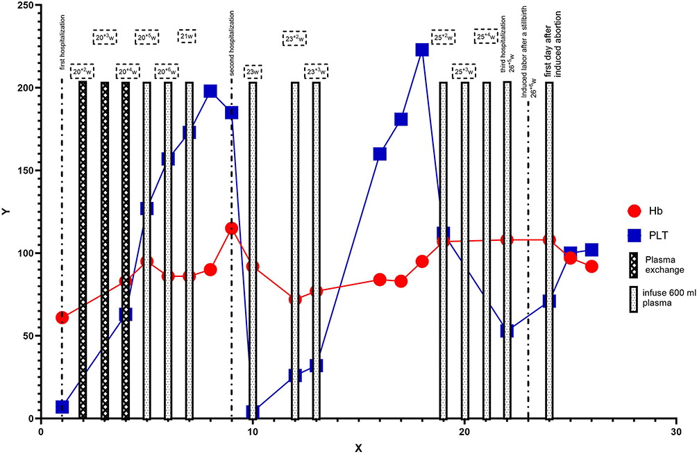 |
Figure 1 During the 3rd pregnancy treatment, the dynamic change curves of platelet count and hemoglobin concentration are as follows. On the X-axis, we listed the key treatment events. Among them, cross-striped bars indicate that plasma exchange was performed on that day, and dotted-striped bars indicate that plasma infusion was carried out on that day. The red line represents the hemoglobin concentration (g/L), and the blue line represents the platelet count (×109/L). |
G4: Before the patient’s 4th pregnancy, the platelet count of the patient was 235×109/L. Due to the cost of the test, the patient refused the ADAMTS13 activity test. In the early stage of pregnancy, 4000u of low-molecular-weight heparin was injected subcutaneously daily, and 100mg of aspirin was taken orally every day. On August 24, 2024 (12+4 weeks), the platelet count was 163×109/L, and we initiated the first preventive infusion (300 mL of fresh frozen plasma). Five days later, the platelet count was 198×109/L. The patient was instructed to stop the low-molecular-weight heparin treatment and continue to take aspirin orally every day until full term.
As the patient refused to test the ADAMTS13 activity level, we mainly determined the next monitoring date based on the platelet count. On October 14, 2024 (19+6 weeks), we monitored that the patient’s platelet count decreased significantly compared with before (118×109/L), and 300 mL of plasma was infused once. Five days later, the platelet count was 188×109/L. On November 4, 2024 (22+6 weeks), the platelets decreased again (110×109/L), and we infused 300 mL of plasma again. After that, even though there was no significant decrease in platelets, we still prophylactically infused 300 mL of plasma at 25+1 weeks,27+4 weeks,29+2 weeks and 31+2 weeks respectively. Even so, on January 14, 2025 (33 weeks), the patient’s platelet count suddenly dropped to 65×109/L. In the following 2 days, we infused 400 mL of plasma daily to stabilize the condition. We realized that the infusion frequency of every two weeks in the 3rd trimester of pregnancy could no longer meet the prevention requirements. Therefore, 400 mL of plasma was infused respectively at 34+6 weeks, 36 weeks, and 36+5 weeks. The patient underwent cesarean section to terminate the pregnancy at February 15th, 2025 (37+4 weeks). The bleeding during the operation was about 500 mL. The newborn’s birth weight was 2660g, and the Apgar score was 10 at both 1 minute and 5 minutes. The blood routine after birth showed that: the platelet count was 294×109/L and the hemoglobin was 168g/L.
Postoperatively, the patient’s platelet count decreased significantly. On the second postoperative day, it was 25×109/L. Then for the following 3 days, we continuously infused plasma (600 mL each time).The patient was discharged on the fifth day after the operation. At the time of discharge, the platelet count was 127×109/L. We conducted a follow-up, the platelet count of the patient was 216×109/L twenty days after the operation. A total of 15 plasma infusions were performed during this pregnancy (including 3 times after delivery), and the total plasma usage was 5500 mL. The changes in the patient’s platelet count and hemoglobin results are shown in Figure 2.
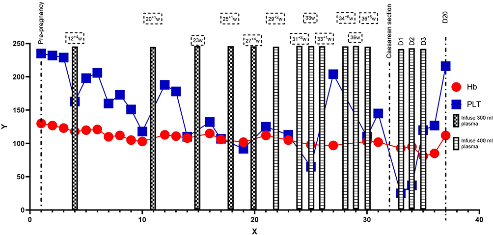 |
Figure 2 During the 4th pregnancy treatment stage, the dynamic change curves of platelet count and hemoglobin concentration are shown as follows. On the x-axis, we listed the key treatment events. Among them, the mosaic stripes indicate that a preventive plasma infusion of 300 mL was carried out on that day, and the horizontal stripes indicate that a preventive plasma infusion of 400 mL was conducted on that day. The red line represents the hemoglobin concentration (g/L), and the blue line represents the platelet count (×109/L). |
Pathological Examination
G3: In the placenta, from the chorionic plate to the basal plate, large areas of fresh and old infarction are observable, along with multifocal calcification. The vessels of the chorionic plate are significantly dilated, and some chorionic stem vessels are necrotic and occluded, with thrombosis present in the microvessels. The villi are overly mature, with an increase in syncytial nodules, and some villi are edematous, and avascular villi can be identified. The manifestations of decidual vascular disease include the absence and poor recasting of decidual vessels, along with fibrinoid necrosis of the vascular wall. See Figure 3.
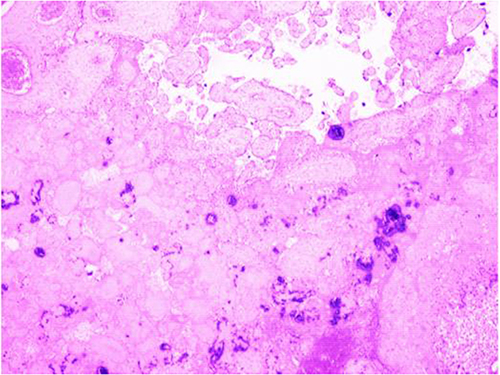 |
Figure 3 Histopathology of placenta (G3, HE×40). |
G4: The vascular of the chorionic plate was dilated and congested; multiple patchy infarctions were observable in the placenta, accompanied by focal calcification, and the villous space of some placenta was narrow, along with cellulose deposition; the syncytial nodules of some placental villi increased, the blood vessels of some villi proliferated and dilated, and the terminal villi of some were dysplastic; a few blood vessels in the decidua basalis plate were poorly recast. See Figure 4.
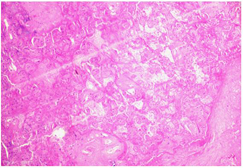 |
Figure 4 Histopathology of placenta (G4, HE×40). |
Discussion
Hereditary thrombotic thrombocytopenic purpura (hTTP) also known as Upshaw-Schulman syndrome (USS), is an autosomal recessive disorder caused by mutations in the ADAMTS13 gene located in the 9q34 region of chromosome 9, which contains 29 exons.6 According to the NCBI ClinVar database, more than 260 ADAMTS13 gene mutation sites have been discovered so far, covering various types such as mutations, substitutions, translocations, deletions, and insertions. Among them, missense mutations (55%) are the most common type of mutation, followed by frameshift mutations (28%). Such gene mutations can lead to different degrees of synthesis disorders of ADAMTS13, or a decrease in activity or an increase in degradation mediated by non-coding splicing of mRNA.7 It is well known that the main role of ADAMTS13 is to cleave von Willebrand factor (vWF) on the surface of vascular endothelial cells in the blood circulation and at the site of vascular injury.8 However, once the above-mentioned synthesis disorders or decreased activity occur, the ability of vascular endothelial cells to clear vWF will be weakened, thereby generating abnormally large vWF. Subsequently, abnormal coagulation cascade reactions, microvascular thrombosis, and hemolysis will occur one after another.9
There are two phenotypes of this disorder, namely early-onset and late-onset. The late-onset type occurs in adulthood, and for the majority of female patients, the first onset is during pregnancy. This different clinical manifestation depends on different mutation sites and mutation frequencies. In our case report, the patient’s ADAMTS13 gene has compound heterozygous nucleotide variations: c.1193G>A (p.R398H) in exon 10, with a mutation frequency of 47.7%, which is a missense variation and was inherited from the patient’s mother, According to the ACMG guidelines, this mutation site is likely pathogenic. The Polyphen2 and SIFT databases predict that it has a harmful effect on protein function. Both the CADD and GERP databases show that this site is evolutionarily conserved and has potential functional implications. The HGMD database records this mutation as a pathogenic mutation, which has been observed in cases of familial thrombotic thrombocytopenic purpura (PMID: 11586351); c.3914G>T (p.G1305V) in exon 28, with a mutation frequency of 49%. This missense mutation is a de novo mutation or was inherited from the patient’s father (as the patient’s father died early and could not be further confirmed).According to the ACMG guidelines, this mutation site is likely pathogenic. The Polyphen2 and SIFT databases predict that it has a harmful effect on protein function. Both the CADD and GERP databases show that this site is evolutionarily conserved and has potential functional implications. Forms a compound heterozygote with the p.R398H mutation. In fact, this patient only experiences the illness during pregnancy, and after terminating the pregnancy, the platelet count can return to normal within a short period of time. At present, it seems that pregnancy is the only factor that triggers this disease, and this phenomenon is worthy of in-depth discussion.
Several studies have focused on pregnancy as an trigger factor of hTTP. Among the patients registered in the hTTP registry in the United Kingdom, 42% of the patients had their first onset or aggravation of the disease during pregnancy;10 A 21-year cohort study conducted in France showed that, nearly 27% of female hTTP patients had their first onset during pregnancy, and 65% of the patients had the onset in the late pregnancy and postpartum period11. The reasons for this situation are undoubtedly complex. Apart from factors such as hormones, complement activation, and immune responses, the most crucial reason lies in that the balance of the interaction between ADAMTS13 and vWF during pregnancy is more likely to be disrupted. Studies have shown that the content of vWF gradually increases with the increase of gestational weeks. This high level of vWF plays an important role in the pathogenesis of TTP.12
In the case report we submitted, the patient was not diagnosed until her 3rd pregnancy, which indicates that making a timely and definite diagnosis in clinical practice remains a challenge. Some clinical features of TTP during pregnancy are difficult to distinguish from more common TMA, such as HELLP syndrome or HUS,13 thus complicating the clinical diagnosis. In addition, many hospitals do not list the detection of ADAMTS13 as a routine item, and the detection period is long. In the absence of the detection result of ADAMTS13, medical staff need to start from the clinical situation (onset time, symptoms and signs) and basic laboratory tests, and conduct detailed evaluation and comparison to achieve a comprehensive differential diagnosis. Just as stated in our case report,although the test result of ADAMTS13 activity has not been obtained for the moment, based on the patient’s medical history, clinical manifestations and laboratory indicators, it is basically possible to diagnose the patient as TTP. During the waiting for the ADAMTS13 test result, we did not delay the treatment but urgently carried out plasma exchange. The patient’s condition then tended to be stable, winning time for the subsequent treatment.
Besides the discoveries obtained in the trigger factors and the diagnosis process, during the treatment progress of the two pregnancies, we also had some interesting findings.
1.This patient experienced fetal death during two pregnancies (the 1st and the 3rd). Meanwhile, in the placental pathologies of the 3rd and 4th pregnancies, we found significant differences (the descriptions of villous vascular infarction and decidual vascular lesions in the placental pathological report of the 3rd pregnancy were more serious). We infer that the earlier the onset time, the more severe the placental function is impaired. Thus, the possibility of secondary PE, FGR and even fetal death is greater. Therefore, the fetal outcome is largely affected by the gestational age at which the disease occurs. A cohort study in France11 also observed this phenomenon: the smaller the gestational age at the onset of the disease, the higher the probability of fetal death. The fetal mortality rate of patients with the disease occurring in the late stage of pregnancy is only 5%. Therefore, if TTP occurs during pregnancy, in addition to considering the condition of the mother and the fetus, whether the pregnancy can continue should also be determined based on the gestational age. For some patients with severe attacks in the early stages of pregnancy, it may be more appropriate to perform early artificial abortion.
2.The treatment of hTTP is relatively simple. The therapeutic goal is to restore the minimum ADAMTS13 activity required in the body. At present,therapeutic drugs are mainly divided into three categories, including plasma infusion, plasma-derived factor VIII concentrates, and recombinant ADAMTS13.14 Plasma transfusion can effectively replenish ADAMTS13, which can both prevent the onset of the disease during pregnancy and treat acute onset. According to relevant literature reports, the recurrence risk of re-pregnancy in patients with hTTP who did not receive preventive treatment is as high as 100%.15 In our report, the patient did not receive preventive treatment in her previous three pregnancies and all developed the disease at about 20 weeks. However, during the 4th pregnancy, we carried out a preventive plasma infusion, and the patient did not develop the disease.
3.Compared with the preventive treatment we carried out during the 4th pregnancy, maintaining the stability of the platelet count during the 3rd pregnancy was more difficult (larger doses and more frequent plasma infusions are needed), we also observed that the patient’s platelet count dropped sharply from 185×109/L to 4×10⁹/L within just two days, which was completely unexpected. The above situation reveals two key issues: First, compared with the treatment after an acute attack, prophylactic plasma infusion may bring better maternal and fetal outcomes. Second, platelet count cannot fully reflect the activity of ADAMTS13 in the body. If conditions permit, dynamically monitoring the activity of ADAMTS13 to guide the timing of plasma infusion may achieve more satisfactory results.
4.At present, the optimal infusion protocol for prophylactic treatment remains unclear, and the main reasons are twofold. Firstly, the level of ADAMTS13 activity required to prevent acute attacks has not been defined. Secondly, for patients receiving regular plasma infusions, the half-life of ADAMTS13 is variable, which depends on body weight, metabolism, and the severity of the disease.6 Based on their own clinical practice, Zununi et al3 proposed that prophylactic treatment should be initiated at 10 weeks of gestation or immediately after pregnancy confirmation, with 10–15 mL/kg of plasma infused every 2 to 3 weeks. Alwan et al10 believed that plasma infusion once every 3 weeks was ineffective for 70% of patients, and these patients needed to receive infusions once a week or once every 2 weeks to benefit.Based on the above experience, for a patient weighing 95 kg, the preventive plasma infusion dose should have been set at 950–1425 mL. However, during the treatment of the 3rd pregnancy, we found that an infusion of 600 mL of plasma each time was sufficient to effectively increase the patient’s platelet count. Therefore, when formulating the preventive infusion plan for the 4th pregnancy, we were more conservative: in the early and middle stages of pregnancy, we found that an infusion of 300 mL every 2 to 3 weeks was sufficient to stabilize the platelet level. However, in the late stage of pregnancy, this infusion dose and frequency were clearly insufficient, and the only severe drop in platelet count during the pregnancy occurred during this period (from 30+6 weeks to 33 weeks: 113×109/L → 65×109/L). After that, we adjusted the infusion plan to 400 mL once a week, and the patient’s platelet level did not show a significant decrease again. Compared with previous literature reports, our plan (3–5 mL/kg of plasma infused every 1 to 3 weeks) not only maintained the therapeutic effect but also significantly reduced the plasma usage.
5.The patient’s 3rd pregnancy was complicated by severe FGR, resulting in a fetal death. Subsequent placental pathology revealed severe microthrombosis and decidual vascular lesions, which are similar to the pathological abnormalities of PE. Relevant research reports also point out that the risk of PE in pregnant women with a history of acquired thrombotic thrombocytopenic purpura (aTTP) is as high as 31%, while this proportion is only 3% in the general population.16 Therefore, in the early stage of the 4th pregnancy, we used low-molecular-weight heparin and guided the patient to take oral aspirin throughout the pregnancy to reduce the risk of thrombosis and infarction in the placental vessels. After receiving these treatments, the patient did not experience any serious complications such as PE and FGR during pregnancy. Some scholars oppose the use of aspirin,they believe that aspirin will not be routinely used unless there are specific obstetric indications. The reason is that the pathogenesis of thrombosis in acute TTP is not dependent on cyclooxygenase-mediated platelet aggregation, but is caused by excessive platelet aggregation mediated by very large molecular weight vWF.2
Limitations
It should be noted that the above series of findings are based on only a single case, and their general applicability has certain limitations. Looking ahead, regarding the management of such patients, we will further verify the proposed speculations and strive to fill the gaps in the observational data related to ADAMTS13 activity monitoring.
Conclusion
Reported a case of pregnancy-triggered hereditary thrombotic thrombocytopenic purpura (hTTP) caused by compound heterozygous mutations in ADAMTS13. This case highlights that pregnancy can act as the only clinical trigger in certain women with hTTP. Obstetricians and hematologists should maintain a high index of suspicion for hTTP in pregnant women presenting with unexplained thrombocytopenia and hemolytic anemia, especially when routine conditions like HELLP or ITP are excluded.During pregnancy, once the diagnosis is confirmed, plasma should be infused as early as possible to supplement the active ADAMTS13. Compared with the treatment after an acute attack, prophylactic plasma infusion may bring better maternal and fetal outcomes.
Ethical Approval
Not applicable. Institutional Review Board (IRB) approval is not applicable for a case report, but the manuscript does not violate patient confidentiality as all information/figures are de-identified.
Consent for Publication
The patient described in this case report has provided written consent for its publication.
Acknowledgments
We are deeply indebted to all the medical staff engaged in the treatment and management of this patient. Precisely because of your endeavors and dedication, a perfect pregnancy outcome has been achieved.
Author Contributions
All authors made a significant contribution to the work reported, whether that is in the conception, study design, execution, acquisition of data, analysis and interpretation, or in all these areas; took part in drafting, revising or critically reviewing the article; gave final approval of the version to be published; have agreed on the journal to which the article has been submitted; and agree to be accountable for all aspects of the work.
Funding
The authors declare that they received no funding from any institutions.
Disclosure
The authors declare that they have no competing interests in this work.
References
1. Page EE, Kremer Hovinga JA, Terrell DR, Vesely SK, George JN. Thrombotic thrombocytopenic purpura: diagnostic criteria, clinical features, and long-term outcomes from 1995 through 2015. Blood Adv. 2017;1(10):590–600. doi:10.1182/bloodadvances.2017005124
2. Fogerty AE, Kuter DJ. How I treat thrombocytopenia in pregnancy. Blood. 2024;143(9):747–756. doi:10.1182/blood.2023020726
3. Zununi Vahed S, Rahbar Saadat Y, Ardalan M. Thrombotic microangiopathy during pregnancy. Microvasc Res. 2021;138:104226. doi:10.1016/j.mvr.2021.104226
4. Joly BS, Coppo P, Veyradier A. An update on pathogenesis and diagnosis of thrombotic thrombocytopenic purpura. Expert Rev Hematol. 2019;12(6):383–395. doi:10.1080/17474086.2019.1611423
5. Stanley M, Killeen RB, Michalski JM. Thrombotic thrombocytopenic purpura. In: StatPearls. Treasure Island (FL):StatPearls Publishing;2023.
6. Nusrat S, Beg K, Khan O, Sinha A, George J. Hereditary thrombotic thrombocytopenic purpura. Genes. 2023;14(10):1956. doi:10.3390/genes14101956
7. Li P, Jiang J, Xi Q, Yang Z. An ADAMTS13 mutation that causes hereditary thrombotic thrombocytopenic purpura: a case report and literature review. BMC Med Genomics. 2021;14(1):252. doi:10.1186/s12920-021-01099-3
8. Yagi H, Yamaguchi N, Shida Y, et al. Highly elevated plasma level of von Willebrand factor accelerates the formation of platelet thrombus under high shear stress in plasma with deficient ADAMTS13 activity. Thromb Res. 2017;159:91–95. doi:10.1016/j.thromres.2017.10.007
9. Zheng XL. ADAMTS13 and von Willebrand factor in thrombotic thrombocytopenic purpura. Annu Rev Med. 2015;66(1):211–225. doi:10.1146/annurev-med-061813-013241
10. Alwan F, Vendramin C, Liesner R, et al. Characterization and treatment of congenital thrombotic thrombocytopenic purpura. Blood. 2019;133(15):1644–1651. doi:10.1182/blood-2018-11-884700
11. Béranger N, Coppo P, Tsatsaris V, et al. Management and follow-up of pregnancy-onset thrombotic thrombocytopenic purpura: the French experience. Blood Adv. 2024;8(1):183–193. doi:10.1182/bloodadvances.2023011972
12. Zheng L, Zhang D, Cao W, Song WC, Zheng XL. Synergistic effects of ADAMTS13 deficiency and complement activation in pathogenesis of thrombotic microangiopathy. Blood. 2019;134(13):1095–1105. doi:10.1182/blood.2019001040
13. Dap M, Romiti J, Dolenc B, Morel O. Thrombotic thrombocytopenic purpura and severe preeclampsia: a clinical overlap during pregnancy and a possible coexistence. J Gynecol Obstet Hum Reprod. 2022;51(7):102422. doi:10.1016/j.jogoh.2022.102422
14. Scully M, Rayment R, Clark A, et al. A British society for haematology guideline: diagnosis and management of thrombotic thrombocytopenic purpura and thrombotic microangiopathies. Br J Haematol. 2023;203(4):546–563. doi:10.1111/bjh.19026
15. Zheng XL, Vesely SK, Cataland SR, et al. ISTH guidelines for the diagnosis of thrombotic thrombocytopenic purpura. J Thromb Haemost. 2020;18(10):2486–2495. doi:10.1111/jth.15006
16. Vesely SK. Life after acquired thrombotic thrombocytopenic purpura: morbidity, mortality, and risks during pregnancy. J Thromb Haemost. 2015;13 Suppl 1:S216-S222. doi:10.1111/jth.12953
 © 2025 The Author(s). This work is published and licensed by Dove Medical Press Limited. The
full terms of this license are available at https://www.dovepress.com/terms
and incorporate the Creative Commons Attribution
- Non Commercial (unported, 4.0) License.
By accessing the work you hereby accept the Terms. Non-commercial uses of the work are permitted
without any further permission from Dove Medical Press Limited, provided the work is properly
attributed. For permission for commercial use of this work, please see paragraphs 4.2 and 5 of our Terms.
© 2025 The Author(s). This work is published and licensed by Dove Medical Press Limited. The
full terms of this license are available at https://www.dovepress.com/terms
and incorporate the Creative Commons Attribution
- Non Commercial (unported, 4.0) License.
By accessing the work you hereby accept the Terms. Non-commercial uses of the work are permitted
without any further permission from Dove Medical Press Limited, provided the work is properly
attributed. For permission for commercial use of this work, please see paragraphs 4.2 and 5 of our Terms.
Recommended articles

Unilateral Twin Ectopic Pregnancy: A Case Report from the Eastern Part of Ethiopia, Harar
Gure Eticha T
International Medical Case Reports Journal 2022, 15:521-527
Published Date: 19 September 2022
Pregnancy Associated Invasive Apocrine Carcinoma of the Breast: Case Report from Ethiopia
Alemu HK, Hammad N, Tola MA, Vanderpuye V
Breast Cancer: Targets and Therapy 2023, 15:429-433
Published Date: 23 June 2023
A Case Report of Pregnancy Complicated with Primary Hemophagocytic Lymphohistiocytosis
Chen Y, Huang X, Chen H, Tong J, Huang L, Su J, Deng L
International Journal of Women's Health 2024, 16:843-851
Published Date: 17 May 2024
Diagnosis and Management Challenges of Spontaneous Quadruplet Pregnancy in a Resource-Limited Setting: A Case Report
Tjandraprawira KD, Santoso DPJ, Bayuaji H, Nugrahani AD, Susandi D
International Medical Case Reports Journal 2025, 18:325-332
Published Date: 11 March 2025


Severe Mitral Stenosis in Term Pregnancy Presenting During Labor in a Resource-Limited Setting: A Case Report from Borama, Awdal Region of Somalia


Jama AM, Osman AA, Deria HM, Mousa MM, Jama AM, Ali AO
International Medical Case Reports Journal 2026, 19:564292
Published Date: 26 February 2026