
Back to Journals » Journal of Pain Research » Volume 19
Pharmacological Pain Management in Adults with Venous Ulceration: A Systematic Review
Authors Saghdaoui LB  , Lampridou S , Baldo L, Onida S, Hohenschurz-Schmidt D, Chumbley GM, Atkin L
Received 13 December 2025
Accepted for publication 25 March 2026
Published 1 April 2026 Volume 2026:19 588661
DOI https://doi.org/10.2147/JPR.S588661
Checked for plagiarism Yes
Review by Single anonymous peer review
Peer reviewer comments 2
Editor who approved publication: Professor Robert B. Raffa
Layla Bolton Saghdaoui,1 Smaragda Lampridou,1 Layla Baldo,1 Sarah Onida,1 David Hohenschurz-Schmidt,2 Gillian M Chumbley,3 Leanne Atkin4
1Section of Vascular Surgery - Department of Surgery and Cancer, Imperial College London / Imperial College Healthcare NHS Trust, London, UK; 2Imperial Clinical Trials Unit, School of Public Health, Faculty of Medicine, Imperial College London, London, UK; 3Pain Service, Department of Surgery and Cancer, Imperial College Healthcare NHS Trust, London, UK; 4School of Human and Health Sciences, University of Huddersfield and Mid Yorkshire NHS Teaching Trust, Huddersfield, West Yorkshire, UK
Correspondence: Layla Bolton Saghdaoui, Email [email protected]
Introduction: Venous ulceration (VU) is a chronic lower limb wound caused by sustained venous hypertension. Treatment includes compression therapy, wound care and surgical intervention. Due to the prolonged nature of wound healing, patients experience a substantial burden on quality of life, largely influenced by pain. Existing literature generalises findings across all chronic wounds and there are no published reviews or specific guidelines informing pharmacological pain management for patients with VU.
Methods: Five databases were searched to identify studies evaluating the effectiveness of pharmacological interventions for VU pain, published in English between 2000 and 2024. The CASP appraisal tools were used to assess the quality of publications, and findings are reporting descriptively and following PRISMA guidance.
Results: Initial literature searches yielded 1,161 references, of which 447 were duplicates. Once screening was complete, thirteen studies were included in this evaluation. The majority (10/13) were randomised controlled trials comparing pharmacological interventions with standard care. Small sample sizes and poor reporting quality were common across all papers, and overall limits the evidence. Eight studies evaluated ibuprofen-impregnated dressings, all reporting some degree of pain reduction. Three studies investigated localized oxygen therapy, demonstrating improved wound healing outcomes. However, pain reduction was not statistically significant and may be secondary to healing. The remaining two studies examined topical sevoflurane and aspirin. Sevoflurane showed pain reduction over time in a single, low-quality retrospective study. The aspirin trial was inconclusive due to poor recruitment and underpowering.
Conclusion: Currently, there is a lack of high-quality studies, evaluating the pharmacological treatment of pain in people experiencing VU. While there may be some evidence to support the use of topical agents such as ibuprofen, further industry-independent efficacy studies and cost analyses are required. Further research is also required to compare standard of care to commonly used agents such as oral paracetamol.
Keywords: venous ulceration, pain, pain management, pharmacological pain management
Introduction
Venous ulceration (VU) develops as a consequence of prolonged and untreated lower limb venous hypertension.1 While primary treatment includes regular wound care, compression therapy and surgical intervention to address superficial venous incompetence, due to its chronic nature and sustained pain, it is crucial that such treatment is supplemented with appropriate pain management.2
Living with a lower limb wound impacts all aspects of a patient’s life, such as their ability to work, socialise, and carry out daily living activities.3 When left unmanaged, pain adds an additional level of complexity to the already substantial burden on quality of life.4 Qualitative exploration of patient experiences paints a bleak picture, illustrating patients’ daily frustration, emotional exhaustion and social isolation. It also highlights the importance and impact of the role of healthcare professionals when it comes to assessment and treatment. Unfortunately, pain is often found to be inadequately addressed.5
As VU healing can often take many months/years, international guidelines published by Wounds UK, the National Institute for Health and Care Excellence (NICE) and the Royal Australian College of General Practitioners (RACGP) outline the importance of ongoing pain assessment, the use of simple analgesics, nonsteroidal anti-inflammatory drugs and the treatment of neuropathic pain were necessary.2,6,7 However, the guideline sections outlining treatment for VU pain are informed by general strategies for pain management, and are not specific to VU.8 This is largely due to the variety of ways VU pain can present, such as nociceptive pain (associated with the damaged tissue),9,10 neuropathic pain (caused by damaged nerve endings)11 and nociplastic pain (chronic pain arising from altered nociceptive signalling and pain perception).12
While guidance provided by the American Venous Forum13 acknowledges the impact of pain, it does not outline specific pharmacological recommendations. Alternatively, they focus on wound healing, the treatment of venous insufficiency and patient lifestyle behaviours, such as limb elevation and physical activity. These recommendations do include considering the use of drug therapy such as pentoxifylline and micronised purified flavonoid fraction; however, the aim is to improve venous return rather than address pain.
Despite qualitative research suggesting that pharmacological treatment is often the first option in practice when addressing pain,5 the most recent large-scale review evaluating pain management predominantly focused on the following: pain assessment, psychological strategies to address pain, the role of exercise and complementary or alternative medicinal approaches.14 A review looking specifically at the clinical management of pain explores topical interventions for treating pain in chronic wounds. However, this concludes that while topical methods may be helpful, the research evaluated should be interpreted with caution due to the quality, of evidence and called for further adequately powered studies.15
Two additional literature reviews contribute to this body of published evidence; however, existing research largely combines different types of chronic wounds (eg. venous ulceration, pressure ulcers, and diabetic foot wounds) together in their evaluations making it difficult to identify population-specific results.16,17 This is significant, as the underlying cause of a wound can have a substantial impact on the type of pain experienced and the appropriateness of treatment modalities.18 Beyond this, typical characteristics of the VU population add an additional layer of complexity. Patients are often elderly and comorbid, which increases their susceptibility to complications related to polypharmacy and adverse drug reactions.19,20 Consequently, treatments that may be appropriate for other types of chronic wounds may not be suitable for individuals with VU.
While there is one publication that focuses specifically on venous ulceration, this explores the prevalence and characteristics of wound-related background pain and factors associated with pain intensity. It did not specifically evaluate methods to manage or alleviate pain.18
It is evident that wound healing and the prevention of recurrence should be the main goal of overall VU management.21 While successful ulcer healing will usually also terminate pain, addressing pain throughout the healing process to support patients’ quality-of-life should be acknowledged as a basic human right.14,22 While previous reviews have explored pharmacological pain management in chronic wounds and include data from patients with venous ulceration, a synthesis focused specifically on this population is lacking. This review, therefore, aims to identify, describe and appraise the existing evidence to inform pain management in adults experiencing VU, providing a clearer understanding of the evidence applicable to this population.
Material and Methods
Objectives
- Identifying medications commonly employed for pain management in patients with venous ulceration.
- Evaluate the effectiveness and risks of pharmacological interventions [eg non-steroidal anti-inflammatory drugs (NSAIDs), anticonvulsants or opioids] prescribed for patients with venous ulceration.
This review is reported in line with the Preferred Reporting Items for Systematic Reviews and Meta-Analyses (PRISMA) checklist23 and was registered on the international database of prospectively registered systematic reviews (CRD420251026023).24
Search Strategy and Study Selection
A systematic search of the literature was conducted using the following databases: Cinahl (via Ebsco), Medline (via Ovid), Embase (via Ovid), British Nursing Index, Cochrane Central Register Controlled Trials, Clinical Trials.gov. Additionally, a search of the grey literature was conducted using OpenGrey (www.opengrey.eu). Reference lists of included studies were also reviewed for further relevant articles.
Searches included words and phrases that would retrieve studies focusing on lower limb wounds caused by venous insufficiency and pain management (see Supplementary File 1 and Appendix 1 for all search strings). Study selection was conducted by four authors, LBS, SL, DHS and LB, using the online platform Covidence (https://www.covidence.org). This enabled removal of duplicates, and independent first-level and full-text screening against eligibility criteria. To start, titles and abstracts of all results were reviewed in line with the inclusion and exclusion criteria. Similarly, full-text articles were independently reviewed by LBS, SL or DHS. Any discrepancies between reviewers were resolved by a third author.
Inclusion and Exclusion Criteria
Design and Language
To gain a comprehensive understanding of the up-to-date available evidence, the review included primary research published between the years 2000 and 2024. The following study designs were included: randomised controlled trials (RCTs), quasi-experimental studies and observational studies such as cross-sectional studies, cohort studies, case-control studies, mixed-methods or qualitative studies.
If a study had a published protocol providing additional methodological information, this was also reviewed. Studies published as abstracts only, conference abstracts, reviews (systematic, narrative, scoping), opinion articles and commentaries on trials were excluded. However, if abstracts were felt to be particularly relevant, authors were contacted to ascertain if the full study had been published (this did not result in any additional papers). Only studies published in English were included due to the limitations on costs and resources available for translation.
Population
Studies were included if they examined the pharmacological treatment of pain in patients (>18 years) diagnosed with venous ulceration of any chronicity, gender, ethnicity or severity or chronicity of ulceration. Chronic venous ulceration was defined as a wound on the lower limb that has developed as a result of venous insufficiency. Studies evaluating mixed populations with wounds multiple causes (eg. arterial, diabetic or pressure ulcers) were excluded unless data from patients with venous ulceration could be isolated.
Types of Intervention
Studies were included if they examined pharmacological interventions delivered via any administrative route with the intention of addressing pain caused by the presence of chronic venous ulceration. This included interventions delivered in any type of healthcare setting (primary care – general/family practice or secondary care – hospital/specialist service). Examples of such interventions include medication, such as non-opioid drugs (eg. paracetamol and non-steroidal anti-inflammatory drugs), anticonvulsants (eg. gabapentin/pregabalin), and tricyclic antidepressants (eg. nortriptyline/amitriptyline/duloxetine), or opioids (eg. codeine/morphine).
Comparators
No limits were applied to the type of comparators used to evaluate an intervention. Any other pain management option was included, as were placebo controls and no intervention/exposure comparators.
Intervention Outcomes
The primary outcome for this review was improvement in pain. Studies were included if they evaluated this using any of the following outcomes: Any assessments of pain severity or interference, such as numerical rating scores, visual analogue scales, verbal rating (eg., none, mild, moderate, severe), composite scores, pain-related functional measures, and any measures of reduction or improvement in pain intensity. Additional outcomes collected included the following:
● Ulcer healing rates by any definition, eg. full wound closure or reduction in wound bed size
● Any measures of pain onset, such as on movement vs at rest
● Any measures of improvement in pain characteristics
● Any measures of improvement in pain time course
● Any measures of effect on exacerbating factors
● Effect on quality of life
● Effect on mental wellbeing
● Sleep quality and duration
● Cognitive functioning
● Adverse outcome
● Cost measures
● All qualitative outcomes
Data Extraction
Using the online platform Covidence (https://www.covidence.org), a data extraction form was developed that covered the outcomes listed above. Initial data extraction included authors, study location, year of publication, study design/methods, data collection method, study aims, number of participants, participant characteristics, intervention details, control details, length of follow-up, study context and study limitations. All other outcome measures were extracted following this. Outcomes were recorded using the numerical data and statistical results presented by the authors, such as relative risk, odds ratio, or analysis of variance and related test results. As with study selection, this process was conducted for each study independently by author LBS, SL or DHS (where discrepancies occurred, these were reviewed by the third author).
Data Synthesis
Criteria for meta-analysis was the inclusion of at least two studies reporting complete data from the same outcome domain of interest. Although many studies initially appeared to assess similar interventions and outcomes, such as a verbal rating score, there was considerable variability in follow-up time points (7–47 days) and data reporting. For instance, baseline pain scores were not consistently reported, and the presentation of pain reduction varied, with some studies reporting absolute pain scores over time and others reporting percentage changes in pain intensity. Due to this heterogeneity, it was not possible to perform a meta-analysis. Narrative synthesis focused on the descriptive presentation of key study criteria and narrative discussions of available studies per intervention type.
Quality Appraisal
Two authors (LBS & SL) independently evaluated the quality of the included papers and their risk of bias using tools published by the Critical Appraisal Skills Programme (CASP).25 Any discrepancies were resolved by an additional member of the study team (LK). The CASP checklists require users to give a rating of “yes”, “no”, or “unsure” to each question. For cohort studies, the checklist also requires users to summarise the study results and detail any clinical implications for practice.25 Guidance for CASP checklists advises that they are designed as pedagogic tools and do not incorporate an overall scoring system. However, user guidance also states that some form of classification for quality is acceptable.26 Given this, although applying a scoring system is not a validated approach, for illustration purposes, it was decided that one point would be given for every question answered as “yes”. Following this, a total score was calculated for each study. Following this, the sample of studies was dichotomised with an arbitrary overall score of 50% cut-off for exploratory purposes. As mentioned, while deemed acceptable, it is important to note that user guidance also states that scores can be misleading26 and should be interpreted cautiously. As a result, no studies were excluded based on assessment scores. A full breakdown of results and scores can be found in the supplementary file 2.
Results
The literature search in this review initially yielded 1,161 references, of which 447 were duplicates. After the screening process, 13 studies were included (Figure 1). There were two potentially relevant abstracts only publications27,28 where the authors were queried for further published data. One author replied and the full paper was provided and included for evaluation.28 The second author did not respond.27
 |
Figure 1 PRISMA Flow chart. |
Sample Description
The majority of studies (8/13) were carried out in Europe,28–36 followed by Australia,37,38 Canada,32 and Brazil.39 Most studies (10/13) were randomised controlled trials (RCTs) comparing a pharmacological agent with standard or usual care.29,30,34,35,37–40 While there were small variations in what standard of care entailed, this largely consisted of compression therapy bandaging, with dressings changed as and when required (approximately every 3–5 days). One study used a class three compression stocking rather than traditional bandaging.34 Full details on participant characteristics and intervention protocols can be found in Table 1.
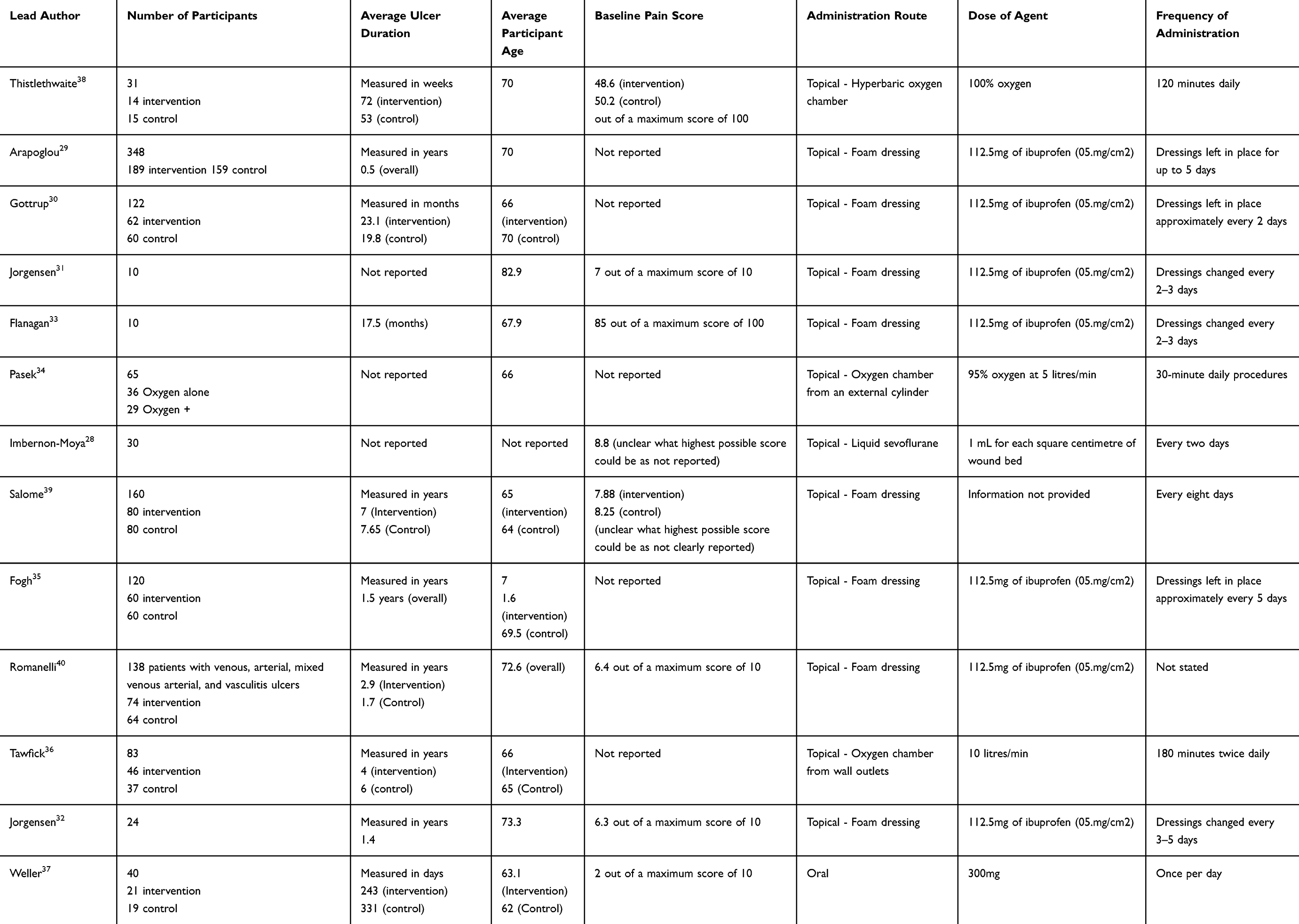 |
Table 1 Participant Characteristics and Intervention Protocols |
When assessing inclusion, only two studies required a formal duplex to diagnose venous incompetence.30,37 Instead, studies relied on patients having an ankle brachial pressure index reading of above 0.831–34,36,38,39 and in some cases, additional clinician directed physical assessment.28,35,37 The two studies including wounds of different aetiologies did not detail specific criteria for determining VU.29,40
Study Reporting
Pain reduction and pain at dressing changes were the most common outcomes, routinely measured using a numerical rating score. Additional outcomes included ulcer healing, adverse events and quality of life/physical functioning. Overall authors deemed the reporting completeness as poor. Five of the thirteen studies did not report baseline pain scores,29,30,34–36 nine did not report any patient co-morbidities28–35,40 and around half (6/46%) did not provide comprehensive recruitment participant flow data, such as how many patients were lost to follow-up or withdrawal information.28,29,32,34,36,39 An additional three studies reported pain in a way (bar graph) that made data difficult to interpret and impossible to extract or compare with the other studies.31,32,36 Additionally, no studies reported any costs or implementation data, such as how much the trial intervention cost or any additional service delivery requirements, for example, longer appointments or more staff. A summary of reporting completeness is depicted in Table 2. Only four studies had either published a protocol or listed the project on a national registry.29,30,37,38 Study methods can be found in Table 3.
 |
Table 2 Reporting Standards for Included Studies |
 |
Table 3 Study Methods |
Ibuprofen
Seven of the eight studies evaluated a foam-based, ibuprofen-impregnated product funded by Coloplast A/S, the manufacturer of the dressing.29–33,35,39,40 Five of these studies were RCTs.29,30,35,39,40 The product is described as being a topically applied, non-adhesive, primary dressing called BIATAIN-Ibu and is intended for use under compression therapy.41 The remaining study did not state the make or model but described it as a non-adherent Ibuprofen foam dressing.39 Each dressing contains 112.5 mg of ibuprofen (05 mg/cm2), comes in 15x15cm foam squares and releases ibuprofen when the dressing encounters wound exudate. One of the eight papers also examined the use of these dressings in combination with a silver-releasing wound contact layer (Physiotulle Ag, Coloplast A/S42) specifically for patients with infected wounds.32
As previously noted, while the eight studies examined the same intervention, combining the data was challenging and carrying out any meaningful meta-analysis was not possible. As briefly mentioned above, not all publications provided baseline pain scores, exact responder counts (eg. events or numbers) or mean ± standard deviations. Additionally, rather than reporting overall pain scores at each time point, some articles reported percentage change in pain intensity.29–31,40 There were also discrepancies across papers when considering follow-up time periods, ranging from 7 to 47 days. A further limitation of the reporting was the insufficient detail provided by some studies regarding dressing changes and compression application. For example, both Arapoglou 2011 and Fogh 2012 did not clearly state if and how often dressings were changed within the follow-up period. Therefore, it is difficult to assess how such changes might have affected overall outcomes.
Despite the heterogeneity of studies, all reported an improvement in pain for patients who used the ibuprofen impregnated dressing. However, as mentioned, it’s important to note that five of these eight studies did not report baseline pain score, and the quality of reporting was generally poor. Therefore, interpretation is difficult, and positive results should be considered with caution.
For those studies that evaluated pain in the days post dressing changes, the effect was highest in the first few hours and days (1–3).29,30,32,33,35,40 Two RCTs29,40 included wounds of different aetiologies (venous, arterial, mixed venous arterial, vasculitis, trauma wounds). Both reported pain outcomes for each subgroup, allowing the extraction of data for patients with VU. While VU patients did experience a statistically significant reduction in pain when compared to the comparator (usual care) (49% summed pain score >50%29/p = 0.01240), both papers found a greater improvement among those with arterial wounds (68% summed pain score >50%/p 0.005).40
Adverse events were reported in five of the eight studies30–32,35,40 but their incidence was low (ranging from 2 to 12 events). These included the development of eczema, blisters, localised skin irritation, maceration of the surrounding skin and infection.30,32,35,39,40 One study measured the presence of ibuprofen in blood serum, detecting it only in two patients who had taken additional ibuprofen orally during the study period.31 The same study found that stinging and bleeding at dressing changes were improved compared with the control group, reaching statistical significance for stinging (p = 0.0047) but not for bleeding (p = 0.061).31 Exudate management was also reported as good or excellent with limited leakage in two studies.31,32
Three studies (one RCT,30 one cohort study31 and one case series33) evaluated effects on patients’ quality of life (QoL). One used a validated tool (the WHO-5 Well-Being Index);29 another employed a questionnaire designed to evaluate daily living factors such as well-being, appetite, sleep, and mood;31 and the third used qualitative methods to explore these aspects in depth.31 All participants utilising ibuprofen dressings reported substantial enhancements in lifestyle domains, encompassing sleep quality, mental health, and activity levels, when compared to standard of care. Beyond its daily impact, the qualitative analysis further detailed the nature and characteristics of VU pain.33 Authors illustrated patient experiences using four key themes: unrelenting pain (shooting pains), the unpredictability of pain, living with restrictions and living with fatigue. When considering the overall effects of the ibuprofen dressing, pain relief was reportedly most effective in the first hours and early days after applications (1–5 days).33,35 As with other studies, this was linked to improved sleep quality and the ability to carry out daily living activities such as household chores, walking longer distances, managing stairs and generally participating in family life.33
Wound healing was described as a secondary outcome in five of the eight studies.30–33,35 Of those that included comparative groups, none reported a statistically significant difference in healing rates. While the two studies without a comparator group reported reduction in wound bed size, it was not clear if this was due to the dressing, nor if it was statistically or clinically significant.31,32
Oxygen
Localised delivery of oxygen to the wound bed was evaluated in three studies (two RCTs34,38 and one cohort study)[36] For treatment, the affected limb was placed in a sealed cylindrical chamber and oxygen was delivered by introducing it either from an external cylinder34,38 or piped wall outlets.36 Concentration of oxygen varied from 95%34 to 100%38 and was administered at 5 L/min34 and 10 L/min.36
Rationales for session times and frequencies were not given by the authors, but they varied across the three studies: 30 minute daily procedures over 4 weeks;34 180 minutes twice daily, five days a week, until full ulcer healing or for 12 weeks (whichever is sooner);36 120 minutes daily (80 minutes of oxygen and 30 minutes of air compression and decompression) five days a week for six weeks or until the ulcer healed.38
Although only one study explicitly stated this,36 descriptions suggest that oxygen treatments were delivered in inpatient/hospital settings. While compression therapy was applied between treatments in two studies,34,38 one left wounds fully exposed unless patients were going outside.36 In the study described by Pasek et al,34 both groups received oxygen therapy, but one group also had physical therapy intervention While authors specify that this was provided by a trained professional, no further detail is given regarding type of exercise or dosage.
Pain was a secondary outcome after healing in all studies. Although all reported some reduction in pain from baseline to study completion, none were statistically significant and it remains unclear whether this improvement was attributable to the treatment itself or to natural wound healing.34,36,38 However, one study (RCT) did report a statistically significant reduction in the intake of analgesic drugs for those in the intervention group (P = 0.009 vs P = 0.015, respectively).38 Ultimately, it’s important to consider that all three studies were underpowered to detect differences in pain, and two did not have pain as a primary outcome. Therefore, no conclusions about the effect of oxygen therapy on pain can be drawn from the data.34,36,38
Pasek et al34 reported improved EQ-5D quality-of-life scores in both groups, although the improvement was statistically greater in the group that also received physical therapy. This suggests that the higher scores may be attributable to additional physical activity. The two remaining studies either did not evaluate QoL36 or did not report a significant improvement.38
Only one study38 reported any adverse events that may have been related to the intervention, including two minor cases of otic barotrauma (ear pain caused by a pressure difference between the middle ear and outside environment)[43] All three studies reported a statistically significant improvement in either reduction in wound bed size34,36,38 or ulcer healing.36
Other Pharmacological Agents
The final two publications evaluated the topical application of sevoflurane and the oral administration of aspirin. Beyond its use in wounds, sevoflurane is routinely delivered as an inhaled halogenated anaesthetic for the induction and maintenance of general anaesthesia. In this small retrospective study,28 wounds were cleaned with saline and irrigated with 1 mL of liquid sevoflurane for each square centimetre of wound bed. Sterile gauze was used to protect surrounding skin. Wounds were covered with a sterile hydrophilic braided cotton pad soaked in saline, and compression was applied. This procedure took place every two days for a month and routine washing and debridement continued throughout. While the analgesic effects were limited when first used, improvements in the length of analgesic effect (measured in minutes and hours) were noted at each dressing change. Time to effect varied between two and seven minutes and the median length of effect was 12 hours (SD: 9 hours; range: 8–18 hours). Concurrent use of oral analgesics progressively diminished across all patients regardless of what agent they were regularly using (paracetamol, non-steroidal anti-inflammatories, metamizole tramadol or morphine). Prior to the study period, patients took oral analgesics a median of 5.3 ± 64.2 hours after wound cleaning, by the second dressing change with sevoflurane this decreased to a median period of 15.4 ± 6 3.7 hours (p = 0.001). Mild adverse reactions were noted in eleven of the patients enrolled and included pruritus (5) erythema (2) heat (3) and irritative dermatitis (1). Given the small sample size and retrospective nature of data collection, additional adequately powered randomised trials are needed to confirm these findings.
The final trial37 explored the addition of 300 mg oral aspirin tablets once per day in addition to standard of care (compression therapy). This RCT aimed to evaluate improvements in wound healing, VU recurrence, wound pain, QoL, adherence and inflammation markers. While this was a well-designed study, poor recruitment and the resultingly small sample (n = 40), prevented strong conclusions, as no significant differences were observed between groups. Despite the researcher’s best efforts, recruitment was hampered by the fact that many patients were already taking aspirin for cardiovascular secondary prevention, making them ineligible. Only two of the nineteen patients randomised to aspirin experienced minor bleeds that were considered adverse events. Of these, two only one was considered to be related to the study medication.
Discussion
This review included thirteen studies with a range of study designs (RCT, Cohort studies, and one Case Control study). Considering the evidence presented, primary dressings impregnated with ibuprofen do appear to provide some benefit when addressing pain concerns in patients with VU.29–33,35,39,40 However, it’s important to note that almost all the studies in this category were funded by the company distributing ibuprofen dressings. Other agents evaluated in this review, such as localised delivery of oxygen34,36,38 and oral aspirin,37 showed less promise, but the numbers of participants and quality were severely limited. While results from the evaluation of sevoflurane was positive, this drug was only tested in a single, small and poorly reported study,28 and further exploration in the form of an RCT is required to support its use in clinical practice.
As previously noted, poor reporting of pain was a theme throughout the publications in this review. While all studies used some form of numerical and/or ordinal pain score in line with the IMMPACT recommendations, the reporting was often inadequate. Many studies did not report baseline scores and others presented data in a table or bar chart, making it impossible to extract and interpret data.14,28,30,34–36,40 The IMMPACT recommendations also outline the importance of measuring QoL in a meaningful way, moving beyond statistical significance and towards clinically meaningful changes, to demonstrate if interventions have broader impact on patients’ lives. In doing so, studies should examine physical functioning, emotional functioning, and global rating of improvement (patients’ global impression of change). Only four of the included studies complied with such advice, either using a validated questionnaire or qualitative methods.30,31,33,34
Of the thirteen papers included, twelve evaluated a topical pharmacological agent.28–36,38–40 Currently, there is limited evidence specifically aimed at patients with VU. However, the focus on such interventions is mirrored in literature evaluating care of chronic wounds in general (arterial wounds, pressure ulcers, diabetic foot and trauma wounds). Two previous systematic reviews specifically evaluated topical agents15,44 and have some overlap with this review, as they include five of the same studies evaluating ibuprofen.29–31,35,40 Similar to the conclusions of this review, while such dressings appear to provide pain relief and show promise, the overall quality of evidence was questionable, and the authors advised that results should be considered with caution.
Ibuprofen dressings also present certain clinical limitations. While rates of adverse events were low across the reviewed studies, maceration and further skin breakdown due to excess moisture, is often a key concern. For this reason, foam dressings are not currently recommended for use under compression.45 Compression has been found to reduce the fluid-handling capacity of foam, leading to saturation. In turn, prolonged contact with exudate may damage healthy skin.45,46 Although not reported as a common occurrence in the reviewed studies, it is conceivable that frequent dressing changes prevented maceration. Most studies redressed wounds every 2–3 days.29–33,35,39,40 Unfortunately, this may not be consistent with routine clinical practice, where some dressings are commonly left for up to a week due to a lack of available clinical capacity to change them more frequently.47,48 Additionally, as ibuprofen dressings require exudate to activate drug release,41 it seems most appropriate that they are used primarily in patients with low to moderate exudate. Other than maceration, the additional adverse events reported were localised rather than systemic; they included eczema, blisters, and skin irritation. This is significant, as a key concern for this elderly population is the effect of polypharmacy on their overall health. Furthermore, as previously mentioned, as no cost-effectiveness data were reported in any of the included studies, it is impossible to assess whether the additional expense for medicated dressings is justified.
Beyond Ibuprofen, additional topical agents evaluated in prior reviews for other types of chronic wounds included morphine gel and lidocaine cream. Such studies were not included in this review, as they enrolled patients with a variety of wounds, and the VU data could not be isolated. While the evidence suggests benefits of lidocaine, this is not the case for morphine gel.15,44 Both reviews (published in 202044 and 202315) called for further high-quality research on the impact of topical agents. However, in the past five years since publication, this does not appear to have taken place.
While there were three studies reviewing the use of oxygen, none show positive results for pain management and were unlikely to be easily implementable. All oxygen therapies required patients to be in a hospital setting over several appointments.34,36,38 This is not in keeping with the delivery of VU care, which is largely provided in a community/home care setting.49 Beyond this, the ease of implementation was largely overlooked in all studies reviewed, and no studies included any cost data, making it difficult to ascertain if they were cost-effective interventions compared to standard of care.
When considering the available evidence, it is worth noting that most studies were industry funded and in some cases industry partners were included in the editorial process of writing publications.29–33,35,36,40 This does not necessarily bias evidence but raises the question of why such agents have not been trialled more widely with the support of public funding bodies or universities.
Current UK clinical practice recommendations suggest simple oral analgesia, such as paracetamol with or without codeine, as the first-line approach for any type of pain experienced by patients with VU.8 Beyond this, healthcare providers are advised to follow general pain guidelines and stepped-care approaches. While this differs slightly between countries, this includes processes such as a progressive analgesic ladder that usually incorporates a three-step process: non opioids, mild opioids (eg. codeine) and strong opioids (eg. morphine).50 While this guidance is useful, the absence of trials evaluating the use of oral analgesics with VU patients, such as paracetamol, represents a fundamental gap that must be addressed, given that these agents are recommended as first-line treatment in clinical practice.50
Despite the evidence presented and outlined in previous reviews,15,44 UK guidelines also note that the use of nonsteroidal anti-inflammatory drugs (NSAIDs) is not recommended for pain control due to some evidence indicating impaired wound healing and worsening of leg oedema.8 In the case of topical ibuprofen, this claim is not consistent with the evidence presented in this review (or previous publications15,44) which found no detrimental effect on healing outcomes.30–33,35 Alternative international guidelines provide limited additional insight into the use of NSAIDs. In the Best Practice Statement jointly published by the European Wound Management Association and Wounds Australia, authors acknowledge their inability to make definitive recommendations for pain management and provide no recommendations specific to NSAIDs.51 Likewise, the American Venous Forum offered limited guidance and called for updated clinical guidelines in 2020.52
Considering the body of literature on topical agents15,44,52 and guideline recommendations,8 the present review largely aimed to identify if there were any additional VU specific publications evaluating alternative pharmacological treatments and their effects. Following general clinical practice and guidance we would have expected studies on the effects of drugs such as paracetamol, codeine, tramadol, and morphine.53 The fact that only one low-quality study of such commonly used agents was identified (aspirin)37 highlights an important gap in the literature. Since wounds of differing aetiologies are likely to produce distinct pain profiles and drugs may have differential effects,17 more work is required to support clinical practice with reliable high-quality evidence. However, this must be specific to VU as they account for up to 80% of all lower limb wounds,54 cost approximately £2 billion per year55 and has a significant impact on patient QoL.56
Limitations
As previously noted, a key limitation of this review is the poor reporting and heterogeneity of study outcomes and follow-up time points, which prevented the conduct of a meta-analysis. Furthermore, apart from studies investigating ibuprofen, there was a lack of high-quality research evaluating alternative pharmacological agents. While the PRISMA guidelines were followed to ensure a systematic search was conducted, there is always a chance that studies could have been missed. Furthermore, only studies published in English were included, and there could be clinically relevant evidence that was not considered. While every effort has been made to make a balanced and unbiased argument for and against the evidence presented, it is also important to note that review articles can be subjective in some respects.
Conclusion
Currently, there is a lack of high-quality, independently funded studies evaluating pharmacological pain management for venous leg ulcers. Preliminary, low-quality evidence from industry-sponsored studies suggests that topical ibuprofen may provide some pain relief, but these findings require independent confirmation before they can inform clinical practice. No VU-specific evidence was identified to support the use of oral agents commonly recommended in guidelines. While clinicians should continue to follow available guidelines, high-quality, adequately powered trials comparing topical and oral agents are urgently needed.
Abbreviations
VU, Venous ulceration; NICE, National Institute for Health and Care Excellence; QoL, Quality of life; PRISMA, Preferred Reporting Items for Systematic Reviews and Meta-Analyses.
Data Sharing Statement
As previously noted, all data presented is publicly available and referenced. The full search strategy is provided in appendix 1. Data extraction was completed using the platform Covidence and exported into excel format. This is available at request to the corresponding author.
Ethical Approval
As this is a review article, no prior ethical approval was required. All data presented is publicly available and referenced.
Acknowledgments
Nothing to note.
Funding
The research time of Layla Bolton Saghdaoui (grant number NIHR302917), Smaragda Lampridou (grant number NIHR303308) and David Hohenschurz-Schmidt (grant number NIHR305077) is funded by the National Institute for Health and Care Research (NIHR). The views expressed are those of the authors and not necessarily those of the NIHR or the Department of Health and Social Care.
Disclosure
DHS received consulting fees from Altern Health Ltd. and income from private osteopathic practice. He reports employment with Health Sciences University and Imperial College London, and or self-employed income from Osteopathie Schule Deutschland and Haute école de santé Fribourg, where he received honoraria for teaching and supervision. He serves on the executive committee and as a Trustee of the Society for Back Pain Research, the Scientific Committee for the 2026 International Association for the Study of Pain (IASP) World Congress, as an editor at BMC Medical Research Methodology, and as TSC/DEMC member on the AIDE-BC trial. He also received research funding from The Osteopathic Foundation, the Alan and Sheila Diamond Charitable Trust, the UK National Institute for Health and Care Research (NIHR305077), the Chelsea and Westminster Hospital Joint Research Council, and personal support through prizes or conference travel awards from IASP, BritSpine, and EFIC. He received personal honoraria for research from the Analgesic, Anesthetic, and Addiction Clinical Trial Translations, Innovations, Opportunities, and Networks (ACTTION). The authors report no other conflicts of interest in this work.
References
1. Vasudevan B. Venous leg ulcers: pathophysiology and Classification. Indian Dermatol Online J. 2014;5(3):366–15. doi:10.4103/2229-5178.137819
2. National Institute for Health and Care Excellence. Leg ulcer - venous. 2019. Available from: https://cks.nice.org.uk/leg-ulcer-venous#!topicSummary.
3. Green J, Jester R, McKinley R, et al. The impact of chronic venous leg ulcers: a systematic review. J Wound Care. 2014;23(12):601–612. doi:10.12968/jowc.2014.23.12.601
4. Carradice D, Mazari FAK, Samuel N, et al. Modelling the effect of venous disease on quality of life. Br J Surg. 2011;98(8):1089–1098. doi:10.1002/bjs.7500
5. Probst S, Schobinger E, Saini C, et al. Unveiling the hidden pain and daily struggles of individuals with a venous leg ulcer: a thematic analysis. J. Tissue Viability. 2025;34(3):100906. doi:10.1016/j.jtv.2025.100906
6. Wounds UK. Best Practice Statement: Holistic Management of Venous Leg Ulceration. 2016; Available from: https://legsmatter.org/wp-content/uploads/2018/04/content_120221.pdf.
7. Sinha S, Sreedharan S. Management of venous leg ulcers in general practice - a practical guideline. Aust Fam Physician. 2014;43(9):594–598.
8. National Institute for Health and Care Excellence, Scenario: Venous leg ulcers. 2013. URL: https://cks.nice.org.uk/topics/leg-ulcer-venous/management/venous-leg-ulcers/.
9. Hwang SS, Chang VT, Kasimis B. Cancer breakthrough pain characteristics and responses to treatment at a VA medical center. Pain. 2003;101(1–2):55–64. doi:10.1016/S0304-3959(02)00293-2
10. Bell A. The neurobiology of acute pain. Vet J. 2018;237:55–62. doi:10.1016/j.tvjl.2018.05.004
11. Eusen M, Brenaut E, Schoenlaub P, et al. Neuropathic pain in patients with chronic leg ulcers. J Eur Acad Dermatol Venereol. 2016;30(9):1603–1605. doi:10.1111/jdv.13647
12. International Association for the Study of Pain. IASP Taxonomy: Nociplastic pain 2018. Available from: http://www.iasp-pain.org/Taxonomy?navItemNumber=576.
13. O’Donnell TF Jr, Passman MA, Marston WA, et al. Management of venous leg ulcers: clinical practice guidelines of the Society for Vascular Surgery ® and the American Venous Forum. J Vasc Surg. 2014;60(2 Suppl):3s–59s. doi:10.1016/j.jvs.2014.04.049
14. Ahmajarvi K, et al. Holistic management of wound-related pain: an overview of the evidence and recommendations for clinical practice. J Wound Manag. 2024;25(1):1–140.
15. Ffrench C, Finn D, Velligna A, et al. Systematic review of topical interventions for the management of pain in chronic wounds. Pain Rep. 2023;8(5):e1073. doi:10.1097/PR9.0000000000001073
16. Mervis JS, Federman DG. Pain management in patients with chronic wounds. Curr Dermatol Rep. 2018;7(3):136–146. doi:10.1007/s13671-018-0220-2
17. Dubský M, Fejfarova V, Bem R, et al. Pain management in older adults with chronic wounds. Drugs Aging. 2022;39(8):619–629. doi:10.1007/s40266-022-00963-w
18. Leren L, Johansen E, Eide H, et al. Pain in persons with chronic venous leg ulcers: a systematic review and meta-analysis. Int Wound J. 2020;17(2):466–484. doi:10.1111/iwj.13296
19. Berenguer Pérez M, López-Casanova P, Sarabia Lavín R, et al. Epidemiology of venous leg ulcers in primary health care: incidence and prevalence in a health centre-A time series study (2010-2014). Int Wound J. 2019;16(1):256–265. doi:10.1111/iwj.13026
20. Agale S. Chronic Leg Ulcers: Epidemiology, Aetiopathogenesis, and Management. Ulcers; 2013:2013.
21. Wounds UK. Best Practice Statement: Addressing complexities in the management of venous leg ulcers. Wounds UK. Available from: https://lohmann-rauscher.co.uk/downloads/lr19_bps_complexvlu_wuk_web.pdf.
22. Ogston-Tuck S. Access to pain management from a legal and human rights perspective. Nurs Stand. 2022;37(9):75–79. doi:10.7748/ns.2022.e11897
23. Page MJ, McKenzie JE, Bossuyt PM, et al. The PRISMA 2020 statement: an updated guideline for reporting systematic reviews. Rev Esp Cardiol. 2021;74(9):790–799. doi:10.1016/j.recesp.2021.06.016
24. Layla Bolton Saghdaoui SL, Chumbley G, Baldo L, Onida S, Atkin L. Pharmacological Pain Management in Adults with Venous Ulceration. PROSPERO; 2025.
25. Critical Appraisal Skills Programme. Critical Appraisal Checklists. 2025. Available from: https://casp-uk.net/casp-tools-checklists/.
26. Critical Appraisal Skills Programme. FAQs. 2005. Available from: https://casp-uk.net/faqs/.
27. Analgesic and healing effect of topical sevoflurane for chronic venous ulcers. J Am Acad Dermatol. 2018;79(3):AB41. doi:10.1016/j.jaad.2018.05.205
28. Imbernon-Moya A, Ortiz-de Frutos FJ, Sanjuan-Alvarez M, et al. Pain and analgesic drugs in chronic venous ulcers with topical sevoflurane use. J Vasc Surg. 2018;68(3):830–835. doi:10.1016/j.jvs.2017.11.071
29. Arapoglou V, Katsenis K, Syrigos KN, et al. Analgesic efficacy of an ibuprofen-releasing foam dressing compared with local best practice for painful exuding wounds. J Wound Care. 2011;20(7):319–325. doi:10.12968/jowc.2011.20.7.319
30. Gottrup F, Jørgensen B, Karlsmark T, et al. Reducing wound pain in venous leg ulcers with Biatain Ibu: a randomized, controlled double-blind clinical investigation on the performance and safety. Wound Repair Regen. 2008;16(5):615–625. doi:10.1111/j.1524-475X.2008.00412.x
31. Jørgensen B, Friis GJ, Gottrup F. Pain and quality of life for patients with venous leg ulcers: proof of concept of the efficacy of BIATAIN-Ibu, a new pain reducing wound dressing. Wound Repair Regen. 2006;14(3):233–239. doi:10.1111/j.1743-6109.2006.00116.x
32. Jorgensen B, Gottrup F, Karlsmark T, Bech-Thomsen N, Sibbald RG. Combined use of an ibuprofen-releasing foam dressing and silver dressing on infected leg ulcers. J Wound Care. 2008;17(5):210–214. doi:10.12968/jowc.2008.17.5.29153
33. Flanagan M, Vogensen H, Haase L. Case Series Investigating the Experience of Pain in Patients with Chronic Venous Leg Ulcers Treated with a Foam Dressing Releasing Ibuprofen. World Wide Wounds; 2006:15p–15p.
34. Pasek J, Szajkowski S, Pietrzak M, et al. Comparison of the efficacy of topical hyperbaric oxygen therapy alone vs a combination of physical methods including topical hyperbaric oxygen therapy, magnetotherapy, and low-energy light therapy in the treatment of venous leg ulcers. Dermatologic Therapy. 2020;33(6):e14474. doi:10.1111/dth.14474
35. Fogh K, Andersen MB, Bischoff-Mikkelsen M, et al. Clinically relevant pain relief with an ibuprofen-releasing foam dressing: results from a randomized, controlled, double-blind clinical trial in exuding, painful venous leg ulcers. Wound Repair Regen. 2012;20(6):815EP–821. doi:10.1111/j.1524-475X.2012.00844.x
36. Tawfick W, Sultan S. Does Topical Wound Oxygen (TWO2) Offer an Improved Outcome Over Conventional Compression Dressings (CCD) in the Management of Refractory Venous Ulcers (RVU)? A Parallel Observational Comparative Study. Eur J Vasc Endovascular Surg. 2009;38(1):125EP–132.
37. Weller CD, Martin C, Bouguettaya A, et al. Once daily 300 mg aspirin with compression versus compression alone in patients with chronic venous leg ulcers (ASPiVLU): a randomised, double-blinded, multicentre, placebo-controlled, clinical trial. J. Tissue Viability. 2021;30(4):509–516. doi:10.1016/j.jtv.2021.07.005
38. Thistlethwaite KR, Finlayson KJ, Cooper PD, et al. The effectiveness of hyperbaric oxygen therapy for healing chronic venous leg ulcers: a randomized, double-blind, placebo-controlled trial. Wound Repair Regen. 2018;26(4):324–331. doi:10.1111/wrr.12657
39. Salome GM, Ferreira LM. Impact of non-adherent Ibuprofen foam dressing in the lives of patients with venous ulcers. Revista Do Colegio Brasileiro de Cirurgioes. 2017;44(2):116EP–124. doi:10.1590/0100-69912017002002
40. Romanelli M, Dini V, Polignano R, et al. Ibuprofen slow-release foam dressing reduces wound pain in painful exuding wounds: preliminary findings from an international real-life study. J Dermatological Treat. 2009;20(1):19EP–26. doi:10.1080/09546630802178232
41. Coloplast. Biatain® Ibu Non-Adhesive. Available from: https://products.coloplast.co.uk/coloplast/wound-care/-biatain-ibu/biatain-ibu-non-adhesive/.
42. Coloplast. Physiotulle® Ag. Available from: https://www.coloplast.co.uk/physiotulle-ag-en-gb.aspx.
43. ONeill OJ, Frank BK. Ear Barotrauma. Treasure Island: StatPearls Publishing; 2023.
44. Purcell A, Buckley T, King J, et al. Topical analgesic and local anesthetic agents for pain associated with chronic leg ulcers: a systematic review. Adv Skin Wound Care. 2020;33(5):240–251. doi:10.1097/01.ASW.0000658572.14692.fb
45. O’Meara S, James MM-S. Foam dressings for venous leg ulcers. Cochrane Database Syst Rev. 2013;2013(5):Cd009907.
46. National Institute for Health and Care Excellence. Advanced wound dressings. Available from: https://bnf.nice.org.uk/wound-management/advanced-wound-dressings/#foam-dressings.
47. Gloucestershire Hospitals NHS Foundation Trust. Treating your leg ulcer. 2023. Available from: https://www.gloshospitals.nhs.uk/your-visit/patient-information-leaflets/treating-your-leg-ulcer/.
48. Gray TA, Wilson P, Dumville JC, et al. What factors influence community wound care in the UK? A focus group study using the Theoretical Domains Framework. BMJ Open. 2019;9(7):e024859. doi:10.1136/bmjopen-2018-024859
49. Scottish Intercollegiate Guidelines Network. Management of chronic venous leg ulcers, A national clinical guideline. 2010. Available from: https://www.sign.ac.uk/media/1058/sign120.pdf.
50. Anekar AA, Cascella HJ. 2025. WHO Analgesic Ladde. Treasure Island (FL): StatPearls Publishing, StatPearls [Internet].
51. Franks PJ, Barker J, Collier M, et al. Management of patients with venous leg ulcers: challenges and current best practice. J Wound Care. 2016;25(Sup6):S1–s67. doi:10.12968/jowc.2016.25.Sup6.S1
52. Shaydakov ME, Ting W, Sadek M, et al. Review of the current evidence for topical treatment for venous leg ulcers. J Vasc Surg Venous Lymph Disord. 2022;10(1):241–247.e15. doi:10.1016/j.jvsv.2021.06.010
53. National Institute for Health and Care Excellence. Analgesia - mild-to-moderate pain. 2025. Available from: https://cks.nice.org.uk/topics/analgesia-mild-to-moderate-pain/.
54. Probst S, Saini C, Gschwind G, et al. Prevalence and incidence of venous leg ulcers-A systematic review and meta-analysis. Int Wound J. 2023;20(9):3906–3921. doi:10.1111/iwj.14272
55. Phillips CJ, Humphreys I, Thayer D, et al. Cost of managing patients with venous leg ulcers. Int Wound J. 2020;17(4):1074–1082. doi:10.1111/iwj.13366
56. Smith J, Carville K, Maguire C, et al. The impact of venous leg ulcers on quality of life. Wound Practice & Research. J Australian Wound Manag Association. 2023;31(4):164–173.
 © 2026 The Author(s). This work is published and licensed by Dove Medical Press Limited. The
full terms of this license are available at https://www.dovepress.com/terms
and incorporate the Creative Commons Attribution
- Non Commercial (unported, 4.0) License.
By accessing the work you hereby accept the Terms. Non-commercial uses of the work are permitted
without any further permission from Dove Medical Press Limited, provided the work is properly
attributed. For permission for commercial use of this work, please see paragraphs 4.2 and 5 of our Terms.
© 2026 The Author(s). This work is published and licensed by Dove Medical Press Limited. The
full terms of this license are available at https://www.dovepress.com/terms
and incorporate the Creative Commons Attribution
- Non Commercial (unported, 4.0) License.
By accessing the work you hereby accept the Terms. Non-commercial uses of the work are permitted
without any further permission from Dove Medical Press Limited, provided the work is properly
attributed. For permission for commercial use of this work, please see paragraphs 4.2 and 5 of our Terms.
Recommended articles

Assessment of the Community Pharmacists’ Knowledge and Attitudes Toward Pain and Pain Management in Saudi Arabia
Alorfi NM, Ashour AM, Algarni AS, Alsolami FA, Alansari AM, Tobaiqy M
International Journal of General Medicine 2022, 15:8527-8537
Published Date: 7 December 2022
Effect of the Evidence-Based and Multidisciplinary Pain Management Program in Patients After Laparoscopic Hepatectomy: A Quasi-Experimental Study
Song X, Liu Y, Zou J, Zhang D, Bai T, Han Z, Wang Y
Journal of Pain Research 2026, 19:582027
Published Date: 24 February 2026
Validation of the Kuopio Questionnaire on Pain Management Attitudes Among Healthcare Personnel in a Finnish Teaching Hospital
Paunonen P, Tyynelä-Korhonen K, Nuutinen R, Haatainen KM, Kervinen M, Wüstefeld M, Pöyhiä R, Lamminmäki A
Journal of Pain Research 2026, 19:532085
Published Date: 18 March 2026