
Back to Journals » Clinical Ophthalmology » Volume 20
Paired Opposite Clear Corneal Incision in Cataract Patients with Different Types of Astigmatism
Authors Icoz M  , Aktas Ozaltun O
Received 6 November 2025
Accepted for publication 22 January 2026
Published 29 January 2026 Volume 2026:20 405677
DOI https://doi.org/10.2147/OPTH.S405677
Checked for plagiarism Yes
Review by Single anonymous peer review
Peer reviewer comments 2
Editor who approved publication: Dr Scott Fraser
Mehmet Icoz,1,* Ozlem Aktas Ozaltun2,*
1Department of Ophthalmology, Yozgat City Hospital, Yozgat, Turkey; 2Department of Ophthalmology, Karabük Training and Research Hospital, Karabuk, Turkey
*These authors contributed equally to this work
Correspondence: Mehmet Icoz, Department of Ophthalmology, Yozgat City Hospital, Yozgat, Turkey, Email [email protected]
Purpose: To compare the refractive effects of paired-opposite clear corneal incision (p-OCCI) with-the-rule (WTR), against-the-rule (ATR) and oblique astigmatism.
Methods: This retrospective study was conducted in 131 patients undergoing uneventful cataract surgery.In all cases, a 2.4 mm p-OCCI was made 180 degrees opposite to the meridian of the vertical axis.Preoperative and 6-month postoperative keratometry values obtained from partial coherence interferometry were used to calculate corrected keratometric astigmatism and vectorial analysis parameters were calculated by Alpins method.These parameters were surgically induced astigmatism (SIA), target induced astigmatism (TIA), correction index (CI), angle of error (AE), difference vector (DV), flattening effect (FE).
Results: There was no significant difference between the three groups in terms of uncorrected and best corrected visual acuities (p> 0.05).Mean corrected keratometric astigmatism values were 1.06± 0.15 D in WTR astigmatism group, 1.21± 0.35 D in ATR astigmatism group and 1.15± 0.29 D in oblique astigmatism group (p> 0.05).SIA value was 1.15± 0.54 D in WTR astigmatism group, 0.79 ± 0.31 D in ATR astigmatism group and 0.53 ± 0.14 D in oblique astigmatism group (p=0.006).In terms of FE,there was no statistically significant difference between the WTR and oblique astigmatism groups, while it was lowest in the ATR astigmatism group (p> 0.05, p=0.03,respectively).The CI value was similar in the WTR and ATR astigmatism groups, but lower in the oblique astigmatism group (p> 0.05, p=0.04,respectively).There was no significant difference between the three groups in terms of TIA, AE and DV (p> 0.05).
Conclusion: Although the corneal astigmatism-correcting effect of p-OCCI is similar in different types of astigmatism,p-OCCI may be less predictable in oblique astigmatism.
Keywords: astigmatism, opposite clear corneal incision, cataract, vectorial analysis
Introduction
Patients undergoing cataract surgery now expect almost-perfect visual acuity and refractive outcomes in the postoperative period. Ferrer-Blasco and De Bernardo reported a corneal astigmatism rate of 24–36% between 1.0 and 2.5 D in cataract patients.1,2 With advances in phacoemulsification and lens technology, refractive errors such as spherical and astigmatism can be corrected. There are different options for these correction procedures. These options include appropriate corneal incision, limbal relaxing incision, astigmatic keratotomy and toric intraocular lens use.3,4 Among these options, toric intraocular lens implantation, which is becoming increasingly common and is known to be effective, has significant advantages.5 One of the most important advantages of toric intraocular implantation is the correction of moderate to high astigmatism.5–7 However, on the other hand, it has significant disadvantages in terms of accessibility, cost-effective, and the use of additional materials.
The astigmatism-reducing effect of corneal incision is known with different studies from past years to the present.6,7 With the paired opposite clear corneal incision (p-OCCI), a vertical quadrant is preferred as the main corneal incision and a corneal incision of the same size-plane is made 180 degrees opposite. It has been shown that p-OCCI performed between 2.4 and 3.5 mm improves astigmatism by 0.50–2.06 D on average.7–10
The magnitude of astigmatism may vary in different corneal meridians. In this context, according to the meridian with the strongest refractive power; it is divided into with-the-rule (WTR), against-the-rule (ATR) and oblique astigmatism. The fact that the collagen fibril orientation of the cornea is not the same in all regions causes differences in terms of refractive characteristics.11 Based on this anatomical variation, the limited data on the efficacy of OCCI in different types of astigmatism prompted the need for this study. The lack of data on the vectorial effect of the p-OCCI application in different types of astigmatism is noteworthy. On the other hand, although it is not the subject of this study, the presence and consideration of posterior corneal astigmatism in modern cataract surgery is also being investigated as an important topic.12
The fact that the p-OCCI option does not require any additional costs or materials, and that it is a method that any surgeon performing cataract surgery can easily apply, reflects the importance of this astigmatism-correcting technique. The primary aim of this study was to compare the refractive effects of p-OCCI in WTR, ATR and oblique astigmatism types.
Method
Before starting this retrospective study, the necessary approvals were obtained from the Karabuk University Ethics committee (ethics committee approval code: 2025/2141). The articles of the Declaration of Helsinki were followed at all stages of the study. The study was conducted using data obtained from patient files and ocular examinations.
This study was conducted in 131 patients who underwent cataract surgery for decreased visual acuity and underwent p-OCCI for preoperative corneal astigmatism (1–2.5 D) at Yozgat City Hospital Ophthalmology Clinic between June 2022 and January 2025. Participants were classified as WTR (group A, n=48) at 90 ± 20 degrees, ATR (group B, n=38) at 180 ± 20 degrees, and oblique astigmatism (group C, n=45) outside these two quadrants.13 The anatomical position of the eye is used as a basis, with the nasal quadrant at 0 degrees and the temporal quadrant at 180 degrees for the right eye, and the temporal quadrant at 0 degrees and the nasal quadrant at 180 degrees for the left eye.
Exclusion criteria were cataract other than senile cataract, pterygium, corneal ectasias (keratoconus, pellucid marginal degeneration), history of ocular surgery, trauma, intraoperative anterior and/or posterior capsule rupture, nucleus drop, implantation of intraocular lens (IOL) in a different location other than bag complex, zonule weakness, and in the postoperative period; intense corneal edema, leakage from the wound site, posterior capsule opacification, vitreous hemorrhage, retinal detachment, cystoid macular edema, vitreoretinal interface diseases.
Detailed ophthalmologic examination was performed in all patients. Visual acuity, intraocular pressure measurements, anterior segment and fundus assessments were performed at preoperative and postoperative 6th month examinations. Spherical and cylindrical refraction values were determined with autorefractometer (Nidek Co., Aichi, Japan). Preoperative IOL measurement was calculated with partial coherence interferometry (IOLMaster 500 - Carl Zeiss, Jena, Germany). SRKT formulation was used for IOL measurement and −0.5 D was chosen as the target. Corneal astigmatism measurements were calculated with keratometry values (flat keratometry (K1), steep keratometry (K2)) obtained from partial coherence interferometry. The difference between preoperative and postoperative 6th month keratometric astigmatism values was considered as corrected keratometric astigmatism value. All cases were performed by a single surgeon and the mean surgically induced astigmatism (SIA) value of 0.50 D was accepted based on the surgeon’s last 100 cases. Vectorial analysis parameters were calculated using the technique described by Alpins and Assort software (Assort Pty, Ltd).14–16 These parameters were SIA, target induced astigmatism (TIA), correction index (CI), angle of error (AE), difference vector (DV) and flattening effect (FE). SIA refers to the change in the axis and power of astigmatism after surgery. TIA represents the surgically targeted astigmatism power and axis. CI is calculated by SIA/TIA. An index greater than one represents overcorrection and less than one represents undercorrection. AE was considered as the difference between the angle of the SIA vector and the angle of the TIA vector. DV indicates the difference between the magnitude of preoperative astigmatism and the target value. FE represents the reduction in astigmatism with the values obtained from SIA in the targeted meridian.
All surgeries were performed by the same surgeon with the same technique under topical anesthesia. Right eye surgeries were performed with the right hand and left eye surgeries were performed with the left hand. The surgeon did not change his position and microscope position during surgery. All patients were asked to sit under the preoperative slit lamp light and look straight ahead in a vertical position. Once the appropriate view was achieved, a dotted mark was made at the 6–12 o’clock position of the cornea with a sterile pen. Following this preoperative marking, the main incision and the quadrant area where p-OCCI would be applied were marked intraoperatively using a Mendez ring, using the 6–12 o’clock position as the reference point, using a sterile pen. Through this ring, the axis of the vertical, oblique and horizontal quadrants was preferred as the main corneal incision. 180 degrees opposite to this axis was marked. Two lateral corneal incisions were made in accordance with these axes. Adrenaline and viscoelastic material were administered through the corneal side incisions, sequentially. A continuous, curvilinear capsulorhexis with a mean size of 5 mm was made through the lateral incision. Corneal main incision was made with a 2.4 mm keratome from the preoperatively determined axis. During the corneal main incision, the keratome was advanced 1–1.5 mm in the stroma in the corneal plane starting from the corneal-alimbal junction and then sloped downward and directed into the anterior chamber cavity. The nucleus-cortex complex was mobilized by hydrodissection and hydrodelineation. Nucleus removal was performed with phacomulsification (Veritas Vision System, Johnson & Johnson Vision, U.S.A) using stop and chop technique. The balance of the cortex was cleared with bimanual irrigation - aspiration. Viscoelastic material was injected through the main corneal incision. A one-piece hydrophobic foldable intraocular lens (Alcon SA60AT, Alcon Laboratories, Inc., Fort Worth, TX) was placed in a cartridge system and implanted into the bag through the injector system. Viscoelastic material was removed by bimanual irrigation - aspiration. At this stage, the irrigation cannula was kept in the anterior chamber and a corneal incision was created 180 degrees opposite to the main corneal incision at the marked point with a 2.4 mm keratoma, identical to the main corneal incision architecture. No stromal hydration or suturing was used to close this incision. The other main and lateral corneal incisions were closed with stromal hydration. At the end of surgery, cefuroxime was given to the anterior chamber. Postoperative, all patients were advised to use topical antibiotic + corticosteroid combination 4*1 dose for 1 month.
For the sample size analysis; when the effect size value was determined as 0.4, type 1 error value as 0.01, and power value as 90%, the total number of participants was found to be 115.
Statistical Package for the Social Sciences (Version 22.0 Chicago, IL, USA) was used for data analysis. Quantitative values were expressed as mean ± standard deviation. The conformity of the data to normal distribution was evaluated visually by histogram and statistically by Kolmogorov–Smirnov test. One-way analysis of variance or Kruskall-Wallis tests were used for the comparison of the groups since there were three independent groups. Significant differences between the three groups were evaluated by post-hoc analysis. p < 0.05 was considered statistically significant. Only surgeries on the right or left eye were included for all participants.
Results
This retrospective study was conducted in 131 eyes of 131 patients. Demographic data and preoperative findings of all participants are presented in Table 1. There was no statistically significant difference between the groups in terms of preoperative visual acuity, spherical, cylindrical and spherical equivalent refraction values (p>0.05 for all values). There was also no statistically significant difference between the groups in terms of preoperative flat and step keratometry values (p>0.05 for all values).
 |
Table 1 Demographic Data and Preoperative Findings of the Groups |
Postoperative 6th month visual acuity, refraction values and keratometry measurements of the groups are presented in Table 2. There was no statistically significant difference between the three groups in terms of postoperative visual acuities, cylindrical refraction, spherical equivalent refraction, corrected keratometric astigmatism value and flat - step keratometry values (p>0.05 for all values).
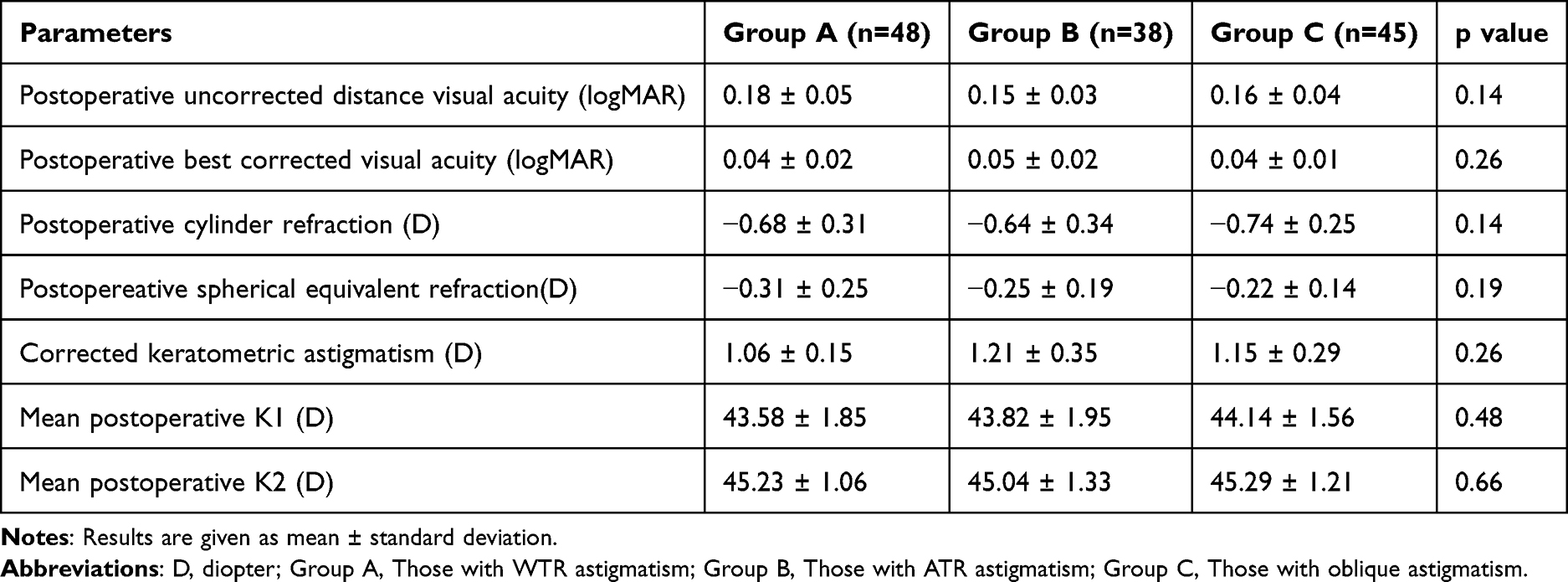 |
Table 2 Distribution of Postoperative Findings of the Groups |
The distribution of the parameters obtained by vectorial analysis between the groups is shown in Table 3 and Figures 1–3. There was a statistically significant difference between the groups in terms of SIA value; the mean SIA value was 1.15 ± 0.54 D in group A, 0.79 ± 0.31 D in group B and 0.53 ± 0.14 D in group C (p=0.006). There was no statistically significant difference between groups A and B in terms of CI, whereas it was statistically lower in group C compared to the other two groups (p>0.05, p=0.04, respectively). In terms of FE, there was no statistically significant difference between groups A and C, while it was statistically lower in group B compared to the other two groups (p>0.05, p=0.03, respectively). There was no significant difference between the three groups in terms of TIA, AE and DV (p>0.05 for all values). Also, the vector analysis parameters of the right and left eyes were compared in all three groups. No statistically significant differences were observed between the two groups for any of the parameters (p>0.05 for all values).
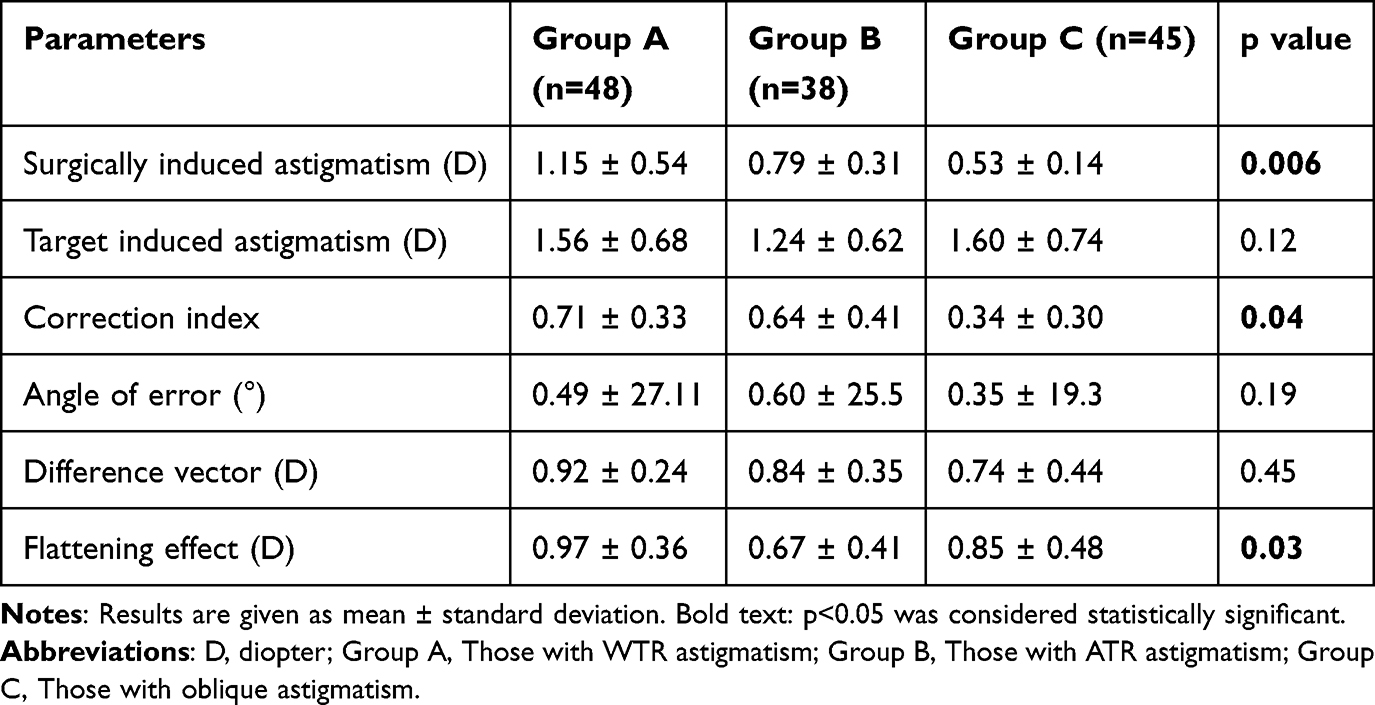 |
Table 3 Distribution of Vectorial Analysis Parameters of Groups |
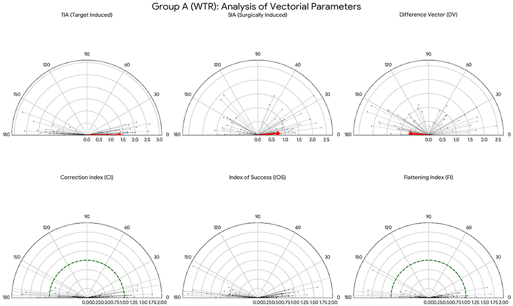 |
Figure 1 Presentation of Group A’s vector analysis parameters. |
 |
Figure 2 Presentation of Group B’s vector analysis parameters. |
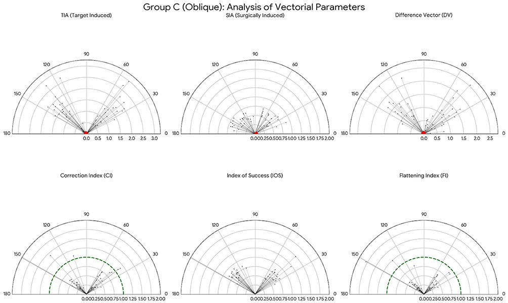 |
Figure 3 Presentation of Group C’s vector analysis parameters. |
Discussion
The purpose of cataract surgery is to achieve near-optimal refractive results after the procedure, which is a goal anticipated by both the patient and the clinician. Although cataract surgery is a common surgery today, the rate of astigmatism requiring refractive correction in patients undergoing cataract surgery is remarkable. p-OCCI efficacy has been evaluated by different investigators and its effect on correcting astigmatism has been demonstrated.6,7,10 However, as far as we know, there are only two studies on the efficacy of p-OCCI in different types of astigmatism.17,18 With this study;
- The mean amount of astigmatism corrected with p-OCCI in three different astigmatism types was 1–1.25 D.
- Postoperative 6th month SIA value was highest in the WTR astigmatism group and lowest in the oblique astigmatism group.
- Postoperative 6th month FE value was not significantly different in ATR and oblique astigmatism groups, while it was the lowest in WTR astigmatism group.
- In terms of postoperative 6th month CI value; there was no significant difference between WTR and ATR astigmatism groups, while oblique astigmatism group had the lowest CI value compared to the other two groups.
p-OCCI is frequently preferred by cataract surgeons as a technique to reduce corneal astigmatism as described by Lever and Dahan.18 In this technique, a second corneal incision of the same architecture is made 180 degrees opposite to the main corneal incision. 2.1 D in 3.5 mm incision, 1.8 D in 3.2 mm incision and 1.1 D in 2.4 mm incision have been reported to improve corneal astigmatism.7,18,19 In this study, a 2.4 mm incision was used and corneal astigmatism was improved at similar diopters (1.06 D, 1.21 D and 1.15 D) in three different types of astigmatism.
SIA was evaluated with incisions of different sizes in this technique and mostly consistent results were reported.7,9,20 When postoperative SIA values were examined; Tadros et al. 1.57 D in a 3.5 mm incision, Sundeshan et al 1.66 D in a 3.2 mm incision, and Icoz et al reported 0.91 D in a 2.4 mm incision.7,9,20 In these studies, no subgroup analysis was performed according to the type of astigmatism. Binayi Faal et al reported a postoperative SIA value of 1.96 D in the WTR astigmatism group and 2.13 D in the ATR astigmatism group with a 4.0 mm incision similar to our study design, while Bazzazi et al reported a postoperative mean SIA value of 0.79 D in the WTR astigmatism group and 0.68 D in the ATR astigmatism group with a 3.2 mm incision.10,17 In both of these studies, no statistically significant difference was reported in terms of postoperative SIA value according to astigmatism type, whereas in Chiam’s study, a significant difference was reported in terms of SIA value at both postoperative 1st month and postoperative 6th month.10,13,17 In Chiam’s study, a 3.2 mm incision was applied and the postoperative 6th month SIA value was 1.6 D in the WTR astigmatism group and 0.8 D in the ATR astigmatism group, while 1.5 D was found in the ATR astigmatism group in which a 3.5 mm incision was applied.13 In this study, the postoperative 6th month SIA value was 1.15 D in the WTR astigmatism group, 0.79 D in the ATR astigmatism group and 0.53 D in the oblique astigmatism group. In adults, the mean horizontal corneal distance is accepted as 11.7 mm and the mean vertical corneal distance as 10.6 mm.21 This geometric difference will cause different refractive changes depending on the incisions to be applied to the cornea. Therefore, corneal incisions made in the horizontal plane will be further from the center of the cornea than incisions made in the vertical plane. This distance difference may directly affect the astigmatism that may occur due to the incision. Merriam et al also supported this hypothesis and showed that incisions made in the superior quadrant may cause more refractive changes than incisions made in the temporal quadrant.22 Although smaller incisions (1.8, 2.0 and 2.2 mm) are now preferred for cataract surgery, our study is the first to evaluate the effect of p-OCCI on SIA in different types of astigmatism with the smallest incision (2.4 mm) in the existing literature.23 These results will be important for every surgeon interested in cataract and refractive surgery.
The distribution of collagen, which constitutes the main skeletal structure of the cornea, has been studied by different investigators from past to present and important inferences have been made. In these studies, the central scattering pattern of the cornea is examined using synchrotron X-ray.24,25 The central scattering pattern provides information about collagen distribution in different quadrants of the cornea. This pattern is seen throughout the cornea and continues in the limbus through a ring formed by collagen. The transition points of these rings are along the superior - inferior axis; thinning towards the limbus and sclera. The width of the rings is shown as 1.5 mm in superior and 2.0 mm in inferior. When the nasal - temporal corneal quadrants are examined; collagen rings start 1.5 mm behind the limbus and terminate at 90 degrees. In the superior - inferior quadrant, these rings start 1 mm behind the limbus and end at 45 degrees.25,26 In addition to this anatomical difference, histologically, it is known that collagen fibrils are more dense and parallel in the nasal and temporal quadrants.13 For these two reasons mentioned above, it is expected that FE would be less in p-OCCI performed in the ATR astigmatism (mostly temporal quadrant) group due to the more intense healing reaction of collagen fibrils in response to incision. In this study, FE was found to be statistically significantly lower in the ATR astigmatism group compared to the other two groups. On the other hand, it is also known that the effect of astigmatism correction treatments performed with transparent corneal incisions may decrease over time.27 However, Merriam et al reported the long-term results and found no significant difference between the early and late periods.22 Although relatively late results of p-OCCI were reported in this study, it provides an idea for studies with longer-term follow-up.
CI was identified as one of the significant vectorial analysis parameters in this study. CI is determined by SIA/TIA. Binayi Faal et al reported a significant difference between the WTR astigmatism group and the ATR astigmatism group in terms of CI value, while no significant relationship was found between the other groups.17 In this study, the lowest CI value was found in the oblique astigmatism group, while no significant difference was found between the other two groups. These different results may be explained by the difference in TIA values, and incisions made in the horizontal quadrant affect the CI less, whereas incisions made in the superior quadrant affect the CI more.21 While we would like to state that the necessary sensitivity and care were exercised regarding preoperative markings, situations such as cyclotorsion and axis misalignment associated with manual marking should be kept in mind. However, we would like to emphasize once again that markings were made under a biomicroscope slit lamp to prevent this situation. Other parameters evaluated such as AE and DV did not differ according to different types of astigmatism as in previous studies.18 The lower CI and SIA values in the oblique astigmatism group mentioned above may be related to the heterogeneity of corneal collagen distribution.24–26 In particular, less dense and less parallel collagen distribution in the oblique quadrants may cause this result.13
Although the application of p-OCCI through an additional incision in cataract surgery was initially intimidating as a possible source of infection, no infections associated with this technique have been reported since its inception (approximately 20 years).18 In this study, this incision was not sutured or stromal hydration was not performed at the end of surgery, but intracameral antibiotics were administered in all cases. Although it was performed in different quadrants such as nasal, inferior, and inferonasal, no infection was detected in this study.
The strength of the study is that it contributes to the limited data on the efficacy of p-OCCI in different types of astigmatism and includes the largest number of participants to our knowledge.
In addition, the study also has limitations. The first one was the retrospective design and not reporting long-term results. Additionally, results from a single center and a single surgeon were reported. Another was that keratometry measurements were obtained from the IOL Master 500. It should also be noted that evaluation of the corneal periphery with the IOL Master 500 device is less effective than corneal topography/tomography imaging. Corneal topography or tomography measurements were not performed. However, in the study by Lee et al keratometry values obtained from topography and partial coherence interferometry were compared and no significant difference was reported.28 In addition, these refractive changes may affect corneal aberrations. Therefore, not evaluating corneal aberrations was another important limitation. Furthermore, the absence of patient-reported subjective or objective reports may also be considered a limitation. In addition, the failure to examine and evaluate posterior corneal astigmatism values can also be considered another limitation. Another significant limitation was that the markings had to be done manually. Furthermore, it is essential to recognize that the outcomes may be influenced by the surgeon’s experience in procedures conducted by an individual surgeon.
As a result; the astigmatism reducing effect of 2.4 mm p-OCCI was observed in three different astigmatism types (1.06 D, 1.21 D and 1.16). Postoperative visual acuity improved significantly in all groups, but there was no significant difference between the three groups. When the vectorial analysis parameters were analyzed, the postoperative 6th month SIA value was highest in the WTR astigmatism group and lowest in the oblique astigmatism group. Postoperative CI value was similar in WTR and ATR astigmatism groups, while it was the lowest in oblique astigmatism group. For this reason, p-OCCI may be less predictable in oblique astigmatism. Postoperative FE was lowest in the ATR astigmatism group, while there was no significant difference between the other two groups. Other vectorial analysis parameters (TIA, AE and DV) did not differ between the groups. Although the efficacy of p-OCCI is similar in different types of astigmatism, the vectorial analysis parameters may differ.
Data Sharing Statement
The data that support the findings of this study are available from the corresponding author [MI], upon reasonable request.
Ethics Approval
This retrospective study was conducted at the Ophthalmology Clinic of Yozgat City Hospital in accordance with the principles of the Declaration of Helsinki. Informed consent forms were obtained from all participants. Approval was received from the Karabuk University Ethics committee (ethics committee approval code: 2025/2141).
Disclosure
The authors report no conflicts of interest in this work.
References
1. Ferrer-Blasco T, Montés-Micó R, Peixoto-de-matos SC, González-Méijome JM, Cerviño A. Prevalence of corneal astigmatism before cataract surgery. J Cataract Refract Surg. 2009;35:70–9. doi:10.1016/j.jcrs.2008.09.027
2. De Bernardo M, Zeppa L, Cennamo M, Iaccarino S, Zeppa L, Rosa N. Prevalence of corneal astigmatism before cataract surgery in Caucasian patients. Eur J Ophthalmol. 2014;24:494–500. doi:10.5301/ejo.5000415
3. Titiyal JS, Khatik M, Sharma N, et al. Toric intraocular lens implantation versus astigmatic keratotomy to correct astigmatism during phacoemulsification. J Cataract Refract Surg. 2014;40:741–747. doi:10.1016/j.jcrs.2013.10.036
4. Monaco G, Scialdone A. Long-term outcomes of limbal relaxing incisions during cataract surgery: aberrometric analysis. Clin Ophthalmol. 2015;9:1581–1587. doi:10.2147/OPTH.S89024
5. Priyamvada PV, Reddy JK, Vijayaraghavan P, Thulasidas M, Sundaram V. Comparison between opposite clear corneal incision versus toric intraocular lens in correcting astigmatism. Indian J Ophthalmol. 2024;72(9):1304–1307. doi:10.4103/IJO.IJO_3191_23
6. Qammar A, Mullaney P. Paired opposite clear corneal incisions to correct preexisting astigmatism in cataract patients. J Cataract Refract Surg. 2005;31(6):1167–1170. doi:10.1016/j.jcrs.2004.11.053
7. Icoz M, Yildirim B, Gurturk Icoz SG. Comparison of different methods of correcting astigmatism in cataract surgery. Clin Exp Optom. 2024;107(4):409–414. doi:10.1080/08164622.2023.2239816
8. Maedel S, Hirnschall N, Chen YA, Findl O. Rotational performance and corneal astigmatism correction during cataract surgery: aspheric toric intraocular lens versus aspheric nontoric intraocular lens with opposite clear corneal incision. J Cataract Refract Surg. 2014;40:1355–1362. doi:10.1016/j.jcrs.2013.11.039
9. Tadros A, Habib M, Tejwani D, Von Lany H, Thomas P. Opposite clear corneal incisions on the steep meridian in phacoemulsification: early effects on the cornea. J Cataract Refract Surg. 2004;30:414–417. doi:10.1016/S0886-3350(03)00649-7
10. Bazzazi N, Barazandeh B, Kashani M, Rasouli M. Opposite clear corneal incisions versus steep meridian incision phacoemulsification for correction of pre-existing astigmatism. J Ophthalmic Vis Res. 2008;3:87–90.
11. Meek KM, Boote C. The organization of collagen in the corneal stroma. Exp Eye Res. 2004;78(3):503–512. doi:10.1016/j.exer.2003.07.003
12. Stewart S, Yeo TK, Moutari S, McNeely RN, Moore JE. Effect of posterior corneal surgically induced astigmatism on toric intraocular lens power calculations. Clin Ophthalmol. 2025;19:35–43. doi:10.2147/OPTH.S488571
13. Chiam PJ. Effect of paired opposite clear corneal incisions on with-the-rule versus against-the-rule astigmatism. Cornea. 2015;34(8):901–905. doi:10.1097/ICO.0000000000000441
14. Alpins NA. Vector analysis of astigmatism changes by flattening, steepening and torque. J Cataract Refract Surg. 1997;23:1503–1514. doi:10.1016/S0886-3350(97)80021-1
15. Alpins NA, Ong J, Stamatelatos G. Asymmetric corneal flattening effect after small incision cataract surgery. J Refract Surg. 2016;32(9):598–603. doi:10.3928/1081597X-20160608-01
16. Alpins N. Astigmatism analysis by the Alpins method. J Cataract Refract Surg. 2001;27:31–49. doi:10.1016/S0886-3350(00)00798-7
17. Binayi Faal N, Ojaghi H, Sadeghieh Ahari S. Paired opposite 4 mm clear corneal incisions on steep meridian during phacoemulsification. J Curr Ophthalmol. 2022;33(4):400–407. doi:10.4103/joco.joco_205_20
18. Lever J, Dahan E. Opposite clear corneal incisions to correct pre-existing astigmatism in cataract surgery. J Cataract Refract Surg. 2000;26:803. doi:10.1016/S0886-3350(00)00378-3
19. Ben Simon GJ, Desatnik H. Correction of pre-existing astigmatism during cataract surgery: comparison between the effects of opposite clear corneal incisions and a single clear corneal incision. Graefes Arch Clin Exp Ophthalmol. 2005;243(4):321–326. doi:10.1007/s00417-004-1035-3
20. Khokhar S, Lohiya P, Murugiesan V, Panda A. Corneal astigmatism correction with opposite clear corneal incisions or single clear corneal incision: comparative analysis. J Cataract Refract Surg. 2006;32:1432–1437. doi:10.1016/j.jcrs.2006.04.010
21. Kim EJ, Rana VK, Barton A, Khatri S, Rana S, Schaefer J. The spiral of Tillaux: dead on accurate; a cadaveric study. Strabismus. 2024;32(2):85–90. doi:10.1080/09273972.2024.2346548
22. Merriam JC, Zheng L, Merriam JE, Zaider M, Lindström B. The effect of incisions for cataract on corneal curvature. Ophthalmology. 2003;110:1807–1813. doi:10.1016/S0161-6420(03)00537-2
23. Kane JX, LaHood BR, Goggin M. Analysis of posterior corneal surgically induced astigmatism following cataract surgery with a 1.8-mm temporal clear corneal incision. J Refract Surg. 2023;39(6):381–386. doi:10.3928/1081597X-20230426-01
24. White TL, Lewis PN, Young RD, et al. Elastic microfibril distribution in the cornea: differences between normal and keratoconic stroma. Exp Eye Res. 2017;159:40–48. doi:10.1016/j.exer.2017.03.002
25. Newton RH, Meek KM. Circumcorneal annulus of collagen fibrils in the human limbus. Invest Ophthalmol Vis Sci. 1998;39:1125–1134.
26. Newton RH, Meek KM. The integration of the corneal and limbal fibrils in the human eye. Biophys J. 1998;75:2508–2512. doi:10.1016/S0006-3495(98)77695-7
27. Kohnen S, Neuber R, Kohnen T. Effect of temporal and nasal unsutured limbal tunnel incisions on induced astigmatism after phacoemulsification. J Cataract Refract Surg. 2002;28:821–825. doi:10.1016/S0886-3350(01)01215-9
28. Lee H, Chung JL, Kim EK, Sgrignoli B, Kim TI. Univariate and bivariate polar value analysis of corneal astigmatism measurements obtained with 6 instruments. J Cataract Refract Surg. 2012;38:1608–1615. doi:10.1016/j.jcrs.2012.04.035
 © 2026 The Author(s). This work is published and licensed by Dove Medical Press Limited. The
full terms of this license are available at https://www.dovepress.com/terms
and incorporate the Creative Commons Attribution
- Non Commercial (unported, 4.0) License.
By accessing the work you hereby accept the Terms. Non-commercial uses of the work are permitted
without any further permission from Dove Medical Press Limited, provided the work is properly
attributed. For permission for commercial use of this work, please see paragraphs 4.2 and 5 of our Terms.
© 2026 The Author(s). This work is published and licensed by Dove Medical Press Limited. The
full terms of this license are available at https://www.dovepress.com/terms
and incorporate the Creative Commons Attribution
- Non Commercial (unported, 4.0) License.
By accessing the work you hereby accept the Terms. Non-commercial uses of the work are permitted
without any further permission from Dove Medical Press Limited, provided the work is properly
attributed. For permission for commercial use of this work, please see paragraphs 4.2 and 5 of our Terms.
Recommended articles

Reproducibility of the Magnitude of Lens Rotation Following Implantation of a Toric Intraocular Lens with Modified Haptics
Quesada GA, Quesada RA, Jones JJ, Straker BJK, Zhao W, Tsai L, Vilupuru S
Clinical Ophthalmology 2022, 16:3213-3224
Published Date: 29 September 2022
Efficacy of a One-Piece Aberration Neutral Hydrophobic Acrylic Toric Intraocular Lens
Buckhurst PJ, Lau G, Williams JI, Packer M
Clinical Ophthalmology 2022, 16:3763-3774
Published Date: 15 November 2022
Visual and Refractive Outcomes After Bilateral Implantation of a Biconvex Aspheric Toric Monofocal Intraocular with a Double C-Loop Haptic Design
Ang RET, Tañá-Rivero P, Pastor-Pascual F, Stodulka P, Tetz M, Fischinger I
Clinical Ophthalmology 2023, 17:2765-2776
Published Date: 20 September 2023
Visual and Refractive Outcomes of a New Hydrophobic Trifocal Toric Intraocular Lens
Daya S, Espinosa Lagana M
Clinical Ophthalmology 2024, 18:997-1007
Published Date: 3 April 2024