
Back to Journals » Journal of Experimental Pharmacology » Volume 17
Neuroprotective Effects of Asiatic Acid on Autophagy and Mitochondrial Integrity in a Parkinson’s Disease Cellular Model
Authors Prommahom A  , Balit T, Somkana S, Manprasong S, Panyasuppakun C, Kijkraikul A, Thawornrungroaj P, Thawornrungroaj P, Dharmasaroja P, Gonmanee T, Khemawoot P , Khwanraj K
Received 25 April 2025
Accepted for publication 2 September 2025
Published 1 October 2025 Volume 2025:17 Pages 687—705
DOI https://doi.org/10.2147/JEP.S536728
Checked for plagiarism Yes
Review by Single anonymous peer review
Peer reviewer comments 2
Editor who approved publication: Prof. Dr. Abdelwahab Omri
Athinan Prommahom,1 Tatcha Balit,2 Sunisa Somkana,1 Satjapot Manprasong,1 Chonlakorn Panyasuppakun,1 Atipha Kijkraikul,1 Preawanit Thawornrungroaj,1 Pitchaya Thawornrungroaj,1 Permphan Dharmasaroja,1 Thanasup Gonmanee,1 Phisit Khemawoot,1 Kawinthra Khwanraj1
1Chakri Naruebodindra Medical Institute, Faculty of Medicine Ramathibodi Hospital, Mahidol University, Samut Prakan, Thailand; 2Department of Medical Science, School of Medicine, Walailak University, Nakhon Si Thammarat, Thailand
Correspondence: Kawinthra Khwanraj, Email [email protected]
Background: Parkinson’s disease (PD) is a progressive neurodegenerative disorder. PD patients mostly exhibit mitochondrial dysfunction and autophagic impairment. Asiatic acid (AA) is a triterpenoid with the highest antioxidant activity used to treat oxidative stress. It has been found to have a neuroprotective effect against mitochondrial dysfunction in cellular models of PD; however, its effect on autophagy has not been investigated.
Purpose: This study aimed to investigate whether AA affects autophagy in a cellular model of PD.
Methods: SH-SY5Y cells were differentiated into dopaminergic neuron-like cells via retinoic acid administration. Differentiated cells were treated with AA for 24 h and then exposed to 1-methyl-4-phenylpyridinium (MPP+). Cell viability was assessed using a 3-(4, 5-dimethylthiazolyl-2)-2, 5-diphenyltetrazolium bromide (MTT) assay. The expression of microtubule-associated protein 1 light chain 3 (LC3)-II/I, Beclin-1, sequestosome-1/ubiquitin-binding protein p62 (SQSTM1/p62), and tyrosine hydroxylase (TH) was analyzed via Western blot. Caspase-3/7 and LC3 expression was measured using immunofluorescence, as was the colocalization of LC3 and mitochondria. MitoTracker and JC-10 were used to assess the mitochondrial morphology and mitochondrial membrane potential (ΔΨm), respectively.
Results: Pretreating cells with AA before MPP+ exposure resulted in significantly higher expression of LC3-II/I and Beclin-1, while the expression of SQSTM1/p62 was slightly lower compared to that in cells not pretreated with AA. Cells pretreated with AA exhibited significantly higher viability and TH expression, but significantly lower caspase-3/7 expression and numbers of apoptotic nuclei compared to cells treated with MPP+ alone. Notably, pretreatment with AA resulted in tubular mitochondria with considerably higher ΔΨm values. The colocalization of LC3 and mitochondria was also significantly higher in the cells pretreated with AA.
Conclusion: AA protected dopaminergic neuron-like cells against MPP+-induced apoptosis via the induction of autophagy and the enhancement of mitochondrial function, suggesting that it could be developed as a therapeutic agent for PD.
Keywords: asiatic acid, dopaminergic neuron-like cells, autophagy, mitochondria, mitophagy
Introduction
At the global level, Parkinson’s disease (PD) is the most prevalent neurodegenerative disorder that results in impaired motor functions. Approximately 1% of individuals over 60 years old are diagnosed with PD, and patients with PD experience significant distress due to bradykinesia, tremor, rigidity, and postural instability.1 The neuropathological changes that occur in PD are characterized by dopaminergic neuronal loss followed by the accumulation of Lewy bodies in cell bodies and neurites, with aggregated α-synuclein being the main protein component.2 It has been reported that α-synuclein may have a neurotoxic effect by interrupting protein trafficking or interfering with mitochondrial activities and autophagy.3
Autophagy is the process cells use to degrade defective cellular components via lysosomes. It plays important roles in cell proliferation, survival, and death. In the context of this study, the autophagic functions that neurons and glial cells perform help maintain homeostasis in the central nervous system. If autophagy in these cells is impaired, toxic proteins (eg, α-synuclein) may accumulate, and the clearance of abnormal mitochondria may be reduced, which can lead to oxidative stress, a key stimulator of PD. Therefore, autophagy is a potential target for PD treatment.4
Pharmacological research has shown that bioactive compounds from medicinal plants have beneficial effects on PD, improving mitochondrial function and decreasing oxidative stress.5 One such bioactive compound is asiatic acid (AA), a natural pentacyclic triterpenoid found in Centella asiatica.6 C. asiatica, also known as “gotu kola”, is a tropical medicinal plant found in South Asian countries (eg, Sri Lanka, India, Malaysia, and Indonesia), as well as China, South Africa, and Madagascar.7 C. asiatica has been shown to have beneficial properties, such as wound-healing, anti-inflammatory, antimicrobial, anticancer, antioxidant, and neuroprotective effects.8 Among its bioactive constituents, AA is the aglycone form of asiaticoside. It has emerged as a compound of great interest. AA has demonstrated potential in various pharmacological activities, including anti-inflammatory, anti-oxidative stress, and regulates apoptosis that provides its therapeutic effects in numerous diseases such as nootropic, neuroprotective, cardioprotective, antihypertensive, antimicrobial, and antitumor activities.6,9
In a similar manner, asiaticoside has previously shown to have wound healing effects,10,11 anti-inflammatory effects,12 promotes liver protective activity,13 and neuroprotective effects by ameliorated cognitive impairment and neurotoxicity in vivo.14 The studies have found that AA attenuates tubular injury and fibrosis in Diabetic nephropathy (DN). Similarly, asiaticoside also improves DN by attenuating inflammation, oxidative stress, and fibrosis through the NRF2/heme oxygenase-1 (HO-1) antioxidant pathway.15 Asiaticoside and AA protected kidney function and alleviated kidney inflammation and fibrosis of DN, which mechanically reduced podocyte damage by autophagic activation.16 Asiaticoside and AA have exerted excellent anti-oxidative and anti-inflammatory properties. However, the differences and effects of these two bioactive compounds on autophagy require further evaluation.
In addition, AA as a predominant bioactive compound in the systemic exposure showed a good correlation to alleviate neurodegenerative and inflammatory metabolomes in these large mammal species.17,18 In comparison to four triterpenes, AA also exhibited the highest viability on fibroblast cells that were exposed to oxidative stress induced by ultraviolet B irradiation.19 In another study, AA exhibited the mitochondrial membrane potential (ΔΨm), and modulating the expression of Bcl-2, Bax, and caspases in SH-SY5Y cells.20 Previous study showed that AA enhanced the ratio of microtubule-associated protein 1 light chain 3 (LC3)-II to LC3-I in lung carcinoma epithelial cells, indicating that autophagy occurred. They also found that treating cells with an autophagic inhibitor and chloroquine increased AA-induced cell death, suggesting that autophagy plays a role in the protective effect of AA.21 However, a previous study conducted in a mouse myocardial ischemia–reperfusion injury model indicated that AA decreased p38 and Beclin‐1 phosphorylation and the ratio of LC3-II to LC3-I, which are markers of autophagy.22
Despite these findings, the effect of AA on autophagy in a PD cellular model has not been investigated. Therefore, in this study, we aimed to investigate the neuroprotective effect of AA on various indicators of autophagy and the underlying pathway related to apoptosis in 1-methyl-4-phenylpyridinium (MPP+)-treated dopaminergic neuron-like cells to develop a novel therapeutic approach against PD.
Materials and Methods
Chemicals and Reagents
AA purity 97% (Batch No. MKCK5217) was purchased from Sigma-Aldrich, China. Ham’s F-12 Nutrient Mixture (F-12), minimum essential medium (MEM), fetal bovine serum (FBS), penicillin-streptomycin, and amphotericin B were acquired from GIBCO Life Technologies (NY, USA). The following compounds were acquired from Sigma-Aldrich (MO, USA): all-trans-retinoic acid (RA), dimethyl sulfoxide (DMSO), and MPP+. All other reagents were obtained from commercial suppliers.
Cell Culture and Differentiation
The human neuroblastoma SH-SY5Y cell line was purchased from the American Type Culture Collection CRL-2266 (Manassas, VA, USA). Cells were grown as a monolayer at 37°C in a humidified atmosphere containing 5% CO2 in a 1:1 mixture of MEM and F-12 culture medium supplemented with 10% FBS, 100 units/mL penicillin–streptomycin, and 5 mL/L amphotericin B. The SH-SY5Y cells were subcultured at 90% confluence in 6-well plates (NY, USA). To differentiate the SH-SY5Y cells into dopaminergic neuron-like cells, the medium was replaced with MEM and F-12 culture medium (1:1 ratio) containing 1% FBS and 10 µM RA, and the cells were cultured for three days.
Cell Treatment
Differentiated SH-SY5Y cells were divided into four groups—the control, AA, MPP+, and AA+MPP+ groups—to examine the neuroprotective effect of AA against MPP+-induced PD in a cellular model. The cells in the AA and AA+MPP+ groups were treated with 10 nM AA, which was included in the culture medium. The cells in the control and MPP+ groups were treated with the same volume of culture medium. The cells were treated for 24 h. Then, 1500 µM MPP+ was administered to the cells in the MPP+ and AA+MPP+ groups. An equal volume of distilled water was applied to the cells in the AA and control groups. The cells were treated with MPP+ for 24 h and then harvested for use in the experiments.
Measurement of Cell Viability
A 3-(4, 5-dimethylthiazolyl-2)-2, 5-diphenyltetrazolium bromide (MTT) assay was employed to assess cell viability. In this assay, cells were added to 96-well plates at 20,000 cells/well, MTT solution was then added, and the cells were incubated at 37°C for 3 h. The culture medium was substituted with DMSO, and the cells were incubated for 15 min to dissolve the dark blue formazan crystals. The absorbance was measured at 570 nm using a microplate reader (BioTek Synergy Neo2). The data are presented as a percentage relative to the untreated control group. Each experiment was conducted at least three times.
Western Blotting Analysis
After the cells were treated, they were lysed using a lysis buffer containing 1% protease inhibitor. They were then kept on ice for 20 min before being centrifuged at 12,000 rpm at 4°C for 20 min. The supernatants were collected, and the protein quantities were assessed using a BCA Protein Assay Kit (Thermo Fisher Scientific, MA, USA). For each sample, 30–40 µg of protein was applied to a precast sodium dodecyl sulfate-polyacrylamide gel with a gradient of 4–15%, and electrophoresis was then performed. The proteins were then transferred onto a nitrocellulose membrane (Bio-Rad). The membranes were blocked with a solution containing 5% skim milk in Tris-buffered saline with 0.1% Tween 20 for 2 h. The membranes were exposed to primary antibodies specific to the following proteins in blocking buffer overnight: tyrosine hydroxylase (TH; rabbit pAb, ABclonl, A0028; 1:1000 dilution), LC3B (mouse mAb, ABclonl, A17424; 1:1000 dilution), phospho-Beclin-1 (rabbit pAb, ABclonal, AP1287; 1:500 dilution), sequestosome-1/ubiquitin-binding protein p62 (SQSTM1/p62; rabbit mAb; ABclonal, A11483; 1:1000 dilution), and β-actin (Abcam, #ab6276; 1:1000 dilution). Subsequently, the membranes were subjected to incubation with a horseradish peroxidase-conjugated antibody (Cell Signaling, #7077; 1:5000 dilution) in a blocking buffer at room temperature for 2 h. The immunoreactivity of the proteins was visualized using ECL chemiluminescent reagents (UVITEC, Cambridge, UK) in accordance with the manufacturer’s instructions. The immunoblot density was measured using ImageJ software (National Institute of Health, MD, USA).
Immunocytochemistry
Cells were seeded onto circular coverslips coated with poly-d-lysine (GIBCO, Thermo Fisher Scientific, USA) in a 24-well plate. Following each experiment, the cells were fixed by exposure to 4% paraformaldehyde in phosphate-buffered saline (PBS) for 15 min at room temperature. Subsequently, they were permeabilized using 0.25% Triton-X-100 in PBS. Nonspecific binding was inhibited by applying a solution containing 1% bovine serum albumin, 10% normal goat serum, and 0.3% glycine in PBS with 0.1% Tween. The immunocytochemistry staining procedure involved incubating the slides with primary antibodies specific for TH, microtubule-associated protein 2 (MAP2), synaptophysin, and LC3B. The incubations were performed in a dark humidity box at 4°C overnight. The slides were then treated with secondary antibodies. Ultimately, the cells were rinsed with PBS and mounted using VECTASHIELD antifade mounting medium with 4′,6-diamidino-2-phenylindole (DAPI) (Vector Laboratories). An LSM 900 Airyscan 2 confocal fluorescence microscope (ZEISS) was used to acquire the images.
Mitochondrial Staining
SH-SY5Y cells were cultured and induced to differentiate on round coverslips placed in 24-well plates. Following the cell treatment, the cells were exposed to MitoTracker Red CMXRos (Cell Signaling, #9082) in culture medium for 30 min at 37°C in a 5% CO2 incubator. Subsequently, the cells were fixed in cold methanol at –20°C for 15 min and washed three times with PBS (5 min per wash). Mitochondria were observed using an LSM 900 Airyscan 2 confocal fluorescence microscope (ZEISS).
Measurement of Δψm
The ΔΨm was quantified using the JC-10 ΔΨm assay kit (ab112134, Abcam) and a microplate reader. SH-SY5Y cells were distributed into 96-well plates at 20,000 cells/well. After the cells were treated, the culture medium was extracted, the JC-10 dye loading solution was added, and the cells were incubated for 30 min at 37°C in a 5% CO2 incubator. Then, 50 µL of assay buffer B was added to each well. A Cytation 5 Imaging Reader (Biotek) was used to detect the fluorescence intensity at 515 and 570 nm (green monomeric and red aggregated forms, respectively).
Apoptotic Nuclear Staining
Apoptotic nuclei were identified using DAPI nuclear labeling. SH-SY5Y cells were placed onto round glass coverslips in 24-well plates. Following the treatment of the cells in each experiment, the culture medium was extracted, and the cells were rinsed with PBS. Subsequently, the cells were treated with 4% paraformaldehyde for 15 min at room temperature to immobilize them, and they were then permeabilized with 0.25% Triton-X-100 in PBS. The cells were rinsed with PBS and then coated with VECTASHIELD Antifade Mounting Medium containing DAPI (Vector Laboratories) to analyze the structure of both healthy and apoptotic nuclei. Nuclei were visualized using an LSM 900 Airyscan 2 confocal fluorescence microscope (ZEISS).
Measurement of Caspase-3/7 Activity
The CellEvent Caspase-3/7 Green ReadyProbes Reagent (R37111, Invitrogen) was used to measure caspase-3 and caspase-7 activity. Cells were placed in 6-well plates and treated, and then the culture medium was extracted. Next, the cells were cultured in the presence of the reagent (2 drops/mL) in a culture medium containing 5% FBS for 30 min at 37°C in a CO2 incubator with a 5% CO2 concentration. Living cells (green fluorescence) were visualized using an Axio version A1 inverted fluorescent microscope (ZEISS). At least 12 fields were viewed before intensity was calculated.
Statistical Analysis
Data are reported as the mean ± standard error of the mean (SEM). The studies were conducted independently, with a minimum of three repetitions. The statistical significance of the data was tested using a one-way analysis of variance (ANOVA) with post hoc Tukey’s multiple comparison tests. An unpaired t-test was used to compare the expression of TH, MAP2, and synaptophysin in the undifferentiated and differentiated cells. The statistical analysis was conducted using GraphPad Prism software. Statistical significance was attributed to a p-value < 0.05.
Results
Evaluation of SH-SY5Y Cell Differentiation
Dopaminergic neuron-like cells were used to study the effect of AA in a cellular model of PD. Neuroblastoma SH-SY5Y cells were induced to differentiate by incubation with 10 μM RA and 1% FBS for 3 days (Figure 1A). Compared to cells that were not subjected to the differentiation conditions (ie, undifferentiated cells), cells that underwent differentiation exhibited extensive neurite outgrowths (Figure 1B). Quantification of the lengths of the neurites showed that the differentiated cells had significantly longer neurites than the undifferentiated cells (P < 0.0001) (Figure 1C). The expression of TH, MAP2, and synaptophysin, which are markers of mature dopaminergic neurons, in the undifferentiated and differentiated cells was examined by immunocytochemistry. The results showed that the expression of TH, MAP2, and synaptophysin was significantly higher in the differentiated cells compared to that in the undifferentiated cells (P < 0.0001) (Figure 1D–I). These results suggested that exposing SH-SY5Y cells to 10 μM RA for three days generated dopaminergic neuron-like cells suitable for our experiments.
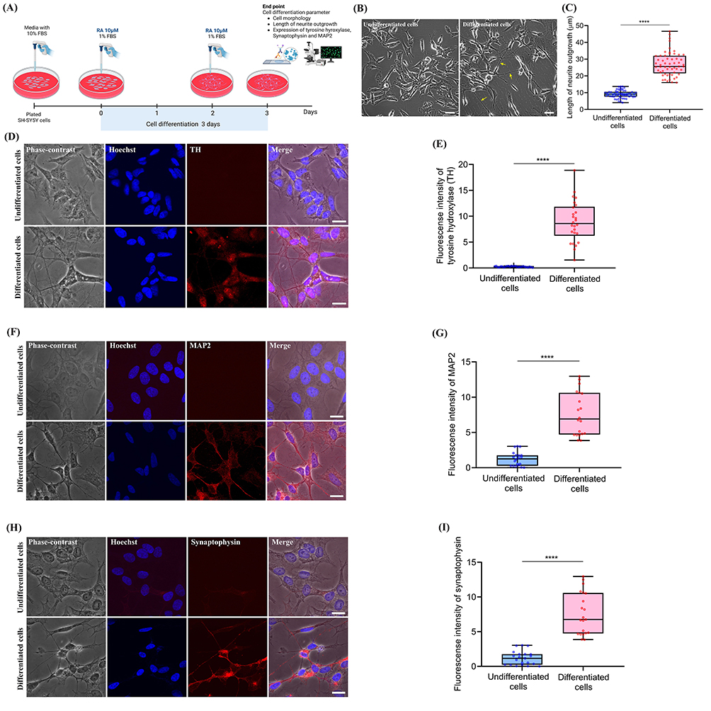 |
Figure 1 Expression of dopaminergic neuronal markers in differentiated SH -SY5Y cells. (A) Timeline of differentiation of SH -SY5Y cells induced by 10 μM retinoic acid in 1% FBS MEM and F12 media. (B) Phase-contrast micrographs show morphological changes in SH -SY5Y cells after three days of differentiation. Yellow arrows indicate neurite outgrowth. Scale bars show 20 μm. (C) Neurite outgrowth length of differentiated cells (****P < 0.0001). (D, H, F) Confocal immunofluorescence images show expression of TH, MAP2, and synaptophysin proteins. Scale bars show 20 μm. (E, G, I) Graph shows fluorescence intensity of TH, MAP2, and synaptophysin, respectively (****P < 0.0001). Data are expressed as mean ± SEM. Each experiment point was performed in triplicate from three different sets of experiments. Hoechst, nuclear staining; TH, tyrosine hydroxylase; MAP2, microtubule associated protein 2; synaptophysin, major protein of synaptic vesicle p38. |
Effect of AA on the Viability of Dopaminergic Neuron-Like Cells
Previous studies have shown that a high concentration of AA reduces cell viability.23 To validate this finding, we treated the dopaminergic neuron-like cells with increasing concentrations of AA (1, 10, 100, and 1000 nM) for 24 h and then analyzed their viability using the MTT assay. The cells treated with AA at 100 nM (P < 0.005) and 1000 nM (P < 0.0001) showed significantly lower viability than the control cells (Figure 2A). Based on these results, in the subsequent experiments, cells were treated with AA at 1 nM and 10 nM for 24 h.
 |
Figure 2 Effect of AA or/and MPP+ on cell viability of dopaminergic neuron-like cells. (A) MTT cell viability assay of dopaminergic neuron-like cells treated with AA (1, 10,100, and 1000 nM) for 24 hours. (B) MTT cell viability assay of dopaminergic neuron-like cells treated with MPP+ (500, 1000, 1500, and 2000 μM) for 24 h. (C) Dopaminergic neuron-like cells were treated with different concentrations of AA (1, 10,100, and 1000 nM) for 24 hours before adding MPP+ to the culture medium for 24 hours, and cell viability was assessed by MTT assay. Data are expressed as mean ± SEM of a percentage to the control group. Each experiment point was performed in triplicate from three different sets of experiments. **P < 0.005, ****P < 0.0001, compared with the control group; #P < 0.05, ####P < 0.0001, compared with the MPP+ treated group. |
Effect of MPP+ on Dopaminergic Neuron-Like Cells
To determine the effect of MPP+ on the viability of the dopaminergic neuron-like cells and select the optimal concentration of MPP+ for use in our experiments, we exposed the cells to different concentrations of MPP+ (500, 1000, 1500, and 2000 μM) for 24 h and determined their viability using the MTT assay. Cell viability decreased as the MPP+ concentration increased in a dose-dependent manner. When compared to the control cells, the cells treated with the following concentrations of MPP+ exhibited significantly lower viability: 500 μM (24%, P < 0.0001), 1000 μM (31%, P < 0.0001), 1500 μM (45%, P < 0.0001), and 2000 μM (59%, P < 0.0001) (Figure 2B). We used 1500 μM MPP+ in our subsequent experiments because this concentration reduced the cell viability to approximately 55%.
Effect of AA on the Viability of MPP+-Treated Dopaminergic Neuron-Like Cells
To investigate the ability of AA to protect dopaminergic neuron-like cells treated with MPP+ against cell death, we incubated cells with AA at different concentrations (1, 10, 100, and 1000 nM) for 24 h before exposing them to 1500 μM MPP+ for 24 h and determining the cell viability. Cells treated with MPP+ for 24 h showed significantly lower viability (approximately 50.91%, P < 0.0001) than the control cells. Pretreatment with AA at 1, 10, and 100 nM before the addition of MPP+ resulted in significantly higher viability (75.22%, P < 0.0001; 81.90%, P < 0.0001; and 63.69%, P < 0.05, respectively) compared with treatment with MPP+ alone. In contrast, pretreatment with AA at 1000 nM before the addition of MPP+ resulted in slightly lower viability (46.64%) (Figure 2C). These results indicated that maximum protection was achieved at an AA concentration of 10 nM, and this concentration was used in the other experiments.
Effect of AA on Autophagy in MPP+-Treated Dopaminergic Neuron-Like Cells
To determine the effect of AA on autophagy, the expression of the autophagy markers LC3, phospho-Beclin-1, and SQSTM1/p62 was assessed via immunocytochemistry and Western blot analysis (Figure 3A). Cells pretreated with AA had significantly higher levels of LC3 and LC3 puncta per cell compared to those in the control group (P < 0.0001). Cells treated with MPP+ did not significantly differ from the control cells in terms of the level of LC3; however, they had significantly less LC3 puncta per cell than the control cells (P < 0.0001). In contrast, the cells in the AA+MPP+ group had significantly more LC3 puncta per cell compared with those in the MPP+ group (P < 0.0001) (Figure 3B–D).
 |
Figure 3 Effect of AA pretreatment on LC3 in MPP+-treated dopaminergic neuron-like cells. (A) After three days of differentiation, cells were pretreated with AA 10 nM for 24 hours before MPP+ treatment for 24 hours and LC3 expression was monitored. (B) Representative confocal immunofluorescence image of LC3 (green) and DAPI nuclear staining (blue). White arrows indicate LC3 puncta in the cytoplasm. Scale bars indicate 10 μm. (C) Quantification of LC3 fluorescence intensity and (D) number of LC3 puncta per cell. Data are shown as mean ± SEM. Each experimental point was performed in triplicate from three different sets of experiments. **P < 0.01, ****P < 0.0001, compared with the control group; ΔΔΔΔP < 0.0001, compared with the AA group; ####P < 0.0001, compared with the MPP+ treated group; n.s, not significance. |
The conversion of LC3-I to LC3-II was examined using Western blot analysis. The LC3-II/LC3-I ratio was significantly lower in the MPP+ group compared to that in the control group (P < 0.005). The AA+MPP+ group had a slightly higher LC3-II/LC3-I ratio than the MPP+ group (Figure 4A). The MPP+ group also exhibited lower phospho-Beclin-1 expression than the control group (P < 0.05), and the AA+MPP+ group showed slightly higher phospho-Beclin-1 expression than the MPP+ group (Figure 4B). When we examined the expression of SQSTM1/p62, it was found that the MPP+ group exhibited significantly higher SQSTM1/p62 expression than the control group (P < 0.05). In contrast, the cells in the AA+MPP+ group showed slightly lower SQSTM1/p62 expression compared with the cells in the MPP+ group (Figure 4C).
 |
Figure 4 Effect of AA pretreatment on LC3-II, phospho-Beclin-1, and SQSTM1/p62 proteins expression in MPP+-treated dopaminergic neuron-like cells. (A) Immunoblot of protein expression in LC3- II /LC3-I ratio. (B) Immunoblot of phosphorylation form of Beclin-1 protein expression. (C) Immunoblot of protein expression of SQSTM1/p62. The density of the bands was normalized with that of β-actin protein. Data are expressed as mean ± SEM of a percentage to the control group. Each experiment point was performed in triplicate from three different sets of experiments. *P < 0.05, **P < 0.005, compared with the control group; ΔP < 0.05, ΔΔP < 0.005 compared with the AA group. LC3, Microtubule-associated protein 1A/1B-light chain 3; phospho-Beclin-1, phosphorylation of Beclin-1; SQSTM1/p62, sequestosome-1/ubiquitin-binding protein p62. |
Together, these results suggested that AA increased autophagy in the MPP+-treated dopaminergic neuron-like cells.
Effect of AA on Mitochondrial Changes in MPP+-Treated Dopaminergic Neuron-Like Cells
We then used confocal microscopy to determine the effect that AA had on mitochondrial changes observed in MPP+-treated dopaminergic neuron-like cells. In the control and AA groups, mitochondria were long and tubular in shape. In contrast, cells in the MPP+ group were found to have punctate and fragmented mitochondria. As shown in Figure 5A, the cells in the AA+MPP+ group exhibited long and tubular mitochondria. Quantification of the intensity of the MitoTracker Red fluorescence revealed that the cells treated with MPP+ had less intense red fluorescence than those in the control group (P < 0.0001). Cells in the AA+MPP+ group showed significantly stronger red fluorescence intensity compared to those in the MPP+ group (P < 0.0001) (Figure 5B). These results indicated that AA diminished mitochondrial alteration in the MPP+-treated dopaminergic neuron-like cells.
 |
Figure 5 Effect of AA pretreatment on mitochondrial morphology in MPP+-treated dopaminergic neuron-like cells. (A) Representative confocal immunofluorescence images of Mitotracker-Red visualizing mitochondria. White arrows indicate the long tubular shape of mitochondria. Yellow arrows indicate fragmented mitochondria. Scale bars indicate 10 μm. (B) Quantification of Mitotracker-Red fluorescence intensity. Data are expressed as mean ± SEM of a percentage to the control group. Each experimental point was performed in triplicate from three different sets of experiments. *P < 0.05, ****P < 0.0001, compared with the control group; ΔΔΔΔP < 0.0001, compared with the AA group; ####P < 0.0001, compared with the MPP+ treated group. |
Effect of AA on the Δψm in MPP+-Treated Dopaminergic Neuron-Like Cells
Next, we measured the effect of AA on the ΔΨm in the MPP+-treated dopaminergic neuron-like cells. For this, we used a JC-10 assay kit and a microplate reader to assess mitochondrial damage. JC-10 exists in a red aggregate form in healthy cells and in a green monomeric form in cells with mitochondrial damage. Figure 6A shows representative images of cells from the different treatment groups, and Figure 6B shows the aggregate-to-monomer ratios calculated for each treatment group. Compared with the cells in the control group, the cells in the MPP+ group exhibited significantly lower ΔΨm values (P < 0.005), and the cells in the AA+MPP+ group showed significantly higher ΔΨm values than those in the MPP+ group (P < 0.05). These results suggested that AA reduced mitochondrial damage in MPP+-treated dopaminergic neuron-like cells.
 |
Figure 6 Effect of AA pretreatment on ΔΨm in MPP+-treated dopaminergic neuron-like cells. The ΔΨm was measured using a JC-10 microplate reader. (A) Representative fluorescence image of live differentiated SH -SY5Y cells stained with JC-10. Red and green fluorescence indicate aggregated (polarized) and monomeric (depolarized) ΔΨm, respectively. Scale bars indicate 20 μm. (B) Quantification of relative JC-10 aggregation/monomer ratio. Data are expressed as mean ± SEM of a percentage to the control group. Each experiment point was performed in triplicate from three different sets of experiments. **P < 0.005, compared with the control group; ΔΔP < 0.005, compared with the AA group; #P < 0.05, compared with the MPP+ treated group. |
Effect of AA on Mitophagy in MPP+-Treated Dopaminergic Neuron-Like Cells
The effect of AA on the colocalization of LC3 and mitochondria in cells treated with MPP+ was investigated using immunofluorescence staining and confocal microscopy. The results are shown in Figure 7. The level of colocalization in the MPP+ group did not differ significantly from that in the control group. Pretreatment with AA was found to result in more colocalization compared to that in the control group (P < 0.0001). Notably, cells in the AA+MPP+ group showed significantly greater colocalization than did cells in the MPP+ group (P < 0.01). These results raised the possibility that AA could protect against and repair mitochondrial damage and increase mitophagy in MPP+-treated dopaminergic neuron-like cells.
 |
Figure 7 Effect of AA pretreatment on mitochondria and mitophagy in MPP+-treated dopaminergic neuron-like cells. (A) Representative confocal immunofluorescence images of Mitotracker-Red and LC3 (green) to visualize mitochondria and autophagy, respectively. White arrows indicate colocalization of LC3 and mitochondria. Scale bars indicate 10 μm. (B) Quantification of colocalization of LC3 and mitochondria. Data are expressed as mean ± SEM of a percentage to the control group. Each experiment point was performed in triplicate from three different sets of experiments. *P < 0.05, ****P < 0.0001, compared with the control group; ΔP < 0.05, ΔΔΔΔP < 0.0001, compared with the AA group; ##P < 0.01, compared with the MPP+ treated group. |
Effect of AA on the Expression of TH in MPP+-Treated Dopaminergic Neuron-Like Cells
We also examined the effect of AA on the expression of TH, the dopaminergic neuron protein essential for survival, in MPP+-treated dopaminergic neuron-like cells. As shown in Figure 8, the cells in the MPP+ group expressed significantly less TH than the cells in the control group (P < 0.0005). Interestingly, the cells in the AA+MPP+ group showed significantly higher TH expression compared to the cells in the MPP+ group (P < 0.05). These results confirmed that AA increased survival in the MPP+-treated dopaminergic neuron-like cells.
 |
Figure 8 Effect of AA pretreatment on TH protein expression in MPP+-treated dopaminergic neuron-like cells. Data are expressed as mean ± SEM of a percentage to the control group. Each experiment point was performed in triplicate from three different sets of experiments. ***P < 0.0005, compared with the control group; ΔP < 0.05, ΔΔΔP < 0.0005 compared with the AA group. #P < 0.05, compared with the MPP+ treated group. TH, tyrosine hydroxylase. |
Effect of AA on Apoptosis in MPP+-Treated Dopaminergic Neuron-Like Cells
To investigate the effect of AA on apoptotic cell death in MPP+-treated dopaminergic neuron-like cells, we employed DAPI nuclear staining and confocal microscopy. The results are shown in Figure 9. The cells in the control and AA groups were found to have normal nuclei, the cells in the MPP+ group showed nuclear condensation and fragmentation, and more normal nuclei were observed in the cells in the AA+MPP+ group than in those in the MPP+ group (Figure 9A). As shown in Figure 9B, there was a significantly greater percentage of apoptotic nuclei in the MPP+ group compared with the control group (P < 0.0001), and there were significantly less apoptotic nuclei in the AA+MPP+ group compared with the MPP+ group (P < 0.0001) (Figure 9B).
 |
Figure 9 Effect of AA pretreatment on apoptotic nuclei and caspase-3/7 activation in MPP+-treated dopaminergic neuron-like cells. (A) Apoptotic nuclear morphology was elucidated using DAPI staining. Apoptotic nuclei were characterized by nuclear condensation (white arrows) and fragmentation (yellow arrows). Scale bars indicate 10 μm and 5 μm in enlarged images. (B) Graph represents percentage of apoptotic cells. (C) Representative live cell fluorescence micrographs of caspase-3/7. White arrows indicate caspase-3/7 activation in cytoplasm of cells. (D) Graph represents the fluorescence intensity of caspase-3/7. Data are expressed as mean ± SEM. Each experiment point was performed in triplicate from three different sets of experiments. **P < 0.005, ****P < 0.0001, compared with the control group; ΔΔΔP < 0.0005, ΔΔΔΔP < 0.0001, compared with the AA group. ####P < 0.0001, compared with the MPP+ treated group. |
We also measured the level of caspase-3/7 activity in live cells to provide further evidence of AA’s ability to protect cells against apoptosis in MPP+-treated dopaminergic neuron-like cells. Cells treated with MPP+ alone were found to have significantly stronger caspase-3/7 activity than cells in the control group (P < 0.0001), whereas cells in the AA+MPP+ group were found to have significantly weaker caspase-3/7 activity than those in the MPP+ group (P < 0.0001) (Figure 9C and D). These results demonstrated that pretreating cells with AA resulted in less apoptotic cell death in MPP+-treated dopaminergic neuron-like cells.
Discussion
In this study, we established a cellular model of PD by treating dopaminergic neuron-like cells with MPP+ and showed that pretreating the cells with AA protected them from cell death. The results suggest that the cytoprotective activity of AA that reduces MPP+-induced apoptosis may be associated with the induction of autophagy and mitophagy, the prevention of mitochondrial damage, and the suppression of caspase-3/7 activation.
Initially, we established a cellular model of PD. For this, we induced neuroblastoma SH-SY5Y cells to differentiate into dopaminergic neuron-like cells by incubation with RA for three days. Similar to previous studies, we observed extensive neurite outgrowth and increased MAP2 and synaptophysin expression, which are markers of mature neurons.24–26 Because both undifferentiated and differentiated SH-SY5Y cells express the dopaminergic neuron marker TH, this cell line is widely used in PD models.27–29 In this study, the differentiated SH-SY5Y cells showed significantly higher expression of TH, which we also observed in our previous studies.30,31
MPP+ is a neurotoxin used to generate animal and cellular models of PD.32,33 It has been shown to exert oxidative stress on cells by inhibiting mitochondrial complex I34 and to induce mitochondrial fission and reduce the ΔΨm by interrupting the mitochondrial electron transport chain.35–37 The presence of antioxidant enzymes, such as superoxide dismutase, protects against MPP+ toxicity in neuronal cell lines38 and dopaminergic neurons in primary culture.39 In this study, incubating cells with 1500 µM MPP+ resulted in punctate and fragmented mitochondria and diminished the ΔΨm in the dopaminergic neuron-like cells. These results are consistent with those of previous studies in which MPP+ and rotenone or H2O2 were used to generate cellular models of PD.20,40
Mitochondria play a central role in apoptosis. Mitochondrial outer membrane (MOM) permeabilization caused by mitochondrial fission leads to the release of proapoptotic factors, such as cytochrome c and apoptosis-inducing factor (AIF).41 Furthermore, studies have indicated that ΔΨm loss contributes to cell death by disrupting normal mitochondrial function42,43 and that a reduction in the ΔΨm initiates the removal of damaged mitochondria, a process known as mitophagy.44,45
In this study, 24-hr pretreating cells with 10 nM AA before incubating them with MPP+ resulted in relatively less mitochondrial damage (ie, less extensive changes to the mitochondrial morphology and ΔΨm). It was also found that treating cells with MPP+ did not significantly affect the colocalization of mitochondria and the autophagy marker LC3 and that pretreating cells with AA before incubating them with MPP+ resulted in notably more colocalization of LC3 and mitochondria compared to that in cells treated with MPP+ only. The elimination of dysfunctional mitochondria is crucial for cell viability; thus, cells have developed diverse mitophagy pathways to ensure that mitophagy occurs under different conditions in a timely manner.46 The concentration of AA is essential for its neuroprotection. In this study, 1–10 nM AA acted perfectly against MPP+ toxicity, however, as the concentrations of AA were increased (100–1000 nM), the protective ability decreased, and even showed toxic effects by reducing cell viability as shown in Figure 2. According to previous studies, at very high pharmacological doses, AA induces apoptosis via production of ROS, increased intracellular Ca2+, alteration of Bax/Bcl-2 ratio and activation of caspase-3.47,48 The present study did not show the different time point of AA treatment. Nevertheless, pretreatment of AA for 24 hr significantly protected MPP+-induced neurotoxicity in dopaminergic neural-like cells. This result correlated with previous studies. 24, 48 and 72 hr of AA administration inhibited A549, H1299 and LLC cell proliferation21 and decreased U87-MG cell viability49 in both dose- and time-dependent manner. Moreover, 24 hr AA pretreatment protected H9c2 cells from ROS‐mediated autophagy via a p38 mitogen‐activated protein kinase/Bcl‐2/beclin‐1 signaling pathway in myocardial ischemia‐reperfusion injury.22 These findings supported that 24 hr pretreatment of AA is sufficient to investigate its effect on cellular activities and underlying molecular mechanisms.
Mitophagy can be categorized into two distinct forms: ubiquitin-mediated mitophagy and receptor-mediated mitophagy. Ubiquitin-mediated mitophagy encompasses the PTEN induced kinase (PINK)/Parkin pathway and additional ubiquitin-mediated mechanisms.50 In this form of mitophagy, PINK1 accumulates on the MOM, recruiting Parkin to ubiquitinate outer membrane components when mitochondria are damaged. Mitochondrial proteins with phosphorylated poly-ubiquitin chains consequently affect other proteins and signal autophagy.51 Beclin-1 is recruited and activated in autophagosomes derived from pre-autophagic structures.52 During a typical autophagic flux, LC3-I is converted to LC3-II via conjugation with phosphatidylethanolamine, and LC3-II then inserts into the autophagosomal membrane.53 The degradation of MOM proteins is initiated by K63-linked ubiquitin, which recruits mitochondrial adaptor proteins and interacts with the LC3-II attached to the autophagosomal membrane. It has been asserted that SQSTM1/p62 preferentially positions itself between mitochondria and facilitates the aggregation of impaired mitochondria via polymerization.54,55 The damaged mitochondria and protein debris are encapsulated within autophagosomes, which subsequently fuse with lysosomes to form autolysosomes that mediate degradation. PINK1, Parkin, and SQSTM1/p62 are degraded through a lysosome-dependent process, whereas LC3-II is recycled.50,56 In the present study, incubating cells with MPP+ increased autophagic flux; however, incomplete autolysosomes were formed, as indicated by the elevated levels of SQSTM1/p62. Pretreating the cells with AA before incubating them with MPP+ significantly enhanced the level of autophagic activity, as evidenced by the increased LC3-II/LC3-I ratio and phospho-Beclin-1 expression, and the presence of complete autolysosomes was indicated by the relatively lower levels of SQSTM1/p62. Our findings align with those of previous studies that found that AA alleviates acute hepatic injury by enhancing autophagy.57
Mechanistically, autophagy is promoted by AMP activated protein kinase (AMPK), which is a key energy sensor and regulates cellular metabolism to maintain energy homeostasis. Conversely, autophagy is inhibited by the mammalian target of rapamycin (mTOR), a central cell-growth regulator that integrates growth factor and nutrient signals. AMPK and mTOR regulate autophagy through direct phosphorylation of Ulk1.58,59 Under glucose starvation, AMPK promotes autophagy by directly activating Ulk1 through phosphorylation of Ser 317 and Ser 777. Under nutrient sufficiency, high mTOR activity prevents Ulk1 activation by phosphorylating Ulk1 Ser 757 and disrupting the interaction between Ulk1 and AMPK.60 Recent study showed AA protects against cardiac hypertrophy by activation of AMPKα, and suppressing mTOR/P70S6K/S6 and ERK, which resulted in restricted hypertrophy and fibrosis, ultimately improving cardiac performance. Moreover, these protective effects of AA were abolished after AMPKα inhibition or deficiency.61 Meanwhile, asiaticoside, a glycoside form, effectively prevented cerebral ischemia‐mediated cognitive impairment and neuronal damage in the rats. Moreover, autophagy was inhibited and the mTOR pathway was activated in rats with cerebral ischemia by asiaticoside treatment.62 The role of mTOR in neurodegenerative diseases is still debated. Because mTOR is critical for protein synthesis and cell survival processes, it is worth noting that mTOR inhibition causes autophagy to activate, which may play a role in neuroprotection. However, long term inhibition can trigger the feedback loop and prevent autophagy. As a result, mTOR is a double-edged sword. It can play both neuroprotective and neurotoxic roles. Hence, the signaling between mTOR and autophagy has to be balanced. If the balance between mTOR and autophagy can be achieved, mTOR might be a possible target for the therapy of PD.63 Therefore, the mechanistic insight of relevant signaling pathways, mTOR/AMPK exes need to be evaluated in the future study for better understanding about AA’s effect on autophagic and mitochondrial functions in PD.
The results of this study also support the notion that AA protects cells against MPP+-induced apoptosis. Caspase-3/7 activation is widely considered the defining molecular marker of apoptotic cell death,64 and it contributes to some mitochondrial events once thought to occur upstream of effector caspases. These caspases control ΔΨm loss and AIF release and may also amplify the initial death signal by promoting cytochrome c release. Although Bax translocation and cytochrome c release eventually occur during the process, it is likely that multiple pathways regulate mitochondrial function in apoptosis.65 In this study, cells incubated with MPP+ showed stronger caspase-3/7 activity and higher numbers of apoptotic nuclei, which indicated that MPP+ induced apoptotic cell death. However, the caspase-3/7 activity and number of apoptotic nuclei were lower in cells pretreated with AA, which suggested that AA protected cells from MPP+-induced apoptosis. These findings are consistent with those of previous studies that found AA had antioxidative and anti-apoptotic effects in Drosophila and cellular models of PD.66 In a recent study, we showed that inhibiting the antioxidant peroxiredoxin (Prdx)-1 promoted apoptosis by increasing reactive oxygen species production, p53 activation (which increased the expression of cleaved caspase-3), and the number of apoptotic nuclei in a cellular model of PD.67
Finally, we analyzed the expression of TH, the enzyme that regulates the rate of dopamine synthesis and is used as a marker of cell function.68 Our results showed that incubating cells with MPP+ resulted in lower cell viability and TH expression. These findings demonstrated that MPP+ had a cytotoxic effect. In cells with lower levels of TH, the ability to synthesize dopamine is reduced, which can lead to dopamine dysfunction in PD patients. However, pretreatment with AA resulted in higher cell viability and TH expression in MPP+-treated dopaminergic neuron-like cells. These findings correlate with those of previous studies that found overexpression of the antioxidant Prdx-2 led to higher TH expression and protected neurons from death after MPP+ treatment in a cellular model.33 Moreover, Prdx-2 overexpression has been shown to prevent the loss of dopaminergic neurons in an 1-methyl-4-phenyl-1,2,3,6-tetrahydropyridine mouse model of PD.69 Previous studies showed that AA has numerous pharmacological actions, including antioxidant, anti-inflammatory, and neuroprotectivity.47,70 The mechanisms may be concerned with reduced ROS, inhibited GSH decrease, and protected mitochondria. The loss of ΔΨm has been identified as the first steps in the apoptotic process.71 Our results were consistent with previous study that MPP+ can disrupt the mitochondrial and autophagic functions together with cellular survival PI3K/AKT/mTOR pathway leading to the dopaminergic neural apoptosis in PD model.30,31,72 AA is shown to act directly on the mitochondria against MPP+ induced depolarization of ΔΨm. Accordingly, we suggest that AA inhibits the apoptosis induced by MPP+ possibly through its antioxidant activity, mitochondrial protection and the autophagy induction. These cellular processes may work synergistically to protect DA neurons from MPP+ induced apoptotic cell death.
Conclusion
The results of this study indicate that AA may protect dopaminergic neuron-like cells from MPP+-induced apoptosis by inducing autophagy, as evidenced by the higher LC3-II/LC3-I ratio and Beclin-1 expression observed in cells pretreated with AA. The lower levels of SQSTM1/p62 found in the cells pretreated with AA suggested that complete autolysosomes were present. Additionally, pretreating cells with AA resulted in tubular mitochondria, improved mitochondrial function, higher levels of TH, and lower caspase-3/7 activity, which led to less apoptotic cell death. Therefore, AA is a potential neuroprotective agent, and further studies should be conducted, including in vivo and clinical studies, to evaluate its use in patients with PD. However, the related molecular mechanisms underlying the autophagic induction of AA require further exploration, which may contribute to uncovering its clinical therapeutic potential in PD management.
Abbreviations
AA, Asiatic acid; C. asiatica, Centella Asiatica; DMSO, Dimethyl sulfoxide; FBS, Fetal bovine serum; F-12, Ham’s F-12 Nutrient Mixture; LC3, Microtubule associated protein 1 light chain 3; MAP2, Microtubule associated protein 2; MEM, Minimum essential medium; MOM, Mitochondrial outer membrane; ΔΨm, Mitochondria membrane potential; MPP+, 1-Methyl-4-phenylpyridinium; MTT, 3-(4, 5-dimethylthiazolyl-2)-2, 5-diphenyltetrazolium bromide; PBS, Phosphate-buffered saline; PD, Parkinson’s disease; Prdx, Peroxiredoxin; PINK, PTEN induced kinase; RA, all-trans-retinoic acid; SQSTM1/p62, Sequestosome-1/ubiquitin-binding protein p62; TH, tyrosine hydroxylase.
Acknowledgments
We thank Kristen Sadler, PhD, from Scribendi (www.scribendi.com) for editing a draft of this manuscript.
Funding
This research project is supported by Faculty of Medicine Ramathibodi Hospital Mahidol University (grant number RF_65090).
Disclosure
The authors declare that they have no competing interests in this work.
References
1. Grunewald A, Kumar KR, Sue CM. New insights into the complex role of mitochondria in Parkinson’s disease. Prog Neurobiol. 2019;177:73–93. doi:10.1016/j.pneurobio.2018.09.003
2. Hou X, Watzlawik JO, Fiesel FC, Springer W. Autophagy in Parkinson’s disease. J Mol Biol. 2020;432(8):2651–2672. doi:10.1016/j.jmb.2020.01.037
3. Zhang J, Culp ML, Craver JG, Darley-Usmar V. Mitochondrial function and autophagy: integrating proteotoxic, redox, and metabolic stress in Parkinson’s disease. J Neurochem. 2018;144(6):691–709. doi:10.1111/jnc.14308
4. Zhang K, Zhu S, Li J, et al. Targeting autophagy using small-molecule compounds to improve potential therapy of Parkinson’s disease. Acta Pharmaceutica Sinica B. 2021;11(10):3015–3034. doi:10.1016/j.apsb.2021.02.016
5. Srivastav S, Fatima M, Mondal AC. Important medicinal herbs in Parkinson’s disease pharmacotherapy. Biomed Pharmacother. 2017;92:856–863. doi:10.1016/j.biopha.2017.05.137
6. Lv J, Sharma A, Zhang T, Wu Y, Ding X. Pharmacological review on asiatic acid and its derivatives: a potential compound. SLAS Technol. 2018;23(2):111–127. doi:10.1177/2472630317751840
7. Orhan IE. Centella asiatica (L.) urban: from traditional medicine to modern medicine with neuroprotective potential. Evid Based Complement Alternat Med. 2012;2012:946259. doi:10.1155/2012/946259
8. Lokanathan Y, Omar N, Ahmad Puzi NN, Saim A, Hj Idrus R. Recent updates in neuroprotective and neuroregenerative potential of centella asiatica. Malays J Med Sci. 2016;23(1):4–14. doi:10.1089/ten.tea.2014.0521
9. Nagoor Meeran MF, Goyal SN, Suchal K, Sharma C, Patil CR, Ojha SK. Pharmacological properties, molecular mechanisms, and pharmaceutical development of asiatic acid: a pentacyclic triterpenoid of therapeutic promise. Front Pharmacol. 2018;9:892. doi:10.3389/fphar.2018.00892
10. Lee JH, Kim HL, Lee MH, et al. Asiaticoside enhances normal human skin cell migration, attachment and growth in vitro wound healing model. Phytomedicine. 2012;19(13):1223–1227. doi:10.1016/j.phymed.2012.08.002
11. Somboonwong J, Kankaisre M, Tantisira B, Tantisira MH. Wound healing activities of different extracts of Centella asiatica in incision and burn wound models: an experimental animal study. BMC Complement Altern Med. 2012;12:103. doi:10.1186/1472-6882-12-103
12. Wan J, Gong X, Jiang R, Zhang Z, Zhang L. Antipyretic and anti-inflammatory effects of asiaticoside in lipopolysaccharide-treated rat through up-regulation of heme oxygenase-1. Phytother Res. 2013;27(8):1136–1142. doi:10.1002/ptr.4838
13. Zhang L, Li HZ, Gong X, et al. Protective effects of Asiaticoside on acute liver injury induced by lipopolysaccharide/D-galactosamine in mice. Phytomedicine. 2010;17(10):811–819. doi:10.1016/j.phymed.2010.01.008
14. Defillipo PP, Raposo AH, Fedoce AG, et al. Inhibition of cPLA2 and sPLA2 activities in primary cultures of rat cortical neurons by Centella asiatica water extract. Nat Prod Commun. 2012;7(7):841–843.
15. Zhuang LG, Zhang R, Jin GX, Pei XY, Wang Q, Ge XX. Asiaticoside improves diabetic nephropathy by reducing inflammation, oxidative stress, and fibrosis: an in vitro and in vivo study. World J Diabet. 2024;15(10):2111–2122. doi:10.4239/wjd.v15.i10.2111
16. Ni Y, Pan Y, Zhou J, et al. Asiaticoside and asiatic acid improve diabetic nephropathy by restoring podocyte autophagy and improving gut microbiota dysbiosis. Biochem Pharmacol. 2025;241:117161. doi:10.1016/j.bcp.2025.117161
17. Boonyarattanasoonthorn T, Kongratanapasert T, Maiuthed A, Hamlin R, Kijtawornrat A, Khemawoot P. Bioenhancing effects of piperine and curcumin on triterpenoid pharmacokinetics and neurodegenerative metabolomes from Centella asiatica extract in beagle dogs. Sci Rep. 2022;12(1):20789. doi:10.1038/s41598-022-24935-7
18. Songvut P, Chariyavilaskul P, Khemawoot P, Tansawat R. Pharmacokinetics and metabolomics investigation of an orally modified formula of standardized Centella asiatica extract in healthy volunteers. Sci Rep. 2021;11(1):6850. doi:10.1038/s41598-021-86267-2
19. Lim J, Lee H, Hong S, Lee J, Kim Y. Comparison of the antioxidant potency of four triterpenes of Centella asiatica against oxidative stress. Antioxidants. 2024;13(4):483. doi:10.3390/antiox13040483
20. Chen D, Zhang XY, Sun J, et al. Asiatic acid protects dopaminergic neurons from neuroinflammation by suppressing mitochondrial ros production. Biomol Ther. 2019;27(5):442–449. doi:10.4062/biomolther.2018.188
21. Wu T, Geng J, Guo W, Gao J, Zhu X. Asiatic acid inhibits lung cancer cell growth in vitro and in vivo by destroying mitochondria. Acta Pharm Sin B. 2017;7(1):65–72. doi:10.1016/j.apsb.2016.04.003
22. Yi C, Si L, Xu J, Yang J, Wang Q, Wang X. Effect and mechanism of asiatic acid on autophagy in myocardial ischemia-reperfusion injury in vivo and in vitro. Exp Ther Med. 2020;20(5):54. doi:10.3892/etm.2020.9182
23. Nataraj J, Manivasagam T, Justin Thenmozhi A, Essa MM. Neuroprotective effect of asiatic acid on rotenone-induced mitochondrial dysfunction and oxidative stress-mediated apoptosis in differentiated SH-SYS5Y cells. Nutr Neurosci. 2017;20(6):351–359. doi:10.1080/1028415x.2015.1135559
24. Rumpansuwon K, Prommahom A, Dharmasaroja P. eEF1A2 knockdown impairs neuronal proliferation and inhibits neurite outgrowth of differentiating neurons. Neuroreport. 2022;33(8):336–344. doi:10.1097/wnr.0000000000001791
25. Haley GE, Eghlidi DH, Kohama SG, Urbanski HF, Raber J. Association of microtubule associated protein-2, synaptophysin, and apolipoprotein E mRNA and protein levels with cognition and anxiety levels in aged female rhesus macaques. Behav Brain Res. 2012;232(1):1–6. doi:10.1016/j.bbr.2012.03.032
26. Lopes FM, Schroder R, da Frota ML Jr, et al. Comparison between proliferative and neuron-like SH-SY5Y cells as an in vitro model for Parkinson disease studies. Brain Res. 2010;1337:85–94. doi:10.1016/j.brainres.2010.03.102
27. Kovalevich J, Langford D. Considerations for the use of SH-SY5Y neuroblastoma cells in neurobiology. Methods Mol Biol. 2013;1078:9–21. doi:10.1007/978-1-62703-640-5_2
28. Nagatsu T, Nakashima A, Ichinose H, Kobayashi K. Human tyrosine hydroxylase in Parkinson’s disease and in related disorders. J Neural Transm. 2019;126(4):397–409. doi:10.1007/s00702-018-1903-3
29. Wang T, Li C, Han B, et al. Neuroprotective effects of Danshensu on rotenone-induced Parkinson’s disease models in vitro and in vivo. BMC Complement Med Ther. 2020;20(1):20. doi:10.1186/s12906-019-2738-7
30. Khwanraj K, Phruksaniyom C, Madlah S, Dharmasaroja P. Differential expression of tyrosine hydroxylase protein and apoptosis-related genes in differentiated and undifferentiated SH-SY5Y neuroblastoma cells treated with MPP(.). Neurol Res Int. 2015;2015:734703. doi:10.1155/2015/734703
31. Prommahom A, Dharmasaroja P. Effects of eEF1A2 knockdown on autophagy in an MPP(+)-induced cellular model of Parkinson’s disease. Neurosci Res. 2021;164:55–69. doi:10.1016/j.neures.2020.03.013
32. Lee G, Joo JC, Choi BY, Lindroth AM, Park SJ, Park YJ. Neuroprotective effects of Paeonia Lactiflora extract against cell death of dopaminergic SH-SY5Y cells is mediated by epigenetic modulation. BMC Complement Altern Med. 2016;16:208. doi:10.1186/s12906-016-1205-y
33. Liu M, Zuo S, Guo X, et al. The study of overexpression of peroxiredoxin-2 reduces MPP(+)-induced toxicity in the cell model of Parkinson’s disease. Neurochem Res. 2023;48(7):2129–2137. doi:10.1007/s11064-023-03880-5
34. Kalivendi SV, Kotamraju S, Cunningham S, Shang T, Hillard CJ, Kalyanaraman B. 1-Methyl-4-phenylpyridinium (MPP+)-induced apoptosis and mitochondrial oxidant generation: role of transferrin-receptor-dependent iron and hydrogen peroxide. Biochem J. 2003;371(1):151–164. doi:10.1042/bj20021525
35. Keane PC, Kurzawa M, Blain PG, Morris CM. Mitochondrial dysfunction in Parkinson’s disease. Parkinsons Dis. 2011;2011:716871. doi:10.4061/2011/716871
36. Trotta AP, Gelles JD, Serasinghe MN, Loi P, Arbiser JL, Chipuk JE. Disruption of mitochondrial electron transport chain function potentiates the pro-apoptotic effects of MAPK inhibition. J Biol Chem. 2017;292(28):11727–11739. doi:10.1074/jbc.M117.786442
37. Verma M, Zhu J, Wang KZQ, Chu CT. Chronic treatment with the complex I inhibitor MPP(+) depletes endogenous PTEN-induced kinase 1 (PINK1) via up-regulation of Bcl-2-associated athanogene 6 (BAG6). J Biol Chem. 2020;295(23):7865–7876. doi:10.1074/jbc.RA119.010474
38. Lai M, Griffiths H, Pall H, Williams A, Lunec J. An investigation into the role of reactive oxygen species in the mechanism of 1-methyl-4-phenyl-1,2,3,6-tetrahydropyridine toxicity using neuronal cell lines. Biochem Pharmacol. 1993;45(4):927–933. doi:10.1016/0006-2952(93)90178-Y
39. Akaneya Y, Takahashi M, Hatanaka H. Involvement of free radicals in MPP+ neurotoxicity against rat dopaminergic neurons in culture. Neurosci lett. 1995;193(1):53–56. doi:10.1016/0304-3940(95)11668-M
40. Xiong Y, Ding H, Xu M, Gao J. Protective effects of asiatic acid on rotenone- or H2O2-induced injury in SH-SY5Y cells. Neurochem Res. 2009;34(4):746–754. doi:10.1007/s11064-008-9844-0
41. van Loo G, Saelens X, van Gurp M, MacFarlane M, Martin SJ, Vandenabeele P. The role of mitochondrial factors in apoptosis: a Russian roulette with more than one bullet. Cell Death Differ. 2002;9(10):1031–1042. doi:10.1038/sj.cdd.4401088
42. Halestrap AP, McStay GP, Clarke SJ. The permeability transition pore complex: another view. Biochimie. 2002;84(2):153–166. doi:10.1016/S0300-9084(02)01375-5
43. Green DR, Kroemer G. The pathophysiology of mitochondrial cell death. Science. 2004;305(5684):626–629. doi:10.1126/science.1099320
44. Vasan K, Clutter M, Fernandez Dunne S, et al. Genes involved in maintaining mitochondrial membrane potential upon electron transport chain disruption. Front Cell Dev Biol. 2022;10:781558. doi:10.3389/fcell.2022.781558
45. Ashrafi G, Schwarz TL. The pathways of mitophagy for quality control and clearance of mitochondria. Cell Death Differ. 2013;20(1):31–42. doi:10.1038/cdd.2012.81
46. Wang S, Long H, Hou L, et al. The mitophagy pathway and its implications in human diseases. Signal Transduct Target Ther. 2023;8(1):304. doi:10.1038/s41392-023-01503-7
47. Park BC, Bosire KO, Lee E-S, Lee YS, Kim J-A. Asiatic acid induces apoptosis in SK-MEL-2 human melanoma cells. Cancer Lett. 2005;218(1):81–90. doi:10.1016/j.canlet.2004.06.039
48. Lee YS, Jin D-Q, Kwon EJ, et al. Asiatic acid, a triterpene, induces apoptosis through intracellular Ca2+ release and enhanced expression of p53 in HepG2 human hepatoma cells. Cancer Lett. 2002;186(1):83–91. doi:10.1016/S0304-3835(02)00260-4
49. Thakor FK, Wan KW, Welsby PJ, Welsby G. Pharmacological effects of asiatic acid in glioblastoma cells under hypoxia. Mol Cell Biochem. 2017;430(1–2):179–190. doi:10.1007/s11010-017-2965-5
50. Li A, Gao M, Liu B, et al. Mitochondrial autophagy: molecular mechanisms and implications for cardiovascular disease. Cell Death Dis. 2022;13(5):444. doi:10.1038/s41419-022-04906-6
51. Heo JM, Ordureau A, Paulo JA, Rinehart J, Harper JW. The PINK1-PARKIN mitochondrial ubiquitylation pathway drives a program of OPTN/NDP52 recruitment and TBK1 activation to promote mitophagy. Mol Cell. 2015;60(1):7–20. doi:10.1016/j.molcel.2015.08.016
52. Menon MB, Dhamija S. Beclin 1 Phosphorylation - at the Center of Autophagy Regulation. Front Cell Dev Biol. 2018;6:137. doi:10.3389/fcell.2018.00137
53. Zhang J. Teaching the basics of autophagy and mitophagy to redox biologists--mechanisms and experimental approaches. Redox Biol. 2015;4:242–259. doi:10.1016/j.redox.2015.01.003
54. Narendra D, Kane LA, Hauser DN, Fearnley IM, Youle RJ. p62/SQSTM1 is required for Parkin-induced mitochondrial clustering but not mitophagy; VDAC1 is dispensable for both. Autophagy. 2010;6(8):1090–1106. doi:10.4161/auto.6.8.13426
55. Yamano K, Kikuchi R, Kojima W, et al. Critical role of mitochondrial ubiquitination and the OPTN-ATG9A axis in mitophagy. J Cell Biol. 2020;219(9). doi:10.1083/jcb.201912144
56. Ni HM, Bockus A, Wozniak AL, et al. Dissecting the dynamic turnover of GFP-LC3 in the autolysosome. Autophagy. 2011;7(2):188–204. doi:10.4161/auto.7.2.14181
57. Pang X, Qiao Q, Vonglorkham S, et al. Asiatic acid ameliorates acute hepatic injury by reducing endoplasmic reticulum stress and triggering hepatocyte autophagy. Biomed Pharmacother. 2020;129:110375. doi:10.1016/j.biopha.2020.110375
58. Glick D, Barth S, Macleod KF. Autophagy: cellular and molecular mechanisms. J Pathol. 2010;221(1):3–12. doi:10.1002/path.2697
59. Alers S, Loffler AS, Wesselborg S, Stork B. Role of AMPK-mTOR-Ulk1/2 in the regulation of autophagy: cross talk, shortcuts, and feedbacks. Mol Cell Biol. 2012;32(1):2–11. doi:10.1128/MCB.06159-11
60. Kim J, Kundu M, Viollet B, Guan KL. AMPK and mTOR regulate autophagy through direct phosphorylation of Ulk1. Nat Cell Biol. 2011;13(2):132–141. doi:10.1038/ncb2152
61. Ma ZG, Dai J, Wei WY, et al. Asiatic acid protects against cardiac hypertrophy through activating AMPKalpha signalling pathway. Int J Biol Sci. 2016;12(7):861–871. doi:10.7150/ijbs.14213
62. Guo M, Xu J, Wang S, Dong B. Asiaticoside reduces autophagy and improves memory in a rat model of dementia through mTOR signaling pathway regulation. Mol Med Rep. 2021;24(3). doi:10.3892/mmr.2021.12284
63. Narayanan L, Thekkekkara D, Kondaveeti SN, et al. Deciphering the intricate role of mTOR signaling and autophagy in Parkinson’s disease and therapeutic prospects. J Appl Pharm Sci. 2024. doi:10.7324/japs.2023.156692
64. Galluzzi L, Vitale I, Aaronson SA. Molecular mechanisms of cell death: recommendations of the nomenclature committee on cell death 2018. Cell Death Differ. 2018;25(3):486–541. doi:10.1038/s41418-017-0012-4
65. Lakhani SA, Masud A, Kuida K, et al. Caspases 3 and 7: key mediators of mitochondrial events of apoptosis. Science. 2006;311(5762):847–851. doi:10.1126/science.1115035
66. Ding H, Xiong Y, Sun J, Chen C, Gao J, Xu H. Asiatic acid prevents oxidative stress and apoptosis by inhibiting the translocation of alpha-synuclein into mitochondria. Front Neurosci. 2018;12:431. doi:10.3389/fnins.2018.00431
67. Wirakiat W, Prommahom A, Dharmasaroja P. Inhibition of the antioxidant enzyme PRDX1 activity promotes MPP(+)-induced death in differentiated SH-SY5Y cells and may impair its colocalization with eEF1A2. Life Sci. 2020;258:118227. doi:10.1016/j.lfs.2020.118227
68. White RB, Thomas MG. Moving beyond tyrosine hydroxylase to define dopaminergic neurons for use in cell replacement therapies for Parkinson’s disease. CNS Neurol Disord Drug Targets. 2012;11(4):340–349. doi:10.2174/187152712800792758
69. Qu D, Rashidian J, Mount MP, et al. Role of Cdk5-mediated phosphorylation of Prx2 in MPTP toxicity and Parkinson’s disease. Neuron. 2007;55(1):37–52. doi:10.1016/j.neuron.2007.05.033
70. Lee MK, Kim SR, Sung SH, et al. Asiatic acid derivatives protect cultured cortical neurons from glutamate-induced excitotoxicity. Res Commun Mol Pathol Pharmacol. 2000;108(1–2):75–86.
71. Zamzami N, Marchetti P, Castedo M, et al. Sequential reduction of mitochondrial transmembrane potential and generation of reactive oxygen species in early programmed cell death. J Exp Med. 1995;182(2):367–377. doi:10.1084/jem.182.2.367
72. Khwanraj K, Prommahom A, Dharmasaroja P. eEF1A2 siRNA suppresses MPP(+)-induced activation of Akt and mTOR and potentiates caspase-3 activation in a Parkinson’s disease model. Sci World J. 2023;2023:1335201. doi:10.1155/2023/1335201
 © 2025 The Author(s). This work is published and licensed by Dove Medical Press Limited. The
full terms of this license are available at https://www.dovepress.com/terms
and incorporate the Creative Commons Attribution
- Non Commercial (unported, 4.0) License.
By accessing the work you hereby accept the Terms. Non-commercial uses of the work are permitted
without any further permission from Dove Medical Press Limited, provided the work is properly
attributed. For permission for commercial use of this work, please see paragraphs 4.2 and 5 of our Terms.
© 2025 The Author(s). This work is published and licensed by Dove Medical Press Limited. The
full terms of this license are available at https://www.dovepress.com/terms
and incorporate the Creative Commons Attribution
- Non Commercial (unported, 4.0) License.
By accessing the work you hereby accept the Terms. Non-commercial uses of the work are permitted
without any further permission from Dove Medical Press Limited, provided the work is properly
attributed. For permission for commercial use of this work, please see paragraphs 4.2 and 5 of our Terms.
Recommended articles

Electroacupuncture Exerts Chondroprotective Effect in Knee Osteoarthritis of Rabbits Through the Mitophagy Pathway
Xing L, Chen X, Guo C, Zhu W, Hu T, Ma W, Du M, Xu Y, Guo C
Journal of Pain Research 2023, 16:2871-2882
Published Date: 21 August 2023
An in-Depth Exploration of the Genetic Interaction Network Between Ferroptosis and Acute Pancreatitis
Li J, Jia Y, Cao F, Wang G, Li F
Journal of Inflammation Research 2023, 16:4425-4439
Published Date: 6 October 2023
Traditional Chinese Medicine Monomers and Their Derivatives as a Promising Therapeutic Tool for Hepatocellular Carcinoma by Activation of Mitophagy
Zhu J, Yin S, Luo S, Yu F, Sun K
Drug Design, Development and Therapy 2025, 19:7069-7087
Published Date: 19 August 2025