Back to Journals » Clinical, Cosmetic and Investigational Dermatology » Volume 15
LncRNA LNCOC1 is Upregulated in Melanoma and Serves as a Potential Regulatory Target of miR-124 to Suppress Cancer Cell Invasion and Migration
Authors Liu C, Ding X, Wei C, Pei Y, Meng F, Zhong Y, Liu Y
Received 24 January 2022
Accepted for publication 12 April 2022
Published 26 April 2022 Volume 2022:15 Pages 751—762
DOI https://doi.org/10.2147/CCID.S359786
Checked for plagiarism Yes
Review by Single anonymous peer review
Peer reviewer comments 2
Editor who approved publication: Dr Jeffrey Weinberg
Changhai Liu,1 Xiangsheng Ding,1 Cuie Wei,1 Yongdong Pei,1 Fanjun Meng,1 Yuren Zhong,1 Yi Liu2
1Department of Burn and Plastic Surgery, The First Affiliated of Hospital of Kangda College of Nanjing Medical University/The First People’s Hospital of Lianyungang, Lianyungang, People’s Republic of China; 2Department of Burn Plastic Surgery and Wound Repair, Second Hospital of Lanzhou University, Lanzhou, People’s Republic of China
Correspondence: Changhai Liu, Department of Burn and Plastic Surgery, The First Affiliated of Hospital of Kangda College of Nanjing Medical University/The First People’s Hospital of Lianyungang, No. 6 Zhenhua East Road, Haizhou District, Lianyungang City, 222000, People’s Republic of China, Email [email protected] Yi Liu, Department of Burn Plastic Surgery and Wound Repair, Second Hospital of Lanzhou University, No. 82 Cuiyingmen, Chengguan District, Lanzhou City, 730030, People’s Republic of China, Email [email protected]
Background: A cascade of genes and pathways have been reported in the precise regulation of malignant melanoma (MM). Previous study has demonstrated that lncRNA LNCOC1 is an oncogenic factor in the pathogenesis and development of various cancers. The present study explored the functionalities of LNCOC1 and its interactions with miR-124 in MM.
Methods: A total of 65 melanoma patients were enrolled in this study. The expression of LNCOC1 and miR-124 after cell transfection were detected by RT-qPCR. The migration rates of SK-MEL-3 and A375 cells after transient transfection with LNCOC1 expression vector and miR-124 mimic was detected by trans-well assay.
Results: LNCOC1 was accumulated to high levels in melanoma, and it was significantly correlated with the low survival rate of melanoma patients. Our bioinformatics data showed that miR-124 could target LNCOC1. Overexpression of miR-124 could downregulate LNCOC1. However, up-regulated the expression of LNCOC1 did not affect the expression of miR-124. Our correlation analysis also revealed that the expression of LNCOC1 and miR-124 were inversely correlated in both melanoma tissues and non-tumor tissues. The trans-well invasion and migration assays indicated that overexpression of miR-124 inhibited the melanoma cell invasion and migration. However, overexpression of LNCOC1 promoted melanoma cell invasion and migration.
Conclusion: LNCOC1 is upregulated in melanoma, which can be considered as a potential target of miR-124 in modulating melanoma cell invasion and migration. LNCOC1 may also be an interfering target of MM therapy.
Keywords: LNCOC1, miR-124, melanoma, invasion, migration
Introduction
MM is a type of skin cancer that originates from pigment-containing cells or melanocytes.1 In rare cases, MM can also invade intestines, mouth or even eyes.2 In 2018, MM affected 287,723 new cases, accounting for 1.6% of all newly diagnosed cases, and caused 60,712 deaths, which are 0.6% of cancer-related deaths.3 At early stages, MM causes obvious symptoms, such as pigment spreading, unhealable sore and skin swelling or redness.4,5 Most MM patients are diagnosed at early stages and the prognosis is generally satisfactory.4,5 It is estimated that more than 90% patients with MM can live long than 5 years after the initial diagnosis.6 However, in cases of thicker melanoma and metastatic melanoma, the overall survival rate is always disappointing.
The main risk factors for MM include sunburn, fair skin, UV light exposure and unusual moles.7 Besides, cancer-related molecular pathways also play critical roles in the pathogenesis of MM.8 Increased understanding of the molecular mechanisms of this disease provides novel insights into the development of targeted therapies.9 One study proposed that obesity is involved in melanoma progression and Adipokines (leptin and resistin) enhanced cell growth and proliferation of melanoma via the Akt signaling pathway.10 Moreover, some molecules have been demonstrated to exert protective effects on the progression of melanoma. Methyl-β-cyclodextrin has been reported to be able to induce anticancer effects by causing cell cycle arrest and induction of apoptosis. Although the functions of various RNAs in melanoma have been investigated, the exact mechanism is still unclear.
Immunotherapy refers to a treatment method that artificially enhances or inhibits the immune function of the body to treat the disease by targeting the low or hyperactive immune state of the body.11 MM is characterized by a higher incidence in immunocompromised patients, active lymphocytic infiltration in primary tumors and metastases, and infiltrating T lymphocytes that recognize melanoma antigens.12 Immunotherapy can be divided into four categories, the first category is biological immunotherapy, such as cytokines, interferon and granulocyte-monocyte colony-stimulating factor.13 The second category is peptide, whole protein, virus, DNA or dendritic cell vaccination.14 The third category is cell therapy, which involves the use of lymphocyte-activated killer cells, tumor-infiltrating lymphocytes and other specific lymphocytes.15 The fourth category is immune checkpoint blockades.16 In the past few years, the immunological origins of MM have led to the discovery of antibodies against specific targets, and the mechanism of action of this immunotherapy has focused on specific targets of counter-regulatory mechanisms of the immune response, such as anti-programmed cell death 1 and anti-cytotoxic T lymphocyte-associated protein 4. These blockers greatly increased and prolonged the overall survival in patients with metastatic MM.17
miRNAs are 18–25 nucleotides in length and function as important modulators of gene expression through binding to the recognition sequence of their target RNAs. It has been reported that miRNAs play important roles in translational repression or mRNA degradation,18,19 and are involved in the pathogenesis and progression of various human cancers.20–22 Recent studies have revealed that miRNAs can promote or suppress the progression of MM.23,24 miR-124 was reported to act as a tumor suppressor through targeting Ral-interacting protein of 76 kDa (RLIP76),25 versican26 and RACK1.27 However, other important molecular targets of miR-124 in MM remain elusive.
As important regulators in the gene expression network, non-coding RNAs (ncRNAs) regulate the expression of cancer-related genes to suppress or promote cancer progression.28 LncRNA-ovarian cancer associated 1 (LNCOC1) has been shown to be an oncogene in ovarian cancer. It was reported that LNCOC1 has abundant expression in OC tissues, and could promote the pathogenesis of OC cells.29,30 However, the precise mechanism of how Lnc-OC1 exerts its roles in the pathogenesis of MM is still elusive.
In this study, our bioinformatics analyses revealed that LNCOC1 might be a regulatory target of miR-124, which was also an important player in cancer biology.31 In addition, we observed that the expression levels of LNCOC1 were elevated in melanoma tissues than that in non-tumor tissues, and up-regulated expression of miR-124 in melanoma cells reduced the expression levels of LNCOC1. Moreover, overexpression of LNCOC1 could promote the invasion and migration of melanoma cells.
Materials and Methods
Research Subjects
A total of 65 melanoma patients (40 males and 25 females, mean age 51.7 ± 5.5 (14–63) years old) were enrolled in the First Affiliated of Hospital of Kangda College of Nanjing Medical University/The First People’s Hospital of Lianyungang between December 2017 and August 2019. The Ethics Committee of this hospital approved this study. All patients signed the informed consent. The clinical features of MM patients are listed in Table 1. The inclusion criteria of patients in this study were as follows: 1) the cases were newly diagnosed; 2) there was no therapies initiated within 3 months before this study; 3) no other systemic diseases were diagnosed, such as other malignancies, severe infections, other types of skin disease. The Exclusion criteria of patients in this study were as follows: 1) the cases were recurrent; 2) multiple chronic diseases were diagnosed. The follow-up strategy was strictly followed by all patients: 1) within 2 years, re-check every 3 months; 2) re-check every half a year within 5 years; 3) re-check when patient feels physically unpleasant after 5 years.
 |
Table 1 Baseline Clinicopathological Characteristics of OSCC Patients |
Bioinformatics Analysis
IntaRNA 2.0 software was used to analyze the interaction site between miR-124 and LNCOC1, the dimer formed by the seed sequence and the two and its stability. The Hazard Ratio was calculated by a multivariate Cox proportional hazard model.
Tissue Samples and Cells
Paired melanoma and adjacent (3 cm around melanoma) non-tumor tissues were obtained from each patient. All tissue samples were subjected to histopathological test. It was shown that all melanoma tissues contained more than 95% cancer cells, and less than 1% of cancer cells were observed in non-tumor tissues. All fresh tissue specimens were frozen at −80°C. Primary neonatal normal human epidermal melanocytes were obtained from Sangon Biotech (Shanghai, China). MM cell lines SK-MEL-3 and A375 (ATCC) were used. DMEM was used to culture the cells in a humidified atmosphere containing 5% CO2 at 37°C. Cells were harvested at confluence of about 85% to perform subsequent experiments.
Cell Transfections
Expression vector of LNCOC1 was constructed using pcDNA3.1 vector as backbone. miR-124 mimic, control miRNA, LNCOC1 knockdown negative control and LNCOC1 shRNA were synthesized by Sangon Biotech (Shanghai, China). Empty vector pcDNA3.1, control miRNA or LNCOC1 knockdown control RNA were used as negative control (NC). Following the manufacturer’s instructions of Lipofectamine 2000 (Lipo2000, Invitrogen), after SK-MEL-3 and A375 cells were harvested and counted, 10 nM expression vector, knockdown vector or 40 nM miRNA using were transfected with Lipo2000. miR-124 mimic, control miRNA, LNCOC1 shRNA and control shRNA were designed by Invitrogen, and the sequences were as following:
Control miRNA: 5’-UUGUACUACACAAAAGUACUG-3’
miR-124 mimic: 5’-UAAGGCACGCGGUGAAUGCC-3’
LNCOC1 shRNA: 5’-AGTGCTCCTAGTGTTACCAGAG-3’
Control shRNA: 5’-ACTAGCTAGGCATCGATATCAG-3’
RNA Preparation and RT-qPCR
Total RNAs (including miRNA) from SK-MEL-3 and A375 cell lines and paired tissue samples were isolated by Trizol reagent (Invitrogen). To retain miRNA, 85% ethanol was used in both washing and precipitation steps. RNA integrity was tested by electrophoresis using 5% urine-PAGE gel. RNA was used to synthesize cDNAs. The cDNA was used as the template for PCR amplification, and the PCR products were purified and diluted as 10-fold gradients (10−1 to 10−6). Each of the six concentration gradients was used as the template, and ddH2O was used to set a negative control. A fluorescence quantitative PCR amplification was performed to establish a standard curve. The reaction conditions were pre-denaturation at 95°C for 3 min, denaturation 95°C for 10 s, annealed for 30 s, and the annealing temperature was 53.9°C for 40 cycles, and fluorescence was collected after each cycle to generate an amplification curve. The expression of LNCOC1 and miR-214 were analyzed by qPCR with GAPDH or U6 as the internal control, respectively. Ct values were calculated by 2−ΔΔCT method, all experiments were conducted in three replicates.
The primers sequences were as follows:
LNCOC1 reverse: 5’-CTGTGGTCACAAAGGCCTGA-3′
LNCOC1 forward: 5’-GGCCTGTGTGTTGAATGCTG-3′
GAPDH reverse: 5’-GTGCTAAGCAGTTGGTGGTG-3′
GAPDH forward: 5’-ATGGGTGTGAACCATGAGAA-3′
miR-124 reverse: 5’-CAGTGCAGGGTCCGAGGTAT-3′
miR-124 forward: 5’-CGACGTAAGGCACGCG-3′
U6 reverse: 5’-CGCTTCACGAATTTGCGTGTCAT-3′
U6 forward: 5’-GCTTCGGCAGCACATATACTAAAAT-3′
Cell Proliferation Analysis
Primary human melanocytes were collected. To prepare single-cell suspensions, 3 × 10⁴ cells were resuspended in 1 mL culture medium containing 10% FBS. Before cell collection, 10% CCK-8 was added, and OD values at 450 nm were measured after 4 h.
Trans-Well Assays
Trans-well insert (8.0 μm pore, Corning) was used to measure cell invasion and migration rates of SK-MEL-3 and A375 cells in each transfection group. Uncoated- and Matrigel (Millipore, USA)-coated membranes were applied in migration and invasion assays, respectively. After cell culture at 37°C for 12 h, 0.5% crystal violet (Sigma-Aldrich) was used to stain the lower surface of membrane for 15 min in dark. Cells were observed under a light microscope and then images were captured.
Statistical Analysis
Paired tissues and multiple groups were compared using paired t-test and ANOVA Tukey’s test, respectively. According to the RT-qPCR data, the 65 melanoma patients were subgrouped into low or high LNCOC1 expression group (n = 31 for low; n = 34 for high), low or high miR-124 expression group (n = 31 for low; n = 34 for high). The median expression level of LNCOC1 or miR-124 in melanoma tissues was used as the cutoff value. Kaplan-Meier and Log rank test were used to analyze the survival curves. P < 0.05 indicated that the difference was significant.
Results
The Upregulated Expression of LNCOC1 in Melanoma Predicts Poor Survival
The expression of LNCOC1 and miR-124 in paired melanoma and non-tumor tissues from the 65 melanoma patients were measured by RT-qPCR. The expression levels of LNCOC1 were significantly higher in melanoma tissues (Figure 1A, p < 0.05) than that in non-tumor tissues. Most patients who died during follow-up had high expression levels of LNCOC1 (Figure 1B, Hazard Ratio = 1.862; p < 0.05), suggesting that the poor survival in melanoma might be due to the high expression levels of LNCOC1, which might be involved in the pathogenesis of melanoma. On the contrary, compared with the non-tumor tissues, the expression levels of miR-124 were remarkably lower in melanoma tissues (Figure 1C, p < 0.05), and lower expression levels of miR-124 indicated poorer survival of melanoma patients (Figure 1D, Hazard Ratio = 0.5371; p < 0.05), indicating that miR-124 was an anti-pathological factor, which has been revealed in previous studies on the functions of miR-124 in melanoma.
 |
Figure 1 LNCOC1 and miR-124 had opposite expression patterns in melanoma. Expression of LNCOC1 and miR-124 in paired tissues was studied with RT-qPCR. The 65 patients with MM were grouped into high and low LNCOC1 or miR-124 level groups (n = 34 for low; n = 31 for high) with the median expression levels of LNCOC1 or miR-124 as cutoff value. Paired tissues and Survival curves of LNCOC1 (A and B) or miR-124 (C and D) were respectively shown. *p < 0.05. |
LNCOC1 Promotes Cell Proliferation of Primary Human Melanocytes
The expression of LNCOC1 in melanoma cell lines and primary human melanocytes was examined. Primary human melanocytes were used as the control group. As shown in Figure 2A, compared with primary human melanocytes, the expression levels of LNCOC1 were significantly increased in melanoma cell lines, while the expression levels of miR-124 were remarkably decreased in melanoma cell lines (Figure 2B, p < 0.05). These data indicated that upregulation of LNCOC1 might be associated with melanoma progression. Thus, LNCOC1 was transfected into human melanocytes. Compared to C and NC (empty pcDNA3.1 vector and control miRNA) groups, overexpression of LNCOC1 increased the proliferation rate of human melanocytes (Figure 2C, p < 0.05). However, overexpression of miR-124 had opposite effects on cell proliferation (Figure 2C, p < 0.05).
 |
Figure 2 The expression levels of LNCOC1 were increased in melanoma cell lines and enhanced cell proliferation of primary human melanocytes. Expression of LNCOC1 and miR-124 in primary human melanocytes and melanoma cell lines were detected by RT-qPCR. The expression levels of LNCOC1 were much higher in SK-MEL-3 and A375 than that in primary melanocytes (A and B). Overexpressed LNCOC1 could promote the proliferation of primary melanocytes. As a suppressive factor in cancer, miR-124 had an inhibitory effect on melanocytes proliferation (C). *p < 0.05. |
miR-124 Targets LNCOC1 to Downregulate Its Expression in Melanoma Cells
IntaRNA 2.0 was used to predict the interaction between miR-124 and LNCOC1. It was observed that miR-124 and LNCOC1 may form multiple-base pairs. The relationship of LNCOC1 with melanoma has not been reported in other literatures (Figure 3A). To further explore the relationship between them, RT-qPCR was used to confirm the overexpression of miR-124 and after transfection into SK-MEL-3 and A375 cells (Figure 3B, p < 0.05). Comparing to the control groups, overexpression of miR-124 led to downregulate expression of LNCOC1 (Figure 3C, p < 0.05), while overexpression of LNCOC1 did not affect the expression of miR-124 (Figure 3D).
 |
Figure 3 MiR-124 targets LNCOC1 to downregulate its expression in melanoma cells. The interaction between miR-124 and LNCOC1 was first predicted by performing RNA interaction prediction software IntaRNA 2.0 (A). SK-MEL-3 and A375 cells were transfected with miR-124 mimic or LNCOC1 expression vector, and the overexpression of miR-124 and LNCOC1 were confirmed by performing RT-qPCR (B). The effect of miR-124 overexpression on LNCOC1 transcription was assessed by RT-qPCR at 48 h post-transfection (C), and the effect of LNCOC1 on the expression of miR-124 was detected by RT-qPCR as well (D). MiR-124 overexpression could inhibit the transcription of LNCOC1, however, on effect was observed in the perturbation of LNCOC on miR-124. *p < 0.05. |
The Correlation of LNCOC1 with miR-124
The correlation of LNCOC1 and miR-124 was evaluated by measuring their expression by RT-qPCR (Figure 1A and C). The expression of LNCOC1 and miR-124 were inversely correlated across melanoma tissues (Figure 4A) and non-tumor tissues (Figure 4B) derived from melanoma patients, which suggested that the correlation between LNCOC1 and miR-124 might be an important regulatory mechanism in the pathogenesis and progression of melanoma.
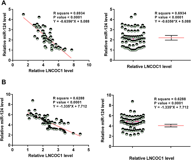 |
Figure 4 The expression levels of LNCOC1 and miR-124 were inversely and significantly correlated. Expression of LNCOC1 and miR-124 in melanoma tissues and non-tumor tissues from 65 MM patients were evaluated by RT-qPCR. Spearman correlation coefficient was used to analyze the correlation between miR-124 and LNCOC1 across melanoma tissues and non-tumor tissues (A and B). The data showed miR-124 was negatively correlated with LNCOC1 in both types of tissues from melanoma patients. |
LNCOC1 Regulates Melanoma Cell Invasion and Migration
Trans-well assays were performed to explore the roles of LNCOC1 and miR-124 on the invasion (Figure 5A and B) and migration (Figure 5C and D) of SK-MEL-3 and A375 cells. Overexpression of LNCOC1 promoted invasion and migration abilities of melanoma cell lines (p < 0.05). In addition, overexpression of miR-124 reversed these effects (p < 0.05), indicating that LNCOC1 and miR-124 play opposite roles in regulating pathological behaviors of melanoma cells. Moreover, knockdown of LNCOC1 (Figure 5E) resulted in a suppressed invasion (Figure 5F and G) and migration (Figure 5H and I) of SK-MEL-3 and A375 cells, which suggested that LNCOC1 is a negative factor in the regulation of melanoma progression.
 |
Figure 5 MiR-124 targeted LNCOC1 to suppress melanoma cell invasion and migration. Trans-well assay was performed to analyze the effects of overexpression of LNCOC1 and miR-124 on the invasion and migration of SK-MEL-3 and A375 cells. LNCOC1 elevation significantly promoted melanoma cell invasion (A and B) and migration (C and D). In the contrary, overexpression of miR-124 remarkably inhibited cell invasion (A and B) and migration (C and D) and could inverse the effects of LNCOC1. LNCOC1 was knocked down in both SK-MEL-3 and A375, and the knocking down efficiency was evaluated by conducting RT-qPCR (E). In addition, suppressing LNCOC1 in melanoma cells could significantly prohibit cell invasion and migration (F–I). *p < 0.05. |
Discussion
The interaction between LNCOC1 and miR-124 in human MM was investigated in this study. We found that the expression of LNCOC1 and miR-124 were altered in melanoma, and miR-124 may target LNCOC1 to suppress cancer cell invasion and migration.
Tumor metastasis is an important reason for the high mortality of malignant melanoma and the poor quality of life in advanced patients.32 Studies have shown that lncRNAs may play a tumor suppressive or tumor-promoting role in the invasion and metastasis of malignant tumor cells.33 LncRNAs can induce EMT by regulating transcription factors such as E-cadherin, N-cadherin, vimentin, β-catenin, Snail, slug, etc., or alter the extracellular matrix (ECM) by changing the activity of ECM degrading enzymes (matrix metallopeptidase, MMP).34 In addition, it can activate the Wnt/β-catenin, PI3K/AKT, MAPK and other signaling pathways, thereby promoting the invasion and metastasis of malignant melanoma.35 A recent study reported the role of oncogenic lncRNA LNCOC1 in ovarian cancer.36 LNCOC1 is abundantly expressed in ovarian cancer tissues and its upregulation led to increased migration and invasion of ovarian cancer cells’.37 Based on current knowledge, the involvement of LNCOC1 in other types of cancer is unclear. This study firstly reported that LNCOC1 is upregulated in melanoma, and high expression levels of LNCOC1 was significantly associated with the poor survival of melanoma patients.
Increasing evidence has demonstrated that miRNAs play important roles as oncogenes or tumor suppressor genes in tumorigenesis and development. Recent studies have found that miRNAs such as miRNA-137,38 miRNA-106b,39 miRNA-20540 and miRNA-125b41 are crucial factors in regulating melanoma cell proliferation. Moreover, miR-124 was remarkably downregulated in melanoma, which was consistent with previous findings that miR-124 is reduced and a suppressor in melanoma.27 Furthermore, our regression analysis also showed that low expression levels of miR-124 predicted poor progression of melanoma patients, which was consistent with previous study that low expression levels of miR-124 in breast cancer was associated with poor prognosis of breast cancer.36 On the other hand, we used primary melanocytes as the control to compare the expression of LNCOC1 and miR-124 in two types of melanoma cell line, A375 and SKMEL3, which showed that the expression of LNCOC1 and miR-124 were similar in A37 and SKMEL3 cells, and both of them had higher expression levels of LNCOC1 and lower expression levels of miR-124 than that in primary melanocytes. To investigate how LNCOC1 and miR-124 affect the primary melanocytes, we next conducted cell proliferation assay to observe this effect, which showed that LNCOC1 could significantly promote cell proliferation, while miR-124 played an opposite role in the primary melanocytes’ proliferation.
Both LNCOC1 and miR-124 are important factors in the progression of melanoma. To clarify the relationship between these two RNAs, we first predicted the interaction between them by bioinformatics analysis, which showed a binding site with miR-124 in the 3’-UTR of LNCOC1. The relationship between LNCOC1 and miR-124 was also evaluated by conducting transfections in melanoma cell lines. The results indicate that overexpression of miR-124 was able to inhibit the expression of LNCOC1. However, LNCOC1 had no effect on miR-124 transcription, which suggests the modifications between LNCOC1 and miR-124 is unidirectional. Besides, in both melanoma tissues and non-tumor tissues, our regression analysis revealed that the expression of LNCOC1 and miR-124 were inversely correlated with each other, which suggested that LNCOC1 and miR-124 are two players that exert different functions in the progression of melanoma.
To clarify how LNCOC1 and miR-124 affect melanoma tumor behaviors, we assessed melanoma cell lines’ invasion and migration with overexpression of LNCOC1 and miR-124 as well as their co-effect. We observed that the overexpression of LNCOC1 could significantly exacerbate the invasion and migration of A375 and SKMEL3 cells. However, miR-124 had the opposite effects. On the other hand, knockdown of LNCOC1 could markedly prohibit the invasion and migration of melanoma cells. It is worth noting that the functionality of miR-124 in cancer biology is controversial. It has been reported that miR-124 can target Interleukin-11 to suppress bone metastasis in breast cancer.31 However, miRNA-124 downregulates PTEN in gastric cancer cells to promote the peritoneal metastasis of tumors.37 A recent study reported that dissemination of melanoma could be suppressed by depleting miR-124, suggesting that miR-124 might exert an oncogenic role in melanoma.42 However, our study showed the downregulation of miR-124 in melanoma and its inhibitory effects on melanoma cell invasion and migration, indicating its suppressive roles in cancer. The opposite observations may be caused by differential roles of miR-124 in different types of cancer. Thus, future studies are needed to confirm the suppressive effects of miR-124 in melanoma.
Conclusion
In summary, this study proved that miR-124 might target LNCOC1 to suppress the tumor behaviors of melanoma. The targeting role of miR-124 to LNCOC1 may also occur in melanoma patients based on the observation that the expression of LNCOC1 and miR-214 were inversely and significantly correlated across both melanoma and non-tumor tissues. For the limitation of our research, we did not reveal how overexpression of miR-124 affects LNCOC1 and why there was no response of the expression of miR-124 to LNCOC1 elevation. Thus, the underlying mechanism on the relationship between these two RNAs needs to be further explored. Accordingly, the mechanism of this research was described as the following scheme (Figure 6).
 |
Figure 6 The scheme of mechanism of the effect of LNCOC1 and miR-124 on melanoma. In MM patients, LNCOC1 displayed an elevated level in their tumor tissues. Also, a decrease in miR-124 was found in those tumor samples, which might contribute to the poor survival of MM patients. On the other hand, overexpression of miR-124 could downregulate LNCOC1 expression, suggesting LNCOC1 is an inhibitory target of miR-124 to suppress the invasion and migration of melanoma cells. In addition, LNCOC1 reduction and miR-124 elevation inhibited primary human melanocyte proliferation to play protective roles in the progression of melanoma. |
Data Sharing Statement
The data that support the findings of this study are available on request from the corresponding author.
Ethical Approval and Consent to Participate
All patients signed the written informed consent. Informed consent was provided by parents or legal guardians of patients under 18 years of age. All procedures were approved by The First Affiliated of Hospital of Kangda College of Nanjing Medical University/The First People’s Hospital of Lianyungang Ethics Committee. Procedures operated in this research were completed in keeping with the standards set out in the Announcement of Helsinki and laboratory guidelines of research in China.
Author Contributions
All authors made substantial contributions to the conception and design, data acquisition, analysis and interpretation, and drafting and editing the manuscript; agreed to submit to the current journal; gave final approval of the version to be published; and agree to be accountable for all aspects of the work.
Funding
There is no funding to report.
Disclosure
All authors have no conflicts of interest.
References
1. Schadendorf D, Fisher DE, Garbe C, et al. Melanoma. Nature Reviews Dis Prim. 2015;1:15003. doi:10.1038/nrdp.2015.3
2. Shields CL, Kels JG, Shields JA. Melanoma of the eye: revealing hidden secrets, one at a time. Clin Dermatol. 2015;33(2):183–196. doi:10.1016/j.clindermatol.2014.10.010
3. Bray F, Ferlay J, Soerjomataram I, Siegel RL, Torre LA, Jemal A. Global cancer statistics 2018: GLOBOCAN estimates of incidence and mortality worldwide for 36 cancers in 185 countries. CA Cancer J Clin. 2018;68(6):394–424. doi:10.3322/caac.21492
4. Tsao H, Olazagasti JM, Cordoro KM, et al. Early detection of melanoma: reviewing the ABCDEs. J Am Acad Dermatol. 2015;72(4):717–723. doi:10.1016/j.jaad.2015.01.025
5. Rat C, Hild S, Rault Sérandour J. Use of smartphones for early detection of melanoma: systematic review. J Med Internet Res. 2018;20(4):e135. doi:10.2196/jmir.9392
6. Robert C, Karaszewska B, Schachter J, et al. Improved overall survival in melanoma with combined dabrafenib and trametinib. N Engl J Med. 2015;372(1):30–39. doi:10.1056/NEJMoa1412690
7. Bränström R, Chang YM, Kasparian N, et al. Melanoma risk factors, perceived threat and intentional tanning: an international online survey. Eur J Cancer Prevent. 2010;19(3):216–226. doi:10.1097/CEJ.0b013e3283354847
8. Liu Y, Sheikh MS. Melanoma: molecular pathogenesis and therapeutic management. Mol Cell Pharmacol. 2014;6(3):228.
9. Smalley KS. Understanding melanoma signaling networks as the basis for molecular targeted therapy. J Invest Dermatol. 2010;130(1):28–37. doi:10.1038/jid.2009.177
10. Malvi P, Chaube B, Pandey V, et al. Obesity induced rapid melanoma progression is reversed by orlistat treatment and dietary intervention: role of adipokines. Mol Oncol. 2015;9(3):689–703. doi:10.1016/j.molonc.2014.11.006
11. Eggenhuizen PJ, Ng BH, Ooi JD. Treg enhancing therapies to treat autoimmune diseases. Int J Mol Sci. 2020;21(19):7015. doi:10.3390/ijms21197015
12. Rosenberg SA, Restifo NP. Adoptive cell transfer as personalized immunotherapy for human cancer. Science. 2015;348(6230):62–68. doi:10.1126/science.aaa4967
13. Peng M, Mo Y, Wang Y, et al. Neoantigen vaccine: an emerging tumor immunotherapy. Mol Cancer. 2019;18(1):128. doi:10.1186/s12943-019-1055-6
14. Gardner A, de Mingo Pulido Á, Ruffell B. Dendritic cells and their role in immunotherapy. Front Immunol. 2020;11:924. doi:10.3389/fimmu.2020.00924
15. Wolf MM, Kimryn Rathmell W, Beckermann KE. Modeling clear cell renal cell carcinoma and therapeutic implications. Oncogene. 2020;39(17):3413–3426. doi:10.1038/s41388-020-1234-3
16. Zappasodi R, Wolchok JD, Merghoub T. Strategies for predicting response to checkpoint inhibitors. Curr Hematol Malig Rep. 2018;13(5):383–395. doi:10.1007/s11899-018-0471-9
17. Uhara H. Recent advances in therapeutic strategies for unresectable or metastatic melanoma and real-world data in Japan. Int J Clin Oncol. 2019;24(12):1508–1514. doi:10.1007/s10147-018-1246-y
18. Wang G, Fu Y, Liu G, Ye Y, Zhang X. miR-218 inhibits proliferation, migration, and EMT of gastric cancer cells by targeting WASF3. Oncol Res. 2017;25(3):355–364. doi:10.3727/096504016X14738114257367
19. Lv H, Zhang Z, Wang Y, Li C, Gong W, Wang X. MicroRNA-92a promotes colorectal cancer cell growth and migration by inhibiting KLF4. Oncol Res. 2016;23(6):283–290. doi:10.3727/096504016X14562725373833
20. Loh HY, Norman BP, Lai KS, Rahman N, Alitheen NBM, Osman MA. The regulatory role of MicroRNAs in breast cancer. Int J Mol Sci. 2019;20(19). doi:10.3390/ijms20194940
21. Liu H, Lei C, He Q, Pan Z, Xiao D, Tao Y. Nuclear functions of mammalian MicroRNAs in gene regulation, immunity and cancer. Mol Cancer. 2018;17(1):64. doi:10.1186/s12943-018-0765-5
22. Adams BD, Kasinski AL, Slack FJ. Aberrant regulation and function of microRNAs in cancer. Current Biol. 2014;24(16):R762–R776. doi:10.1016/j.cub.2014.06.043
23. Zhou J, Xu D, Xie H, et al. miR-33a functions as a tumor suppressor in melanoma by targeting HIF-1α. Cancer Biol Ther. 2015;16(6):846–855. doi:10.1080/15384047.2015.1030545
24. Yu H, Yang W. MiR-211 is epigenetically regulated by DNMT1 mediated methylation and inhibits EMT of melanoma cells by targeting RAB22A. Biochem Biophys Res Commun. 2016;476(4):400–405. doi:10.1016/j.bbrc.2016.05.133
25. Zhang D, Han Y, Xu L. Upregulation of miR-124 by physcion 8-O-β-glucopyranoside inhibits proliferation and invasion of malignant melanoma cells via repressing RLIP76. Biomed Pharmacother. 2016;84:166–176. doi:10.1016/j.biopha.2016.09.022
26. Yang P, Bu P, Li C. miR-124 inhibits proliferation, migration and invasion of malignant melanoma cells via targeting versican. Exp Ther Med. 2017;14(4):3555–3562. doi:10.3892/etm.2017.4998
27. Shen C, Hua H, Gu L, et al. miR-124 functions as a melanoma tumor suppressor by targeting RACK1. Onco Targets Ther. 2019;12:9975–9986. doi:10.2147/OTT.S225120
28. Anastasiadou E, Jacob LS, Slack FJ. Non-coding RNA networks in cancer. Nat Rev Cancer. 2018;18(1):5–18. doi:10.1038/nrc.2017.99
29. Wu G, Yang Z, Chen Y, Li X, Yang J, Yin T. Downregulation of Lnc-OC1 attenuates the pathogenesis of polycystic ovary syndrome. Mol Cell Endocrinol. 2020;506:110760. doi:10.1016/j.mce.2020.110760
30. Tao F, Tian X, Lu M, Zhang Z. A novel lncRNA, Lnc-OC1, promotes ovarian cancer cell proliferation and migration by sponging miR-34a and miR-34c. J gene genom. 2018;45(3):137–145. doi:10.1016/j.jgg.2018.03.001
31. Cai WL, Huang WD, Li B, et al. microRNA-124 inhibits bone metastasis of breast cancer by repressing Interleukin-11. Mol Cancer. 2018;17(1):9. doi:10.1186/s12943-017-0746-0
32. Lin Y, Xu J, Lan H. Tumor-associated macrophages in tumor metastasis: biological roles and clinical therapeutic applications. J Hematol Oncol. 2019;12(1):76. doi:10.1186/s13045-019-0760-3
33. Huang D, Chen J, Yang L, et al. NKILA lncRNA promotes tumor immune evasion by sensitizing T cells to activation-induced cell death. Nat Immunol. 2018;19(10):1112–1125. doi:10.1038/s41590-018-0207-y
34. Li C, Ma X, Ni C, et al. LncRNA NEAT1 promotes nucleus pulposus cell matrix degradation through regulating Nrf2/ARE axis. Eur J Med Res. 2021;26(1):11. doi:10.1186/s40001-021-00481-2
35. Song ZY, Wang F, Cui SX, et al. Knockdown of CXCR4 inhibits CXCL12-Induced angiogenesis in HUVECs through downregulation of the MAPK/ERK and PI3K/AKT and the Wnt/beta-Catenin pathways. Cancer Invest. 2018;36(1):10–18. doi:10.1080/07357907.2017.1422512
36. Yan G, Li Y, Zhan L, et al. Decreased miR-124-3p promoted breast cancer proliferation and metastasis by targeting MGAT5. Am J Cancer Res. 2019;9(3):585–596.
37. Xin R, Bai F, Feng Y, et al. MicroRNA-214 promotes peritoneal metastasis through regulating PTEN negatively in gastric cancer. Clin Res Hepatol Gastroenterol. 2016;40(6):748–754. doi:10.1016/j.clinre.2016.05.006
38. Hao S, Luo C, Abukiwan A, et al. miR-137 in hibits proliferation of melanoma cells by targeting PAK2. Exp Dermatol. 2015;24(12):947–952. doi:10.1111/exd.12812
39. Prasad R, Katiyar SK. Down-regulation of miRNA-106b inhibits growth of melanoma cells by promoting G1-phase cell cycle arrest and reactivation of p21/WAF1/Cip1 protein. Oncotarget. 2014;5(21):10636–10649. doi:10.18632/oncotarget.2527
40. Dar AA, Majid S, de Semir D, et al. miRNA-205 suppresses melanoma cell proliferation and induces senescence via regulation of E2F1 protein. J Biol Chem. 2011;286(19):16606–16614. doi:10.1074/jbc.M111.227611
41. Zhang J, Lu L, Xiong Y, et al. MLK3 promotes melanoma proliferation and invasion and is a target of microRNA-125b. Clin Exp Dermatol. 2014;39(3):376–384. doi:10.1111/ced.12286
42. Orso F, Quirico L, Virga F, et al. miR-214 and miR-148b targeting inhibits dissemination of melanoma and breast cancer. Cancer Res. 2016;76(17):5151–5162. doi:10.1158/0008-5472.CAN-15-1322
 © 2022 The Author(s). This work is published and licensed by Dove Medical Press Limited. The
full terms of this license are available at https://www.dovepress.com/terms
and incorporate the Creative Commons Attribution
- Non Commercial (unported, 3.0) License.
By accessing the work you hereby accept the Terms. Non-commercial uses of the work are permitted
without any further permission from Dove Medical Press Limited, provided the work is properly
attributed. For permission for commercial use of this work, please see paragraphs 4.2 and 5 of our Terms.
© 2022 The Author(s). This work is published and licensed by Dove Medical Press Limited. The
full terms of this license are available at https://www.dovepress.com/terms
and incorporate the Creative Commons Attribution
- Non Commercial (unported, 3.0) License.
By accessing the work you hereby accept the Terms. Non-commercial uses of the work are permitted
without any further permission from Dove Medical Press Limited, provided the work is properly
attributed. For permission for commercial use of this work, please see paragraphs 4.2 and 5 of our Terms.
Recommended articles
Integrated Analysis of Altered lncRNA, circRNA, microRNA, and mRNA Expression in Hepatocellular Carcinoma Carrying TERT Promoter Mutations
Zhang H, Zhang X, Yu J
Journal of Hepatocellular Carcinoma 2022, 9:1201-1215
Published Date: 29 November 2022
Identification Invasion-Related Long Non-Coding RNAs in Lung Adenocarcinoma and Analysis of Competitive Endogenous RNA Regulatory Networks
Mao Y, Cai F, Jiang T, Zhu X
International Journal of General Medicine 2023, 16:1817-1831
Published Date: 15 May 2023
Serine/Threonine Protein Kinase-3 Promotes Oral Squamous Cell Carcinoma by Activating Ras-MAPK Mediated Cell Cycle Progression
Yue L, Xu Y, Lu P
International Journal of General Medicine 2023, 16:3115-3124
Published Date: 21 July 2023
Advances in the Study of Non-Coding RNA in the Signaling Pathway of Pulmonary Fibrosis
Pan D, Di X, Yan B, Su X
International Journal of General Medicine 2024, 17:1419-1431
Published Date: 10 April 2024
A New Cuproptosis-Related lncRNAs Model for Predicting the Prognosis of Hepatitis B Virus-Associated Hepatocellular Carcinoma and Experimental Validation of LINC01269

Shi C, Sun Y, Sha L, Gu X
International Journal of General Medicine 2024, 17:6009-6027
Published Date: 10 December 2024