Back to Journals » International Journal of Nanomedicine » Volume 21
Engineered Nanomaterials for Drug Delivery in Temporomandibular Joint Osteoarthritis: Translational Insights and Current Advances
Authors Sun S, Jiang Y, Zhang J, Zhang S, Xu Y
Received 16 January 2026
Accepted for publication 30 May 2026
Published 5 June 2026 Volume 2026:21 596836
DOI https://doi.org/10.2147/IJN.S596836
Checked for plagiarism Yes
Review by Single anonymous peer review
Peer reviewer comments 2
Editor who approved publication: Professor Lijie Grace Zhang
Shoufu Sun,1,2,* Yunan Jiang,2,* Jichun Zhang,2,* Shanyong Zhang,1 Yuanjin Xu1
1Department of Oral Surgery, Shanghai Ninth People’s Hospital, Shanghai Jiao Tong University School of Medicine; College of Stomatology, Shanghai Jiao Tong University School of Medicine; National Center for Stomatology; National Clinical Research Center for Oral Diseases; Shanghai Key Laboratory of Stomatology; Shanghai Research Institute of Stomatology, Shanghai, People’s Republic of China; 2Shanghai Tongren Hospital, Shanghai Jiao Tong University School of Medicine, Shanghai, People’s Republic of China
*These authors contributed equally to this work
Correspondence: Shanyong Zhang, Shanghai Ninth People’s Hospital, Shanghai Jiao Tong University School of Medicine, Shanghai, People’s Republic of China, Email [email protected] Yuanjin Xu, Shanghai Ninth People’s Hospital, Shanghai Jiao Tong University School of Medicine, Shanghai, People’s Republic of China, Email [email protected]
Abstract: Temporomandibular joint osteoarthritis (TMJ OA) is a degenerative disorder involving cartilage degeneration, synovial inflammation, and abnormal bone remodeling. Current clinical treatments are limited by low delivery efficiency and short intra-articular drug retention. This review summarizes recent advances in nanotechnology-based drug delivery systems for TMJ OA therapy. Various nanocarriers, including liposomes, polymer nanoparticles, nanomicelles, metal-organic frameworks, and hydrogels, have been investigated for their ability to improve joint targeting, extend intra-articular residence time, and enable sustained or stimuli-responsive drug release. In addition, emerging nanomaterials, such as nanozymes, inorganic nanomaterials, and carbon-based materials, show potential in modulating the inflammatory microenvironment and supporting cartilage repair. However, most current evidence is derived from non-TMJ OA models or related nanoplatform studies, and direct TMJ-specific validation remains limited, which constrains translational interpretation. Despite these advances, several challenges remain, including limited penetration across physiological barriers, insufficient durability of therapeutic effects, and unresolved safety issues in clinical translation. Future studies should place greater emphasis on TMJ-specific validation, the development of intelligent responsive delivery systems, and the establishment of standardized evaluation approaches. The integration of patient-specific factors may further support precision treatment and facilitate the clinical translation of nanotherapeutic strategies for TMJ OA.
Keywords: temporomandibular joint osteoarthritis, nanomaterials, delivery systems, targeted therapy, precision medicine
Introduction
Temporomandibular joint osteoarthritis (TMJ OA) is a common degenerative disorder characterized by chronic synovitis, progressive cartilage degeneration, and abnormal remodeling of subchondral bone.1 Clinically, TMJ OA presents with recurrent joint pain, restricted mouth opening and functional impairment, significantly affecting patients’ quality of life.2,3 Despite its high prevalence, effective clinical management of TMJ OA remains challenging. Currently, conventional treatment strategies, such as oral administration or intra-articular (IA) injection of non-steroidal anti-inflammatory drugs (NSAIDs), are commonly used to alleviate symptoms. However, oral administration is often associated with systemic side effects and insufficient drug concentration in the joint.4 IA injection provides a more direct approach by improving local bioavailability and reducing systemic exposure.5,6 Nevertheless, rapid clearance of drugs through synovial capillaries and lymphatic drainage limits their retention, often necessitating repeated injections. This may lead to pain, tissue damage, infection risk, and poor patient compliance.7 Although some sustained-release systems have been developed, many are readily captured by synovial tissues, resulting in premature drug release and limited penetration into deeper targets such as cartilage.6 These limitations highlight the need for novel delivery systems capable of prolonging intra-articular drug retention while improving tissue-specific targeting.
At the same time, TMJ OA involves well-defined therapeutic targets, including inflammatory mediators, catabolic enzymes, and chondrocyte dysfunction.8 Recent advances in biomaterials and nanotechnology have enabled the development of novel drug delivery systems for TMJ OA (Figure 1). Nanomaterials offer tunable physicochemical properties and versatile functionalization, making them suitable for improving drug retention, controlled release, and targeting efficiency.7 For instance, metal–organic frameworks (MOFs) have been explored for antioxidant delivery,9 while hydrogels enable sustained release through their three-dimensional networks,4 and liposomes can encapsulate both hydrophilic and hydrophobic agents.10 These systems have gradually evolved from simple carriers into multifunctional platforms that integrate delivery, regulation, and, in some cases, monitoring capabilities. This review focuses on the design, advantages, and limitations of nanomaterial-based intra-articular delivery systems, and discusses their potential to improve therapeutic efficacy and clinical translation. Given the limited number of TMJ-specific studies, this review incorporates evidence from other osteoarthritis models where appropriate, while highlighting the need for cautious interpretation.
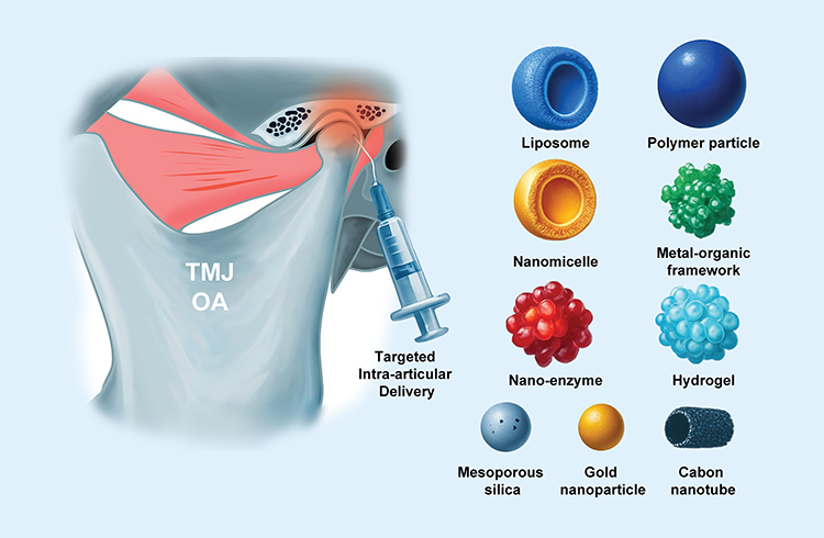 |
Figure 1 Schematic overview of emerging nanomaterial-based drug delivery systems for the treatment of TMJ OA. |
TMJ-Specific Structural and Pathological Features Guiding Intra-Articular Drug Delivery
Anatomical and Biomechanical Characteristics of the TMJ Relevant to Drug Delivery
The TMJ has distinct anatomical and physiological features that are closely related to intra-articular drug delivery. Unlike weight-bearing joints covered by hyaline cartilage, the TMJ is lined with fibrocartilage, which is more resistant to shear stress but differs in permeability and intrinsic repair capacity.11 The TMJ is exposed to complex mechanical loading, including compression, shear, and friction generated during mastication and mandibular movement. In addition, the presence of an articular disc and a relatively confined joint cavity creates a unique microenvironment that directly affects drug distribution and retention.12
TMJ homeostasis depends on continuous remodeling in response to mechanical stimuli. However, excessive or unbalanced loading disrupts this process and initiates degenerative changes.13 From the perspective of drug delivery, several structural and functional characteristics present notable challenges. The dense extracellular matrix and fibrocartilaginous architecture can limit drug penetration into cartilage. Meanwhile, joint motion and synovial fluid turnover may accelerate drug clearance, reducing retention time. Furthermore, pathological alterations such as inflammation and oxidative stress may affect drug stability and release kinetics. These characteristics highlight the need for delivery systems that improve tissue penetration, prolong intra-articular residence, and adapt to the local microenvironment.
Pathological Characteristics and Molecular Mechanisms of TMJ OA
The core pathological changes in TMJ OA include progressive destruction of articular cartilage, subchondral bone sclerosis, and synovial inflammation.14 Among these, synovial inflammation plays a central role in driving disease progression. In TMJ OA, synovial tissue shows angiogenesis and inflammatory cell infiltration, accompanied by increased levels of pro-inflammatory cytokines such as interleukin-1β (IL-1β) and tumor necrosis factor-α (TNF-α) in the synovial fluid.15 These cytokines activate the NF-κB signaling pathway, leading to upregulation of matrix metalloproteinases (MMP-3 and MMP-13), which degrade type II collagen and proteoglycans in the cartilage matrix.16,17
At the cellular level, cartilage degeneration is largely driven by an imbalance between chondrocyte anabolism and apoptosis.18 Type II collagen (COL2A1), a major structural component of the extracellular matrix, also regulates chondrocyte hypertrophy through the integrin β1/SMAD1 pathway.19 The transcription factor SOX9 is essential for chondrocyte proliferation, differentiation and phenotype maintenance.20 In TMJ OA, reduced expression of these molecules, together with increased apoptosis induced by aging, oxidative stress, and inflammation, accelerates cartilage degeneration.21–23
Impaired cartilage repair further contributes to disease progression. Dysfunction of mesenchymal stem cells (MSCs), particularly their reduced chondrogenic differentiation capacity, limits tissue regeneration. Recent studies show that Kartogenin (KGN) can promote chondrogenesis and restore the expression of SOX9 and COL2A.24,25 In the clinical treatment strategies for TMJ OA, targeted regulation of cartilage and subchondral bone tissues is of great significance. However, its poor water solubility restricts clinical application. Exosome-mediated delivery has been shown to improve the bioavailability of KGN and enhance the chondrogenic potential of synovial fluid-derived MSCs (SF-MSCs), suggesting a promising strategy for TMJ OA treatment.24
Nanomaterial-Based Intra-Articular Drug Delivery Systems for TMJ OA
The treatment of TMJ OA remains challenging, largely due to the limited efficacy of conventional drug delivery approaches. Systemic administration often fails to achieve sufficient drug concentrations within the joint, while intra-articular injection, although more direct, is hindered by rapid drug clearance and short retention time. The residence time of free drugs after injection is typically less than 24 hours26 and the half-life of therapeutic proteins such as TGF-β is only 2–4 hours within the joint cavity.27 These limitations make it difficult to maintain effective drug levels for sustained therapeutic action. To address these challenges, a range of nanomaterial-based delivery systems has been developed. These systems can be broadly categorized based on their material composition and functional properties. Table 1 summarizes the key features of representative nanomaterial-based delivery platforms.
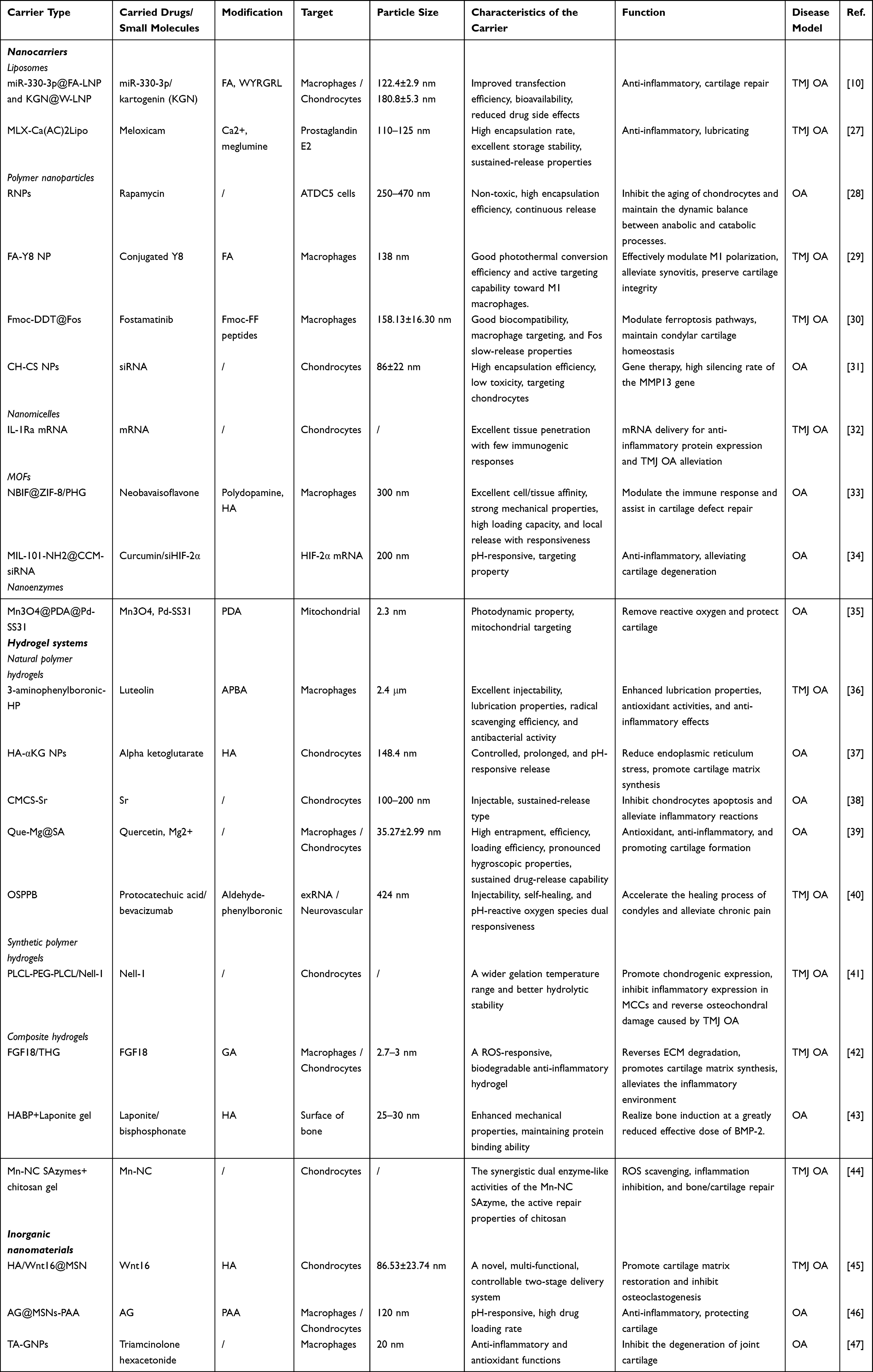 |
Table 1 Overview of Drug Delivery Systems Developed for Intra-Articular Treatment of TMJ OA |
Nanocarriers
Nanocarriers represent a major class of delivery systems for TMJ OA. Their nanoscale size enables penetration across the synovial barrier, with particles in the range of 50–200 nm showing favorable transport properties.48 Common nanocarriers include liposomes, polymeric nanoparticles, nanomicelles, and metal–organic frameworks (MOFs). In addition, surface modification strategies allow these carriers to interact more effectively with target tissues, such as cartilage or chondrocytes, thereby improving local drug retention.48–50
Liposomes
Liposomes are spherical vesicles composed of phospholipid bilayers, with structures similar to cell membranes, which confer excellent biocompatibility.51 Their ability to encapsulate both hydrophilic and hydrophobic agents makes them versatile carriers for TMJ OA therapy. In addition, surface modification enables improved interaction with target cells or tissues within the joint. Recent studies have explored functionalized liposomes for combined anti-inflammatory and cartilage-protective effects. Yang et al10 developed a dual-targeted lipid nanoparticle system co-delivering miR-330-3p and kartogenin (KGN), which was shown to regulate macrophage polarization and support chondrocyte homeostasis, thereby alleviating synovial inflammation and cartilage degeneration (Figure 2A). Liposomes have also been used to improve the delivery of clinically relevant drugs. Meloxicam (MLX), a commonly used anti-inflammatory agent for TMJ OA, suffers from poor solubility and limited intra-articular retention. To address this, Zhong et al27 developed a liposomal formulation that enhances drug loading and stability. Both in vitro and in vivo results demonstrated that this system can reduce chondrocyte apoptosis and extracellular matrix degradation while providing additional lubrication within the joint (Figure 2B). While these findings highlight the potential of liposome-based systems, most evidence is derived from general OA, and their long-term stability, drug leakage, and in vivo reproducibility in TMJ-specific settings remain to be further validated.
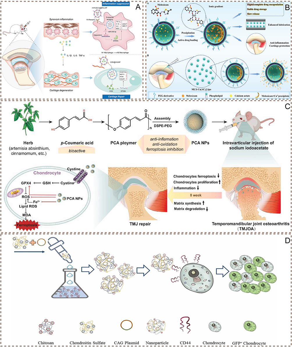 |
Figure 2 Targeted nanocarrier strategies for TMJ OA therapy. (A) Schematic representation of two ligand-modified lipid nanoparticle systems (FA-LNP and W-LNP) designed to target macrophages and chondrocytes, respectively, enabling combined anti-inflammatory and tissue repair effects through molecular and gene regulation (Reproduced with permission from Ref.10 Copyright 2024, Elsevier B.V). (B) Actively loaded meloxicam liposome system (MLX Ca(AC)2Lipo) for TMJ OA therapy, designed for local meloxicam release to exert anti-inflammatory and cartilage-protective effects (Reproduced with permission from Ref.27 Copyright 2023, Yingqian Zhong et al). (C) Poly(p-coumaric acid) nanoparticles alleviate TMJ OA by inhibiting chondrocyte ferroptosis while exerting antioxidant and anti-inflammatory effects to promote joint repair (Reproduced with permission from Ref.52 Copyright 2024, Jiaxin Guo et al). (D) Chitosan–chondroitin sulfate nanoparticle platform for gene delivery applications in joint disease treatment (Reproduced with permission from Ref.31 Copyright 2022, Elsevier B.V). |
Polymeric Nanoparticles
Polymeric nanoparticles are widely used drug delivery platforms, with materials such as poly (lactic-co-glycolic acid) (PLGA), chitosan (CS), and poly (lactic acid) (PLA) being commonly employed.51 These materials offer good biocompatibility and biodegradability, and are particularly suitable for sustained drug release. Moreover, polymeric systems can be designed to respond to specific microenvironmental cues, such as pH or enzymatic activity, which may further enhance their performance in joint diseases.53
Several studies have demonstrated the therapeutic potential of polymeric nanoparticles in TMJ OA. Ma et al28 developed PLGA-based nanoparticles for rapamycin delivery, which promoted chondrogenic differentiation and reduced oxidative stress–induced chondrocyte senescence. In vivo, this system alleviated cartilage damage, synovial inflammation, and pain. Similarly, Guo et al52 reported poly(p-coumaric acid)-based nanoparticles with antioxidant and anti-inflammatory properties, which showed protective effects on cartilage and subchondral bone in TMJ models (Figure 2C).
Polymeric nanoparticles have also been engineered for targeted or externally regulated therapies. Wang et al29 developed a folic acid-conjugated Y8 nanocomposite capable of modulating synovial macrophage polarization through photothermal effects, thereby reducing inflammation and cartilage degeneration. Zhang et al30 designed a composite delivery system enabling sustained release of anti-inflammatory agents and promoting macrophage reprogramming, which contributed to maintaining cartilage homeostasis.
Natural polymer-based systems further expand the application scope of polymeric nanoparticles. Chitosan-based carriers, for instance, have been explored for gene delivery due to their favorable biocompatibility and functional versatility.54–56 Moghadam et al31 developed a chitosan–chondroitin sulfate nanoparticle system that enhanced cellular uptake and enabled efficient delivery of nucleic acids, including siRNA targeting MMP13, providing a potential strategy for gene-based therapy in TMJ OA (Figure 2D).
Nanomicelles
Nanomicelles are nanoscale structures formed by the self-assembly of amphiphilic polymers.57 These polymers contain both hydrophilic and hydrophobic segments, enabling the formation of a core–shell architecture in aqueous environments: the hydrophobic core facilitates the encapsulation of poorly soluble drugs, while the hydrophilic shell enhances stability and biocompatibility.58 Recent studies have explored nanomicelles as carriers for advanced therapeutic modalities. Deng et al32 developed a polymeric nanomicelle system with excellent tissue penetration and minimal immunogenicity for mRNA delivery. This platform enabled efficient protein expression in cartilage tissues and alleviated TMJ OA progression by suppressing pro-inflammatory cytokines such as IL-6 and TNF-α, highlighting its potential in nucleic acid–based therapies. In addition, surface modification strategies can further improve the targeting capability of nanomicelles, thereby enhancing therapeutic precision and minimizing off-target effects.59,60
Emerging Nanocarriers: MOFs and Nanoenzymes
In addition to the nanocarriers discussed above, several emerging nanomaterials, including MOFs and nanoenzymes, have attracted increasing attention for their unique physicochemical properties and potential applications in osteoarthritis treatment.61–64 However, studies specifically focusing on TMJ OA remain limited, and current evidence is largely derived from other OA models. MOFs are characterized by high surface area, tunable pore structures, and versatile functionalization capabilities, enabling efficient drug loading and controlled release. In OA-related studies, MOF-based systems have been explored for delivering anti-inflammatory agents and nucleic acids with stimulus-responsive release profiles, contributing to improved regulation of the joint microenvironment.33,34 Similarly, nanoenzymes exhibit enzyme-like catalytic activities that can modulate oxidative stress and inflammation. By mimicking antioxidant enzymes, they can reduce reactive oxygen species and regulate immune responses, thereby alleviating OA progression.65
Recent advances further highlight the potential of these materials in more sophisticated therapeutic strategies. For instance, mitochondria-targeted or externally responsive nanozyme systems have been developed to enhance subcellular regulation and improve therapeutic precision.35 These approaches, although not yet validated in TMJ-specific models, provide important insights into the design of multifunctional and microenvironment-responsive delivery systems. Taken together, while direct evidence in TMJ OA is still lacking, these emerging nanomaterials offer valuable conceptual and technical frameworks that may inform future developments in targeted and combinatorial therapies for TMJ disorders.
Hydrogel Drug Delivery System
Hydrogels are three-dimensional network materials widely used in drug delivery due to their favorable biocompatibility, tunable degradability, and capacity for encapsulating diverse therapeutic agents.66,67 Their crosslinked structures can be readily engineered to achieve specific physicochemical properties, enabling applications in tissue repair and controlled drug delivery.68 For intra-articular applications, hydrogels offer distinct advantages. Injectable precursors allow minimally invasive administration and conform to irregular joint spaces, while in situ gelation enables localized retention and sustained release of encapsulated drugs, proteins, or cells.69,70 These features make hydrogels particularly suitable for improving drug residence time and therapeutic efficacy in OA treatment. Based on their material origin and design strategies, hydrogels can be broadly classified into natural polymer hydrogels, synthetic polymer hydrogels, and composite hydrogels (Figure 3).71,72 Nevertheless, most studies primarily focus on drug release behavior and short-term efficacy, while systematic evaluation of long-term performance and comparative effectiveness across different hydrogel systems remains limited.
 |
Figure 3 Classification of hydrogels based on material origin and composition. |
Natural Polymer Hydrogels
Natural polymer hydrogels, including gelatin, hyaluronic acid (HA), chitosan, and sodium alginate, are widely explored in drug delivery due to their favorable biocompatibility, low immunogenicity, and intrinsic bioactivity. These materials can provide a supportive microenvironment for cell survival and tissue repair, making them particularly suitable for intra-articular applications. However, their relatively weak mechanical strength and susceptibility to rapid enzymatic degradation often limit their long-term performance.72 To overcome these limitations, strategies such as chemical modification, cross-linking, or incorporation of functional nanomaterials have been employed to enhance their stability and functionality.73
Among these materials, HA-based hydrogels have attracted considerable attention due to their natural presence in joint tissues and their lubrication and anti-inflammatory properties.74,75 Similarly, Liu et al36 designed HA-based microparticles capable of dynamically binding diol-containing drugs, demonstrating antioxidant and anti-inflammatory effects and promoting cartilage regeneration in TMJ OA (Figure 4A). In addition, a composite HA hydrogel incorporating mesoporous silica nanoparticles achieved dual-layer sustained release of Wnt16, effectively modulating the Wnt/β-catenin signaling pathway and enhancing cartilage matrix restoration in TMJ OA (Figure 4B).45 Although not specific to TMJ OA, HA-based delivery systems have also shown therapeutic potential in other OA models, such as HA-αKG nanoparticles that improve cartilage anabolism and alleviate endoplasmic reticulum stress,37 highlighting their broader applicability.
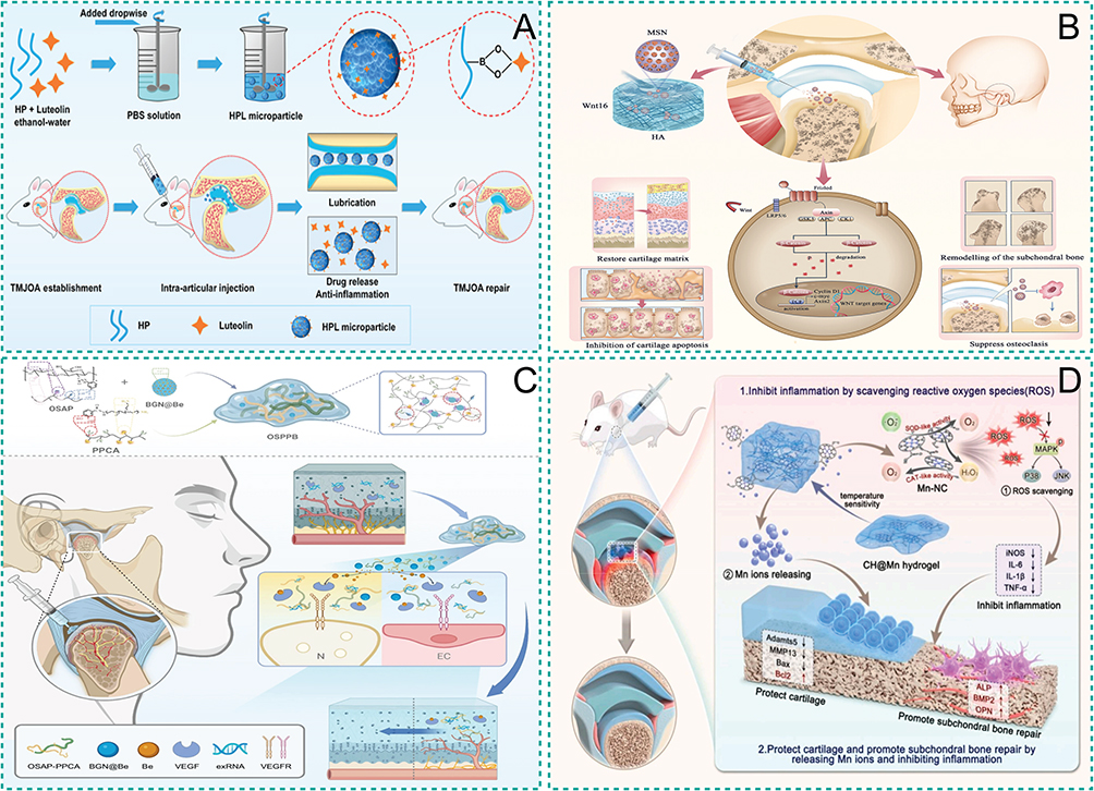 |
Figure 4 Hydrogel-based systems for responsive drug delivery in TMJ OA. (A) Fabrication process of luteolin-loaded hyaluronic acid microparticles and their intra-articular application for TMJ OA therapy, combining lubrication, sustained drug release, and anti-inflammatory effects (Reproduced with permission from Ref.36 Copyright 2024, Lei Liu et al). (B) Schematic representation of the HA/Wnt16@MSN delivery system for synergistic therapy of TMJ OA through regulation of the Wnt/β-catenin signaling pathway (Reproduced with permission from Ref.45 Copyright 2024, Yan Zhu et al). (C) Injectable, self-healing, and dual pH/ROS-responsive multicationic hydrogel designed to modulate the osteochondral interface and alleviate TMJ OA-associated pathology by inhibiting neuroangiogenesis (Reproduced with permission from Ref.40 Copyright 2025, Wenpin Qin et al). (D) An Mn-NC single-atom nanozyme composite hydrogel exhibits dual SOD-like and CAT-like activities to scavenge ROS, while releasing Mn ions to protect chondrocytes and promote cartilage and subchondral bone regeneration in TMJ OA (Reproduced with permission from Ref.44 Copyright 2026, Min Xing et al). |
Chitosan-based hydrogels are another important class of natural polymers with favorable biodegradability and bioadhesive properties.76 Owing to these characteristics, they have been widely applied in drug delivery and tissue engineering.77,78 Recent studies have expanded their functionality beyond passive delivery. Besides, chitosan derivatives such as carboxymethyl chitosan strontium (CMCS-Sr) have been shown to enhance chondrocyte proliferation and matrix synthesis while reducing inflammatory responses in OA models.38 Dual-drug delivery systems based on chitosan nanoparticles have also demonstrated synergistic anti-inflammatory and pro-regenerative effects, supporting their role in cartilage protection and regeneration.79
Sodium alginate-based hydrogels have also been widely investigated for controlled drug delivery due to their mild gelation properties and good biocompatibility.80–84 Composite systems incorporating bioactive nanoparticles, such as quercetin–Mg2⁺ complexes, have demonstrated the ability to reduce oxidative stress, suppress inflammatory cytokine production, and maintain cartilage metabolic balance in OA models.39 In addition, alginate-based multifunctional hydrogels capable of scavenging extracellular RNA and continuously releasing therapeutic agents have shown potential in modulating neurovascularization and alleviating TMJ OA-associated pain (Figure 4C).40
Overall, natural polymer hydrogels provide a versatile platform for integrating multiple therapeutic functions, including drug delivery, microenvironment modulation, and tissue regeneration. Nevertheless, their inherent mechanical limitations and variability in degradation behavior still necessitate further optimization for long-term and site-specific applications in TMJ OA.
Synthetic Polymer Hydrogels
Synthetic polymer hydrogels are three-dimensional hydrophilic networks capable of retaining large amounts of water while maintaining structural stability. Their mechanical properties and degradation behavior can be tuned by adjusting polymer molecular weight, cross-linking density, or chemical composition, enabling controlled drug release.85,86 However, many synthetic polymers lack intrinsic bioactivity or cell-recognition sites, which may limit their interaction with cartilage tissue.
Commonly used materials include polyacrylamide (PAAm), polyethylene glycol (PEG), and PLGA.87 PAAm-based hydrogels are easy to prepare and allow flexible structural design. Dual-responsive PNIPAm-co-PAAm hydrogels can achieve temperature- and pH-sensitive drug release with high loading efficiency, although most studies are limited to in vitro experiments.88–90 PEG-based hydrogels are highly biocompatible and water-soluble, and their molecular weight and properties are readily adjustable.86 Functionalized PEG systems, such as cartilage-targeted PEG (PCFMN), have shown the ability to upregulate anabolic genes and downregulate catabolic genes in chondrocytes, delaying OA progression in vivo.91 Notably, TMJ OA evidence shows that a PLCL-PEG-PLCL injectable hydrogel combined with Nell-1 can achieve sustained release and promote cartilage repair while improving subchondral bone structure in vivo.41 However, this evidence is still limited to preclinical models, and further validation is needed to assess long-term efficacy, reproducibility, and safety in clinically relevant TMJ settings. PLGA-based polymer nanoparticles have been described in previous sections. Generally, synthetic polymer hydrogels for TMJ OA are still in an early exploratory stage, and their combination with bioactive modifications or hybridization with natural polymers may enhance therapeutic efficacy in the unique TMJ environment.
Composite Hydrogels
Composite hydrogels combine the biocompatibility of natural polymers with the tunability of synthetic polymers, offering both improved mechanical properties and biological activity.92 Interactions among components can support cell adhesion, signaling, and growth factor delivery, which may facilitate cartilage regeneration.93 Typical designs include gelatin–PEG complexes and hyaluronic acid–nanoclay systems. In TMJ OA, a ROS-responsive HA-GA-based hydrogel has been developed to deliver FGF18, enabling simultaneous ROS scavenging and sustained release of pro-regenerative factors, thereby overcoming the inhibitory effects of inflammation on cartilage repair and effectively delaying disease progression.42 Kim YH et al43 developed a self-assembled hydrogel with functionalized clay nanoparticles was constructed by crosslinking hyaluronic acid modified with bisphosphonate side chains using low-concentration nanoclay. This hydrogel showed enhanced mechanical properties, maintained protein-binding capacity, and preserved BMP-2 activity in vivo for over six weeks. A nanozyme-functionalized chitosan hydrogel integrating Mn–N–C single-atom catalysts demonstrated efficient ROS scavenging and suppression of MAPK signaling, thereby inhibiting chondrocyte apoptosis and extracellular matrix degradation while promoting osteochondral repair in TMJ OA (Figure 4D).44 Future studies may focus on tailoring composite hydrogels specifically for TMJ cartilage, optimizing mechanical resilience, growth factor delivery, and stem cell support to match the joint’s unique microenvironment.
Inorganic Nanomaterials
Inorganic nanomaterials have unique physicochemical properties, including stability, biocompatibility, and high drug-loading capacity, making them attractive carriers for drug delivery and potential therapeutic applications.94 They can efficiently transport drugs, proteins, or genes to target sites, enhancing efficacy while reducing side effects. Common inorganic nanomaterials in drug delivery include mesoporous silica nanoparticles (MSNs), gold nanoparticles (GNPs), and quantum dots (QDs).95 Nevertheless, direct evidence in TMJ OA remains limited, and most current understanding is extrapolated from other OA models or related biomedical applications. This highlights the need for more TMJ-specific preclinical validation.
Mesoporous Silica Nanoparticles
MSNs are characterized by tunable mesoporous structures, high surface area, and facile surface functionalization.96,97 These features allow efficient drug loading and the development of multifunctional theranostic systems. To date, only limited TMJ OA-specific studies are available. Zhu et al45 designed a double-layer Wnt16 delivery system based on MSNs encapsulated in a hyaluronic acid hydrogel. This HA/Wnt16@MSN hydrogel promoted pre-cartilage matrix repair, inhibited osteoclastogenesis, and suppressed over-activation of the Wnt/β-catenin pathway in TMJ OA models. Other MSN-based systems, though not specific to TMJ OA, provide useful mechanistic insights. He et al reported that AG@MSNs-PAA carrying silver ions reduced inflammation and protected cartilage in IL-1β-stimulated chondrocytes and ACLT-induced OA rat models.46 These studies highlight MSNs’ versatile delivery capacity and suggest potential applications for TMJ-targeted therapy.
Gold Nanoparticle Carriers
GNPs are valued for their optical, electronic, and surface-modifiable properties.98–100 They can achieve targeted drug delivery and stable release, with photophysical properties enabling precise spatiotemporal control. In general OA models, TA-GNPs (triamcinolone acetonide conjugated with gold nanoparticles) reduced pro-inflammatory factors, alleviated oxidative stress, and improved cartilage structure.47 While TMJ OA-specific studies are lacking, these findings from other OA models suggest that GNP-based carriers could be adapted for targeted TMJ applications.
Quantum Dots
QDs offer size-tunable luminescence, high surface area, and versatile surface functionalization, making them suitable for both drug delivery and in vivo tracking.101–103 Although research on QDs in OA is limited, recent studies provide promising references for TMJ tissue engineering. Jeong et al104 used QD-labeled bioactive scaffolds for TMJ disc regeneration in mini-pigs, achieving anatomically correct tissue with region-specific mechanical properties and enabling non-invasive monitoring of scaffold degradation. This work, although not an OA model, illustrates the potential of QDs not only for delivery but also for real-time tracking in TMJ-related applications.
Carbon-Based Nanomaterials
Carbon-based nanomaterials, including carbon nanotubes (CNTs), nanographene, and fullerenes, exhibit high mechanical strength, conductivity, and surface area.105,106 CNTs can deliver drugs, DNA, or siRNA, and enhance composite materials’ mechanical properties, while nanographene offers large surface area and stability for biomedical applications.107 However, current studies directly addressing TMJ OA are scarce, indicating that translational work is still at an early stage.
Trend Outlook: Personalized and Precision Approaches for TMJ OA
The management of TMJ OA is increasingly moving toward personalized treatment and precision medicine, driven by the heterogeneity of disease progression and inter-individual variability. Effective therapy requires tailoring strategies to patient-specific pathological features, including gene expression profiles, metabolic status, and local joint microenvironment. For instance, some patients may benefit more from immunomodulatory approaches, while others require interventions targeting cartilage degeneration or specific inflammatory pathways. Emerging evidence also suggests that certain immune cell subsets can protect TMJ cartilage by modulating synovium–cartilage interactions, highlighting opportunities for targeted immunotherapy.
Intelligent biomaterial-based drug delivery systems, such as responsive hydrogels and exosome carriers, offer spatially and temporally controlled release, potentially enhancing therapeutic efficacy while minimizing systemic side effects. These platforms could be engineered to match the TMJ’s complex biomechanical environment, including limited joint space, irregular cartilage geometry, and heterogeneous loading patterns, thereby improving retention and therapeutic precision.
RNA-based therapies, including siRNA and mRNA approaches, expand the possibilities for precision intervention by enabling selective regulation of key genes. Advances in genome-wide association studies and transcriptomic analyses provide a foundation for identifying disease-relevant targets, paving the way for personalized RNA therapeutics that modulate molecular pathways in vivo.108 For TMJ OA, these strategies may allow patient-specific modulation of inflammatory and regenerative pathways, offering a targeted approach that complements conventional biomaterial-based delivery systems.
Artificial intelligence (AI) offers additional potential to accelerate the rational design of TMJ OA therapies. Machine learning and deep learning can assist in optimizing drug formulations, predicting pharmacokinetics, and guiding individualized treatment strategies.109 Integration of AI with nanomaterial carriers, hydrogel systems, and RNA therapeutics could enable dynamic, adaptive treatment plans that respond to temporal changes in the joint microenvironment, ultimately enhancing efficacy while reducing off-target effects.
Taken together, the integration of personalized medicine, intelligent drug delivery, RNA therapeutics, and AI-driven optimization represents a promising frontier for TMJ OA management. Despite the current progress, the available evidence remains heterogeneous and is often still at a preliminary stage. Direct comparisons between different delivery systems are uncommon, and negative or inconclusive findings are seldom discussed, which may introduce potential bias in evaluating overall efficacy. At the same time, key issues such as long-term biosafety, reproducibility, manufacturing scale-up, and regulatory feasibility have yet to be fully examined in most preclinical studies. These factors make it necessary to interpret the reported therapeutic potential with appropriate caution in the context of TMJ OA. Future research should focus on developing integrative platforms that combine these approaches, validated in TMJ-specific preclinical models, to achieve precise, durable, and patient-tailored therapies. Such efforts may also inform guidelines for combinatorial strategies that address the multifactorial nature of TMJ OA, including inflammation, cartilage degeneration, and subchondral bone remodeling.
Conclusions
Recent advances in drug delivery systems have significantly improved strategies for TMJ OA therapy. Recent advances include targeted nanocarriers, responsive hydrogels for sustained intra-articular release, and gene-based approaches for modulating inflammatory and catabolic pathways. These systems improve drug retention and tissue-specific delivery while potentially reducing systemic exposure.
Despite these advances, clinical translation remains challenging. Physiological barriers, including the synovial membrane and dense cartilage matrix, limit effective penetration, while insufficient retention and potential immunogenicity or off-target effects pose additional hurdles. In addition, concerns related to long-term biosafety, batch-to-batch reproducibility, and the feasibility of large-scale manufacturing remain insufficiently addressed in current studies. Importantly, the lack of TMJ-specific evidence remains a central limitation in the field.
Future research should prioritize the design of intelligent, TMJ-specific delivery platforms with improved targeting, immune evasion, and stability, coupled with standardized preclinical evaluation frameworks. Incorporating patient-specific molecular profiles will further enable personalized and precision therapies.
Overall, novel drug delivery systems provide promising strategies for improving TMJ OA management, but their clinical translation will require further validation in TMJ-specific models and closer alignment with clinical needs.
Acknowledgments
The authors would like to thank all the colleagues for their assistance in accomplishing this study.
Funding
This work was supported by the National Natural Science Foundation of China (Grant No.82571100, 82370921, 82370979 and 82301108); the Program of Shanghai Technology Research Leader (23XD1430800); the Shanghai Leading Talent Program of Eastern Talent Plan (LJ2024096) and Shanghai Huangpu district leading talents.
Disclosure
The authors declare that they have no conflict of interest.
References
1. Liu Q, Yang H, Zhang M, et al. Initiation and progression of dental-stimulated temporomandibular joints osteoarthritis. Osteoarthritis Cartilage. 2021;29(5):633–17. doi:10.1016/j.joca.2020.12.016
2. Sperry MM, Kartha S, Winkelstein BA, Granquist EJ. Experimental methods to inform diagnostic approaches for painful TMJ osteoarthritis. J Dent Res. 2019;98(4):388–397. doi:10.1177/0022034519828731
3. Shi J, Lee S, Pan HC, et al. Association of condylar bone quality with TMJ osteoarthritis. J Dent Res. 2017;96(8):888–894. doi:10.1177/0022034517707515
4. Chapa-Villarreal FA, Stephens M, Pavlicin R, Beussman M, Peppas NA. Therapeutic delivery systems for rheumatoid arthritis based on hydrogel carriers. Adv Drug Deliv Rev. 2024;208:115300. doi:10.1016/j.addr.2024.115300
5. Cao Y, Ma Y, Tao Y, Lin W, Wang P. Intra-articular drug delivery for osteoarthritis treatment. Pharmaceutics. 2021;13(12):2166. doi:10.3390/pharmaceutics13122166
6. Liang Y, Xu X, Xu L, et al. Non-surgical osteoarthritis therapy, intra-articular drug delivery towards clinical applications. J Drug Target. 2021;29(6):609–616. doi:10.1080/1061186X.2020.1870231
7. Zong L, Wu Q, Dong Z, Huang L, Yang H. Research progress of nanomaterials for intra-articular targeted drug delivery in treatment of osteoarthritis. Zhongguo Xiu Fu Chong Jian Wai Ke Za Zhi. 2022;36(7):908–914. doi:10.7507/1002-1892.202203033
8. Liu C, Sun Y, Li D, et al. A multifunctional nanogel encapsulating layered double hydroxide for enhanced osteoarthritis treatment via protection of chondrocytes and ECM. Mater Today Bio. 2024;26:101034. doi:10.1016/j.mtbio.2024.101034
9. Huang L, Yao Y, Ruan Z, et al. Baicalin nanodelivery system based on functionalized metal-organic framework for targeted therapy of osteoarthritis by modulating macrophage polarization. J Nanobiotechnology. 2024;22(1):221. doi:10.1186/s12951-024-02494-5
10. Yang K, Zhao Y, Wang C, et al. Dual-targeted lipid nanoparticles system for synergistic anti-inflammation and cartilage repair in the treatment of temporomandibular joint osteoarthritis. Chem Eng J. 2024;481:148769. doi:10.1016/j.cej.2024.148769
11. Lu K, Ma F, Yi D, Yu H, Tong L, Chen D. Molecular signaling in temporomandibular joint osteoarthritis. J Orthop Translat. 2022;32:21–27. doi:10.1016/j.jot.2021.07.001
12. Cardoneanu A, Macovei LA, Burlui AM, et al. Temporomandibular joint osteoarthritis: pathogenic mechanisms involving the cartilage and subchondral bone, and potential therapeutic strategies for joint regeneration. Int J Mol Sci. 2022;24(1):171. doi:10.3390/ijms24010171
13. Liu Y, Jia F, Li K, et al. Critical signaling molecules in the temporomandibular joint osteoarthritis under different magnitudes of mechanical stimulation. Front Pharmacol. 2024;15:1419494. doi:10.3389/fphar.2024.1419494
14. Liu X, Li Y, Zhao J, et al. Pyroptosis of chondrocytes activated by synovial inflammation accelerates TMJ osteoarthritis cartilage degeneration via ROS/NLRP3 signaling. Int Immunopharmacol. 2023;124(Pt A):110781. doi:10.1016/j.intimp.2023.110781
15. Li Y, Sun H, Liu X, et al. Transglutaminase 2 inhibitors attenuate osteoarthritic degeneration of TMJ-osteoarthritis by suppressing NF-kappaB activation. Int Immunopharmacol. 2023;114:109486. doi:10.1016/j.intimp.2022.109486
16. Liao S, Liu Z, Lv W, et al. Efficient delivery of siRNA via tetrahedral framework nucleic acids: inflammation attenuation and matrix regeneration in temporomandibular joint osteoarthritis. ACS Appl Mater Interfaces. 2024;16(40):53499–53514. doi:10.1021/acsami.4c11089
17. Cafferata EA, Monasterio G, Castillo F, et al. Overexpression of MMPs, cytokines, and RANKL/OPG in temporomandibular joint osteoarthritis and their association with joint pain, mouth opening, and bone degeneration: a preliminary report. Oral Dis. 2021;27(4):970–980. doi:10.1111/odi.13623
18. Lee YT, Mohd Yunus MH, Yazid MD, Ugusman A. Unraveling the path to osteoarthritis management: targeting chondrocyte apoptosis for therapeutic intervention. Front Cell Dev Biol. 2024;12:1347126. doi:10.3389/fcell.2024.1347126
19. Lian C, Wang X, Qiu X, et al. Collagen type II suppresses articular chondrocyte hypertrophy and osteoarthritis progression by promoting integrin beta1-SMAD1 interaction. Bone Res. 2019;7(1):8. doi:10.1038/s41413-019-0046-y
20. Lefebvre V, Dvir-Ginzberg M. SOX9 and the many facets of its regulation in the chondrocyte lineage. Connect Tissue Res. 2017;58(1):2–14. doi:10.1080/03008207.2016.1183667
21. Ansari MM, Ghosh M, Lee DS, Son YO. Senolytic therapeutics: an emerging treatment modality for osteoarthritis. Ageing Res Rev. 2024;96:102275. doi:10.1016/j.arr.2024.102275
22. Hu W, Yao X, Li Y, et al. Injectable hydrogel with selenium nanoparticles delivery for sustained glutathione peroxidase activation and enhanced osteoarthritis therapeutics. Mater Today Bio. 2023;23:100864. doi:10.1016/j.mtbio.2023.100864
23. Li H, Wang X, Pan H, et al. The mechanisms and functions of IL-1beta in intervertebral disc degeneration. Exp Gerontol. 2023;177:112181. doi:10.1016/j.exger.2023.112181
24. Xu X, Liang Y, Li X, et al. Exosome-mediated delivery of kartogenin for chondrogenesis of synovial fluid-derived mesenchymal stem cells and cartilage regeneration. Biomaterials. 2021;269:120539. doi:10.1016/j.biomaterials.2020.120539
25. Bhuyan S, Swain S, Misra RDK, Rautray TR. Nanomedicine-driven approaches for kartogenin delivery: advancing chondrogenic differentiation and cartilage regeneration in tissue engineering. Int J Nanomed. 2025;20:7443–7468. doi:10.2147/IJN.S525580
26. Schipper JAM, Tuin AJ, Van Dongen JA, van Bakelen NB, Harmsen MC, Spijkervet FKL. Intra-articular injection of adipose-derived stromal vascular fraction in osteoarthritic temporomandibular joints: study design of a randomized controlled clinical trial. Bioengineering. 2024;11(2). doi:10.3390/bioengineering11020171
27. Zhong Y, Zhou Y, Ding R, et al. Intra-articular treatment of temporomandibular joint osteoarthritis by injecting actively-loaded meloxicam liposomes with dual-functions of anti-inflammation and lubrication. Mater Today Bio. 2023;19:100573. doi:10.1016/j.mtbio.2023.100573
28. Ma JC, Luo T, Feng B, et al. Exploring the translational potential of PLGA nanoparticles for intra-articular rapamycin delivery in osteoarthritis therapy. J Nanobiotechnology. 2023;21(1):361. doi:10.1186/s12951-023-02118-4
29. Wang Y, Ren J, Ren T, et al. Photothermal reprogramming of synovial M1 macrophages reshapes the pro-inflammatory microenvironment to reverse temporomandibular joint osteoarthritis. J Nanobiotechnology. 2026.
30. Zhang Y, Zhang D, Yue X, et al. Fmoc-DDT@Fos hydrogel mitigates temporomandibular joint osteoarthritis through regulating macrophage reprogramming and ferroptosis. Mater Today Bio. 2026;37:102906. doi:10.1016/j.mtbio.2026.102906
31. Moghadam NA, Bagheri F, Eslaminejad MB. Chondroitin sulfate modified chitosan nanoparticles as an efficient and targeted gene delivery vehicle to chondrocytes. Colloids Surf B Biointerfaces. 2022;219:112786. doi:10.1016/j.colsurfb.2022.112786
32. Deng J, Fukushima Y, Nozaki K, et al. Anti-inflammatory therapy for temporomandibular joint osteoarthritis using mRNA medicine encoding interleukin-1 receptor antagonist. Pharmaceutics. 2022;14(9):1785. doi:10.3390/pharmaceutics14091785
33. Jiang Y, Liao H, Yan L, et al. A metal-organic framework-incorporated hydrogel for delivery of immunomodulatory neobavaisoflavone to promote cartilage regeneration in osteoarthritis. ACS Appl Mater Interfaces. 2023;15(40):46598–46612. doi:10.1021/acsami.3c06706
34. Zhang ZJ, Hou YK, Chen MW, et al. A pH-responsive metal-organic framework for the co-delivery of HIF-2alpha siRNA and curcumin for enhanced therapy of osteoarthritis. J Nanobiotechnology. 2023;21(1):18. doi:10.1186/s12951-022-01758-2
35. Li Y, Yang J, Chen X, et al. Mitochondrial-targeting and NIR-responsive Mn(3)O(4)@PDA@Pd-SS31 nanozymes reduce oxidative stress and reverse mitochondrial dysfunction to alleviate osteoarthritis. Biomaterials. 2024;305:122449. doi:10.1016/j.biomaterials.2023.122449
36. Liu L, He G, Li Y, et al. Hyaluronic acid-based microparticles with lubrication and anti-inflammation for alleviating temporomandibular joint osteoarthritis. Biomater Res. 2024;28:0073. doi:10.34133/bmr.0073
37. Wang X, Xue Y, Hao K, et al. Sustained therapeutic effects of self-assembled hyaluronic acid nanoparticles loaded with alpha-Ketoglutarate in various osteoarthritis stages. Biomaterials. 2025;314:122845. doi:10.1016/j.biomaterials.2024.122845
38. Liu Z, Mo X, Ma F, et al. Synthesis of carboxymethyl chitosan-strontium complex and its therapeutic effects on relieving osteoarthritis. Carbohydr Polym. 2021;261:117869. doi:10.1016/j.carbpol.2021.117869
39. Chen J, Wu G, Wu J, Jiao Z. Sodium alginate microspheres loaded with Quercetin/Mg nanoparticles as novel drug delivery systems for osteoarthritis therapy. J Orthop Surg Res. 2025;20(1):300. doi:10.1186/s13018-025-05698-z
40. Qin W, Ma Z, Bai G, et al. Neurovascularization inhibiting dual responsive hydrogel for alleviating the progression of osteoarthritis. Nat Commun. 2025;16(1):1390. doi:10.1038/s41467-025-56727-8
41. Wang C, Wang Y, Wang C, et al. Therapeutic application of 3B-PEG injectable hydrogel/Nell-1 composite system to temporomandibular joint osteoarthritis. Biomed Mater. 2021;17(1). doi:10.1088/1748-605X/ac367f
42. Kuang Y, Hua B, Ye X, Zhao Y, Yu M, Liu X. Dual-functional ROS-responsive hydrogel alleviates temporomandibular joint osteoarthritis by enhancing cartilage repair and mitigating inflammation. Mater Today Bio. 2025;33:102103. doi:10.1016/j.mtbio.2025.102103
43. Kim YH, Yang X, Shi L, et al. Bisphosphonate nanoclay edge-site interactions facilitate hydrogel self-assembly and sustained growth factor localization. Nat Commun. 2020;11(1):1365. doi:10.1038/s41467-020-15152-9
44. Xing M, Chen S, Zhu M, et al. Nanozyme hydrogels remodel pathological microenvironment for temporomandibular joint osteoarthritis therapy via inhibiting MAPK signal pathway. Bioact Mater. 2026;60:216–242. doi:10.1016/j.bioactmat.2026.01.031
45. Zhu Y, Cao L, Yuan M, et al. Microgel encapsulated mesoporous silica nanoparticles for releasing Wnt16 to synergistically treat temporomandibular joint osteoarthritis. Adv Sci. 2024;11(41):e2404396. doi:10.1002/advs.202404396
46. He M, Qin Z, Liang X, et al. A pH-responsive mesoporous silica nanoparticles-based drug delivery system with controlled release of andrographolide for OA treatment. Regen Biomater. 2021;8(4):rbab020. doi:10.1093/rb/rbab020
47. Dos Santos Haupenthal DP, Resmini MB, Da silva LA, et al. Intra-articular treatment with triamcinolone hexacetonide associated with gold nanoparticles reduces cartilage degeneration in an animal model of osteoarthritis. Curr Drug Targets. 2023;24(3):287–296. doi:10.2174/1389450124666221212090319
48. Morici L, Allemann E, Rodriguez-Nogales C, Jordan O. Cartilage-targeted drug nanocarriers for osteoarthritis therapy. Int J Pharm. 2024;666:124843. doi:10.1016/j.ijpharm.2024.124843
49. Mancipe Castro LM, Sequeira A, Garcia AJ, Guldberg RE. Articular cartilage- and synoviocyte-binding Poly(ethylene glycol) nanocomposite microgels as intra-articular drug delivery vehicles for the treatment of osteoarthritis. ACS Biomater Sci Eng. 2020;6(9):5084–5095. doi:10.1021/acsbiomaterials.0c00960
50. Deng R, Zhao R, Zhang Z, et al. Chondrocyte membrane-coated nanoparticles promote drug retention and halt cartilage damage in rat and canine osteoarthritis. Sci Transl Med. 2024;16(735):eadh9751. doi:10.1126/scitranslmed.adh9751
51. Edis Z, Wang J, Waqas MK, Ijaz M, Ijaz M. Nanocarriers-mediated drug delivery systems for anticancer agents: an overview and perspectives. Int J Nanomed. 2021;16:1313–1330. doi:10.2147/IJN.S289443
52. Guo J, Su K, Wang L, et al. Poly(p-coumaric acid) nanoparticles alleviate temporomandibular joint osteoarthritis by inhibiting chondrocyte ferroptosis. Bioact Mater. 2024;40:212–226. doi:10.1016/j.bioactmat.2024.06.007
53. Pandey P, Verma M, Lakhanpal S, et al. An updated review summarizing the anticancer potential of Poly(Lactic-co-Glycolic Acid) (PLGA) based curcumin, epigallocatechin gallate, and resveratrol nanocarriers. Biopolymers. 2025;116(1):e23637. doi:10.1002/bip.23637
54. Yang K, Han HS, An SH, et al. Mucoadhesive chitosan microcapsules for controlled gastrointestinal delivery and oral bioavailability enhancement of low molecular weight peptides. J Control Release. 2024;365:422–434. doi:10.1016/j.jconrel.2023.10.021
55. Li Y, Zhang Z, Zhang Z. Porous Chitosan/Nano-Hydroxyapatite composite scaffolds incorporating simvastatin-loaded PLGA microspheres for bone repair. Cells Tissues Organs. 2018;205(1):20–31. doi:10.1159/000485502
56. Ezzaki C, Chaari A, Al-Othman A. Recent advances on chitosan-based nanoparticles for brain drug delivery. Polymers. 2025;17(22):3055. doi:10.3390/polym17223055
57. Kashapov R, Gaynanova G, Gabdrakhmanov D, et al. Self-assembly of amphiphilic compounds as a versatile tool for construction of nanoscale drug carriers. Int J Mol Sci. 2020;21(18):6961. doi:10.3390/ijms21186961
58. Liu J, Cabral H, Mi P. Nanocarriers address intracellular barriers for efficient drug delivery, overcoming drug resistance, subcellular targeting and controlled release. Adv Drug Deliv Rev. 2024;207:115239. doi:10.1016/j.addr.2024.115239
59. Mahmud MM, Pandey N, Winkles JA, Woodworth GF, Kim AJ. Toward the scale-up production of polymeric nanotherapeutics for cancer clinical trials. Nano Today. 2024;56:102314. doi:10.1016/j.nantod.2024.102314
60. Jeong EJ, Kim C, Lee YC, Rhim T, Lee SK, Lee KY. Tumor-specific cytolysis by peptide-conjugated echogenic polymer micelles. Biomed Pharmacother. 2024;172:116272. doi:10.1016/j.biopha.2024.116272
61. Xia N, Chang Y, Zhou Q, Ding S, Gao F. An overview of the design of metal-organic frameworks-based fluorescent chemosensors and biosensors. Biosensors. 2022;12(11):928.
62. Lawson HD, Walton SP, Chan C. Metal-organic frameworks for drug delivery: a design perspective. ACS Appl Mater Interfaces. 2021;13(6):7004–7020. doi:10.1021/acsami.1c01089
63. Li L, Qi Z, Han S, Li X, Liu B, Liu Y. Advances and applications of metal-organic framework nanomaterials as oral delivery carriers: a review. Mini Rev Med Chem. 2022;22(20):2564–2580. doi:10.2174/1389557522666220330152145
64. Gao X, Zhang J, Gong Y, Yan L. The biomedical applications of nanozymes in orthopaedics based on regulating reactive oxygen species. J Nanobiotechnology. 2024;22(1):569. doi:10.1186/s12951-024-02844-3
65. Yang X, Tan M, Guo J, et al. PdZn/CoSA‐NC nanozymes with highly efficient SOD/CAT activities for treatment of osteoarthritis via regulating immune microenvironment. Adv Funct Mater. 2024;34(41).
66. Baniasadi H, Abidnejad R, Fazeli M, et al. Innovations in hydrogel-based manufacturing: a comprehensive review of direct ink writing technique for biomedical applications. Adv Colloid Interface Sci. 2024;324:103095. doi:10.1016/j.cis.2024.103095
67. Tong S, Li Q, Liu Q, Song B, Wu J. Recent advances of the nanocomposite hydrogel as a local drug delivery for diabetic ulcers. Front Bioeng Biotechnol. 2022;10:1039495. doi:10.3389/fbioe.2022.1039495
68. Vashist A, Perez Alvarez G, Andion Camargo V, et al. Recent advances in nanogels for drug delivery and biomedical applications. Biomater Sci. 2024;12(23):6006–6018. doi:10.1039/D4BM00224E
69. Xie C, Liu G, Wang L, et al. Synthesis and properties of injectable hydrogel for tissue filling. Pharmaceutics. 2024;16(3):430. doi:10.3390/pharmaceutics16030430
70. Hou S, Niu X, Li L, et al. Simultaneous nano- and microscale structural control of injectable hydrogels via the assembly of nanofibrous protein microparticles for tissue regeneration. Biomaterials. 2019;223:119458. doi:10.1016/j.biomaterials.2019.119458
71. Wang P, Cai F, Li Y, et al. Emerging trends in the application of hydrogel-based biomaterials for enhanced wound healing: a literature review. Int J Biol Macromol. 2024;261(Pt 1):129300. doi:10.1016/j.ijbiomac.2024.129300
72. Shukla A, Syaifie PH, Rochman NT, Jaya Syaifullah S, Jauhar MM, Mardliyati E. A recent study of natural hydrogels: improving mechanical properties for biomedical applications. Biomed Mater. 2025;20(2):022010. doi:10.1088/1748-605X/adb2cd
73. Wang M, Deng Z, Guo Y, Xu P. Engineering functional natural polymer-based nanocomposite hydrogels for wound healing. Nanoscale Adv. 2022;5(1):27–45. doi:10.1039/D2NA00700B
74. Gandla K, Islam F, Zehravi M, et al. Natural polymers as potential P-glycoprotein inhibitors: pre-ADMET profile and computational analysis as a proof of concept to fight multidrug resistance in cancer. Heliyon. 2023;9(9):e19454. doi:10.1016/j.heliyon.2023.e19454
75. Li B, Song S, Zhou Y, et al. Biopolymer hydrogels in biomedicine: bridging chemistry, biology, and clinical translation. Int J Biol Macromol. 2025;318(Pt 2):145048. doi:10.1016/j.ijbiomac.2025.145048
76. Zhao J, Qiu P, Wang Y, et al. Chitosan-based hydrogel wound dressing: from mechanism to applications, a review. Int J Biol Macromol. 2023;244:125250. doi:10.1016/j.ijbiomac.2023.125250
77. Yildirim M, Poyraz S, Acet O, Acet BO, Karakoc V, Odabasi M. Chitosan hydrogels: versatile platforms for drug delivery in cancer treatment, wound dressing, and 3D bioprinting applications. Int J Biol Macromol. 2025;314:144367. doi:10.1016/j.ijbiomac.2025.144367
78. Pang J, Bi S, Kong T, et al. Mechanically and functionally strengthened tissue adhesive of chitin whisker complexed chitosan/dextran derivatives based hydrogel. Carbohydr Polym. 2020;237:116138. doi:10.1016/j.carbpol.2020.116138
79. Nabizadeh Z, Nasrollahzadeh M, Kruppke B, Nasrabadi D. A combination of chitosan nanoparticles loaded with celecoxib and kartogenin has anti-inflammatory and chondroprotective effects: results from an in vitro model of osteoarthritis. Heliyon. 2024;10(10):e31058. doi:10.1016/j.heliyon.2024.e31058
80. He Q, Tong T, Yu C, Wang Q. Advances in algin and alginate-hybrid materials for drug delivery and tissue engineering. Mar Drugs. 2022;21(1):14. doi:10.3390/md21010014
81. Li S, Zhang H, Chen K, et al. Application of chitosan/alginate nanoparticle in oral drug delivery systems: prospects and challenges. Drug Deliv. 2022;29(1):1142–1149. doi:10.1080/10717544.2022.2058646
82. Lai J, Azad AK, Sulaiman W, Kumarasamy V, Subramaniyan V, Alshehade SA. Alginate-based encapsulation fabrication technique for drug delivery: an updated review of particle type, formulation technique, pharmaceutical ingredient, and targeted delivery system. Pharmaceutics. 2024;16(3):370. doi:10.3390/pharmaceutics16030370
83. Luo C, Guo A, Zhao Y, Sun X. A high strength, low friction, and biocompatible hydrogel from PVA, chitosan and sodium alginate for articular cartilage. Carbohydr Polym. 2022;286:119268. doi:10.1016/j.carbpol.2022.119268
84. Nayak AK, Hasnain MS, Nanda SS, Yi DK. Hydroxyapatite-alginate based matrices for drug delivery. Curr Pharm Des. 2019;25(31):3406–3416. doi:10.2174/1381612825666190906164003
85. Gul K, Gan RY, Sun CX, et al. Recent advances in the structure, synthesis, and applications of natural polymeric hydrogels. Crit Rev Food Sci Nutr. 2022;62(14):3817–3832. doi:10.1080/10408398.2020.1870034
86. Darwish MA, Abd-Elaziem W, Elsheikh A, Zayed AA. Advancements in nanomaterials for nanosensors: a comprehensive review. Nanoscale Adv. 2024;6(16):4015–4046. doi:10.1039/D4NA00214H
87. Poyraz Y, Baltaci N, Hassan G, Alayoubi O, Uysal BO, Pekcan O. Composite hydrogel of polyacrylamide/starch/gelatin as a novel amoxicillin delivery system. Gels. 2024;10(10):625. doi:10.3390/gels10100625
88. Zhang Q, Lv Y, Liu M, Wang X, Mi Y, Wang Q. Nanoinitiator for enzymatic anaerobic polymerization and graft enhancement of gelatin-PAAM hydrogel. J Mater Chem B. 2018;6(9):1402–1409. doi:10.1039/C7TB03244G
89. Lin CX, Zhan HY, Liu MH, Fu SY, Lucia LA. Novel preparation and characterization of cellulose microparticles functionalized in ionic liquids. Langmuir. 2009;25(17):10116–10120. doi:10.1021/la9008703
90. Santhamoorthy M, Vy Phan TT, Ramkumar V, Raorane CJ, Thirupathi K, Kim SC. Thermo-sensitive Poly (N-isopropylacrylamide-co-polyacrylamide) hydrogel for pH-responsive therapeutic delivery. Polymers. 2022;14(19):4128. doi:10.3390/polym14194128
91. Xiong W, Lan Q, Liang X, et al. Cartilage-targeting poly(ethylene glycol) (PEG)-formononetin (FMN) nanodrug for the treatment of osteoarthritis. J Nanobiotechnology. 2021;19(1):197. doi:10.1186/s12951-021-00945-x
92. Huang S, Hong X, Zhao M, et al. Nanocomposite hydrogels for biomedical applications. Bioeng Transl Med. 2022;7(3):e10315. doi:10.1002/btm2.10315
93. Kim SA, Sur YJ, Cho ML, et al. Atelocollagen promotes chondrogenic differentiation of human adipose-derived mesenchymal stem cells. Sci Rep. 2020;10(1):10678. doi:10.1038/s41598-020-67836-3
94. Urie R, Ghosh D, Ridha I, Rege K. Inorganic nanomaterials for soft tissue repair and regeneration. Annu Rev Biomed Eng. 2018;20(1):353–374. doi:10.1146/annurev-bioeng-071516-044457
95. Chen S, Hao X, Liang X, et al. Inorganic nanomaterials as carriers for drug delivery. J Biomed Nanotechnol. 2016;12(1):1–27. doi:10.1166/jbn.2016.2122
96. Xu B, Li S, Shi R, Liu H. Multifunctional mesoporous silica nanoparticles for biomedical applications. Signal Transduct Target Ther. 2023;8(1):435. doi:10.1038/s41392-023-01654-7
97. Jain P, Hassan N, Iqbal Z, Dilnawaz F. Mesoporous silica nanoparticles: a versatile platform for biomedical applications. Recent Pat Drug Deliv Formul. 2018;12(4):228–237. doi:10.2174/1872211313666181203152859
98. Golchin K, Golchin J, Ghaderi S, et al. Gold nanoparticles applications: from artificial enzyme till drug delivery. Artif Cells Nanomed Biotechnol. 2018;46(2):250–254. doi:10.1080/21691401.2017.1305393
99. Duncan B, Kim C, Rotello VM. Gold nanoparticle platforms as drug and biomacromolecule delivery systems. J Control Release. 2010;148(1):122–127. doi:10.1016/j.jconrel.2010.06.004
100. Dykman L, Khlebtsov B, Khlebtsov N. Drug delivery using gold nanoparticles. Adv Drug Deliv Rev. 2025;216:115481. doi:10.1016/j.addr.2024.115481
101. Qi L, Gao X. Emerging application of quantum dots for drug delivery and therapy. Expert Opin Drug Deliv. 2008;5(3):263–267. doi:10.1517/17425247.5.3.263
102. Han Y, Hao H, Zeng H, et al. Harnessing the potential of graphene quantum dots for multifunctional biomedical applications. Chem Rec. 2024;24(12):e202400185. doi:10.1002/tcr.202400185
103. Noel KJ, Umashankar MS, Narayanasamy D. Exploring research on the drug loading capacity of quantum dots. Cureus. 2024;16(8):e67869. doi:10.7759/cureus.67869
104. Jeong HJ, Koch A, Park S, Tarafder S, Lee CH. Bioactive scaffolds integrated with micro-precise spatiotemporal delivery and in vivo degradation tracking for complex tissue regeneration. Eng Regen. 2025;6:34–44. doi:10.1016/j.engreg.2025.01.001
105. Taghavi S, Abnous K, Taghdisi SM, Ramezani M, Alibolandi M. Hybrid carbon-based materials for gene delivery in cancer therapy. J Control Release. 2020;318:158–175. doi:10.1016/j.jconrel.2019.12.030
106. Lv T, Liu J, Li F, et al. Label-free and ultrasensitive detection of cartilage acidic protein 1 in osteoarthritis using a single-walled carbon nanotube field-effect transistor biosensor. ACS Appl Mater Interfaces. 2024;16(28):36804–36810. doi:10.1021/acsami.4c05638
107. Itoo AM, Vemula SL, Gupta MT, et al. Multifunctional graphene oxide nanoparticles for drug delivery in cancer. J Control Release. 2022;350:26–59. doi:10.1016/j.jconrel.2022.08.011
108. Veiga N, Diesendruck Y, Peer D. Targeted lipid nanoparticles for RNA therapeutics and immunomodulation in leukocytes. Adv Drug Deliv Rev. 2020;159:364–376. doi:10.1016/j.addr.2020.04.002
109. Bae H, Ji H, Konstantinov K, et al. Artificial intelligence-driven nanoarchitectonics for smart targeted drug delivery. Adv Mater. 2025;37(42):e10239. doi:10.1002/adma.202510239
 © 2026 The Author(s). This work is published and licensed by Dove Medical Press Limited. The
full terms of this license are available at https://www.dovepress.com/terms
and incorporate the Creative Commons Attribution
- Non Commercial (unported, 4.0) License.
By accessing the work you hereby accept the Terms. Non-commercial uses of the work are permitted
without any further permission from Dove Medical Press Limited, provided the work is properly
attributed. For permission for commercial use of this work, please see paragraphs 4.2 and 5 of our Terms.
© 2026 The Author(s). This work is published and licensed by Dove Medical Press Limited. The
full terms of this license are available at https://www.dovepress.com/terms
and incorporate the Creative Commons Attribution
- Non Commercial (unported, 4.0) License.
By accessing the work you hereby accept the Terms. Non-commercial uses of the work are permitted
without any further permission from Dove Medical Press Limited, provided the work is properly
attributed. For permission for commercial use of this work, please see paragraphs 4.2 and 5 of our Terms.
Recommended articles
Awareness and Predictors of the Use of Bioinformatics in Genome Research in Saudi Arabia
Alomair L, Abolfotouh MA
International Journal of General Medicine 2023, 16:3413-3425
Published Date: 11 August 2023
Novel Molecular Targets in Endometrial Cancer: Mechanisms and Perspectives for Therapy
Soberanis Pina P, Lheureux S
Biologics: Targets and Therapy 2024, 18:79-93
Published Date: 21 March 2024
The Emerging Roles of Nano Drug Delivery Systems in Treatment of Osteoporosis-Current Knowledge, Challenges and Future Perspectives
Yin P, Dong S, Yu J, Zhao Z, Hu Y
International Journal of Nanomedicine 2025, 20:11061-11079
Published Date: 10 September 2025
Nanomaterial-Enhanced Immunotherapy: Advancing T-Cell-Based Treatments for Bladder Cancer
Chen J, Fu Y, Zhang Z, Zhao J, Zuo J, Ye X, Xiong Q, Nie Z, Dong H, Shi H, Tan Z, Wang C, Chen B, Wang Z, Li X, Chen P, Wang H, Fu S
International Journal of Nanomedicine 2025, 20:15235-15275
Published Date: 18 December 2025
The Application of Nanomaterials in Kidney Stone Disease: Emerging Strategies for Early Diagnosis, Targeted Therapy, and Prevention
Zuo J, Zhang Z, Chen J, Gou K, Zhou J, Wen L, Wei H, Li X, Zhan P, Chen P, Li H, Zhao J, Wang H, Fu S, Chen J, Wang J
International Journal of Nanomedicine 2026, 21:610906
Published Date: 16 June 2026