
Back to Journals » International Journal of Chronic Obstructive Pulmonary Disease » Volume 20

Different Properties of the Erector Spinae and Multifidus Muscles on Physical Performance in Patients With Chronic Obstructive Pulmonary Disease
Authors Imashiro K, Nakanishi M, Minoshima Y, Kouda K  , Sasaki S, Takahashi K , Asai K , Hirano T , Matsunaga K , Minakata Y
Received 21 January 2025
Accepted for publication 26 March 2025
Published 29 April 2025 Volume 2025:20 Pages 1299—1308
DOI https://doi.org/10.2147/COPD.S513153
Checked for plagiarism Yes
Review by Single anonymous peer review
Peer reviewer comments 2
Editor who approved publication: Dr Jill Ohar
Kyosuke Imashiro,1,2 Masanori Nakanishi,3 Yuta Minoshima,1,2 Ken Kouda,2 Seigo Sasaki,4 Koichiro Takahashi,5 Kazuhisa Asai,6 Tsunahiko Hirano,7 Kazuto Matsunaga,7 Yoshiaki Minakata4
1Division of Rehabilitation, Wakayama Medical University Hospital, Wakayama, 641-8510, Japan; 2Department of Rehabilitation Medicine, Wakayama Medical University, Wakayama, 641-8509, Japan; 3Third Department of Internal Medicine, Wakayama Medical University, Wakayama, 641-8509, Japan; 4Department of Respiratory Medicine, NHO Wakayama Hospital, Hidaka-Gun, Wakayama, 644-0044, Japan; 5Division of Hematology, Respiratory Medicine and Oncology, Department of Internal Medicine, Faculty of Medicine, Saga University, Saga, 840-0805, Japan; 6Department of Respiratory Medicine, Graduate School of Medicine, Osaka Metropolitan University, Osaka, 545-8585, Japan; 7Department of Respiratory Medicine and Infectious Disease, Graduate School of Medicine, Yamaguchi University, Yamaguchi, 755-8505, Japan
Correspondence: Yoshiaki Minakata, Department of Respiratory Medicine, NHO Wakayama Hospital, Hidaka-Gun, Wakayama, 644-0044, Japan, Tel +81-738-22-3256, Fax +81-738-22-2126, Email [email protected]
Background: Muscle atrophy in the dorsal muscle group (DMG) is associated with physical activity (PA) in patients with chronic obstructive pulmonary disease (COPD). However, no studies have separately evaluated the erector spinae muscle (ESM) and the multifidus muscle (MM) within the DMG, leaving the distinct impact of each muscle on PA unclear.
Purpose: This study evaluated the differences in muscle characteristics between ESM and MM in stable patients with COPD.
Patients and Methods: In Study 1, we evaluated the relationship between the cross-sectional area of ESM (ESMCSA) and MM (MMCSA) on chest computed tomography and PA parameters. In Study 2, as a pilot study, we analyzed the muscle fatigue characteristics of ESM and MM using a trunk holding test and electromyographic (EMG) power spectrum analysis to evaluate the median frequency (MF) slope. We then evaluated the differences in the MF slopes of both muscles in patients with COPD compared with healthy subjects.
Results: Of 77 patients with COPD, the MMCSA was positively associated with the duration of PA at ≥ 3.0 metabolic equivalents (METs) (r=0.279, p=0.014), whereas the ESMCSA was negatively associated with the duration of behavior at 1.0– 1.5 METs (r=− 0.429, p< 0.001). The MF slopes of the MM were significantly lower in COPD patients (n=7) than in healthy subjects (n=28) (p< 0.01), indicating greater fatigue, with no significant differences in MF slopes for ESM or trunk extension holding time.
Conclusion: These results indicate that the functional characteristics of the ESM and MM differ in COPD patients. MM was mainly associated with moderate-to-vigorous PA and involved greater fatigue in COPD patients compared to healthy subjects, while the ESM was mainly associated with sedentary behavior.
Plain Language Summary: This study investigated the differences in the functional characteristics of the erector spinae muscles (ESM) and multifidus muscles (MM) in patients with chronic obstructive pulmonary disease (COPD) and the differences in the relationship between both muscles and physical activity (PA). In Study 1, the cross-sectional area (CSA) of MM analyzed by chest CT was positively correlated with the duration of moderate to vigorous PA (≥ 3.0 metabolic equivalents (METs)), suggesting a role in dynamic trunk stability such as walking. On the other hand, the CSA of ESM was negatively correlated with the duration of sedentary behavior (1.0-1.5 METs), reflecting its role in maintaining a static posture such as sitting. In Study 2, the muscle fatigue of MM was greater in patients of COPD than in healthy subjects, but that of ESM was not significantly different between both groups. These findings suggest that ESM and MM may play different roles in physical performance. This study provides valuable insight into the role of spinal muscles in patients with COPD and the implementation of rehabilitation to improve PA. Understanding the characteristics of ESM and MM in patients with COPD may help to optimize treatment interventions and improve quality of life.
Keywords: COPD, dorsal muscle group, physical activity, electromyography, muscle fatigue, trunk holding test
Introduction
Patients with stable chronic obstructive pulmonary disease (COPD) show structural changes in muscle fibers and muscle atrophy.1,2 Muscle weakness due to muscle atrophy directly affects patients’ quality of life (QOL) and has a prognostic impact on life expectancy.3 Various factors contribute to muscle atrophy in patients with COPD, including smoking, aging, genetic background, chronic hypoxia, hypercapnia (acidosis), chronic systemic inflammation, poor nutrition, and consequent physical inactivity.4 Both muscle mass and strength are involved in physical activity (PA),5–7 which is considered a strong predictor of all-cause mortality in COPD,8–10 and improvement in these facets is an important management goal.11
Recently, it has been reported that the cross-sectional area (CSA) of the dorsal muscle group (DMG) measured by chest computed tomography (CT) is associated with PA and disease prognosis in patients with COPD,12 and the rate of decrease in the CSA of the DMG (DMGCSA) is associated with the COPD prognosis.13 The DMG mainly includes the erector spinae muscle (ESM) and the multifidus muscle (MM). In previous reports on COPD, both muscles were included as measurement targets,12–14 but no reports have investigated the relationship between the CSA of ESM (ESMCSA) and MM (MMCSA) and physical activity.
The trunk holding test was developed by Sorensen to assess the functional capacity of the DMG.15 It is a noninvasive and reliable method to measure muscle fatigue of ESM and MM separately when combined with electromyographic power spectrum analysis.16 However, the differences in muscle properties of ESM and MM measured by the trunk holding test in patients with COPD are still unknown.
Although, there are no reports examining the characteristics of ESM and MM separately in COPD, in Parkinson’s disease, muscle fatigue in ESM is caused by a combination of increased type 1 fibers, a decreased mitochondrial function, and muscle weakness.17 In patients with lower back pain, MM muscle atrophy is associated with chronic lower back pain18,19 and in asymptomatic subjects, MM is significantly larger than in patients with chronic low back pain.20
This study investigated the differences in ESM and MM characteristics in patients with stable COPD. Study 1 examined PA-related differences between both muscles in terms of CSA on chest CT. Furthermore, Study 2 examined the differences in fatigue characteristics between the two muscles in a pilot study.
Subjects and Methods
Patients
Study 1
The data were obtained from the baseline data of a multicenter, randomized, parallel group, controlled study. Patients with stable COPD ≥40 years old were recruited for this study. COPD was defined as a post-bronchodilator forced expiratory volume in one second (FEV1)/forced vital capacity (FVC) < 70% and exclusion other diseases. The exclusion criteria were as follows: 1) patients who required oxygenation during the daytime, 2) patients with clinically obvious bronchial asthma symptoms, 3) patients with a history of exacerbation within three months, 4) patients whose PA was extremely suppressed by diseases other than COPD, and 5) patients judged unsuitable for registration by the physician. Recruitment was performed at five institutes, including Osaka Metropolitan University, Saga University, Yamaguchi University, Wakayama Medical University, and NHO Wakayama Hospital, from June 2022 to January 2023.
Written informed consent was obtained from all participants. This study was approved by the ethics committee of NHO Wakayama Hospital (approval number: 03-06; approval date: October 26, 2021) and registered with UMIN-CTR (UMIN000046390, November 24, 2021).
Study 2
Patients with COPD who attended Wakayama Medical University Hospital were recruited. The inclusion criterion was a post-bronchodilator FEV1/ FVC < 70%, and the exclusion criteria were as follows: 1) patients who could not fulfill the purpose of this study; 2) patients who could not walk independently; 3) patients who had comorbidities affecting walking, such as cardiac, peripheral arterial, cerebral, or neurological diseases; 4) patients who could not tolerate performing the trunk holding test; 5) patients who had other diseases that may cause airflow obstruction, such as bronchial asthma, bronchiectasis, or pulmonary tuberculosis; and 6) patients who were currently being treated for thoracolumbar spine diseases.
This study was conducted between December 2019 and November 2021. It was approved by the Wakayama Medical University Ethics Review Committee (Approval No. 2789) and was conducted in accordance with the Declaration of Helsinki. Written informed consent was obtained from all participants. For healthy subjects, data were used from the, “Investigation of the Efficacy of Exercise Intervention for Degenerative Degeneration of Paraspinal Muscles”, conducted under the approval of the Ethics Review Committee of Wakayama Medical University in September 2015 (Approval No. 1677) with written informed consent.
Outcome & Measure
Study 1
Patient demographics, the smoking history, modified Medical Research Council (mMRC), a chronic obstructive pulmonary disease assessment test, post-bronchodilator spirometry, chest high-resolution computed tomography (HRCT), and PA assessed using accelerometers were evaluated. The correlation between PA or sedentary behavior (SB) parameters and the DMGCSA, ESMCSA, MMCSA, or CSA of the pectoralis muscle (PMCSA) was evaluated. Differences in the relationship of PA or SB parameters between the ESMCSA and MMCSA were assessed.
Study 2
This pilot study evaluated the differences in fatigue properties between ESM and MM. For patients with COPD, demographics, smoking history, mMRC dyspnea scale, and post-bronchodilator spirometry were evaluated. Only patient demographics were obtained for healthy subjects. To evaluate the fatigue characteristics of the ESM and MM separately, a trunk holding test was performed. The blood pressure, heart rate, and transcutaneous blood oxygen saturation were measured before and after the trunk holding test. The endurance time and median frequency (MF) slope from an electromyogram in the ESM and MM were compared between patients with COPD and healthy subjects.
CSA of Muscles on HRCT
The CSA of these muscles was measured according to previous reports.12,21,22 In brief, the DMGCSA, ESMCSA, and MMCSA were measured using a single axial slice of the CT scan obtained at the inferior margin of the 12th thoracic vertebra (Th12), and the PMCSA was measured above the aortic arch. An analysis was performed using Synapse Vincent (FUJIFILM Medical Co., Ltd., Tokyo, Japan). The left and right muscles were identified using predefined attenuation ranges of −50 and 90 hounsfield units and were manually shaded. The DMGCSA, ESMCSA, MMCSA, and PMCSA were calculated as the sum of the left and right muscles, respectively (Figure 1).
 |
Figure 1 Technique for measuring the cross-sectional area of the erector spinae and multifidus muscles. The green region is used as an indicator of each muscle. (A) The cross-sectional area of the dorsal muscle group, (B) cross-sectional area of the erector spinae muscle, and (C) cross-sectional area of the multifidus muscle. |
PA and SB Measurement
A tri-axial accelerometer (HJA-750C Active style Pro; Omron Healthcare, Kyoto, Japan) was used to evaluate PA and SB. Subjects were instructed to wear an accelerometer on their waist for 14 consecutive days, with measurements made from the time they woke up to the time they went to bed (except during bathing and underwater activities), and to fill out a diary at the same time. Data from rainy days, days with special activities, and days with <8 h of accelerometer wearing time were excluded, and patients who had data from at least 3 valid days were considered valid cases. The non-wearing time was defined as the condition of 0 metabolic equivalents (METs) for more than 90 consecutive minutes (activity recordings within 2 minutes of 0 METs for 30 consecutive minutes before and after were evaluated as artifacts) based on the triaxial accelerometer data every 10 seconds.23 As PA parameters, the step count and the durations at ≥3.0 METs and ≥2.0 METs were employed, and as an SB parameter, the duration at 1.0–1.5 METs was employed. The mean value of each parameter from valid days was defined as the representative data for each patient.
Trunk Holding Test
The trunk holding test was performed as previously reported.17 In brief, subjects were placed in a prone position on two connected elevated treatment beds, with the upper body placed on one treatment bed and the lower body below the superior anterior iliac spine on the other treatment bed, with the thigh, lower leg, and ankle joints secured with three straps. Only the upper-body treatment bed was lowered, and the subject was instructed to hold their trunk horizontally with the upper body off the treatment bed. The study was terminated when the participant was unable to maintain the upper body in a horizontal plane despite strong verbal encouragement (defined as a decrease in height of at least 2 cm in 2 seconds).
The endurance time was recorded with a stopwatch and used as an indicator of isometric endurance of the trunk extensors. During the trunk extension retention test, electromyographic activity was monitored using an MQ air (Marq-Medical of Denmark, Farum, Denmark) according to previously published methods.24 In brief, a pair of silver chloride surface electrodes were placed on the ESM located 3 cm lateral to the lumbar spinous process at the L1 level and on the MM located at the L5 level parallel to the line connecting the posterior end of the superior spinous process and the L1-L2 spine (Figure S1).25 The diameter of the recording electrodes was 10 mm, and the bipolar electrodes were 2 cm apart. Signals were digitized with a bandpass filter (8–500 hz) and A/D converter/Vital Recorder2 (Kissei Comtec, Nagano, Japan) and stored on a computer at a sampling rate of 2000 hz. The MF indices from the electromyogram signal were plotted for each time point using the KinemaTracer fast Fourier transform spectral analysis software program (Kissei Comtec). Linear regression analysis was applied to the MF time series (MF as a function of time) to calculate the MF slope, which was the intercept of the regression line between MF and time (Figure S2).
Statistical Analysis
The normality of the data distribution was assessed using the D’Agostino-Pearson test. Comparisons of patient characteristics were made using Fisher’s exact test or Mann–Whitney U-Test. The relationship between the PA or SB parameters and CSA for each muscle obtained via CT was assessed using Pearson’s or Spearman’s correlation coefficients. Comparisons of the MF slope of the MM or ESM and endurance time in the trunk holding test between COPD and healthy subjects were assessed using the Mann–Whitney U-test. Statistical analyses were performed using the GraphPad Prism 7 (GraphPad Software Inc., La Jolla, CA, USA) or IBM SPSS Statistics (IBM Japan, Tokyo, Japan) software program, with a significance level of <5%.
Results
In Study 1, 80 patients with COPD were recruited, of whom 3 were excluded, so 77 were included in the analysis (Figure 2). Included patients were 74.3 years old, there were 73 males (94.8%), and they had an FEV1% of the predicted (FEV1%pred) 65.9% with the Global Initiative for Chronic Obstructive Lung Disease (GOLD) stage of I/II/III/IV in 14/48/10/5, respectively (Table 1). Regarding the relationship between PA or SB parameters and muscle CSA on CT, the DMGCSA was positively correlated with the duration at ≥3.0 METs and ≥2.0 METs and negatively correlated with the duration at 1.0–1.5 METs. Regarding the DMGCSA, the ESMCSA was only correlated with the duration at 1.0–1.5 METs, and the MMCSA was only correlated with the duration at ≥3.0 METs. The PMCSA was not correlated with any of these parameters (Table 2).
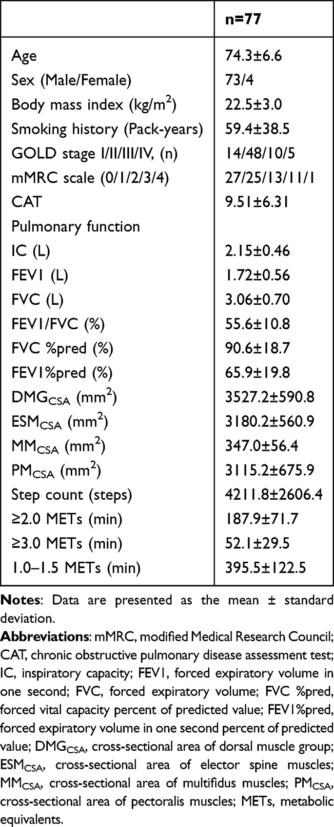 |
Table 1 Patient’s Characteristics (Study 1) |
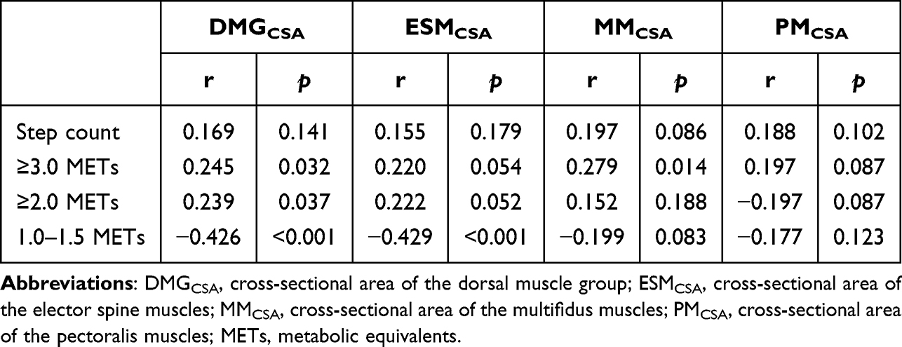 |
Table 2 Correlation Between Physical Activity and the Cross-Sectional Area of Muscles |
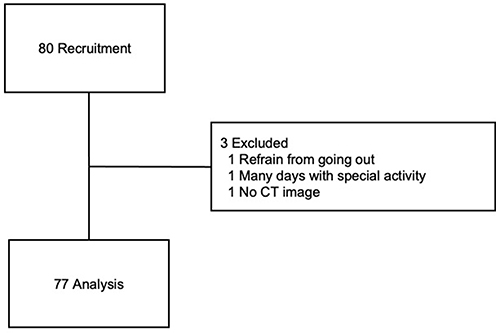 |
Figure 2 Flow diagram. |
In Study 2, there was a significant difference in sex ratios between patients with COPD and healthy subjects (p<0.001), but there was no marked difference in age. In cases of COPD, FEV1%pred was 81.3%, and the GOLD stage was I/II/III/IV in 3/3/1/0, respectively (Table 3). The MF slopes, calculated as the intercept of the regression line between MF and time, of both the right and left MM were significantly lower in COPD patients than in healthy subjects, but those of the ESM showed no significant difference (Figure 3). Furthermore, the endurance time in the trunk holding test was not significantly different between patients with COPD and healthy subjects (Figure S3).
 |
Table 3 Patient’s Characteristics (Study 2) |
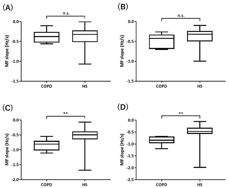 |
Figure 3 A comparison of the MF slope in the erector spinae and multifidus muscles. (A) Right spinal erector spinae, (B) Left spinal erector spinae, (C) Right multifidus muscle, (D) Left multifidus muscle. Abbreviations: MF slope, median frequency slope; HS, healthy subjects; n.s., not significant. Note: ** p<0.01. |
Discussion
In this present study, we found that the MMCSA was positively correlated with the duration of moderate-to-vigorous PA (MVPA, ≥3 METs), while the ESMCSA was negatively correlated with the duration of SB (1.0–1.5 METs). The DMGCSA correlated with both PA and SB, suggesting that the MM and ESM may have distinct roles in the DMG. In addition, the muscle fatigue characteristics indicated by the slope of the MF were lower in the MM of COPD patients than in healthy people. However, no significant difference was found in the ESM.
The MMCSA was positively correlated with the duration at ≥3 METs, which may highlight its role in dynamic trunk stabilization during MVPA, such as walking and cycling. Conversely, the ESMCSA was negatively correlated with the duration at 1.0–1.5 METs, such as lying down or sitting, which may reflect reduced muscle activation in these static postures.26,27 Prolonged inactivity can lead to chronic deactivation of the ESM, contributing to muscle atrophy,28 which aligns with our findings of a negative correlation between the ESMCSA and SB.
Tanimura et al12 reported that correlations between the ESMCSA and step count; however, “their ESMCSA” measurement included both the MMCSA and ESMCSA, meaning that “their ESMCSA” was the same as the DMGCSA in the current study. Although the DMGCSA did not correlate with the step count in our report, it did correlate with the duration at ≥3 METs, one of the PA parameters, which was compatible with their results.
We observed no significant correlation between the PMCSA and any PA or SB parameter. Previous studies have shown that upper limb muscle strength is less affected by PA than lower limb or trunk muscle strength in healthy subjects.29 In patients with COPD, lower limb muscle weakness is more likely to occur than upper limb muscle weakness than in healthy subjects,2 and lower limb muscle strength in COPD is strongly associated with a reduced exercise capacity.30 These previous reports suggested that the upper limb muscle may be less strongly associated with PA in COPD than in healthy patients. This is consistent with our findings, in which the PMCSA was not correlated with PA or SB. Another study suggested that grip strength correlates with PA in patients with COPD and a good nutritional status.7 However, they also demonstrated no correlation between grip strength and PA in patients with COPD and a poor nutritional status. Although we did not assess the nutritional status, our cohort might have included patients with a relatively poor nutritional status.
The MF slope of the MM in COPD patients was reduced compared to that in healthy subjects, suggesting that the MM is more prone to fatigue in this population. MM is classified as a local muscle primarily responsible for stabilizing the spine during dynamic activities. Unlike the ESM, which generates significant force for spinal extension, the MM collaborates with other trunk muscles, such as the transversus abdominis, diaphragm, and pelvic floor muscles, to resist lumbar flexion.31–33 In patients with COPD, dynamic hyperinflation flattens the diaphragm, making it difficult to maintain intra-abdominal pressure and stabilize the trunk.34 The observed decline in the MF slope of the MM in COPD patients might reflect decreased spinal stability and a compromised diaphragm function, both of which are key characteristics of COPD. Although the MM typically comprises fatigue-resistant type I muscle fibers, the decline in MF slope may indicate a shift in the muscle fiber composition or quality in patients with COPD.35–37 This may be influenced by the fact that fatty infiltration within the muscle occurs even in cases of mild COPD.38
The MF slope of the ESM did not differ markedly between COPD patients and healthy subjects, suggesting that ESM endurance was maintained in COPD patients. In addition, there was no marked difference in endurance time between COPD patients and healthy subjects, suggesting that muscle endurance in the trunk holding test is mainly determined by ESM activity. The ESM is a global muscle that supports spinal extension and functions as a muscle that maintains an anti-gravity posture.31 Most of the recruited patients with COPD in Study 2 had mild or moderate stages of the disease. In the early stages of COPD, the ESM function might be maintained to the same extent as in healthy subjects, but muscle fibers in the MM, which are affected by the diaphragm function, might be altered.
Sung et al reported significant improvements in the MM function and reductions in low back pain in patients with low back pain through a 4-week spinal stabilization exercise program designed to improve MM contractile capacity.39 On the other hand, there are reports that the muscle activity of the ESM is increased by strength training such as back extension and deadlift.40 Therefore, it may be important to evaluate MM and ESM separately, and to implement a spinal stabilization program for patients with decreased MM and a strength training program for patients with decreased ESM.
Several limitations associated with the present study warrant mention. First, In study 1, the MMCSA at the Th12 level was considerably smaller than that of the ESMCSA, so measurement errors may have occurred. In the present studies, the MMCSA and ESMCSA were measured at the Th12 level, as reported in previous studies on DMGCSA. However, since some studies measured the MMCSA at the lumbar spine level, it is possible that the MMCSA needs to be measured at the lumbar spine level to obtain more accurate measurements.41 This point should be considered in future studies. Second, Study 1 focused on the relationship between masses and PA or SB in patients with COPD, but other factors, such as the cardiopulmonary function, nutritional status, and medication use, may also influence PA or SB. As these factors were not accounted for, conclusions regarding the direct impact of muscle changes on PA or SB may be limited. Third, the sample size of the COPD patients in Study 2 was small, which may have limited the generalizability of the results. A larger sample size would provide more robust results. Fourth, there was a significant sex imbalance between the COPD and healthy groups, with more men in the COPD group and more women in the healthy group in Study 2. Previous studies have indicated that sex differences in MF slope are negligible in the elderly,42 suggesting that the observed differences in the MF slope of MM were likely disease-related rather than gender-related. Fifth, although the recruited patients with COPD in Study 2 were primarily in the mild to moderate stages, it is possible that muscle fatigue patterns differ in patients with more severe COPD. Although it may be difficult to conduct the trunk holding test in patients with severe or very severe COPD, evaluating muscle fatigue in these patients remains important.
Conclusion
In COPD patients, MM was associated with a duration at ≥3.0 METs whereas ESM was associated with a duration at 1.0–1.5 METs, and MM in COPD patients was more likely to cause fatigue than in healthy subjects, thus indicating that the two muscles have different characteristics in physical activity.
Acknowledgments
We thank Brian Quinn, Managing Editor for Japan Medical Communication, for proofreading this manuscript.
Author Contributions
All authors made a significant contribution to the work reported, whether that is in the conception, study design, execution, acquisition of data, analysis, and interpretation, or in all these areas, took part in drafting, revising, or critically reviewing the article; gave final approval of the version to be published; have agreed on the journal to which the article has been submitted; and agree to be accountable for all aspects of the work.
Funding
This study was funded by the Environmental Restoration and Conservation Agency of Japan.
Disclosure
KT received lecture fees from AstraZeneca, GlaxoSmithKline, Nippon Boehringer Ingelheim and Sanofi. KA received lecture fees from AstraZeneca, GlaxoSmithKline, Nippon Boehringer Ingelheim, Novartis Pharma and Sanofi. TH received lecture fees from AstraZeneca, Sanofi, GlaxoSmithKline and Kyorin Pharmaceuticals. KM received lecture fees from AstraZeneca, Sanofi, GlaxoSmithKline, Novartis Pharma, Kyorin Pharmaceutical and Nippon Boehringer Ingelheim. Y. Minakata received lectures from Nippon Boehringer Ingelheim. The authors report no other conflicts of interest in this work.
References
1. Maltais F, Decramer M, Casaburi R. et al. An official American Thoracic Society/European Respiratory Society statement: update on limb muscle dysfunction in chronic obstructive pulmonary disease. Am J Respir Crit Care Med. 2014;189(9):e15–e62. doi:10.1164/rccm.201402-0373ST
2. Bernard S, LeBlanc P, Whittom F, et al. Peripheral muscle weakness in patients with chronic obstructive pulmonary disease. Am J Respir Crit Care Med. 1998;158(2):629–634. doi:10.1164/ajrccm.158.2.9711023
3. Pepin V, Saey D, Whittom F, LeBlanc P, Maltais F. Walking versus cycling: sensitivity to bronchodilation in chronic obstructive pulmonary disease. Am J Respir Crit Care Med. 2005;172(12):1517–1522. doi:10.1164/rccm.200507-1037OC
4. Barreiro E, Jaitovich A. Muscle atrophy in chronic obstructive pulmonary disease: molecular basis and potential therapeutic targets. J Thorac Dis. 2018;10(Suppl 12):S1415–S1424. doi:10.21037/jtd.2018.04.168
5. Schols AM, Broekhuizen R, Weling-Scheepers CA, Wouters EF. Body composition and mortality in chronic obstructive pulmonary disease. Am J Clin Nutr. 2005;82(1):53–59. doi:10.1093/ajcn/82.1.53
6. Yoshimura K, Sato S, Muro S, et al. Interdependence of physical inactivity, loss of muscle mass and low dietary intake: extrapulmonary manifestations in older chronic obstructive pulmonary disease patients. Geriatr Gerontol Int. 2018;18(1):88–94. doi:10.1111/ggi.13146
7. Kawai T, Asai K, Miyamoto A, et al. Distinct contributions of muscle mass and strength stratified by nutritional status to physical activity in patients with chronic obstructive pulmonary disease. Respir Investig. 2023;61(4):389–397. doi:10.1016/j.resinv.2023.03.001
8. Garcia-Aymerich J, Lange P, Benet M, Schnohr P, Antó JM. Regular physical activity reduces hospital admission and mortality in chronic obstructive pulmonary disease: a population based cohort study. Thorax. 2006;61(9):772–778. doi:10.1136/thx.2006.060145
9. Waschki B, Kirsten A, Holz O, et al. Physical activity is the strongest predictor of all-cause mortality in patients with COPD: a prospective cohort study. Chest. 2011;140(2):331–342. doi:10.1378/chest.10-2521
10. Garcia-Rio F, Rojo B, Casitas R, et al. Prognostic value of the objective measurement of daily physical activity in patients with COPD. Chest. 2012;142(2):338–346. doi:10.1378/chest.11-2014
11. GOLD. Global Strategy for the Diagnosis, Management and Prevention of COPD, Global Initiative for Chronic Obstructive Lung Disease (GOLD) 2018. 2018. https://goldcoped.org/.
12. Tanimura K, Sato S, Fuseya Y, et al. Quantitative Assessment of Erector Spinae Muscles in Patients with Chronic Obstructive Pulmonary Disease. Novel Chest Computed Tomography-derived Index for Prognosis. Ann Am Thorac Soc. 2016;13(3):334–341. doi:10.1513/AnnalsATS.201507-446OC
13. Tanimura K, Sato S, Sato A, et al. Accelerated Loss of Antigravity Muscles Is Associated with Mortality in Patients with COPD. Respiration. 2020;99(4):298–306. doi:10.1159/000506520
14. Higashimoto Y, Shiraishi M, Sugiya R, et al. Effect of Pulmonary Rehabilitation on Erector Spinae Muscles in Individuals With COPD. Respir Care. 2021;66(9):1458–1468. doi:10.4187/respcare.08678
15. Biering-Sorensen F. Physical measurements as risk indicators for low-back trouble over a one-year period. Spine. 1984;9(2):106–119. doi:10.1097/00007632-198403000-00002
16. Ng JK, Richardson CA. Reliability of electromyographic power spectral analysis of back muscle endurance in healthy subjects. Arch Phys Med Rehabil. 1996;77(3):259–264. doi:10.1016/s0003-9993(96)90108-2
17. Nishimura Y, Tsuboi H, Murata KY, et al. Comparison of erector spinae fatigability between female patients with Parkinson’s disease and healthy individuals: a cross sectional pilot study. BMC Neurol. 2022;22(1):189. doi:10.1186/s12883-022-02719-w
18. Freeman MD, Woodham MA, Woodham AW. The role of the lumbar multifidus in chronic low back pain: a review. PM R. 2010;2(2):142–167. doi:10.1016/j.pmrj.2009.11.006
19. Danneels LA, Vanderstraeten GG, Cambier DC, Witvrouw EE, De Cuyper HJ, Danneels L. CT imaging of trunk muscles in chronic low back pain patients and healthy control subjects. Eur Spine J. 2000;9(4):266–272. doi:10.1007/s005860000190
20. Hides J, Gilmore C, Stanton W, Bohlscheid E. Multifidus size and symmetry among chronic LBP and healthy asymptomatic subjects. Man Ther. 2008;13(1):43–49. doi:10.1016/j.math.2006.07.017
21. Lee CS, Cron DC, Terjimanian MN, et al. Dorsal muscle group area and surgical outcomes in liver transplantation. Clin Transplant. 2014;28(10):1092–1098. doi:10.1111/ctr.12422
22. McDonald ML, Diaz AA, Ross JC, et al. Quantitative computed tomography measures of pectoralis muscle area and disease severity in chronic obstructive pulmonary disease. A cross-sectional study. Ann Am Thorac Soc. 2014;11(3):326–334. doi:10.1513/AnnalsATS.201307-229OC
23. Choi L, Liu Z, Matthews CE, Buchowski MS. Validation of accelerometer wear and nonwear time classification algorithm. Med Sci Sports Exerc. 2011;43(2):357–364. doi:10.1249/MSS.0b013e3181ed61a3
24. Hermens HJ, Freriks B, Merletti R, et al. European Recommendations for Surface Electromyography; Results of the SENIAM Project. Enschede: Roessingh Research and Development b.v; 1999.
25. Tsuboi H, Nishimura Y, Sakata T, et al. Properties of Paraspinal Muscles in Japanese High School Baseball Players With Terminal-Stage Lumbar Spondylolysis. PM R. 2018;10(2):175–182. doi:10.1016/j.pmrj.2017.06.018
26. Claus AP, Hides JA, Moseley GL, Hodges PW. Thoracic and lumbar posture behaviour in sitting tasks and standing: progressing the biomechanics from observations to measurements. Appl Ergon. 2016;53(Pt A):161–168. doi:10.1016/j.apergo.2015.09.006
27. Callaghan JP, McGill SM. Low back joint loading and kinematics during standing and unsupported sitting. Ergonomics. 2001;44(3):280–294. doi:10.1080/00140130118276
28. Mörl F, Bradl I. Lumbar posture and muscular activity while sitting during office work. J Electromyogr Kinesiol. 2013;23(2):362–368. doi:10.1016/j.jelekin.2012.10.002
29. Leblanc A, Pescatello LS, Taylor BA, et al. Relationships between physical activity and muscular strength among healthy adults across the lifespan. Springerplus. 2015;4(1):557. doi:10.1186/s40064-015-1357-0
30. Mador MJ, Bozkanat E. Skeletal muscle dysfunction in chronic obstructive pulmonary disease. Respir Res. 2001;2(4):216–224. doi:10.1186/rr60
31. Bergmark A. Stability of the lumbar spine. A study in mechanical engineering. Acta Orthop Scand Suppl. 1989;230(sup230):1–54. doi:10.3109/17453678909154177
32. Norris CM. Spinal stabilisation: 3. Stabilisation mechanisms of the lumbar spine. Physiotherapy. 1995;81(2):72–79. doi:10.1016/S0031-9406(05)67048-4
33. Malátová R, Rokytová J, Stumbauer J. The use of muscle dynamometer for correction of muscle imbalances in the area of deep stabilising spine system. Proc Inst Mech Eng H. 2013;227(8):896–903. doi:10.1177/0954411913486078
34. De Troyer A. Effect of hyperinflation on the diaphragm. Eur Respir J. 1997;10(3):708–713. doi:10.1183/09031936.97.10030708
35. Jørgensen K, Nicholaisen T, Kato M, Kato M. Muscle fiber distribution, capillary density, and enzymatic activities in the lumbar paravertebral muscles of young men. Significance for isometric endurance. Spine. 1993;18(11):1439–1450. doi:10.1097/00007632-199318110-00007
36. Abe T, Loenneke JP, Thiebaud RS, Fukunaga T. Age-related site-specific muscle wasting of upper and lower extremities and trunk in Japanese men and women. Age. 2014;36(2):813–821. doi:10.1007/s11357-013-9600-5
37. Mannion AF, Dumas GA, Stevenson JM, Cooper RG. The influence of muscle fiber size and type distribution on electromyographic measures of back muscle fatigability. Spine. 1998;23(5):576–584. doi:10.1097/00007632-199803010-00010
38. Jeon YJ, Han S, Park GM, et al. Intramuscular and Intermuscular Abdominal Fat Infiltration in COPD: a Propensity Score Matched Study. Int. J Chron Obstruct Pulmon Dis. 2021;16:1989–1999. doi:10.2147/COPD.S312888
39. Sung PS. Multifidi muscles median frequency before and after spinal stabilization exercises. Arch Phys Med Rehabil. 2003;84(9):1313–1318. doi:10.1016/S0003-9993(03)00139-4
40. Oliva-Lozano JM, Muyor JM. Core Muscle Activity During Physical Fitness Exercises: a Systematic Review. Int J Environ Res Public Health. 2020;17(12):4306. doi:10.3390/ijerph17124306
41. Bouche KG, Vanovermeire O, Stevens VK, et al. Computed tomographic analysis of the quality of trunk muscles in asymptomatic and symptomatic lumbar discectomy patients. BMC Musculoskelet Disord. 2011;12:65. doi:10.1186/1471-2474-12-65
42. Tsuboi H, Nishimura Y, Sakata T, et al. Age-related sex differences in erector spinae muscle endurance using surface electromyographic power spectral analysis in healthy humans. Spine J. 2013;13(12):1928–1933. doi:10.1016/j.spinee.2013.06.060
 © 2025 The Author(s). This work is published and licensed by Dove Medical Press Limited. The
full terms of this license are available at https://www.dovepress.com/terms
and incorporate the Creative Commons Attribution
- Non Commercial (unported, 4.0) License.
By accessing the work you hereby accept the Terms. Non-commercial uses of the work are permitted
without any further permission from Dove Medical Press Limited, provided the work is properly
attributed. For permission for commercial use of this work, please see paragraphs 4.2 and 5 of our Terms.
© 2025 The Author(s). This work is published and licensed by Dove Medical Press Limited. The
full terms of this license are available at https://www.dovepress.com/terms
and incorporate the Creative Commons Attribution
- Non Commercial (unported, 4.0) License.
By accessing the work you hereby accept the Terms. Non-commercial uses of the work are permitted
without any further permission from Dove Medical Press Limited, provided the work is properly
attributed. For permission for commercial use of this work, please see paragraphs 4.2 and 5 of our Terms.
Recommended articles

Bibliometric Analysis of Exercise and Chronic Obstructive Pulmonary Disease
Chen M, Zhang Y, Mao Y, Lian Y, Ye P, Liu C, Zhang Z, Fu X
International Journal of Chronic Obstructive Pulmonary Disease 2023, 18:1115-1133
Published Date: 8 June 2023
Determinants of Activity Phenotype in Patients with Chronic Obstructive Pulmonary Disease
Murakami Y, Minakata Y, Kato M, Sasaki S, Azuma Y, Kawabe K, Ono H
International Journal of Chronic Obstructive Pulmonary Disease 2023, 18:1919-1929
Published Date: 31 August 2023
Communication About Sexuality (COSY) Encourages Physical Activity in COPD: A Randomised Trial
Dalla Lana K, Frei A, Radtke T, Braun J, Puhan MA, Steurer-Stey C
International Journal of Chronic Obstructive Pulmonary Disease 2025, 20:3535-3544
Published Date: 30 October 2025