
Back to Journals » Infection and Drug Resistance » Volume 19
Clinical Features and Risk Factors for Rhabdomyolysis in Patients with Chlamydia psittaci Pneumonia: A Multicenter Study
Authors Xu L, Zhang A, Zou J, Sun C, Lin Y, Fang C 
Received 6 March 2026
Accepted for publication 15 May 2026
Published 22 May 2026 Volume 2026:19 607267
DOI https://doi.org/10.2147/IDR.S607267
Checked for plagiarism Yes
Review by Single anonymous peer review
Peer reviewer comments 2
Editor who approved publication: Dr Hazrat Bilal
Limin Xu,1 Anbing Zhang,2 Jialiu Zou,3 Cheng Sun,4 Yuejiao Lin,5 Changquan Fang6
1Department of Geriatrics, Huizhou First Hospital, Huizhou, Guangdong, People’s Republic of China; 2Department of Pulmonary and Critical Care Medicine, Zhongshan People’s Hospital, Zhongshan, Guangdong, People’s Republic of China; 3Department of Pulmonary and Critical Care Medicine, Dongkeng Hospital of Dongguan, Dongguan, Guangdong, People’s Republic of China; 4Department of Pulmonary and Critical Care Medicine, Guangzhou First People’s Hospital, South China University of Technology, Guangzhou, Guangdong, People’s Republic of China; 5Department of Pulmonary and Critical Care Medicine, Shaoguan First People’s Hospital, Shaoguan, Guangdong, People’s Republic of China; 6Department of Pulmonary and Critical Care Medicine, Huizhou Central People’s Hospital, Huizhou, Guangdong, People’s Republic of China
Correspondence: Changquan Fang, Department of Pulmonary and Critical Care Medicine, Huizhou Central People’s Hospital, No. 41 Eling North Road, Huicheng District, Huizhou, 516000, Guangdong, People’s Republic of China, Tel + 86 137-5155-1071, Email [email protected]
Purpose: Chlamydia psittaci infection can lead to rhabdomyolysis (RM); however, systematic characterization of RM in patients with Chlamydia psittaci pneumonia has not been performed. This study analyzed the clinical characteristics of and risk factors for RM in patients with Chlamydia psittaci pneumonia.
Patients and Methods: Clinical data of patients with Chlamydia psittaci pneumonia who were admitted to hospitals in Guangdong Province between January 2020 and December 2025 were retrospectively collected. Serum muscle enzyme activities at admission and factors associated with RM development were investigated.
Results: Elevated creatine kinase (CK) levels were observed in 46.8% of patients, and 20.5% developed RM. Compared with the non-severe pneumonia group, the severe pneumonia group included significantly more patients with elevated CK levels and RM (P < 0.001). Compared with patients without RM, those with RM experienced higher incidences of myalgia, dyspnea, and altered consciousness, and they had a higher Pneumonia Severity Index (PSI), a greater likelihood of acute kidney injury, and longer hospital stays; additionally, they more commonly required mechanical ventilation. Systemic inflammatory markers (such as C-reactive protein [CRP]), skeletal muscle and myocardial injury markers, D-dimer levels, and hepatic and renal dysfunction indicators were substantially escalated in patients with RM; however, their serum sodium and albumin levels were significantly lower (P < 0.05). High PSI, elevated CRP level, dyspnea, and hyponatremia were considered independent risk factors for RM in patients with Chlamydia psittaci pneumonia.
Conclusion: RM is an important complication of Chlamydia psittaci pneumonia. Early monitoring and intervention are warranted to improve the prognosis.
Keywords: Chlamydia psittaci, pneumonia, rhabdomyolysis, risk factors
Introduction
Chlamydia psittaci pneumonia is a rare zoonotic infectious disease caused by the transmission of Chlamydia psittaci bacterium.1,2 Recently, because of the extensive application of high-throughput sequencing technologies and increased clinical awareness, the reported incidence of Chlamydia psittaci pneumonia has significantly increased.3,4 Clinical manifestations of Chlamydia psittaci pneumonia are heterogeneous and range from mild influenza-like symptoms to severe community-acquired pneumonia; additionally, Chlamydia psittaci pneumonia is often accompanied by high fever, dry cough, headache, and prominent extrapulmonary manifestations such as hepatic dysfunction and neurological symptoms.4,5 Diverse symptoms frequently result in a misdiagnosis and delayed treatment.
Clinical observations have indicated that the occurrence of rhabdomyolysis (RM) in patients with Chlamydia psittaci pneumonia is not only prevalent but also often indicates severe disease and a poor prognosis.6 Compared with patients with uncomplicated Chlamydia psittaci pneumonia, those with Chlamydia psittaci pneumonia and concurrent RM tend to experience more severe myalgia and higher levels of inflammatory markers, and they are at an increased risk for multiorgan dysfunction.6,7 Nevertheless, the current understanding of RM is limited because its clinical epidemiological characteristics, such as its incidence and typical clinical presentation, in large multicenter cohorts have not been systematically described. Additionally, key risk factors that predispose certain patients with Chlamydia psittaci pneumonia to RM are unclear. Whether these factors are related to pathogen virulence, host-specific responses, or other clinical or therapeutic factors requires further elucidation. In-depth analyses of clinical outcomes of this patient population, including the development of acute kidney injury (AKI) and the need for intensive care, have been insufficient, thus limiting assessments of the impact of RM on treatment strategies and prognostic evaluations.
Therefore, systematically characterizing the clinical characteristics of Chlamydia psittaci pneumonia complicated by RM and identifying associated risk factors are of critical importance to early risk stratification, which enables timely intervention and enhances clinical prognosis. Therefore, this multicenter retrospective study aimed to comprehensively describe the clinical characteristics of and risk factors for RM in patients with Chlamydia psittaci pneumonia to provide evidence-based support for clinical decision-making.
Materials and Methods
Study Design and Participants
Clinical data of patients with Chlamydia psittaci pneumonia who were admitted to eight hospitals in Guangdong Province between January 2020 and December 2025 were retrospectively collected. The inclusion criteria were as follows: clinical manifestations and chest imaging findings consistent with the diagnostic criteria for community-acquired pneumonia;8 negative results of routine microbiological examinations (including cultures and smears of blood, sputum, and bronchoalveolar lavage fluid); and detection of Chlamydia psittaci nucleic acid sequences in bronchoalveolar lavage fluid or blood samples via high-throughput sequencing techniques (eg., metagenomic next-generation sequencing [mNGS] or targeted next-generation sequencing [tNGS]). The exclusion criteria were age younger than 18 years and incomplete clinical data.
The diagnosis of severe community-acquired pneumonia required meeting8 either (1) any one of the major criteria, namely, septic shock requiring vasopressors and respiratory failure requiring mechanical ventilation or (2) at least three of the following minor criteria: respiratory rate ≥ 30/min, PaO2/FiO2 ≤ 250 mmHg, multilobar infiltrates, confusion and/or disorientation, blood urea nitrogen ≥ 20 mg/dL, white blood cell count < 4×10⁹/L, platelet count < 100×109/L, temperature < 36 °C, or hypotension requiring rapid fluid resuscitation. RM was diagnosed according to the following established criteria:9 presence of clinical symptoms such as myalgia and muscle weakness; elevated creatine kinase (CK) levels exceeding five-times the upper limit of normal (310 U/L for male patients and 200 U/L for female patients); and exclusion of common etiologies, such as trauma, exercise, cerebrovascular or cardiovascular events, and drug use. Participants were categorized into the RM group or non-RM group based on these diagnostic criteria. AKI was defined as an increase in serum creatinine level ≥ 0.3 mg/dL within 48 h; an increase to ≥ 1.5 times the known or presumed baseline value within the previous 7 days; or urine output persistently < 0.5 mL/kg/h for 6 h.10
Next-Generation Sequencing
In accordance with standard clinical procedures, 5–10 mL of bronchoalveolar lavage fluid or venous blood was collected from each patient and submitted to designated laboratories for mNGS or tNGS. Specifically, mNGS was performed by Daan Gene Co., Ltd. (Guangzhou, China), Vision Medicals Co., Ltd. (Guangzhou, China), BGI Genomics Co., Ltd. (Shenzhen, China), and Seegene Biotech Co., Ltd. (Guangzhou, China). tNGS was conducted by Daan Gene Co., Ltd. (Guangzhou, China), KingMed Diagnostics Group Co., Ltd. (Guangzhou, China), and Huayin Medical Laboratory (Guangzhou, China). All procedures, including nucleic acid extraction and purification, library construction, high-throughput sequencing, and bioinformatics analysis, followed standardized laboratory workflows, and formal pathogen detection reports were issued.
Data Collection
A standardized case report form was developed to record data. This form included the following: baseline characteristics such as age, sex, interval from symptom onset to hospital admission, length of hospital stay, and history of underlying diseases; clinical manifestations and vital signs at admission; laboratory findings upon admission; and treatment regimens and clinical outcomes. Additionally, the Pneumonia Severity Index (PSI) was calculated based on clinical parameters obtained on the day of admission.
Statistical Analysis
Statistical analyses were performed using SPSS version 25.0. Continuous variables with normal distribution were expressed as mean ± standard deviation (SD) ( ). For normally distributed data, comparisons between groups were performed using the independent samples t-test, and correlations were assessed using Pearson’s correlation analysis. Continuous variables that were not normally distributed were presented as medians (interquartile range [IQR]). For non-normally distributed data, comparisons between groups were conducted using the Mann–Whitney U-test, and correlations were evaluated using Spearman’s rank correlation analysis. Finally, categorical variables were expressed as frequencies (percentages), and comparisons between groups were performed using the χ2 test or Fisher’s exact test.
To identify risk factors associated with RM, variables with P < 0.05 in univariate analysis were included in a multivariable logistic regression model for further analysis. Receiver-operating characteristic (ROC) curves were plotted to evaluate the diagnostic performance of relevant indicators of RM. A two-sided P < 0.05 was considered statistically significant.
Results
General Information of Patients with Chlamydia psittaci Pneumonia
A total of 231 patients with Chlamydia psittaci pneumonia were initially included; after excluding one patient younger than 18 years and 10 patients with incomplete clinical data, 220 patients were finally included. Furthermore, 132 (60.0%) were male and 88 (40.0%) were female. The mean age of the participants was 58.9 years (SD, ±12.1 years). The median interval from symptom onset to hospital admission was 5 days (IQR, 3–7 days). Additionally, 113 patients (51.4%) had underlying comorbidities, including hypertension in 56 (25.5%), diabetes mellitus in 44 (20.0%), coronary heart disease in 21 (9.5%), and chronic liver disease in 15 (6.8%) [Table 1]. Furthermore, of these 220 patients, 103 (46.8%) had elevated CK levels and 45 (20.5%) developed RM. Of the 120 patients with non-severe pneumonia, 34 (28.3%) had elevated CK levels and 7 (5.8%) developed RM. In contrast, among the 100 patients with severe pneumonia, 69 (69.0%) exhibited elevated CK levels and 38 (38.0%) developed RM. The proportions of patients with elevated CK levels and those who developed RM were significantly higher in the severe pneumonia group than in the non-severe pneumonia group (P < 0.001) [Figure 1].
 |
Table 1 Clinical Features of Rhabdomyolysis in Patients with C. psittaci Pneumonia |
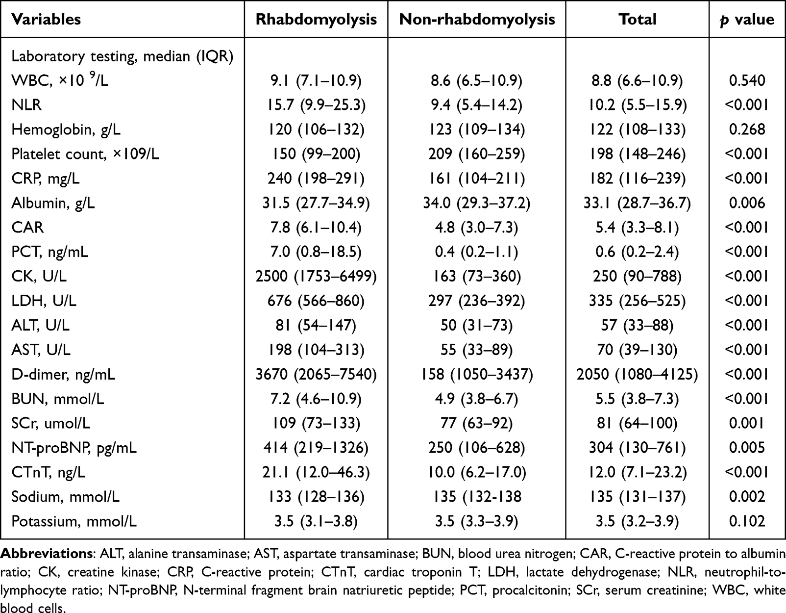 |
Table 2 Analysis of Laboratory Test Results for Patients with C. psittaci Pneumonia Accompanied with Rhabdomyolysis |
 |
Figure 1 Creatine kinase abnormality at admission in patients with Chlamydia psittaci pneumonia by severity of disease. ***Comparison between severe and non-severe pneumonia, P<0.001. Bars represent the number of patients. |
Among the 45 patients with RM, CK levels ranged from 1,000 to 4,999 U/L in 28 patients and from 5,000 to 14,999 U/L in 10 patients. Additionally, CK levels were higher than 15,000 U/L in seven patients (Figure 2).
 |
Figure 2 Distribution of creatine kinase levels in 45 patients with rhabdomyolysis. Bars represent the number of patients. |
Clinical Characteristics of RM in Patients with Chlamydia psittaci Pneumonia
Compared with patients without RM, those with RM were predominantly male (Tables 1–3). Furthermore, those with RM exhibited significantly higher frequencies of myalgia, dyspnea, and altered consciousness and had a higher PSI. Patients with RM were also more likely to develop AKI, require prolonged hospital stays, and require invasive mechanical ventilation. Additionally, compared with patients without RM, those with RM had significantly elevated neutrophil-to-lymphocyte ratios (NLRs), C-reactive protein (CRP)-to-albumin ratios (CARs), and levels of CRP, procalcitonin (PCT), CK, lactate dehydrogenase, D-dimer, alanine aminotransferase, aspartate aminotransferase, blood urea nitrogen, serum creatinine, N-terminal pro-B-type natriuretic peptide, and cardiac troponin T. In contrast, platelet counts and serum sodium and albumin levels were significantly lower in patients with RM (P < 0.05).
 |
Table 3 Treatment and Prognosis for Patients with C. psittaci Pneumonia Accompanied with Rhabdomyolysis |
A correlation heatmap analysis demonstrated that CK and lactate dehydrogenase levels were significantly positively correlated with the PSI (r = 0.467, P < 0.001; r = 0.587, P < 0.001), NLR (r = 0.359, P < 0.001; r = 0.363, P < 0.001), CAR (r = 0.393, P < 0.001; r = 0.468, P < 0.001), and levels of PCT (r = 0.559, P < 0.001; r = 0.606, P < 0.001), alanine aminotransferase (r = 0.382, P < 0.001; r = 0.533, P < 0.001), aspartate aminotransferase (r = 0.549, P < 0.001; r = 0.743, P < 0.001), and D-dimer (r = 0.429, P < 0.001; r = 0.628, P < 0.001). Conversely, CK and lactate dehydrogenase levels were significantly negatively correlated with the platelet count (r = −0.393, P < 0.001; r = −0.380, P < 0.001) [Table 4].
 |
Table 4 Correlation of Muscle Enzyme-Related Indicators |
Risk Factors for RM in Patients with Chlamydia psittaci Pneumonia
Based on previous literature on RM, both the number of events (ie., number of patients with concurrent RM) and potential collinearity among variables were analyzed to identify factors associated with RM. A multivariable logistic regression analysis of the identified factors was subsequently conducted. RM development was included as the dependent variable, and the PSI, NLR, CRP level, dyspnea, and hyponatremia were included as independent variables. The results demonstrated that a higher PSI (odds ratio [OR], 1.182; 95% confidence interval [CI], 1.029–1.316; P = 0.022), dyspnea (OR, 6.002; 95% CI, 1.212–29.717; P = 0.028), elevated CRP levels (OR, 1.217; 95% CI, 1.182–1.475; P = 0.007), and hyponatremia (OR, 2.249; 95% CI, 1.063–4.762; P = 0.034) were independent risk factors for RM in patients with Chlamydia psittaci pneumonia (Table 5).
 |
Table 5 Risk Factors for Rhabdomyolysis in Multivariate Logistic Regression Model |
To evaluate the predictive values of the PSI and CRP levels for RM in Chlamydia psittaci pneumonia, a ROC curve analysis was performed. The area under the ROC curve values for both the PSI and CRP levels were significantly greater than 0.7 (P < 0.001), indicating good diagnostic performance (Table 6 and Figure 3). Using a cutoff value of 107, the sensitivity and specificity of the PSI for predicting RM were 77.1% and 72.0%, respectively. Alternatively, using a cutoff value of 196 mg/L, the sensitivity and specificity of the CRP level were 75.6% and 64.6%, respectively.
 |
Table 6 Predictive Efficacy of PSI Score and CRP for Rhabdomyolysis |
 |
Figure 3 ROC curve of PSI and CRP in predicting Chlamydia psittaci Pneumonia complicated with rhabdomyolysis. Abbreviations: CRP, C-reactive protein; PSI, pneumonia severity index. |
Discussion
To our knowledge, this is the first study to systematically investigate the clinical characteristics of and risk factors for RM in patients with Chlamydia psittaci pneumonia. This multicenter retrospective analysis indicated that RM is an important complication of Chlamydia psittaci pneumonia that is closely associated with aggravated disease severity and adverse clinical outcomes. Additionally, several independent risk factors with early warning potential were identified in this study.
RM developed in 20.5% of patients with Chlamydia psittaci pneumonia in the study cohort, and nearly half exhibited elevated CK levels. This proportion was considerably higher than that reported for pneumococcal pneumonia and coronavirus disease 2019 (COVID-19).11–13 These findings suggest that Chlamydia psittaci infection may have a stronger propensity for direct skeletal muscle involvement or triggering an intense systemic inflammatory response, thus leading to myocyte injury. The results of this study further revealed that patients with Chlamydia psittaci pneumonia complicated by RM generally experienced more severe disease, as reflected by the increased incidences of dyspnea and altered consciousness, higher PSI, increased need for invasive mechanical ventilation, and prolonged hospital stays. These observations are largely consistent with the findings in cases of RM associated with Legionella infection and COVID-19.14,15 Additionally, the incidence of AKI in patients with RM was significantly higher than that in patients without RM, suggesting that nephrotoxic substances released during RM, such as myoglobin, may contribute to renal injury.16,17 This mechanism may play an important role in exacerbating disease severity and poor prognoses. Therefore, RM should be regarded as a clinically significant complication of Chlamydia psittaci pneumonia that warrants heightened attention and proactive management.
The precise mechanisms underlying RM in Chlamydia psittaci pneumonia are unclear. However, based on the findings of this study and the previous literature,18–22 the following potential mechanisms have been indicated: direct invasion of skeletal muscle cells by the pathogen, immune-mediated injury driven by proinflammatory mediators and cytokines, damage caused by microvascular obstruction, and hypoxia and metabolic disturbances.
Chlamydia psittaci, which is characterized by high pathogenicity, may cause widespread damage to host cells, including myocytes, thereby leading to systemic infection.23 Participants with RM exhibited significantly elevated inflammatory markers, including the NLR, CAR, and levels of CRP and PCT. Chlamydia psittaci infection can trigger a robust immune response with the release of large amounts of inflammatory cytokines.19 These inflammatory cytokines may directly injure muscle cells or facilitate the release of reactive oxygen species and proteolytic enzymes through the activation of immune cells (eg., neutrophils and macrophages), further disrupting muscle cell membranes and intracellular structures. Substantially elevated D-dimer levels observed in our cohort suggest a hypercoagulable state and possible endothelial injury.24,25 Severe Chlamydia psittaci pneumonia can impair pulmonary gas exchange, resulting in systemic hypoxia. Under hypoxic conditions, skeletal muscle cells may develop energy metabolism dysfunction and membrane instability. Notably, although previous studies have reported cases of quinolone-induced RM,26 no such cases were identified in this study.
Infection is a relatively uncommon cause of RM.22 No studies have specifically addressed risk factors for RM in Chlamydia psittaci pneumonia. In this study, based on the results of multivariable logistic regression analysis, several independent risk factors for RM in patients with Chlamydia psittaci pneumonia were identified, including a high PSI, elevated CRP levels, dyspnea, and hyponatremia. These findings indicate that RM development is not driven by a single mechanism; instead, RM development reflects the combined effects of disease severity, systemic inflammation, and internal homeostatic disturbances. Further analysis showed that PSI ≥ 107 or CRP level ≥ 196 mg/L can serve as risk thresholds for predicting concurrent rhabdomyolysis, and they can be used for early clinical warning.
This study investigated the association between a specific pathogen, Chlamydia psittaci, and RM in a large, multicenter cohort. This study systematically characterized the clinical features of RM in Chlamydia psittaci pneumonia and its associated risk factors, thus addressing a significant knowledge gap in this field. Nevertheless, because of its retrospective observational design, this study had certain inherent limitations. The effects of confounding factors (eg., concomitant medications and pre-existing muscle disorders) could not be eliminated. Documentation of clinical symptoms, such as myalgia, relied on medical records and may have been subject to information bias. In addition, the small number of patients in the case group may affect the stability of the prediction model. All data were derived from hospitals within a single province, which may have limited the generalizability of the findings to other populations, and there was no a priori sample size calculation. Future prospective multicenter studies across different regions are warranted to confirm the risk factors identified in this study and further elucidate the molecular mechanisms underlying muscle injury induced by Chlamydia psittaci.
Conclusion
RM is a common and serious complication of Chlamydia psittaci pneumonia that is associated with adverse clinical outcomes. During the treatment of patients with Chlamydia psittaci pneumonia, particularly those with critical illness, physicians should maintain a high level of vigilance. For patients with a high PSI, significantly elevated CRP levels, prominent dyspnea, or hyponatremia, routine and dynamic monitoring of muscle enzyme profiles is recommended to facilitate the early detection of RM.
Abbreviations
ALT, alanine transaminase; AST, aspartate transaminase; AUC, Area Under the Curve; BMI, body mass index; BUN, blood urea nitrogen; CAR, C-reactive protein to albumin ratio; CK, creatine kinase; CI, confidence interval; CRP, C-reactive protein; CTnT, cardiac troponin T; ECMO, extracorporeal membrane oxygenation; HFNC, high-flow nasal cannula oxygen therapy; LDH, lactate dehydrogenase; mNGS, metagenomic next-generation sequencing; NLR, neutrophil-to-lymphocyte ratio; NT-proBNP, N-terminal fragment brain natriuretic peptide; OR, odds ratio; PCT, procalcitonin; PLT, platelet; PSI, pneumonia severity index; RM, rhabdomyolysis; ROC, receiver-operating characteristic; SCr, serum creatinine; tNGS, targeted next-generation sequencing; WBC, white blood cells.
Data Sharing Statement
The datasets generated and analyzed during the current study are available from the corresponding author (Changquan Fang) upon reasonable request.
Ethics Approval and Informed Consent
The Ethics Committees of the Huizhou First Hospital approved this study (Approval No. KYLL-2025-110-01). All patients and legal guardians provided informed consent. This study was performed in accordance with the Helsinki Declaration.
Author Contributions
All authors made a significant contribution to the work reported, whether that is in the conception, study design, execution, acquisition of data, analysis and interpretation, or in all these areas; took part in drafting, revising or critically reviewing the article; gave final approval of the version to be published; have agreed on the journal to which the article has been submitted; and agree to be accountable for all aspects of the work.
Funding
This study was supported by the Medical Scientific Research Foundation of Guangdong Province of China (No. B2024131).
Disclosure
The authors report no conflicts of interest in this work.
References
1. Hogerwerf L, De Gier B, Baan B, Hoek VANDER. Chlamydia psittaci (psittacosis) as a cause of community-acquired pneumonia: a systematic review and meta-analysis. Epidemiol Infect. 2017;145(15):3096–10. doi:10.1017/S0950268817002060
2. Wu X, Li Y, Zhang M, et al. Etiology of severe community-acquired pneumonia in adults based on metagenomic next-generation sequencing: a prospective multicenter study. Infect Dis Ther. 2020;9(4):1003–1015. doi:10.1007/s40121-020-00353-y
3. Wen Y, Zhang W, Li Y, et al. Epidemiological characteristics of human psittacosis in Guangzhou, China, January 2021 to June 2024. Front Public Health. 2025;13:1526990. doi:10.3389/fpubh.2025.1526990
4. Cui Z, Meng L. Psittacosis pneumonia: diagnosis, treatment and interhuman transmission. Int J Gen Med. 2023;16:1–6. doi:10.2147/IJGM.S396074
5. Fang C, Xu L, Lu J, Li Y, Zhao Z. Liver function abnormalities in patients with Chlamydia psittaci pneumonia: a multicenter retrospective study. Infect Drug Resist. 2025;18:3207–3217. doi:10.2147/IDR.S535247
6. Zhang A, Xia X, Yuan X, et al. Severe Chlamydia psittaci pneumonia complicated by rhabdomyolysis: a case series. Infect Drug Resist. 2022;15:873–881. doi:10.2147/IDR.S355024
7. Qi YF, Huang JL, Chen JH, Huang CP, Li YH, Guan WJ. Chlamydia psittaci pneumonia complicated with rhabdomyolysis: a case report and literature review. Zhonghua Jie He He Hu Xi Za Zhi. 2021;44(9):806–811. doi:10.3760/cma.j.cn112147-20210228-00137
8. Metlay JP, Waterer GW, Long AC, et al. Diagnosis and treatment of adults with community-acquired pneumonia: an official clinical practice guideline of the American Thoracic Society and Infectious Diseases Society of America. Am J Respir Crit Care Med. 2019;200(7):e45–e67. doi:10.1164/rccm.201908-1581ST
9. Stahl K, Rastelli E, Schoser B. A systematic review on the definition of rhabdomyolysis. J Neurol. 2020;267(4):877–882. doi:10.1007/s00415-019-09185-4
10. Khwaja A. KDIGO clinical practice guidelines for acute kidney injury. Nephron Clin Pract. 2012;120(4):c179–c184. doi:10.1159/000339789
11. Takayanagi N, Tokunaga D, Kubota M, et al. Community-acquired pneumonia with rhabdomyolysis. Nihon Kokyuki Gakkai Zasshi. 2005;43(12):731–735.
12. Hashemi B, Farhangi N, Toloui A, et al. Prevalence and predictive factors of rhabdomyolysis in COVID-19 patients: a cross-sectional study. Indian J Nephrol. 2024;34(2):144–148. doi:10.4103/ijn.ijn_311_22
13. Albaba I, Chopra A, Al-Tarbsheh AH, et al. Incidence, risk factors, and outcomes of rhabdomyolysis in hospitalized patients with COVID-19 infection. Cureus. 2021;13(11):e19802. doi:10.7759/cureus.19802
14. Hill J, Li J, Li Y. Incidence and influence of hyperCKemia in Legionella infection. J Neurol Sci. 2022;436:120252. doi:10.1016/j.jns.2022.120252
15. Mokhtari AK, Maurer LR, Christensen MA, et al. Rhabdomyolysis in severe COVID-19: male sex, high body mass index, and prone positioning confer high risk. J Surg Res. 2021;266:35–43. doi:10.1016/j.jss.2021.03.049
16. Młynarska E, Krzemińska J, Wronka M, Franczyk B, Rhabdomyolysis-induced RJ. (RIAKI) including the role of COVID-19. Int J Mol Sci. 2022;23(15):8215. doi:10.3390/ijms23158215
17. Liu C, Shi J, Wang F, et al. Development and validation of an interpretable multi-task model to predict outcomes in patients with rhabdomyolysis: a multicenter retrospective cohort study. EClinicalmedicine. 2025;87:103438. doi:10.1016/j.eclinm.2025.103438
18. Kaler J, Mukhtar O, Khan B, et al. Rhabdomyolysis: an unusual presentation of Mycoplasma pneumoniae infection in an adult—a case report and literature review. Case Rep Med. 2018;2018:6897975. doi:10.1155/2018/6897975
19. Zhang Z, Wang P, Ma C, et al. Host inflammatory response is the major factor in the progression of Chlamydia psittaci pneumonia. Front Immunol. 2022;13:929213. doi:10.3389/fimmu.2022.929213.
20. Yang H, Sun P, Zhou S, et al. Chlamydia psittaci infection induces IFN-I and IL-1β through the cGAS-STING-IRF3/NLRP3 pathway via mitochondrial oxidative stress in human macrophages. Vet Microbiol. 2024;299:110292. doi:10.1016/j.vetmic.2024.110292
21. Narita M. Pathogenesis of extrapulmonary manifestations of Mycoplasma pneumoniae infection with special reference to pneumonia. J Infect Chemother. 2010;16(3):162–169. doi:10.1007/s10156-010-0044-X
22. Kodadek L, Carmichael SP, Seshadri A, et al. Rhabdomyolysis: an American Association for the Surgery of Trauma Critical Care Committee clinical consensus document. Trauma Surg Acute Care Open. 2022;7(1):e000836. doi:10.1136/tsaco-2021-000836
23. Knittler MR, Sachse K. Chlamydia psittaci: update on an underestimated zoonotic agent. Pathog Dis. 2015;73(1):1–15. doi:10.1093/femspd/ftu007
24. Franchini M, Focosi D, Pezzo MP, Mannucci PM. How we manage a high D-dimer. Haematologica. 2024;109(4):1035–1045. doi:10.3324/haematol.2023.283966
25. Huang X, Li D, Liu F, Zhao D, Zhu Y, Tang H. Clinical significance of D-dimer levels in refractory Mycoplasma pneumoniae pneumonia. BMC Infect Dis. 2021;21(1):14. doi:10.1186/s12879-020-05700-5
26. Wu M, Yang D. Levofloxacin-induced acute rhabdomyolysis: a case report. Int J Clin Pharmacol Ther. 2023;61(12):572–574. doi:10.5414/CP204377
 © 2026 The Author(s). This work is published and licensed by Dove Medical Press Limited. The
full terms of this license are available at https://www.dovepress.com/terms
and incorporate the Creative Commons Attribution
- Non Commercial (unported, 4.0) License.
By accessing the work you hereby accept the Terms. Non-commercial uses of the work are permitted
without any further permission from Dove Medical Press Limited, provided the work is properly
attributed. For permission for commercial use of this work, please see paragraphs 4.2 and 5 of our Terms.
© 2026 The Author(s). This work is published and licensed by Dove Medical Press Limited. The
full terms of this license are available at https://www.dovepress.com/terms
and incorporate the Creative Commons Attribution
- Non Commercial (unported, 4.0) License.
By accessing the work you hereby accept the Terms. Non-commercial uses of the work are permitted
without any further permission from Dove Medical Press Limited, provided the work is properly
attributed. For permission for commercial use of this work, please see paragraphs 4.2 and 5 of our Terms.
Recommended articles

Omadacycline for the Treatment of Severe Chlamydia psittaci Pneumonia Complicated with Multiple Organ Failure: A Case Report
Fang C, Xu L, Tan J, Tan H, Lin J, Zhao Z
Infection and Drug Resistance 2022, 15:5831-5838
Published Date: 4 October 2022
Chlamydia Psittaci Pneumonia-Induced Myocarditis: A Case Report
Yang X, Liu Z, Liu X, Li Q, Huang H, Li R, He M
Infection and Drug Resistance 2023, 16:4259-4264
Published Date: 29 June 2023
A Nomogram for Predicting Mortality in Patients with Pneumonia-Associated Acute Respiratory Distress Syndrome (ARDS)
Huang D, He D, Gong L, Jiang W, Yao R, Liang Z
Journal of Inflammation Research 2024, 17:1549-1560
Published Date: 8 March 2024
Clinical Characteristics and Analysis of Risk Factors Associated with Rhabdomyolysis in Snakebite Victims
Wang FZ, Xiang SH, Lin SQ, Qian FQ, Zhang QX, Geng MH, Deng JX, Wu SC
International Journal of General Medicine 2024, 17:5535-5546
Published Date: 26 November 2024
Liver Function Abnormalities in Patients with Chlamydia psittaci Pneumonia: A Multicenter Retrospective Study
Fang C, Xu L, Lu J, Li Y, Zhao Z
Infection and Drug Resistance 2025, 18:3207-3217
Published Date: 28 June 2025