Back to Journals » International Journal of Nanomedicine » Volume 21
Carbohydrate-Based Hydrogels: Weaving Nature’s Versatility into Biomedical Innovation
Authors Cova CM, Zuliani A, Khiar N ![]()
Received 2 September 2025
Accepted for publication 15 January 2026
Published 6 March 2026 Volume 2026:21 564814
DOI https://doi.org/10.2147/IJN.S564814
Checked for plagiarism Yes
Review by Single anonymous peer review
Peer reviewer comments 10
Editor who approved publication: Dr Kamakhya Prakash Misra
Camilla Maria Cova, Alessio Zuliani, Noureddine Khiar
Asymmetric Synthesis and Nanosystems Group (Art&Fun), Institute for Chemical Research (IIQ), CSIC-University of Seville, Seville, 41092, Spain
Correspondence: Noureddine Khiar, Asymmetric Synthesis and Nanosystems Group (Art&Fun), Institute for Chemical Research (IIQ), CSIC-University of Seville, Seville, 41092, Spain, Email [email protected]
Abstract: During the past few years, the development of innovative hydrogels for biomedical applications has undergone significant advancements. Among the diverse classes of soft biomaterials, carbohydrate-based hydrogels have attracted particular attention due to their intrinsic biocompatibility, biodegradability, and high versatility in chemical modification. Their structural diversity enables finely tunable biological interactions, and recent approaches increasingly focus on receptor-mediated targeting to improve cellular recognition and therapeutic precision. These properties position carbohydrate-based hydrogels as promising platforms in three major application areas: drug delivery, tissue engineering, and wound healing. In addition, their high water-retention capacity supports favourable healing environments and allows sustained drug release, while their natural origin helps reduce production costs and environmental impact. Despite these advantages, important challenges remain—such as achieving controlled degradation, ensuring long-term mechanical stability, and balancing bioactivity with safety—to fully exploit their clinical potential. To better align with emerging trends, this review also highlights recent advancements involving the integration of carbohydrate-based hydrogels with smart materials and nanocomposites, which are expected to further enhance their performance and expand their biomedical applications. Overall, this review provides a comprehensive overview of current progress in carbohydrate-based hydrogels, emphasizing their bio-interactions, existing limitations, and future directions in this rapidly evolving field.
Keywords: hydrogels, carbohydrates, drug delivery, tissue engineering, wound healing, biopolymer
Introduction
Recent advancements in biomaterials research have catalysed extensive investigations into alternative biocompatible materials, with a focus on understanding their properties, benefits, limitations, and the potential of alternative sources such as carbohydrates and proteins for their synthesis and/or functionalization1–6 Among the numerous explored biomaterials, hydrogels have emerged as particularly promising due to their unique combination of biocompatibility, biodegradability, favourable mechanical properties, and responsiveness to external stimuli.
Structurally, hydrogels consist of three-dimensional networks of hydrophilic polymers, cross-linked either physically or chemically, that have the capacity to absorb significant amounts of water—ranging from 10% to several thousand times their own weight—while maintaining their structural integrity (Figure 1A). This ability to retain water while preserving stability and their unique properties makes hydrogels ideal for a wide array of biomedical applications,7–11 from tissue engineering to smart drug delivery systems.8,12–20
 |
Figure 1 Schematic representation of the two type of hydrogels: (A) physically and chemically crosslinked polymer hydrogels. Taken from ref.20 published by Elsevier. (B) Supramolecular hydrogels form LMWGs. |
A distinct and widely studied subclass of hydrogels is supramolecular hydrogels, which are composed of low molecular weight gelators (LMWGs) that self-assemble into fibrillar networks, trapping water within their structure. These hydrogels form through non-covalent interactions such as hydrogen bonding, hydrophobic interactions, π–π stacking and van der Waals forces (Figure 1B). Their reversible nature and tunable properties offer significant advantages, making them highly promising for biomedical applications such as drug delivery, wound healing, and tissue regeneration.
The development of hydrogels originated with the pioneering work of Wichterle and Lim in the 1960s, who first introduced poly-2-hydroxyethyl methacrylate for use in contact lenses and ocular applications.21 Since then, hydrogels have expanded significantly, evolving into implantable, injectable, and sprayable forms, each specifically designed for distinct medical applications. Injectable hydrogels, for example, are widely used for localized drug delivery, while sprayable hydrogels provide innovative wound healing solutions by forming protective barriers over injured tissues.22 Beyond the medical field, recent advancements have demonstrated the potential of hydrogels in environmental engineering and soft robotics, showcasing their versatility. Despite these expanding applications, the biomedical sector continues to dominate research, with over 25,000 references to hydrogels in biomedical contexts published in recent years. This surge in scientific interest highlights the immense potential of hydrogels in medicine, as illustrated in Figure 2.
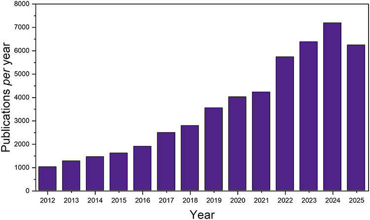 |
Figure 2 Publications related to hydrogels in the biomedical field over the last 14 years (2012 to 2025). The citation report graphic was generated using Clarivate Web of Science by searching for “hydrogels” and refining the results to the “biomedical” research area (updated to February 2026). © Clarivate, 2026. All rights reserved. |
The adaptability of hydrogels for such many different applications lies in their tuneable physical properties, which can be precisely engineered by modifying structural components. Key parameters influencing these characteristics include the ratio of hydrophilic to hydrophobic elements, polymer concentration, and reaction conditions like temperature and time.23 By manipulating these factors, researchers can tailor hydrogels to exhibit specific properties—such as controlled swelling and deswelling rates, enhanced mechanical strength, and customizable degradation profiles—making them suitable for a broad range of biomedical applications.24 This versatility has been essential in advancing their use in drug delivery, tissue engineering, wound healing, and regenerative medicine. In particular, in biomedical applications, the three critical characteristics of hydrogels are:25
Biocompatibility, which ensures that the hydrogel interacts with biological tissues without causing adverse effects like toxicity or inflammation, a feature commonly seen in hydrogels made from natural polymers, which helps minimize immune responses and promotes healing.
Biodegradability, which is essential particularly in tissue engineering, where the hydrogel must break down into non-toxic by-products that are safely absorbed or eliminated by the body, while supporting cellular migration and growth.26
Biological recognition, involving the hydrogel ability to mimic the natural extracellular matrix by incorporating ligands that facilitate cell interaction and adherence to the gel matrix.
Among the various materials utilized in hydrogel engineering, carbohydrates have received significant attention due to their inherent biocompatibility, biodegradability, and versatility.27–30 For example, common carbohydrates like alginate, chitosan, cellulose, and hyaluronic acid are abundant in nature and exhibit desirable properties for biomedical applications.31,32 Alginate, derived from brown seaweed, is extensively used in wound dressings and drug delivery systems due to its biocompatibility and mild gelation process. Chitosan, a cationic polysaccharide with antibacterial properties, is valuable in tissue engineering for promoting wound healing and biodegradability.33 Cellulose, known for its mechanical strength and water retention, is utilized in wound dressings, drug delivery, and scaffolds for tissue engineering.34 Hyaluronic acid, found in connective tissues and synovial fluid, is appreciated for its viscoelastic properties and water-binding capacity, making it useful in clinical fields such as cosmetic surgery and orthopaedics.35 The versatility of carbohydrate-based hydrogels is further enhanced by the functional groups—hydroxyl, amino, and carboxyl—that are abundant on carbohydrate chains.26 These groups provide numerous sites for chemical and physical cross-linking, allowing the modification of hydrogels to achieve specific physical and chemical properties tailored to different biomedical needs. By adjusting these features, hydrogels can be designed to exhibit controlled swelling behaviours, tuneable mechanical strength, and customized degradation rates.36
More in general, the merge of carbohydrates and hydrogels, offers a range of exceptional characteristics including:11
Superior biocompatibility, minimizing the likelihood of adverse immune reactions when introduced into the body, supporting cell adhesion, proliferation, differentiation, and facilitating more effective healing and seamless integration with surrounding tissues.24
Biodegradability, forming non-toxic by-products that can be absorbed or eliminated via natural metabolic pathways. This characteristic is particularly valuable in tissue engineering, where temporary scaffolds need to degrade over time, providing support only until natural tissue regeneration occurs.37
Sustainability, supported by the natural availability of carbohydrates which reduces the environmental impact, particularly when compared to conventional hydrogels that often depend on petrochemical-derived polymers with significant ecological footprints.
Versatility and tunability, through various chemical and physical modifications.38 Factors such as cross-linking density, polymer concentration, and mechanical strength can be customized to meet the specific requirements. This tunability allows for precise control over swelling behaviour, degradation rates, and mechanical properties.
Excellent water retention, making them ideal for applications like wound dressings, where maintaining a moist environment is crucial for promoting healing, reducing infection risk, and facilitating cell migration.24 This water retention also enhances their effectiveness as carriers for controlled and sustained drug release.
Functional properties, allowing incorporation of functional groups, bioactive molecules, or targeting ligands, which enhance their interaction with biological systems.39 This functionalization enables hydrogels to release growth factors or therapeutic agents in a controlled manner, improving treatment outcomes and expanding their potential for targeted therapies and personalized medicine.
Bioactivity: Many carbohydrate-based hydrogels exhibit inherent bioactivity, promoting cell adhesion and growth, which makes them particularly suited for use as scaffolds in tissue engineering.40 This bioactivity is essential for supporting cellular functions that are critical for successful tissue regeneration.
Cost-effectiveness, thanks to the widespread availability of carbohydrates. This affordability is especially advantageous in resource-limited settings, where access to low-cost biomedical materials can significantly impact patient care.
It should be highlighted that many of these features of carbohydrate-based hydrogels can aid in the shift towards a sustainable society, as called for by several national and international policies and public pressure.41,42 In particular, some characteristics are directly connected to various United Nations Sustainable Development Goals (SDGs).43 Firstly, they contribute to the SDG 3, “Good Health and Well-being”, by offering advanced medical treatments, enhancing healing processes, and providing sustained release of therapeutic agents, all of which are essential for maintaining and improving health outcomes. Moreover, advancements in carbohydrate-based hydrogels represent significant innovations in the biomedical field, aligning with the SDG 9, “Industry, Innovation, and Infrastructure”. Indeed, these advancements involve the development of new materials that can be integrated into healthcare infrastructure, fostering innovation and supporting the creation of resilient healthcare systems. Furthermore, the natural origin of carbohydrate-based hydrogels aligns with the SDG 12, “Responsible Consumption and Production” and the SDG 13, “Climate Action”, by reducing the carbon footprint associated with biomedical material production. Lastly, the use of natural, biodegradable materials derived from carbohydrates promotes the sustainable use of terrestrial ecosystems, supporting the SDG 15, “Life on Land”, by aiding in the conservation of biodiversity and encouraging the use of renewable biological resources. Nevertheless, the successful translation of carbohydrate-based hydrogels into clinical practice44 still requires addressing several challenges, primarily related to a deeper understanding of bio-interactions at the molecular level, scalability, and improved mechanical strength. In particular, the in-depth use of carbohydrate chemistry for the rational design of compounds for specific targeting remains practically unexplored in this field, significantly limiting the full potential of these hydrogels. Consequently, continued research and innovation remain essential.
Numerous recent reviews in the literature explored the diverse biomedical applications of hydrogels, including their roles in drug delivery, tissue engineering, and wound healing treatment.23,25,45 For instance, some studies focus on sprayable hydrogels for biomedical applications, such as the work by Liao et al46 and others investigate hydrogel-based devices for biomedical applications, as reviewed by Deligkaris et al.47 Other reviews specifically addressing carbohydrates and hydrogels focus on chitosan-based hydrogels,33 and alginate-based hydrogels for biomedical applications.48 Additionally, a book chapter explores carbohydrate-based nanohydrogels for drug delivery systems.36
As a tool for guiding research in this area, the present review offers an in-depth look at recent advancements in carbohydrate-hydrogels, emphasizing their bio-interactions, structural modifications, and functional capabilities. Unlike broader hydrogel reviews, this manuscript specifically focuses on carbohydrate-based systems, providing a detailed analysis of how their molecular composition and architecture influence biological performance and material properties. Supported by selected case studies, it not only highlights current limitations in synthesis, stability, and functionality but also critically evaluates innovative strategies aimed at overcoming these challenges. Furthermore, the review synthesizes insights from interdisciplinary studies—spanning chemistry, biology, and materials science—to propose future research directions that may expand the applicability of carbohydrate hydrogels in areas such as drug delivery, tissue engineering, and biosensing. By offering both a comprehensive overview and a targeted critique, this work serves as a unique reference for researchers seeking to leverage the distinct advantages of carbohydrate-based hydrogels while addressing gaps in current knowledge (Figure 3).
 |
Figure 3 Scheme of the key topics of the review. |
Fundamentals of Carbohydrate-Based Hydrogels
Carbohydrates play a crucial role in living organisms, serving structural functions, providing energy, and participating in cellular signalling and communication.49 Among the most studied carbohydrates for the preparation of hydrogel are polymeric materials, including chitosan, alginate, dextran, cellulose, and hyaluronic acid.50 Only a few examples involve the use of monosaccharides of small molecules, such as mannose, sucrose, fructose51 and galactose. This substantial limitation highlights the challenge of designing hydrogels with specific bio-interactions, which can be addressed by creating carbohydrate-based molecules tailored for targeted interactions exploiting the chemistry of carbohydrates. Their individual contributions to hydrogel performance are discussed below, with an emphasis on biological interactions (summarized in Figure 4) and applications (summarized in Table 1).
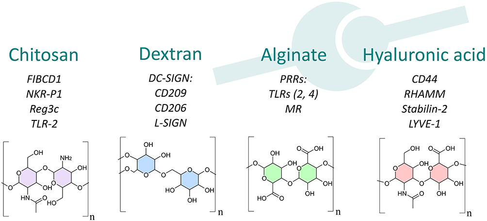 |
Figure 4 Chemical structure and main biological interactions of the most widely studied carbohydrates used in hydrogel preparation. |
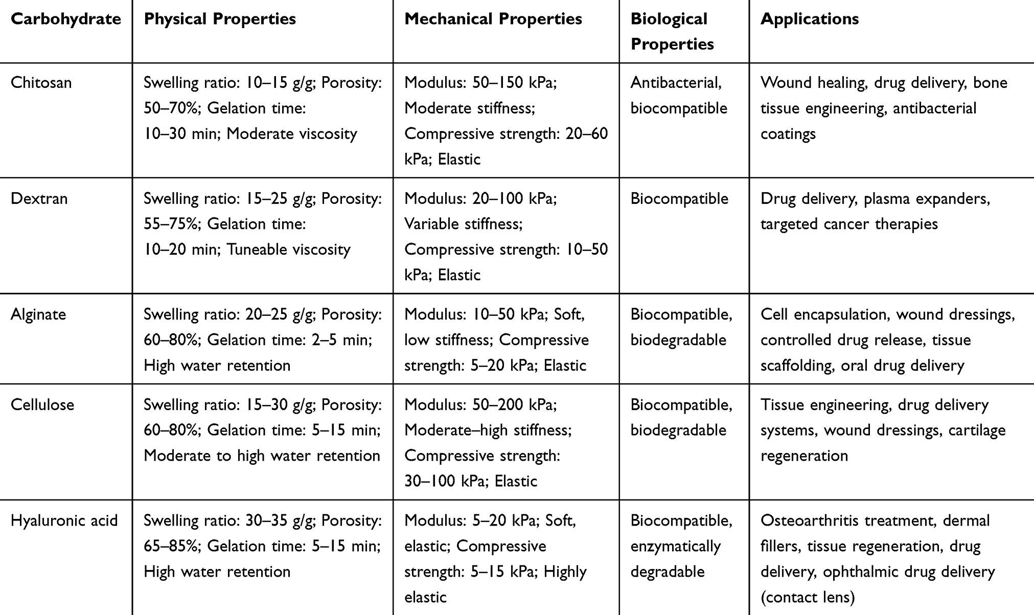 |
Table 1 Properties and Applications of Key Carbohydrate-Based Hydrogels in Biomedical and Therapeutic Fields |
Chitosan
Chitosan is a linear polysaccharide derived from the deacetylation of chitin, which is abundant in crustacean shells,52 consists of linear chains of β-(1→4)-linked D-glucosamine (deacetylated unit) and N-Acetyl-D-glucosamine units (acetylated unit).53 Its unique properties, including the presence of reactive amino and hydroxyl groups, enable its modification for specific applications. Chitosan hydrogels are pH-responsive and exhibit high biocompatibility, biodegradability, and water retention, making them versatile for drug delivery, wound healing, and tissue engineering.33 In drug delivery, these hydrogels control the release of therapeutic agents, enhancing their bioavailability and reducing side effects. In wound healing, chitosan hydrogels promote moisture retention and create barriers against infection, while in tissue engineering, they act as scaffolds supporting cell growth and regeneration.33,54–57
Chitosan is a biopolymer with significant biological activity due to its unique chemical structure, particularly its abundant amino groups (-NH2). These groups enable chitosan to interact effectively with cellular receptors, influencing cell behaviour. Its positive charge at physiological pH enhances its affinity for negatively charged cell membranes and specific receptors, crucial for cell adhesion, migration, and proliferation. This interaction can activate intracellular signalling pathways that promote tissue repair.58
Chitosan is not naturally present in mammals, giving it the capacity to cause recognition by the mammalian immune system.59 Several chitin-binding receptors have been identified in mammals, including fibrinogen C domain containing 1(FIBCD1), natural killer cell inhibitory receptor (NKR-P1), and Regenerating islet-derived protein 3-c (Reg3c).60–62 For instance, FIBCD1 is a transmembrane receptor that specifically detects chitin and chitosan. Its expression has been observed in various tissues, such as the gastrointestinal tract and lungs, and it plays a significant role in activating immune cells and host defence mechanisms.63 When chitosan interacts with FIBCD1, it triggers several signalling pathways. One potential pathway involves the stimulation of intracellular signalling molecules, such as protein kinases, leading to the phosphorylation and activation of downstream signalling proteins. This cascade of events regulates immune cell activation and cytokine production, which are critical for the immune response. The activation of FIBCD1 can enhance the phagocytic capacity of immune cells like macrophages and neutrophils, contributing to the body defence mechanisms.60
Moreover, the interplay between chitosan and Toll-like receptor 2 (TLR-2) initiates signalling cascades that are crucial for immune reactions. When chitosan binds to TLR-2, it activates various signalling pathways, notably the Myeloid Differentiation Factor 88 (MyD88)-dependent pathway. This pathway utilizes the adaptor protein MyD88 to trigger the activation of downstream signalling molecules, including interleukin-1 receptor-associated kinase (IRAK) and tumour necrosis factor receptor-associated factor 6 (TRAF6).60,64 Upon activation, these molecules initiate a cascade of reactions that amplify the activity of transcription factors like nuclear factor kappa-light-chain-enhancer of activated B cells (NF-kB) and activator protein 1 (AP-1).65 The subsequent production and release of pro-inflammatory cytokines such as Interleukin 6 (IL-6), tumor necrosis factor-alpha (TNF-α), and Interleukin 1 beta (IL-1β) are critical for initiating and regulating immune responses, promoting inflammation, and coordinating the recruitment and activation of immune cells. Interestingly, chitin has been shown to expedite the wound healing process through pathways that involve MyD88, which is subsequently linked to the Transforming growth factor beta (TGFβ)/ small mother against decapentaplegic (Smad) signalling pathway.66
Dextran
Dextran is a branched polysaccharide composed of glucose units linked primarily by α-(1→6) glucosidic linkages, with occasional α-(1→3) branches. Dextran is notable for its excellent biocompatibility and biodegradability. These properties make dextran-based hydrogels ideal for drug delivery and tissue engineering. In drug delivery, dextran hydrogels provide controlled release of encapsulated drugs, such as anticancer agents, ensuring sustained therapeutic efficacy while minimizing toxicity. In tissue engineering, they serve as scaffolds that support cell attachment and growth, contributing to tissue regeneration.67–69
Dextran interacts with several receptors and proteins in the body, particularly in the immune system and the vascular system. In humans, receptors that bind to dextran include a group known as the DC-SIGN (dendritic cell–specific intercellular adhesion molecule 3-grabbing nonintegrin) family. This group comprises receptors such as DC-SIGN (CD209) and L-SIGN, which is a homologue of DC-SIGN found in the liver and lymphatic endothelium. Additionally, the mannose receptor (CD206) and langerin are also part of this receptor family. These receptors play a crucial role in the uptake of pathogens by dendritic cells and macrophages. Furthermore, they may influence the modulation of immune responses, typically favouring the pathogens themselves rather than the host organisms.70
Alginate
Alginate is an anionic polysaccharide primarily extracted from the cell walls of brown seaweeds and algae. It consists of two monomeric units, β-D-mannuronic acid (M blocks) and α-L-guluronic acid (G blocks), linked by β-(1→4) glycosidic bonds.71 Alginate hydrogels are known for their ease of gelation in the presence of divalent cations like calcium and their ability to encapsulate and release drugs in a controlled manner. Their biocompatibility and biodegradability make them suitable for drug delivery, wound healing, and tissue scaffolds.72 Alginate hydrogels support cell growth and tissue regeneration by creating a porous structure that facilitates nutrient and oxygen diffusion.
In mammals, alginate does not naturally occur, enabling it to be recognized by the immune system through various receptors. Notable among these are the pattern recognition receptors (PRRs), which include Toll-like receptors (TLRs) and the mannose receptor (MR). TLRs, particularly TLR2 and TLR4, are essential for detecting foreign materials and initiating immune responses.59 The mannose receptor is crucial for recognizing glycoproteins and polysaccharides, including alginate, facilitating endocytosis and immune activation.
When alginate interacts with these receptors, several signalling pathways are activated. For example, the binding of alginate to TLR2 and TLR4 initiates a cascade of events leading to the activation of the MyD88-dependent signalling pathway. This pathway triggers the production of pro-inflammatory cytokines such as IL-6, TNF-α, and IL-1β, which play vital roles in modulating immune responses. The engagement of alginate with the mannose receptor can also enhance phagocytosis, enabling immune cells to recognize and eliminate foreign materials effectively.
Moreover, alginate can influence cellular behaviour through its interactions with integrins and other adhesion receptors. By modulating the extracellular matrix (ECM), alginate affects cell attachment, proliferation, and differentiation, which are critical for tissue regeneration. The ability of alginate to form hydrogels can mimic the ECM, providing a supportive environment for cell growth and development.
Cellulose
Cellulose is a natural polymer composed of β-D-glucose units linked by β (1→4) glycosidic bonds. It is the primary structural component of the cell walls in plants and is the most abundant organic polymer on Earth.73 Cellulose-based hydrogels are highly hydrophilic and provide excellent water retention, making them effective for wound healing and tissue engineering. Chemical modifications, such as carboxymethylation, enhance cellulose gel-forming properties, allowing for controlled drug release and scaffold formation in tissue engineering applications. Cellulose itself does not interact with specific receptors and is typically used as a matrix for loading other compounds or as a hydration source.74
Hyaluronic Acid
Hyaluronic acid, a non-sulphated glycosaminoglycan, is composed of alternative units of D-glucuronic acid and N-acetyl glucosamine linked by β-1,3 and β-1,4 glycosidic bonds. Hyaluronic acid a naturally occurring biopolymer in connective tissues and synovial fluid, is prized for its biocompatibility, viscoelasticity, and high water-binding capacity.75 In biomedical applications, hyaluronic acid hydrogels serve as scaffolds for tissue engineering, promoting cell proliferation and differentiation. Their hydrophilic nature is also advantageous in wound healing, as they maintain a moist environment that accelerates tissue repair. In ophthalmology and orthopaedics, hyaluronic acid is used for eye surgeries and joint lubrication, respectively.76
Beyond its physicochemical properties, hyaluronic acid exhibits significant biological effects through specific interactions with hyaluronic acid -binding proteins, known as hyaladherins, including CD44, Receptor for hyaluronan-mediated motility (RHAMM), Stabilin-2 and lymphatic vessel endothelial hyaluronan receptor 1 (LYVE-1).77,78 These interactions are pivotal in influencing cell behaviour, immune responses, and tissue regeneration, making hyaluronic acid a critical component in both health and disease.
Hyaluronic acid interacts with several key receptors, primarily CD44, which is a glycoprotein expressed on the surface of most cell types, including skin keratinocytes and fibroblasts.79 CD44 is involved in various signal transduction pathways, making it a critical mediator of hyaluronic acid biological activities. The binding of hyaluronic acid to CD44 initiates multiple intracellular signalling pathways that regulate essential biological processes, such as cell proliferation, migration, wound healing, and tissue regeneration. These pathways are particularly important in contexts like wound healing, where hyaluronic acid promotes cellular movement to injury sites and stimulates growth factor production. CD44 involvement in cancer is of significant interest, as overexpression of CD44 is observed in various cancer types, including breast, pancreatic, gastric, prostate, ovarian, and colon cancers.80,81 Consequently, CD44 hyaluronic acids emerged as a biomarker for cancer cells, making targeting overexpressed CD44 an important strategy in cancer therapy.
Furthermore, since hyaluronic acid receptors were found to be overexpressed on chondrocytes, different hyaluronic-based hydrogels have been broadly developed for these purposes, showing promising results for osteoarthritis therapies.82
In addition to CD44, hyaluronic acid also binds to RHAMM,83 which affects cell migration and tissue remodelling, and LYVE-1, which mediates hyaluronic acid uptake in lymphatic vessels, playing a role in fluid homeostasis and immune responses.84
Summarizing Table 1 shows the key properties of the carbohydrate-based hydrogels presented above.85,86
Monosaccharides
While polysaccharides such as chitosan, hyaluronic acid, and alginate have been extensively studied for hydrogel applications, monosaccharides-such as mannose, sucrose, fructose, and galactose-also offer promising opportunities for the design of functional hydrogels with targeted bio-interactions.87 A common strategy entails conjugating specific sugars, notably mannose and galactose, to nanoparticles, which are subsequently embedded within the hydrogel matrix to enhance its biological functionality and targeting capabilities.88,89 This strategy enables the creation of hydrogels with specific bioactivity, as the sugars on the nanoparticle surface can interact with cell surface receptors, promoting cellular uptake, migration, and other biologically relevant processes. For instance, a study involving D-mannose, a monosaccharide, demonstrated its incorporation into an injectable nanocomposite hydrogel for anti-inflammatory applications in osteoarthritis treatment.90 Network-pharmacology analyses help clarify how the stereochemical configuration of D-mannose, particularly the orientation of its hydroxyl groups, underlies its selective interaction with specific cellular receptors and signalling proteins. Based on its 2D molecular structure, 84 potential pharmacological targets were identified, with 39 overlapping genes associated with osteoarthritis. Another approach involves crosslinking hydrogels whose structures are based on hydrogen bonds, hydrophobic interactions, or ionic interactions. In this context, the use of natural crosslinkers, such as monosaccharides like glucose, sucrose, and fructose, is becoming an increasing trend.51,91
Concerning specifical interactions, D-mannose, for example, have been demonstrated to increase the receptor mediated uptake of nanocarriers of drugs. Indeed, mannose receptors overexpressed on antigen presenting cells such as macrophages or dendritic cells and are necessary targets for treating cancer and other infectious diseases.92
Galactose has been shown to specifically target the asialoglycoprotein receptor (ASGP-R), which is abundantly expressed on the membrane of HepG2 cells.93 Indeed, the ASGP-R displays high specificity for galactose. The ligand-binding site is formed by a cluster of residues—Asp241, Asp265, Asn264, Glu252, Gln239, and Trp243—within the H1 subunit of the receptor. Binding is initiated through coordination between a receptor-associated calcium ion and the oxygen atoms of the galactose molecule. The spatial arrangement of amide and carboxylate side chains in the binding pocket enforces a strict requirement for hydrogen bonding with the 3- and 4-hydroxyl groups of galactose. In addition, galactose recognition is further stabilized by hydrophobic contacts between its C3, C4, C5, and C6 positions and Trp243, along with the formation of four hydrogen bonds involving residues of the H1 subunit.94
Methodologies for the Synthesis of Carbohydrate-Based Hydrogels
The design of carbohydrate-based hydrogels involves both physical and chemical crosslinking strategies, each contributing differently to the material properties and potential applications.23,25,95
Physical Crosslinking
Physical crosslinking relies on non-covalent interactions that create reversible bonds, allowing the hydrogel to maintain its structure without permanent chemical modifications. Several approaches are employed to design carbohydrate-based hydrogels through physical crosslinking, including hydrogen bonding, amphiphilic grafting, crystallization, ionic interactions, and protein interactions.24
Hydrogen Bonding
Hydrogen bonds form principally between molecules with -OH and -NH groups, such as those found in polysaccharides like chitosan, alginate, and dextran. These interactions stabilize the hydrogel network and contribute to its water retention capacity. The establishment of hydrogen bonds between the polysaccharide chains and water molecules enhances the swelling behaviour and overall mechanical stability of the hydrogel, making it particularly suitable for applications requiring high moisture retention.96
Amphiphilic Grafts and Block Polymers
Amphiphilic grafts and block copolymers, which consist of both hydrophilic and hydrophobic segments, can self-assemble into micellar structures within the hydrogel matrix. This structural organization enhances both water retention (due to hydrophilic domains) and mechanical strength (due to hydrophobic regions). This dual functionality is particularly useful for creating hydrogels with tuneable properties for applications such as drug delivery and tissue scaffolding.97,98
Crystallization
Under controlled conditions, some polysaccharides can form crystalline domains within the hydrogel matrix, further stabilizing the network. Methods like freeze-thaw cycles or heat-induced crystallization can promote the formation of such crystalline regions, enhancing the hydrogel mechanical properties and structural integrity.99
Ionic Interactions
Carbohydrate-based hydrogels, especially those derived from anionic polysaccharides like alginate, often rely on ionic interactions for crosslinking. When exposed to divalent cations (eg, calcium ions), the negatively charged carboxylate groups on alginate form ionic bonds, leading to gelation. This method is advantageous because it operates under mild conditions, preserving the bioactivity of sensitive molecules or cells encapsulated within the hydrogel matrix.100,101
Protein Interactions
Carbohydrates can form physical crosslinks through interactions with proteins. Some polysaccharides have specific binding sites for proteins, enabling the formation of hydrogels through protein-carbohydrate complexation. This approach is particularly beneficial in biomedical applications, where the bioactive properties of both proteins and carbohydrates can be exploited for drug delivery or tissue engineering purposes.102,103
Supramolecular Self-Assembly of Amphiphiles
Supramolecular hydrogels form through the self-assembly of amphiphilic molecules driven by non-covalent interactions, such as hydrogen bonding, van der Waals forces, and hydrophobic interactions. These amphiphiles organize into structured networks without requiring covalent modifications, leading to highly dynamic and reversible hydrogel systems. This method is particularly advantageous for biomedical applications, including drug delivery, wound healing, and tissue engineering, where controlled degradation and responsiveness to environmental stimuli are desirable.104,105
Chemical Crosslinking
Chemical crosslinking involves the formation of covalent bonds between polymer chains, resulting in a stable and irreversible hydrogel network. Various methods are used to chemically crosslink carbohydrate-based hydrogels, offering precise control over the material properties.106
Enzymatic Reactions
Enzymes such as transglutaminase or laccase facilitate the formation of covalent bonds between carbohydrate molecules.107 Enzymatic crosslinking is biocompatible and allows for precise control over the crosslinking density, enabling the design of hydrogels with tailored mechanical properties and degradation rates for specific biomedical applications.108,109
Chemical Reactions
Chemical crosslinking can be achieved through reactions like esterification or amidation, where functional groups (eg, carboxyl, hydroxyl, or amine groups) on the polysaccharide chains are modified to form stable covalent bonds. This method provides a high degree of control over the hydrogel properties, including its mechanical strength and responsiveness to environmental conditions.110,111
High-Energy Radiation
High-energy radiation (eg, gamma rays or electron beams) induces crosslinking by generating free radicals in the polymer chains, which then form covalent bonds. This technique offers the dual benefit of sterilizing the hydrogel while simultaneously enhancing its structural integrity without the need for chemical additives.112,113
Free-Radical Polymerization
Free-radical polymerization is a widely used chemical crosslinking method where a radical initiator triggers the polymerization of monomers or oligomers, leading to the formation of a cross-linked network. This approach is commonly used in hydrogel design due to its versatility and ability to produce hydrogels with tuneable mechanical and swelling properties.114,115
Click Reactions
Click chemistry, particularly azide-alkyne cycloaddition116 offers a highly selective and efficient method for crosslinking carbohydrate-based hydrogels.117 This technique operates under mild conditions, allowing for the incorporation of bioactive molecules, such as drugs or growth factors, into the hydrogel network. The precision of click chemistry makes it ideal for creating hydrogels for drug delivery and tissue engineering, where controlled functionality and biodegradability are essential.117
In conclusion, Table 2 provides a comparative overview of the different methods used for hydrogel synthesis.
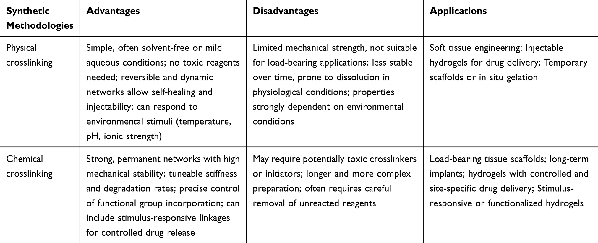 |
Table 2 Comparison of Hydrogel Synthesis Methodologies |
Biomedical Applications and Selected Examples
Various hydrogels made from carbohydrates have been developed as drug carriers, cell delivery systems, scaffolds for tissue engineering, and more, offering versatile platforms for medical applications.
Drug Carriers for Drug Delivery
Drug delivery systems offer innovative approaches to disease treatment through the encapsulation of active pharmaceutical ingredients within carriers. This encapsulation allows for controlled and sustained release at targeted sites, thereby minimizing the risk of overdose and reducing adverse side effects.52,118 Among these systems, carbohydrate-based hydrogels are primarily owing to their excellent biocompatibility and biodegradability, which closely mimic the extracellular matrix found in biological tissues.119 Their inherent nontoxicity ensures patient safety, while their high water content and absorption capacity create a hydrated microenvironment that enhances drug stability and facilitates molecular transport.
From a mechanistic perspective, the transfer of drug molecules from carbohydrate-based hydrogels to the surrounding bulk solution is predominantly governed by diffusion through the hydrated polymer network. This process is strongly influenced by key structural parameters of the hydrogel, including mesh size, crosslinking density, and polymer chain mobility. Highly crosslinked networks with smaller mesh sizes typically restrict molecular diffusion, leading to prolonged and sustained release profiles, whereas more loosely crosslinked structures allow faster drug transport.
In addition to diffusion-driven mechanisms, hydrogel swelling and polymer relaxation play a significant role in modulating drug release kinetics. Upon exposure to the release medium, water uptake induces network expansion, increasing chain flexibility and facilitating molecular migration. This phenomenon is particularly relevant during the initial stages of release and may account for the presence of an early burst effect, especially for drug molecules weakly bound or located near the hydrogel surface.
Drug–polymer interactions further contribute to the controlled release behaviour observed in carbohydrate-based hydrogels. Hydrogen bonding, electrostatic interactions, and hydrophilic affinity between the carbohydrate matrix and the encapsulated drug can temporarily retain the active molecules within the network, delaying their diffusion into the bulk solution. These interactions, together with the tunable degradation rates of carbohydrate-based hydrogels, provide an effective means to tailor release kinetics for specific therapeutic requirements. For instance, in some forms of cancer, carbohydrates can bind to receptors like CD44, which is commonly associated with tumour progression and metastatic invasion. This interaction not only enhances drug accumulation in target tissues but also promotes a targeted and controlled release of the active ingredient, thereby optimizing treatment efficacy and minimizing systemic side effects.
One significant area of application for carbohydrate-based hydrogels is in anti-inflammatory treatments. Non-steroidal anti-inflammatory drugs (NSAIDs) are commonly employed to manage pain and inflammation associated with conditions such as arthritis, muscle injuries, and post-surgical recovery. However, the prolonged use of these medications often leads to adverse effects, including gastrointestinal irritation and cardiovascular risks. Carbohydrate-based hydrogels can enhance NSAID delivery by minimizing side effects while maximizing therapeutic efficacy.24,120,121 For instance, a study demonstrated the development of an alginate hydrogel designed for localized ibuprofen delivery. This hydrogel was engineered to swell in response to inflammatory signals, allowing for the rapid release of ibuprofen directly at the site of inflammation. Such localized delivery not only improved the drug efficacy but also mitigated the systemic side effects typically associated with oral administration.118,122 Similarly, research by Suhail et al involved the development of an alginate hydrogel for the delivery of diclofenac. The hydrogel exhibited significant swelling in response to physiological conditions, enabling controlled and sustained diclofenac release (Figure 5A), which improved drug availability at the site of action while reducing gastrointestinal side effects.123
 |
Figure 5 (A) Kinetic of release of diclofenac loaded into alginate hydrogel at different pH and surface morphology of alginate hydrogels. Taken from ref.123 (B) Kinetic of release of bovine serum albumin loaded into alginate/guar gum hydrogel crosslinked with glutaraldehyde at different pH and scanning electron micrographs of air-dried beads. Taken from ref124 (C) A hydrogel containing dual glucose sensors and dual glucose-responding elements based on phenylboronic acid-modified chitosan, formyl terminated polyethylene glycol and polyvinylacohol through the cross-linking of two dynamic covalent bonds, phenylborate ester and imine, and glucose oxidase and catalase. Taken from ref.125 |
Proteins and peptides present unique challenges in drug delivery due to their larger molecular sizes and complex structures, which can be destabilized by environmental factors such as temperature, pH, and enzymatic degradation. These destabilizing conditions can significantly reduce the bioavailability and therapeutic efficacy of these biomolecules. Thus, careful formulation is essential to ensure that proteins and peptides maintain their integrity from the point of delivery to their target site within the body.126,127 Carbohydrate-based hydrogels provide an innovative solution for encapsulating these biomolecules, creating a protective matrix that shields them from harsh environmental conditions.125 For instance, Abraham et al developed a pH-sensitive alginate/guar gum hydrogel crosslinked with glutaraldehyde for the controlled delivery of bovine serum albumin, achieving an optimal drug release of approximately 90% at pH 7.4 (Figure 5B).128
Carbohydrate-based hydrogels also play a crucial role in hormonal therapies, which are essential for managing conditions such as diabetes, hormonal imbalances, and certain cancers. These hydrogels provide sustained release of hormones, thus improving therapeutic outcomes and enhancing patient adherence.129 For example, insulin, a critical hormone for regulating blood glucose levels, is typically delivered via injections, which can be inconvenient and painful for patients.130 Zhang et al formulated a chitosan hydrogel that encapsulated insulin and demonstrated its capacity to maintain stable blood glucose levels in diabetic animal models. This hydrogel was designed to swell in response to glucose concentration, facilitating controlled insulin release in correlation with rising blood sugar levels, thereby mimicking physiological insulin release (Figure 5C).131 Similarly, Burova et al124 utilized dextran as a hydrogel to protect insulin, yielding promising results that contribute to the development of oral protein pharmaceutical preparations.
As therapeutic applications become more complex, nucleic acid-based therapies, including DNA and RNA, have garnered attention for their potential in treating genetic disorders and cancers. However, delivering these molecules safely and effectively poses challenges due to their inherent instability.132 Carbohydrate-based hydrogels can address these challenges by facilitating the delivery of nucleic acids while protecting them from degradation.133,134 For example, Yalcin et al utilized dextran-coated iron oxide nanoparticles to deliver miR-29a to tumors, aiming to inhibit the expression of tumor-associated microRNAs. Despite miRNA short half-life and susceptibility to degradation, the developed platform effectively protected miR-29a using dextran-coated magnetic nanoparticles for cancer therapy. The results demonstrated that this formulation is a safe delivery system, with the magnetic nanoparticles safeguarding the unstable miR-29a from decomposition while facilitating its uptake by cancer cells.135 In another innovative application, Segura et al developed a hyaluronic acid-based hydrogel for targeted gene therapy delivery. This hydrogel encapsulated plasmid DNA and exhibited stability under physiological conditions. It was engineered to degrade in response to elevated levels of hyaluronidase, an enzyme presented in tumor tissues. Upon degradation, the hydrogel released the encapsulated DNA, enabling its uptake by cancer cells and leading to the expression of therapeutic proteins that inhibit tumor growth.136 Xiao et al137 designed a supramolecular hydrogel formed by spiropyran conjugated with galactose for the release of miR-122 and the delivery of the miRNA into HepG2. The presence of galactose, which target the asialoglycoprotein receptor (ASGP-R) that is highly expressed on the membrane of HepG2 cells, enables targeted delivery of miRNA (Figure 6A).
 |
Figure 6 (A) Dual-functional supramolecular hydrogel, able to release encapsulated miRNA in a light-controlled manner. The presence of galactose targets the asialoglycoprotein receptor. Details of the merocyanine moiety that undergoes photo-isomerization into spiropyran. Taken from ref.137 (B) Doxorubicin-loaded hyaluronic acid nanogels synthesized using a methacrylated strategy. Cellular uptake studies show preferential internalization by CD44 or CD168-overexpressed cancer cells in vitro. In vivo antitumor evaluations demonstrate distinct suppression of tumor growth by these nanogels. Taken from ref.138 |
Carbohydrate-based hydrogels also allow for the incorporation of various antimicrobial agents, enabling localized treatment strategies. For instance, curcumin, a natural compound derived from turmeric, demonstrates significant antimicrobial and anti-inflammatory properties. Researchers have developed alginate and chitosan-based hydrogels to encapsulate curcumin, enhancing its delivery while protecting the compound from degradation and providing controlled release.139 Another study by Prusty et al reported on a dextran-based hydrogel system that incorporated covalently bound silver nanoparticles. The researchers evaluated the swelling, deswelling, and water retention characteristics of these hydrogel composites to assess the release rate of ornidazole, an antibacterial medication. The in vitro results indicated a remarkable release rate of 98.5% of ornidazole within six hours.140
Furthermore, the effective delivery of chemotherapeutic agents is crucial for maximizing therapeutic efficacy while minimizing side effects. Several polysaccharides have the unique, innate ability to recognize specific receptors overexpressed on the surfaces of diseased cells, enabling the design of targeted drug delivery systems that can selectively deliver therapeutic agents through receptor-mediated endocytosis.141 Yang et al developed a range of hyaluronic acid-based nanogels through the copolymerization of methacrylated hyaluronic acid with di(ethylene glycol) diacrylate. The resulting nanogels, which exhibited a spherical shape and measured approximately 70 nm in diameter, were subsequently loaded with doxorubicin using an incubation method. In vitro experiments demonstrated that cellular uptake of these nanogels was dependent on CD44, leading to enhanced internalization in tumor cell lines that overexpress the CD44 receptor (Figure 6B).138
An outstanding study reported the encapsulation of doxorubicin within a pH-responsive chitosan/poly (acrylamide-co-maleic acid) hydrogel system. This system was designed to preferentially release doxorubicin in the acidic microenvironment of tumours, thereby enhancing drug efficacy and minimizing systemic side effects. In vitro experiments demonstrated that the chitosan hydrogel could sustain the release of doxorubicin over an extended period, effectively inhibiting cancer cell proliferation.142 Additionally, biodegradable carboxymethyl cellulose/graphene oxide nanocomposite hydrogel beads prepared by Namazi et al using physical crosslinking with FeCl3·6H2O were evaluated as a drug delivery system for doxorubicin. Their findings indicated that hydrogen bonding interactions were stronger in basic conditions than in acidic conditions, resulting in higher drug release at pH 6.8 compared to pH 7.4 (Figure 7A).143 Moreover, hyaluronic acid-based hydrogels have been developed for encapsulating paclitaxel, facilitating controlled release for intraperitoneal chemotherapy. However, despite the prolonged local retention of paclitaxel observed in vivo (Figure 7B), this approach did not translate into an enhanced anti-tumor effect. This discrepancy may stem from limitations inherent to the hydrogel system, such as insufficient penetration of paclitaxel into peritoneal tumor nodules due to the hydrogel’s viscosity and limited diffusivity, premature degradation or dilution within the peritoneal cavity, or suboptimal release kinetics that fail to achieve therapeutic drug concentrations at the tumor site. These considerations highlight the need for further optimization—including modulation of crosslinking density, tuning of mesh size, or incorporation of responsive degradation mechanisms—to improve the therapeutic performance of hyaluronic acid–based intraperitoneal drug delivery platforms.144 A supramolecular gel based on structurally related diazobenzene/diacetylene glycolipids was also reported to encapsulate and release hydrophobic drugs such as docetaxel in a controlled manner. The system relies on the photoswitching of azobenzene-based amphiphiles, allowing for reversible gel–sol transitions under UV irradiation, which could enable precise spatiotemporal drug release. The presence of mannose in the gel have demonstrated selective interactions with lectins, which could be exploited for targeted drug delivery in cancer therapy.145 Poursadegh et al89 reported the synthesis of a MIL-88 (Fe) metal-organic framework using the in situ method in the presence of hydroxyapatite nanoparticles (HAP). It was then functionalized with mannose as an anticancer receptor through the Steglich esterification method. For drug release investigation, 5-fluorouracil was loaded into the as synthetized structure, which was then coated with a hyaluronic acid hydrogel film (Figure 7C). Cytotoxicity tests on HT29 cancer cells showed increased effectiveness due to the presence of mannose and hyaluronic acid, highlighting the dual-targeted delivery system. Mannose-binding lectins are commonly overexpressed in many cancer cells, making them an ideal target for drug delivery. Additionally, hyaluronic acid selectively binds to the overexpressed CD44 receptor on tumor cells, further enhancing the targeted delivery of the drug.
 |
Figure 7 (A) Superior drug loading capacity of carboxymethylcellulose/Zn-based metal-organic framework/graphene oxide bio-nanocomposite compared to graphene oxide. Taken from ref.143 (B) Hyaluronic acid-based in-situ crosslinkable hydrogel developed as a paclitaxel (PTX) carrier. In tumor-bearing nude mice, PTX-gel demonstrated superior retention in the peritoneal cavity, with microparticulate PTX entrapped in the hyaluronic acid gel, while Taxol-gel and other Taxol-based formulations showed negligible PTX levels in the cavity after 14 days. Taken from ref.144 (C) Dual-targeted oral anticancer delivery system for 5-fluorouracil delivery, composed of mannose-functionalized hydroxyapatite nanoparticles/metal organic frameworks embedded into a hyaluronic acid edible hydrogel film. Taken from ref.89 |
Finally, the role of carbohydrate-based hydrogels extends to vaccine delivery, which is crucial for preventing infectious diseases. These hydrogels can function as adjuvants, enhancing immune responses while allowing for controlled release of vaccine components. Hyaluronic acid-based hydrogels have been effectively employed for delivering vaccine antigens, such as those targeting influenza and COVID-19. The sustained release of antigens from these hydrogels promotes stronger and longer-lasting immune responses. Studies indicate that encapsulating vaccine components within carbohydrate-based hydrogels can enhance immunogenicity, resulting in improved antibody responses and potentially reducing the number of doses required for effective vaccination. This innovative approach not only boosts vaccine efficacy but also simplifies vaccination schedules, ultimately contributing to better public health outcomes.146
Some of the most recent and relevant publications related to carbohydrate-based hydrogels for drug delivery are reported in Table 3.
 |
Table 3 Selected Publications Related to Carbohydrate-Based Hydrogel for Drug Delivery |
The field of drug delivery is undergoing significant advancements, driven by the unique properties of carbohydrate-based hydrogels. These hydrogels, with their highly porous structures, serve as versatile carriers capable of encapsulating a wide range of therapeutic agents, including small molecules, peptides, proteins, and nucleic acids. The porous network of these hydrogels not only enables efficient drug entrapment but also provides a suitable microenvironment that preserves the stability of the encapsulated therapeutics, ensuring their integrity throughout the delivery process.
A key advantage of carbohydrate-based hydrogels in drug delivery is their ability to offer controlled and sustained release profiles.163 This feature is especially critical in the management of chronic diseases, where prolonged therapeutic intervention is required to maintain efficacy and avoid frequent dosing.
Moreover, carbohydrate-based hydrogels can be functionalized by integrating bioactive molecules, further enhancing their therapeutic capabilities. For example, by incorporating growth factors or anti-inflammatory agents, hydrogels can be designed to actively promote tissue regeneration and healing in addition to delivering drugs.
Recent advances in this field have also seen the incorporation of nanomaterials into carbohydrate-based hydrogels, further increasing their drug delivery potential. Nanoparticles can be embedded within the hydrogel matrix to enhance its mechanical strength, improving the stability of the hydrogel in physiological environments.164 Additionally, these nanomaterials can provide multifunctional properties, such as targeted drug delivery or enhanced antimicrobial activity.
Tissue Engineering
Tissue engineering is an innovative and rapidly evolving field that merges concepts from biology, materials science, and engineering. Its primary focus is on creating biological substitutes that can restore, maintain, and enhance the function of damaged tissues. This interdisciplinary approach hinges on the development of scaffolds-structural frameworks that facilitate cellular growth and tissue regeneration. By replicating the natural extracellular matrix, scaffolds play a pivotal role in guiding cellular behaviour, enabling attachment, proliferation, and differentiation.165–168 The design and materials of scaffolds are fundamental to the success of tissue engineering. Ideal scaffolds exhibit several key characteristics: they must be biocompatible to prevent immune responses, biodegradable to allow for the seamless integration of new tissue, and possess mechanical properties that mimic those of the target tissue. For instance, while bone scaffolds require stiffness and strength to endure physiological forces, those designed for softer tissues may need to be more flexible. Moreover, the porosity and surface topography of scaffolds significantly influence cellular processes such as nutrient exchange and migration. High porosity facilitates these essential functions, while surface features enhance cell attachment and proliferation.
Recent advancements in biomaterials have transformed the landscape of tissue engineering, particularly with the emergence of biopolymer-based hydrogels. Unlike traditional scaffold materials that often struggle to replicate the hydrated, dynamic environment of native tissues, hydrogels offer a compatible and moist environment conducive to cell growth. Carbohydrate-based hydrogels, including alginate, chitosan, and hyaluronic acid, are particularly promising in tissue engineering due to their intrinsic biocompatibility and bioactivity. Hydrogels derived from these materials can promote cell adhesion, proliferation, and differentiation by mimicking native extracellular matrix cues; reduce the risk of immune reactions compared to synthetic polymers; facilitate the incorporation of growth factors or signaling molecules, supporting functional tissue regeneration.169,170
Cartilage tissue engineering faces significant challenges due to the limited regenerative capacity of cartilage, particularly in the context of injuries and degenerative conditions such as osteoarthritis. Since the 1990s, researchers have explored a variety of biomaterials, including natural and synthetic materials, chondrocytes, stem cells, and growth factors, to develop effective strategies for cartilage repair through the injection or implantation of scaffolds. Among the various types of scaffolds studied, hydrogels derived from natural resources have gained increasing importance due to their structural and biological similarities to the native extracellular matrix. These properties facilitate cell transplantation, proliferation, and differentiation, making hydrogels a central topic in contemporary CTE research.171–174 Recent advancements have shown that combining alginate with growth factors like transforming growth factor-beta (TGF-β) significantly enhances chondrocyte proliferation and matrix production.175 Innovations in alginate formulations, such as incorporating bioactive materials like hydroxyapatite nanowires (HAPNWs), have significantly improved the mechanical properties and cellular interactions of hydrogels, enhancing their clinical applicability for cartilage and bone defects (Figure 8A and B). Specifically, studies on Bovine serum albumin /alginate/HAPNW scaffolds demonstrate that the addition of HAPNWs increases the compressive modulus from 389.7 ± 29.6 kPa to 682.6 ± 51.0 kPa, highlighting a ~75% improvement in stiffness. This enhancement is largely due to the high aspect ratio of the nanowires, which facilitates effective stress transfer, formation of a percolating reinforcement network, and energy dissipation under cyclic loading, analogous to steel reinforcement in concrete. Furthermore, hydroxyapatite concentration plays a key role in tuning mechanical performance: optimized loadings increase stiffness and toughness without compromising elasticity or porosity, while excessive concentrations may reduce flexibility or alter the interconnected pore network essential for nutrient transport and cell infiltration. These structure–property relationships emphasize that both the morphology and loading of hydroxyapatite must be carefully controlled to achieve scaffolds with robust mechanical behavior and excellent biocompatibility.176
 |
Figure 8 (A) representation of the structures of bovine serum albumin/sodium alginate hydrogel scaffolds incorporated with hydroxyapatite. Taken from ref.176 (B) Macroscopic comparison of defects (white dotted circles) across three groups at 6- and 12-weeks post-surgery. Taken from ref.176 A novel bovine serum albumin and sodium alginate hydrogel scaffold doped with hydroxyapatite nanowires for cartilage defects repair. (C) Scanning electron micrographs of collagen/chitosan and collagen/chitosan/hyaluronic acid hydrogels crosslinked with genipin at 2, 10, and 20 mM concentrations. Taken from ref.177 (D) Infarct size and wall thickness. Representative Masson’s trichrome-stained myocardial sections from PBS group (i), chitosan group (ii), PBS/ADSCs group (iii), and chitosan/ADSCs group (iv). Photomicrographs show two cases of representative myocardial sections in each group (Scale bar = 200 μm). Infarct size (v) and infarct wall thickness (vi) were statistically compared across different groups. Data are mean ±SEM *p < 0.05 versus PBS group; #p < 0.05 versus chitosan group; $p < 0.05 versus PBS/ADSCs group. Taken from ref.178 |
Chitosan, derived from chitin, is notable for its structural similarity to glycosaminoglycans found in cartilage. It can encapsulate chondrocytes and deliver growth factors, facilitating cartilage repair while exhibiting antimicrobial properties that reduce infection risks during implantation.174 For instance, hydrogels prepared with glycerol phosphate, chitosan, and hydroxyethylcellulose as a cross-linker were used to prepare an injectable hydrogel for cartilage repair.179
Hyaluronic acid also exhibits chondroprotective and anti-inflammatory properties. Additionally, several studies have reported an overexpression of the CD44 receptor on human articular chondrocytes. Consequently, hyaluronic acid-based hydrogels are extensively studied and applied as viscosupplements and drug delivery systems for treating osteoarthritis.180
Transitioning to bone tissue engineering, the focus shifts to addressing critical defects that arise from trauma, disease, or surgical intervention.181,182 A recent study explores the synthesis and properties of novel injectable hydrogels made from collagen, chitosan, and hyaluronic acid, chemically crosslinked with genipin (Figure 8C). The hydrogels exhibit good biocompatibility, supporting the proliferation and adhesion of MG-63 cells, indicating potential for bone regeneration, especially for small bone losses. The findings suggest that these materials could serve as effective injectable scaffolds.177
In the field of neural tissue engineering, unique challenges arise, particularly concerning directional growth and functional recovery following nerve injuries. Chitosan, a carbohydrate polymer derived from chitin, has emerged as a promising material for supporting neuronal growth. According to reports by Cao et al,183 neural cells showed improved attachment and growth when exposed to a blended hydrogel containing agarose and chitosan. They proposed that the steric hindrance effect of chitosan may contribute to the morphological variations and promote optimal cell attachment and outgrowth. Alizadeh et al184 synthetized a hydrogel composed of chitosan-aniline pentamer, alginate, and agarose, which was loaded with olfactory stem cells for neural tissue engineering. The chitosan-aniline component enhanced the hydrogel conductivity. This hydrogel promoted cellular activity and facilitated the differentiation of olfactory stem cells into neuron-like cells when exposed to various neurotrophic factors. Additionally, the hydrogel was fully resorbable, and the aniline oligomer could be readily eliminated through the kidneys.
In cardiac tissue engineering, repairing myocardial tissue damaged by infarction is critical for restoring heart function. In light of the limited efficacy of current treatments for cardiac regeneration, tissue engineering approaches have been explored for their potential to provide mechanical support to injured cardiac tissues, deliver cardio-protective molecules, and improve cell-based therapeutic techniques.185 Enhancing the microenvironment of myocardial infarction to improve the engraftment, survival, and homing of stem cells presents a significant challenge. In this context, Liu et al178 created an injectable chitosan hydrogel designed for delivering adipose-derived mesenchymal stem cells (ADSC) into the ischemic heart. They found that reactive oxygen species (ROS) in damaged tissue may adversely affect the adhesion molecules of ADSC (αV, β1, p-FAK, and p-Src) that are crucial for engraftment and homing. Additionally, ROS can disrupt host myocardial ligands such as ICAM1 and VCAM1, which play a role in stem cell engraftment. The chitosan hydrogel developed could enhance cell engraftment by reducing ROS levels and increasing cytokines associated with stem cell homing, such as SDF-1 (Figure 8D).
The regeneration of periodontal tissues186 is equally critical for maintaining oral health, particularly in cases of periodontal disease. In recent years, hydrogels have been utilized as carriers for delivering and releasing bioactive molecules, such as growth factors and anti-inflammatory agents. These bioactive molecules facilitate tissue regeneration and repair, speeding up the healing process of periodontal tissues. Some research has also explored loading both growth factors and antibacterial drugs into hydrogels. This dual-purpose hydrogel can control infection and inflammation while also enhancing the regeneration and repair of periodontal tissues.187 Tan et al188 developed a chitosan-based thermosensitive hydrogel containing β-tricalcium phosphate, confirming its three-dimensional network structure and demonstrating notable biocompatibility with pre-osteoblastic MC3T3-E1 cells and human gingival fibroblasts. This hydrogel shows considerable potential for periodontal tissue regeneration. Hyaluronic acid hydrogels have proven effective in enhancing cell migration and supporting tissue integration in periodontal applications. Clinical studies have shown that patients treated with hyaluronic acid hydrogels experience improved outcomes regarding tissue regeneration and reduced inflammation.186 The development of composite hydrogels that combine hyaluronic acid with other biomaterials, such as collagen or chitosan, is currently being explored to further augment the regenerative potential in periodontal treatments, providing a multifaceted approach to restore periodontal health. Recent research reported the synthesis of a chitosan-hyaluronic acid hydrogel scaffold for periodontal tissue regeneration without extraneous chemical agents from the crosslinking reactions.189 Hyaluronic acid-based scaffolds and their interactions with CD44 have been primarily studied in stem cells190 and cancer stem cells.191 These interactions have been shown to promote cell migration and tissue regeneration. Additionally, research indicates that the binding of hyaluronic acid to CD44 can inhibit the activation of matrix metalloproteinases (MMPs) such as MMP-1, MMP-3, and MMP-13 in chondrocytes, as well as reduce MMP-9 expression in osteoclasts. Since MMPs are key players in periodontal disease, these findings support the idea that CD44-hyaluronic acid interactions are important for regulating MMP activity, making hyaluronic acid -based scaffolds promising materials for periodontal regeneration.192
Finally, corneal tissue engineering aims to restore vision by repairing or regenerating damaged corneal tissue. Hyaluronic acid hydrogels have demonstrated considerable potential due to their excellent biocompatibility and moisture-retaining capabilities. Research indicates that hyaluronic acid hydrogels can enhance the proliferation of corneal epithelial cells, facilitating the healing of corneal wounds and improving visual outcomes.193 Notably, studies have shown that hyaluronic acid-based hydrogels can serve as effective drug delivery systems, releasing therapeutic agents such as anti-inflammatory drugs or growth factors to promote corneal regeneration. This localized treatment enhances the healing process while maintaining the optical properties essential for vision, an aspect that is critical in clinical applications. For instance, Kang et al194 developed an in-situ hydrogel composed of collagen and hyaluronic acid, enriched with growth factors, and evaluated its potential for corneal repair. Upon gelation, the hydrogel exhibited strong mechanical and biological properties (Figure 9). To assess its cytocompatibility, the researchers seeded corneal endothelial cells onto the scaffold and observed nearly 100% cell viability and proliferation. In their in vivo rat model, the treated corneas showed full recovery of corneal thickness, minimal scarring, and no epithelial hyperplasia within just 7 days.
 |
Figure 9 Schematic illustration of bio-orthogonally crosslinked hyaluronic acid/collagen-based hydrogel treatment. The gelation and photoactivated release of hydrogel. The application of hydrogel pre-solution for corneal reparation. Taken from ref.194 |
A summary of recent and key studies on carbohydrate-based hydrogels for tissue engineering can be found in Table 4.
 |
Table 4 Selected Publications Related to Carbohydrate-Based Hydrogel for Tissue Engineering |
Carbohydrate-based hydrogels are becoming crucial in tissue engineering, helping create biomimetic scaffolds that support cell growth and tissue regeneration. These hydrogels mimic the natural extracellular matrix, fostering key cellular behaviours like adhesion, proliferation, and differentiation.207 Their properties, such as porosity, stiffness, and degradation rates, can be customized for specific tissues—stiffer hydrogels for cartilage repair and softer ones for soft tissue regeneration. Advanced techniques like 3D bioprinting enable precise control over hydrogel structure, creating complex microstructures that replicate natural tissue. Incorporating bioactive factors like growth factors, cytokines, or cell adhesion peptides enhances cell survival and differentiation, boosting the regenerative potential of stem cells. Stimuli-responsive hydrogels, which change properties in response to signals like pH or temperature, provide dynamic control over cellular behaviour. Integrating stem cell therapy with carbohydrate-based hydrogels supports stem cell viability and differentiation, offering promising solutions for treating degenerative diseases and injuries through regenerative therapies.
Wound Dressings for Wound Healing
Wound healing is a complex biological process designed to restore tissue integrity and function following an injury. This multifaceted process unfolds through several coordinated phases: haemostasis, inflammation, proliferation, and remodelling.208 Wounds can generally be categorized into two main groups based on the duration of the injury: acute and chronic. Acute wounds arise suddenly and typically heal rapidly, often resolving without complications. Common examples include surgical incisions, minor abrasions, and moderate burns. In contrast, chronic wounds are characterized by prolonged healing periods. These include severe burns, pressure ulcers, diabetic foot ulcers, and ulcers resulting from radiation or chemotherapy.
While traditional wound care methods have been effective for many cases, they often fall short in creating optimal conditions for healing, particularly in chronic or complex wounds. This inadequacy has led to the exploration of advanced materials, such as carbohydrate-based hydrogels, which have emerged as promising candidates for enhancing wound healing outcomes. These hydrogels possess high biocompatibility, reducing the risk of immune responses and ensuring safe integration within biological systems. Their hydrophilic nature allows them to retain substantial moisture at the wound site, which is essential for cellular activities such as migration, proliferation, and differentiation—key processes in effective healing.209 Moreover, they are characterized by additional characteristic such as anti-inflammatory, antibacterial and antioxidant abilities that can help the process of healing.164
Chitosan, in its topical formulation, plays a significant role in promoting wound healing. Its mechanism of action involves several key processes, including the infiltration of inflammatory cells, such as polymorphonuclear leukocytes, and the secretion of inflammatory mediators like TNF-α. This process is accompanied by the migration of macrophages and an increase in collagen production, which are essential for tissue repair. The presence of N-acetyl-D-glucosamine, a component of chitosan, enhances the binding to specific receptors in the body, leading to increased activation of macrophages. This activation triggers a cascade of events, including the release of various biological mediators that facilitate the healing process. Furthermore, chitosan has been shown to activate the complement system, an integral part of the immune response, while also stimulating fibroblasts to produce interleukin-8 (IL-8) and other cytokines,210 thereby contributing to the inflammatory response and tissue regeneration. For this scope, Del Olmo et al211 prepared chitosan hydrogel crosslinked with genipin. These hydrogels demonstrated antibacterial properties and proved effective in treating ulcerative wounds in both in vitro and in vivo assays. To enhance their therapeutic potential, hydrogels were loaded with acetylsalicylic acid and antibiotics, including cefuroxime, tetracycline, and amoxicillin. The combination of acetylsalicylic acid with these antibiotics exhibited significant anti-inflammatory activity. Zhang et al212 developed a novel alginate-chitosan oligosaccharide-ZnO composite hydrogel (Figure 10A) with antibacterial and healing-promoting properties. This innovative polysaccharide composite hydrogel showed significant advantages in wound healing combined with antibacterial infection and water retention (Figure 10B). In a more recent research, Mohanty et al213 developed a multifunctional film containing silver-doped zinc oxide nanoparticles, using chitosan and guar gum, for potential applications in wound healing, antibacterial activity, and haemostasis. The nanoparticle-infused film demonstrated 90% and 94% inhibition of gram-negative and gram-positive bacteria, respectively. It also promoted enhanced wound closure, showing 100% cell migration at 24 hours, compared to just 66% in the control group. In vitro testing revealed 80% inhibition of bacterial biofilm after 72 hours, with good cytocompatibility and hemocompatibility in L929 mouse fibroblast cells, making it an excellent candidate for wound healing applications.
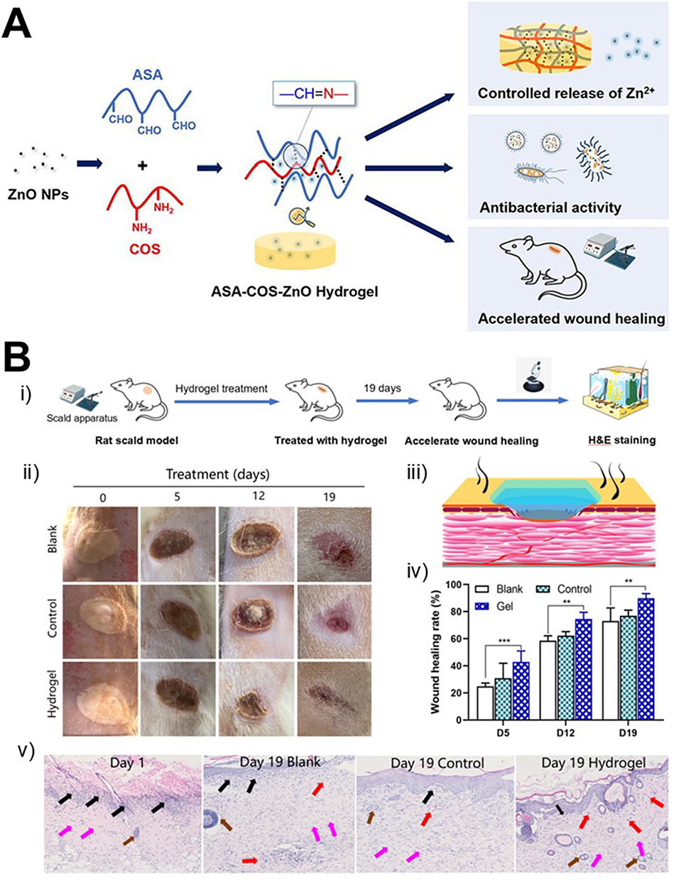 |
Figure 10 (A) Schematic illustration of preparation of sodium alginate-chitosan oligosaccharide‑zinc oxide hydrogel with controlled release of Zn2+, antibacterial activity, and accelerated wound healing. (B) (i) Wound formation and treatment; (ii) representative digital images of the wound at specific time points for the blank, positive control, and sodium alginate-chitosan oligosaccharide‑zinc oxide hydrogel groups; (iii) interactions between the hydrogel and wound tissues; (iv) quantification of wound closure over 19 days (** and *** indicate levels of statistical significance p < 0.01 and p < 0.001, respectively); v) photomicrographs of histological staining of wound sites on day 0 and day 19 (inflammatory cells: black arrows, blood vessels: red arrows, hair follicles: brown arrows, fibroblasts: purple arrows). Taken from ref.212 |
Hyaluronic acid plays numerous critical physiological roles for wound healing due to its distinct molecular structure and physicochemical characteristics.214 These roles include joint lubrication, modulation of vascular wall permeability, and facilitation of the diffusion and function of proteins, water, and electrolytes. As a vital extracellular matrix component, it promotes initial inflammation, supports cell infiltration, accelerates the formation of granulation tissue matrices, and aids in tissue remodelling. For example, Liu et al215 developed an electrospun thioether-grafted hyaluronic acid nanofiber that can spontaneously form a nanofibrous hydrogel in situ on wound sites, thereby modulating the inflammatory microenvironment and accelerating the healing of chronic diabetic wounds. As illustrated in Figure 11A, the acute wound model was established in Kunming mice, while Figure 11B shows markedly improved closure following nanofiber dressing and subsequent hydrogel treatment.
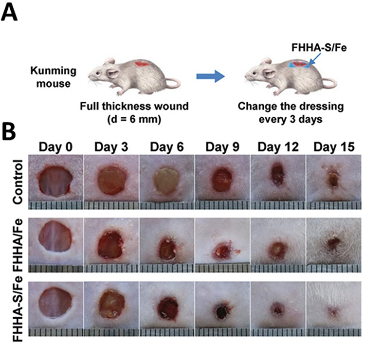 |
Figure 11 Electro spun thioether-grafted hyaluronic acid nanofiber hydrogel enhanced the healing effect in an acute wound model. (A) Schematic representation of the establishment and treatment of the acute wound model. (B) Representative images of wounds following different treatments at the specified days. Taken from ref.215 |
Shah et al216 developed an injectable hydrogel composed of hyaluronic acid, pullulan-grafted Pluronic F127, designed as a sustained and targeted delivery system for curcumin to promote skin regeneration in diabetic wounds. Biological testing demonstrated that curcumin within the hydrogel boosted cell proliferation, reduced inflammatory cell presence, and ultimately improved wound closure. Additionally, these studies indicated that curcumin release was enhanced with higher levels of hyaluronic acid. Wang et al217 developed an advanced wound dressing by incorporating hyaluronic acid-grafted pullulan succinate with chitosan (Figure 12), aiming to enhance antimicrobial properties and expedite skin wound healing. The resulting film exhibited a highly organized three-dimensional structure, along with a superior swelling index compared to the individual polymers, enabling efficient absorption of exudates from the wound site. When tested in vitro with L929 cells, the film displayed no cytotoxicity and actively promoted cell proliferation. Moreover, the composite demonstrated significant antibacterial efficacy against Staphylococcus aureus and Escherichia coli. In in vivo studies, the film dressings exhibited superior performance in reducing inflammation and accelerating the healing of skin wounds in rat models.
 |
Figure 12 Synthetic route of hyaluronic acid-grafted pullulan succinate and the preparation of the chitosan/ hyaluronic acid-grafted pullulan succinate film. Taken from ref.217 |
Alginate also play a key role in the healing of chronic wounds. Their high M block content effectively stimulates cytokine production by human monocytes.218 Alginate properties include strong biological activity, high adsorption capacity, bio-stability, anionic nature, hydrophilicity, non-toxicity, lack of immunogenic responses, hydrogel-forming ability, and cost-effectiveness. Alginate can form bonds with metal ions through various mechanisms, including electrostatic interactions and coordination. When guluronate blocks interact with calcium ions (Ca2⁺), they create an egg-box structure that facilitates cross-linking, making alginate suitable for numerous advanced applications, especially in wound healing.219 Alginate maintains a moist wound environment and reduces bacterial infection, both essential for rapid healing. Upon contact with wound exudates, alginates partially dissolve, forming a gel matrix that protects against microbes while providing an optimal moist environment. This process promotes cell proliferation and granulation tissue formation, accelerating wound healing.220 Additionally, alginate-based dressings demonstrate excellent haemostatic properties and can be easily removed from the wound site. An example of this is the alginate-fibrinogen-based composite hydrogels created by Soleimanpour et al221 for skin wound healing. The findings indicated that this synthesized system possesses excellent characteristics for wound healing applications, thanks to its suitable mechanical properties and biocompatibility. Furthermore, in vivo studies using Wistar rat models demonstrated that the bio composite hydrogel exhibited significant antibacterial effects and efficacy in treating chronic wounds. In a recent study, sodium alginate and arginine were crosslinked with zinc to form a hydrogel for use in skin wound dressings. The hydrogel demonstrated an enhanced swelling capacity, which was advantageous for absorbing wound exudate. Additionally, it exhibited notable antioxidant activity and strong inhibitory effects against Escherichia coli and Staphylococcus aureus.222
Dextran exhibits several distinctive properties, including enhanced water absorption, non-toxicity, and the capacity for polymer cross-linking. These characteristics promote angiogenesis, stimulate cell proliferation, and accelerate wound healing, thereby facilitating superior skin regeneration.223 Furthermore, dextran plays a critical role in the removal of wound exudates, metabolic by products, and other detrimental substances, thus creating an optimal environment for healing. It also supports angiogenesis, fostering the formation of new blood vessels, and protects the epidermis from ischemic damage. Additionally, dextran contributes to collagen deposition and influences tissue remodelling during the late stages of wound healing.220
Due to their excellent biocompatibility, cellulose-based hydrogel bio-adhesives are commonly used for loading and releasing various substances in wound treatment.208
For example, a cellulose-based hydrogel film was synthesized through a multistep process involving cellulose dissolution in a NaOH/urea system, crosslinking with chitosan, and subsequent incorporation of carboxylated multi-walled carbon nanotubes, resulting in a conductive composite hydrogel (Figure 13). This system exhibited enhanced structural integrity and functional performance, highlighting its potential for advanced biomedical applications.224
 |
Figure 13 Schematic representation of the multistep synthesis of a cellulose–chitosan hydrogel, including cellulose dissolution, chitosan-mediated crosslinking, incorporation of carboxylated multi-walled carbon nanotubes, and formation of the final hydrogel film. Taken from ref.224 |
Table 5 presents a selection of the most recent and significant publications on carbohydrate-based hydrogels for wound healing.
 |
Table 5 Selected Publications Related to Carbohydrate-Based Hydrogel for Wound Healing |
Recent research has focused on enhancing the healing properties of carbohydrate-based hydrogels through innovative approaches. One significant advancement involves the incorporation of growth factors into these hydrogels. For example, chitosan-based hydrogels loaded with epidermal growth factor have been developed to promote faster epithelialization and granulation tissue formation. In preclinical models, these epidermal growth factor -loaded hydrogels have demonstrated significantly enhanced healing rates compared to control groups without growth factors.237
Moreover, composite hydrogels that combine carbohydrate-based materials with synthetic polymers have shown promise in improving mechanical strength and flexibility.24
In recent years, the development of smart hydrogels has garnered considerable attention. These responsive materials can adapt to changes in their environment, such as pH, temperature, or ionic strength. This dynamic behaviour enables them to optimize drug release and hydration based on the specific conditions of the wound, providing a more personalized approach to wound management.238–242
Antimicrobial properties are another focal point in the advancement of carbohydrate-based hydrogels. By integrating antimicrobial agents—such as silver nanoparticles or essential oils—into the hydrogel matrix, researchers aim to combat bacterial infections that can complicate wound healing. This is particularly important for chronic and complex wounds, where the risk of infection is significantly elevated.243
Advantages and Challenges
Carbohydrate-based hydrogels are increasingly recognized for their significant advantages in biomedical applications, particularly due to their tuneable properties. The structure of carbohydrates can be easily modified through chemical or physical methods, enabling researchers to customize characteristics such as swelling behaviour, degradation rates, and mechanical strength. This versatility is essential for tailoring hydrogels for specific biomedical applications, ranging from drug delivery systems to tissue engineering scaffolds. The ease of modification further enhances the appeal of carbohydrate-based hydrogels, as functional groups inherent in saccharides provide multiple sites for chemical alterations, facilitating the incorporation of bioactive compounds, growth factors, or nanoparticles. Biocompatibility is another critical advantage. Many carbohydrate-based hydrogels are derived from natural sources, which reduces the risk of adverse immune responses when used in vivo. Their biodegradability ensures that they can be safely absorbed by the body, minimizing the need for surgical removal post-application.
The increasing number of research articles and patents underscores the growing interest and significant industrial investment in this field. For instance, EP4188468A1 discusses a method for enhancing the mechanical properties of carbohydrate-based hydrogels through specific crosslinking agents, demonstrating their potential for improved performance in tissue engineering. Similarly, CN113797385B is related to a chitosan/polyethylene glycol hydrogel with potential applications in promoting drug release and wound healing.
However, despite their advantages, carbohydrate-based hydrogels must address several challenges to achieve broader biomedical use.
- Firstly, the rational design of carbohydrate-based compounds within the hydrogel matrix for specific biological interaction is rarely investigated. The precise design of carbohydrate components—such as selecting the right monosaccharides, modifying the monosaccharides, or functionalizing them with bioactive molecules—is essential for promoting desired cellular behaviours and improving the performance of hydrogels in applications like drug delivery and tissue engineering. A more systematic approach to designing these hydrogels could enhance their biological activity, allowing for better targeting, controlled release, and integration with biological environments.244
- One other significant limitation is related to their mechanical strength. While suitable for soft tissue applications, many carbohydrate hydrogels lack the robustness required for hard tissue engineering. Indeed, on the contrary of many organogels245 natural polymer-based hydrogel possesses low mechanical strength which restricts its application in various fields.246,247 This shortcoming often necessitates reinforcement through composites or crosslinking, which can complicate the fabrication process. Therefore, improving their mechanical strength has become very challenging.248–250
- Stability is another challenge. Environmental factors such as temperature, pH, and ionic strength can adversely affect hydrogel properties, leading to variability in performance. This instability can be particularly concerning in applications requiring precise control over release rates or mechanical properties.85
- Scalability in production is a crucial issue as well. While laboratory synthesis of carbohydrate-based hydrogels can yield desirable properties, translating these methods to large-scale production, ie, multi-kilo, without compromising quality remains a challenge.251
- Sustainability. To mitigate this challenge, researchers are exploring greener synthesis methods that employ low-energy consumption processes and less hazardous solvents. Such approaches not only enhance sustainability but also aim to preserve the biological properties of the resulting hydrogels.25 To this end, sustainability characteristics should always be demonstrated and described using green metrics calculations and, where applicable, life cycle assessment (LCA).252
- Sterilization: hydrogels intended for biomedical applications must be sterilized without compromising their structure, functionality, or bioactivity. Common sterilization methods, such as autoclaving or gamma irradiation, can alter the mechanical properties, swelling capacity, or degradation profile of carbohydrate-based hydrogels, requiring careful optimization.253
- Regulatory Compliance: Translation to clinical use requires adherence to strict regulatory standards regarding safety, efficacy, and biocompatibility. Demonstrating long-term stability, predictable degradation, absence of immunogenicity, and consistent performance under physiological conditions are essential for regulatory approval.254
To address these issues, recent studies have explored a variety of design strategies.
One widely studied approach is the incorporation of nanoparticles or nanofibers into the polysaccharide matrix.85 For example, adding nano-silica to an alginate–chitosan–gelatin hydrogel improved mechanical strength and promoted osteogenic differentiation of pre-osteoblastic cells.255 Similarly, embedding nano-hydroxyapatite into alginate scaffolds enhanced structural stability and bioactivity, making them suitable for bone tissue engineering.256 While these nanocomposite hydrogels improve stiffness, durability, and porosity, challenges remain, such as ensuring uniform dispersion of fillers and avoiding potential cytotoxicity or immunogenicity.
Another effective strategy involves double-network or interpenetrating polymer networks, where two polymer networks interpenetrate each other. One network provides stiffness, while the second contributes elasticity, resulting in hydrogels with significantly enhanced mechanical strength and resilience.257 For example, dual-network hydrogels combining modified alginate and chitosan demonstrated self-healing, injectability, and mechanical robustness suitable for drug and cell delivery.258 Although highly effective, these hydrogels require careful synthesis to maintain porosity and uniformity.
Chemical crosslinking using bio-orthogonal reactions, such as Schiff-base formation, thiol–ene chemistry, or click reactions, offers another route to improve hydrogel stability. Covalent networks are more resistant to physiological degradation than ionically crosslinked hydrogels and allow for precise control over degradation kinetics and bioactive molecule incorporation.259 However, the choice of crosslinking chemistry must balance stability with biocompatibility, and complex chemistries can complicate large-scale production.
Self-assembling peptide–polysaccharide hybrids represent an innovative approach to enhance hydrogel performance. Here, peptides form a nanofibrillar network within the polysaccharide matrix, providing mechanical reinforcement and bioactive domains for cell adhesion. These hybrids allow tunable degradation and hydration while maintaining biocompatibility.260 The main limitations are higher cost and complexity in synthesis, as well as the need for thorough in vivo validation.
Finally, incorporating biodegradable micro- or nanoparticles within the hydrogel provides both reinforcement and controlled release of therapeutic agents. For instance, poly(lactic-co-glycolic acid) microspheres embedded in polysaccharide hydrogels allow sustained drug release while enhancing mechanical stability.261 Care must be taken to maintain uniform particle distribution and preserve hydrogel porosity and swelling properties.
In conclusion, while each of these strategies addresses specific limitations, trade-offs remain between mechanical strength, biocompatibility, degradability, and manufacturability. No single approach provides a universal solution; the optimal design depends on the intended clinical application. Successful translation requires application-specific optimization, reproducible synthesis, and thorough in vitro and in vivo validation to ensure safety, functionality, and scalability.
Conclusions and Outlook
Looking ahead, the potential for clinical translation of carbohydrate-based hydrogels is vast. As these materials become increasingly sophisticated, they can be tailored to meet specific medical needs, enhancing patient outcomes across various therapeutic domains.26 The integration of smart technologies into hydrogel design offers a pathway for innovation that could fundamentally change how treatments are administered.239 For example, hydrogels embedded with biosensors could monitor wound conditions in real-time, providing healthcare providers with critical information regarding the status of a wound and the need for intervention.25 The development of multifunctional hydrogels that integrate various therapeutic properties—such as haemostatic capabilities, antimicrobial activity, and the delivery of growth factors—holds great promise for simplifying wound management.262,263 Such multifunctional dressings could address multiple aspects of healing, improving overall treatment efficiency and patient comfort, while also reducing the need for additional interventions. Despite these promising advances, several challenges must still be addressed before widespread clinical adoption can be achieved. Issues of reproducibility, batch-to-batch variability, and the lack of standardized evaluation protocols remain significant barriers. Establishing robust manufacturing processes and harmonized testing criteria will be essential to ensure consistent performance and regulatory approval. Looking toward the future, emerging trends are expected to shape the next generation of carbohydrate-based hydrogels. AI-guided material design could accelerate the discovery of optimized formulations with enhanced biological responses. Another promising avenue for future research involves the customization and personalization of hydrogel.264 Continued progress in 3D and 4D bioprinting, organ-on-chip systems, organoid biomanufacturing enable the fabrication of patient-specific hydrogel architectures with complex geometries and spatially controlled properties, while organ-on-chip systems and organoid biomanufacturing facilitate in vitro modelling of tissue physiology and personalized therapeutic testing. Additionally, the incorporation of advanced biosensing capabilities and responsive behaviours will further expand their functionality, paving the way for intelligent, dynamic wound-care systems.265 Overall, carbohydrate-based hydrogels stand at the forefront of biomedical innovation, and ongoing interdisciplinary research will continue to unlock their full potential.
Acknowledgments
Financial support was provided by the Spanish Ministry of Science and Innovation (Ref: PID2020-119949RB-I00 and PID2023-152689OB-I00 to NK), the Andalusian Ministry of Economy, Science and Innovation cofinanced by the European Regional Development Fund (ERDF) from FEDER and the European Social Fund (ESF) (PY20_00882 and CV20-04221 to NK, and postdoctoral fellowship “POSTDOC_21_00594” to AZ), the CSIC (CSIC-COV19-047 to NK), the PAIDI Program from the Andalusian Government (FQM-313 to NK). The COST action CA-18132 “Functional Glyconanomaterials for the Development of Diagnostic and Targeted Therapeutic Probe” is also acknowledged. This project has received funding from the European Union’s Horizon Europe research and innovation programme under the Marie Skłodowska-Curie grant agreement No 101150385.
Disclosure
There are no conflicts to declare.
References
1. Khiar N, Suárez B, Valdivia V, Fernández I. Phosphinite thioglycosides derived from natural d-sugars as useful P/S ligands for the synthesis of both enantiomers in palladium-catalyzed asymmetric substitution. Synlett. 2005;2005:2963–39. doi:10.1055/s-2005-918963
2. Datta S, Bhattacharya S. Multifarious facets of sugar-derived molecular gels: molecular features, mechanisms of self-assembly and emerging applications. Chem Soc Rev. 2015;44(15):5596–5637. doi:10.1039/C5CS00093A
3. Zuliani A, Castillejos MC, Khiar N. Continuous flow synthesis of PCN-222 (MOF-545) with controlled size and morphology: a sustainable approach for efficient production. Green Chem. 2023;25(24):10596–10610. doi:10.1039/D3GC02774K
4. Cova CM, Ramos V, Escudero A, Holgado JP, Khiar N, Zuliani A. A sustainable lecithin-based ligand for the bio-functionalization of iron and hybrid metal organic frameworks (MOFs) nanoparticles with the sugar mannose. Green Chem. 2024;26(23):11563–11575. doi:10.1039/D4GC03743J
5. Rosales-Barrios C, González-Sánchez ZI, Zuliani A, et al. PSMA-targeted delivery of docetaxel in prostate cancer using small-sized PDA-based micellar nanovectors. J Control Release. 2025;379:890–905. doi:10.1016/j.jconrel.2025.01.062
6. Khiar N, Salvador Á, Chelouan A, Alcudia A, Fernández I. “Sulfolefin”: highly modular mixed S/Olefin ligands for enantioselective Rh-catalyzed 1,4-addition. Org Biomol Chem. 2012;10(12):2366–2368. doi:10.1039/C2OB07132K
7. Tønnesen HH, Karlsen J. Alginate in drug delivery systems. Drug Dev Ind Pharm. 2002;28(6):621–630. doi:10.1081/ddc-120003853
8. Chai Q, Jiao Y, Yu X. Hydrogels for biomedical applications: their characteristics and the mechanisms behind them. Gels. 2017;3(1):6. doi:10.3390/gels3010006
9. Carter P, Narasimhan B, Wang Q. Biocompatible nanoparticles and vesicular systems in transdermal drug delivery for various skin diseases. Int J Pharm. 2019;555:49–62. doi:10.1016/j.ijpharm.2018.11.032
10. Huang K, Wu J, Gu Z. Black phosphorus hydrogel scaffolds enhance bone regeneration via a sustained supply of calcium-free phosphorus. ACS Appl Mater Interfaces. 2019;11(3):2908–2916. doi:10.1021/acsami.8b21179
11. Sharma S, Bhende M, Goel A. A review: polysaccharide-based hydrogels and their biomedical applications. Polym Bull. 2024;81(10):8573–8594. doi:10.1007/s00289-023-05130-8
12. Coviello T, Matricardi P, Marianecci C, Alhaique F. Polysaccharide hydrogels for modified release formulations. J Control Release. 2007;119(1):5–24. doi:10.1016/j.jconrel.2007.01.004
13. Pescosolido L, Piro T, Vermonden T, et al. Biodegradable IPNs based on oxidized alginate and dextran-HEMA for controlled release of proteins. Carbohydr Polym. 2011;86(1):208–213. doi:10.1016/j.carbpol.2011.04.033
14. Das D, Das R, Ghosh P, Dhara S, Baran Panda A, Pal S. Dextrin cross linked with poly(HEMA): a novel hydrogel for colon specific delivery of ornidazole. RSC Adv. 2013;3(47):25340–25350. doi:10.1039/C3RA44716B
15. Das D, Das R, Mandal J, Ghosh A, Pal S. Dextrin crosslinked with poly(lactic acid): a novel hydrogel for controlled drug release application. J Appl Polym Sci. 2014;131(7). doi:10.1002/app.40039
16. Das D, Pal S. Dextrin/poly (HEMA): pH responsive porous hydrogel for controlled release of ciprofloxacin. Int J Biol Macromol. 2015;72:171–178. doi:10.1016/j.ijbiomac.2014.08.007
17. Chalard A, Vaysse L, Joseph P, et al. Simple Synthetic Molecular Hydrogels from Self-Assembling Alkylgalactonamides as Scaffold for 3D Neuronal Cell Growth. ACS Appl Mater Interfaces. 2018;10(20):17004–17017. doi:10.1021/acsami.8b01365
18. Cao H, Duan L, Zhang Y, Cao J, Zhang K. Current hydrogel advances in physicochemical and biological response-driven biomedical application diversity. Signal Transduct Target Ther. 2021;6(1):1–31. doi:10.1038/s41392-021-00830-x
19. Morris J, Bietsch J, Bashaw K, Wang G. Recently developed carbohydrate based gelators and their applications. Gels. 2021;7(1):24. doi:10.3390/gels7010024
20. Mehta P, Mahadik K, Kadam S, Dhapte-Pawar V. Chapter 4 - Advanced applications of green hydrogels in drug delivery systems. In: Ahmed S, editor. Applications of Advanced Green Materials. Woodhead Publishing; 2021:89–130. doi:10.1016/B978-0-12-820484-9.00004-0.
21. Wichterle O, Lím D. Hydrophilic gels for biological use. Nature. 1960;185(4706):117–118. doi:10.1038/185117a0
22. Caló E, Khutoryanskiy VV. Biomedical applications of hydrogels: a review of patents and commercial products. Eur Polym J. 2015;65:252–267. doi:10.1016/j.eurpolymj.2014.11.024
23. Ho TC, Chang CC, Chan HP, et al. Hydrogels: properties and applications in biomedicine. Molecules. 2022;27(9):2902. doi:10.3390/molecules27092902
24. Thang NH, Chien TB, Cuong DX. Polymer-based hydrogels applied in drug delivery: an overview. Gels. 2023;9(7):523. doi:10.3390/gels9070523
25. Sánchez-Cid P, Jiménez-Rosado M, Romero A, Pérez-Puyana V. Novel trends in hydrogel development for biomedical applications: a review. Polymers. 2022;14(15):3023. doi:10.3390/polym14153023
26. Zhao L, Zhou Y, Zhang J, Liang H, Chen X, Tan H. Natural polymer-based hydrogels: from polymer to biomedical applications. Pharmaceutics. 2023;15(10):2514. doi:10.3390/pharmaceutics15102514
27. Khiar N. Determination of the absolute configuration of sulfinyl glycosides: the role of the exo-anomeric effect. Tetrahedron Lett. 2000;41(47):9059–9063. doi:10.1016/S0040-4039(00)01651-8
28. de la Harpe KM, Kondiah PPD, Marimuthu T, Choonara YE. Advances in carbohydrate-based polymers for the design of suture materials: a review. Carbohydr Polym. 2021;261:117860. doi:10.1016/j.carbpol.2021.117860
29. Benalaya I, Alves G, Lopes J, Silva LR. A review of natural polysaccharides: sources, characteristics, properties, food, and pharmaceutical applications. Int J Mol Sci. 2024;25(2):1322. doi:10.3390/ijms25021322
30. Hariprasad BS, Lakshmi Priya R, Ganesh Babu S. Recent advances in the synthesis, catalytic, and adsorption applications of carbohydrate polymers. Eur Polym J. 2024;214:113178. doi:10.1016/j.eurpolymj.2024.113178
31. Ainani AF, Darmawan, Rubiyanto JT, et al. Hemicellulose-based hydrogel composite: enhanced properties and diverse applications. Carbohydr Polym Technol Appl. 2024;8:100558. doi:10.1016/j.carpta.2024.100558
32. Awere CO, Anadebe VC, Sabapathi D, et al. From waste to wealth: plant-Derived cellulose nanocrystal hydrogel for anticancer, antibacterial, and anticorrosion studies. Carbohydr Polym Technol Appl. 2025;9:100656. doi:10.1016/j.carpta.2024.100656
33. Pellá MCG, Lima-Tenório MK, Tenório-Neto ET, Guilherme MR, Muniz EC, Rubira AF. Chitosan-based hydrogels: from preparation to biomedical applications. Carbohydr Polym. 2018;196:233–245. doi:10.1016/j.carbpol.2018.05.033
34. Selvaraj S, Chauhan A, Dutta V, et al. A state-of-the-art review on plant-derived cellulose-based green hydrogels and their multifunctional role in advanced biomedical applications. Int J Biol Macromol. 2024;265:130991. doi:10.1016/j.ijbiomac.2024.130991
35. Buckley C, Murphy EJ, Montgomery TR, Major I. Hyaluronic acid: a review of the drug delivery capabilities of this naturally occurring polysaccharide. Polymers. 2022;14(17):3442. doi:10.3390/polym14173442
36. Prusty K, Swain SK. Chapter 7 - carbohydrate-based nanohydrogels for drug-delivery applications. In: Swain SK, Jawaid M, editors. Nanostructured Polymer Composites for Biomedical Applications. Elsevier; 2019:117–137. doi:10.1016/B978-0-12-816771-7.00007-7.
37. Peters JT, Wechsler ME, Peppas NA. Advanced biomedical hydrogels: molecular architecture and its impact on medical applications. Regen Biomater. 2021;8(6):rbab060. doi:10.1093/rb/rbab060
38. Zheng Q, Shang M, Li X, et al. Advances in intelligent response and nano-enhanced polysaccharide-based hydrogels: material properties, response types, action mechanisms, applications. Food Hydrocoll. 2024;146:109190. doi:10.1016/j.foodhyd.2023.109190
39. Revete A, Aparicio A, Cisterna BA, et al. Advancements in the use of hydrogels for regenerative medicine: properties and biomedical applications. Int J Biomater. 2022;2022:3606765. doi:10.1155/2022/3606765
40. Junaid PM, Dar AH, Dash KK, et al. Polysaccharide-based hydrogels for microencapsulation of bioactive compounds: a review. J Agric Food Res. 2024;15:101038. doi:10.1016/j.jafr.2024.101038
41. Zuliani A, Cova CM. Green synthesis of heterogeneous visible-light-active photocatalysts: recent advances. Photochem. 2021;1(2):147–166. doi:10.3390/photochem1020009
42. Zuliani A, Kikhtyanin O, Cova CM, Rodriguez-Padron D, Kubička D, Luque R. Boosting the Ni-Catalyzed Hydrodeoxygenation (HDO) of anisole using scrap catalytic converters. Adv Sustain Syst. 2022;6(4):2100394. doi:10.1002/adsu.202100394
43. Available from: https://sdgs.un.org/.
44. Damiri F, Fatimi A, Liu Y, et al. Recent advances in 3D bioprinted polysaccharide hydrogels for biomedical applications: a comprehensive review. Carbohydr Polym. 2024:122845. 10.1016/j.carbpol.2024.122845.
45. Gull N, Khan SM, Islam A, Butt MTZ. Hydrogels used for biomedical applications. In: Bio Monomers for Green Polymeric Composite Materials. John Wiley & Sons, Ltd; 2019:175–199. doi:10.1002/9781119301714.ch9
46. Liao Y, Xie L, Ye J, et al. Sprayable hydrogel for biomedical applications. Biomater Sci. 2022;10(11):2759–2771. doi:10.1039/D2BM00338D
47. Deligkaris K, Tadele TS, Olthuis W, van den Berg A. Hydrogel-based devices for biomedical applications. Sens Actuators B. 2010;147: 765–774. doi:10.1016/j.snb.2010.03.083
48. Ren Y, Wang Q, Xu W, et al. Alginate-based hydrogels mediated biomedical applications: a review. Int J Biol Macromol. 2024;279(Pt 1):135019. doi:10.1016/j.ijbiomac.2024.135019
49. Moss GP, Smith PAS, Tavernier D. Glossary of class names of organic compounds and reactivity intermediates based on structure (IUPAC Recommendations 1995). Pure Appl Chem. 1995;67(8):1307–1375. doi:10.1351/pac199567081307
50. Yang L, Wang H, Yang Y, Li Y. Self-healing cellulose-based hydrogels: from molecular design to multifarious applications. Carbohydr Polym. 2025;347:122738. doi:10.1016/j.carbpol.2024.122738
51. Sánchez-Cid P, Gónzalez-Ulloa G, Alonso-González M, et al. Influence of natural crosslinkers on chitosan hydrogels for potential biomedical applications. Macromol Mater Eng. 2023;308(12):2300195. doi:10.1002/mame.202300195
52. Soares PIP, Sousa AI, Silva JC, Ferreira IMM, Novo CMM, Borges JP. Chitosan-based nanoparticles as drug delivery systems for doxorubicin: optimization and modelling. Carbohydr Polym. 2016;147:304–312. doi:10.1016/j.carbpol.2016.03.028
53. Arteche Pujana M, Pérez-Álvarez L, Cesteros Iturbe LC, Katime I. Biodegradable chitosan nanogels crosslinked with genipin. Carbohydr Polym. 2013;94(2):836–842. doi:10.1016/j.carbpol.2013.01.082
54. Mou D, Yu Q, Zhang J, et al. Intra-articular injection of chitosan-based supramolecular hydrogel for osteoarthritis treatment. Tissue Eng Regen Med. 2021;18(1):113–125. doi:10.1007/s13770-020-00322-z
55. Qureshi MAUR, Arshad N, Rasool A, et al. Chitosan and carrageenan‐based biocompatible hydrogel platforms for cosmeceutical, drug delivery, and biomedical applications. Starch-Starke. 2022;76:2200052. doi:10.1002/star.202200052
56. Taokaew S, Kaewkong W, Kriangkrai W. Recent development of functional chitosan-based hydrogels for pharmaceutical and biomedical applications. Gels. 2023;9(4):277. doi:10.3390/gels9040277
57. Mishra A, Omoyeni T, Singh PK, Anandakumar S, Tiwari A. Trends in sustainable chitosan-based hydrogel technology for circular biomedical engineering: a review. Int J Biol Macromol. 2024;276:133823. doi:10.1016/j.ijbiomac.2024.133823
58. Santiago TSA, Delezuk JAM, Bataglioli RA, Baratti MO, Carvalho HF, Beppu MM. Influence of hyaluronic acid and chitosan molecular weight on the adhesion of circulating tumor cell on multilayer films. Int J Biol Macromol. 2024;281:136180. doi:10.1016/j.ijbiomac.2024.136180
59. Tripathi AS, Zaki MEA, Al-Hussain SA, et al. Material matters: exploring the interplay between natural biomaterials and host immune system. Front Immunol. 2023;14:1269960. doi:10.3389/fimmu.2023.1269960
60. Bueter CL, Specht CA, Levitz SM. Innate sensing of chitin and chitosan. PLOS Pathog. 2013;9(1):e1003080. doi:10.1371/journal.ppat.1003080
61. Elieh Ali Komi D, Sharma L, Dela Cruz CS. Chitin and its effects on inflammatory and immune responses. Clin Rev Allergy Immunol. 2018;54(2):213–223. doi:10.1007/s12016-017-8600-0
62. Farhadihosseinabadi B, Zarebkohan A, Eftekhary M, Heiat M, Moghaddam MM, Gholipourmalekabadi M. Crosstalk between chitosan and cell signaling pathways. Cell Mol Life Sci CMLS. 2019;76(14):2697. doi:10.1007/s00018-019-03107-3
63. von Huth S, Moeller JB, Schlosser A, et al. Immunohistochemical localization of fibrinogen C domain containing 1 on epithelial and mucosal surfaces in human tissues. J Histochem Cytochem. 2018;66(2):85–97. doi:10.1369/0022155417743694
64. Zheng C, Chen J, Chu F, Zhu J, Jin T. Inflammatory role of TLR-MyD88 signaling in multiple sclerosis. Front Mol Neurosci. 2020;12:314. doi:10.3389/fnmol.2019.00314
65. Liu T, Zhang L, Joo D, Sun SC. NF-κB signaling in inflammation. Signal Transduct Target Ther. 2017;2(1):1–9. doi:10.1038/sigtrans.2017.23
66. Zhong Z, Huang Y, Hu Q, et al. Elucidation of molecular pathways responsible for the accelerated wound healing induced by a novel fibrous chitin dressing. Biomater Sci. 2019;7(12):5247–5257. doi:10.1039/C9BM00404A
67. Sinha VR, Kumria R. Polysaccharides in colon-specific drug delivery. Int J Pharm. 2001;224(1):19–38. doi:10.1016/S0378-5173(01)00720-7
68. Ruel-Gariépy E, Leroux JC. In situ-forming hydrogels—review of temperature-sensitive systems. Eur J Pharm Biopharm. 2004;58(2):409–426. doi:10.1016/j.ejpb.2004.03.019
69. Mano JF, Silva GA, Azevedo HS, et al. Natural origin biodegradable systems in tissue engineering and regenerative medicine: present status and some moving trends. J R Soc Interface. 2007;4(17):999–1030. doi:10.1098/rsif.2007.0220
70. Pustylnikov S, Sagar D, Jain P, Khan ZK. Targeting the C-type lectins-mediated host-pathogen interactions with dextran. J Pharm Pharm Sci. 2014;17(3):371. doi:10.18433/j3n590
71. Lee KY, Mooney DJ. Alginate: properties and biomedical applications. Prog Polym Sci. 2012;37(1):106. doi:10.1016/j.progpolymsci.2011.06.003
72. Tomić SL, Babić Radić MM, Vuković JS, Filipović VV, Nikodinovic-Runic J, Vukomanović M. Alginate-based hydrogels and scaffolds for biomedical applications. Mar Drugs. 2023;21(3):177. doi:10.3390/md21030177
73. Zou P, Yao J, Cui YN, et al. Advances in cellulose-based hydrogels for biomedical engineering: a review summary. Gels. 2022;8(6):364. doi:10.3390/gels8060364
74. Fu LH, Qi C, Ma MG, Wan P. Multifunctional cellulose-based hydrogels for biomedical applications. J Mater Chem B. 2019;7(10):1541–1562. doi:10.1039/C8TB02331J
75. Burdick JA, Prestwich GD. Hyaluronic acid hydrogels for biomedical applications. Adv Mater. 2011;23(12):H41–56. doi:10.1002/adma.201003963
76. Luo Z, Wang Y, Li J, Wang J, Yu Y, Zhao Y. Tailoring hyaluronic acid hydrogels for biomedical applications. Adv Funct Mater. 2023;33(49):2306554. doi:10.1002/adfm.202306554
77. Misra S, Hascall VC, Markwald RR, Ghatak S. Interactions between Hyaluronan and Its Receptors (CD44, RHAMM) regulate the activities of inflammation and cancer. Front Immunol. 2015;6:201. doi:10.3389/fimmu.2015.00201
78. Duan H, Donovan M, Foucher A, Schultze X, Lecommandoux S. Multivalent and multifunctional polysaccharide-based particles for controlled receptor recognition. Sci Rep. 2018;8(1):14730. doi:10.1038/s41598-018-32994-y
79. Bhattacharya D, Svechkarev D, Souchek JJ, et al. Impact of structurally modifying hyaluronic acid on CD44 interaction. J Mater Chem B. 2017;5(41):8183. doi:10.1039/C7TB01895A
80. Mesrati MH, Syafruddin SE, Mohtar MA, Syahir A. CD44: a multifunctional mediator of cancer progression. Biomolecules. 2021;11(12):1850. doi:10.3390/biom11121850
81. Castillejos MC, Zuliani A, Cova CM, et al. Assessing small-sized D-fructose-functionalized micelles for active targeting of triple-negative breast cancer. J Drug Deliv Sci Technol. 2025;112:107281. doi:10.1016/j.jddst.2025.107281
82. Grishko V, Xu M, Ho R, et al. Effects of hyaluronic acid on mitochondrial function and mitochondria-driven apoptosis following oxidative stress in human chondrocytes. J Biol Chem. 2009;284(14):9132. doi:10.1074/jbc.M804178200
83. Xu Y, Benedikt J, Ye L. Hyaluronic acid interacting molecules mediated crosstalk between cancer cells and microenvironment from primary tumour to distant metastasis. Cancers. 2024;16(10):1907. doi:10.3390/cancers16101907
84. Leng Y, Abdullah A, Wendt MK, Calve S. Hyaluronic Acid, CD44 and RHAMM regulate myoblast behavior during embryogenesis. Matrix Biol. 2018;78-79:236. doi:10.1016/j.matbio.2018.08.008
85. Rumon MM, Akib AA, Sarkar SD, et al. Polysaccharide-based hydrogels for advanced biomedical engineering applications. ACS Polymers Au. 2024;4(6):463–486. doi:10.1021/acspolymersau.4c00028
86. Fu J, Yang F, Guo Z. The chitosan hydrogels: from structure to function. New J Chem. 2018;42(21):17162–17180. doi:10.1039/C8NJ03482F
87. Recio R, Lerena P, Pozo E, et al. Carbohydrate-based NK1R antagonists with broad-spectrum anticancer activity. J Med Chem. 2021;64(14):10350–10370. doi:10.1021/acs.jmedchem.1c00793
88. Poursadegh H, Amini-Fazl MS, Javanbakht S, Kazeminava F. Magnetic nanocomposite through coating mannose-functionalized metal-organic framework with biopolymeric pectin hydrogel beads: a potential targeted anticancer oral delivery system. Int J Biol Macromol. 2024;254:127702. doi:10.1016/j.ijbiomac.2023.127702
89. Poursadegh H, Bakhshi V, Amini-Fazl MS, Adibag Z, Kazeminava F, Javanbakht S. Incorporating mannose-functionalized hydroxyapatite/metal-organic framework into the hyaluronic acid hydrogel film: a potential dual-targeted oral anticancer delivery system. Int J Biol Macromol. 2024;274:133516. doi:10.1016/j.ijbiomac.2024.133516
90. Wu H, Wang J, Lin Y, et al. Injectable ozone-rich nanocomposite hydrogel loaded with D-mannose for anti-inflammatory and cartilage protection in osteoarthritis treatment. Small. 2024;20(25):2309597. doi:10.1002/smll.202309597
91. Wang JW, Hon MH. Preparation and characterization of pH sensitive sugar mediated (polyethylene glycol/chitosan) membrane. J Mater Sci Mater Med. 2003;14(12):1079–1088. doi:10.1023/B:JMSM.0000004005.52762.ea
92. Patil TS, Deshpande AS. Mannosylated nanocarriers mediated site-specific drug delivery for the treatment of cancer and other infectious diseases: a state of the art review. J Control Release. 2020;320:239–252. doi:10.1016/j.jconrel.2020.01.046
93. Shi DT, Zhou D, Zang Y, et al. Selective fluorogenic imaging of hepatocellular H2S by a galactosyl azidonaphthalimide probe. Chem Commun. 2015;51(17):3653–3655. doi:10.1039/C4CC09771H
94. D’Souza AA, Devarajan PV. Asialoglycoprotein receptor mediated hepatocyte targeting - strategies and applications. J Control Release. 2015;203:126–139. doi:10.1016/j.jconrel.2015.02.022
95. Santander-Ortega MJ, Stauner T, Loretz B, et al. Nanoparticles made from novel starch derivatives for transdermal drug delivery. J Control Release. 2010;141(1):85–92. doi:10.1016/j.jconrel.2009.08.012
96. Maiti S, Maji B, Yadav H. Progress on green crosslinking of polysaccharide hydrogels for drug delivery and tissue engineering applications. Carbohydr Polym. 2024;326:121584. doi:10.1016/j.carbpol.2023.121584
97. Lu A, Li S. Polysaccharides as a hydrophilic building block of amphiphilic block copolymers for the conception of nanocarriers. Pharmaceutics. 2024;16(4):467. doi:10.3390/pharmaceutics16040467
98. Perin F, Motta A, Maniglio D. Amphiphilic copolymers in biomedical applications: synthesis routes and property control. Mater Sci Eng C. 2021;123:111952. doi:10.1016/j.msec.2021.111952
99. Duggal I, Martinez AN, Richbourg NR, Peppas NA. Network theory and recent developments in networks formed by the freeze–thawing process. Polymer. 2025:129385. doi:10.1016/j.polymer.2025.129385
100. Masoumi Shahrbabak S, Jalali SM, Fathabadi MF, et al. Modified alginates for precision drug delivery: advances in controlled-release and targeting systems. Int J Pharm X. 2025;10:100381. doi:10.1016/j.ijpx.2025.100381
101. Wang W, Huang Y, Pan Y, et al. Sodium alginate modifications: a critical review of current strategies and emerging applications. Foods. 2025;14(22):3931. doi:10.3390/foods14223931
102. Babu A, Shams R, Dash KK, Shaikh AM, Kovács B. Protein-polysaccharide complexes and conjugates: structural modifications and interactions under diverse treatments. J Agric Food Res. 2024;18:101510. doi:10.1016/j.jafr.2024.101510
103. Xue H, Wang P, Ji L, Zhang K, Ge S, Tan J. Polysaccharide-based hydrogels: materials, preparation, and applications in medicine, food, adsorption, and agriculture. J Agric Food Res. 2025;24:102395. doi:10.1016/j.jafr.2025.102395
104. Assali M, Cid JJ, Fernández I, Khiar N. Supramolecular diversity through click chemistry: switching from nanomicelles to 1D-nanotubes and tridimensional hydrogels. Chem Mater. 2013;25(21):4250–4261. doi:10.1021/cm4022613
105. Liu J, Zhang Y, van Dongen K, et al. Hepatic spheroid formation on carbohydrate-functionalized supramolecular hydrogels. Biomacromolecules. 2023;24(6):2447–2458. doi:10.1021/acs.biomac.2c01390
106. Paiva MTP, Kishima JOF, Silva JBMD, Mantovan J, Colodi FG, Mali S. Crosslinking methods in polysaccharide-based hydrogels for drug delivery systems. Biomed Mater Devices. 2024;2(1):288–306. doi:10.1007/s44174-023-00118-4
107. Carrillo-Carrión C, Comaills V, Visiga AM, Gauthier BR, Khiar N. Enzyme-responsive Zr-based metal–organic frameworks for controlled drug delivery: taking advantage of clickable PEG-phosphate ligands. ACS Appl Mater Interfaces. 2023;15(23):27600–27611. doi:10.1021/acsami.3c03230
108. Segneanu AE, Bejenaru LE, Bejenaru C, et al. Advancements in hydrogels: a comprehensive review of natural and synthetic innovations for biomedical applications. Polymers. 2025;17(15):2026. doi:10.3390/polym17152026
109. Kaur N, Hamid, Choudhary P, Jaiswal AK. Recent progress in bioactive loaded hydrogels for food applications. J Agric Food Res. 2025;20:101756. doi:10.1016/j.jafr.2025.101756
110. Alavarse AC, Frachini ECG, da Silva RLCG, Lima VH, Shavandi A, Petri DFS. Crosslinkers for polysaccharides and proteins: synthesis conditions, mechanisms, and crosslinking efficiency, a review. Int J Biol Macromol. 2022;202:558–596. doi:10.1016/j.ijbiomac.2022.01.029
111. Di J, Li J, Sun C, Xu L, Li X. Advances in cellulose-based hydrogels for drug delivery: preparation, modification and challenges. Gels. 2025;11(12):938. doi:10.3390/gels11120938
112. Bhuyan MM, Jeong JH. Preparation of hydrogel by crosslinking and multi-dimensional applications. Gels. 2025;11(11):896. doi:10.3390/gels11110896
113. Jeong JO, Park JS, Kim YA, et al. Gamma Ray-Induced Polymerization and Cross-Linking for Optimization of PPy/PVP Hydrogel as Biomaterial. Polymers. 2020;12(1):111. doi:10.3390/polym12010111
114. de Lima CSA, Balogh TS, Varca JPRO, et al. An updated review of macro, micro, and nanostructured hydrogels for biomedical and pharmaceutical applications. Pharmaceutics. 2020;12(10):970. doi:10.3390/pharmaceutics12100970
115. Liu J, Su C, Chen Y, et al. Current understanding of the applications of photocrosslinked hydrogels in biomedical engineering. Gels. 2022;8(4):216. doi:10.3390/gels8040216
116. Martina K, Calsolaro F, Zuliani A, et al. Sonochemically-promoted preparation of silica-anchored cyclodextrin derivatives for efficient copper catalysis. Molecules. 2019;24(13):2490. doi:10.3390/molecules24132490
117. Li X, Xiong Y. Application of “Click” chemistry in biomedical hydrogels. ACS Omega. 2022;7(42):36918–36928. doi:10.1021/acsomega.2c03931
118. Lima-Tenório MK, Pineda EAG, Ahmad NM, Fessi H, Elaissari A. Magnetic nanoparticles: in vivo cancer diagnosis and therapy. Int J Pharm. 2015;493(1–2):313–327. doi:10.1016/j.ijpharm.2015.07.059
119. Hamidi M, Azadi A, Rafiei P. Hydrogel nanoparticles in drug delivery. Adv Drug Deliv Rev. 2008;60(15):1638–1649. doi:10.1016/j.addr.2008.08.002
120. Di X, Liang X, Shen C, Pei Y, Wu B, He Z. Carbohydrates used in polymeric systems for drug delivery: from structures to applications. Pharmaceutics. 2022;14(4):739. doi:10.3390/pharmaceutics14040739
121. Cui L, Pi J, Qin B, et al. Advanced application of carbohydrate-based micro/nanoparticles for rheumatoid arthritis. Int J Biol Macromol. 2024;269:131809. doi:10.1016/j.ijbiomac.2024.131809
122. Caballero F, Foradada M, Miñarro M, et al. Characterization of alginate beads loaded with ibuprofen lysine salt and optimization of the preparation method. Int J Pharm. 2014;460(1):181–188. doi:10.1016/j.ijpharm.2013.10.034
123. Suhail M, Khan A, Rosenholm JM, Minhas MU, Wu PC. Fabrication and characterization of diclofenac sodium loaded hydrogels of sodium alginate as sustained release carrier. Gels. 2021;7(1):10. doi:10.3390/gels7010010
124. Burova TV, Grinberg NV, Tur DR, et al. Ternary Interpolyelectrolyte Complexes Insulin-Poly(methylaminophosphazene)-Dextran Sulfate for Oral Delivery of Insulin. Langmuir. 2013;29(7):2273–2281. doi:10.1021/la303860t
125. Vermonden T, Censi R, Hennink WE. Hydrogels for protein delivery. Chem Rev. 2012;112(5):2853–2888. doi:10.1021/cr200157d
126. Akbarian M, Chen SH. Instability challenges and stabilization strategies of pharmaceutical proteins. Pharmaceutics. 2022;14(11):2533. doi:10.3390/pharmaceutics14112533
127. Wu J, Sahoo JK, Li Y, Xu Q, Kaplan DL. Challenges in delivering therapeutic peptides and proteins: a silk-based solution. J Control Release. 2022;345:176–189. doi:10.1016/j.jconrel.2022.02.011
128. George M, Abraham TE. pH sensitive alginate-guar gum hydrogel for the controlled delivery of protein drugs. Int J Pharm. 2007;335(1–2):123–129. doi:10.1016/j.ijpharm.2006.11.009
129. Chen S, Miao Q, Liu Y, et al. Construction and functional evaluation of oral long-acting insulin hydrogel microparticles based on physical and chemical double crosslinking. Int J Biol Macromol. 2023;253(Pt 3):126915. doi:10.1016/j.ijbiomac.2023.126915
130. Rahman MS, Hossain KS, Das S, et al. Role of insulin in health and disease: an update. Int J Mol Sci. 2021;22(12):6403. doi:10.3390/ijms22126403
131. Zhang J, Chen F, Yu D, et al. Chitosan-based injectable hydrogels with dual glucose sensors for precise control of insulin release and diabetes mellitus therapy. Int J Pharm. 2023;643:123246. doi:10.1016/j.ijpharm.2023.123246
132. Jacob S, Nair AB, Shah J, Sreeharsha N, Gupta S, Shinu P. Emerging role of hydrogels in drug delivery systems, tissue engineering and wound management. Pharmaceutics. 2021;13(3):357. doi:10.3390/pharmaceutics13030357
133. Sizovs A, McLendon PM, Srinivasachari S, Reineke TM. Carbohydrate polymers for nonviral nucleic acid delivery. Top Curr Chem. 2010;296:131. doi:10.1007/128_2010_68
134. Zhong R, Talebian S, Mendes BB, et al. Hydrogels for RNA delivery. Nat Mater. 2023;22(7):818–831. doi:10.1038/s41563-023-01472-w
135. Yalcin S. Dextran-coated iron oxide nanoparticle for delivery of miR-29a to breast cancer cell line. Pharm Dev Technol. 2019;24(8):1032–1037. doi:10.1080/10837450.2019.1623252
136. Segura T, Chung PH, Shea LD. DNA delivery from hyaluronic acid-collagen hydrogels via a substrate-mediated approach. Biomaterials. 2005;26(13):1575. doi:10.1016/j.biomaterials.2004.05.007
137. Xiao X, Hu J, Wang X, et al. A dual-functional supramolecular hydrogel based on a spiropyran–galactose conjugate for target-mediated and light-controlled delivery of microRNA into cells. Chem Commun. 2016;52(84):12517–12520. doi:10.1039/C6CC07386G
138. Yang C, Wang X, Yao X, Zhang Y, Wu W, Jiang X. Hyaluronic acid nanogels with enzyme-sensitive cross-linking group for drug delivery. J Control Release. 2015;205:206–217. doi:10.1016/j.jconrel.2015.02.008
139. Chopra H, Bibi S, Mohanta YK, et al. In vitro and in silico characterization of curcumin-loaded chitosan–PVA hydrogels: antimicrobial and potential wound healing activity. Gels. 2023;9(5):394. doi:10.3390/gels9050394
140. Prusty K, Swain SK. Nano silver decorated polyacrylamide/dextran nanohydrogels hybrid composites for drug delivery applications. Mater Sci Eng C Mater Biol Appl. 2018;85:130–141. doi:10.1016/j.msec.2017.11.028
141. Dattilo M, Patitucci F, Prete S, Parisi OI, Puoci F. Polysaccharide-Based Hydrogels and Their Application as Drug Delivery Systems in Cancer Treatment: a Review. J Funct Biomater. 2023;14(2):55. doi:10.3390/jfb14020055
142. Akkaya B, Akkaya R, Celikkaya SI, Sarıaydin N, Raheem KY. Doxorubucin loaded pH-responsive chitosan-poly(acrylamide-maleic acid) composite hydrogel for anticancer targeting. J Mol Struct. 2023;1274:134536. doi:10.1016/j.molstruc.2022.134536
143. Javanbakht S, Pooresmaeil M, Namazi H. Green one-pot synthesis of carboxymethylcellulose/Zn-based metal-organic framework/graphene oxide bio-nanocomposite as a nanocarrier for drug delivery system. Carbohydr Polym. 2019;208:294–301. doi:10.1016/j.carbpol.2018.12.066
144. Bajaj G, Kim MR, Mohammed SI, Yeo Y. Hyaluronic acid-based hydrogel for regional delivery of paclitaxel to intraperitoneal tumors. J Control Release. 2011;158(3):386. doi:10.1016/j.jconrel.2011.12.001
145. Romero-Ben E, Carmen Castillejos M, Rosales-Barrios C, et al. Divergent approach to nanoscale glycomicelles and photo-responsive supramolecular glycogels. Implications for drug delivery and photoswitching lectin affinity. J Mater Chem B. 2023;11(42):10189–10205. doi:10.1039/D3TB01713C
146. Chen J, Wang B, Caserto JS, et al. Sustained delivery of SARS-CoV-2 RBD subunit vaccine using a high affinity injectable hydrogel scaffold. Adv Healthc Mater. 2022;11(2):e2101714. doi:10.1002/adhm.202101714
147. Supramaniam J, Adnan R, Mohd Kaus NH, Bushra R. Magnetic nanocellulose alginate hydrogel beads as potential drug delivery system. Int J Biol Macromol. 2018;118:640–648. doi:10.1016/j.ijbiomac.2018.06.043
148. Yu S, Zhang X, Tan G, et al. A novel pH-induced thermosensitive hydrogel composed of carboxymethyl chitosan and poloxamer cross-linked by glutaraldehyde for ophthalmic drug delivery. Carbohydr Polym. 2017;155:208–217. doi:10.1016/j.carbpol.2016.08.073
149. Paradee N, Thanokiang J, Sirivat A. Conductive poly(2-ethylaniline) dextran-based hydrogels for electrically controlled diclofenac release. Mater Sci Eng C Mater Biol Appl. 2021;118:111346. doi:10.1016/j.msec.2020.111346
150. Dabiri SMH, Lagazzo A, Barberis F, Shayganpour A, Finocchio E, Pastorino L. New in-situ synthetized hydrogel composite based on alginate and brushite as a potential pH sensitive drug delivery system. Carbohydr Polym. 2017;177:324–333. doi:10.1016/j.carbpol.2017.08.046
151. Fan M, Ma Y, Tan H, et al. Covalent and injectable chitosan-chondroitin sulfate hydrogels embedded with chitosan microspheres for drug delivery and tissue engineering. Mater Sci Eng C Mater Biol Appl. 2017;71:67–74. doi:10.1016/j.msec.2016.09.068
152. Huang J, Deng Y, Ren J, et al. Novel in situ forming hydrogel based on xanthan and chitosan re-gelifying in liquids for local drug delivery. Carbohydr Polym. 2018;186:54–63. doi:10.1016/j.carbpol.2018.01.025
153. Chen SC, Wu YC, Mi FL, Lin YH, Yu LC, Sung HW. A novel pH-sensitive hydrogel composed of N,O-carboxymethyl chitosan and alginate cross-linked by genipin for protein drug delivery. J Control Release. 2004;96(2):285–300. doi:10.1016/j.jconrel.2004.02.002
154. Mohammadi A, Sahabi M, Beigi-Boroujeni S, et al. Alginate hydrogel with enhanced curcumin release through HPβCD assisted host-guest interaction. Biomater Adv. 2022;141:213130. doi:10.1016/j.bioadv.2022.213130
155. Gratieri T, Gelfuso GM, de Freitas O, Rocha EM, Lopez RFV. Enhancing and sustaining the topical ocular delivery of fluconazole using chitosan solution and poloxamer/chitosan in situ forming gel. Eur J Pharm Biopharm. 2011;79(2):320–327. doi:10.1016/j.ejpb.2011.05.006
156. Ren Y, Zhao X, Liang X, Ma PX, Guo B. Injectable hydrogel based on quaternized chitosan, gelatin and dopamine as localized drug delivery system to treat Parkinson’s disease. Int J Biol Macromol. 2017;105:1079–1087. doi:10.1016/j.ijbiomac.2017.07.130
157. Malik NS, Ahmad M, Minhas MU, et al. Chitosan/Xanthan Gum based hydrogels as potential carrier for an antiviral drug: fabrication, characterization, and safety evaluation. Front Chem. 2020;8:50. doi:10.3389/fchem.2020.00050
158. Lin Y, Chen Q, Luo H. Preparation and characterization of N-(2-carboxybenzyl)chitosan as a potential pH-sensitive hydrogel for drug delivery. Carbohydr Res. 2007;342(1):87–95. doi:10.1016/j.carres.2006.11.002
159. Rasoulzadeh M, Namazi H. Carboxymethyl cellulose/graphene oxide bio-nanocomposite hydrogel beads as anticancer drug carrier agent. Carbohydr Polym. 2017;168:320–326. doi:10.1016/j.carbpol.2017.03.014
160. Madhusudana Rao K, Kumar A, Han SS. Polysaccharide based bionanocomposite hydrogels reinforced with cellulose nanocrystals: drug release and biocompatibility analyses. Int J Biol Macromol. 2017;101:165–171. doi:10.1016/j.ijbiomac.2017.03.080
161. Qu J, Zhao X, Ma PX, Guo B. pH-responsive self-healing injectable hydrogel based on N-carboxyethyl chitosan for hepatocellular carcinoma therapy. Acta Biomater. 2017;58:168–180. doi:10.1016/j.actbio.2017.06.001
162. Zhang W, Jin X, Li H, Zhang RR, Wu CW. Data on the experiments of temperature-sensitive hydrogels for pH-sensitive drug release and the characterizations of materials. Data Brief. 2018;17:419–423. doi:10.1016/j.dib.2018.01.042
163. Lee CS, Hwang HS. Starch-based hydrogels as a drug delivery system in biomedical applications. Gels. 2023;9(12):951. doi:10.3390/gels9120951
164. Alshangiti DM, El-damhougy TK, Zaher A, Madani M, Ghobashy MM. Revolutionizing biomedicine: advancements, applications, and prospects of nanocomposite macromolecular carbohydrate-based hydrogel biomaterials: a review. RSC Adv. 2023;13(50):35251. doi:10.1039/d3ra07391b
165. Langer R, Vacanti JP. Tissue engineering. Science. 1993;260(5110):920–926. doi:10.1126/science.8493529
166. Boehnke N, Cam C, Bat E, Segura T, Maynard HD. Imine hydrogels with tunable degradability for tissue engineering. Biomacromolecules. 2015;16(7):2101–2108. doi:10.1021/acs.biomac.5b00519
167. Janmohammadi M, Nourbakhsh MS. Electrospun polycaprolactone scaffolds for tissue engineering: a review. Int J Polym Mater Polym Biomater. 2019;68(9):527–539. doi:10.1080/00914037.2018.1466139
168. Jia L, Zhang P, Ci Z, et al. Acellular cartilage matrix biomimetic scaffold with immediate enrichment of autologous bone marrow mononuclear cells to repair articular cartilage defects. Mater Today Bio. 2022;15:100310. doi:10.1016/j.mtbio.2022.100310
169. Slaughter BV, Khurshid SS, Fisher OZ, Khademhosseini A, Peppas NA. Hydrogels in regenerative medicine. Adv Mater. 2009;21:3307. doi:10.1002/adma.200802106
170. Lin Y, Lin Y, Hu H, et al. Nature-inspired macromolecular biocomposites based on decellularized extracellular matrix. Macromol Rapid Commun. 2025;46(14):2401049. doi:10.1002/marc.202401049
171. Kwon JS, Yoon SM, Kwon DY, et al. Injectable in situ-forming hydrogel for cartilage tissue engineering. J Mater Chem B. 2013;1(26):3314–3321. doi:10.1039/C3TB20105H
172. Zou Y, Zhang L, Yang L, et al. “Click” chemistry in polymeric scaffolds: bioactive materials for tissue engineering. J Control Release. 2018;273:160–179. doi:10.1016/j.jconrel.2018.01.023
173. Hafezi M, Nouri Khorasani S, Zare M, Esmaeely Neisiany R, Davoodi P. Advanced hydrogels for cartilage tissue engineering: recent progress and future directions. Polymers. 2021;13(23):4199. doi:10.3390/polym13234199
174. Ansari M, Darvishi A, Sabzevari A. A review of advanced hydrogels for cartilage tissue engineering. Front Bioeng Biotechnol. 2024;12:1340893. doi:10.3389/fbioe.2024.1340893
175. Liu W, Madry H, Cucchiarini M. Application of alginate hydrogels for next-generation articular cartilage regeneration. Int J Mol Sci. 2022;23(3):1147. doi:10.3390/ijms23031147
176. Yuan H, Zheng X, Liu W, et al. A novel bovine serum albumin and sodium alginate hydrogel scaffold doped with hydroxyapatite nanowires for cartilage defects repair. Colloids Surf B Biointerfaces. 2020;192:111041. doi:10.1016/j.colsurfb.2020.111041
177. Gilarska A, Lewandowska-łańcucka J, Horak W, Nowakowska M. Collagen/chitosan/hyaluronic acid – based injectable hydrogels for tissue engineering applications – design, physicochemical and biological characterization. Colloids Surf B Biointerfaces. 2018;170:152–162. doi:10.1016/j.colsurfb.2018.06.004
178. Liu Z, Wang H, Wang Y, et al. The influence of chitosan hydrogel on stem cell engraftment, survival and homing in the ischemic myocardial microenvironment. Biomaterials. 2012;33(11):3093–3106. doi:10.1016/j.biomaterials.2011.12.044
179. Naderi-Meshkin H, Andreas K, Matin MM, et al. Chitosan-based injectable hydrogel as a promising in situ forming scaffold for cartilage tissue engineering. Cell Biol Int. 2014;38(1):72–84. doi:10.1002/cbin.10181
180. Cai Y, López-Ruiz E, Wengel J, Creemers LB, Howard KA. A hyaluronic acid-based hydrogel enabling CD44-mediated chondrocyte binding and gapmer oligonucleotide release for modulation of gene expression in osteoarthritis. J Control Release. 2017;253:153–159. doi:10.1016/j.jconrel.2017.03.004
181. Hernández-González AC, Téllez-Jurado L, Rodríguez-Lorenzo LM. Alginate hydrogels for bone tissue engineering, from injectables to bioprinting: a review. Carbohydr Polym. 2020;229:115514. doi:10.1016/j.carbpol.2019.115514
182. Yue S, He H, Li B, Hou T. Hydrogel as a biomaterial for bone tissue engineering: a review. Nanomaterials. 2020;10(8):1511. doi:10.3390/nano10081511
183. Cao Z, Gilbert RJ, He W. Simple Agarose−Chitosan Gel Composite System for Enhanced Neuronal Growth in Three Dimensions. Biomacromolecules. 2009;10(10):2954–2959. doi:10.1021/bm900670n
184. Alizadeh R, Zarrintaj P, Kamrava SK, et al. Conductive hydrogels based on agarose/alginate/chitosan for neural disorder therapy. Carbohydr Polym. 2019;224:115161. doi:10.1016/j.carbpol.2019.115161
185. Peña DB, Jett S, Rowland DTJ, et al. Injectable hydrogels for cardiac tissue engineering. Macromol Biosci. 2018;18(6):e1800079. doi:10.1002/mabi.201800079
186. Sun H, Luan J, Dong S. Hydrogels promote periodontal regeneration. Front Bioeng Biotechnol. 2024;12:1411494. doi:10.3389/fbioe.2024.1411494
187. Guo W, Dong H, Wang X. Emerging roles of hydrogel in promoting periodontal tissue regeneration and repairing bone defect. Front Bioeng Biotechnol. 2024;12:1380528. doi:10.3389/fbioe.2024.1380528
188. Tan N, Sabalic-Schoener M, Nguyen L, D’Aiuto F. β-tricalcium phosphate-loaded chitosan-based thermosensitive hydrogel for periodontal regeneration. Polymers. 2023;15(20):4146. doi:10.3390/polym15204146
189. Miranda DG, Malmonge SM, Campos DM, Attik NG, Grosgogeat B, Gritsch K. A chitosan-hyaluronic acid hydrogel scaffold for periodontal tissue engineering. J Biomed Mater Res B Appl Biomater. 2016;104(8):1691–1702. doi:10.1002/jbm.b.33516
190. Ballios BG, Cooke MJ, Donaldson L, et al. A hyaluronan-based injectable hydrogel improves the survival and integration of stem cell progeny following transplantation. Stem Cell Rep. 2015;4(6):1031–1045. doi:10.1016/j.stemcr.2015.04.008
191. Martínez-Ramos C, Lebourg M. Three-dimensional constructs using hyaluronan cell carrier as a tool for the study of cancer stem cells. J Biomed Mater Res B Appl Biomater. 2015;103(6):1249–1257. doi:10.1002/jbm.b.33304
192. Giannobile WV. Host-response therapeutics for periodontal diseases. J Periodontol. 2008;79(8 Suppl):1592–1600. doi:10.1902/jop.2008.080174
193. Wu KY, Qian SY, Faucher A, Tran SD. Advancements in hydrogels for corneal healing and tissue engineering. Gels. 2024;10(10):662. doi:10.3390/gels10100662
194. Kang NW, Seo YA, Jackson KJ, et al. Photoactivated growth factor release from bio-orthogonally crosslinked hydrogels for the regeneration of corneal defects. Bioact Mater. 2024;40:417–429. doi:10.1016/j.bioactmat.2024.05.045
195. Phatchayawat PP, Khamkeaw A, Yodmuang S, Phisalaphong M. 3D bacterial cellulose-chitosan-alginate-gelatin hydrogel scaffold for cartilage tissue engineering. Biochem Eng J. 2022;184:108476. doi:10.1016/j.bej.2022.108476
196. He Y, Derakhshanfar S, Zhong W, et al. Characterization and application of carboxymethyl chitosan-based bioink in cartilage tissue engineering. J Nanomater. 2020;2020(1):2057097. doi:10.1155/2020/2057097
197. Barceló X, Eichholz KF, Garcia O, Kelly DJ. Tuning the degradation rate of alginate-based bioinks for bioprinting functional cartilage tissue. Biomedicines. 2022;10(7):1621. doi:10.3390/biomedicines10071621
198. Cao Z, Bai X, Wang C, Ren L, Ma D. A simple polysaccharide based injectable hydrogel compositing nano-hydroxyapatite for bone tissue engineering. Mater Lett. 2021;293:129755. doi:10.1016/j.matlet.2021.129755
199. Zhao X, Li P, Zhu J, et al. Polygonatum polysaccharide modified montmorillonite/chitosan/glycerophosphate composite hydrogel for bone tissue engineering. Int J Polym Mater Polym Biomater. 2022;71(15):1176–1187. doi:10.1080/00914037.2021.1960336
200. Tseng TC, Tao L, Hsieh FY, Wei Y, Chiu IM, Hsu SH. An injectable, self-healing hydrogel to repair the central nervous system. Adv Mater. 2015;27(23):3518–3524. doi:10.1002/adma.201500762
201. Fang Y, Zhang T, Song Y, Sun W. Assessment of various crosslinking agents on collagen/chitosan scaffolds for myocardial tissue engineering. Biomed Mater. 2020;15(4):045003. doi:10.1088/1748-605X/ab452d
202. Torabi H, Mehdikhani M, Varshosaz J, Shafiee F. An innovative approach to fabricate a thermosensitive melatonin-loaded conductive pluronic/chitosan hydrogel for myocardial tissue engineering. J Appl Polym Sci. 2021;138(18):app50327. doi:10.1002/app.50327
203. Peng L, Li M, Zhao K, Ma C, Tang H, Li Y. Evaluation of an injectable hydrogel based on hyaluronic acid–Chitosan/β-glycerophosphate-loaded mesenchymal stem cells in enhancing the therapeutic efficacy of myocardial infarction. Macromol Biosci. 2022;22(4):2100286. doi:10.1002/mabi.202100286
204. Kishen A, Cecil A, Chitra S. Fabrication of hydroxyapatite reinforced polymeric hydrogel membrane for regeneration. Saudi Dent J. 2023;35(6):678–683. doi:10.1016/j.sdentj.2023.05.021
205. Feng L, Liu R, Zhang X, et al. Thermo-gelling dendronized chitosans as biomimetic scaffolds for corneal tissue engineering. ACS Appl Mater Interfaces. 2021;13(41):49369–49379. doi:10.1021/acsami.1c16087
206. Xu W, Liu K, Li T, et al. An in situ hydrogel based on carboxymethyl chitosan and sodium alginate dialdehyde for corneal wound healing after alkali burn. J Biomed Mater Res A. 2019;107(4):742–754. doi:10.1002/jbm.a.36589
207. Li L, Wang S, Chen Y, et al. Hydrogels mimicking the viscoelasticity of extracellular matrix for regenerative medicine: design, application, and molecular mechanism. Chem Eng J. 2024;498:155206. doi:10.1016/j.cej.2024.155206
208. Yang J, Wang S. Polysaccharide-based multifunctional hydrogel bio-adhesives for wound healing: a review. Gels. 2023;9(2):138. doi:10.3390/gels9020138
209. Bombaldi de Souza RF, Bombaldi de Souza FC, Bierhalz ACK, Pires ALR, Moraes ÂM. Chapter 7 - Biopolymer-based films and membranes as wound dressings. In: de Moraes MA, da Silva CF, Vieira RS, editors. Biopolymer Membranes and Films. Elsevier; 2020:165–194. doi:10.1016/B978-0-12-818134-8.00007-9.
210. Jayakumar R, Prabaharan M, Sudheesh Kumar PT, Nair SV, Tamura H. Biomaterials based on chitin and chitosan in wound dressing applications. Biotechnol Adv. 2011;29(3):322–337. doi:10.1016/j.biotechadv.2011.01.005
211. Andrade Del Olmo J, Pérez-Álvarez L, Sáez-Martínez V, et al. Wound healing and antibacterial chitosan-genipin hydrogels with controlled drug delivery for synergistic anti-inflammatory activity. Int J Biol Macromol. 2022;203:679–694. doi:10.1016/j.ijbiomac.2022.01.193
212. Zhang M, Qiao X, Han W, Jiang T, Liu F, Zhao X. Alginate-chitosan oligosaccharide-ZnO composite hydrogel for accelerating wound healing. Carbohydr Polym. 2021;266:118100. doi:10.1016/j.carbpol.2021.118100
213. Mohanty S, Bharadwaj T, Verma D, Paul S. Development of Ag doped ZnO nanostructure and tranexamic acid infused chitosan-guargum film: a multifunctional antimicrobial dressing with haemostatic and wound closure potential. Chem Eng J. 2023;472:144976. doi:10.1016/j.cej.2023.144976
214. Liu M, Jin J, Zhong X, Liu L, Tang C, Cai L. Polysaccharide hydrogels for skin wound healing. Heliyon. 2024;10(15):e35014. doi:10.1016/j.heliyon.2024.e35014
215. Liu S, Zhang Q, Yu J, et al. Absorbable Thioether grafted hyaluronic acid nanofibrous hydrogel for synergistic modulation of inflammation microenvironment to accelerate chronic diabetic wound healing. Adv Healthc Mater. 2020;9(11):2000198. doi:10.1002/adhm.202000198
216. Shah SA, Sohail M, Minhas MU, et al. Curcumin-laden hyaluronic acid-co-Pullulan-based biomaterials as a potential platform to synergistically enhance the diabetic wound repair. Int J Biol Macromol. 2021;185:350–368. doi:10.1016/j.ijbiomac.2021.06.119
217. Wang Z, Li K, Xu Q, Fu G, Li H, Yang W. Preparation and evaluation of chitosan- and hyaluronic acid-grafted pullulan succinate films for skin wound healing. Int J Biol Macromol. 2022;223(Pt A):1432–1442. doi:10.1016/j.ijbiomac.2022.11.100
218. Varaprasad K, Jayaramudu T, Kanikireddy V, Toro C, Sadiku ER. Alginate-based composite materials for wound dressing application:A mini review. Carbohydr Polym. 2020;236:116025. doi:10.1016/j.carbpol.2020.116025
219. Goh CH, Heng PWS, Chan LW. Alginates as a useful natural polymer for microencapsulation and therapeutic applications. Carbohydr Polym. 2012;88(1):1–12. doi:10.1016/j.carbpol.2011.11.012
220. Raina N, Pahwa R, Thakur VK, Gupta M. Polysaccharide-based hydrogels: new insights and futuristic prospects in wound healing. Int J Biol Macromol. 2022;223(Pt A):1586–1603. doi:10.1016/j.ijbiomac.2022.11.115
221. Soleimanpour M, Mirhaji SS, Jafari S, et al. Designing a new alginate-fibrinogen biomaterial composite hydrogel for wound healing. Sci Rep. 2022;12(1):7213. doi:10.1038/s41598-022-11282-w
222. Mao G, Tian S, Shi Y, et al. Preparation and evaluation of a novel alginate-arginine-zinc ion hydrogel film for skin wound healing. Carbohydr Polym. 2023;311:120757. doi:10.1016/j.carbpol.2023.120757
223. Priya S, Choudhari M, Tomar Y, et al. Exploring polysaccharide-based bio-adhesive topical film as a potential platform for wound dressing application: a review. Carbohydr Polym. 2024;327:121655. doi:10.1016/j.carbpol.2023.121655
224. Forero-Doria O, Polo E, Marican A, et al. Supramolecular hydrogels based on cellulose for sustained release of therapeutic substances with antimicrobial and wound healing properties. Carbohydr Polym. 2020;242:116383. doi:10.1016/j.carbpol.2020.116383
225. Chanmontri M, Swilem AE, Mutch AL, Grøndahl L, Suwantong O. Physicochemical and in vitro biological evaluation of an injectable self-healing quaternized chitosan/oxidized pectin hydrogel for potential use as a wound dressing material. Int J Biol Macromol. 2023;242:124984. doi:10.1016/j.ijbiomac.2023.124984
226. Chang L, Chen Y, Zhou M, et al. Photothermal enhanced antibacterial chitosan-based polydopamine composite hydrogel for hemostasis and burn wound repairing. Carbohydr Polym. 2024;345:122568. doi:10.1016/j.carbpol.2024.122568
227. Wang J, Xu W, Zhang W, et al. UV cross-linked injectable non-swelling dihydrocaffeic acid grafted chitosan hydrogel for promoting wound healing. Carbohydr Polym. 2023;314:120926. doi:10.1016/j.carbpol.2023.120926
228. Chen LJ, Yu TC, Huang BH, et al. Synthesis of novel chitosan/sodium hyaluronate/iridium hydrogel nanocomposite for wound healing application. Int J Biol Macromol. 2024;270:132351. doi:10.1016/j.ijbiomac.2024.132351
229. Khaliq T, Sohail M, Minhas MU, et al. Hyaluronic acid/alginate-based biomimetic hydrogel membranes for accelerated diabetic wound repair. Int J Pharm. 2023;643:123244. doi:10.1016/j.ijpharm.2023.123244
230. Yang C, Zhang Y, Zhang X, et al. An injectable, self-healing, and antioxidant collagen- and hyaluronic acid-based hydrogel mediated with gallic acid and dopamine for wound repair. Carbohydr Polym. 2023;320:121231. doi:10.1016/j.carbpol.2023.121231
231. Sideek SA, El-Nassan HB, Fares AR, Elkasabgy NA, ElMeshad AN. Cross-linked alginate dialdehyde/chitosan hydrogel encompassing curcumin-loaded bilosomes for enhanced wound healing activity. Pharmaceutics. 2024;16(1):90. doi:10.3390/pharmaceutics16010090
232. Zhao L, Feng Z, Lyu Y, et al. Electroactive injectable hydrogel based on oxidized sodium alginate and carboxymethyl chitosan for wound healing. Int J Biol Macromol. 2023;230:123231. doi:10.1016/j.ijbiomac.2023.123231
233. Lin SP, Kung HN, Tsai YS, Tseng TN, Hsu KD, Cheng KC. Novel dextran modified bacterial cellulose hydrogel accelerating cutaneous wound healing. Cellulose. 2017;24(11):4927–4937. doi:10.1007/s10570-017-1448-x
234. Wu S, Yang Y, Wang S, et al. Dextran and peptide-based pH-sensitive hydrogel boosts healing process in multidrug-resistant bacteria-infected wounds. Carbohydr Polym. 2022;278:118994. doi:10.1016/j.carbpol.2021.118994
235. Deng P, Chen F, Zhang H, Chen Y, Zhou J. Conductive, self-healing, adhesive, and antibacterial hydrogels based on lignin/cellulose for rapid MRSA-infected wound repairing. ACS Appl Mater Interfaces. 2021;13(44):52333–52345. doi:10.1021/acsami.1c14608
236. Zhang J, Zhang G, Wang Y, et al. l-Arginine carboxymethyl cellulose hydrogel releasing nitric oxide to improve wound healing. Eur Polym J. 2023;189:111940. doi:10.1016/j.eurpolymj.2023.111940
237. Guo W, Ding X, Zhang H, et al. Recent advances of chitosan-based hydrogels for skin-wound dressings. Gels. 2024;10(3):175. doi:10.3390/gels10030175
238. Stan D, Tanase C, Avram M, et al. Wound healing applications of creams and “smart” hydrogels. Exp Dermatol. 2021;30(9):1218–1232. doi:10.1111/exd.14396
239. Jaiswal R, Sherje AP. Recent advances in biopolymer-based smart hydrogel for wound healing. J Drug Deliv Sci Technol. 2024;99:105990. doi:10.1016/j.jddst.2024.105990
240. Liu W, Long L, Wang Z, et al. A whole-course-repair system based on stimulus-responsive multifunctional hydrogels for myocardial tissue regeneration. Small Methods. 2024;8(12):e2400121. doi:10.1002/smtd.202400121
241. Wu Y, Zhang W, Huang L, et al. Microenvironment-regulated hydrogels prepared with a brand-new small molecule cross-linker for stepwise treatment of myocardial infarction. Adv Healthc Mater. 2025;14(11):2500804. doi:10.1002/adhm.202500804
242. Bai L, Luo Q, Gao Z, et al. Bioinspired conductive structural color hydrogels: a theragnostic platform for spatiotemporal monitoring and repair of myocardial infarction. Adv Funct Mater. 2025;35(49):10548. doi:10.1002/adfm.202510548
243. Ahmad N, Bukhari SNA, Hussain MA, Ejaz H, Munir MU, Amjad MW. Nanoparticles incorporated hydrogels for delivery of antimicrobial agents: developments and trends. RSC Adv. 2024;14(19):13535. doi:10.1039/d4ra00631c
244. Zuliani A, Ramos V, Escudero A, Khiar N. “Sweet MOFs”: exploring the potential and restraints of integrating carbohydrates with metal–organic frameworks for biomedical applications. Nanoscale Horiz. 2025;10(2):258–278. doi:10.1039/D4NH00525B
245. Zuliani A, Chen S, Giorgi R. Re-usable cross-linked poly(ethyl methacrylate) gels for cleaning purposes of artworks. Appl Mater Today. 2023;30:101716. doi:10.1016/j.apmt.2022.101716
246. Moulisová V, Poveda-Reyes S, Sanmartín-Masiá E, Quintanilla-Sierra L, Salmerón-Sánchez M, Gallego Ferrer G. Hybrid protein–glycosaminoglycan hydrogels promote chondrogenic stem cell differentiation. ACS Omega. 2017;2(11):7609–7620. doi:10.1021/acsomega.7b01303
247. Abdalla TH, Nasr AS, Bassioni G, Harding DR, Kandile NG. Fabrication of sustainable hydrogels-based chitosan Schiff base and their potential applications. Arab J Chem. 2022;15(1):103511. doi:10.1016/j.arabjc.2021.103511
248. Zhao X. Multi-scale multi-mechanism design of tough hydrogels: building dissipation into stretchy networks. Soft Matter. 2014;10(5):672–687. doi:10.1039/C3SM52272E
249. Lin S, Yuk H, Zhang T, et al. Stretchable hydrogel electronics and devices. Adv Mater. 2016;28(22):4497–4505. doi:10.1002/adma.201504152
250. Zheng Y, Huang K, You X, Huang B, Wu J, Gu Z. Hybrid hydrogels with high strength and biocompatibility for bone regeneration. Int J Biol Macromol. 2017;104(Pt A):1143–1149. doi:10.1016/j.ijbiomac.2017.07.017
251. Pushpamalar J, Meganathan P, Tan HL, et al. Development of a Polysaccharide-Based Hydrogel Drug Delivery System (DDS): an update. Gels. 2021;7(4):153. doi:10.3390/gels7040153
252. Cova CM, Zuliani A, Manno R, Sebastian V, Luque R. Scrap waste automotive converters as efficient catalysts for the continuous-flow hydrogenations of biomass derived chemicals. Green Chem. 2020;22(4):1414–1423. doi:10.1039/C9GC04091A
253. Zhou Y, Ma Y, Samad A, et al. Effects of irradiation sterilization on the physicochemical and functional properties of commercial biomedical hydrogels. Biomater Sci. 2025;13(22):6270–6284. doi:10.1039/D5BM01305D
254. Catoira MC, González-Payo J, Fusaro L, Ramella M, Boccafoschi F. Natural hydrogels R&D process: technical and regulatory aspects for industrial implementation. J Mater Sci Mater Med. 2020;31(8):64. doi:10.1007/s10856-020-06401-w
255. Chen X, Sun L, Wang H, et al. Nano-SiO2 reinforced alginate-chitosan-gelatin nanocomposite hydrogels with improved physicochemical properties and biological activity. Colloids Surf B Biointerfaces. 2023;228:113413. doi:10.1016/j.colsurfb.2023.113413
256. Mohammadpour M, Samadian H, Moradi N, et al. Fabrication and characterization of nanocomposite hydrogel based on alginate/nano-hydroxyapatite loaded with linum usitatissimum extract as a bone tissue engineering scaffold. Mar Drugs. 2021;20(1):20. doi:10.3390/md20010020
257. Mondal P, Chatterjee K. Injectable and self-healing double network polysaccharide hydrogel as a minimally-invasive delivery platform. Carbohydr Polym. 2022;291:119585. doi:10.1016/j.carbpol.2022.119585
258. Ma Y, Tang Y, Fan J, et al. A pH-responsive dual-network biopolysaccharide hydrogel with enhanced self-healing and controlled drug release properties. RSC Adv. 2024;14(52):38353–38363. doi:10.1039/D4RA05775A
259. Zhao J, Zhao X, Guo B, Ma PX. Multifunctional interpenetrating polymer network hydrogels based on methacrylated alginate for the delivery of small molecule drugs and sustained release of protein. Biomacromolecules. 2014;15(9):3246–3252. doi:10.1021/bm5006257
260. Ghilan A, Croitoriu A, Chiriac AP, Nita LE, Bercea M, Rusu AG. Injectable networks based on a hybrid synthetic/natural polymer gel and self-assembling peptides functioning as reinforcing fillers. Polymers. 2023;15(3):636. doi:10.3390/polym15030636
261. Cheng H, Guo Q, Zhao H, et al. An injectable hydrogel scaffold loaded with dual-drug/sustained-release PLGA microspheres for the regulation of macrophage polarization in the treatment of intervertebral disc degeneration. Int J Mol Sci. 2022;24(1):390. doi:10.3390/ijms24010390
262. Barrett-Catton E, Ross ML, Asuri P. Multifunctional hydrogel nanocomposites for biomedical applications. Polymers. 2021;13(6):856. doi:10.3390/polym13060856
263. Yin B, Gosecka M, Bodaghi M, et al. Engineering multifunctional dynamic hydrogel for biomedical and tissue regenerative applications. Chem Eng J. 2024;487:150403. doi:10.1016/j.cej.2024.150403
264. Kong F, Mehwish N, Niu X, et al. Personalized hydrogels for individual health care: preparation, features, and applications in tissue engineering. Mater Today Chem. 2021;22:100612. doi:10.1016/j.mtchem.2021.100612
265. Chen L, Duan G, Zhang C, Cheng P, Wang Z. 3D printed hydrogel for soft thermo-responsive smart window. Int J Extreme Manuf. 2022;4(2):025302. doi:10.1088/2631-7990/ac5ae3
 © 2026 The Author(s). This work is published and licensed by Dove Medical Press Limited. The
full terms of this license are available at https://www.dovepress.com/terms
and incorporate the Creative Commons Attribution
- Non Commercial (unported, 4.0) License.
By accessing the work you hereby accept the Terms. Non-commercial uses of the work are permitted
without any further permission from Dove Medical Press Limited, provided the work is properly
attributed. For permission for commercial use of this work, please see paragraphs 4.2 and 5 of our Terms.
© 2026 The Author(s). This work is published and licensed by Dove Medical Press Limited. The
full terms of this license are available at https://www.dovepress.com/terms
and incorporate the Creative Commons Attribution
- Non Commercial (unported, 4.0) License.
By accessing the work you hereby accept the Terms. Non-commercial uses of the work are permitted
without any further permission from Dove Medical Press Limited, provided the work is properly
attributed. For permission for commercial use of this work, please see paragraphs 4.2 and 5 of our Terms.
Recommended articles
Biomembrane-Based Nanostructure- and Microstructure-Loaded Hydrogels for Promoting Chronic Wound Healing
Liu WS, Liu Y, Gao J, Zheng H, Lu ZM, Li M
International Journal of Nanomedicine 2023, 18:385-411
Published Date: 19 January 2023
Stem Cell-Derived Exosomes: A Comprehensive Review of Biomedical Applications, Challenges, and Future Directions

Zhang J, Tian X, Li Y, Fang C, Yang F, Dong L, Shen Y, Pu S, Li J, Chang D, Lei L, Yu X
International Journal of Nanomedicine 2025, 20:10857-10905
Published Date: 4 September 2025
Advances in the Prevention and Treatment of Radiation Skin Injury: Mechanisms, Pharmacological Interventions, and Applications of Novel Dressings
Liu Y, Xu J, Yang S, Song S, Zhao S, Dang Y
International Journal of Nanomedicine 2026, 21:572134
Published Date: 21 January 2026
Cell Membrane-Coated Nanoparticles: Advanced Drug Delivery Systems for Chronic Wound Healing Therapy
Zhao X, Liu Q, Xu Z, Yang F, Li Z, Bai X, Zhu H, Liu Y, Wang Y
International Journal of Nanomedicine 2026, 21:563555
Published Date: 26 February 2026
Biopolymer Hydrogel-Based Nanocomposites Functionalized with Natural Products for Wound Dressings: Translational Advances in Drug Design, Development, and Therapeutic Wound Care
Ranatunga B, Sekar M, Hashmi AR, Zahra F, Ravi RN, Prashantha Kumar BR, Al Hamod M, Al Hamood N, Begum MY, Wong LS, Kumarasamy V, Molugulu N
Drug Design, Development and Therapy 2026, 20:578261
Published Date: 20 March 2026