Back to Journals » Drug Design, Development and Therapy » Volume 20
Biopolymer Hydrogel-Based Nanocomposites Functionalized with Natural Products for Wound Dressings: Translational Advances in Drug Design, Development, and Therapeutic Wound Care
Authors Ranatunga B, Sekar M ![]() , Hashmi AR, Zahra F, Ravi RN
, Hashmi AR, Zahra F, Ravi RN ![]() , Prashantha Kumar BR, Al Hamod M, Al Hamood N, Begum MY
, Prashantha Kumar BR, Al Hamod M, Al Hamood N, Begum MY ![]() , Wong LS
, Wong LS ![]() , Kumarasamy V
, Kumarasamy V ![]() , Molugulu N
, Molugulu N
Received 31 October 2025
Accepted for publication 19 January 2026
Published 20 March 2026 Volume 2026:20 578261
DOI https://doi.org/10.2147/DDDT.S578261
Checked for plagiarism Yes
Review by Single anonymous peer review
Peer reviewer comments 4
Editor who approved publication: Prof. Dr. Georgios Panos
Binu Ranatunga,1 Mahendran Sekar,2,3 Ahmed Raza Hashmi,2 Farwa Zahra,4 Ram Narayanan Ravi,5 BR Prashantha Kumar,6 Mona Al Hamod,7 Noura Al Hamood,8 M Yasmin Begum,8 Ling Shing Wong,9 Vinoth Kumarasamy,10 Nagashekhara Molugulu2
1Department of Chemistry, University of Colombo, Colombo, Sri Lanka; 2School of Pharmacy, Monash University Malaysia, Subang Jaya, Selangor, Malaysia; 3Faculty of Pharmacy and Health Sciences, Royal College of Medicine Perak, Universiti Kuala Lumpur, Ipoh, Perak, Malaysia; 4Department of Pharmacy, Superior University Sargodha Campus, Sargodha, Punjab, Pakistan; 5Jeffrey Cheah School of Medicine and Health Sciences, Monash University Malaysia, Subang Jaya, Selangor, Malaysia; 6Department of Pharmaceutical Chemistry, JSS College of Pharmacy, JSS Academy of Higher Education & Research, Mysuru, Karnataka, India; 7Department of Pharmaceutics, Faculty of Pharmacy, Northern Border University, Rafhaa, Saudi Arabia; 8Department of Pharmaceutics, Faculty of Pharmacy, King Khalid University, Abha, Saudi Arabia; 9Faculty of Health and Life Sciences, INTI International University, Nilai, Malaysia; 10Department of Parasitology & Medical Entomology, Faculty of Medicine, Universiti Kebangsaan Malaysia, Kuala Lumpur, Malaysia
Correspondence: Mahendran Sekar; Vinoth Kumarasamy, Email [email protected]; [email protected]
Abstract: Biopolymer hydrogel-based nanocomposites functionalized with natural products have emerged as advanced therapeutic platforms for next-generation wound dressings, addressing the multifactorial challenges associated with acute and chronic wounds. By integrating biocompatible hydrogel scaffolds with natural product-derived bioactives and nanoparticle-enabled delivery systems, these multifunctional constructs provide a moist wound microenvironment, promote gas exchange, regulate exudates, and support cellular adhesion, proliferation, and tissue regeneration. This review critically examines recent advances in the design, fabrication, and functionalization of biopolymer hydrogels with natural product-based nanoparticles, highlighting their synergistic roles in enhancing antimicrobial efficacy, antioxidant defense, anti-inflammatory responses, angiogenesis, and controlled drug release. Key structure–property–function relationships are discussed, with emphasis on hydrogel composition, crosslinking strategies, physicomechanical performance, and release kinetics in relation to wound healing outcomes. Furthermore, emerging technologies such as bioinspired nanocomposites, smart and stimuli-responsive hydrogels, and advanced fabrication approaches are evaluated. Importantly, translational considerations including scalability, sterilization, batch-to-batch consistency, regulatory pathways, and available preclinical and clinical evidence are addressed to bridge laboratory research with clinical implementation. Collectively, this review underscores natural product-functionalized biopolymer hydrogel nanocomposites as a promising, sustainable, and patient-centric strategy for therapeutic wound care and translational drug design and development.
Keywords: biopolymer hydrogels, nanocomposite wound dressings, natural products, nanoparticle-based drug delivery, antimicrobial and antioxidant therapy, chronic wound healing, translational biomaterials, controlled release systems
Introduction
Wound healing is a vital biological process that restores the skin and underlying tissues, ensuring survival as a natural response to damage.1,2 It occurs through four continuous and overlapping phases, namely hemostasis, inflammation, proliferation, and remodeling. Wounds can be broadly categorized as acute or chronic wounds.1 Acute wounds typically result from incidents like surgical incisions, bites, deep lacerations, or abrasions. They usually heal in an orderly, predictable manner within a relatively short time, generally 8 to 12 weeks,3 supported by low bacterial load, intact matrix, and high cellular activity.4 On the other hand, in contrast, chronic wounds fail to heal properly, often persisting for months or years or recurring frequently.5,6 Injuries, like diabetic foot ulcers (DFUs), venous leg ulcers, pressure ulcers, burn wounds, and necrotic wounds, are considered a significant global health challenge, causing pain, reduced mobility, infection risks, and significant healthcare burdens.7 Accordingly, this review will target the nanocomposite biopolymer hydrogel-based wound dressings to impede wound-related health risks.
The effectiveness of wound healing can be impaired by multiple factors such as infection and biofilm formation,8 prolonged inflammation,9 oxidative stress,8 impaired cell and stem cell functions,3 insufficient growth factors or angiogenesis,10 tissue necrosis, presence of foreign material,5,11 comorbidities,12 and excessive scar formation.9 By considering these complex pathological conditions, it is essential to focus on advanced wound dressings for acquiring both physical support and active biological intervention. Wound dressings have evolved significantly since the 19th century, with modern occlusive types emerging in the late 20th century.13 The primary goal of wound management is to facilitate rapid healing, minimize patient discomfort, and reduce scarring.14 Wound dressings are crucial in achieving this, acting as external variables in wound repair.15 The appropriate selection of a dressing is very important and depends on factors such as the type, depth, location, extent, amount of discharge of the wound, presence of infection, and wound adhesion.16 An ideal wound dressing should allow oxygen transmission, maintain a moist environment, manage exudates, protect against infection, provide mechanical protection, be biocompatible and non-toxic, conform to wound shape, minimize pain, promote hemostasis, and potentially deliver therapeutic agents to enhance repair.17–19
Traditional wound care with gauze and bandages mainly offered protection and absorption but often caused pain, secondary trauma, and failed to provide a moist environment for healing.20 In contrast, modern wound dressings, such as hydrogels, films, foams, and hydrocolloids, are designed to overcome these limitations by maintaining moisture, regulating temperature, removing exudates, protecting from infection, permitting oxygen exchange, and reducing trauma during changes.21 Hydrogels, in particular, are promising due to their capacity to hold large amounts of water, mimic native tissue, allow gas exchange, absorb exudates, and facilitate cell migration and proliferation and their capacity for targeted drug delivery, making them attractive platforms for advanced wound care applications.12
In order to address the multifactorial behavior of chronic wounds, recent advancements have focused on natural product-derived nanocomposite biopolymer hydrogels. Natural bioactive ingredients, along with their biocompatibility and functional versatility, exhibited antimicrobial, antioxidant, anti-inflammatory, and regenerative properties22,23 that are highly relevant to wound healing. Moreover, the integration of natural bioactives into nanoparticles and their subsequent incorporation into biopolymeric hydrogel matrices facilitate physical stability, drug release, and improved wound healing in next-generation hydrogel formulations.24 Biopolymer hydrogels stand out for creating optimal healing environments, enabling drug delivery, and offering smart monitoring, making them a promising solution for personalized wound care solutions in the future.25,26 Despite these advances, a universal wound dressing that can be readily applied to all wounds does not yet exist and remains challenging.27 Furthermore, to enable the successful clinical adaptation, the key translational hurdles, such as scalability, batch-to-batch consistency, and regulatory consideration, should be addressed appropriately.28,29
Accordingly, this review provides a comprehensive and critical appraisal of biopolymer hydrogel-based nanocomposites functionalized with natural products as advanced wound dressing platforms, with a particular emphasis on their design rationale, biofunctional mechanisms, and translational relevance. Integrating current evidence on extracellular matrix–mimicking biopolymer hydrogels, natural product-derived bioactives, and nanoparticle-enabled delivery systems, the review elucidates how their synergistic interactions govern antimicrobial, antioxidant, anti-inflammatory, angiogenic, and regenerative responses during wound healing. Key structure–property–function relationships influencing physicomechanical performance, release kinetics, and therapeutic efficacy are critically examined. In parallel, emerging strategies including smart and stimuli-responsive hydrogels, bioinspired nanocomposites, multicellular approaches, and advanced fabrication technologies such as 3D/4D printing are discussed to underscore their growing relevance in next-generation wound dressings. Furthermore, aspects related to characterization and evaluation, therapeutic performance, preclinical and clinical evidence, translational challenges, and future perspectives are addressed to bridge laboratory innovation with real-world therapeutic wound care, ultimately providing a roadmap for the rational development of multifunctional, patient-centric, and clinically translatable wound dressing systems.
Biopolymer Hydrogels: Foundations and Applications
Types of Biopolymer Hydrogels: Natural and Synthetic
Hydrogels are three-dimensional cross-linked polymeric networks that can absorb and retain significant amounts of water, typically containing 70–90% water. This high-water content allows them to develop a moist environment that is crucial for wound healing, stimulating cell proliferation, angiogenesis, and collagen synthesis, while preventing dehydration and eschar formation.30 Their porous structure also facilitates oxygen transfer, enabling tissue to breathe, and they can act as a protective barrier against infections.14,15 Hydrogels are classified on the basis of incorporated polymeric nature, natural and synthetic polymers.31
Natural Biopolymer Hydrogels
Natural hydrogels, also known as biopolymer hydrogels, are derived from living organisms such as plants, animals, fungi, bacteria, and algae.32 These materials are preferred for biomedical applications due to their inherent beneficial properties.33 Furthermore, Figure 1 demonstrated some frequently employed natural polymers for hydrogel synthesis. Ordinarily employed natural or biopolymers in hydrogels are enlisted in Table 1. Natural biopolymer hydrogels are inherently biocompatible, non-toxic, and biodegradable, mimicking the ECM of the native tissue. Many possess intrinsic biological activity that enhances tissue-biomaterial interaction, promoting cell adhesion, spreading, and differentiation.34 They are often renewable and easily accessible. Natural biopolymer hydrogels have low antigenicity and generally trigger reduced immune responses.14
 |
Figure 1 Illustrating major categories of commonly employed biopolymers for hydrogel synthesis, majorly including protein- and polysaccharide-based biopolymers. |
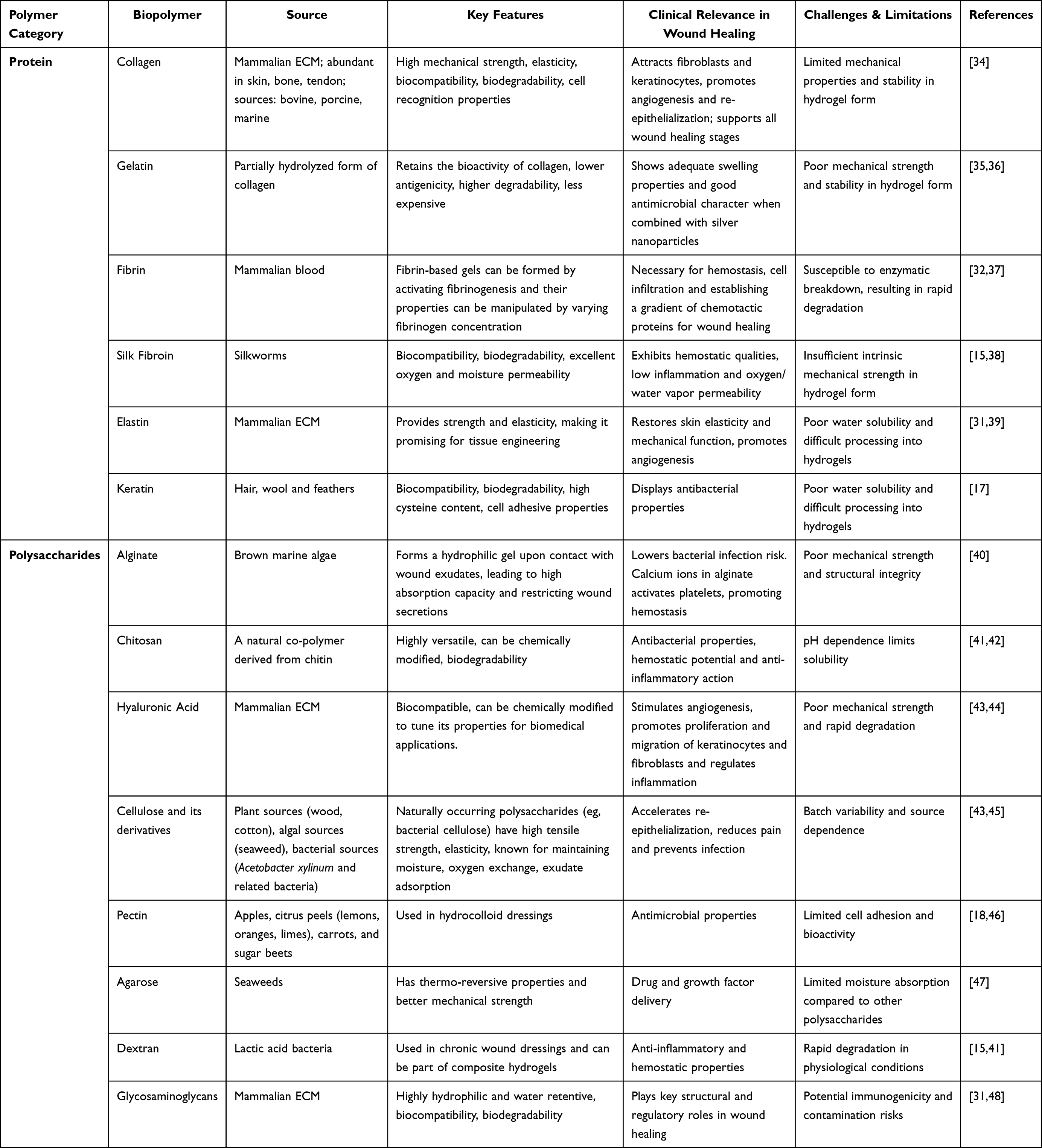 |
Table 1 Comparative Assessment of Biopolymers: Sources, Key Features, Clinical Implications, and Challenges in Wound Healing |
Nonetheless, they often have poor mechanical strength and stability, limiting their use in load-bearing applications or for preventing secondary damage. It is often challenging to control degradation rates.17 Natural biopolymer hydrogels exhibit high variability due to arduous isolation procedures from variable sources,31 and they may also exhibit issues like adherence, opacity, and lack of multifunctionality.15 They can also be prone to microbial contamination.49
Synthetic Polymer Hydrogels
Synthetic hydrogels are chemically modified in laboratories, offering controlled and reproducible properties.40 Commonly used synthetic polymers in hydrogels are enlisted in Table 2. Moreover, Figure 2 represents chemical structures of frequently utilized synthetic polymers in hydrogel synthesis. The chemistry and structure of synthetic hydrogels can be easily controlled, allowing for tailored properties, controlled degradation rates, and industrial-scale reproducibility. They often possess good physical strength, water retention ability, and exudate absorption capacity50 and can be developed with superior transparency.19
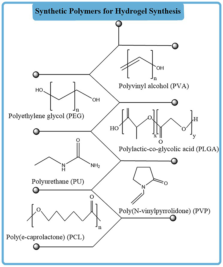 |
Figure 2 Chemical structures of commonly applied synthetic polymers in hydrogel formulation. |
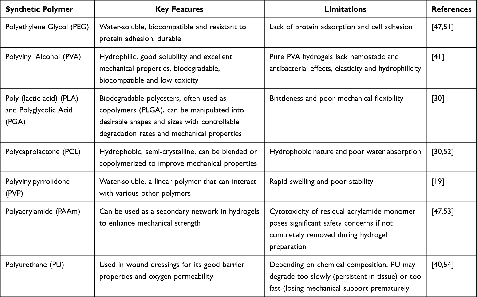 |
Table 2 Comparative Summary of Key Features and Limitations of Commonly Employed Synthetic Polymers in Hydrogels |
Nevertheless, they typically lack inherent cell adhesion sites and biological functions, often requiring chemical modifications. Their accumulation in tissues can potentially cause toxic effects or inflammatory reactions.25 Synthetic hydrogels may have structures very different from the ECM. Additionally, they can sometimes be more expensive to use.14
Role of Hydrogel Dressings in Wound Care
Hydrogel dressings play a critical role in modern wound management by providing an optimal environment for healing. They are designed to absorb wound exudates while maintaining a moist wound bed, which is essential for autolytic debridement and preventing scar formation. The porous structure allows for gas exchange, facilitating tissue breathing and promoting cell migration and epithelization. Many hydrogels offer a soothing or cooling effect and low adhesion to the wound tissue, minimizing pain and trauma during dressing changes.15
Beyond passive protection, advanced hydrogels are engineered to actively participate in the healing process. They can be formulated to deliver therapeutic agents by acting as carriers for antibiotics, growth factors, stem cells, anti-inflammatory agents, or natural compounds to accelerate and improve wound repair,43 manage exudates and infection by effectively removing excess exudates, protection against microbial invasion and inhibit bacterial growth,17 provide mechanical and bio-recognition support by mimicking the ECM, offering a scaffold for tissue regeneration and promoting cell adhesion, migration and proliferation,47 and smart hydrogels can be designed to respond to changes in the wound microenvironment (eg, pH and temperature) for on-demand drug release or to monitor healing progress.17
Key Properties of Biopolymer Hydrogels for Wound Care
Biopolymer hydrogels possess a range of key properties that make them highly advantageous for wound care, particularly in facilitating the complex process of wound healing.1 They are comprised of cross-linked polymer networks that form a three-dimensional structure. This structure allows them to swell and retain significant volumes of water or other aqueous fluids, often up to thousands of times their dry weight.1 This high water content is essential for preserving a moist wound environment, which promotes epithelialization, prevents scab formation, and ensures a consistent supply of growth factors and other molecules essential for healing. The visual inspection of the wounds is facilitated by the transparency of many hydrogels without removing the dressing.20
Biopolymers are naturally derived from sources such as plants, animals, or microbes. They are highly favored due to their inherent biocompatibility, low immunogenicity, and non-toxicity.55 Their degradation products are typically non-harmful and can be metabolized by the body.56 This minimizes adverse reactions and improves biocompatibility with various tissues.57 They are naturally degradable into non-toxic substances, eliminating the need for surgical removal after treatment.58 The degradation rate can often be tailored to match the rate of tissue regeneration.57
Another primary benefit of biopolymer-based hydrogels is their ability to mimic the native ECM, providing a supportive scaffold for cell adhesion, migration, proliferation, and differentiation. This accelerates the healing process, especially in chronic wounds where cell functions might be impaired.1 Biopolymer hydrogels can effectively absorb excess wound exudates while preventing dehydration of the wound bed. This helps to prevent tissue maceration and removes cell debris.20 They can be designed to be soft, flexible, and elastic, allowing them to conform to the contours of irregular-shaped wounds.57 This ensures better contact with the wound bed and maximizes therapeutic effects. Their mechanical strength can also be tailored to match the surrounding tissue, which is important for promoting integration and functionality.1
Biopolymer hydrogels are excellent carriers for encapsulating and delivering bioactive agents such as growth factors, antimicrobial agents, anti-inflammatory drugs, and cells. They can release these agents in a controlled and sustained manner, enhancing therapeutic outcomes and minimizing complications by mitigating infection, reducing inflammation, and stimulating angiogenesis.1 This also reduces the need for frequent dressing changes.57 Many biopolymer hydrogels possess intrinsic properties or can be loaded with agents that reduce inflammation. This is particularly beneficial for chronic wounds, which often exhibit prolonged inflammatory phases. Mechanisms include scavenging excessive ROS and promoting polarization of macrophages from pro-inflammatory (M1) to anti-inflammatory (M2) phenotypes.10 Some natural biopolymers, like chitosan, have inherent antibacterial, bacteriostatic, and anti-biofilm properties.56 Hydrogels can also be combined with antimicrobial agents or nanoparticles to prevent and treat wound infections, which are a major impediment to healing.4 Certain biopolymers, such as chitosan and alginate, are known for their hemostatic capabilities, promoting blood clotting to stop bleeding.1 They can also promote angiogenesis, which is crucial for delivering oxygen and nutrients to the wound site and accelerating tissue regeneration.58 Smart hydrogels can possess self-healing capabilities, meaning they can restore their structural and functional integrity after damage. This is due to reversible dynamic linkages within their network, making them more durable and effective for long-term applications.56 Hydrogels are soft and flexible, minimizing pain and discomfort during application and dressing changes.58 Sprayable forms offer convenient, quick, and large-scale application, particularly beneficial for irregular-shaped wounds like burns.12 They can modulate various biological functions that are important for tissue proliferation and differentiation.59 This is achieved by providing the necessary biophysical and biochemical conditions for optimal cellular behavior.1 Collectively, these biological and physicochemical attributes establish a foundational rationale for hydrogel-based wound dressings and highlight their subsequent functionalization with bioactives and nanoparticles, explored in the following segments.
Biocompatibility and Bioactivity of Biopolymer Hydrogels
Biopolymer hydrogels are increasingly recognized as optimal wound dressings due to their biocompatibility and bioactivity, which promote an accelerated and more efficient healing process.15 They are well tolerated by the body without eliciting adverse reactions, and their biodegradability allows them to naturally break down over time.17 Many natural polymers, such as chitosan, cellulose, alginate, and hyaluronic acid, are suitable for prolonged use,40 as confirmed by the cytocompatibility of various hydrogel composites in cell line studies such as NIH/3T3, HEK-293, and RAW264.7 cells.19,60 Their high elasticity and structure resemble the native ECM, providing a suitable environment for cell proliferation and tissue regeneration.17 However, their low adherence to tissue reduces discomfort and prevents secondary injury upon removal.60
Beyond being inert physical barriers, biopolymer hydrogels actively contribute to the wound healing process by maintaining a moist wound environment and promoting oxygen transfer to the tissue, thereby facilitating rapid healing and reduced scar formation. They can absorb excess wound exudates, restricting secretions and lowering the risk of bacterial infection. Many biopolymers, especially chitosan, possess intrinsic antibacterial, antiviral, and antifungal properties. A strong antimicrobial activity has been shown by chitosan-based hydrogels against common wound-infecting bacteria, ie, Staphylococcus aureus and Escherichia coli. This property is enhanced when combined with nanoparticles, such as silver or zinc oxide.40
Hydrogels promote cell migration, proliferation and epithelialization.41 Collagen and gelatin hydrogels provide structural support, stimulate growth factor release, and promote collagen deposition for tissue remodeling, while hyaluronic acid enhances angiogenesis and tissue regeneration.26 Moreover, hydrogels can incorporate various bioactive agents such as growth factors (eg, EGF, bFGF, and VEGF), stem cells, and platelet-rich plasma to further enhance healing by stimulating cell proliferation, differentiation, and neovascularization.21 Hydrogels containing active compounds like curcumin, α-lipoic acid, exhibit inflammatory and antioxidant properties, which are vital for chronic wound healing, especially in diabetic patients.41
Hemostatic activity represents another important biofunctional attribute of biopolymer hydrogels. Alginate dressings, derived from brown seaweed, are particularly effective in blood clotting due to their calcium component, while hydrogels incorporating components like cellulose nanofibers and gelatin also demonstrate good hemostatic properties.61 Additionally, hydrogels are excellent drug carriers, allowing for the controlled and sustained release of therapeutic agents, such as antibiotics, growth factors, or anti-inflammatory drugs, directly to the wound site. For example, a hydrogel containing curcumin encapsulated in micelles has been shown to improve cutaneous wound healing.62 Some advanced hydrogels are designed with self-healing capabilities, enabling them to repair minor damage to the dressing and maintain wound protection.41 Some hydrogels are designed to be pH-responsive, which can be beneficial as pH changes are often indicative of wound infection or progression.14 Overall, the biocompatibility, non-toxicity, and diverse bioactive properties of biopolymer hydrogels make them highly promising materials for advanced wound care and treatment.50
Natural Products in Wound Healing: A Bioactive Resource
Overview of Natural Products with Wound Healing Potential
Natural products from diverse sources, including plants, animals, and fungi, hold significant potential for wound healing.63 These products have been utilized for centuries in traditional medicine due to their inherent bioactive properties.64 The process of wound healing is complex, involving distinct, yet overlapping, phases: hemostasis, inflammation, proliferation, and remodeling.65 Natural products contribute to various aspects of this process, with their effects often being dose-dependent, related to the nature of wounded tissue and application time.62 Natural products with wound-healing potential are listed in Table 3.
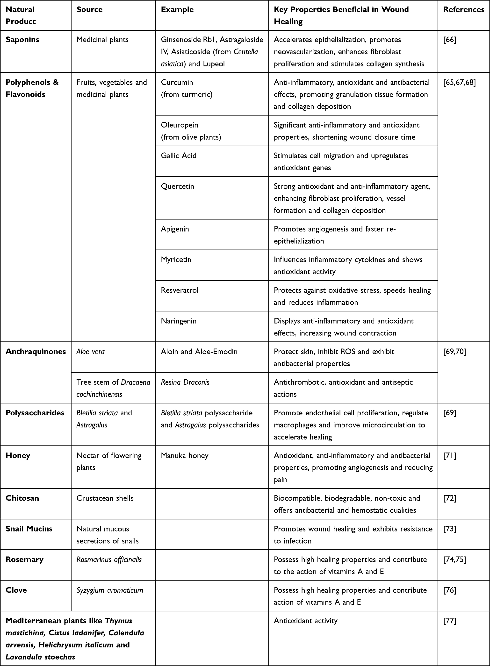 |
Table 3 Natural Products and Their Key Properties Presenting to Wound Healing |
The use of natural products in wound care offers several advantages, including ease of access, relatively low cost, and generally limited side effects compared to synthetic drugs. However, there are limitations such as poor water solubility, leading to issues with stability and bioavailability (eg, myricetin, curcumin, quercetin, and gallic acid).65 Some natural products also show toxicity at higher concentrations or under specific conditions (eg, aloe-emodin, comfrey extract, certain honey types, and high concentrations of Ganoderma lucidum polysaccharide).64
Recent advancements in nanotechnology are addressing many of these limitations. Nanomaterials, such as electrospun nanofibers, green-synthesized metal nanoparticles (eg, AgNPs, gold, copper oxide, and titanium dioxide), nanoemulsions, nanohydrogels, and nanoliposomes, can prominently increase the efficacy, bioavailability, stability, controlled release, and permeability of natural compounds at the wound site. These advanced delivery systems, including composite dressings and microneedles, are proving promising in improving wound healing, even for chronic wounds.62 Multiple fabrication strategies for acquiring the plant-based natural product-derived nanoparticles have been reported from international research groups. In 2025, a study was conducted in South Asia that integrated the green synthesis techniques for designing the silver nanoparticles and embedded them into a hydrogel matrix, thereby acquiring the effective wound healing characteristics.78 Similarly, the significance of plant-derived nanoparticles in wound recovery was acknowledged by South African researchers.79 An East Asia study also highlighted the therapeutic effectiveness of nanocomposite hydrogels via incorporating the bioactive-loaded nanoparticles into hydrogel matrices.80 Collectively, international studies underscore the global relevance and therapeutic significance of natural bioactive nanoparticles in hydrogel.
Future research should focus on standardizing protocols for measuring and evaluating wound healing to enable better comparison of different natural products.65 Further pharmacological experiments and well-designed clinical trials are necessary to fully confirm the safety and efficacy of these natural product-based formulations in clinical practice.62,64
Mechanisms of Action of Natural Products in Wound Healing
Natural products contribute to wound healing through a multifaceted array of mechanisms, targeting the complex and dynamic phases.69 Figure 3 illustrates the sequential phases of wound recovery. These botanical and animal-derived agents, often perceived to cause minimal unwanted side effects and be affordable, induce healing and tissue regeneration via multiple interconnected processes.63,69 One primary mechanism involves anti-inflammatory activity, where natural products reduce the duration of the inflammatory phase, which, if prolonged, can hinder healing and increase pain. They achieve this by decreasing pro-inflammatory factors such as interleukin-1β (IL-1β), IL-6, tumor necrosis factor-alpha (TNF-α), monocyte chemoattractant protein-1 (MCP-1), interferon-gamma (INFγ), nitric oxide, and prostaglandin E2 (PGE2), while simultaneously increasing anti-inflammatory factors like IL-10.63 They also modulate macrophage transition from pro-inflammatory (M1) to anti-inflammatory (M2) phenotypes and inhibit key signaling pathways like NF-κB and AP-1, blocking lipoxygenase activity and leukotriene B4 production and reducing inflammatory cell infiltration.
 |
Figure 3 The stages of wound healing illustrate the molecular-level activities involved in repairing the damaged tissue. [1] In hemostasis, immediately after injury platelet aggregation facilitate to stop bleeding, [2] immune cells (neutrophils and macrophages), infiltrate the wound site to remove debris and pathogens, while epithelial cells begin to migrate beneath the scab, [3] in proliferation, fibroblasts proliferate and deposit extracellular matrix, forming granulation tissue, and [4] during remodeling, regenerated epidermis and formation of scar tissue help to restore structural integrity. |
Antimicrobial activity is another critical mechanism, as infections significantly impede wound healing. Natural products reduce bacterial load and prevent infection by inhibiting vital bacterial processes such as dihydrofolate enzymes, nucleic acid synthesis, the ADP/ATP cycle, and efflux pumps.64 They can also disrupt bacterial cell membranes, suppress protein synthesis, alter sulfhydryl groups (eg, allicin) and create unfavorable environments like low pH or high sugar concentrations (eg, honey).64,65 Honey also produces antimicrobial compounds such as hydrogen peroxide and methylglyoxal (Manuka honey).64,74
Antioxidant activity is crucial for wound healing, as ROS must be balanced to prevent excessive inflammation and impaired healing.65 Natural products maintain this redox balance by promoting Nrf2 pathways, inhibiting MAPK/NF-kB and NADPH oxidase activity, and increasing antioxidant enzymes such as superoxide dismutase (SOD), catalase (CAT), glutathione peroxidase (GPx), and heme oxygenase-1 (HO-1);64 they also inhibit lipid peroxidation.81
In the proliferative phase, natural products enhance tissue formation and remodeling by increasing collagen content and synthesis, improving its cross-linking and deposition, and boosting the wound’s tensile strength.69 They also promote wound contraction, reducing wound size, and can reduce scar formation and hyperplasia while promoting softening and absorption of existing scars. Furthermore, they regulate matrix metalloproteinases and tissue inhibitors of metalloproteinases, which are crucial for ECM degradation and remodeling and increase hydroxyproline content, a marker of collagen production.82
Angiogenesis and neovascularization, the development of new blood vessels, are vital for granulation tissue to receive adequate nutrients and oxygen.69,81 Natural products stimulate angiogenesis by promoting endothelial cell migration and increasing the expression of key growth factors such as vascular endothelial growth factor (VEGF), fibroblast growth factor (FGF), and platelet-derived growth factor (PDGF).82 They also activate cellular pathways like ERK/CREB, mTOR, and HIF-1.74
Natural products also exert effects through the modulation of cellular activity, specifically influencing fibroblasts and keratinocytes, which are essential for tissue repair. They promote cell proliferation and enhance cell migration to the wound site. Some natural products also possess analgesic properties, which can improve patient comfort and aid in recovery.82 All these biological mechanisms highlight the influence of natural bioactives and nanoparticles in hydrogel matrices to support wound recovery, as discussed in subsequent sections. Botanical sources that are providing these aforementioned attributes to facilitate wound healing majorly include Azadirachta indica, Aloe vera, Chamomilla recutita, Achillea millefolium, Centella asiatica, Polygonum cuspidatum, Angelica sinensis, Helianthus annuus, Rosmarinus officinalis, curcumin, resveratrol, quercetin, tannins, honey, and bilirubin.63–65,69,82 Figure 4 presented the general mechanisms to treat inflammation and wound closure via natural product-mediated biological response.
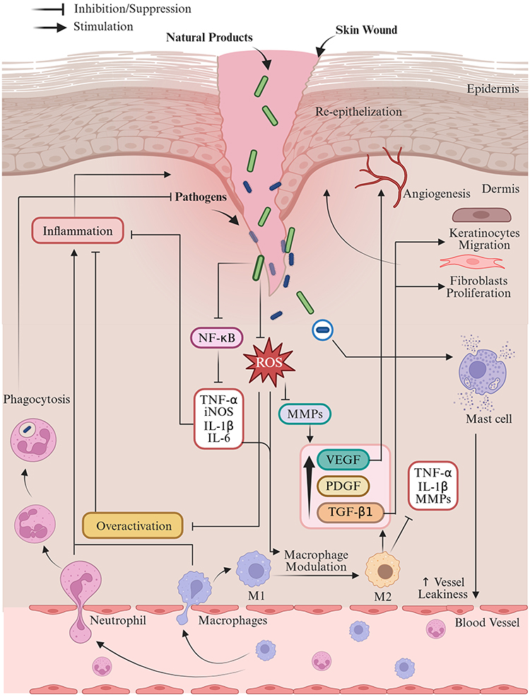 |
Figure 4 General mechanistic illustration of natural product-mediated regulation of wound healing. Wounds exhibit persistent pathogen entry, excessive inflammation, and oxidative stress, which lead to impaired tissue repair. Moreover, pathogens enhance the production of reactive oxygen species (ROS), pro-inflammatory cytokines (TNF-α, IL-1β, IL-6), and matrix metalloproteinases, followed by extracellular matrix degradation and vascular leakage (release neutrophils and macrophages). However, natural compounds facilitate the suppression of NF-κB pathway signaling and counter the oxidative stress via reducing the ROS. This action assists in inhibiting pro-inflammatory mediators and macrophage modulation (M1 phenotype to the reparative M2 phenotype), enhancing the release of growth factors such as VEGF, PDGF, and TGF-β1. Consequently, re-epithelialization and wound closure are supported by improved angiogenesis, extracellular matrix preservation, fibroblast proliferation, and keratinocyte migration. Abbreviation: ROS, Reactive oxygen species, NF-κB, Nuclear factor kappa B cells, TNF-α, Tumor necrosis factor alpha, IL-1β, Interleukin-1 beta, IL-6, Interleukin-6, iNOS, Inducible nitric oxide synthase, MMPs, Matrix metalloproteinases, VEGF, Vascular endothelial growth factor, PDGF, Platelet-derived growth factor, TGF-β1, Transforming growth factor beta 1, M1, Classically activated (pro-inflammatory) macrophages, M2, Alternatively activated (anti-inflammatory/reparative) macrophages. |
Nanotechnology further enhances the efficacy of natural products for wound healing by improving their bioavailability, enabling controlled and sustained release of active compounds, and increasing their permeability into deeper skin layers. This approach, utilizing nanomaterials in hydrogels, nanofibers, nanoemulsions, and nanoliposomes, allows for a multi-phase directed tissue regeneration that precisely targets different stages of wound healing for better therapeutic outcomes, particularly for chronic wounds.62
Selection Criteria for Natural Bioactives in Hydrogel Functionalization
The selection criteria for natural bioactive compounds used in hydrogel functionalization for wound healing focus on their proven biological activities, safety, and compatibility with advanced delivery systems. A primary consideration is the mechanism of action of these natural products, as they need to effectively target the complex, interlinked phases of wound healing.74 Compounds are chosen for their anti-inflammatory, antimicrobial, and antioxidant properties, which are crucial for managing the initial stages of wound repair and preventing complications.62,63,74 For instance, natural products can reduce pro-inflammatory factors like IL-1β, IL-6, and TNF-α, while increasing anti-inflammatory factors such as IL-10. Their antimicrobial activity helps prevent infection by disrupting bacterial processes, and antioxidant effects maintain redox balance by boosting enzymes like SOD and CAT.62,74
Beyond these foundational bioactivities, selected natural products should also promote tissue regeneration during the proliferative and remodeling phases, which includes enhancing cellular proliferation, collagen synthesis, and angiogenesis. This involves stimulating growth factors like VEGF, FGF, and PDGF and improving collagen deposition and wound contraction, while ideally reducing scar formation.64,74 Safety and minimal side effects are significant criteria, as natural products are often preferred for their perceived affordability and reduced adverse reactions compared to synthetic drugs.63 However, scientific standardization, validation, and systematic safety evaluation are necessary, as some natural compounds can exhibit toxicity at certain concentrations (eg, aloe-emodin, Ganoderma lucidum extracts, honey extract, and comfrey extract).69 This necessitates careful determination of the right dosage and timely use.74
The physical and chemical characteristics of natural compounds are also critical, especially when integrating them into hydrogel systems. Many beneficial natural products suffer from poor water solubility (eg, myricetin, quercetin, gallic acid, curcumin, and thymol), which limits their bioavailability and topical application.63,64 Nanotechnology is a key enabler in this regard, as it can enhance bioavailability, enable controlled and sustained release, and increase permeability into deeper skin layers, thereby overcoming these limitations and allowing for a multi-phase directed tissue regeneration approach. The choice of encapsulation method within hydrogels (eg, emulsion/gelation, conjugation, and soaking) depends on the specific chemical interactions possible (eg, ionic bonding, hydrogen bonding, and hydrophobic interactions). Furthermore, research emphasizes selecting compounds or extracts where their mechanisms of action are well understood to facilitate matching them to specific wound healing phases. The potential for synergistic effects when combining multiple natural bioactives in a single hydrogel system is also an important consideration for achieving comprehensive wound healing outcomes.74
Nanoparticles Derived from Natural Products
Types of Natural Product-Based Nanoparticles
Natural product-based nanomedicines represent a significant advancement in wound healing, offering a promising approach to overcome limitations of traditional therapies by enhancing drug delivery, bioavailability, and stability of active compounds, particularly those with poor water solubility.11,62,83 These nanostructures enable targeted and controlled release of therapeutic agents to the wound site, thereby minimizing systemic side effects.7 Various forms of natural product-based nanoparticles are employed, often fabricated through advanced techniques like electrospinning.11 Figure 5 shows some of the major applications of nanoparticles in wound healing.
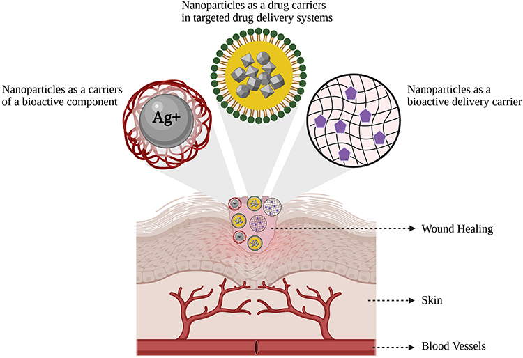 |
Figure 5 Demonstrating the contribution of nanoparticles as a drug carrier in effective wound healing. |
One prominent category is polymeric nanoparticles, which serve as nanocarriers composed of natural polymers such as chitosan and alginates or synthetic polymers like poly(lactic-co-glycolic acid). These systems are designed to encapsulate drugs and offer advantages such as controlled drug release, non-toxicity, and biocompatibility with target tissues. For example, polymeric nanoparticles encapsulating enoxaparin have shown improvements in wound healing and skin penetration. Similarly, those loaded with host defense peptides (LL37) have modulated inflammatory responses and promoted granulation tissue formation, re-epithelialization, and collagen content. Antifungal agents like amphotericin B, when encapsulated in silane-hydrogel nanoparticles, demonstrated high killing efficiency against Candida spp. and inhibited fungal biofilm formation, accelerating wound healing.11 Figure 6 denotes the mechanisms by which polymeric nanoparticles perform controlled and sustained drug release.
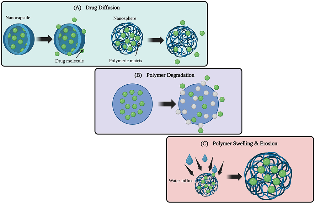 |
Figure 6 Usually performed drug release mechanisms by polymeric nanoparticles. (A) In drug diffusion, bioactive molecules tend to diffuse into the surrounding medium from the polymeric network, (B) showing the polymeric degradation that assist to release drug content, and (C) water influx in the polymeric network helps to swell the network, which directly facilitates its erosion and release of drug molecules. |
Lipid-based nanostructures constitute another major class, including nanoemulsions, liposomes, and solid lipid nanoparticles (SLNs), and micelles. Nanoemulsions are capable of nano-sizing essential oils, which, upon application, form a film that facilitates the penetration of lipophilic drugs into deeper skin layers, thereby improving their solubility.62 Noteworthy examples include nanoemulsions formulated with licorice and lavender essential oils, which promote wound closure and epithelialization, and curcumin nanoemulsions, which exhibit anti-inflammatory activity and healing effects comparable to fusidic acid. Liposomes, formed from phospholipid bilayers, have been utilized to encapsulate daptomycin for antibacterial action against biofilms and quercetin and curcumin nano-vesicles for inhibiting ROS and reducing edema, while stimulating fibroblast proliferation. Curcumin-loaded liposomes have also demonstrated an improved rate of wound closure.11 Solid lipid nanoparticles and nanostructured lipid carriers (NLCs) are also lipid-based systems;83 for instance, morphine-loaded lipid nanoparticles provide high drug loading capacity and sustained release, promoting re-epithelialization.11 Micelles, composed of phospholipid unilayers,83 specifically polymeric micelles loaded with curcumin, have shown anti-diabetic and wound-healing benefits in diabetic rats by reducing blood glucose levels.11
Natural products can be incorporated to provide intrinsic therapeutic properties for sustained release.74 Examples include cyclodextrin–eugenol hydrogels, which offer antimicrobial, anti-inflammatory, and angiogenesis-promoting effects, and sacran hydrogels, known for their moisturizing abilities, anti-inflammatory activity, and acceleration of wound closure.11,83 Hydrogels loaded with compounds like berberine or betaine have demonstrated reduced inflammation and oxidative stress, alongside accelerated re-epithelialization.74
Another distinct type involves green-synthesized metal nanoparticles, where plant extracts are used to synthesize nanoparticles, which are then incorporated into wound dressings. These plant-mediated biosynthesized nanoparticles, such as silver, gold, and copper oxide nanoparticles, exhibit potent antibacterial and anti-inflammatory activities. They also contribute to accelerated wound healing by enhancing cell proliferation, connective tissue formation, and re-epithelialization. For instance, silver nanoparticles synthesized using Momordica charantia extract showed enhanced antimicrobial activity and reduced cytotoxicity against fibroblasts.62
Finally, electrospun nanofibers represent a significant delivery platform that often integrates the above-mentioned nanoparticle types.7 These nanoscale fine fibers, frequently derived from plant-based materials like cellulose, alginate, and plant proteins, mimic the ECM, providing an ideal scaffold for cell adhesion, migration, and proliferation.11 Electrospinning enables the direct encapsulation of active natural products, including flavonoids, polyphenols, and various plant extracts, allowing for precise control over their release and effectively addressing challenges such as poor water solubility and instability. These versatile nanofiber systems are transforming modern wound care by offering enhanced therapeutic outcomes through their unique physical and chemical properties.7 All these studies from multiple regions illustrate the global significance of nanocomposite hydrogel matrices that not only exhibit the versatile biological responses but also support their clinical potential in advanced wound healing.
From a comparative perspective, the strong anti-inflammatory and antibacterial behavior has been exhibited by metal nanoparticles, which is mandatory to control microbial load in case of chronic wounds.84 However, in order to get controlled drug release behavior, polymeric nanoparticles are more suitable, thereby prolonging the therapeutic effects.85 In lipid-based nanocarriers, better encapsulation and penetration abilities are achieved, while electrospun nanofibers provide targeted delivery along with structural support.86 Overall, this comparison suggests the selection of nanoparticles according to pathological condition and wound type to acquire effective responses.
Antibacterial, Anti-Inflammatory, and Healing Properties of Natural Nanoparticles
Antibacterial Properties
Many natural product-based nanomaterials exhibit unique antimicrobial properties, which are crucial for controlling infection and preventing biofilm formation, common challenges in wound healing.11,62,87–89 Bacteria often produce extracellular polymers that act as a shield, forming biofilms that inhibit epithelial cell growth and protect colonized bacteria from antimicrobial therapies.11 Natural nanoparticles counteract this by mechanisms such as reactive oxygen species (ROS) generation and membrane disruption.83,87
Specifically, AgNPs are widely studied for their potent antibacterial activity, especially when green-synthesized using plant extracts.88 AgNPs prepared from Momordica charantia fruit extract, which showed enhanced antimicrobial activity and reduced cytotoxicity, and those from Piper nigrum leaf extracts, which inhibited bacterial colonization.62 Curcumin-loaded nanoemulsions and hydrogels have also demonstrated significant antimicrobial effects.11 Beyond metals, certain plant-derived essential oils like tea tree oil, lavender oil, and cinnamon essential oil exhibit strong antimicrobial properties.7 Honey, a traditional medicinal product, prevents bacterial biofilm formation due to its acidic nature and peroxidases,11,63 while chitosan, a polysaccharide, possesses natural antibacterial properties and can be incorporated into nanofibers to enhance antimicrobial activity.90
Anti-Inflammatory Properties
Inflammatory diseases are characterized by dysregulated inflammatory reactions and oxidative stress, which traditional therapies often fail to address without adverse effects. Natural product-based nanomedicines offer a solution by possessing anti-inflammatory and antioxidant properties.83 They achieve this by suppressing the production of pro-inflammatory mediators like interleukin (IL)-1β, IL-6, tumor necrosis factor (TNF)-α, and nitric oxide (NO), while enhancing anti-inflammatory cytokines such as IL-10.62
Curcumin-based nanostructures are notably potent in modulating the inflammatory phase, regulating levels of TNF-α, IL-10, and transforming growth factor (TGF)-β1.62 Resveratrol-loaded nanoparticles have been shown to suppress inflammatory responses by decreasing pro-inflammatory cytokines and increasing anti-inflammatory cytokines, promoting macrophage differentiation towards an M2 (anti-inflammatory) phenotype.83 Quercetin, a flavonoid, also demonstrates anti-inflammatory effects by inhibiting edema formation.11 Other flavonoids like naringenin, astragaloside IV, chrysin, and puerarin are integrated into nanofibers to reduce inflammation and oxidative stress. Essential oils encapsulated in nanofiber dressings provide anti-inflammatory benefits, as seen with peppermint extract in polyurethane nanofibers for diabetic wounds.7
Healing Properties
Beyond managing infection and inflammation, natural product-based nanomedicines actively promote various aspects of the wound healing cascade. They are designed to cure wounds without affecting normal skin function or causing scar formation, while speeding up the healing mechanism and keeping the wound moisturized.11
These nanomedicines enhance the growth of fibroblasts and keratinocytes, which are vital for tissue repair and re-epithelialization.11 For example, asiaticoside from Centella asiatica extract accelerates tissue regeneration by promoting skin fibroblast proliferation.7 They promote the formation of new connective tissues and blood vessels (angiogenesis or vascularization).89 Recombinant human EGF (rhEGF)-loaded lipid nanoparticles, for instance, enhance wound closure and stimulate new connective tissues and blood vessels.11 Similarly, chitosan and collagen are recognized for providing mechanical support and acting as excellent healing scaffolds.90 Techniques like electrospinning, which produces nanofiber dressings mimicking the extracellular matrix, support cell adhesion, migration, and proliferation, leading to accelerated wound healing with reduced scar formation.7 Nanotechnology significantly improves the delivery of natural compounds by increasing their solubility, stability, and permeability to deeper skin layers.11 This controlled release ensures that the therapeutic agents are available at the wound site over a sustained period, maximizing efficacy and minimizing systemic side effects.62
Many plant-based nano-formulations, such as those incorporating curcumin, fenugreek, and tragacanth gum, have shown remarkable activity in promoting collagen synthesis and fibroblast proliferation, leading to accelerated re-epithelialization.62 Aloe vera-loaded nanofiber scaffolds have demonstrated good cell compatibility and significant antibacterial activity, making them effective for wound healing.82 The combination of nanomaterials with traditional products like honey also enhances their efficiency for targeted delivery and controlled release.11
Overall, the integration of natural products with nanotechnology presents a powerful approach, offering multi-functional therapeutic systems that can address various wound healing challenges simultaneously, making them the future of pharmaceuticals in wound care and management.7,11,74
Functionalization of Biopolymer Hydrogels with Natural Product Nanoparticles
The association of biocompatibility and biodegradability attributes with biopolymers makes them an enticing option for hydrogel fabrication.91 A versatile biopolymer, ie, bacterial cellulose,61 chitosan,92 guar gum,93 alginate,94 gelatin, and others, is incorporated effectively for hydrogel formation to expedite wound dressings.95 For wound dressings, hydrogels are considered to be the auspicious contender because they facilitate the wound’s healing by sustaining the moist microclimate at the injury site.96 Additionally, natural product-loaded nanoparticles along with biopolymeric hydrogels develop hydrogel-nanoparticle composites that accelerate the wound healing potential of hydrogels. These composites not only assist the healing but also contribute to delivering the anti-inflammatory and antimicrobial activity to manage the bioburden and phases of the healing cascade at the wound site that directly influence the healing duration.93,97 This segment inquires about the approaches/strategies engaged to consolidate such nanoparticles into biopolymer hydrogels, drawing attention to their synergistic contribution in foremost wound healing applications.
Strategies for Incorporating Natural Nanoparticles into Hydrogel Matrices
In order to potentiate the wound healing efficiency of hydrogels, natural product-based nanoparticles are engaged into the hydrogels. Consequently, to achieve this, different strategies/approaches that assist in acquiring the suitable nanoparticles (NPs)-impregnated hydrogel-based wound dressings are employed, as demonstrated in Table 4. Additionally, Figure 7 illustrates the hydrogel functionalization via NPs and presents the wound healing.
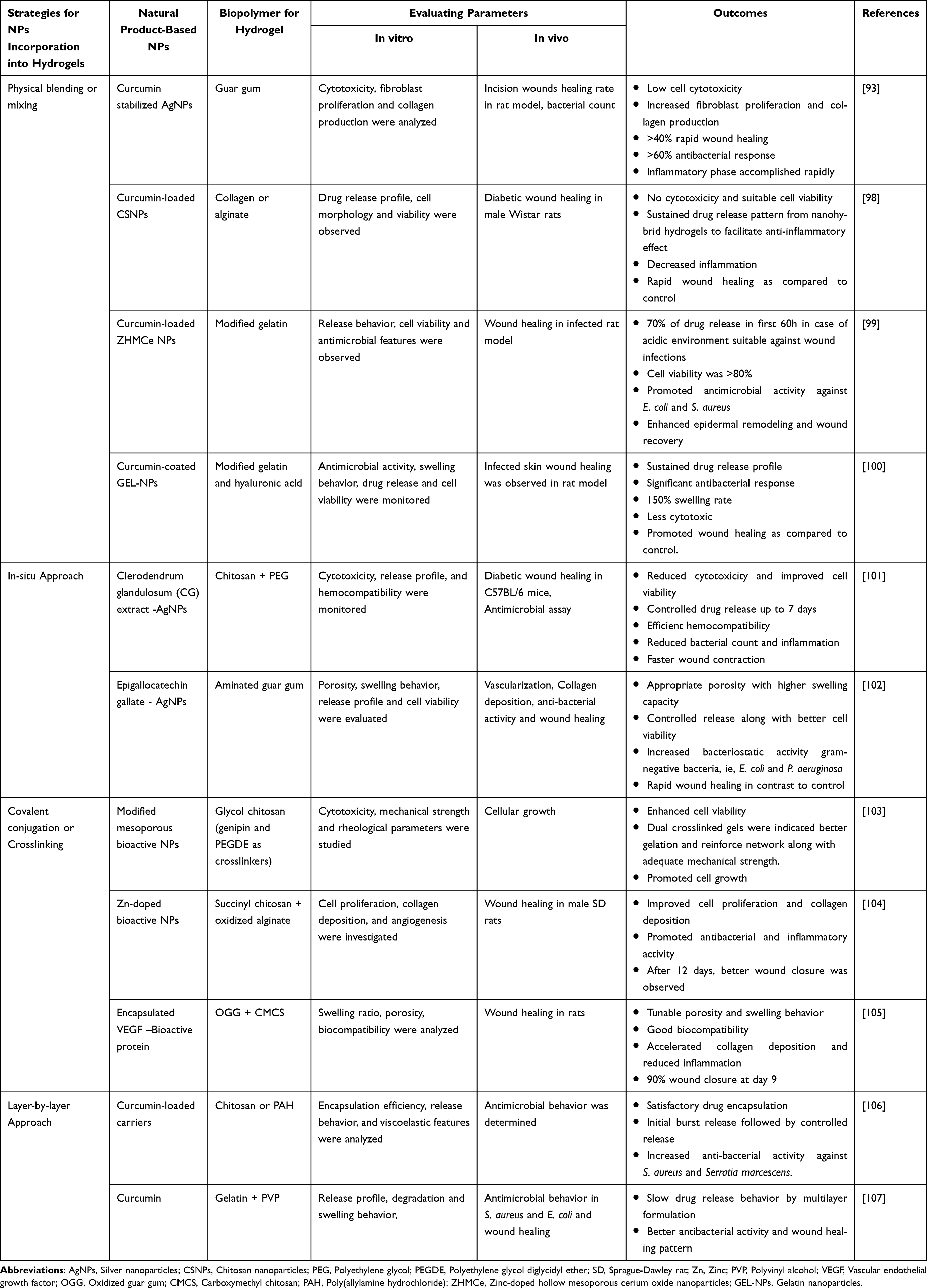 |
Table 4 Commonly Employed Strategies for Hydrogel Functionalization with Natural Product-Based Nanoparticles |
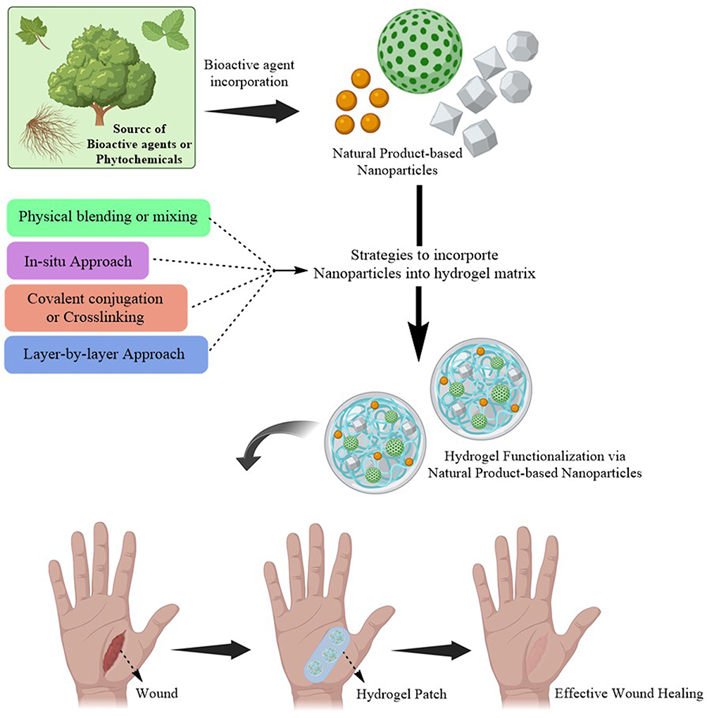 |
Figure 7 Functionalization of hydrogels via natural product-based nanoparticles to facilitate efficient wounds healing. Strategies such as physical blending, in-situ technique, covalent conjugation and layer-by-layer phenomena, effectively support the natural product-based nanoparticles’ integration into hydrogels matrix. |
The most frequently operated and straightforward strategy is the physical blending or mixing, in which NPs are directly dispersed into the premeditated hydrogel solution via non-covalent interactions; to avail uniform dispersion, pertinent stirring is performed.108 Respectively, physical mixing preserves the functionality of NPs and no chemical reactions are involved. Karri et al conducted a study98 in which researchers designed the curcumin-loaded NPs along with collagen-alginate blends separately via the freeze-drying method. Subsequently, NPs were combined with collagen-alginate blends directly by overnight stirring; as a result, nanohybrid hydrogel scaffolds were prepared. After evaluation, it was observed that the designed formulation delivers high biocompatibility, enhanced stability, and a sustained release profile. Moreover, in animal studies, it was monitored that the nanohybrid hydrogel scaffolds have a significant potential for rapid and efficient diabetic wound healing, in contrast to controlled and placebo groups. Collectively, the physical blending of NPs into the hydrogel is a promising technique to facilitate effective wound healing.98 A scientific report was contributed by S. Bhubhanil et al,93 in which curcumin-loaded silver nanoparticles (Cu-AgNPs) were prepared and via physical blending, loaded into the guar gum hydrogel solution, followed by physical crosslinking. It was analyzed that nano-hydrogel composites facilitate wound healing by enhancing fibroblast formation along with antibacterial activity. This finding clarifies the effectiveness of this approach for nanoparticle impregnation in hydrogels for satisfactory wound dressings.
The functionalization of natural product-based NPs in biopolymer hydrogels via the in-situ method proposes conspicuous benefits, including uniform NP dispersion and enhanced stability; moreover, pre-designed nanocarriers are not required. In this method, NP formation occurs directly inside the hydrogel precursor solution by utilizing metal salt precursors, ie, silver nitrate, and reducing agents,109 ie, ascorbic acid, sodium borohydride, sodium citrate, and plant extract, can also be used. Reducing agents assist the nucleation of metal ions and grow them into NPs inside the hydrogel matrix.110 A study was performed by A. Majie et al,101 in which Clerodendrum glandulosum (CG) plant extract was employed to synthesize the silver nanoparticles (AgNPs) into a hydrogel matrix, composed of chitosan and polyethylene glycol (PEG). Consequently, it was analyzed that the CG-AgNPs-loaded chitosan–PEG hydrogel exhibited the controlled release and delivered the significant and rapid diabetic wound treatment along with antioxidant, antimicrobial, and anti-inflammatory features. The importance of the in-situ method for NP impregnation into the hydrogel matrix was also reported recently in another study that showed its value for wound healing along with antimicrobial behavior. Usually, green synthesis was preferred to design the metal nanoparticles because of its safety profile. In this study,111 in-situ photochemical synthesis technique was incorporated to synthesize the AgNPs into hydrogels. A remarkable increase in antimicrobial activity with significantly enhanced wound healing was presenting the potential of NP functionalization in hydrogels via an in-situ strategy. Sometimes, the in-situ technique engages the development of NP inside the biopolymeric solution rather than the hydrogel itself. An aminated guar gum biopolymeric solution was employed along with AgNO3 solution to design AgNPs because it acts as a stabilizer and reducing agent. Subsequently, this mixture was mixed with sodium alginate and gelatin and accordingly, crosslinked via glycerol and calcium chloride, followed by gelation to develop hydrogel patches. In vitro and in vivo studies endorsed that the designed hydrogel patches have an adequate potential for wound healing via increased vascularization and collagen deposition.102
Hydrogel-nanoparticle composites have developed as a potential podium for controlled release with enhanced drug stability for wound healing, thereby also serving as a dual drug delivery system.112 Besides the simplicity and biocompatibility of physical blending, the covalent conjugation, another method for NP incorporation into hydrogels, delivers an efficient drug release profile along with improved structural stability. In this approach, NPs established the chemical interactions with the hydrogel matrix.113 Furthermore, NPs become an integral part of the hydrogel matrix because of covalent conjugation, a primary benefit of this technique that ensures a consistent therapeutic response, while in physical mixing, NPs may leach out from the matrix.114 However, byproduct purification and judicious chemical reaction optimization are required.115 A study was accomplished by Q. Min et al,103 in which a team of researchers designed surface-modified mesoporous bioactive NPs; for modification, amino-containing compounds were employed with different spacer lengths to generate amino-functionalized bioactive NPs. Afterwards, crosslinkers like genipin alone or a combination of genipin and poly(ethylene glycol) diglycidyl ether were utilized, and surface-modified NPs were incorporated with glycol chitosan to conceive hydrogels. Moreover, J. Zhu et al104 designed a hydrogel composed of succinyl chitosan and oxidized alginate. Then, zinc-doped bioactive glass NPs were inserted into the composite hydrogel. The Schiff-base covalent conjugation assists in stabilizing NPs inside the composite hydrogel matrix. Schiff-based interactions help to generate a suitable environment for cell proliferation at the injury site. Additionally, various attributes, including collagen deposition, myofibril buildup, granulation tissue formation, and anti-inflammatory factors, have been scrutinized to evaluate the potential of Schiff-based composite hydrogels for wound dressings.104 Apart from the advantages of covalent conjugation, few notable limitations transform the nanocomposites’ therapeutic effectiveness. Primarily, the bioactivity of fragile natural compounds gets compromised because of harsh reaction conditions, ie, reactive crosslinking agents and extreme pH, which reduce structural integrity and modify functional groups required for bioactivity.116 Moreover, the permanent nature of covalent bonds can restrict diffusion-based and stimuli-responsive release, limiting the ability of hydrogel systems to dynamically respond to changes in the wound microenvironment.117
Layer-by-layer assembly, or multilayer hydrogels, is a bottom-up fabrication approach that involves the alternate deposition of materials to develop multilayered structures,118 another cost-effective, versatile, and precise method for NP incorporation in the hydrogel matrix. The nanoparticle hydrogel composite via the layer-by-layer assembly technique helps to set up the thin layers of polymers and nanoparticles one after the other. The oppositely charged species and electrostatic interactions, ie, hydrogen and covalent bonding, can be employed to adhere these layers together.119 A straightforward preparation procedure, high biocompatibility, and tunable morphological aspects of multilayer hydrogels via layer-by-layer assembly provide the controlled drug release from the matrix.120 Different deposition procedures, including dip coating,121 spray deposition,122 and spin coating,123 can be utilized for multilayer fabrication. E. Tamahkar et al120 developed a multilayer wound dressing combination of synthetic and biopolymers that were utilized to develop a multilayer structure of hydrogel. The biopolymer-based middle layer was loaded with drug molecules, and the lower layer served as a controlling membrane for drug release. The resulting hydrogel-based wound dressing delivered sustained drug release for over 7 days and exhibited remarkable therapeutic activity.
In one more study,106 a layer-by-layer approach was utilized to develop curcumin-loaded microcarrier multilayer hydrogel structures. The internal alginate cores and outer chitosan or poly(allyl amine hydrochloride) coatings delivered sustained drug release and enhanced pharmacological activity. This study demonstrates how natural product-based NPs can be embedded efficiently into hydrogel matrices via layer-by-layer. Nano-hydrogel multilayered wound dressings were designed by using the electrospinning technique by S-M Huang et al107 Polyvinyl alcohol and gelatin were used to design a multilayer structure. The middle gelatin layer served as a reservoir for curcumin-loaded nanoparticles, while outer hydrogel layers of polyvinyl alcohol loaded with gentamicin delivered controlled drug release and exudate absorption. Overall, it was observed that the multilayer hydrogel membrane development via the layer-by-layer strategy aids in supporting wound healing along with the prevention of microbial infections. Similarly, the layer-by-layer strategy is utilized efficiently for the functionalization of biopolymer hydrogels with natural product-based nanoparticles.
All aforementioned strategies are used for hydrogel functionalization via nanoparticles; however, each has its own benefits and drawbacks, which facilitates the choice of a suitable approach for better consequences. Physical blending or entrapment offers operational simplicity and compatibility for sensitive bioactives along with minimal chemical modifications. But leaching of nanoparticles reduces their retention within the matrix and leads to an inconsistent release profile. While in covalent conjugation, chemical crosslinking encourages the retention of nanoparticles in the hydrogel matrix due to chemical interaction, but harsh reaction conditions are not suitable for bioactive agents; subsequently, the compatibility of bioactives gets compromised.112 However, hybrid systems that integrate multiple strategies can help balance these trade-offs, leveraging the simplicity of physical entrapment with the stability of covalent bonds to tailor release kinetics and functional outcomes for diverse wound healing applications.124
Synergistic Effects of Nanoparticles and Natural Bioactives in Wound Healing
In the area of wound healing, using a hydrogel platform comprising natural nanoparticles is one of the newly emerging innovative approaches for addressing the multifactorial nature of wound healing.125 The nanoparticles used for the treatment of wounds can be of two types, such as nanoparticles with inherent wound-healing properties and nanoparticles loaded with natural bioactives, enabling synergistic biological and structural functions within the hydrogel platform.126 Nanoparticles obtained from various materials such as metals, polymers, polysaccharides, and bioactive compounds derived from plants have exhibited excellent potential in wound healing compared to traditional wound dressings. Plant-based bioactives, such as terpenoids, polyphenols, and alkaloids, have gained huge attraction for developing nanoparticles owing to their benefits of natural origin and reduced toxicity.127
The nanoparticles used in the hydrogel matrix for wound healing can be of different types, such as metal-based/metal oxide (green synthesis from plants), carbon-based, and polymeric. The different types of metal-based and metal oxide nanoparticles derived from various plant sources include gold, silver, zinc oxide, copper oxide, and titanium dioxide. They offer numerous advantages to the hydrogel network, such as enhanced swelling capacity by increasing the crosslinking density and increased mechanical strength in terms of strain, elasticity, and tensile strength; increased antioxidant/antibacterial effects, and accelerated wound healing.128
Nandhini et al129 fabricated zinc oxide nanoparticles derived from Ocimum americanum and Euphorbia hirta extract for treating wounds. The developed nanoparticles exhibited significant photothermic effects, antioxidant, and antibacterial (S. aureus, E. coli, and Pseudomonas sp.) properties, minimal toxicity (Zebrafish), and healing of infected wounds via enhancement of fibroblast cell migration/proliferation in 3T3-L1 cells. Wu et al130 fabricated a hydrogel made of PF-127 (Pluronic F-127) with gold nanoparticles derived from Andrographis paniculata extract for treating wounds. The developed nanoparticles exhibited significant antioxidant effects, antibacterial effects (P. aeruginosa, S. aureus, S. pneumoniae, and E. coli), no skin irritation, and effective wound closure (within 10 days) in male albino mice. Ragab et al131 fabricated a hydrogel network made of chitosan and PVA loaded with silver nanoparticles derived from Aloe Vera and green tea for wound healing applications. The developed hydrogel exhibited significant antibacterial activity (E. coli and S. aureus), sustained release, good moisture content, biodegradability, and minimal toxicity (HSF (human skin fibroblast) cell lines). Su et al132 fabricated a hydrogel network made of gellan gum with titanium dioxide nanoparticles (TiO2) derived from Morus alba wound healing. The developed hydrogel exhibited significant antibacterial activity (E. coli and S. aureus), an enhanced cell survival rate, and cell migration (3T3 fibroblast cells).
Like metal nanoparticles, polymeric nanoparticles made of chitosan, sodium alginate, carboxymethyl cellulose (CMC), and PLGA provide superior benefits to the hydrogel scaffold. Narissepali et al133 fabricated a hydrogel network made of polymeric nanoparticles loaded with asiaticoside (Centella asiatica) and neurotensin for diabetic wound healing. The developed hydrogel exhibited significant cell adhesion/proliferation (L929 cells), sustained release, and quick wound closure of nearly 99.7% via epithelial regeneration and collagen formation in Wistar rats. The wound closure is also attributed to the inhibition of the inflammatory marker (TNF-α) and elevating the levels of VEGF, α-SMA, and COL-1. Saboori et al134 developed a traditional hydrogel made of alginate/carboxymethyl cellulose (CMC) and a hydrogel made of alginate/carboxymethyl cellulose (CMC) nanoparticles for wound healing. Both the hydrogel formulations were loaded with Satureja khuzestanica essential oil. Both the formulations exhibited significant antibacterial effects (S. aureus and P. aeruginosa). Nanoparticle-loaded hydrogels showed better antibacterial effects when compared to traditional hydrogels. These findings highlight the enormous potential of nanoparticles, either synthesized from plants or loaded with phytochemicals incorporated into hydrogel, in the landscape of wound healing research.
Recently, Zhang et al performed a study to design a responsive multifunctional hydrogel for chronic wound healing, which highlights the synergistic therapeutic potential of natural product-derived nanocomposite hydrogels.135 Initially, the biopolymers, ie, methylcellulose and quaternized chitosan, were chemically modified for hydrogel formation. The polydopamine nanoparticles were designed and co-loaded with silver ions and curcumin for providing antibacterial and anti-inflammatory responses simultaneously. Subsequently, designed nanoparticles along with vascular endothelial growth factor (for angiogenesis) were integrated into hydrogel for the final design via a direct physical incorporation approach. After adequate evaluation, it was confirmed that the multifunctional features and targeted response of the designed formulation facilitated the efficient wound healing. Similarly, accelerated and efficient tissue regeneration activity was observed in metal polyphenolic nanocomposite hybrid hydrogel,136 in which nanoparticles were co-loaded with salvianolic acid B and glucose oxidase and embedded in a polysaccharide hydrogel matrix to reduce oxidative stress and inflammation and regulate metabolic dysregulation within the wound microenvironment. The significant antimicrobial and antioxidant activities were monitored along with remarkably improved angiogenesis and tissue regeneration in diabetic wounds. These studies illustrate how the synergistic interplay between natural bioactives, nanoparticles, and hydrogel matrices can effectively hamper the multifactorial pathology of chronic wounds. Furthermore, the therapeutic benefits of advanced hydrogel systems via combining natural bioactives and nanocomposites to acquire synergistic wound healing are clearly presented. Li et al also illustrated the multifaceted therapeutic activities of hydrogels established via dihydromyricetin encapsulation into Pluronic F127 micelles followed by crosslinking with amine-rich polyethyleneimine, resulting in better management of diabetic wounds.137
From a contextual perspective, biopolymers effectively mimic the extracellular matrix, consequently providing adequate biocompatibility and moisture regulation via biopolymer hydrogels during wound recovery but often lacking the desired therapeutic functionality.1 However, versatile biological activities are offered by natural product-derived nanoparticles but face hurdles associated with stability, consistency, and standardization.138 In nanocomposite hydrogels, nanoparticle integration facilitated the improvement of mechanical strength, system stability, and responsiveness; however, it may raise concerns regarding regulatory aspects.139 Ultimately, integrating these distinct approaches within a unified platform enables complementary advantages while mitigating individual limitations, highlighting the rationale for multifunctional nanocomposite hydrogel systems in advanced wound care.140
Case Studies: Functionalized Hydrogels in Preclinical and Clinical Research
Different kinds of in vitro cell lines and in vivo animal models are utilized for investigating the potential of functionalized hydrogels in the field of wound healing research.141 Clinical trial142 data are also available for the hydrogels investigated for wound healing applications to some extent. Wei et al143 fabricated a hydrogel network made of gelatin loaded with silver nanoparticles derived from Mentha pulegium extract for treating methicillin-resistant S. aureus (MRSA)-infected wounds. The developed hydrogel exhibited significant photothermic effects, antioxidant and antibacterial features, and healing of infected wounds via enhancement of collagen deposition in SD rats. Khalid et al144 fabricated silver and gold nanoparticles derived from Cichorium intybus extract for treating wounds and investigated their properties against clinically approved ointments. The developed hydrogel exhibited significant wound healing properties, such as quick closure of wounds in the 21 days of study in albino mice. Both silver and gold nanoparticles show excellent wound healing, in which silver exhibited better wound healing in mice when compared to gold.
Abdel et al145 fabricated a hydrogel network made of curcumin-cyclodextrin hybrid nanoparticles for wound healing and tissue regeneration applications. The developed hydrogel exhibited significant anti-inflammatory (protein denaturation assay), antioxidant (ABTS assay), antibacterial (E. coli, S. aureus, P. aeruginosa, and Bacillus subtilis), biocompatibility, enhancement in fibroblast proliferation and migration (HSF cells); and healing of wounds via rapid epithelialization, enhanced collagen remodeling, and robust neovascularization in rats. Aashiba et al146 fabricated a hydrogel patch made of xanthan gum loaded with copper oxide nanoparticles derived from Breynia androgyna extract for treating wounds. The developed hydrogel patch exhibited significant antibacterial (S. aureus, P. aeruginosa, K. pneumoniae, and Streptococcus sp.), antifungal (Candida albicans and Aspergillus niger), and healing of wounds via enhancement of granulation tissue formation, re-epithelialization, and collagen deposition in Wister albino rats. Sharma et al78 developed a hydrogel made of Carbopol loaded with silver nanoparticles derived from Cyperus rotundus for advanced wound care applications. The developed hydrogel exhibited significant antibacterial potency against E. coli and S. epidermidis, anti-inflammatory potency tested via protein denaturation assay, anti-oxidant potential evaluated via DPPH assay, biocompatiblity in HEK293 cells, promotion of wound healing in HDF cells, no redness or dermal reactions (skin tolerance test), and adequate wound coverage and healing in comparison to the standard Muprocin ointment in male Wistar rats.
Moshfeghi et al147 reported the fabrication of a hydrogel made of alginate/gelatin loaded with zinc oxide nanoparticles derived from Falcaria vulgaris extract for wound healing applications. The fabricated hydrogel exhibited superior mechanical properties in terms of tensile strength, degradation rate, elongation at break, swelling capacity, and elastic modulus. For biological properties, it exhibited significant antibacterial effects (Escherichia coli, Staphylococcus aureus), high cell viability (L929 cells), and better wound healing efficacy via faster wound closure rate and enhanced collagen deposition in male Wistar rats. All these studies in different animals highlight the significance of functionalized hydrogels in wound healing applications. There are a lot of hydrogels already approved for clinical use, especially for wound healing, by the Food and Drug Administration (FDA) (Table 5).
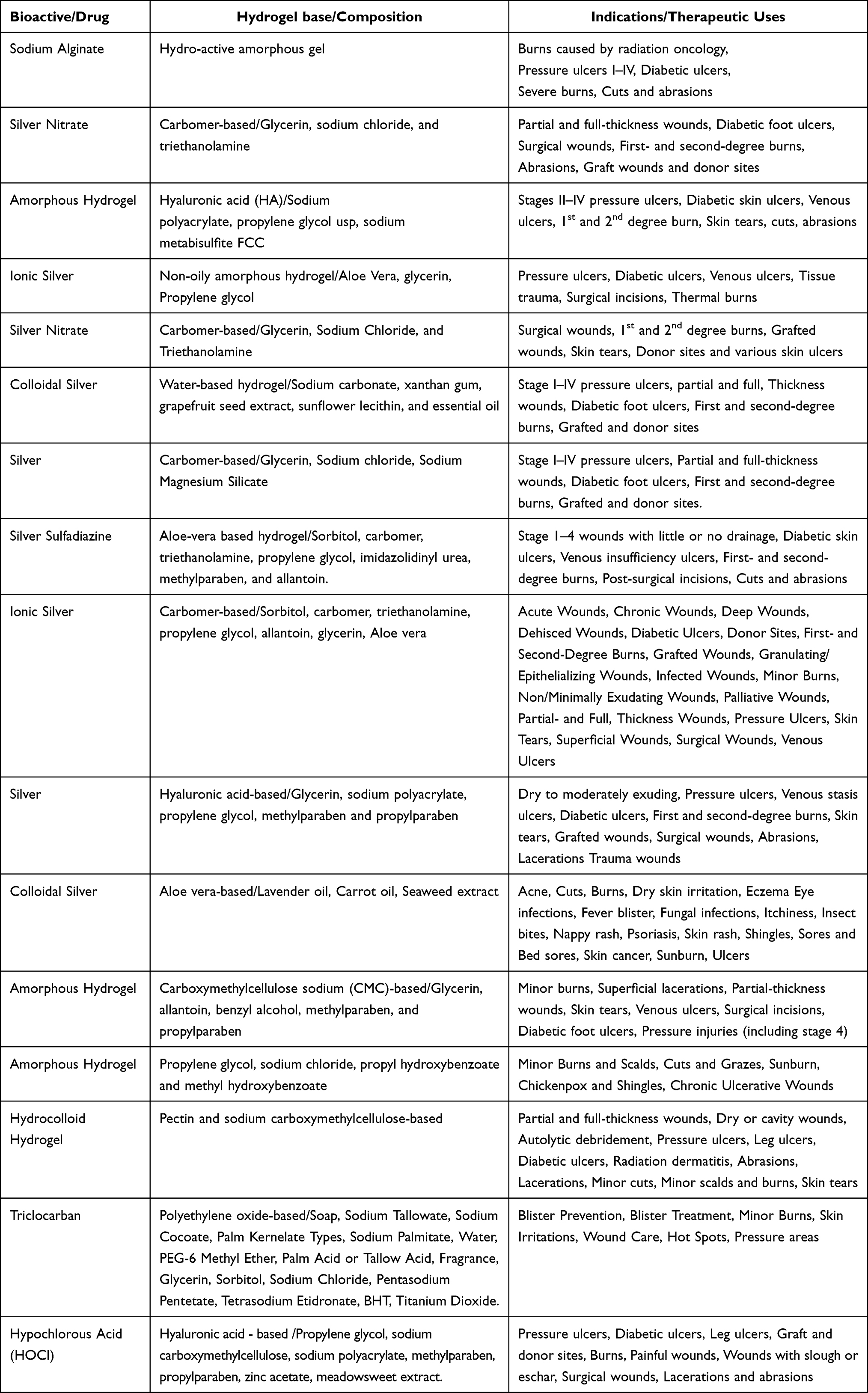 |
Table 5 List of Clinically Approved Hydrogels for Wound Healing Applications |
Many hydrogels are also investigated in humans (Table 6) to evaluate their safety and efficacy after being pre-clinically tested (animal models). Since the objective of the preclinical trials is to transition to the next phase, ie, human trials, this indicates that the formulation of interest has huge clinical transferability. A representative clinical trial evaluating hydrogel formulations is the study titled “Safety and Efficacy Evaluation of Tissue Engineered Construct Based on Allogeneic Adipose-derived Multipotent Mesenchymal Stromal Cells and Platelet-poor Plasma Fibrin Hydrogel to Treat the Patients with Burn Wounds. (ClinicalTrials.gov Identifier: NCT03113747)”. The primary objective of this Phase I open-label randomized controlled trial was to check how safe and effective it was to use a tissue-engineered construct made from allogeneic cultured adipose-derived multipotent mesenchymal stromal cells (ALLO-ADSCs) and platelet-poor plasma fibrin hydrogel to treat patients with 2nd-B and 3rd-degree burn wounds compared to standard care. The study is currently recruiting. Twenty adult patients (aged 18–65 years) with 2nd -B and 3-d degree burn wounds are expected to be enrolled. They will be randomized into two groups, with the hydrogel application procedure carried out twice – once simultaneously with a skin grafting procedure and 2–3 days following autodermoplasty, while bandaging. The primary endpoint was to monitor how well the healing of the skin flap and epithelialization of burn wounds in the perforations of a skin graft occurred, while secondary outcomes included dynamics of healing of the skin flap, dynamics of the phagocytic activity of neutrophils in the area of burn wounds, according to the NBT test, and duration of treatment (days) to complete epithelialization of burn wounds. The cultured multipotent mesenchymal stromal cells are anticipated to positively influence the epithelization process of burn wounds and enhance both the extent and speed of healing in skin flaps during autologous skin grafting.
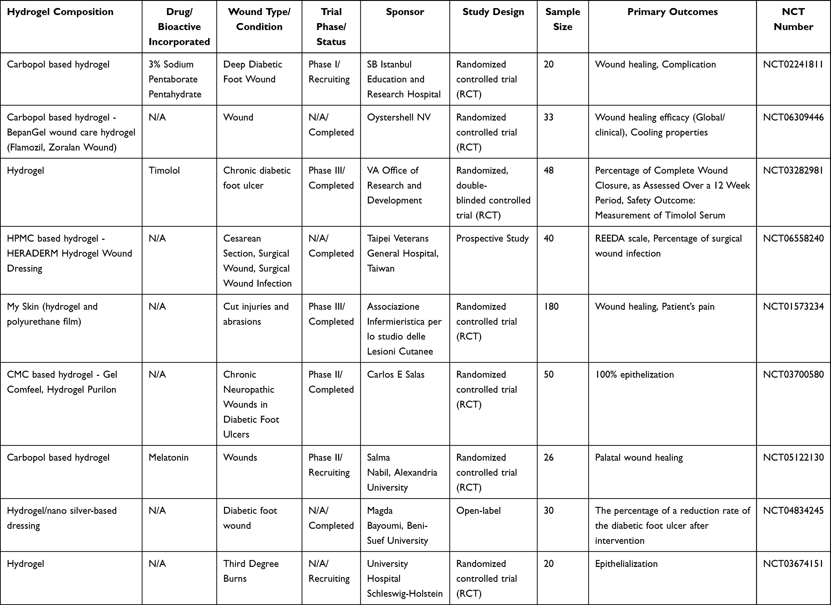 |
Table 6 Summary of Clinical Trials Investigating Hydrogels for Wound Healing |
Another clinical trial evaluating hydrogel formulation titled “Prospective, Multicenter, Single-blind, Randomized, Controlled Clinical Trial on Safety and Efficacy of a Novel Topical Formulation Containing Erythropoietin for the Treatment of Diabetic Foot Ulcers. (ClinicalTrials.gov Identifier: NCT02361931)”. The primary objective of this Phase I randomized controlled trial is to evaluate the safety and efficacy of topical hydrogel treatment for diabetic foot ulcers. This study is an exploratory proof-of-concept study on hydrogel treatment for diabetic foot ulcers. The study was completed. The researcher has created a patented technology (RMD-G1) that includes erythropoietin as the active pharmaceutical ingredient (API) within a carbopol-based hydrogel featuring a fibronectin matrix. RMD-G1 was created to preserve erythropoietin’s stability and effectiveness for extended durations and to enhance the delivery of erythropoietin to the wound bed. Twenty adult patients (aged 18–80 years) with non-infected diabetic hard-to-heal wounds (ulcers/foot ulcers) were enrolled. They were randomized into two groups, with RMD-G1 (gel with 2000 IU/mL of erythropoietin) as an adjunct therapy to standard of care (SOC) topically applied on the wound bed daily for 12 weeks. The primary endpoint was the number of participants without adverse events following RMD-G1 treatment and the number of participants with the reduction of wound area by 75% or more, while secondary outcomes included the number of patients with hypersensitivity at the wound site, speed of healing, reduction of wound area, partial wound closure, rate of wound closure, and recurrence of closed wounds. The cultured multipotent mesenchymal stromal cells are anticipated to demonstrate a beneficial impact on the epithelization of burn wounds and to influence the rate and extent of healing of skin flaps in autologous skin graft procedures. Results indicated that patients treated with hydrogel demonstrated no safety issues compared to standard treatment. Moreover, the hydrogel group exhibited a higher rate of complete wound closure. These findings suggest that the patented hydrogel is a safe and effective therapeutic option for enhancing wound healing in diabetic foot ulcers, warranting further large-scale Phase II studies to validate its clinical applicability. These clinical trials indicate the huge potential of hydrogels for wound healing, but still large studies are required to validate the safety and efficacy of the functionalized hydrogel formulations for wound healing applications.
Characterization and Evaluation of Functionalized Hydrogel Wound Dressings
The structural and physical characteristics, biological testing, and in vivo attributes influenced the critical evaluation to analyze the performance of functionalized biopolymer hydrogels for wound dressings. The optimized level of moisture retention, mechanical strength, exudate absorption, low immunogenicity, and biocompatibility are highly considerable features that ultimately influence the therapeutic utility of hydrogels for infection prevention and tissue regeneration for efficient wound healing.148 The protracted characterization not only legitimizes the well-off natural product-based NPs incorporation but also ensures their safety, efficacy, and clinical relevance.
Characterization of Hydrogel Nanocomposites
Characterization of nanoparticles functionalized hydrogels is required to verify their different parameters that either directly or indirectly control the formulation efficacy. In this section, we summarized various parameters that are mandatory for hydrogel nanocomposite characterization. Majorly include particle size and size distribution, FTIR, XRD, thermogravimetric analysis, and structural morphology via SEM and TEM.
Physical Characterization Parameters: Particle Size, FTIR, XRD, and TGA
β-chitin/nano hydroxyapatite (nHAp) nanocomposite hydrogel scaffolds were synthesized by Kumar et al,149 thereby scrutinizing the bone and wound tissue engineering. For nHAp particle size and size distribution in the hydrogel matrix, the dynamic light scattering technique (Nicomp™ 380 ZLS) was utilized, which demonstrated the size range of 50–120 nm. FTIR spectroscopy (Spectrophotometer Perkin-Elmer RX1) was engaged for confirming the chemical structure of nHAp and nanocomposite hydrogel, over 400–4000 cm−1. In the FTIR spectrum, three major peaks, 565 and 602 showed the phosphate group stretching vibration and 3570 cm−1 showed the OH group stretching vibration in nHAp. While in the FTIR spectrum of nanocomposite hydrogel (β-chitin + nHAp) a peak at 3450 cm−1 confirmed the H-bonding interaction between polymer (β-chitin) and nHAp. For determining the nHAp crystal structure, the XRD pattern was analyzed via a Panalytical diffractometer (XPERT PRO powder) at room temperature at a 40 kV voltage. In the results, it was monitored that the XRD pattern of nHAp precisely matched with the standard spectrum of HAp. In the nanocomposite hydrogel, peaks for both β-chitin and nHAp were visible; however, the peak for the polymer became weaker upon nHAp addition, which indicated their interaction. Furthermore, the TG/DTA instrument (SII TG-DTA6200) was used for thermogravimetric analysis at a range of 25–500°C. Initially, nHAp showed 10% weight loss because of moisture evaporation. However, incorporation of nHAp slightly improved the thermal stability of the polymer in nanocomposite hydrogels.149
Structural Morphology of Hydrogel Nanocomposites
Dang et al150 prepared biofunctional nanocomposite hydrogel via engaging nanocurcumin and chitosan-g-pluronic copolymer for treating the burn wounds. For morphological characterization of this formulation, scanning electron and transmission electron microscopy were performed via SEM, JEM-1400, TEM, and JSM 7401F, respectively. In detail, freeze-dried hydrogel samples were utilized; for SEM, these samples were fractured for interpretation of internal structure. While, in the TEM sample, it was added into the water, and thereby, a thin film was prepared on the disk for morphological evaluation. The SEM images disclosed the existence of interconnected pores with irregular shapes along with a uniform distribution of nanocurcumin having an average size between 20 and 50 nm present in nanocomposite hydrogels, which can promote the water absorption capacity of hydrogels. Furthermore, with TEM analysis, the nanocurcumin particles appeared spherical with the size range of 20–50 nm, also evidenced by the light scattering experiments.150
Evaluation Parameters of Hydrogel Nanocomposites
A number of physicochemical and functional parameters are engaged for evaluating the performance of nanocomposite hydrogels that influence their therapeutic efficiency. The crucial assessments include drug encapsulation efficiency, mechanical features, drug release profile, and swelling behavior. Collectively, these attributes dictate the delivery of therapeutic ingredients.
Drug Encapsulation Efficiency (EE%)
Drug encapsulation efficiency (EE%) is an important evaluation parameter that aids in verifying the amount of entrapped drug inside the nanoparticles because drug concentration has an impact on its therapeutic response. Mageed et al145 processed curcumin-cyclodextrin hybrid nanoparticles and incorporated them into a hydrogel matrix for wound treatment. Centrifugation was employed to analyze the EE of curcumin inside hybrid NPs. The assessment of the calibration curve and EE% of curcumin was monitored at the 420 nm absorption peak. The EE% was calculated by the given equation.151

Mechanical Attributes
For specific clinical relevancy, the mechanical features of hydrogels are crucial for ensuring their efficacy across various wounds. The maintenance of structural integrity and provision of adequate mechanical support are essential for hydrogel functionality throughout the healing process.152 Different approaches, ie, utilization of reinforcing agents, crosslinking, and blending with other materials like NPs, assist in promoting the mechanical aspects of natural hydrogels.153 Additionally, it was reported that the mechanical strength of biopolymers, ie, chitosan, cellulose, and starch, in hydrogel matrices increased in the case of nanocomposite hydrogels. This combination makes hydrogels ideal for the treatment of various wounds and improving patient outcomes.154 Chin et al155 designed a nanocomposite hydrogel dressing loaded with phomopsidione for pressure ulcers and wounds associated with microbial infection; chitosan and polyethylene glycol were utilized for hydrogel formation. In order to analyze the mechanical aspects of the designed nanocomposite hydrogel, three key features were monitored, including tensile strength, elongation at break, and Young’s modulus via a universal testing machine (INSTRON 3366). For comparison, nanocomposite hydrogels were compared with a control sample (hydrogel without phomopsidione). In contrast to the control sample, the nanocomposite hydrogels demonstrated 29.62% enhanced tensile strength, 5.41% improved elongation at break, and a Young’s modulus that was 13.76% higher. These findings justified that the mechanical features of nanocomposite hydrogels are much better than the plain ones. Moreover, the therapeutic utility of these hydrogels is also increased remarkably. Consequently, researchers claimed that the nanocomposite hydrogels have a promising potential to treat the chronic wounds.155
Swelling Behavior
The swelling behavior of nanocomposite hydrogels plays an integral role in proficient wound healing. It was reported that the increased concentration of monomers for hydrogel formation developed a significant improvement in the swelling ratio. The release of drugs from hydrogel leans on the swelling behavior.156 The following equation can be employed to evaluate the swelling behavior of hydrogels, whether having nanoparticles or not:157

In this ratio (Q), We indicates the weight of swollen hydrogel, and Wd indicates the dry weight of pure hydrogel.
Another simple equation that assists in determining the degree of swelling is;

Moreover, the swelling behavior of nanocomposite hydrogels expresses their absorption extent for wound exudates and blood. Raghavendra et al conducted a study,158 in which a team of researchers designed the nanocomposite hydrogels. Evaluators monitored the designed formulation, indicating the high absorptive capacity, which is contributed due to the incorporation of green synthesized core-shell nanoparticles into the hydrogel matrix. As a result, the swelling behavior of the formulation is 12 to 22 g/g; ultimately, hydrogels can absorb 1200–2200% of their dry weight, which demonstrates the effective absorbent ability of nanocomposite hydrogels for wound-related fluids. Overall, the high swelling capacities of hydrogels directly impact the moisture retention for cell mitigation, tissue regeneration, and effective wound healing. The tunable swelling ability of hydrogels helps to develop the formulations with optimal hydration with injured area stability.148
Biological Testing: Cell Viability, Hemocompatibility, and in vitro Antibacterial Analysis
The biological evaluation of nanocomposite hydrogel wound dressings is pivotal for certifying the safety and therapeutic utility. In this particular segment, we elaborate on the cytocompatibility, hemocompatibility, and in vitro antimicrobial activity for hydrogel wound dressings. To ensure the therapeutic effectiveness of wound dressing, they should support cell mitigation and proliferation, avoid undesired interaction with blood and the immune system, and restrict microbial loads that affect the healing process.
Keihan et al159 designed nanobiocomposite hydrogels for wound dressings, in which magnesium hydroxide NPs were uniformly dispersed into a hydrogel matrix that contains silk fibroin and carboxymethyl cellulose. The cell viability assay and hemolysis activity summarized that the nanobiocomposite hydrogels are biocompatible and safe for blood contact. The MTT assay was performed to monitor the cell viability, for which Hu02 cells (human fibroblast cells) were treated with nanobiocomposite hydrogel and cisplatin. The findings showed that the Hu02 cells in the presence of nanobiocomposite hydrogel maintained 84.5% viability after 7 days, in contrast to the untreated control group that indicated 95.7% viability. However, cisplatin reduced Hu02 cell concentration by >85%, and only 3.76% of cells became viable at a dose of 1 g/mL. Consequently, it was justified that the nanobiocomposite hydrogels are significantly biocompatible for human cells and not generating any compelling harm to cells. Additionally, only 8.3% hemolysis was recorded, which confirmed the remarkable hemocompatibility of the nanobiocomposite hydrogels for wound dressings.159
The antimicrobial activity is also essential for nanocomposite hydrogel wound dressings. The bacterial load influences the wound healing process; therefore, efficient control of the bacterial count assists in better wound recovery and enhances clinical relevancy. Miao et al160 prepared chitosan and recombinant keratin-31 contained cryogels, a specialized type of hydrogel with tunable mechanical features and high water absorption capacity. Gallic acid-reduced AgNPs were designed, and they were incorporated into cryogels for their functionalization. The agar diffusion and turbidimetric methods were utilized to evaluate the in vitro antibacterial activity of these particularly designed nanocomposite hydrogels against S. aureus and E. coli. The findings clearly indicated the bactericidal activity of designed nanocomposite hydrogels against S. aureus and E. coli. In the agar diffusion test, the well-defined zones of inhibition were monitored against S. aureus, while the zones were a little larger against E. coli. Furthermore, significant bacterial growth reduction was monitored against both kinds of bacteria in the turbidimetric assay. Approximately 99.9% of bacterial inhibition was monitored at 0.5–1 mM concentration of NPs-loaded cryogels.160 The antimicrobial activity of nanocomposite hydrogel wound dressings facilitates better patient outcomes.
In vivo Performance and Healing Efficiency
The in vivo effectiveness of hydrogel nanocomposites for wound healing is adequately evidenced in reported literature.161 The in vivo performance delivers the direct evidence of hydrogels’ wound-dressing therapeutic aptitude under physiological conditions. For ensuring the collagen deposition and tissue regeneration, the visual inspection, wound closure rate, and histological evaluation are usually employed.
In order to analyze the healing efficiency of hydrogel nanocomposites, biopolymer hydrogel was prepared and functionalized via curcumin-AgNPs.93 Subsequently, human skin fibroblast cells were employed for the scratch wound-healing test, and two concentrations of curcumin-AgNPs, ie, 0.03 and 0.06 nM, were utilized for evaluation. However, injured cells have reduced cell-to-cell communication and are much weaker in strength; that’s why the higher dose (0.06 nM) may provoke the toxicity for injured tissues. Meanwhile, the lower concentration (0.03 nM) of nanoparticles demonstrated the remarkable enhancement in cell mitigation by 100% and 50% at days 7 and 14, respectively, relative to untreated cells. Findings indicated the growth of fibroblast cells and efficiency of nanocomposite hydrogels for tissue repair.93
For evaluating the in vivo effectiveness of nanocomposite hydrogels, lignin-based green-synthesized AgNPs were designed and incorporated into the carrageenan-based hydrogel (holding CaCl2 and MgCl2 as crosslinkers) for hydrogel functionalization.162 After the ethical approval of the Animal Ethics Committee, researchers engaged 6-week-old male SD rats for testing the wound-healing effectiveness of the designed formulation. A sterile biopsy punch was used to generate the circular wound of 8 mm in diameter and immediately covered with the hydrogel dressings. For wound closure assessment, SD rats were monitored on days 3, 7, and 14. The following equation facilitates calculating the wound area correctly:
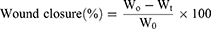
Where Wo was the initial wound area and Wt was the wound area at time t. After quantitative and photographic evaluation, it was observed that nanocomposite hydrogels significantly reduced the wound area, which indicated the healing efficiency of this system. Formulation having MgCl2 showed rapid healing, with wound area reducing from 100% at day 0–56%, 18%, and 3% at days 3, 7, and 14, respectively. The experimental outcomes demonstrate the promising potential of NP-based hydrogel wound dressings for wound recovery.162
For more clinically relevant evaluation, the effect of nanocomposite hydrogel wound dressings on internal tissue repair and the quality of skin regeneration in the wound area, the skin is evaluated after days 6 and 12 of treatment via Hematoxylin & Eosin (HE) staining.163 The nanocomposite hydrogel formulation was designed by utilizing zinc-doped bioactive glass NPs and biopolymers, ie, succinyl chitosan and oxidized alginate, by Zhu et al.104 Consequently, histomorphological assessment was performed for analyzing the tissue repair and skin regeneration. By day 6, researchers came to know that the nanocomposite hydrogel-treated wounds demonstrated better tissue repair progress in terms of granulation and fibroblast activity as compared to the control group. Additionally, angiogenesis was remarkably monitored via the designed formulation on day 6, while on day 12, blood vessels and skin glands were clearly monitored, presenting the internal regeneration. For wound closure, at day 12 it was observed that the size of the unhealed area in the treatment group is about 1.25 mm in diameter, in contrast to the control group that showed the unhealed area of about 2.84 mm. These findings indicate the promising potential of nanocomposite hydrogels for significant wound healing.104
Dong et al performed a study to evaluate the therapeutic effect of hydrogels on the recovery of oral ulcers.164 The stem cells of human exfoliated deciduous teeth were used to get the cell lysate, followed by encapsulation in the injectable fibrinogen/thrombin hydrogel. In the animal model, fabricated hydrogels exhibited improved ulcer recovery and clearly highlight the anti-inflammation response and epithelial integrity by days 3 and 5, respectively. Additionally, the hydrogel enhanced epithelial cell growth and vascular remodeling.
Clinical Implications and Challenges
Regulatory and Safety Considerations for Natural Product-Based Wound Dressings
Each component, ie, natural product and wound dressings, adds its own regulatory challenges for commercialization. Wound dressings incorporated with natural products have a synergistic approach, enhancing wound healing while helping to overcome side effects.62,165 Despite their proved therapeutic potential in preclinical studies, some of the regulatory challenges hinder their approval as commercially available products.166 Natural bioactives and nanoparticles are major constituents for multifunctional hydrogel dressings; a primary regulatory hurdle resides in their classification because these nanocomposite hydrogels may be regulated as advanced therapy products, medical devices, and drug-device combinations, which makes it difficult to categorize them properly. For manufacturers, it is essential to submit appropriate documentation regarding safety, biocompatibility, and performance characteristics for approval; however, water content variability and potential structural changes over time impede the standardization of testing protocols and acceptance criteria. Consequently, these ambiguities make approval pathways complicated, and development timelines suffer.167 Regulatory guidelines entail safety, efficacy, standardization, and quality aspects of the natural products.168
Increased demand for natural products has highlighted the public health issues related to their quality, efficacy, and safety.169 In the past, it was considered that natural products have no harm, while in recent advancements, research has proven that natural products with poor quality have interactions and adverse effects with co-administered drugs.170,171 As the herbal products are of a complex nature, consisting of many constituents, which makes their evaluation difficult.172 Therefore, counterfeited, adulterated, and poor-quality natural products pose serious health issues to patients due to lack of standardization and authentication problems.173 According to European guidelines, regulation of natural products depends on their level of indication, available clinical literature, and nature of product.170 To evaluate their safety and efficacy regarding their traditional use, there should be evidence from experimental data except for their minor use.174 There is also a requirement of good agricultural practices for natural products.175 The regulatory bodies require the detailed indication, detail of constituents, qualitative nature of constituents, route of administration, adverse reactions, and clinical literature regarding different characteristics of natural products.168 All these requirements make the marketing authorization of natural products difficult and create regulatory hindrance for their approval.170
Different regulatory bodies classify natural product-based wound dressings differently; some consider them as drugs for having the antioxidant, antimicrobial, and pro-regenerative properties, while others categorize them as medical devices for retaining the moisture content.167,176 The difference in their classification by the regulatory authorities creates a hindrance in their documentation necessary for approval of wound dressing incorporated with natural products.167
Commercialization of Functionalized Hydrogel Dressings
In recent years, hydrogels have offered various clinical applications.177 To develop novel hydrogels incorporated with natural products, extensive research is being implemented.178 Pharma companies are highly investing in research activities to conduct the clinical trials to explore their therapeutic potential, ie, wound healing properties.179,180 Thus, to commercialize the hydrogel dressings, many of the clinical studies had been conducted to bring out the positive results that increase the growth of the market.181
In 2024, the market size of hydrogel dressings was USD 791.3 million, which is expected to increase from USD 829.0 million to USD 1,209.7 million from 2025 to 2032, respectively, with a compound annual growth rate (CAGR) of 5.5% over the estimated span of 2025 to 2032. North America in 2024 predominated the market of hydrogel dressing with a market share of 40.17%.182
Multiple factors are involved in the market rise of hydrogel wound dressings, such as the rising number of geriatric populations, increasing incidence rate of surgical procedures, and higher prevalence of chronic and acute wounds.183 Surgical wounds pose a notable risk in the health care system, reported by the Wound Healing Society (WHS), causing mortality in 75% of cases due to surgical site infections.182 The high prevalence rate puts an economic burden globally and demands the launch of advanced technologies such as nanotechnology and 3D printing, which introduced advanced hydrogel dressings with enhanced functions and properties, which are also believed to increase the market growth.183–185 Hydrogel dressings are also recommended strongly by the healthcare professionals due to their effectiveness, which also increases their demand in the market.186
However, the strict regulatory framework regarding the use of natural products creates hindrance in their marketing authorization.187 During their acceptance, there is a requirement to submit the characteristic detail of natural products regarding their indication, list of constituents they contain, adverse reactions, contraindications, and evidence from relevant clinical literature.170,188 All these requirements make it expensive to authorize the natural products for many manufacturers, particularly for underdeveloped countries.170 Therefore, many of the natural products remain unrecognized because of the strict regulatory requirements, and the commercial market of natural product-based hydrogel dressings tends to decline.170,189
Barriers to Clinical Translation and Potential Solutions
Despite the remarkable achievement in the research area, still there are issues that need attention in the clinical translation. Hydrogel-based wound dressings loaded with natural products consist of two different components, such as hydrogels and nanoparticles.62 Wound dressings face obstacles from both components individually, and their integration also results in additional complexities, hindering their clinical translation.190
Wound healing is a multistage process that requires different hydrogel dressings for each stage.191 Moreover, each patient shows a different response to hydrogel dressings, as having different causes for impaired healing. These markers show that there is a need for personalized hydrogel dressings, which are in the early stages of development, as the single hydrogel dressing is not sufficient for all types of wound healing phases.192 After developing individualized dressings, hydrogel materials could be able to collect the data about the wound, helping in tailoring the specific design of dressings for each patient.4
Another barrier for clinical translation of hydrogel dressings incorporated with natural products is intra-inter species variation in clinical studies.193 Despite having strong therapeutic potential, still there is a need for clinical trials on humans due to intra- and inter-species variations.193 Researchers often use mice or rats as animal models for in vivo studies. But disparity exists between human and rodent skin, eg, the skin of rodents shows contractions during the healing process, causing variability in experimental results.194 However, larger animals that show similar characteristics or show similarity in skin morphology, such as pigs, could be used for experimental purpose, but the accessibility and practicality issues have restricted their extensive use as animal models.195
Although the synergistic applications could be obtained by combining the hydrogels with nanoparticles, it requires extensive research to find the underlying mechanisms involved and the safety and efficacy of the material used, which increases the production costs, acting as a barrier for their clinical translation.4
Clinical translation of the nanomedicines presents some practical difficulties when formulating on a large scale, because minute differences cause significant changes in their physicochemical parameters, affecting their safety, efficacy, and therapeutic potential, which hinder their commercial production for wound dressings.196–198 Other challenges involved are their characterization, finding a suitable animal model, surface properties, and lacking clinical trials that prevent their transformation from laboratory research to their commercialization.199,200
Another barrier is the high cost, which limits the use of hydrogel dressings in the market. National Health Services spend almost USD 6.48 billion per year on wound care, as reported by the Institute of Pressure Injury Prevention. Due to higher cost, these are associated with the unavailability and inaccessibility of these in developing countries, which may deter the health care professionals from recommending them to their patients, ultimately affecting the market growth. Furthermore, batch-to-batch variation in plant-derived systems is a significant barrier, as it directly affects product consistency and regulatory compliance. Lack of large-scale clinical validation protocols impedes scalability and clinical translation. It is essential to fabricate regulatory protocols to conquer the scalability and translational challenges.
Future Trends in Hydrogel-Based Wound Care
Emerging Technologies in Wound Healing
Traditional wound dressings primarily offered basic benefits, such as protecting the wound and reducing pain, but they were limited in promoting efficient tissue repair.201 The advent of hydrogel-based systems addressed many of these drawbacks by providing a moist healing environment, enhanced biocompatibility, and improved patient comfort.202 More recently, the development of functionalized hydrogels has demonstrated superior outcomes compared to conventional hydrogel dressings, particularly in accelerating wound closure and improving patient compliance.203 Nonetheless, large-scale clinical evidence on their long-term safety and efficacy remains limited and warrants further investigation. Emerging advancements, such as 3D-printed hydrogel systems,204 represent the next frontier in wound healing, offering the potential for personalized, structurally tailored, and multifunctional dressings. Mujawar et al205 designed a 3D-printed hydrogel scaffold made of sodium alginate-gelatin loaded with Aloe barbadensis for accelerated wound healing. The developed hydrogel scaffold exhibited moderate swellability and excellent mechanical properties in terms of Young’s modulus and tensile strength. In terms of biological properties, the scaffold showed significant antibacterial activity (Staphylococcus aureus, Pseudomonas aeruginosa), hemocompatibility, biocompatibility, biodegradability enhancement in fibroblast proliferation, migration, and effective viability in Normal Human Dermal Fibroblast cells (NHDF). In Wistar rats, the designed hydrogel scaffold showed enhanced wound closure, minimized wound contraction, accelerated epithelial regeneration with minimal inflammation, and reduced scar formation after 14 days of treatment.
Yang et al206 developed a 3D-printed hydrogel network made of hyaluronic acid loaded with deferoxamine for accelerated healing in diabetic wounds. The fabricated hydrogel network exhibited optimum swellability and excellent mechanical properties. In terms of biological properties, the scaffold showed reasonable degradation time, optimum biodegradability, significant antioxidant activity, anti-inflammatory activity, and promoted migration and angiogenesis in human immortalized keratinocytes (HaCaT). In SD rats, the developed hydrogel network exhibited accelerated healing of the chronic diabetic wounds via vascular regeneration. These studies highlight the promise of 3D-printed hydrogels for wound healing. However, further studies on long-term safety and efficacy assessments in preclinical models and ultimately clinical trials are needed to determine the full potential of these innovative and emerging hydrogel systems in the landscape of wound care.
Additionally, hydrogels incorporating multicellular approaches facilitate personalized wound care so as to integrate the distinct cells to promote neovascularization, reepithelization, and collagen deposition for healing.207 The properties of changing behavior of 4D printing hydrogels in response to stimuli such as temperature, light, and moisture revolutionize the healing domain. These hydrogels are capable of inducing post-production alteration for better medical applications. Multifunctional wound dressings along with 4D-printed techniques seem to be more efficacious in comparison to traditional hydrogel dressings.208 Furthermore, bioinspired nanocomposites facilitate wound recovery via DNA-induced biomineralization, which enables the preparation of specialized ink for 3D printing dressings. The robust therapeutic potential was achieved by incorporating this technique in the treatment of diabetic wounds.209 Collectively, all these modern and emerging strategies highlight the growing relevance of advanced hydrogel dressings in wound care.208
Role of Smart Hydrogel Dressings and Controlled Release Systems
There is a massive shift in the paradigm of strategies for wound care, especially for treating infected and chronic wounds, with the deployment of controlled release systems and the use of real-time monitoring systems for diagnostic purposes. Smart or intelligent systems are used for both diagnostic and therapeutic purposes. The foremost challenge with these systems is to make them function continuously without any interruption, since the framework (feedback loop) will not function if the two modulus bundles are just bundled; instead, they should be integrated. The latest innovative hydrogel systems210,211 bridge the gap between the traditional wound dressing and intelligent therapeutic systems by the utilization of modern technologies such as data processing, stimuli-responsive drug delivery, and biosensing to create the closed-loop system providing better wound care and management. For instance, Liu et al212 designed a sprayable hydrogel network made of cyclodextrin loaded with human umbilical cord mesenchymal stem cell-derived exosomes and borneol for accelerated healing in diabetic wounds. The developed hydrogel network exhibited excellent biocompatibility, anti-inflammatory activity, antioxidant activity, and broad-spectrum antibacterial activity (Staphylococcus aureus, Pseudomonas aeruginosa). In human immortalized keratinocytes (HaCaT), the hydrogel exhibited enhancement of keratinocyte migration and stimulated angiogenesis in RAW 264.7 cells and HUVECs. In BALB/c mice, the developed hydrogel network exhibited superior healing of diabetic wounds in comparison to blank hydrogel and exosomes (standalone treatment). These studies highlight the promise of 3D-printed hydrogels for wound healing. The hydrogel system was sprayed onto irregularly shaped wounds of the mouse model, with its crosslinking density and degradation kinetics precisely controlled by modifying the photocuring duration. This tunable feature allows the controlled release of borneol and exosomes for nearly 2 to 7 days. This study shows the promising potential of the intelligent and innovative hydrogel systems that can precisely control the release of the bioactive loaded into the hydrogel matrix, providing increased therapeutic outcomes and patient compliance; however, the engineering and clinical challenges still exist.
Moreover, smart hydrogels are multifaceted and can be fabricated according to the desired microenvironment, as they have an ability to respond according to physiological stimuli like enzymes, pH, temperature, and redox condition. For precision drug delivery, they have gained significant attention, highlighting their broader translational potential.213 The integration of both nanotechnology and bioengineering principles constitutes smart hydrogel dressings, which enables responsive and controlled delivery for wound recovery.208
Potential for Personalized Medicine in Wound Care
Gene hydrogels214 are one of the transformative strategies to accelerate the wound healing process by combining the regenerative properties of hydrogels with targeted gene delivery systems, enabling a tailored and personalized approach. These gene-modified hydrogels215 have exhibited numerous advantages during the process of wound healing, such as mitigation of inflammatory responses, reduction of cytotoxicity induced by drugs, and acceleration of tissue regeneration. There are numerous preclinical studies reported on utilizing these gene hydrogels. For example, Xie et al216 fabricated a gene hydrogel (Janus hydrogel (ABC/PEM) formed by genetically engineered polypeptide) loaded with Mn-based mineralized erythrocyte for diabetic wound healing. The developed hydrogel promoted cutaneous healing and regeneration of skin structure. In male SD rats, it also showed the accelerated rate of reconstruction of skin structure by breaking the oxygen diffusion barrier and supplying oxygen on demand to promote angiogenesis and functionalization. It has inhibited the inflammatory processes in the fibroblasts and keratinocytes. One of the interesting advantages of this hydrogel is its accelerated wound healing potential, where it has shown a healing rate of nearly 99% after 21 days of treatment. This study demonstrates the enormous potential of gene hydrogel, which is an emerging strategy for the wound healing landscape. However, in order to advance to the next stage of commercialization and wide application, some of the critical challenges, such as scale-up difficulties, stability issues of the hydrogel, and lack of large-scale human studies to validate safety and efficacy, have to be resolved.
Conclusion and Future Orientation
For addressing the complex pathophysiology of acute and chronic wounds, the multifunctional therapeutic systems contributed adequately; consequently, wound management evolves continuously. This review consolidates recent advancements in biopolymer hydrogel-based dressings functionalized with natural product-containing nanoparticles. This combination brings versatile biological activities, including enhanced antimicrobial protection, inflammation recovery, active tissue regeneration, and minimized oxidative stress, emphasizing the growing relevance of nanocomposite hydrogel-based dressings in next-generation wound care. Furthermore, integration of nanotechnology and biopolymer hydrogels together exhibited the targeted biological activities and structural support. Despite these significant advancements, several unresolved challenges and knowledge gaps remain. Further investigation is required to analyze the long-term biocompatibility and safety evidence for the nanocomposite hydrogel complex, along with the standardization and reproducibility protocols. Moreover, for complete understanding of emerging hydrogel platforms, a thorough knowledge of structure–function relationships and the conduction of extensive comparative clinical studies are mandatory. New insights have been contributed in this review that highlights the translational aspects of natural product-derived nanoparticle-functionalized hydrogels to mention the growing relevance of their therapeutic performance and clinical applicability.
For the future, researchers should consider the integration of advanced fabrication techniques like 3D printing for the development of smart and stimuli-responsive hydrogels, along with scalable manufacturing strategies. Consequently, the precise drug release and real-time wound monitoring could be achieved. Future research should also prioritize economical and eco-friendly methodologies in wound dressing development. Addressing translational challenges such as batch-to-batch consistency, regulatory classification of natural product-based combination dressings, and cost-effective large-scale manufacturing will be essential to enable clinical adoption of multifunctional hydrogel systems.
Data Sharing Statement
All data supporting the findings of this study are available from the corresponding author upon reasonable request.
Acknowledgments
The authors acknowledge the School of Pharmacy, Monash University Malaysia, for providing institutional support and access to research infrastructure that facilitated the preparation of this manuscript. The authors also appreciate the availability of scientific resources and research facilities that contributed to the quality and comprehensiveness of this work.
Funding
This work was supported by the Deanship of Scientific Research at Northern Border University, Arar, Kingdom of Saudi Arabia, and by the Deanship of Research and Graduate Studies at King Khalid University, Saudi Arabia, through the Large Group Research Project under grant number RGP2/59/46.
Disclosure
The authors declare that they have no conflicts of interest.
References
1. Mishra R, Bajpai S. Natural biopolymer-based hydrogels: an advanced material for diabetic wound healing. Diabetol Int. 2024;15(4):719–44. doi:10.1007/s13340-024-00737-2
2. Arany P. Craniofacial wound healing with photobiomodulation therapy: new insights and current challenges. J Dent Res. 2016;95(9):977–984. doi:10.1177/0022034516648939
3. Tavakoli S, Klar AS. Advanced hydrogels as wound dressings. Biomolecules. 2020;10(8):1169. doi:10.3390/biom10081169
4. Zhang W, Liu L, Cheng H, et al. Hydrogel-based dressings designed to facilitate wound healing. Mater Adv. 2024;5(4):1364–1394. doi:10.1039/D3MA00682D
5. Ramzan M. Natural products loaded with nanoparticles for wound healing.
6. Raina N, Rani R, Pahwa R, et al. Biopolymers and treatment strategies for wound healing: an insight view. Int J Polym Mater Polym Biomater. 2022;71(5):359–375. doi:10.1080/00914037.2020.1838518
7. Liu Q, Luo S, Peng J, et al. Electrospun nanofibers from plant natural products: a new approach toward efficient wound healing. Int J Nanomed. 2024;Volume 19:13973–13990. doi:10.2147/IJN.S501970
8. Ibrahim NI, Wong SK, Mohamed IN, et al. Wound healing properties of selected natural products. Int J Environ Res Public Health. 2018;15(11):2360.
9. Fauzian F, Garmana AN, Mauludin R. Applications of nanotechnology-based drug delivery system for delivering natural products into acute and chronic wounds: a review. Biointerface Res Appl Chem. 2023;13:426.
10. Fu YJ, Shi Y-F, Wang L-Y, et al. All‐natural immunomodulatory bioadhesive hydrogel promotes angiogenesis and diabetic wound healing by regulating macrophage heterogeneity. Adv Sci. 2023;10(13):2206771. doi:10.1002/advs.202206771
11. Lokare P, Keshamma E, Kumar A, Alsowadi YA, Hossain MM, Pallathadka LK. Applications of plant-based nanomedicines for wound healing–An emerging paradigm for effective therapy. Mater Today. 2023;73:494–501.
12. Serpico L, Dello Iacono S, Cammarano A, et al. Recent advances in stimuli-responsive hydrogel-based wound dressing. Gels. 2023;9(6):451. doi:10.3390/gels9060451
13. Rezvani Ghomi E, Khalili S, Nouri Khorasani S, et al. Wound dressings: current advances and future directions. J Appl Polym Sci. 2019;136(27):47738. doi:10.1002/app.47738
14. Prete S, Dattilo M, Patitucci F, et al. Natural and synthetic polymeric biomaterials for application in wound management. J Funct Biomater. 2023;14(9):455. doi:10.3390/jfb14090455
15. Sheokand B, Vats M, Kumar A, et al. Natural polymers used in the dressing materials for wound healing: past, present and future. J Polym Sci. 2023;61(14):1389–1414. doi:10.1002/pol.20220734
16. Maessen-Visch MB, van Montfrans C. Wound dressings, does it matter and why? Phlebology. 2016;31(1_suppl):63–67. doi:10.1177/0268355516633383
17. Aderibigbe BA. Hybrid-based wound dressings: combination of synthetic and biopolymers. Polymers. 2022;14(18):3806. doi:10.3390/polym14183806
18. Obagi Z, Damiani G, Grada A, Falanga VI. Principles of wound dressings: a review. Surg Technol Int. 2019;35(5):0–57.
19. Divyashri G, Badhe RV, Sadanandan B, et al. Applications of hydrogel-based delivery systems in wound care and treatment: an up-to-date review. Polym Adv Technol. 2022;33(7):2025–2043. doi:10.1002/pat.5661
20. Shi C, Wang C, Liu H, et al. Selection of appropriate wound dressing for various wounds. Front Bioeng Biotechnol. 2020;8:182. doi:10.3389/fbioe.2020.00182
21. Tran HQ, Shahriar SMS, Yan Z, et al. Recent advances in functional wound dressings. Adv Wound Care. 2023;12(7):399–427. doi:10.1089/wound.2022.0059
22. Guo J, Hu B, Wei Y, et al. Hydrogel delivering self-assembled herbal nanoparticles accelerates diabetic wound healing through mitochondrial regulation. Mater Today Bio. 2025;35:102417. doi:10.1016/j.mtbio.2025.102417
23. Liu L, Ma W, Wang J, Wang X, Li S. Recent progress in polyphenol-based hydrogels for wound treatment and monitoring. Biosensors. 2025;15(10):657.
24. Jangra N, Singla A, Puri V, et al. Herbal bioactive-loaded biopolymeric formulations for wound healing applications. RSC Adv. 2025;15(16):12402–12442. doi:10.1039/d4ra08604j
25. Sari MHM, Cobre ADF, Pontarolo R, et al. Status and future scope of soft nanoparticles-based hydrogel in wound healing. Pharmaceutics. 2023;15(3):874. doi:10.3390/pharmaceutics15030874
26. Elangwe CN, Morozkina SN, Olekhnovich RO, et al. A review on chitosan and cellulose hydrogels for wound dressings. Polymers. 2022;14(23):5163. doi:10.3390/polym14235163
27. Kusnadi K, Herdiana Y, Rochima E, et al. Collagen-based nanoparticles as drug delivery system in wound healing applications. Int J Nanomed. 2024;Volume 19:11321–11341. doi:10.2147/IJN.S485588
28. Muslim MRF, Chabib L, Suryaningsih BE, et al. Green-synthesized silver nanoparticle hydrogels for biofilm-infected wounds: bridging sustainability and clinical translation. Front Pharmacol. 2025;16. doi:10.3389/fphar.2025.1694144
29. Rodríguez-Gómez FD, Monferrer D, Penon O, et al. Regulatory pathways and guidelines for nanotechnology-enabled health products: a comparative review of EU and US frameworks. Front Med. 2025;12. doi:10.3389/fmed.2025.1544393
30. Tottoli EM, Dorati R, Genta I, et al. Skin wound healing process and new emerging technologies for skin wound care and regeneration. Pharmaceutics. 2020;12(8):735. doi:10.3390/pharmaceutics12080735
31. Catoira MC, Fusaro L, Di Francesco D, et al. Overview of natural hydrogels for regenerative medicine applications. J Mater Sci. 2019;30(10):115. doi:10.1007/s10856-019-6318-7
32. Gomez-Florit M, Pardo A, Domingues RMA, et al. Natural-based hydrogels for tissue engineering applications. Molecules. 2020;25(24):5858. doi:10.3390/molecules25245858
33. Klein M, Poverenov E. Natural biopolymer‐based hydrogels for use in food and agriculture. J Sci Food Agric. 2020;100(6):2337–2347. doi:10.1002/jsfa.10274
34. Joyce K, Fabra GT, Bozkurt Y, et al. Bioactive potential of natural biomaterials: identification, retention and assessment of biological properties. Signal Transduct Target Ther. 2021;6(1):122. doi:10.1038/s41392-021-00512-8
35. Kumar SSD, Rajendran NK, Houreld NN, et al. Recent advances on silver nanoparticle and biopolymer-based biomaterials for wound healing applications. Int J Biol Macromol. 2018;115:165–175. doi:10.1016/j.ijbiomac.2018.04.003
36. Mushtaq F, Raza ZA, Batool SR, et al. Preparation, properties, and applications of gelatin-based hydrogels (GHs) in the environmental, technological, and biomedical sectors. Int J Biol Macromol. 2022;218:601–633. doi:10.1016/j.ijbiomac.2022.07.168
37. Li S, Dan X, Chen H, et al. Developing fibrin-based biomaterials/scaffolds in tissue engineering. Bioact Mater. 2024;40:597–623. doi:10.1016/j.bioactmat.2024.08.006
38. Zheng H, Zuo B. Functional silk fibroin hydrogels: preparation, properties and applications. J Mat Chem B. 2021;9(5):1238–1258. doi:10.1039/D0TB02099K
39. Romano S, Yazdanpanah S, Petillo O, et al. Sustainable hydrogels for medical applications: biotechnological innovations supporting one health. Gels. 2025;11(7):559. doi:10.3390/gels11070559
40. Gobi R, Ravichandiran P, Babu RS, et al. Biopolymer and synthetic polymer-based nanocomposites in wound dressing applications: a review. Polymers. 2021;13(12):1962. doi:10.3390/polym13121962
41. Güiza-Argüello VR, Solarte-David V, Pinzón-Mora A, et al. Current advances in the development of hydrogel-based wound dressings for diabetic foot ulcer treatment. Polymers. 2022;14(14):2764. doi:10.3390/polym14142764
42. Ahmed S, Ikram S. Chitosan based scaffolds and their applications in wound healing. Achiev Life Sci. 2016;10(1):27–37. doi:10.1016/j.als.2016.04.001
43. Falbo F, Spizzirri UG, Restuccia D, et al. Natural compounds and biopolymers-based hydrogels join forces to promote wound healing. Pharmaceutics. 2023;15(1):271. doi:10.3390/pharmaceutics15010271
44. Collins MN, Birkinshaw C. Hyaluronic acid based scaffolds for tissue engineering—A review. Carbohydr Polym. 2013;92(2):1262–1279. doi:10.1016/j.carbpol.2012.10.028
45. Klemm D, Kramer F, Moritz S, et al. Nanocelluloses: a new family of nature‐based materials. Angew Chem Int Ed. 2011;50(24):5438–5466. doi:10.1002/anie.201001273
46. Munarin F, Tanzi MC, Petrini P. Advances in biomedical applications of pectin gels. Int J Biol Macromol. 2012;51(4):681–689. doi:10.1016/j.ijbiomac.2012.07.002
47. Dhand AP, Galarraga JH, Burdick JA. Enhancing biopolymer hydrogel functionality through interpenetrating networks. Trends Biotechnol. 2021;39(5):519–538. doi:10.1016/j.tibtech.2020.08.007
48. Burdick JA, Prestwich GD. Hyaluronic acid hydrogels for biomedical applications. Adv Mater. 2011;23(12):H41–H56. doi:10.1002/adma.201003963
49. Reddy MSB, Ponnamma D, Choudhary R, et al. A comparative review of natural and synthetic biopolymer composite scaffolds. Polymers. 2021;13(7):1105. doi:10.3390/polym13071105
50. Mir M, Ali MN, Barakullah A, et al. Synthetic polymeric biomaterials for wound healing: a review. Progress Biomater. 2018;7(1):1–21. doi:10.1007/s40204-018-0083-4
51. Lin -C-C, Anseth KS. PEG hydrogels for the controlled release of biomolecules in regenerative medicine. Pharm Res. 2009;26(3):631–643. doi:10.1007/s11095-008-9801-2
52. Dash TK, Konkimalla VB. Poly-є-caprolactone based formulations for drug delivery and tissue engineering: a review. J Control Release. 2012;158(1):15–33. doi:10.1016/j.jconrel.2011.09.064
53. Rao JP, Geckeler KE. Polymer nanoparticles: preparation techniques and size-control parameters. Prog Polym Sci. 2011;36(7):887–913.
54. Santerre J, Woodhouse K, Laroche G, et al. Understanding the biodegradation of polyurethanes: from classical implants to tissue engineering materials. Biomaterials. 2005;26(35):7457–7470. doi:10.1016/j.biomaterials.2005.05.079
55. Sahana T, Rekha P. Biopolymers: applications in wound healing and skin tissue engineering. Mol Biol Rep. 2018;45(6):2857–2867. doi:10.1007/s11033-018-4296-3
56. Ilić-Stojanović S, Nikolić L, Cakić S. A review of patents and innovative biopolymer-based hydrogels. Gels. 2023;9(7):556. doi:10.3390/gels9070556
57. He JJ, McCarthy C, Camci-Unal G. Development of hydrogel‐based sprayable wound dressings for second‐and third‐degree burns. Adv Nanobiomed Res. 2021;1(6):2100004. doi:10.1002/anbr.202100004
58. Ribeiro M, Simões M, Vitorino C, et al. Hydrogels in cutaneous wound healing: insights into characterization, properties, formulation and therapeutic potential. Gels. 2024;10(3):188. doi:10.3390/gels10030188
59. Hama R, Ulziibayar A, Reinhardt JW, et al. Recent developments in biopolymer-based hydrogels for tissue engineering applications. Biomolecules. 2023;13(2):280. doi:10.3390/biom13020280
60. Barbu A, Neamtu B, Zăhan M, et al. Current trends in advanced alginate-based wound dressings for chronic wounds. J Pers Med. 2021;11(9):890. doi:10.3390/jpm11090890
61. Portela R, Leal CR, Almeida PL, et al. Bacterial cellulose: a versatile biopolymer for wound dressing applications. Microb Biotechnol. 2019;12(4):586–610. doi:10.1111/1751-7915.13392
62. Hajialyani M, Tewari D, Sobarzo-Sánchez E, et al. Natural product-based nanomedicines for wound healing purposes: therapeutic targets and drug delivery systems. Int J Nanomed. 2018;Volume 13:5023–5043. doi:10.2147/IJN.S174072
63. Trinh X-T, Long N-V, Van Anh LT, et al. A comprehensive review of natural compounds for wound healing: targeting bioactivity perspective. Int J Mol Sci. 2022;23(17):9573. doi:10.3390/ijms23179573
64. Liu E, Gao H, Zhao Y, et al. The potential application of natural products in cutaneous wound healing: a review of preclinical evidence. Front Pharmacol. 2022;13:900439. doi:10.3389/fphar.2022.900439
65. Chorti-Tripsa E, Galanis V-Z, Constantinides TC, et al. Natural products from the mediterranean area as wound healing agents—in vitro studies: a systematic review. Antioxidants. 2025;14(4):484. doi:10.3390/antiox14040484
66. Men SY, Huo Q-L, Shi L, et al. Panax notoginseng saponins promotes cutaneous wound healing and suppresses scar formation in mice. J Cosmet Dermatol. 2020;19(2):529–534. doi:10.1111/jocd.13042
67. Utpal BK, Sutradhar B, Zehravi M, et al. Polyphenols in wound healing: unlocking prospects with clinical applications. Naunyn-Schmiedeberg’s Arch Pharmacol. 2025;398(3):2459–2485. doi:10.1007/s00210-024-03538-1
68. Zulkefli N, Che Zahari CNM, Sayuti NH, et al. Flavonoids as potential wound-healing molecules: emphasis on pathways perspective. Int J Mol Sci. 2023;24(5):4607. doi:10.3390/ijms24054607
69. Maver T, Maver U, Stana kleinschek K, et al. A review of herbal medicines in wound healing. Int J Dermatol. 2015;54(7):740–751. doi:10.1111/ijd.12766
70. Reda R, Assar DH, Al-Hawary II, et al. Evaluation of nanocellulose-based anthraquinone from marine fungi Penicillium flavidorsum as an alternative therapy for skin wound healing: histopathological and immunohistochemical evidences from a rat model. Beni-Suef Univ J Basic Appl Sci. 2024;13(1):63. doi:10.1186/s43088-024-00522-9
71. Ebadi P, Fazeli M. Evaluation of the potential in vitro effects of propolis and honey on wound healing in human dermal fibroblast cells. S Afr J Bot. 2021;137:414–422. doi:10.1016/j.sajb.2020.10.003
72. Abdel-Gawad R, Osman R, Awad GAS, et al. Wound healing potential of silver nanoparticles embedded in optimized bio-inspired hybridized chitosan soft and dry hydrogel. Carbohydr Polym. 2024;324:121526. doi:10.1016/j.carbpol.2023.121526
73. Singh N, Brown AN, Gold MH. Snail extract for skin: a review of uses, projections, and limitations. J Cosmet Dermatol. 2024;23(4):1113–1121. doi:10.1111/jocd.16269
74. Chummun I, Bekah D, Goonoo N, et al. Assessing the mechanisms of action of natural molecules/extracts for phase-directed wound healing in hydrogel scaffolds. RSC Med Chem. 2021;12(9):1476–1490. doi:10.1039/D1MD00100K
75. de Macedo LM, Santos ÉMD, Militão L, et al. Rosemary (Rosmarinus officinalis L. syn Salvia rosmarinus Spenn.) and its topical applications: a review. Plants. 2020;9(5):651. doi:10.3390/plants9050651
76. Banerjee K, Madhyastha H, Sandur V R, et al. Anti-inflammatory and wound healing potential of a clove oil emulsion. Colloids Surf B. 2020;193:111102. doi:10.1016/j.colsurfb.2020.111102
77. Maleki H, Doostan M, Khoshnevisan K, et al. Zingiber officinale and thymus vulgaris extracts co-loaded polyvinyl alcohol and chitosan electrospun nanofibers for tackling infection and wound healing promotion. Heliyon. 2024;10(1):e23719. doi:10.1016/j.heliyon.2023.e23719
78. Sharma S, Bose A, Biswas S, et al. Cyperus rotundus mediated green synthesis of silver nanoparticles for antibacterial wound dressing applications. Sci Rep. 2025;15(1):18394. doi:10.1038/s41598-025-03555-x
79. Oselusi SO, Sibuyi NRS, Meyer M, et al. Phytonanotherapeutic applications of plant extract-synthesized silver nanoparticles in wound healing—a prospective overview. BioNanoScience. 2024;14(3):3455–3475. doi:10.1007/s12668-024-01535-5
80. Che J, Huang D, Wang Y, et al. Natural multi-active nanoparticles integrated biological hydrogel microcarriers for wound healing. J Nanobiotechnol. 2025;23(1):582. doi:10.1186/s12951-025-03666-7
81. Tsala DE, Amadou D, Habtemariam S. Natural wound healing and bioactive natural products. Phytopharmacology. 2013;4(3):532–560.
82. Wan X, Chen Y, Geng F, et al. Narrative review of the mechanism of natural products and scar formation in wound repair. Ann Translat Med. 2022;10(4):236. doi:10.21037/atm-21-7046
83. Cao F, Gui SY, Gao X, et al. Research progress of natural product-based nanomaterials for the treatment of inflammation-related diseases. Mater Design. 2022;218:110686.
84. Shalaby MA, Anwar MM, Saeed H. Nanomaterials for application in wound healing: current state-of-the-art and future perspectives. J Polym Res. 2022;29(3):91. doi:10.1007/s10965-021-02870-x
85. Koliqi R, Uskokovic V, Selmani PB, et al. Polymeric nanoparticles for wound healing. Pharm Nanotechnol. 2025;13(4):775–793. doi:10.2174/0122117385307311240506104035
86. Girija AR, Balasubramanian S, Cowin AJ. Nanomaterials-based drug delivery approaches for wound healing. Curr Pharm Des. 2022;28(9):711–726. doi:10.2174/1381612828666220328121211
87. Hano C, Abbasi BH. Plant-based green synthesis of nanoparticles: production, characterization and applications. Biomolecules. 2021;12(1):31. doi:10.3390/biom12010031
88. Ijaz I, Gilani E, Nazir A, et al. Detail review on chemical, physical and green synthesis, classification, characterizations and applications of nanoparticles. Green Chem Lett Rev. 2020;13(3):223–245. doi:10.1080/17518253.2020.1802517
89. Wang C, Gao X, Chen Z, et al. Preparation, characterization and application of polysaccharide-based metallic nanoparticles: a review. Polymers. 2017;9(12):689. doi:10.3390/polym9120689
90. Ganguly DS. Food processing and quality control 2013.
91. Sood A, Dev A, Das SS, et al. Curcumin-loaded alginate hydrogels for cancer therapy and wound healing applications: a review. Int J Biol Macromol. 2023;232:123283. doi:10.1016/j.ijbiomac.2023.123283
92. Hamedi H, Moradi S, Hudson SM, et al. Chitosan based hydrogels and their applications for drug delivery in wound dressings: a review. Carbohydr Polym. 2018;199:445–460. doi:10.1016/j.carbpol.2018.06.114
93. Bhubhanil S, Talodthaisong C, Khongkow M, et al. Enhanced wound healing properties of guar gum/curcumin-stabilized silver nanoparticle hydrogels. Sci Rep. 2021;11(1):21836. doi:10.1038/s41598-021-01262-x
94. Kamar SS, Abdel-Kader DH, Rashed LA. Beneficial effect of curcumin nanoparticles-hydrogel on excisional skin wound healing in type-I diabetic rat: histological and immunohistochemical studies. Ann Anat. 2019;222:94–102. doi:10.1016/j.aanat.2018.11.005
95. Tao G, Wang Y, Cai R, et al. Design and performance of sericin/poly(vinyl alcohol) hydrogel as a drug delivery carrier for potential wound dressing application. Mater Sci Eng C Mater Biol Appl. 2019;101:341–351. doi:10.1016/j.msec.2019.03.111
96. Xue H, Hu L, Xiong Y, et al. Quaternized chitosan-Matrigel-polyacrylamide hydrogels as wound dressing for wound repair and regeneration. Carbohydr Polym. 2019;226:115302. doi:10.1016/j.carbpol.2019.115302
97. Gupta A, Briffa SM, Swingler S, et al. Synthesis of silver nanoparticles using curcumin-cyclodextrins loaded into bacterial cellulose-based hydrogels for wound dressing applications. Biomacromolecules. 2020;21(5):1802–1811. doi:10.1021/acs.biomac.9b01724
98. Karri VV, Kuppusamy G, Talluri SV, et al. Curcumin loaded chitosan nanoparticles impregnated into collagen-alginate scaffolds for diabetic wound healing. Int J Biol Macromol. 2016;93(Pt B):1519–1529.
99. Zhao C, Yang J, Chen W, et al. Gelatin/Dopamine/Zinc-Doped Ceria/Curcumin nanocomposite hydrogels for repair of chronic refractory wounds. Int J Pharm. 2024;663:124575. doi:10.1016/j.ijpharm.2024.124575
100. Fu Z, Zou J, Zhong J, et al. Curcumin-loaded nanocomposite hydrogel dressings for promoting infected wound healing and tissue regeneration. Int J Nanomed. 2024;Volume 19:10479–10496. doi:10.2147/IJN.S479330
101. Majie A, Saha R, Sarkar A. A novel chitosan-PEG hydrogel embedded with in situ silver nanoparticles of Clerodendrum glandulosum Lindl. extract: evaluation of its in vivo diabetic wound healing properties using an image-guided machine learning model. Biomater Sci. 2024;12(16):4242–4261.
102. Kar AK, Singh A, Dhiman N, et al. Polymer-assisted in situ synthesis of silver nanoparticles with epigallocatechin gallate (EGCG) impregnated wound patch potentiate controlled inflammatory responses for brisk wound healing. Int J Nanomed. 2019;9837–9854.
103. Min Q, Wang C, Zhang Y, Tian D, Wan Y, Wu J. Strong and elastic hydrogels from dual-crosslinked composites composed of glycol chitosan and amino-functionalized bioactive glass nanoparticles. Nanomaterials. 2022;12(11):1874.
104. Zhu J, Jiang G. Incorporation of ZnO/Bioactive glass nanoparticles into alginate/chitosan composite hydrogels for wound closure. ACS Appl Bio Mater. 2019;2(11):5042–5052.
105. Kong B, Liu R, Cheng Y, et al. Natural biopolymers derived hydrogels with injectable, self-healing, and tissue adhesive abilities for wound healing. Nano Res. 2023;16(2):2798–2807. doi:10.1007/s12274-022-4936-8
106. Szczęsna W, Tsirigotis-Maniecka M, Lamch Ł, et al. Multilayered curcumin-loaded hydrogel microcarriers with antimicrobial function. Molecules. 2022;27(4):1415.
107. Huang SM, Liu SM, Tseng HY, Chen WC. Development and in vitro analysis of layer-by-layer assembled membranes for potential wound dressing: electrospun curcumin/gelatin as middle layer and gentamicin/polyvinyl alcohol as outer layers. Membranes. 2023;13(6):564.
108. Yang J, Chen Y, Zhao L, et al. Constructions and properties of physically cross-linked hydrogels based on natural polymers. Polymer Rev. 2023;63(3):574–612. doi:10.1080/15583724.2022.2137525
109. Aldakheel FM, Sayed MME. Green synthesis of silver nanoparticles loaded hydrogel for wound healing; systematic review. Gels. 2023;9(7):530.
110. Masood N, Ahmed R, Tariq M, et al. Silver nanoparticle impregnated chitosan-PEG hydrogel enhances wound healing in diabetes induced rabbits. Int J Pharm. 2019;559:23–36.
111. Begum F, Mahto AK, Kumari S, Dewangan RP. In-situ synthesis of silver nanoparticle within self-assembling ultrashort peptide hydrogel as antibacterial with wound healing properties. Curr Protein Pep Sci. 2025;26:6687–81.
112. Yoon MS, Lee JM, Jo MJ, et al. Dual-drug delivery systems using hydrogel–nanoparticle composites: recent advances and key applications. Gels. 2025;11(7):520.
113. Mohammed AA, Merrild NG, Li S, Pinna A, Jones JR. Double-network hydrogels reinforced with covalently bonded silica nanoparticles via 1-Ethyl-3-(3-dimethylaminopropyl) carbodiimide chemistry. ACS Omega. 2022;7(48):43904–43914.
114. Erfani A, Diaz AE, Doyle PS. Hydrogel-enabled, local administration and combinatorial delivery of immunotherapies for cancer treatment. Mater Today. 2023;65:227–243.
115. Ma X, Li SJ, Liu Y, et al. Bioengineered nanogels for cancer immunotherapy. Chem Soc Rev. 2022;51(12):5136–5174.
116. Torchilin VP. Multifunctional, stimuli-sensitive nanoparticulate systems for drug delivery. Nat Rev Drug Discov. 2014;13(11):813–827.
117. Caló E, Khutoryanskiy VV. Biomedical applications of hydrogels: a review of patents and commercial products. Eur Polym J. 2015;65:252–267.
118. Zhang Z, Zeng J, Groll J, Matsusaki M. Layer-by-layer assembly methods and their biomedical applications. Biomater Sci. 2022;10(15):4077–4094.
119. Shende P, Patil A, Prabhakar B. Layer-by-layer technique for enhancing physicochemical properties of actives. J Drug Delivery Sci Technol. 2020;56:101519.
120. Tamahkar E, Özkahraman B, Süloğlu AK, İdil N, Perçin I. A novel multilayer hydrogel wound dressing for antibiotic release. J Drug Delivery Sci Technol. 2020;58:101536.
121. Srisang S, Nasongkla N. Layer-by-layer dip coating of Foley urinary catheters by chlorhexidine-loaded micelles. J Drug Delivery Sci Technol. 2019;49:235–242.
122. Hsu BB, Hagerman SR, Hammond PT. Rapid and efficient sprayed multilayer films for controlled drug delivery. J Appl Polym Sci. 2016;133(25).
123. Kulikouskaya V, Chyshankou I, Pinchuk S, Vasilevich I, Volotovski I, Agabekov V. Fabrication and characterization of ultrathin spin-coated poly (L-lactic acid) films suitable for cell attachment and curcumin loading. Biomed Mater. 2020;15(6):065022.
124. Choi W, Kohane DS. Hybrid nanoparticle–hydrogel systems for drug delivery depots and other biomedical applications. ACS Nano. 2024;18(34):22780–22792.
125. Zhang X, Wei P, Yang Z, et al. Current progress and outlook of nano-based hydrogel dressings for wound healing. Pharmaceutics. 2023;15(1):68.
126. Hamdan S, Pastar I, Drakulich S, et al. Nanotechnology-driven therapeutic interventions in wound healing: potential uses and applications. ACS Cent Sci. 2017;3(3):163–175. doi:10.1021/acscentsci.6b00371
127. Vijapur LS, Shalavadi M, Desai AR, et al. Wound healing potential of green synthesized silver nanoparticles of Glycyrrhiza glabra linn root extract: a preclinical study. J Trace Elem Med Biol. 2025;11:100214. doi:10.1016/j.jtemin.2025.100214
128. Iqbal Y, Amin F, Aziz MH, et al. Flexible sodium alginate-gelatin hydrogel membrane incorporated with green synthesized bimetallic ZnO:CeO2 nanocomposite for antioxidant, antibacterial and biocompatibility studies. React Funct Polym. 2025;212:106228. doi:10.1016/j.reactfunctpolym.2025.106228
129. Nandhini J, Karthikeyan E, Sheela M, et al. Optimization of microwave-assisted green synthesis of zinc oxide nanoparticles using Ocimum americanum and Euphorbia hirta extracts: in vitro evaluation of antioxidant, anti-inflammatory, antibacterial, cytotoxicity, and wound healing properties. Intelligent Pharm. 2025;3(1):90–109. doi:10.1016/j.ipha.2024.09.003
130. Wu C, Huang L. Andrographis paniculata mediated AuNPs as anti-microbial, antioxidant, anti-diabetic agents and their efficacy in wound healing of mice. BioResources. 2025;20(2):2687–2710. doi:10.15376/biores.20.2.2687-2710
131. Ragab A, El-Badry N, Tamer N, et al. Biodegradable chitosan/PVA-based hydrogel incorporating green synthesized silver nanoparticles for wound healing applications. BMC Chemistry. 2025;19(1):190. doi:10.1186/s13065-025-01564-5
132. Su Y, Zhu X, Xu G, et al. Green biosynthesis of titanium dioxide nanoparticles incorporated gellan gum hydrogel for biomedical application as wound dressing. Front Chem. 2025;13. doi:10.3389/fchem.2025.1560213
133. Narisepalli S, Kommera SP, Mittal A. Composite scaffold containing asiaticoside polymeric nanoparticles and neurotensin porous microparticles demonstrates accelerated diabetic wound healing. Mol Pharmaceut. 2025;22(7):3757–3770. doi:10.1021/acs.molpharmaceut.5c00019
134. Saboori K, Nassira M, Safari M, et al. Antibacterial activity of a conventional hydrogel and a nanoparticle based hydrogel containing Satureja khuzestanica essential oil. Sci Rep. 2025;15(1):21267. doi:10.1038/s41598-025-06160-0
135. Zhang W, Liu W, Long L, et al. Responsive multifunctional hydrogels emulating the chronic wounds healing cascade for skin repair. J Control Release. 2023;354:821–834. doi:10.1016/j.jconrel.2023.01.049
136. Gong H, Yang L, Li Y, et al. Metal-polyphenol nanocomposite hybrid hydrogel: a multifunctional platform for treating diabetic foot ulcers through metabolic microenvironment reprogramming. Biomaterials. 2025;322:123414. doi:10.1016/j.biomaterials.2025.123414
137. Li H, Wen H, Zhang H, et al. A multifunctional Dihydromyricetin-loaded hydrogel for the sequential modulation of diabetic wound healing and Glycemic control. Burns Trauma. 2025;13:tkaf024. doi:10.1093/burnst/tkaf024
138. Olorunkosebi M, Yaro GO, Kwara PA, et al. Standardization and safety challenges of green nanoparticles in food systems: a comprehensive review. J Adv Food Sci Technol. 2025;12(4):166–183. doi:10.56557/jafsat/2025/v12i410020
139. Zhang S, Gao H, Mayo KH, et al. Multifunctional metal composite hydrogels for diabetic wound therapy. Gels. 2025;11(12):960. doi:10.3390/gels11120960
140. Park J, De R. Next-generation wound healing materials: role of biopolymers and their composites. Polymers. 2025;17(16):2244. doi:10.3390/polym17162244
141. Parmanik A, Bose A, Panigrahi LL, et al. Garlic extract-mediated SPIONs-incorporated nanohydrogel for enhanced wound healing potential. Curr Drug Deliv. 2025. doi:10.2174/0115672018263115250212075106
142. Zhang L, de Salvo R, Trapp S, et al. Evaluation of BepanGel hydrogel efficacy and tolerability using an abrasive wound model in a within-person, single-center, randomized, investigator-blind clinical investigation. Dermatol Ther. 2020;10(5):1075–1088. doi:10.1007/s13555-020-00432-5
143. Wei S, Wang Y, Wang M, et al. Fabrication of photothermal silver nanoparticles for accelerating MRSA-infected wound healing. New J Chem. 2025;49(11):4647–4657. doi:10.1039/D4NJ05492J
144. Khalid A, Khurshid S, Uroos M, et al. Green synthesis of silver and gold nanoparticles using seeds extract of Cichorium intybus and their comparative analysis with commercially available ointment for wound healing activity. J Mol Histol. 2025;56(3):145. doi:10.1007/s10735-025-10426-2
145. Abdel-Mageed HM, AbuelEzz NZ, Ali AA, et al. Newly designed curcumin-loaded hybrid nanoparticles: a multifunctional strategy for combating oxidative stress, inflammation, and infections to accelerate wound healing and tissue regeneration. BMC Biotech. 2025;25(1):49. doi:10.1186/s12896-025-00989-z
146. Aashiba D, Malar AJ. Bioinspired xanthan gum hydrogel patch embedded with green synthesized CuO nanoparticles: a novel approach for antimicrobial, anticancer and in-vivo wound healing studies. J Indian Chem Soc. 2025;102(8):101824. doi:10.1016/j.jics.2025.101824
147. Moshfeghi T, Najmoddin N, Arkan E, Hosseinzadeh L. A novel bilayer hydrogel/ fibrous wound dressing with Falcaria vulgaris extract and green synthesized ZnO nanoparticles for enhanced wound healing. J Sci. 2025;10(2):100896.
148. Baniasadi H. State-of-the-art in natural hydrogel-based wound dressings: design, functionalization, and fabrication approaches. Adv Colloid Interface Sci. 2025;342:103527. doi:10.1016/j.cis.2025.103527
149. Kumar PT, Srinivasan S, Lakshmanan V-K, et al. Synthesis, characterization and cytocompatibility studies of α-chitin hydrogel/nano hydroxyapatite composite scaffolds. Int J Biol Macromol. 2011;49(1):20–31. doi:10.1016/j.ijbiomac.2011.03.006
150. Dang LH, Nguyen TH, Tran HLB, Doan VN, Tran NQ. Injectable Nanocurcumin-Formulated Chitosan-g-Pluronic Hydrogel Exhibiting a Great Potential for Burn Treatment. J Healthc Eng. 2018;2018:5754890. doi:10.1155/2018/5754890
151. Lv Y, He H, Qi J, et al. Visual validation of the measurement of entrapment efficiency of drug nanocarriers. Int J Pharm. 2018;547(1):395–403. doi:10.1016/j.ijpharm.2018.06.025
152. Farazin A, Shirazi FA, Shafiei M. Natural biomarocmolecule-based antimicrobial hydrogel for rapid wound healing: a review. Int J Biol Macromol. 2023;244:125454. doi:10.1016/j.ijbiomac.2023.125454
153. Saberian M, Safari Roudsari R, Haghshenas N, et al. How the combination of alginate and chitosan can fabricate a hydrogel with favorable properties for wound healing. Heliyon. 2024;10(11):e32040. doi:10.1016/j.heliyon.2024.e32040
154. Alshangiti DM, El-Damhougy TK, Zaher A, et al. Revolutionizing biomedicine: advancements, applications, and prospects of nanocomposite macromolecular carbohydrate-based hydrogel biomaterials: a review. RSC Adv. 2023;13(50):35251–35291. doi:10.1039/d3ra07391b
155. Chin IBI, Yenn TW, Ring LC, et al. Phomopsidione-Loaded Chitosan Polyethylene Glycol (PEG) Nanocomposite Dressing for Pressure Ulcers. J Pharm Sci. 2020;109(9):2884–2890. doi:10.1016/j.xphs.2020.06.005
156. Hebeish A, Sharaf S. Novel nanocomposite hydrogel for wound dressing and other medical applications. RSC Adv. 2015;5(125):103036–103046. doi:10.1039/C5RA07076G
157. Hashem M, Sharaf S, Abd El-Hady MM, et al. Synthesis and characterization of novel carboxymethylcellulose hydrogels and carboxymethylcellulolse-hydrogel-ZnO-nanocomposites. Carbohydr Polym. 2013;95(1):421–427. doi:10.1016/j.carbpol.2013.03.013
158. Raghavendra G, Jayaramudu T, Varaprasad K, et al. Antibacterial nanocomposite hydrogels for superior biomedical applications: a facile eco-friendly approach. RSC Adv. 2015;5(19):14351–14358. doi:10.1039/C4RA15995K
159. Eivazzadeh-Keihan R, Khalili F, Khosropour N, et al. Hybrid Bionanocomposite Containing Magnesium Hydroxide Nanoparticles Embedded in a Carboxymethyl Cellulose Hydrogel Plus Silk Fibroin as a Scaffold for Wound Dressing Applications. ACS Appl Mater Interfaces. 2021;13(29):33840–33849. doi:10.1021/acsami.1c07285
160. Miao N, Jiang T, Li Y. Recombinant Keratin-Chitosan Cryogel Decorated with Gallic Acid-Reduced Silver Nanoparticles for Wound Healing. Int J Nanomed. 2024;19:10369–10385. doi:10.2147/IJN.S479637
161. Liu Y, Kim HI. Characterization and antibacterial properties of genipin-crosslinked chitosan/poly(ethylene glycol)/ZnO/Ag nanocomposites. Carbohydr Polym. 2012;89(1):111–116. doi:10.1016/j.carbpol.2012.02.058
162. Jaiswal L, Shankar S, Rhim J-W, et al. Lignin-mediated green synthesis of AgNPs in carrageenan matrix for wound dressing applications. Int J Biol Macromol. 2020;159:859–869. doi:10.1016/j.ijbiomac.2020.05.145
163. Tan H, Jin D, Qu X, et al. A PEG-Lysozyme hydrogel harvests multiple functions as a fit-to-shape tissue sealant for internal-use of body. Biomaterials. 2019;192:392–404. doi:10.1016/j.biomaterials.2018.10.047
164. Dong N, Zhang C, Zhang Q, et al. A cell-free SHED lysate-hydrogel system for oral ulcer healing with anti-inflammatory and pro-angiogenic effects. J Nanobiotechnol. 2025;23(1):547. doi:10.1186/s12951-025-03597-3
165. Spoială A, Ilie C-I, Ficai D, et al. Synergic effect of honey with other natural agents in developing efficient wound dressings. Antioxidants. 2022;12(1):34. doi:10.3390/antiox12010034
166. Hossain CM, Gera M, Ali KA. Current status and challenges of herbal drug development and regulatory aspect: a global perspective. Asian J Pharm Clin Res. 2022;15(2):31–41. doi:10.22159/ajpcr.2022.v15i12.46134
167. Srivastava GK, Martinez-Rodriguez S, Md Fadilah NI, et al. Progress in wound-healing products based on natural compounds, stem cells, and microRNA-based biopolymers in the European, USA, and Asian markets: opportunities, barriers, and regulatory issues. Polymers. 2024;16(9):1280. doi:10.3390/polym16091280
168. Calixto J. Efficacy, safety, quality control, marketing and regulatory guidelines for herbal medicines (phytotherapeutic agents). Braz J Med Biol Res. 2000;33:179–189. doi:10.1590/S0100-879X2000000200004
169. Ekor M. The growing use of herbal medicines: issues relating to adverse reactions and challenges in monitoring safety. Front Pharmacol. 2014;4:177. doi:10.3389/fphar.2013.00177
170. Verma N. Current regulatory challenges and approaches in the registration of herbal drugs in Europe. Clin Res Regul Affairs. 2016;33(1):9–24. doi:10.3109/10601333.2016.1130717
171. Ulbricht C, Chao W, Costa D, et al. Clinical evidence of herb-drug interactions: a systematic review by the natural standard research collaboration. Curr Drug Metab. 2008;9(10):1063–1120. doi:10.2174/138920008786927785
172. Wei X-C, Cao B, Luo C-H, et al. Recent advances of novel technologies for quality consistency assessment of natural herbal medicines and preparations. ChinMed. 2020;15(1):56. doi:10.1186/s13020-020-00335-9
173. Warude D, Patwardhan B. Botanicals: quality and regulatory issues. J Sci Ind Res. 2005;64:83–92.
174. Yuan H, Ma Q, Ye L, et al. The traditional medicine and modern medicine from natural products. Molecules. 2016;21(5):559. doi:10.3390/molecules21050559
175. Saha S, Mandal A, Dutta A. Good agricultural practices: requirement for the production of quality herbal medicines. In: Natural Products and Drug Discovery. Elsevier; 2018:607–631.
176. Uhljar LÉ, Ambrus R. Electrospinning of potential medical devices (wound dressings, tissue engineering scaffolds, face masks) and their regulatory approach. Pharmaceutics. 2023;15(2):417. doi:10.3390/pharmaceutics15020417
177. Mandal A, Clegg JR, Anselmo AC, et al. Hydrogels in the clinic. Bioeng Transl Med. 2020;5(2):e10158. doi:10.1002/btm2.10158
178. Lai W-F, Rogach AL. Hydrogel-based materials for delivery of herbal medicines. ACS Appl Mater Interfaces. 2017;9(13):11309–11320. doi:10.1021/acsami.6b16120
179. Shu Y-Z. Recent natural products based drug development: a pharmaceutical industry perspective. J Natural Prod. 1998;61(8):1053–1071. doi:10.1021/np9800102
180. Öhnstedt E, Lofton Tomenius H, Vågesjö E, et al. The discovery and development of topical medicines for wound healing. Expert Opin Drug Discov. 2019;14(5):485–497. doi:10.1080/17460441.2019.1588879
181. Brumberg V, Astrelina T, Malivanova T, et al. Modern wound dressings: hydrogel dressings. Biomedicines. 2021;9(9):1235. doi:10.3390/biomedicines9091235
182. Insights FB. Hydrogel dressing market size, share & industry analysis, by product (Amorphous, Impregnated Dressings, and Sheet Dressings), By Material (Natural, Synthetic, and Semi-synthetic), By application (Diabetic Foot Ulcer, Pressure Ulcer, Venous Leg Ulcer, Burn Wounds, and Others), by end-user (Hospitals & Clinics, Home Care Settings, and Others), and regional forecast, 2025-2032. 2024. Available from: https://www.fortunebusinessinsights.com/amp/hydrogel-dressing-market-106252.
183. Research B. Hydrogel dressings market - a global and regional analysis: focus on region, country-level analysis, and competitive landscape - analysis and forecast, 2023-2030. 2024. Available from: https://bisresearch.com/industry-report/hydrogel-dressings-market.html#:~:text=Global%20Hydrogel%20Dressings%20Market%20Industry,to%20643%20million%20by%202030.
184. Zhou X, Yu X, You T, et al. 3D printing‐based hydrogel dressings for wound healing. Adv Sci. 2024;11(47):2404580. doi:10.1002/advs.202404580
185. Alberts A, Tudorache D-I, Niculescu A-G, et al. Advancements in wound dressing materials: highlighting recent progress in hydrogels, foams, and antimicrobial dressings. Gels. 2025;11(2):123. doi:10.3390/gels11020123
186. Moradifar F, Sepahdoost N, Tavakoli P, et al. Multi-functional dressings for recovery and screenable treatment of wounds: a review. Heliyon. 2025;11(1):e41465. doi:10.1016/j.heliyon.2024.e41465
187. Santini A, Cammarata SM, Capone G, et al. Nutraceuticals: opening the debate for a regulatory framework. Br J Clin Pharmacol. 2018;84(4):659–672. doi:10.1111/bcp.13496
188. Tsai HH, Lin H-W, Simon Pickard A, et al. Evaluation of documented drug interactions and contraindications associated with herbs and dietary supplements: a systematic literature review. Int J Clin Pract. 2012;66(11):1056–1078. doi:10.1111/j.1742-1241.2012.03008.x
189. Talalay P, Talalay P. The importance of using scientific principles in the development of medicinal agents from plants. Acad Med. 2001;76(3):238–247. doi:10.1097/00001888-200103000-00010
190. Wang C, Shirzaei Sani E, Shih C-D, et al. Wound management materials and technologies from bench to bedside and beyond. Nature Rev Mater. 2024;9(8):550–566. doi:10.1038/s41578-024-00693-y
191. Wang S, Wu W-Y, Yeo JCC, et al. Responsive hydrogel dressings for intelligent wound management. BMEMat. 2023;1(2):e12021. doi:10.1002/bmm2.12021
192. Wang L, Zhou M, Xu T, et al. Multifunctional hydrogel as wound dressing for intelligent wound monitoring. Chem Eng J. 2022;433:134625. doi:10.1016/j.cej.2022.134625
193. Ali SM, Patrawalla NY, Kajave NS, et al. Species-based differences in mechanical properties, cytocompatibility, and printability of methacrylated collagen hydrogels. Biomacromolecules. 2022;23(12):5137–5147. doi:10.1021/acs.biomac.2c00985
194. Zomer HD, Trentin AG. Skin wound healing in humans and mice: challenges in translational research. J Dermatological Sci. 2018;90(1):3–12. doi:10.1016/j.jdermsci.2017.12.009
195. Mukherjee P, Roy S, Ghosh D, et al. Role of animal models in biomedical research: a review. Lab Animal Res. 2022;38(1):18. doi:10.1186/s42826-022-00128-1
196. Manohar G, Srivastava P. Translational potential of nanoemulsion in wound healing–From discovery to product commercialization. Next Nanotechnol. 2025;7:100187. doi:10.1016/j.nxnano.2025.100187
197. Mishra B, Bharti K. Nanomedicine development: methods, scale-up, and challenges. In: Nanomedicine, Nanotheranostics and Nanobiotechnology. CRC Press:10–21
198. Zheng C, Li M, Ding J. Challenges and opportunities of nanomedicines in clinical translation. Bio Integration. 2021;2(2):57. doi:10.15212/bioi-2021-0016
199. Khopade AJ, Shah M. Challenges for commercial translation of nanomedicines: from lab scale to production scale. In: Commercial Scale Production of Nanomedicines. CRC Press; 2026:1–40.
200. Agrahari V, Agrahari V. Facilitating the translation of nanomedicines to a clinical product: challenges and opportunities. Drug Discov Today. 2018;23(5):974–991. doi:10.1016/j.drudis.2018.01.047
201. Souliotis K, Kalemikerakis I, Saridi M, et al. A cost and clinical effectiveness analysis among moist wound healing dressings versus traditional methods in home care patients with pressure ulcers. Wound Repair Regener. 2016;24(3):596–601. doi:10.1111/wrr.12433
202. Jebran AF, Schleicher U, Steiner R, et al. Rapid healing of cutaneous leishmaniasis by high-frequency electrocauterization and hydrogel wound care with or without DAC N-055: a randomized controlled phase iia trial in Kabul. PLoS Negl Trop Dis. 2014;8(2):e2694. doi:10.1371/journal.pntd.0002694
203. Rodríguez-Acosta H, Tapia- Rivera JM, Guerrero-Guzmán A, et al. Chronic wound healing by controlled release of chitosan hydrogels loaded with silver nanoparticles and calendula extract. J Tissue Viability. 2022;31(1):173–179. doi:10.1016/j.jtv.2021.10.004
204. Li S, Xu Y, Zheng L, et al. High self-supporting chitosan-based hydrogel ink for in situ 3d printed diabetic wound dressing. Adv Funct Mater. 2025;35(5):2414625. doi:10.1002/adfm.202414625
205. Mujawar SS, Arbade GK, Bisht N, et al. 3D printed Aloe barbadensis loaded alginate-gelatin hydrogel for wound healing and scar reduction: in vitro and in vivo study. Int J Biol Macromol. 2025;296:139745. doi:10.1016/j.ijbiomac.2025.139745
206. Yang H, Wang Y, Li R, et al. A 3D-printed grid-like hyaluronic acid based hydrogel loaded with deferoxamine as wound dressing promotes diabetic wound healing. Int J Biol Macromol. 2025;303:140598. doi:10.1016/j.ijbiomac.2025.140598
207. Wang H, Sun D, Lin W, et al. One-step fabrication of cell sheet-laden hydrogel for accelerated wound healing. Bioact Mater. 2023;28:420–431. doi:10.1016/j.bioactmat.2023.06.005
208. Ahmad U, Hanaffi WNW, Islam A, et al. Cutting edge strategies for diabetic wound care: nanotechnology, bioengineering, and beyond. BMEMat;2025. e70033. doi:10.1002/bmm2.70033
209. Ding X, Yu Y, Li W, et al. In situ 3D-bioprinting MoS2 accelerated gelling hydrogel scaffold for promoting chronic diabetic wound healing. Matter. 2023;6(3):1000–1014. doi:10.1016/j.matt.2023.01.001
210. Ge Z, Guo W, Tao Y, et al. Wireless and closed-loop smart dressing for exudate management and on-demand treatment of chronic wounds. Adv Mater. 2023;35(47):e2304005. doi:10.1002/adma.202304005
211. Xu G, Lu Y, Cheng C, et al. Battery-free and wireless smart wound dressing for wound infection monitoring and electrically controlled on-demand drug delivery. Adv Funct Mater. 2021;31(26):2100852. doi:10.1002/adfm.202100852
212. Liu B, Chen L, Huang C, et al. A sprayable exosome-loaded hydrogel with controlled release and multifunctional synergistic effects for diabetic wound healing. Mater Today Bio. 2025;34:102159. doi:10.1016/j.mtbio.2025.102159
213. Kulbay M, Wu KY, Truong D, et al. Smart molecules in ophthalmology: hydrogels as responsive systems for ophthalmic applications. Smart Mol. 2024;2(1):e20230021. doi:10.1002/smo.20230021
214. Hu D, Li Y, Yuan W, et al. Bioactive cationic polymer-based hydrogel with Engrailed-1 gene silencing and microenvironment modulation for enhanced scarless diabetic wound healing. Chem Eng J. 2025;504:158713. doi:10.1016/j.cej.2024.158713
215. Li X, Yi M, Song Z, et al. A calcitonin gene-related peptide co-crosslinked hydrogel promotes diabetic wound healing by regulating M2 macrophage polarization and angiogenesis. Acta Biomater. 2025;196:109–122. doi:10.1016/j.actbio.2025.02.046
216. Xie X-T, Gao C-H, Tan L-F, et al. Gene-engineered polypeptide hydrogels with on-demand oxygenation and ECM-cell interaction mimicry for diabetic wound healing. Biomaterials. 2025;316:122984. doi:10.1016/j.biomaterials.2024.122984
 © 2026 The Author(s). This work is published and licensed by Dove Medical Press Limited. The
full terms of this license are available at https://www.dovepress.com/terms
and incorporate the Creative Commons Attribution
- Non Commercial (unported, 4.0) License.
By accessing the work you hereby accept the Terms. Non-commercial uses of the work are permitted
without any further permission from Dove Medical Press Limited, provided the work is properly
attributed. For permission for commercial use of this work, please see paragraphs 4.2 and 5 of our Terms.
© 2026 The Author(s). This work is published and licensed by Dove Medical Press Limited. The
full terms of this license are available at https://www.dovepress.com/terms
and incorporate the Creative Commons Attribution
- Non Commercial (unported, 4.0) License.
By accessing the work you hereby accept the Terms. Non-commercial uses of the work are permitted
without any further permission from Dove Medical Press Limited, provided the work is properly
attributed. For permission for commercial use of this work, please see paragraphs 4.2 and 5 of our Terms.
Recommended articles
Plant Exosome-Loaded Intelligent Hydrogels for Osteoporotic Bone Regeneration: Mechanisms and Applications
Li L, Ye C, Wu ZQ, Wu R
International Journal of Nanomedicine 2025, 20:15863-15881
Published Date: 29 December 2025
Carbohydrate-Based Hydrogels: Weaving Nature’s Versatility into Biomedical Innovation
Cova CM, Zuliani A, Khiar N
International Journal of Nanomedicine 2026, 21:564814
Published Date: 6 March 2026
Hydrogels in Neurological Disorders: Emerging Diagnostic and Therapeutic Applications
Wang NN, Cao F, Xu D
International Journal of Nanomedicine 2026, 21:618081
Published Date: 8 June 2026
Natural Product-Based Nanomicelles: Properties, Fabrication Strategies, and Therapeutic Applications in Diseases
Yang J, Feng Z, Fu J, Huang J, Wang S, Li L
International Journal of Nanomedicine 2026, 21:615500
Published Date: 30 June 2026