
Back to Journals » International Journal of Nanomedicine » Volume 20
Plant Exosome-Loaded Intelligent Hydrogels for Osteoporotic Bone Regeneration: Mechanisms and Applications
Authors Li L  , Ye C, Wu ZQ, Wu R
Received 14 September 2025
Accepted for publication 23 December 2025
Published 29 December 2025 Volume 2025:20 Pages 15863—15881
DOI https://doi.org/10.2147/IJN.S567471
Checked for plagiarism Yes
Review by Single anonymous peer review
Peer reviewer comments 7
Editor who approved publication: Prof. Dr. RDK Misra
Lang Li,1,2 Cong Ye,1,2 Zhong-qing Wu,1,2 Rong Wu1,2
1The First Affiliated Hospital of Huzhou University, Huzhou, Zhejiang, 313000, People’s Republic of China; 2Department of Orthopaedics, The First People’s Hospital of Huzhou, Huzhou Key Laboratory of Early Diagnosis and Treatment of Osteoarthritis, Huzhou, Zhejiang, 313000, People’s Republic of China
Correspondence: Rong Wu, Email [email protected]
Abstract: Osteoporotic bone defects (OBDs), characterized by disrupted bone metabolic homeostasis, insufficient vascularization, and a persistent inflammatory microenvironment, exhibit poor intrinsic regenerative capacity and remain a pressing clinical challenge in orthopedic practice. Plant-derived exosomes (P-Exos)—a unique class of bioactive nanovesicles enriched in regulatory miRNAs, lipids, proteins, and phytoactive metabolites—have emerged as promising natural modulators capable of enhancing osteogenic differentiation, suppressing excessive osteoclast activity, promoting angiogenesis, and mitigating inflammation. Intelligent hydrogels, with their tunable physicochemical properties, high biocompatibility, and extracellular matrix–mimicking architecture, provide a versatile platform for stabilizing P-Exos and achieving controlled, spatiotemporally regulated release. This review systematically summarizes the biological characteristics of P-Exos and elucidates their roles in orchestrating osteoporotic bone repair. Particular emphasis is placed on the design principles of environmentally responsive hydrogels—including thermosensitive, pH-responsive, photocrosslinkable, and other stimuli-adaptive systems—and their capacity to efficiently encapsulate and precisely deliver P-Exos. Furthermore, the synergistic effects of P-Exos–hydrogel composites in modulating the osteoimmune microenvironment, reinforcing angiogenesis–osteogenesis coupling, and accelerating functional bone regeneration are highlighted. Finally, the review addresses the major challenges that impede clinical translation, including the lack of standardized large-scale production of P-Exos, incomplete pharmacokinetic profiles under hydrogel-mediated release, and limited long-term in vivo data. Overall, this work provides a comprehensive conceptual framework and technical perspective to guide the development of safe, efficient, and precision-engineered therapeutic strategies for the treatment of osteoporotic bone defects.
Keywords: angiogenesis, bone defect, bone regeneration, drug delivery, hydrogel, osteoimmunomodulation, osteoporosis, plant-derived exosomes
Introduction
Osteoporosis(OP) is a progressive skeletal disorder characterized by a decline in bone mass, deterioration of trabecular microarchitecture, and increased fracture susceptibility.1 With the rapid growth of the aging population, osteoporotic fractures have become a global health burden, leading to substantial morbidity, disability, and socioeconomic costs.2 Beyond fracture risk itself, the impaired regenerative capacity of osteoporotic bone represents a major clinical challenge. Compared with non-pathological bone defects, osteoporotic defects exhibit significantly compromised healing due to intrinsic alterations in metabolism, vasculature, immunity, and oxidative balance.3 These pathological features create a hostile microenvironment that fundamentally limits successful regeneration, making osteoporotic bone defects a unique and unresolved problem in regenerative medicine.
The biological basis of impaired bone repair in osteoporosis is multifactorial and self-reinforcing. Metabolic homeostasis is severely disrupted: osteoblast differentiation declines with aging and oxidative stress, osteoclast activity remains persistently elevated through RANKL–RANK signaling, and the expression of osteoprotegerin is insufficient to counteract excessive bone resorption.4 These changes lead to sustained suppression of key osteogenic pathways, including Wnt/β-catenin and BMP/Smad signaling. Concurrently, angiogenesis is markedly impaired. The reduction of H-type vessels, endothelial dysfunction, decreased vascular density, and disrupted angiogenesis–osteogenesis coupling result in a substantial limitation of oxygen, nutrient, and progenitor cell supply at the defect site.5 This vascular deficiency significantly delays the early-stage regenerative cascade.
Inflammation and oxidative stress further exacerbate the pathogenesis. Osteoporotic bone is characterized by chronic low-grade inflammation and an accumulation of reactive oxygen species, which impair osteoblast function, sensitize nociceptors, promote extracellular matrix degradation, and prolong the inflammatory phase of healing.6 These factors not only diminish regenerative potential but also contribute to persistent pain, creating an often-overlooked barrier to functional recovery. Taken together, these metabolic, vascular, inflammatory, and sensory disturbances underscore the need to develop therapeutic systems tailored specifically for osteoporotic bone, rather than applying strategies designed for healthy bone.7
Current pharmacological therapies—including bisphosphonates, RANKL inhibitors, PTH analogs, and recombinant growth factors—provide partial benefits but fail to comprehensively address the complex microenvironment of osteoporotic defects.8 Their limitations include systemic toxicity, short half-life, limited local bioavailability, and the inability to simultaneously regulate osteogenesis, angiogenesis, immune homeostasis, and oxidative stress.9 Meanwhile, bone grafts and synthetic implants often integrate poorly in osteoporotic settings due to inadequate vascularization and reduced cellular activity.10 These limitations highlight an unmet clinical need for multifunctional, locally acting regenerative systems capable of microenvironmental modulation.
Extracellular vesicle–based therapies have emerged as powerful cell-free alternatives capable of orchestrating multi-level biological processes. Mammalian exosomes can promote osteogenesis, modulate immune cells, and enhance angiogenesis, yet their clinical translation is constrained by donor variability, ethical concerns, biosafety risks, and low production yield.11 In contrast, plant-derived exosomes (P-Exos), though structurally distinct from endosomal exosomes, have gained increasing attention as a safe, scalable, and biologically active nanotherapeutic platform.12 P-Exos are enriched with functional plant miRNAs, proteins, lipids, antioxidants, and anti-inflammatory metabolites that can exert cross-kingdom regulatory effects on mammalian cells.11 Their unique advantages—extremely low immunogenicity, absence of zoonotic risk, low cost, stability under gastrointestinal and enzymatic conditions, and feasibility of large-scale production—make them an appealing alternative to mammalian exosomes.13
Emerging evidence suggests that P-Exos can enhance osteoblast differentiation, modulate macrophage polarization, inhibit excessive osteoclastogenesis, and stimulate endothelial activity.14 Many of these effects are mediated by intrinsic plant-derived metabolites such as flavonoids and polyphenols, which remain biologically active within the exosomal lipid bilayer. These multifunctional regulatory properties make P-Exos particularly well suited to counteracting the complex pathological microenvironment of osteoporotic bone defects.15
Despite their promise, free P-Exos face challenges including rapid systemic clearance, limited local accumulation, and susceptibility to degradation before reaching target tissues.16 Intelligent hydrogels therefore represent an ideal delivery platform. Their tunable mechanical properties, high water content, biocompatibility, and ECM-mimicking architecture make them highly suitable for local implantation into bone defects.17 More importantly, thermosensitive, pH-responsive, enzyme-degradable, and photocrosslinkable hydrogels can respond to pathological cues within the osteoporotic microenvironment, enabling sustained and on-demand release of P-Exos.18 These materials can protect exosomes from premature degradation, improve spatial retention, and create a provisional matrix that supports cell adhesion, migration, and extracellular matrix deposition.
Importantly, hydrogels also provide additional clinical benefits beyond drug delivery. Their anti-inflammatory properties, capacity to modulate neuroinflammation, and physical buffering effect help alleviate defect-associated pain—an aspect often overlooked in bone regeneration research but highly relevant for elderly patients.19 Accordingly, combining P-Exos with intelligent hydrogels offers an integrated solution that addresses metabolic imbalance, vascular deficiency, inflammation, oxidative stress, and pain in a single platform.20,21
Given these advantages, P-Exos/hydrogel composite systems represent a promising and innovative strategy for osteoporotic bone regeneration. However, their translation remains at an early stage, and a comprehensive understanding of their biological basis, material design strategies, and therapeutic mechanisms is essential for advancing clinical application. This review aims to: (1) summarize the biological characteristics, composition, and extraction strategies of P-Exos; (2) elucidate their regulatory mechanisms in osteogenesis and angiogenesis; (3) outline the material foundations, responsive behaviors, and controlled-release properties of intelligent hydrogels; (4) highlight the synergistic therapeutic effects of P-Exos/hydrogel composite systems in reprogramming the osteoporotic microenvironment; and (5) discuss current challenges and future opportunities, including AI-assisted biomaterial design, gene-editing strategies, microfluidic manufacturing, and 3D bioprinting (Figures 1 and 2).
 |
Figure 1 Schematic illustration of plant-derived exosome–hydrogel composite system in osteoporotic bone repair. Plant-derived exosomes (P-Exos) combined with intelligent hydrogels (ROS-responsive, thermosensitive, and photocrosslinkable types) exert multi-target effects in osteoporotic bone defects. This system regulates macrophage polarization to promote immune homeostasis, inhibits osteoclast activity, enhances angiogenesis, and stimulates osteogenesis, thereby accelerating bone regeneration in an osteoporotic microenvironment. |
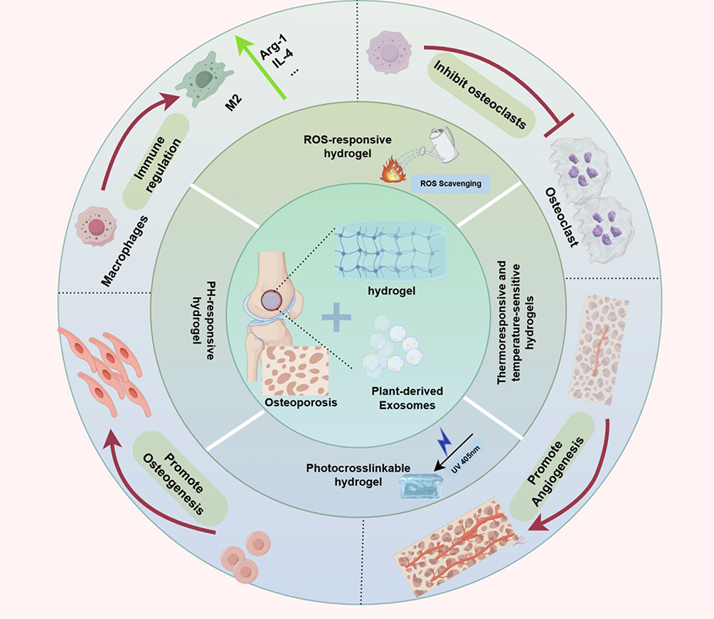 |
Figure 2 Schematic of pathology and synergistic mechanisms of P-Exos-intelligent hydrogel composite for osteoporotic bone defects. Osteoporotic bone defects have imbalanced bone metabolism, inflammation, oxidative stress and insufficient angiogenesis; the composite (with thermosensitive, pH-responsive, photocrosslinkable and liposomal hydrogels) improves pathological microenvironment and achieves bone regeneration by regulating macrophage polarization, inhibiting osteoclasts, promoting osteogenesis and H-type vessel formation. |
Together, this work provides a mechanistic and materials-science framework for developing next-generation, multifunctional, and clinically translatable therapies for osteoporotic bone defects.
Biological Characteristics and Extraction Methods of Plant-Derived Exosomes (P-Exos)
Composition and Biological Functions of P-Exos
Plant-derived exosomes (P-Exos) are nanoscale bilayered vesicles, typically 30–150 nm in diameter, released by a variety of plant tissues.22 Analogous to their mammalian counterparts, P-Exos encapsulate a diverse repertoire of regulatory molecules— including microRNAs, proteins, lipids, secondary metabolites, and antioxidant enzymes—forming a biologically active cargo system capable of mediating intercellular and even cross-kingdom communication.23 Accumulating evidence demonstrates that P-Exos modulate multiple cellular and molecular processes, such as inflammatory signaling cascades, oxidative stress responses, macrophage polarization, osteogenic differentiation, and angiogenesis.22 Their intrinsic stability, low immunogenicity, and naturally derived bioactivity endow them with unique advantages for tissue repair and therapeutic delivery applications.24
Proteomic profiling and Western blot analyses have identified conserved extracellular vesicle (EV)–associated markers in P-Exos, including ESCRT complex proteins (eg, TSG101, ALIX) and heat shock proteins (eg, HSP70), confirming their vesicular identity and evolutionary conservation.25 Functionally, P-Exos attenuate inflammation by downregulating NF-κB–related proinflammatory mediators, mitigate excessive reactive oxygen species via SOD-enriched antioxidative cargo, and exert antitumor effects by modulating apoptosis-associated signaling pathways.26 Together, these multifunctional properties position P-Exos as a promising natural nanoplatform for regenerative medicine, immune modulation, and targeted therapeutic interventions.
Extraction Techniques: Principles, Advantages, and Limitations
Efficient, high-purity isolation of P-Exos is essential for mechanistic investigation and translational application. However, unlike mammalian exosomes, the inherent rigidity of plant cell walls and the abundance of polysaccharides, pigments, and secondary metabolites introduce unique technical barriers.27 Current workflows typically involve sequential steps including plant tissue pretreatment, mechanical and enzymatic disruption, clarification, enrichment, purification, and long-term storage,28 A schematic overview of common PELNs extraction workflows is presented in Figure 3.
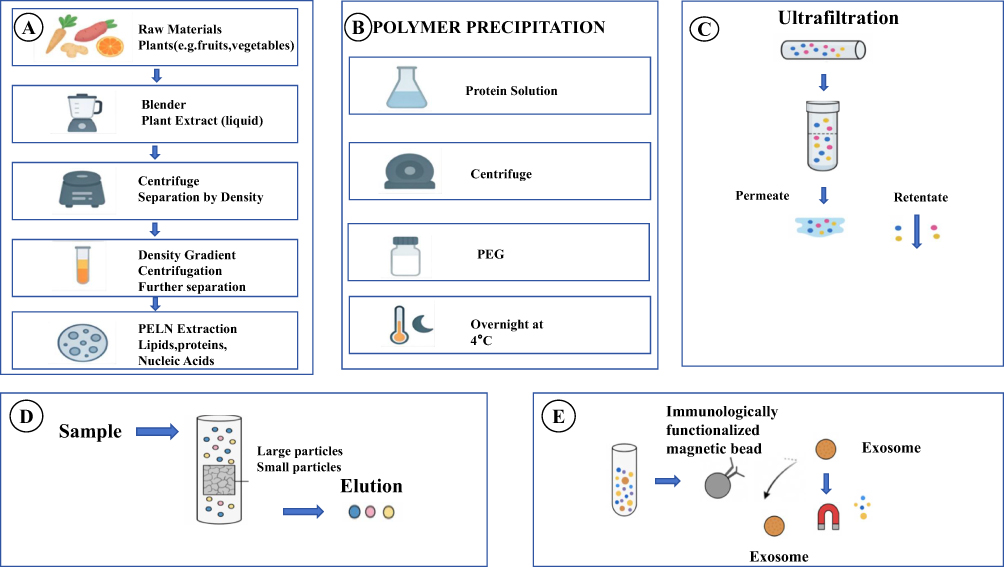 |
Figure 3 Extraction processes of plant-derived exosome-like nanovesicles (PELNs) (A) Basic extraction process: Using plants (eg, fruits, vegetables) as raw materials, plant extract is obtained by blending, then separated via centrifugation and density gradient centrifugation, finally yielding PELNs containing lipids, proteins and nucleic acids. (B) Polymer precipitation: After centrifuging the protein solution, PEG is added and incubated overnight at 4°C to precipitate PELNs. (C) Ultrafiltration: The sample is separated by ultrafiltration membrane—small molecules pass through as permeate, while PELNs are retained as retentate. (D) Size-exclusion chromatography: After sample loading, particles of different sizes are separated by elution to obtain target PELNs. (E) Immunomagnetic bead method: Immunologically functionalized magnetic beads bind specifically to PELNs in the sample, and target vesicles are isolated via magnetic separation. |
Extraction of P-Exos generally relies on several core methodologies that often require combined implementation and protocol optimization. The workflow begins with the selection of fresh, uncontaminated plant tissues (eg, fruits, leaves, roots), followed by thorough washing to remove surface impurities and microorganisms.29 Mechanical homogenization together with enzymatic digestion (eg, cellulase, pectinase) is frequently necessary to disrupt the robust cell wall and release intracellular vesicles.30 Buffer pH, ionic strength, and the inclusion of protease and RNase inhibitors are critical parameters that preserve the bioactive cargo of P-Exos.29
Subsequently, the homogenate is clarified by successive low-speed centrifugation (∼300–2000 g) and/or filtration through 0.45 μm or 0.22 μm membranes to remove coarse debris and organelles.31 The clarified supernatant is then subjected to one or more enrichment techniques—commonly in combination—including differential ultracentrifugation (DUC), polymer-based precipitation, or size-exclusion chromatography (SEC).32 Crude pellets or enriched fractions are resuspended in PBS and further purified by repeated washing or SEC-based desalting to eliminate residual polymers, salts, and soluble contaminants.33 When processing large sample volumes or dilute preparations, ultrafiltration may be applied to concentrate vesicle suspensions. Final preparations are aliquoted in sterile PBS or designated storage buffers and stored at –80 °C, avoiding repeated freeze–thaw cycles.31
The choice of extraction strategy must balance purity, yield, bioactivity preservation, sample availability, and equipment accessibility. Below is a comparative summary of the principal methodologies:Differential ultracentrifugation (DUC):33,34 The most widely adopted technique owing to its accessibility and acceptable purity, but limited by low recovery, long processing time, and potential vesicle deformation due to high shear forces. Polymer-based precipitation:35 High throughput and rapid, yet prone to co-precipitating abundant impurities that compromise downstream functional assays. Size-exclusion chromatography (SEC):36 Achieves excellent purity with minimal structural disruption and is increasingly preferred for mechanistic studies; however, its limited loading capacity and sample dilution necessitate additional concentration steps. Ultrafiltration:37 Effective for concentrating exosomes but may retain similarly sized contaminants and induce vesicle deformation. Immunoaffinity isolation:38 Offers theoretical high specificity but remains constrained by high cost, low throughput, and the lack of well-defined surface markers for plant-derived vesicles.
Despite significant methodological progress, major barriers persist, including the lack of standardized cross-species protocols, yield–purity trade-offs, contamination by polysaccharides and phenolic compounds, and difficulties scaling laboratory processes to clinical-grade production. Quality control typically incorporates nanoparticle tracking analysis (NTA), TEM or cryo-EM, and Western blotting for vesicle characterization. A comparative summary of the principles, advantages, and limitations of these key extraction methodologies is provided in Table 1.
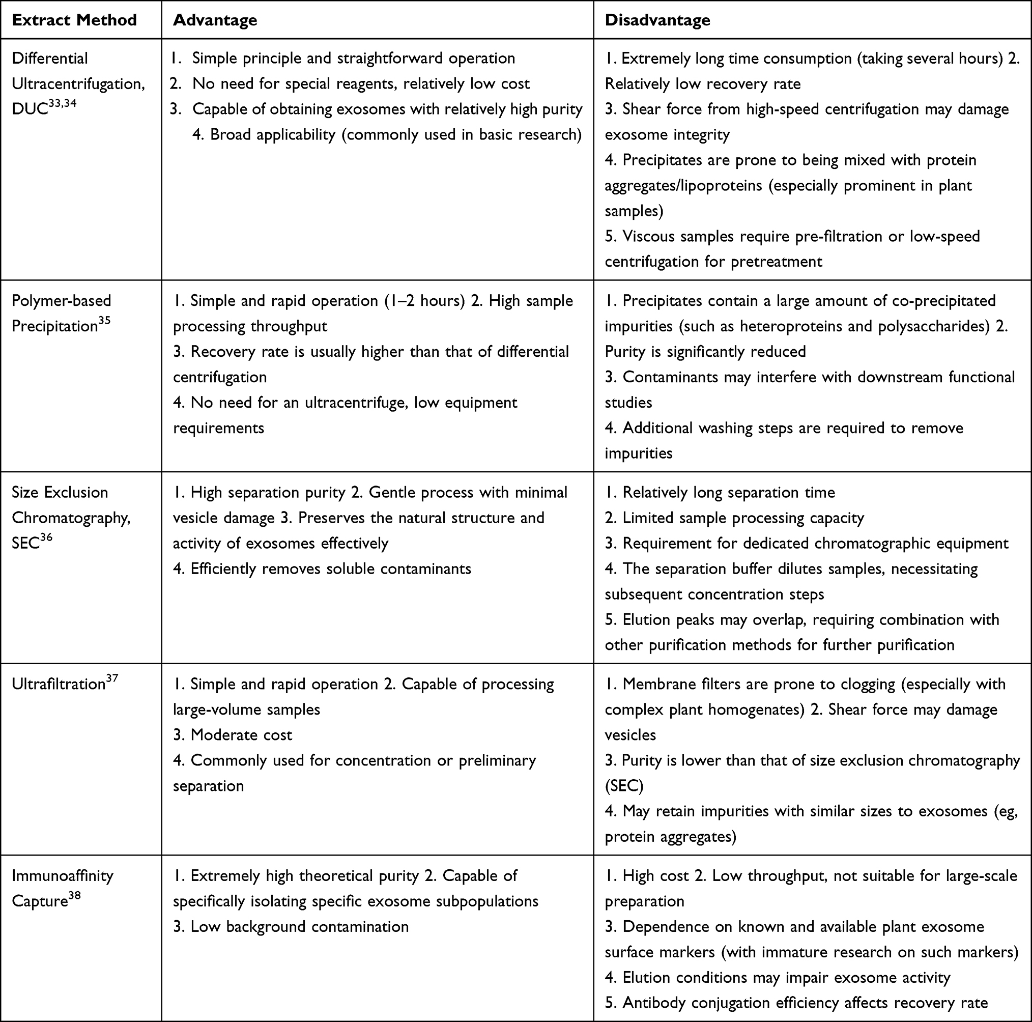 |
Table 1 Extract Method |
Key Considerations and Technical Bottlenecks
Despite significant advances, several unresolved challenges continue to restrict the broader application of P-Exos. Mechanical and chemical stresses generated during extraction may compromise vesicle integrity or degrade miRNA/protein cargo, thereby undermining reproducibility and obscuring mechanistic conclusions.39 Plant-specific impurities—particularly polysaccharides, polyphenols, and pigments—further complicate vesicle characterization and introduce confounding effects in downstream functional assays.40 The absence of universally accepted plant exosome markers remains another critical limitation, reducing the sensitivity and specificity of Western blot–based quality control. In addition, scalable, GMP-compliant manufacturing pipelines remain largely undeveloped, posing a key bottleneck for clinical translation.41
Addressing these technical barriers will require the integration of optimized disruption strategies, high-resolution chromatographic purification, advanced proteomic profiling, and emerging microfluidic-based isolation technologies. Establishing standardized, cross-species extraction–characterization workflows is imperative for accelerating the development and clinical translation of P-Exos–based therapeutics.
Mechanism of Action of P-Exos in Bone Repair 3. P-Exos
Promotion of Osteogenic Differentiation
P-Exos significantly enhance the osteogenic potential of bone marrow mesenchymal stem cells (BMSCs) through multilayered regulatory mechanisms.42 Their bioactive cargos—including plant-derived miRNAs, proteins, lipids, and phytoactive metabolites—activate key osteogenic pathways such as BMP/Smad, Wnt/β-catenin, and MAPK/ERK, consequently upregulating osteogenic markers (Runx2, ALP, COL-I) and promoting osteoblastic lineage commitment.43 Plant-specific miRNAs (eg, miR-26a and autophagy-associated miRNAs) target critical regulators including Smad1 and components of the TGF-β signaling cascade, suppressing adipogenic differentiation and restoring bone metabolic homeostasis.44 In parallel, P-Exos modulate the osteoimmune microenvironment by inhibiting NF-κB signaling, promoting M2 macrophage polarization, and alleviating oxidative stress, thereby establishing a pro-regenerative milieu that supports bone formation.45,46 Notably, exosomes derived from yam and Pueraria lobata have demonstrated the ability to increase trabecular bone mass and improve microarchitecture in osteoporotic models.
Promotion of Angiogenesis
P-Exos also exert potent pro-angiogenic effects by delivering angiogenesis-associated cargos such as miR-126 and miR-210, which activate the PI3K/Akt and VEGF signaling pathways.47 These effects enhance endothelial cell proliferation, migration, and tube formation. Through the modulation of macrophage polarization and attenuation of inflammatory cytokines, P-Exos further facilitate vascular remodeling and endothelial repair.48 When incorporated into intelligent hydrogels, sustained and spatially controlled release of P-Exos upregulates osteogenic–angiogenic coupling mediators (eg, VEGF, BMP-2, ANGPT family), promotes the formation of H-type vessels, and accelerates coordinated angiogenesis and osteogenesis.49,50 This dual-regulatory capacity not only enhances perfusion and nutrient supply but also establishes a vascular microenvironment essential for subsequent matrix deposition and structural bone regeneration.
Multi-Pathway Regulation of Bone Metabolism and Bone-Cartilage Defect Repair
Increasing evidence demonstrates that exosome-like nanovesicles derived from various plants possess unique osteogenic and chondroprotective regulatory functions. A South Korean research group isolated yam-derived nanovesicles (YNVs) that activate the BMP-2/p-p38/Runx2 pathway,51 markedly enhancing proliferation, differentiation, and mineralization of primary mouse osteoblasts and increasing the expression of osteogenic markers including osteopontin (OPN), ALP, and type I collagen (COL-I).52 In ovariectomized (OVX) mouse models, orally administered YNVs significantly increased tibial bone mineral density without inducing systemic toxicity. The process from yam extraction to in vivo osteogenic enhancement is depicted in Figure 4.
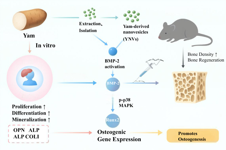 |
Figure 4 Process and mechanism of yam-derived nanovesicles (YNVs) promoting osteoporotic bone regeneration. Yam is used as raw material to obtain yam-derived nanovesicles (YNVs) via extraction and isolation. YNVs can activate bone morphogenetic protein-2 (BMP-2), regulate the expression of osteogenic transcription factor Runx2 through the p-p38 MAPK pathway, and then upregulate the expression of osteogenic genes (OPN, ALP, COLI). This promotes cell proliferation↑, differentiation↑, and mineralization↑ in vitro. After injection administration, it enhances osteogenesis in vivo, ultimately increasing bone density↑ and achieving bone regeneration. |
Chinese researchers extracted Pueraria lobata–derived exosome-like nanovesicles (PELNs), which exert bone-protective effects by modulating the microbiota–gut–bone axis.53 These vesicles reduce circulating levels of the gut-derived metabolite trimethylamine N-oxide (TMAO) in osteoporotic rats,54 thereby enhancing autophagy and promoting the osteogenic differentiation of BMSCs. This protective mechanism via the microbiota-gut-bone axis is schematically summarized in Figure 5.
 |
Figure 5 Mechanism of Pueraria lobata-derived exosome-like nanovesicles (PELNs) protecting osteoporotic bone via the microbiota-gut-bone axisPueraria is used as raw material to obtain Pueraria lobata-derived exosome-like nanovesicles (PELNs) (loaded with autophagy-regulating miRNAs) via extraction and isolation. PELNs regulate gut microbiota, affect trimethylamine N-oxide (TMAO) levels, and activate autophagy by downregulating TMAO, thereby promoting osteogenic differentiation (upregulating osteogenic factors such as OPN, ALP, COLI). After injection administration, it improves the bone microarchitecture in the ovariectomized (OVX) rat model, ultimately achieving bone protection (improved bone density and structure). |
Studies on grapefruit-derived nanovesicles (GEVs) highlight the multi-pathway regulatory capacity of P-Exos. In an in vitro osteoarthritis model, GEV treatment downregulated inflammatory mediators (COX-2, PTGS2) while upregulating antioxidant genes (SOD2, GPX).55 Importantly, GEVs balanced cartilage metabolism by suppressing catabolic (ADAMTS-5) and hypertrophic (COL10) markers while elevating chondrogenic markers (ACAN, COL2, SOX9).55,56 This dual-pathway regulatory action balancing cartilage metabolism is illustrated in Figures 6 and 7. Such multi-target regulatory properties position P-Exos as promising candidates for repairing bone–cartilage composite defects. The key characteristics and mechanisms of these representative P-Exos with bone repair activities are summarized in Table 2.
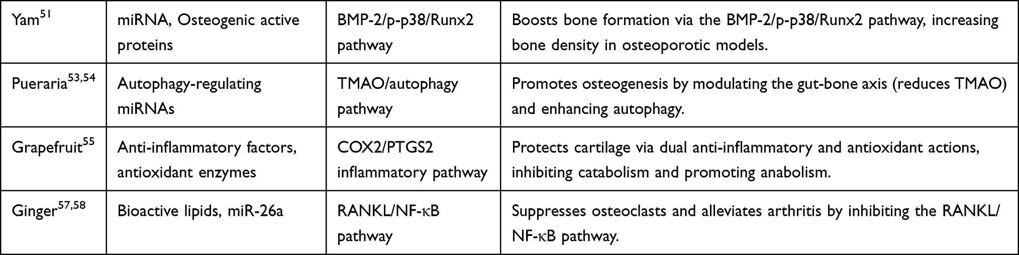 |
Table 2 Representative Plant Exosomes with Bone Repair Activity |
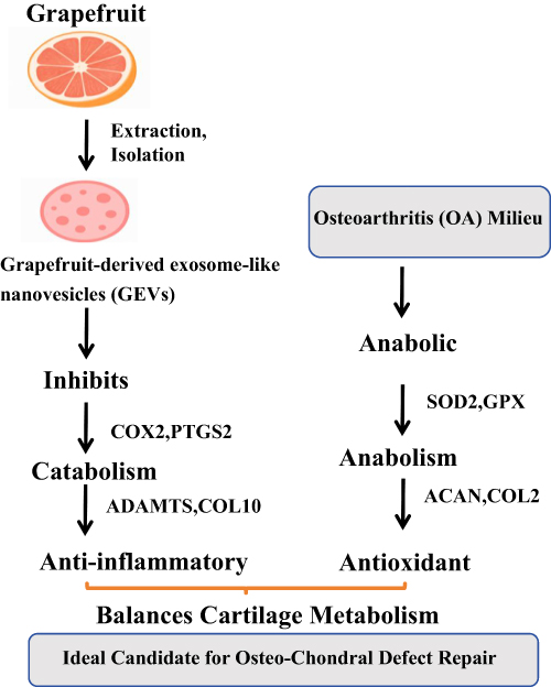 |
Figure 6 Mechanism of grapefruit-derived exosome-like nanovesicles (GEVs) balancing cartilage metabolism for osteo-chondral defect repairGrapefruit is used as raw material to obtain grapefruit-derived exosome-like nanovesicles (GEVs) via extraction and isolation. In the osteoarthritis (OA) milieu, GEVs inhibit catabolism-related factors (COX2, PTGS2, ADAMTS, COL10) to exert anti-inflammatory effects, while upregulate anabolism-related factors (SOD2, GPX, ACAN, COL2) for antioxidant activity. Ultimately, it balances cartilage metabolism and serves as an ideal candidate for osteo-chondral defect repair. |
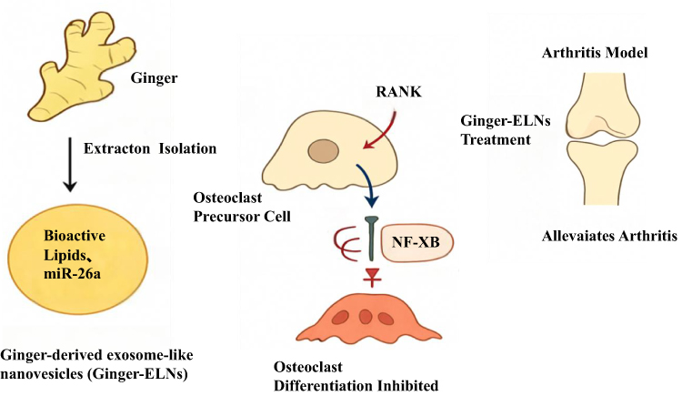 |
Figure 7 Yam-derived exosome-like nanovesicles (YNVs) activate the BMP-2/p-p38/Runx2 signaling pathway to stimulate osteogenic differentiation and increase bone mineral density. |
Advantages of Smart Hydrogels as P-Exos Delivery Carriers
Limitations of Standalone Exosome Therapy
Despite their promising regenerative potential, the clinical translation of plant-derived exosomes (P-Exos) remains hindered by three major limitations. First, free exosomes exhibit rapid in vivo clearance; once systemically administered, they are readily recognized and removed by the mononuclear phagocyte system,59 resulting in a short circulation half-life and insufficient accumulation within bone defect sites. Second, P-Exos display limited responsiveness to the pathological microenvironment of osteoporotic defects, which is characterized by localized acidity due to osteoclast-secreted metabolites, elevated oxidative stress, and enhanced protease activity.60 Free vesicles lack the capacity to sense and adapt to these pathological cues. Third, P-Exos administered as a single bolus cannot achieve the spatiotemporally coordinated release required to match the sequential inflammatory, reparative, and remodeling phases of bone healing.43
To address these challenges, the development of advanced carrier systems has become crucial for enhancing the therapeutic efficacy of P-Exos. An optimal delivery platform should provide: (1) high loading efficiency and robust protection to prevent premature degradation of exosomal cargo; (2) targeted delivery to increase local vesicle concentration at defect sites; (3) microenvironment-responsive release behavior, enabling on-demand cargo delivery; and (4) a three-dimensional scaffold capable of supporting cell infiltration, migration, and new bone matrix deposition.
Hydrogels—owing to their tunable physicochemical properties, excellent biocompatibility, and extracellular matrix–mimicking 3D architecture—are particularly well suited as delivery carriers for P-Exos.11 Notably, intelligent responsive hydrogels can sense exogenous stimuli (eg, temperature, pH, light, or enzymes) and undergo sol–gel transitions or structural reorganization, providing a versatile platform for precise, controlled exosome release.
Design Strategies for Smart Responsive Hydrogels (Thermosensitive, pH-Responsive, Photocrosslinkable, etc.) and Controlled Release Efficiency
Thermosensitive Hydrogels
Thermosensitive hydrogels exhibit temperature-dependent sol–gel transitions, enabling in situ encapsulation and controlled release of P-Exos.61 Representative thermoresponsive polymers—such as poly(N-isopropylacrylamide) (PNIPAM), poloxamers, and chitosan/β-glycerophosphate systems—remain injectable at room temperature and rapidly form crosslinked gel networks at physiological temperature. Zhang et al developed a chitosan/β-glycerophosphate/collagen composite hydrogel,62 which completed gelation within 60 seconds at 37 °C after loading yam-derived exosomes. This system significantly slowed exosome diffusion within bone defects and provided sustained release for at least 14 days in vitro.
pH-Responsive Hydrogels
Given the acidic microenvironment (pH 5.5–6.5) of osteoporotic bone defects, pH-responsive hydrogels achieve acid-triggered release by incorporating functional groups such as carboxyl,63 imine, or sulfonamide moieties. Poly(acrylic acid) (PAA)/chitosan composite hydrogels remain stable under physiological pH but undergo protonation-induced swelling under acidic conditions,64 accelerating the release of encapsulated vesicles. A histidine-modified sodium alginate hydrogel developed by Liu et al, after loading Pueraria lobata–derived exosomes, exhibited a 3.2-fold higher release rate at pH 6.0 than at pH 7.4,65 substantially improving targeted delivery to osteoclast-active bone resorption surfaces.
Photocrosslinkable Hydrogels
Photocurable hydrogels—such as methacrylated gelatin (GelMA) and methacrylated hyaluronic acid (HAMA)—crosslink into 3D networks via photo-initiated free-radical polymerization,66 offering precise control over gelation kinetics and mechanical properties. Under blue or UV light, photoinitiators (eg, riboflavin or LAP) trigger polymer crosslinking, encapsulating exosomes within the hydrogel matrix.67 For example, GelMA hydrogels loaded with grapefruit-derived exosomes form gels within 20 seconds under 405-nm visible light. Their compressive modulus can be tuned between 1–5 MPa to match cancellous bone stiffness,68 thereby supporting osteoblast infiltration and bone integration. The design strategies, release characteristics, and exemplary applications of these intelligent hydrogels for P-Exos delivery are comparatively outlined in Table 3.
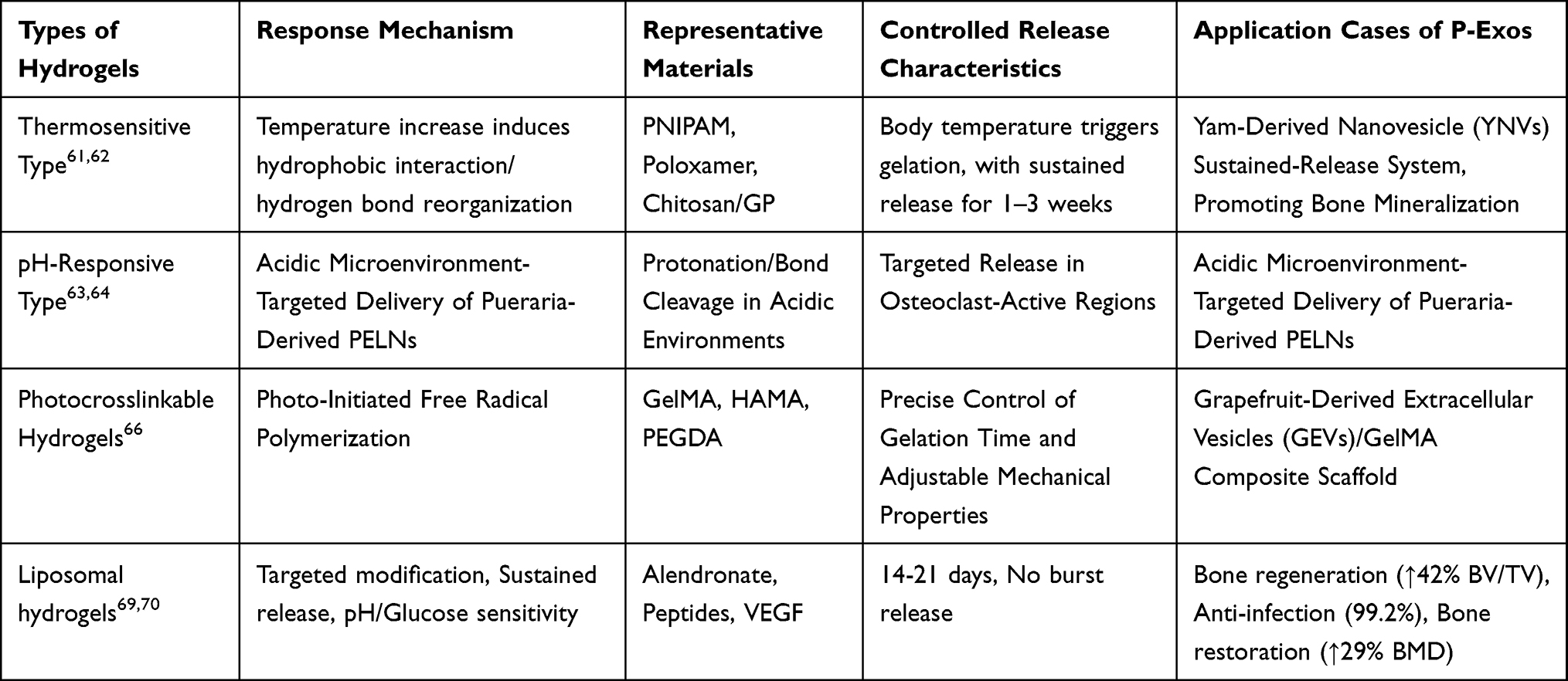 |
Table 3 Comparison of Properties of Three Types of Smart Hydrogels and Their Applications in Plant Exosome Delivery |
Liposomal Hydrogels
Liposomal intelligent hydrogels enable highly precise targeted delivery and controlled release of anti-osteoporotic therapeutics through three principal mechanisms:Bone-targeted modification: Alendronate or bone-specific peptides conjugated onto liposomal surfaces increase hydroxyapatite binding affinity,71 enhancing bone accumulation by 3–5-fold. Enhanced stability and sustained release: The dual barrier of liposome-in-hydrogel systems extends release profiles to 14–21 days, mitigates burst release, and protects fragile growth factors such as VEGF.69 Microenvironment responsiveness: pH- or glucose-sensitive designs enable on-demand release in acidic osteoporotic defects. In preclinical studies, these hydrogels have been shown to:enhance bone regeneration (↑BV/TV by 42%, ↑ALP activity by 58%),exhibit potent anti-infective effects (99.2% S. aureus clearance, recurrence < 8%),70 and restore osteoporotic bone structure (↑BMD by 29%, ↑Tb.N by 31% in OVX rats).72 Such multifunctional systems integrate high targeting specificity, biocompatibility, and microenvironment adaptability, offering a promising platform for osteoporotic bone defect repair.
Synergistic Effects of the P-Exos/Hydrogel Composite System
P-Exos/Hydrogel
The immunocompatibility between the implanted material and the host is a key factor for the successful repair of bone defects. Traditional biomaterials often trigger a foreign body reaction, leading to the formation of a fibrous capsule and the failure of osseointegration. The P-Exos/hydrogel composite system actively regulates macrophage polarization to construct an immune microenvironment conducive to bone regeneration.43 Studies have shown that thermosensitive hydrogels loaded with yam exosomes can promote the polarization of macrophages to the M2 anti-inflammatory phenotype, significantly downregulating the expression of pro-inflammatory factors such as TNF-α and IL-6, while upregulating the levels of repair-related cytokines such as IL-10 and TGF-β.21 This immunomodulatory effect is closely related to the miRNAs (eg, miR-24, miR-146a) carried by P-Exos. These miRNAs inhibit excessive inflammatory responses by targeting the Toll-like receptor signaling pathway, thereby creating favorable conditions for bone tissue regeneration.
Successful bone regeneration relies on the spatiotemporal coordination of vascularization and osteogenesis, namely “angiogenesis-osteogenesis coupling”. The P-Exos/hydrogel system plays a dual promotional role in this process: on the one hand, the three-dimensional porous structure of the hydrogel provides a physical scaffold for the migration of vascular endothelial cells;73 on the other hand, pro-angiogenic factors (eg, miR-126, miR-210) carried by plant exosomes (such as Pueraria lobata PELNs) can significantly promote angiogenesis by activating the PI3K/Akt signaling pathway in endothelial cell.74 In addition, studies have shown that oyster-derived exosomes can effectively promote osteogenic differentiation by activating the PI3K/Akt/β-catenin signaling pathway, thereby synergizing with the angiogenesis process to further enhance bone regeneration effects.75 This characteristic of synchronized angiogenesis and osteogenic differentiation enables the composite system to exhibit significant advantages in repairing large-segment bone defects.
Intelligent hydrogels can accurately simulate the physicochemical properties of the bone extracellular matrix, providing an ideal microenvironment for the proliferation and differentiation of stem cells. For example, photocrosslinkable GelMA hydrogels loaded with grapefruit exosomes can adjust their elastic modulus to match that of normal bone tissue (~15 kPa),76 thereby providing appropriate mechanical stimulation to induce the differentiation of mesenchymal stem cells toward the osteogenic direction. At the same time, the hydrogel network can maintain the effective local concentration of active molecules in exosomes,77 significantly extending their action time. In vitro experimental results show that this composite system can increase the expression levels of osteogenesis-related genes (Runx2, Osterix, Osteocalcin) by 3–5 times, and significantly promote the formation of calcium nodules and matrix mineralization.
Regulation of the Bone Immune Microenvironment
Immunocompatibility between implanted materials and host tissues is fundamental for successful bone defect regeneration. Conventional biomaterials frequently elicit foreign-body reactions that result in fibrous encapsulation and impaired osseointegration.78 In contrast, the P-Exos/hydrogel composite system actively modulates macrophage polarization to establish a pro-regenerative immune microenvironment. Studies have demonstrated that thermosensitive hydrogels loaded with yam-derived exosomes promote polarization toward the M2 anti-inflammatory phenotype,79,80 markedly decreasing pro-inflammatory cytokines such as TNF-α and IL-6 while elevating repair-associated cytokines including IL-10 and TGF-β. These immunoregulatory effects are closely mediated by P-Exos-derived miRNAs (eg, miR-24, miR-146a),81 which attenuate excessive inflammation through targeted inhibition of Toll-like receptor signaling pathways, thereby creating favorable conditions for subsequent bone regeneration.
Angiogenesis–Osteogenesis Coupling
Bone regeneration critically depends on the spatiotemporal coupling between angiogenesis and osteogenesis. The P-Exos/hydrogel system simultaneously facilitates both processes. The 3D porous hydrogel architecture provides structural guidance for endothelial cell migration, whereas pro-angiogenic cargos (eg, miR-126 and miR-210) within P-Exos—such as Pueraria lobata-derived PELNs—activate the PI3K/Akt pathway to promote vascular formation.82 Additionally, oyster-derived exosomes have been shown to stimulate osteogenic differentiation via PI3K/Akt/β-catenin signaling,64 further enhancing the coordinated development of vasculature and bone tissue. This synchronized promotion of angiogenesis and osteogenesis confers significant advantages for repairing large-segment or poorly vascularized bone defects.
Construction of a 3D Biomimetic Microenvironment
Intelligent hydrogels can accurately mimic the physicochemical characteristics of the bone extracellular matrix, thereby providing an ideal microenvironment for stem cell proliferation and osteogenic differentiation. The selection of scaffold materials for bone defect repair requires refined design driven by several key indicators. (1) Pore architecture: Biomimetic trabecular scaffolds typically require an optimal pore size distribution of 200–1200 μm, with an average pore size of approximately 700 μm and a porosity of 70%.83 Gradient pore architectures (eg, Ti6Al4V alloy scaffolds) can enhance bone tissue ingrowth, while multi-scale hierarchical porous structures such as those in PCL/SF/β-TCP-NaOH scaffolds enable personalized defect reconstruction. Morphological parameters should be mechanically benchmarked against natural trabeculae (eg, bovine iliac crest) to ensure a stable osteogenic microenvironment84 by recapitulating the three-dimensional topology of native bone. (2) Mechanical properties: High-performance scaffolds should possess a compressive strength of approximately 85.99 ± 10.03 MPa,85 and their elastic modulus and yield strength must match that of cancellous bone. The stiffness and porosity of the porous network should be adjustable depending on the anatomical site (eg, load-bearing requirements of jaw defect repair).86 Moreover, long-term mechanical retention after implantation must be verified through multi-stage mechanical testing. (3) Biocompatibility: Scaffolds should support robust cell adhesion (eg, ALP activity of 1.71 ± 0.25 U/mg protein) and mineralization, exhibit antioxidant and anti-inflammatory capabilities (eg, Hydrogel-Exos-SeNPs systems),87 and upregulate osteogenic markers such as CD31, RUNX2, and BMP2, as validated in rat calvarial defect models. (4) Osteogenic and angiogenic performance: Composite scaffolds should concurrently promote angiogenesis (eg, icariin-loaded scaffolds) and osteogenic differentiation (eg, EVs-3D-HA scaffolds).88 Incorporation of alkaline-earth-metal-containing materials can enhance osteoinductivity, while mineralized collagen (MC) can synergistically accelerate bone regeneration. Precise spatial control of growth factor release and real-time monitoring of scaffold degradation are also desirable for optimizing repair outcomes.
Compared with metallic and ceramic materials, hydrogels exhibit distinct advantages. Their three-dimensional polymeric networks can fully conform to irregular defect geometries while more closely resembling the extracellular matrix.89 Hydrogels can encapsulate metal nanoparticles (eg, Au@hydrogels) while preserving catalytic function, and exhibit excellent drug-loading and sustained-release capacities (eg, Exo-Gel systems). The high water content facilitates nutrient transport, and metal-ion-hybrid hydrogels—such as those incorporating selenium nanoparticles—exhibit unique osteoinductive activity.76 Hydrogels also avoid the stress-shielding effects associated with metals and the inherent brittleness of ceramics, enabling minimally invasive implantation via injection.
Translational Advantages of Plant-Exosome–Intelligent Hydrogel Systems in Clinical Wound Pain Management
The plant exosome–intelligent hydrogel composite system exhibits substantial novelty and remarkable translational potential in clinical wound pain management. Its innovation lies in overcoming the limitations of traditional single-agent therapies by integrating the intrinsic biological activities of plant-derived exosomes with the environmental responsiveness of intelligent hydrogels,90 thereby establishing a unified “active analgesia–synergistic healing” therapeutic platform. As an emerging bioactive carrier, plant exosomes deliver miRNAs, flavonoids, and other functional molecules capable of modulating macrophage polarization to exert anti-inflammatory effects.66 Meanwhile, intelligent hydrogels enable on-demand drug release in response to microenvironmental cues such as temperature and pH. For instance,60 temperature-responsive hydrogels can attenuate pain signaling through localized cooling, while the hydrogel matrix simultaneously provides a physical protective barrier that minimizes secondary mechanical irritation to the wound. This dual strategy of “biological regulation + intelligent delivery” transforms the traditional passive paradigm of wound dressings that merely cover the wound without actively addressing pain.
In clinical contexts, the composite system directly addresses several major therapeutic challenges. The hydrogel scaffold markedly prolongs the local retention and bioavailability of exosomes, while the incorporated anti-inflammatory bioactive constituents can inhibit the NF-κB signaling pathway and suppress pro-inflammatory mediators such as TNF-α and IL-6,91 thereby interrupting the inflammation–pain vicious cycle at its source. For refractory wounds—including burns and diabetic ulcers—injectable hydrogels can readily adapt to irregular wound geometries and maintain an optimal moist environment for tissue repair. Coupled with the pro-angiogenic and regenerative activities of exosomes,92 the system accelerates wound closure while reducing pain hypersensitivity. Notably, similar clinical studies have reported up to a 60% reduction in wound dressing–associated pain scores (VAS). Importantly, the low immunogenicity of plant-derived exosomes, together with the excellent biocompatibility of hydrogels formulated from recombinant human collagen and hyaluronic-acid derivatives,93 provides a strong foundation for clinical translation. This combined system represents a safe and efficient next-generation therapeutic strategy for managing pain and promoting healing in complex wounds.In diabetic wound repair, this bitter melon-derived exosome-hydrogel system intelligently adapts to the pathological microenvironment. It inhibits inflammation, scavenges ROS, reduces AGEs, and polarizes M1 to M2 macrophages while promoting angiogenesis, thus achieving superior wound healing compared to single therapies. Notably, the pro-angiogenic and immunomodulatory principles demonstrated in diabetic wound healing by systems such as the bitter melon exosome-hydrogel (Figure 8) are directly relevant to addressing the vascular deficiency and chronic inflammation in osteoporotic bone defects.
 |
Figure 8 Figure: Process and regulatory mechanism of bitter melon-derived exosome-hydrogel system promoting diabetic wound healing Bitter melon (MC) is used as raw material; MC exosomes are obtained via juicing and ultracentrifugation, and then combined with dopamine and GelMA to construct a hydrogel. When applied to diabetic wounds, this system exerts anti-inflammatory effects (inhibiting inflammation), resists oxidative stress (scavenging ROS), reduces advanced glycation end products (AGEs), and modulates macrophage polarization from M1 to M2. These effects synergistically promote angiogenesis, ultimately achieving wound healing and tissue repair. |
Current Challenges and Future Prospects
The collective evidence reviewed in this article demonstrates that plant-derived exosomes (P-Exos) and intelligent hydrogel systems are emerging as a powerful and conceptually transformative strategy for osteoporotic bone regeneration. P-Exos possess intrinsic biological advantages—including multi-target molecular cargo, exceptional biocompatibility, low immunogenicity, and natural stability—that enable them to regulate osteoimmunity, inhibit osteoclast overactivation, enhance angiogenesis–osteogenesis coupling, alleviate oxidative stress, and restore the impaired metabolic balance characteristic of osteoporotic bone. Intelligent hydrogels complement these biological properties by providing a biomimetic extracellular matrix–like niche with controllable physicochemical features, tunable degradation profiles, and spatiotemporally programmable release dynamics. Their integration results in a synergistic therapeutic system capable of addressing the complex microenvironmental disruptions that hinder bone repair in osteoporosis, surpassing the limitations of conventional pharmacological treatments and single-factor regenerative approaches. As accumulating high-quality studies reveal, this composite strategy provides a compelling biological rationale and a robust materials foundation to support future guideline development and the advancement of precision orthopedic interventions.
Despite these notable advances, the field remains at an early yet rapidly accelerating stage. Many of the promising findings have been validated predominantly in small-animal models or simplified in vitro environments, leaving important questions concerning stability, biodistribution, immunoregulatory compatibility, long-term biosafety, and therapeutic durability unresolved. The next developmental phase of P-Exos/hydrogel systems will require an expanded evidence base supported by rigorous mechanistic studies and translationally relevant biomaterial engineering. At the biological level, a deeper understanding of the molecular determinants that govern P-Exos functionality is essential. Their heterogeneous cargo—which includes miRNAs, lipids, proteins, flavonoids, polyphenols, and antioxidant metabolites—exerts multifaceted regulatory influences on bone-resident cells, endothelial networks, immune cells, and sensory fibers. Yet the precise cross-kingdom gene-regulatory interactions, target specificity, intracellular trafficking mechanisms, and cell-type–selective responses remain insufficiently characterized. Multi-omics integration, single-cell atlasing, live-cell RNA tracking, and high-resolution proteomics will provide crucial insights into how P-Exos orchestrate osteogenic signaling, reprogram macrophage polarization, coordinate angiogenic–osteogenic interactions, and mitigate chronic inflammatory signaling in aged and osteoporotic bone.
Concurrently, material science innovations will play a decisive role in elevating the therapeutic performance and clinical applicability of P-Exos. Intelligent hydrogels have evolved from simple aqueous networks into sophisticated, digitally designed, and stimulus-responsive biomaterials capable of recognizing disease-associated physicochemical cues. Advanced 3D and 4D bioprinting technologies now enable the fabrication of spatially graded, mechanically adaptive, multi-layered hydrogel constructs that mirror the hierarchical architecture of trabecular and cortical bone. Low-temperature extrusion printing preserves P-Exos bioactivity, while magneto-responsive and shape-morphing 4D hydrogels offer opportunities to create dynamic scaffolds capable of conforming to irregular defects or responding to external magnetic or thermal stimuli. Microfluidic platforms further enhance the manufacturing precision of both P-Exos and hydrogels, enabling high-throughput exosome isolation, cargo enrichment, uniform vesicle encapsulation, and the construction of hydrogel microspheres or microfibers with precisely defined release kinetics. These technologies collectively overcome major barriers such as uncontrolled burst release, low loading efficiency, heterogeneous distribution, and insufficient retention in osteoporotic bone defects.
In addition to structural and manufacturing innovations, chemical and genetic engineering strategies hold significant promise for augmenting the therapeutic potency of P-Exos/hydrogel systems. Surface-functionalized hydrogels incorporating collagen-binding domains, bone-targeting peptides, or integrin-recognition motifs can enhance defect localization, cell–material interactions, and microenvironment sensing. Chemical conjugation strategies, such as catechol-based adhesion, dynamic imine bonding, or phenylboronic acid crosslinking, confer improved tissue adhesion, ROS scavenging, or cell-mediated remodeling capacity. Meanwhile, gene-editing technologies—including CRISPR–Cas platforms and miRNA engineering—enable the design of P-Exos with customized cargo profiles tailored to specific pathological features of osteoporotic bone, such as impaired Wnt/β-catenin signaling, elevated RANKL/RANK activation, or endothelial senescence. The combination of engineered exosomes with microenvironment-responsive hydrogels represents a promising direction for developing programmable, next-generation regenerative systems.
Given the increasing complexity of material–biological interactions, artificial intelligence (AI) will become a central enabler of future development. Machine-learning models can analyze extensive datasets involving rheological parameters, crosslinking chemistries, printing properties, degradation curves, and in vivo regeneration outcomes. These data-driven frameworks will help predict optimal hydrogel formulations, refine P-Exos release kinetics, and design patient-specific constructs with tailored mechanical stiffness and biological performance. AI tools may eventually support clinical decision-making by predicting which composite formulations are best suited for specific patient populations or defect conditions, thereby enhancing precision medicine in orthopedic care.
For successful clinical translation, future studies must extend beyond bone formation endpoints to consider broader functional outcomes such as pain alleviation, mobility restoration, complication reduction, and postoperative wound healing—metrics of particular relevance for elderly osteoporotic patients. The ability of hydrogels and exosomes to modulate neuroinflammation, reduce nociceptive sensitization, and promote rapid soft-tissue healing represents an underexplored but clinically important frontier. Furthermore, large-animal studies that incorporate real-world challenges—such as load-bearing conditions, comorbidities, infection risks, and chronic inflammation—are essential to validate therapeutic robustness. Addressing regulatory, manufacturing, and standardization barriers will require establishing Good Manufacturing Practice (GMP)–compatible workflows, robust quality-control matrices, and reproducible purification–characterization pipelines that ensure batch-to-batch consistency of P-Exos used for clinical applications.
In summary, P-Exos–loaded intelligent hydrogels are positioned to become a next-generation therapeutic modality for osteoporotic bone regeneration, supported by synergistic biological efficacy, adaptive material design, and rapidly advancing fabrication technologies. The convergence of biomaterials engineering, stem cell biology, exosome science, microfluidics, gene editing, and AI-driven material informatics will catalyze the development of more potent, predictable, and clinically translatable regenerative platforms. As research continues to elucidate mechanistic pathways and overcome translational challenges, P-Exos/hydrogel systems hold significant promise not only for improving bone regeneration outcomes but also for informing future guideline development, shaping multidisciplinary clinical care strategies, and ultimately transforming therapeutic paradigms for patients suffering from osteoporotic bone defects.
Conclusion
Plant-derived exosomes (P-Exos) combined with intelligent hydrogels represent a promising strategy for osteoporotic bone regeneration. This composite system enables sustained and targeted delivery of therapeutic cargo, effectively addressing the complex bone microenvironment through synergistic regulation of osteogenesis, angiogenesis, and immunomodulation. Despite challenges in standardization and clinical translation, continued development of these systems holds significant potential for advancing the treatment of osteoporotic bone defects.
Author Contributions
All authors made a significant contribution to the work reported, whether that is in the conception, study design, literature retrieval, analysis and interpretation, or in all these areas; took part in drafting, revising or critically reviewing the article; gave final approval of the version to be published; have agreed on the journal to which the article has been submitted; and agree to be accountable for all aspects of the work.
Funding
This study was supported by the Huzhou Science and Technology Plan Public Welfare Application Research Project (No. 2022GY13).
Disclosure
The authors declare no competing interests.
References
1. Jiménez-Ortega RF, Aparicio-Bautista DI, Becerra-Cervera A, et al. The regulatory role of long non-coding RNAs in the development and progression of osteoporosis. Int J Mol Sci. 2025;26(9):4273. doi:10.3390/ijms26094273
2. Li J, Li L, Wu T, et al. An injectable thermosensitive hydrogel containing resveratrol and dexamethasone-loaded carbonated hydroxyapatite microspheres for the regeneration of osteoporotic bone defects. Small Methods. 2024;8(1):e2300843. doi:10.1002/smtd.202300843
3. Yang L, Chen X, Chen L, et al. Study of injectable hydrogel based on ALN/nHA promoting osteogenesis and inhibiting osteoclasts in osteoporotic bone defects repair. Macromol Biosci. 2024;24(4):e2300416. doi:10.1002/mabi.202300416
4. Cui Y, Guo Y, Kong L, et al. A bone-targeted engineered exosome platform delivering siRNA to treat osteoporosis. Bioact Mater. 2022;10:207–221. doi:10.1016/j.bioactmat.2021.09.015
5. Li Y, Zha Y, Hu W, et al. monoporous microsphere as a dynamically movable drug carrier for osteoporotic bone remodeling. Adv Healthc Mater. 2023;12(16):e2201242. doi:10.1002/adhm.202201242
6. Wang W, Zhang X, Zhang L, et al. An immunomodulating regenerating hydrogel that rescues the oxidative microenvironment and reverses cell senescence for osteoporotic bone defects. ACS Nano. 2025;19(31):28353–28371. doi:10.1021/acsnano.5c06549
7. Min Z, Zou Y, Meng Y, et al. Harnessing redox: biocomposites modulate macrophage-stem cell dynamics in osteo-inflammation. Tissue Eng Part B Rev. 2025. doi:10.1177/19373341251377651
8. Jiang L, Dong J, Wei J, Liu L. Comparison of denosumab and oral bisphosphonates for the treatment of glucocorticoid-induced osteoporosis: a systematic review and meta-analysis. BMC Musculoskelet Disord. 2022;23(1):1027. doi:10.1186/s12891-022-05997-0
9. Noh M, Che X, Jin X, et al. Dimeric (R25C)PTH(1-34) activates the parathyroid hormone-1 receptor in vitro and stimulates bone formation in osteoporotic female mice. Elife. 2025;13:RP97579. doi:10.7554/eLife.97579.5
10. He P, Zhao Y, Wang B, et al. A biodegradable magnesium phosphate cement incorporating chitosan and rhBMP-2 designed for bone defect repair. J Orthop Translat. 2024;49:167–180. doi:10.1016/j.jot.2024.08.004
11. Hu W, Xie X, Xu J. Epimedium-derived exosome-loaded gelma hydrogel enhances MC3T3-E1 osteogenesis via PI3K/Akt pathway. Cells. 2025;14(15):1214. doi:10.3390/cells14151214
12. Dayanandan AP, Bello AB, Arai Y, Lee SJ, Lee SH. Therapeutic strategy for exosome-based bone regeneration to osteoporosis: challenges and potential solutions. J Adv Res. 2025;
13. Wang L, Yang L, Tian L, et al. Exosome-capturing scaffold promotes endogenous bone regeneration through neutrophil-derived exosomes by enhancing fast vascularization. Biomaterials. 2025;319:123215. doi:10.1016/j.biomaterials.2025.123215
14. Zhu F, Wang T, Wang G, Yan C, He B, Qiao B. The exosome-mediated bone regeneration: an advanced horizon toward the isolation, engineering, carrying modalities, and mechanisms. Adv Healthc Mater. 2024;13(19):e2400293. doi:10.1002/adhm.202400293
15. Peng S, Liu X, Chang L, et al. Exosomes derived from rejuvenated stem cells inactivate NLRP3 inflammasome and pyroptosis of nucleus pulposus cells via the transfer of antioxidants. Tissue Eng Regen Med. 2024;21(7):1061–1077. doi:10.1007/s13770-024-00663-z
16. Amini H, Namjoo AR, Narmi MT, et al. Exosome-bearing hydrogels and cardiac tissue regeneration. Biomater Res. 2023;27(1):99. doi:10.1186/s40824-023-00433-3
17. Cao J, Yuan P, Wu B, Liu Y, Hu C. Advances in the research and application of smart-responsive hydrogels in disease treatment. Gels. 2023;9(8):662. doi:10.3390/gels9080662
18. Xing Z, Guo L, Li S, et al. Skeletal muscle-derived exosomes prevent osteoporosis by promoting osteogenesis. Life Sci. 2024;357:123079. doi:10.1016/j.lfs.2024.123079
19. Chen W, Zhang H, Zhou Q, Zhou F, Zhang Q, Su J. Smart hydrogels for bone reconstruction via modulating the microenvironment. Research. 2023;6:0089. doi:10.34133/research.0089
20. Lin SH, Hsu SH. Smart hydrogels for in situ tissue drug delivery. J Biomed Sci. 2025;32(1):70. doi:10.1186/s12929-025-01166-2
21. Chen D, Yang Y, Li B, et al. Nanocomposite hydrogels optimize the microenvironment by exterior/interior crosstalk for reprogramming osteoporotic homeostasis in bone defect healing. J Control Release. 2025;380:976–993. doi:10.1016/j.jconrel.2025.02.048
22. Mizusaki M, Endo T, Nakahata R, Morishima Y, Yusa SI. pH-induced association and dissociation of intermolecular complexes formed by hydrogen bonding between diblock copolymers. Polymers. 2017;9(8):367. doi:10.3390/polym9080367
23. Ma S, Zhang Y, Li S, Li A, Li Y, Pei D. Engineering exosomes for bone defect repair. Front Bioeng Biotechnol. 2022;10:1091360. doi:10.3389/fbioe.2022.1091360
24. Hu S, Wang S, Yang X, et al. Exosomes promise better bone regeneration. Regen Ther. 2025;30:389–402. doi:10.1016/j.reth.2025.06.020
25. Chen X, An H, Du Y, et al. hucMSC-derived exosomes targeting macrophage polarization attenuate systemic inflammation in T1DM via INS/SOD1 delivery. Stem Cell Res Ther. 2025;16(1):384. doi:10.1186/s13287-025-04521-0
26. Liu H, Zhu D, Lang L, et al. Umbilical cord blood-derived exosomes deliver miR-182-5p to Therapeutically target the MYD88/NF-κB signaling pathway in rat peri-implantitis. Mater Today Bio. 2025;34:102246. doi:10.1016/j.mtbio.2025.102246
27. Huang J, Xu Y, Wang Y, et al. Advances in the study of exosomes as drug delivery systems for bone-related diseases. Pharmaceutics. 2023;15(1):220. doi:10.3390/pharmaceutics15010220
28. Samal S, Dash P, Dash M. Drug delivery to the bone microenvironment mediated by exosomes: an axiom or enigma. Int J Nanomed. 2021;16:3509–3540. doi:10.2147/IJN.S307843
29. Xu T, Gao S, Yang N, et al. A personalized biomimetic dual-drug delivery system via controlled release of PTH(1-34) and simvastatin for in situ osteoporotic bone regeneration. Front Bioeng Biotechnol. 2024;12:1355019. doi:10.3389/fbioe.2024.1355019
30. Chen ZH, Du DY, Fu YF, et al. Citric acid-modified pH-sensitive bone-targeted delivery of estrogen for the treatment of postmenopausal osteoporosis. Mater Today Bio. 2023;22:100747. doi:10.1016/j.mtbio.2023.100747
31. Hoang HT, Jo SH, Phan QT, et al. Dual pH-/thermo-responsive chitosan-based hydrogels prepared using “click” chemistry for colon-targeted drug delivery applications. Carbohydr Polym. 2021;260:117812. doi:10.1016/j.carbpol.2021.117812
32. Lu YT, Zeng K, Fuhrmann B, Woelk C, Zhang K, Groth T. Engineering of stable cross-linked multilayers based on thermo-responsive PNIPAM-grafted-chitosan/heparin to tailor their physiochemical properties and biocompatibility. ACS Appl Mater Interfaces. 2022;14(26):29550–29562. doi:10.1021/acsami.2c05297
33. Rahmatinejad F, Kharat Z, Jalili H, Renani MK, Mobasheri H. Comparison of morphology, protein concentration, and size distribution of bone marrow and Wharton’s jelly-derived mesenchymal stem cells exosomes isolated by ultracentrifugation and polymer-based precipitation techniques. Tissue Cell. 2024;88:102427. doi:10.1016/j.tice.2024.102427
34. Gao J, Li A, Hu J, Feng L, Liu L, Shen Z. Recent developments in isolating methods for exosomes. Front Bioeng Biotechnol. 2022;10:1100892. doi:10.3389/fbioe.2022.1100892
35. Huang Q, Wang J, Ning H, Liu W, Han X. Exosome isolation based on polyethylene glycol (PEG): a review. Mol Cell Biochem. 2025;480(5):2847–2861. doi:10.1007/s11010-024-05191-x
36. Shaabani N, Meira SR, Marcet-Palacios M, Kulka M. Multiparametric biosensors for characterizing extracellular vesicle subpopulations. ACS Pharmacol Transl Sci. 2023;6(3):387–398. doi:10.1021/acsptsci.2c00207
37. Wang HY, Xie PJ, Qiao XQ, Zhang LY. Typical strategy and research progress of efficient isolation methods of exosomes based on affinity interaction. Se Pu. 2025;43(5):413–423. doi:10.3724/SP.J.1123.2024.11004
38. Shen J, Ma Z, Xu J, et al. Exosome isolation and detection: from microfluidic chips to nanoplasmonic biosensor. ACS Appl Mater Interfaces. 2024;16(18):22776–22793. doi:10.1021/acsami.3c19396
39. Wang W, Sun H, Duan H, et al. Isolation and usage of exosomes in central nervous system diseases. CNS Neurosci Ther. 2024;30(3):e14677. doi:10.1111/cns.14677
40. Rehman TU, Li H, Martuscelli M, et al. Plant-derived exosomes: nano-inducers of cross-kingdom regulations. Pharmaceuticals. 2025;18(7):1005. doi:10.3390/ph18071005
41. Madhan S, Dhar R, Devi A. Plant-derived exosomes: a green approach for cancer drug delivery. J Mater Chem B. 2024;12(9):2236–2252. doi:10.1039/D3TB02752J
42. Wu H, Chen G, Zhang G, Lv Q, Gu D, Dai M. Mechanism of vascular endothelial cell-derived exosomes modified with vascular endothelial growth factor in steroid-induced femoral head necrosis. Biomed Mater. 2023;18(2):025017. doi:10.1088/1748-605X/acb412
43. Zhao X, Chen X, Deng Y, et al. A novel adhesive dual-sensitive hydrogel for sustained release of exosomes derived from M2 macrophages promotes repair of bone defects. Mater Today Bio. 2023;23:100840. doi:10.1016/j.mtbio.2023.100840
44. Zhang D, Xiao W, Liu C, et al. Exosomes derived from adipose stem cells enhance bone fracture healing via the activation of the wnt3a/β-catenin signaling pathway in rats with type 2 diabetes mellitus. Int J Mol Sci. 2023;24(5):4852. doi:10.3390/ijms24054852
45. He L, Zhou Q, Zhang H, Zhao N, Liao L. PF127 hydrogel-based delivery of exosomal CTNNB1 from mesenchymal stem cells induces osteogenic differentiation during the repair of alveolar bone defects. Nanomaterials. 2023;13(6):1083. doi:10.3390/nano13061083
46. Li B, Thebault P, Labat B, et al. Implants coating strategies for antibacterial treatment in fracture and defect models: a systematic review of animal studies. J Orthop Translat. 2024;45:24–35. doi:10.1016/j.jot.2023.12.006
47. Zhang Y, Xie Y, Hao Z, et al. Umbilical mesenchymal stem cell-derived exosome-encapsulated hydrogels accelerate bone repair by enhancing angiogenesis. ACS Appl Mater Interfaces. 2021;13(16):18472–18487. doi:10.1021/acsami.0c22671
48. Yang J, Zhang L, Ding Q, et al. Flavonoid-loaded biomaterials in bone defect repair. Molecules. 2023;28(19):6888. doi:10.3390/molecules28196888
49. Xiang X, Pathak JL, Wu W, et al. Human serum-derived exosomes modulate macrophage inflammation to promote VCAM1-mediated angiogenesis and bone regeneration. J Cell Mol Med. 2023;27(8):1131–1143. doi:10.1111/jcmm.17727
50. Zhang S, Lu C, Zheng S, Hong G. Hydrogel loaded with bone marrow stromal cell-derived exosomes promotes bone regeneration by inhibiting inflammatory responses and angiogenesis. World J Stem Cells. 2024;16(5):499–511. doi:10.4252/wjsc.v16.i5.499
51. Hwang JH, Park YS, Kim HS, et al. Yam-derived exosome-like nanovesicles stimulate osteoblast formation and prevent osteoporosis in mice. J Control Release. 2023;355:184–198. doi:10.1016/j.jconrel.2023.01.071
52. Chen X, Xing X, Lin S, et al. Plant-derived nanovesicles: harnessing nature’s power for tissue protection and repair. J Nanobiotechnol. 2023;21(1):445. doi:10.1186/s12951-023-02193-7
53. Zhang X, Chen M, Zhu H, et al. Advances of Chinese medicine-derived exosomes in disease intervention and drug delivery system. Phytomedicine. 2025;146:157104. doi:10.1016/j.phymed.2025.157104
54. Feng Z, Huang J, Fu J, Li L, Yu R, Li L. Medicinal plant-derived exosome-like nanovesicles as regulatory mediators in microenvironment for disease treatment. Int J Nanomed. 2025;20:8451–8479. doi:10.2147/IJN.S526287
55. Kim JS, Song BJ, Cho YE. Pomegranate-derived exosome-like nanovesicles containing ellagic acid alleviate gut leakage and liver injury in MASLD. Food Sci Nutr. 2025;13(4):e70088. doi:10.1002/fsn3.70088
56. Yang Z, Deng Z, Gao W, et al. Research progress on exosomes from different sources in osteoarthritis and cartilage injury. J Orthop Surg Res. 2025;20(1):582. doi:10.1186/s13018-025-06000-x
57. Huang S, Zhang M, Li X, et al. Formulation, characterization, and evaluation of curcumin-loaded ginger-derived nanovesicles for anti-colitis activity. J Pharm Anal. 2024;14(12):101014. doi:10.1016/j.jpha.2024.101014
58. Pang X, Li J, Liu Z, et al. Targeted elimination of the oral pathogen to overcome chemoresistance of oral squamous cell carcinoma by biologically derived nanotherapeutics. ACS Nano. 2024;18(46):31794–31808. doi:10.1021/acsnano.4c07000
59. Ye C, Xu J, Wang Y, et al. Injectable exosome-reinforced konjac glucomannan composite hydrogel for repairing cartilage defect: activation of endogenous antioxidant pathways. Regen Biomater. 2025;12:rbaf060. doi:10.1093/rb/rbaf060
60. Meng H, Su J, Shen Q, et al. A Smart MMP-9-responsive hydrogel releasing M2 macrophage-derived exosomes for diabetic wound healing. Adv Healthc Mater. 2025;14(10):e2404966. doi:10.1002/adhm.202404966
61. Chen CH, Kao HH, Lee YC, Chen JP. Injectable thermosensitive hyaluronic acid hydrogels for chondrocyte delivery in cartilage tissue engineering. Pharmaceuticals. 2023;16(9):1293. doi:10.3390/ph16091293
62. Pan Y, Ouchi M. Stereospecific radical polymerization of a side-chain transformable bulky acrylamide monomer and subsequent post-polymerization modification for syntheses of isotactic polyacrylate and polyacrylamide. Angew Chem Int Ed Engl. 2023;62(35):e202308855. doi:10.1002/anie.202308855
63. Erikci S, van den Bergh N, Boehm H. Kinetic and mechanistic release studies on hyaluronan hydrogels for their potential use as a ph-responsive drug delivery device. Gels. 2024;10(11):731. doi:10.3390/gels10110731
64. Xiao P, Shi G, Lu Z, et al. Microenvironment-responsive hydrogels for spatiotemporal delivery of epigallocatechin gallate and BMP-2 to promote osteoporotic bone defect repair. Mater Today Bio. 2025;35:102290. doi:10.1016/j.mtbio.2025.102290
65. Hwang HS, Lee CS. Exosome-integrated hydrogels for bone tissue engineering. Gels. 2024;10(12):762. doi:10.3390/gels10120762
66. Weng J, Chen Y, Zeng Y, et al. A novel hydrogel loaded with plant exosomes and stem cell exosomes as a new strategy for treating diabetic wounds. Mater Today Bio. 2025;32:101810. doi:10.1016/j.mtbio.2025.101810
67. Li X, Si Y, Liang J, et al. Enhancing bone regeneration and immunomodulation via gelatin methacryloyl hydrogel-encapsulated exosomes from osteogenic pre-differentiated mesenchymal stem cells. J Colloid Interface Sci. 2024;672:179–199. doi:10.1016/j.jcis.2024.05.209
68. Chen C, Wang B, Zhao X, et al. Lithium Promotes Osteogenesis via Rab11a-facilitated exosomal wnt10a secretion and β-catenin signaling activation. ACS Appl Mater Interfaces. 2024;16(24):30793–30809. doi:10.1021/acsami.4c04199
69. Chen L, Ai Y, Wu R, et al. Cationized decalcified bone matrix for infected bone defect treatment. BME Front. 2024;5:0066. doi:10.34133/bmef.0066
70. Ren K, Ke X, Zhang M, et al. A Janus adhesive hydrogel with integrated attack and defense for bacteria killing and antifouling. BME Front. 2024;5:0059. doi:10.34133/bmef.0059
71. Jiang T, Yang T, Bao Q, Sun W, Yang M, Mao C. Construction of tissue-customized hydrogels from cross-linkable materials for effective tissue regeneration. J Mater Chem B. 2022;10(25):4741–4758. doi:10.1039/D1TB01935J
72. Nirwan N, Nikita Sultana Y, Vohora D, Vohora D. Liposomes as multifaceted delivery system in the treatment of osteoporosis. Expert Opin Drug Deliv. 2021;18(6):761–775. doi:10.1080/17425247.2021.1867534
73. Lu W, Zeng M, Liu W, et al. Human urine-derived stem cell exosomes delivered via injectable GelMA templated hydrogel accelerate bone regeneration. Mater Today Bio. 2023;19:100569. doi:10.1016/j.mtbio.2023.100569
74. Si Y, Dong S, Li M, et al. Curcumin-encapsulated exosomes in bisphosphonate-modified hydrogel microspheres promote bone repair through macrophage polarization and DNA damage mitigation. Mater Today Bio. 2025;32:101874. doi:10.1016/j.mtbio.2025.101874
75. Hu Y, Hou Z, Liu Z, et al. Oyster mantle-derived exosomes alleviate osteoporosis by regulating bone homeostasis. Biomaterials. 2024;311:122648. doi:10.1016/j.biomaterials.2024.122648
76. Zhang Y, Zheng Q, Sun Z, et al. Selenium nanoparticles-encapsulated exosomes that mitigate acute liver injury via an oral carboxymethyl chitosan/oxidized dextran hydrogel. ACS Appl Mater Interfaces. 2025.
77. Xv D, Cao Y, Hou Y, et al. Polyphenols and functionalized hydrogels for osteoporotic bone regeneration. Macromol Rapid Commun. 2025;46(2):e2400653. doi:10.1002/marc.202400653
78. Wang Z, Yan B, Tang M, Jin D, Lai P. Tuberous sclerosis complex 1 targeted depletion in macrophages promotes osteogenesis by modulating secretion of Oncostatin M in the inflammatory stage of bone healing. Int Immunopharmacol. 2023;124(Pt A):110895. doi:10.1016/j.intimp.2023.110895
79. Zhang Y, Yang F, Sun D, et al. rFSAV promotes Staphylococcus aureus-infected bone defect healing via IL-13- mediated M2 macrophage polarization. Clin Immunol. 2023;255:109747. doi:10.1016/j.clim.2023.109747
80. Yu D, Wang D, Yu Y, et al. Platelet-derived exosomes in situ reprogramming macrophages for rheumatoid arthritis treatment. Cell Commun Signal. 2025;23(1):471. doi:10.1186/s12964-025-02473-9
81. He YN, Zhu HH, Zhou ZH, Qu KK. Exosomal microRNAs in common mental disorders: mechanisms, biomarker potential and therapeutic implications. World J Psychiatry. 2025;15(8):108933. doi:10.5498/wjp.v15.i8.108933
82. Liu L, Zhou N, Fu S, et al. Endothelial cell-derived exosomes trigger a positive feedback loop in osteogenesis-angiogenesis coupling via up-regulating zinc finger and BTB domain containing 16 in bone marrow mesenchymal stem cell. J Nanobiotechnol. 2024;22(1):721. doi:10.1186/s12951-024-03002-5
83. Wang X, Zhang D, Peng H, Yang J, Li Y, Xu J. Optimize the pore size-pore distribution-pore geometry-porosity of 3D-printed porous tantalum to obtain optimal critical bone defect repair capability. Biomater Adv. 2023;154:213638. doi:10.1016/j.bioadv.2023.213638
84. Qin T, Lian X, Ullah A, et al. The multi-scale porous and hydrophilic 3D printed polycaprolactone/silk fibroin/β-tricalcium phosphate bone scaffolds effect on femoral defect repair. Int J Biol Macromol. 2025;330(Pt 4):148230. doi:10.1016/j.ijbiomac.2025.148230
85. Lekhavadhani S, Shanmugavadivu A, Selvamurugan N. Role and architectural significance of porous chitosan-based scaffolds in bone tissue engineering. Int J Biol Macromol. 2023;251:126238. doi:10.1016/j.ijbiomac.2023.126238
86. Ma Y, Wang Y, Tong S, et al. Porous metal materials for applications in orthopedic field: a review on mechanisms in bone healing. J Orthop Translat. 2024;49:135–155. doi:10.1016/j.jot.2024.08.003
87. Zhang H, Zhao Z, Wu C. Bioactive inorganic materials for innervated multi-tissue regeneration. Adv Sci. 2025;12(13):e2415344. doi:10.1002/advs.202415344
88. Zhao Q, Yang D, Chen S, et al. Carbon nanotube bacterial cellulose polycaprolactone scaffolds for bone tissue engineering using top-heating fused deposition three-dimensional printing. Int J Biol Macromol. 2025;318(Pt 1):144588. doi:10.1016/j.ijbiomac.2025.144588
89. Ma S, Ma B, Yang Y, et al. Functionalized 3D hydroxyapatite scaffold by fusion peptides-mediated small extracellular vesicles of stem cells for bone tissue regeneration. ACS Appl Mater Interfaces. 2024;16(3):3064–3081. doi:10.1021/acsami.3c13273
90. Jin E, Yang Y, Cong S, et al. Lemon-derived nanoparticle-functionalized hydrogels regulate macrophage reprogramming to promote diabetic wound healing. J Nanobiotechnol. 2025;23(1):68. doi:10.1186/s12951-025-03138-y
91. Wang X, Zhang X, Chen X, et al. Hierarchical conductive hydrogel for smart detection and killing of gram-positive bacteria in wound healing. Biomacromolecules. 2025;26(9):6258–6272. doi:10.1021/acs.biomac.5c01233
92. Song X, Xiao J, Ai X, Li Y, Sun L, Chen L. An injectable thermosensitive hydrogel delivering M2 macrophage-derived exosomes alleviates osteoarthritis by promoting synovial lymphangiogenesis. Acta Biomater. 2024;189:130–142. doi:10.1016/j.actbio.2024.09.034
93. Liu Y, Teng J, Huang R, et al. Injectable plant-derived polysaccharide hydrogels with intrinsic antioxidant bioactivity accelerate wound healing by promoting epithelialization and angiogenesis. Int J Biol Macromol. 2024;266(Pt 1):131170. doi:10.1016/j.ijbiomac.2024.131170
 © 2025 The Author(s). This work is published and licensed by Dove Medical Press Limited. The
full terms of this license are available at https://www.dovepress.com/terms
and incorporate the Creative Commons Attribution
- Non Commercial (unported, 4.0) License.
By accessing the work you hereby accept the Terms. Non-commercial uses of the work are permitted
without any further permission from Dove Medical Press Limited, provided the work is properly
attributed. For permission for commercial use of this work, please see paragraphs 4.2 and 5 of our Terms.
© 2025 The Author(s). This work is published and licensed by Dove Medical Press Limited. The
full terms of this license are available at https://www.dovepress.com/terms
and incorporate the Creative Commons Attribution
- Non Commercial (unported, 4.0) License.
By accessing the work you hereby accept the Terms. Non-commercial uses of the work are permitted
without any further permission from Dove Medical Press Limited, provided the work is properly
attributed. For permission for commercial use of this work, please see paragraphs 4.2 and 5 of our Terms.
Recommended articles

Synergy of Polydopamine Nanovaccine and Endostar Alginate Hydrogel for Improving Antitumor Immune Responses Against Colon Tumor

Yang Y, Wang N, Tian XX, Wang X, Yang J, Leng XG, Zhang HL
International Journal of Nanomedicine 2022, 17:4791-4805
Published Date: 12 October 2022
Cell Sheet Technology as an Engineering-Based Approach to Bone Regeneration
You Q, Lu M, Li Z, Zhou Y, Tu C
International Journal of Nanomedicine 2022, 17:6491-6511
Published Date: 20 December 2022
Sericin/Nano-Hydroxyapatite Hydrogels Based on Graphene Oxide for Effective Bone Regeneration via Immunomodulation and Osteoinduction

Fu M, Li J, Liu M, Yang C, Wang Q, Wang H, Chen B, Fu Q, Sun G
International Journal of Nanomedicine 2023, 18:1875-1895
Published Date: 6 April 2023
Hollow Hydroxyapatite Microspheres Loaded with rhCXCL13 to Recruit BMSC for Osteogenesis and Synergetic Angiogenesis to Promote Bone Regeneration in Bone Defects
Zeng J, Xiong S, Zhou J, Wei P, Guo K, Wang F, Ouyang M, Long Z, Yao A, Li J, Xiong L, Wu D
International Journal of Nanomedicine 2023, 18:3509-3534
Published Date: 29 June 2023
Enhancing Bone Repair with β-TCP-Based Composite Scaffolds: A Review of Design Strategies and Biological Mechanisms
Ni X, Feng J, Liang M, Zhou F, Xia Y, Dong Z, Xue Q, Li Z, Pu F, Xia P
Orthopedic Research and Reviews 2025, 17:313-340
Published Date: 14 July 2025