
Back to Journals » International Journal of Nanomedicine » Volume 21

Natural Product-Based Nanomicelles: Properties, Fabrication Strategies, and Therapeutic Applications in Diseases
Authors Yang J  , Feng Z, Fu J, Huang J, Wang S, Li L
Received 8 April 2026
Accepted for publication 16 June 2026
Published 30 June 2026 Volume 2026:21 615500
DOI https://doi.org/10.2147/IJN.S615500
Checked for plagiarism Yes
Review by Single anonymous peer review
Peer reviewer comments 2
Editor who approved publication: Professor Eng San Thian
Jiaxin Yang1,*, Zhiying Feng1,*, Jingmin Fu1, Jiawang Huang2, Siyu Wang2, Ling Li2,3
1College of Traditional Chinese Medicine, Hunan University of Chinese Medicine, Changsha, Hunan, People’s Republic of China; 2College of Integrated Traditional Chinese and Western Medicine, Hunan University of Chinese Medicine, Changsha, Hunan, People’s Republic of China; 3Hunan Provincial Key Laboratory of Integrated Traditional Chinese and Western Medicine, Hunan University of Chinese Medicine, Changsha, Hunan, People’s Republic
of China
*These authors contributed equally to this work
Correspondence: Ling Li, Email [email protected]
Abstract: Many bioactive natural products suffer from poor water solubility, rapid metabolism, and low bioavailability, which severely limit their clinical application. Natural product‑based nanomicelles (NSNs), which integrate the delivery functions of nanocarriers with the intrinsic therapeutic activities of the materials themselves, offer a promising solution to this dilemma. NSNs are self‑assembled nanoscale delivery systems constructed from natural small molecules, natural polymers, and their derivatives. This review provides a systematic overview of NSNs. We first summarize their main fabrication strategies, including nanoprecipitation, dialysis, thin‑film hydration, chemical conjugation, and electrostatic interaction, and compare the biological characteristics of NSNs with those of synthetic polymer micelles. The core advantages of NSNs include excellent biocompatibility, intrinsic lesion‑targeting capability, and drug‑excipient synergy. Subsequently, we detail the mechanisms and therapeutic applications of NSNs in six major disease areas: anti‑infection, oncology, inflammation and autoimmune diseases, metabolic diseases, fibrotic diseases, and nervous system disorders. A cross‑disease analysis reveals that modulation of oxidative stress is a common core mechanism underlying the therapeutic benefits of NSNs. Finally, we critically discuss the major barriers to clinical translation, such as batch‑to‑batch variability of natural materials and good manufacturing practice compliance, and propose future directions including mechanism‑driven rational design, theranostics, and manufacturing scalability.
Keywords: natural products, nanomicelles, drug delivery, disease treatment, clinical translation, stimuli-responsive
Background
Nanomicelles are thermodynamically stable aggregates formed by the self-assembly of amphiphilic molecules when their concentration exceeds the critical micelle concentration (CMC).1 Their size typically ranges from 10 to 100 nm, which is smaller than most liposomes and other nanoparticles, enabling them to evade capture by the reticuloendothelial system and achieve passive targeting through the enhanced permeability and retention effect of tumor tissues.2 Notably, nanoparticles smaller than 10 nm are rapidly cleared by the kidneys, whereas those larger than 100 nm are readily captured by the liver and spleen; thus, the 10–100 nm size range represents an ideal choice for achieving prolonged circulation and passive targeting.3 Amphiphilic molecules possess both hydrophilic and hydrophobic groups and are the most common building blocks for supramolecular assembly systems such as liposomes and micelles.4 Functionally, the hydrophobic core efficiently encapsulates poorly soluble drugs, while the hydrophilic shell acts as a protective barrier that enhances circulatory stability and reduces serum protein adsorption and systemic clearance.5 The CMC—the minimum concentration at which surfactants begin to self-assemble into micelles—directly determines the stability of nanomicelles under physiological conditions.6
The clinical translation of nanoformulations has also provided important support for the development of micelle technology. In 1990, the liposomal formulation of amphotericin B (AmBisome®) was approved in Europe for severe fungal infections, validating the clinical feasibility of such carriers even though it did not fully establish the technical framework for nanomedicines.7 In 1995, the pegylated liposomal doxorubicin (Doxil®) was approved based on passive targeting, becoming a milestone in the history of nanomedicine.8 Subsequently, research on polymeric micelles has progressed steadily, with some formulations—such as paclitaxel micelles—being approved in China for the treatment of non‑small cell lung cancer.9 Currently, synthetic polymer materials still dominate nanomicelle research.10 These achievements have convincingly demonstrated that nanoformulations can improve drug solubility, reduce toxicity, and enhance therapeutic efficacy, providing a solid rationale for extending nanomicelle technology to natural products.
According to statistics, nearly half of all clinically used small‑molecule drugs are derived directly or indirectly from natural products.11 As a “green pharmacy”, natural products and their derivatives exhibit unique value in treating complex diseases such as cancer, inflammation, and metabolic disorders.12–14 Natural active ingredients typically act through multi‑target, multi‑pathway synergistic mechanisms and possess excellent biosafety.15 However, many therapeutically potent natural products (eg, paclitaxel, berberine, curcumin) suffer from poor water solubility, rapid metabolism, and low bioavailability, making effective delivery difficult.16 Meanwhile, conventional synthetic polymeric micelles are mostly inert carriers lacking intrinsic therapeutic activity. Therefore, balancing the engineering advantages of nanocarriers with the therapeutic properties of natural products has become a compelling research direction. The combination of these two aspects has given rise to the emerging field of natural product‑based nanomicelles (NSNs).
Although research on NSNs has grown exponentially in recent years, the existing literature mainly focuses on specific materials or single diseases, lacking a systematic integration of this emerging field. In particular, the core biological characteristics that distinguish NSNs from conventional synthetic nanomicelles, the common mechanisms of action across different diseases, and the unique translational challenges faced by NSNs have not been thoroughly elucidated. Accordingly, this review first systematically summarizes the key biological properties of NSNs and provides a horizontal comparison with synthetic nanomicelles. Second, it describes the main preparation methods. Finally, it classifies and elaborates on the mechanisms and therapeutic strategies of NSNs in six major disease categories—anti‑infection, oncology, inflammatory and autoimmune diseases, metabolic diseases, fibrosis, and nervous system disorders—and on this basis clarifies the core challenges in clinical translation. The overall framework of this review is illustrated in Figure 1.
 |
Figure 1 Dual functionality of natural product-based nanomicelles (NSNs). (A) Schematic illustration of the self-assembly of amphiphilic natural molecules into micellar structures. (B) Overview of the six major disease categories covered in this review: anti-infection, oncology, inflammatory and autoimmune diseases, metabolic diseases, fibrotic diseases, and neurological disorders. (C) Classification of NSNs based on material origin:(i)Natural small-molecule self-assembled micelles (ii) Natural polymer-based micelles (iii) Natural small-molecule and polymer hybrid nanomicelles. (D) Key carrier advantages of NSNs: (i)High biocompatibility (ii) Targeted delivery, (iii) Synergy therapy. |
Natural Product-Based Nanomicelles
Biological Characteristics of Natural Product-Based Nanomicelles
As a novel class of nanomicelle systems constructed primarily from natural materials, NSNs inherit the classic drug delivery advantages of nanomicelles. Leveraging the bio-origin characteristics and inherent activities of natural materials, they exhibit three main aspects: excellent biocompatibility and low toxicity; inherent targeting and lesion microenvironment responsiveness; and synergistic therapeutic properties of carriers and drugs. These characteristics are interrelated and synergistic, collectively determining the in vivo behavior and therapeutic efficacy of NSNs. The essence of natural product-based nanomicelles is a nanomicelle system constructed from natural small molecules, natural polymers, and their derivatives as core assembly units, which possess both drug delivery functions and natural biological activities.
NSNs can be classified into three main categories according to their material source and assembly method: natural small-molecule self-assembled nanomicelles, natural polymer-based nanomicelles, and natural small-molecule and polymer hybrid nanomicelles. Natural-synthetic hybrid micelles, as an extension for the clinical translation of NSNs, are not purely natural source micelles, and their core characteristics and assembly units are distinctly different. The assembly units of natural small-molecule‒polymer hybrid micelles are all derived from natural sources and are formed by the cooperative assembly of naturally active small molecules with natural polysaccharide or protein polymers. Natural-synthetic hybrid micelles, on the other hand, use natural materials as the functional core and introduce synthetic polymers as structural modification units, representing a composite system of natural and synthetic materials. Specifically, natural small-molecule self-assembled nanomicelles are directly self-assembled from natural amphiphilic small molecules such as saponins, phospholipids, and polyphenols.17 Natural polymer-based nanomicelles are constructed using natural polysaccharides or proteins such as chitosan,18 dextran,19 hyaluronic acid,20 and sodium alginate21 as the backbone, offering greater stability and greater modifiability. Natural small-molecule and polymer hybrid nanomicelles are formed by the cooperative assembly of natural small molecules and polymers, combining the structural characteristics and functional advantages of the first two types. Furthermore, because natural-synthetic hybrid micelles retain the core advantages of NSNs while improving their preparation performance, they have become an important exploration direction for clinical translation.
Biocompatibility
Natural product-based nanomicelles exhibit inherent and systemic advantages in terms of biocompatibility. The fundamental reason lies in the fact that the materials constituting NSNs are typically derived from the organisms themselves or from the food chain. Their chemical structures and metabolic pathways have been recognized by the host immune system throughout long-term biological evolution, increasing their likelihood of being identified as “self” or “friendly” substances. This greatly reduces the risk of inducing nonspecific immune responses, chronic inflammation, or long-term toxicity. This not only ensures the safety of the treatment process but also minimizes interference with the therapeutic regimen caused by side effects from the carrier material. For example, chitosan is a cationic polysaccharide and a classic natural polymer material for constructing NSNs. Its structure is similar to that of glycosaminoglycans in the extracellular matrix, and it has been widely proven to have excellent biocompatibility, biodegradability, and mucoadhesive properties, making it an ideal material for constructing NSNs for oral or mucosal administration.22,23
Lesion Targeting and Microenvironment Responsiveness
Inherent targeting and intelligent response to the lesion microenvironment are core mechanisms enabling NSNs to achieve precise drug delivery. This approach is currently the most extensively studied method for tumor therapy,24 but its application has been successfully expanded to various nonneoplastic pathological environments, such as fibrosis and inflammatory diseases. Specifically, this can be summarized into three progressive levels: First, passive targeting. NSNs can passively accumulate at tumor sites via enhanced permeability and retention, which are characteristic of solid tumor tissues.25 This is the primary guarantee for NSNs to achieve lesion targeting, which relies on their physical size rather than the chemical properties of the material. Second, there is inherent active targeting. Many natural polymers themselves are endogenous ligands in vivo and can specifically recognize receptors that are overexpressed on diseased cells. For example, hyaluronic acid (HA), a natural anionic polysaccharide, can specifically bind to the CD44 receptor, which is highly expressed on various tumor cells, activated macrophages, or hepatic stellate cells.26–28 NSNs constructed with HA (eg, HA-drug conjugate micelles) can achieve active targeting without the need for additional chemical modifications. This innate targeting simplifies nanocarrier design and avoids the complexity and potential immunogenicity introduced by synthetic ligands. Additionally, some natural materials exhibit inherent tropism toward specific pathological environments on the basis of their physicochemical properties. Positively charged chitosan derivatives show electrostatic adsorption to inflamed or damaged negatively charged mucosal/endothelial tissues, enabling preliminary enrichment at inflammation or injury sites.29 Although this tropism is not classic “ligand–receptor” recognition, it still achieves active, selective distribution. The third factor is lesion microenvironment responsiveness. Pathological sites typically possess unique microenvironmental features distinct from those of normal tissues,30,31 such as weak acidity, high reactive oxygen species levels, specific enzyme overexpression, and glutathione. NSNs can be cleverly designed to utilize these features to trigger responses. Many natural materials themselves possess the ability to respond to these signals. For example, natural polyphenols containing catechol or thioether structures can respond to ROS; the protonation of the amino groups of chitosan confers pH sensitivity; and hyaluronic acid can be degraded by hyaluronidase, which is highly expressed in tumor sites. This responsiveness, stemming directly from the material’s chemistry, enables specific drug release at the lesion site.
Synergistic Therapeutic Properties
Another core characteristic of natural product-based nanomicelles is that the carrier material itself often possesses clear pharmacological activity. This elevates NSNs beyond the realm of traditional inert carriers, evolving into a dual-functional platform that combines delivery functions with therapeutic activity.32 This characteristic is particularly prominent in natural small-molecule self-assembled micelles. Components such as saponins, polyphenols, and flavonoids have anti-inflammatory, antioxidant, antibacterial, or antitumor effects while forming a micellar framework, creating functional complementarity or mechanistic synergy with the loaded drug. This synergy manifests at two levels. First, pharmacodynamic synergy: the carrier and drug act on different nodes of the same disease network, achieving multidimensional intervention in the pathological process. Second, pharmacokinetic synergy: while the carrier improves drug solubility and stability and promotes absorption, its own activity can protect normal tissues from drug damage or enhance intracellular drug accumulation by modulating efflux pump function, achieving the dual goals of enhancing efficacy and reducing toxicity. This characteristic of the “carrier as a drug” allows NSNs to be upgraded from simple delivery tools to intelligent platforms with therapeutic functions, providing new design ideas for combination therapy for complex diseases.
Comparison of Natural and Synthetic Nanomicelles
This section will conduct a direct critical comparison of NSNs and their synthetic counterparts on the basis of several key parameters crucial for biological performance and clinical translation, aiming to clarify the positioning of NSNs within the broader landscape of nanocarriers. As shown in Table 1, considering the current status of clinical translation, NSNs and synthetic nanomicelles are not competing substitutes but rather form a complementary ecological niche. In long-term administration scenarios for chronic diseases such as rheumatoid arthritis and diabetes, the low immunogenicity and biodegradability of NSNs offer unparalleled safety advantages over synthetic micelles.33 However, in intravenous chemotherapy scenarios requiring strict batch consistency, synthetic micelles currently dominate. This is not due to the insufficient efficacy of NSNs but to the batch-to-batch variability of natural materials, which poses a fundamental obstacle to meeting the stringent uniformity requirements of good manufacturing practices for pharmaceutical preparations. The natural-synthetic hybrid strategy is emerging as a key approach to overcome this bottleneck. For example, in antioxidant therapy research for atherosclerosis, biodegradable polymer micelles loaded with simvastatin were constructed via the use of a natural hyaluronic acid coating on synthetic PEG-poly(tyrosine ethyl oxalate) copolymers. This hybrid system retains the targeting properties of natural HA while leveraging the structural controllability of synthetic polymers to improve the reproducibility of micelle preparation. It can also effectively inhibit proinflammatory macrophage accumulation and alleviate oxidative stress at the lesion site, suggesting an innovative direction for atherosclerosis treatment.34
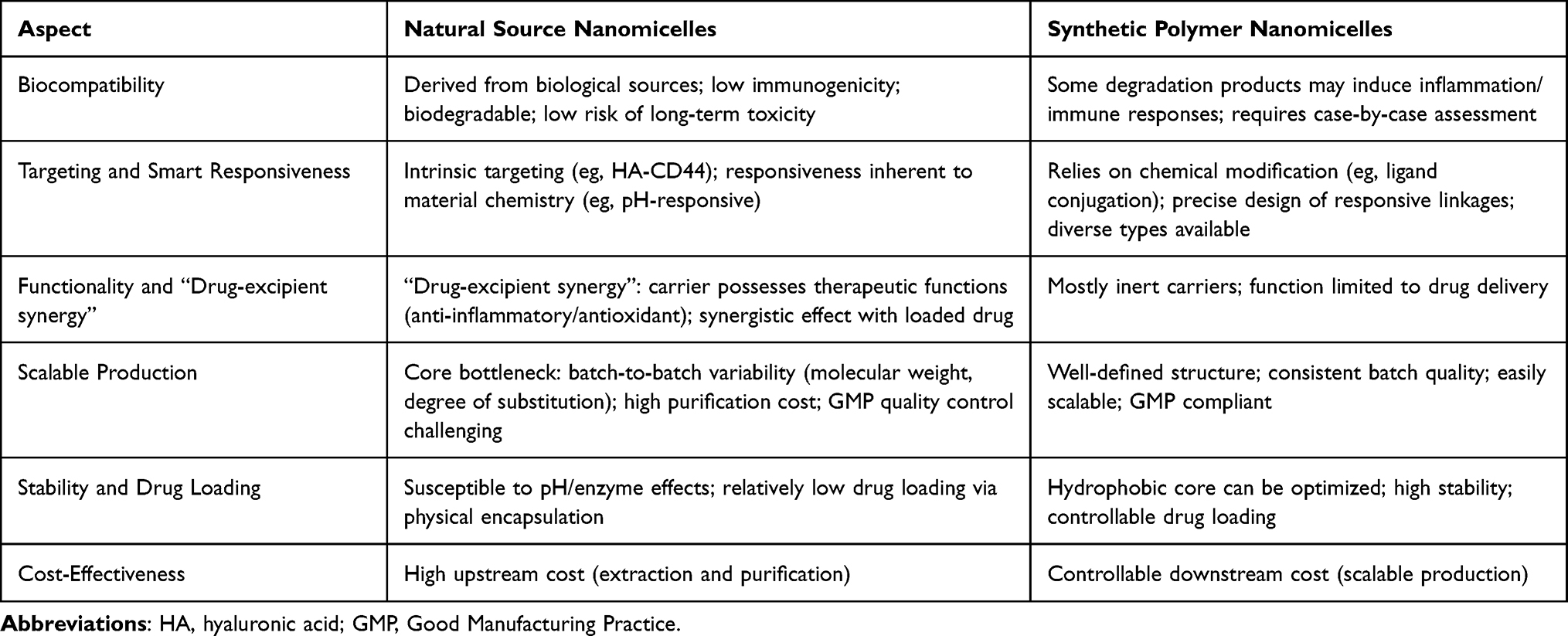 |
Table 1 Comparative Analysis of Natural and Synthetic Nanomicelles |
Preparation of Natural Product-Based Nanomicelles
The preparation of NSNs significantly impacts the micelle size, morphology, and drug loading efficiency. The preparation methods for natural product-based nanomicelles can be classified into three main categories: physical self-assembly methods, chemical conjugation methods, and electrostatic interaction methods. Self-assembly is the process by which individual building blocks spontaneously organize into ordered superstructures.35 Common methods include dialysis and nanoprecipitation, both of which trigger self-assembly by modulating solvent polarity and are suitable for most amphiphilic natural molecule systems. Additionally, the thin-film hydration method is sometimes used as an auxiliary assembly technique in some studies; its essence is still primarily molecule-driven self-assembly with minimal external force. The second category applies to natural polymers lacking sufficient amphiphilicity; hydrophobic segments can be introduced via chemical conjugation (eg, chemical coupling) to construct amphiphilic polymers, followed by self-assembly. In these two main methods, ultrasonication is often introduced as an auxiliary method to obtain more uniform dispersions. The third category involves preparation through electrostatic interactions. Schematic representation of the main preparation methods for NSNs is presented in Figure 2.
 |
Figure 2 Schematic representation of the main preparation methods for natural product-based nanomicelles. (A) Direct dissolution method. (B) Nanoprecipitation (solvent displacement) method. (C) Thin-film hydration method. (D) Dialysis method. (E) Chemical conjugation (covalent linkage) method. Ultrasonication is often applied as an auxiliary step to improve size uniformity. Note: Check mark (✓) indicates advantage; cross mark (✗) indicates disadvantage. |
Nanoprecipitation
Nanoprecipitation (also known as the solvent displacement method) is a simple and rapid preparation technique. Its principle involves injecting an organic solvent containing the drug and polymer into a large volume of the aqueous phase. Upon mixing, the organic solvent rapidly diffuses into the aqueous phase, causing a sharp decrease in polymer solubility, leading to precipitation and encapsulation of the drug, resulting in the formation of nanoformulations.36 This method is generally performed at room temperature, avoiding damage to natural active ingredients by high temperatures, making it more suitable for NSNs. However, the resulting micelles often have a broad size distribution, and drug loading efficiency is significantly influenced by the mixing speed of the solvent and aqueous phase.
Dialysis
In the dialysis method, the hydrophobic drug and amphiphilic material are codissolved in a water-miscible organic solvent (eg, DMF or DMSO) and placed in a dialysis bag. The organic solvent gradually exchanges with water, causing the hydrophobic segments of the copolymer to gradually associate and form micelles of relatively uniform size.37 Owing to its simple operation and ability to avoid the formation of large micellar aggregates, dialysis is a common method for preparing drug-loaded micelles in the laboratory. However, its disadvantages include being time-consuming and inefficient, making it unsuitable for large-scale production. Furthermore, some natural active ingredients may be lost along with the organic solvent during dialysis, reducing drug loading efficiency, which is also a core bottleneck for industrial application.
Thin-Film Hydration
The thin-film hydration method primarily utilizes the characteristics of amphiphilic molecules to spontaneously form ordered aggregates in an aqueous phase after removal of the organic solvent. The material is first dissolved in a volatile organic solvent and then formed into a thin film via rotary evaporation. Under conditions above the material’s phase transition temperature, hydration subsequently induces self-assembly into nanoscale ordered structures.38,39 This method, which was originally a classic technique for liposome preparation, is now also widely used for micelle preparation and offers high encapsulation efficiency for hydrophobic drugs. However, its disadvantages include that the initially hydrated micelles often have large sizes and broad size distributions, and there may be residual organic solvent issues. Therefore, subsequent processing steps such as sonication or membrane extrusion are often required in practical applications.
Chemical Conjugation
The chemical conjugation method fundamentally involves covalently linking hydrophobic drugs to natural carrier materials, artificially constructing amphiphilic precursors with defined structures, which then self-assemble into nanomicelles. Under specific conditions, the covalent bonds linking the drug to the micelle break, releasing the drug. This allows for effective control over the drug release rate, enabling intelligent responsive drug release. Drug loading is typically much greater than physical encapsulation, making it an important approach for achieving intelligent applications of NSNs. However, this also means that higher experimental conditions and control requirements, complex preparation processes, and covalent modification processes may disrupt the biological activity of natural materials, limiting their widespread application.
Electrostatic Interaction
The electrostatic interaction method involves the tight binding of drug molecules with oppositely charged micelles through electrostatic forces, which encapsulate the drug within the micelle. It is primarily used as a drug carrier for nucleic acid drugs such as DNA.40 The prepared micelles are stable and can largely preserve the physiological activity of biological macromolecules. However, the preparation conditions are relatively stringent, requiring the drug molecule itself to carry a sufficiently strong charge and precise control over the pH and ionic strength of the assembly environment. This method is mostly applicable to the delivery of specific nucleic acid drugs.
Mechanisms of the Use of Natural Product-Based Nanomicelles in Disease Treatment
As novel functional drug delivery systems, NSNs have significant application potential in the treatment of various major diseases. This section systematically reviews the therapeutic advantages of these nanomicelles across a range of pathological processes, including anti-infection, cancer, metabolic, inflammation, autoimmune, fibrotic, nervous system, and ischemia‒reperfusion injury.
Anti-Infective Effects
This section systematically elaborates on the latest research progress on the use of natural nanomicelles in the fields of antibacterial and antiviral therapy, focusing on their multiple mechanism synergistic therapeutic advantages and future development directions, as summarized in Table 2 and Figure 3.
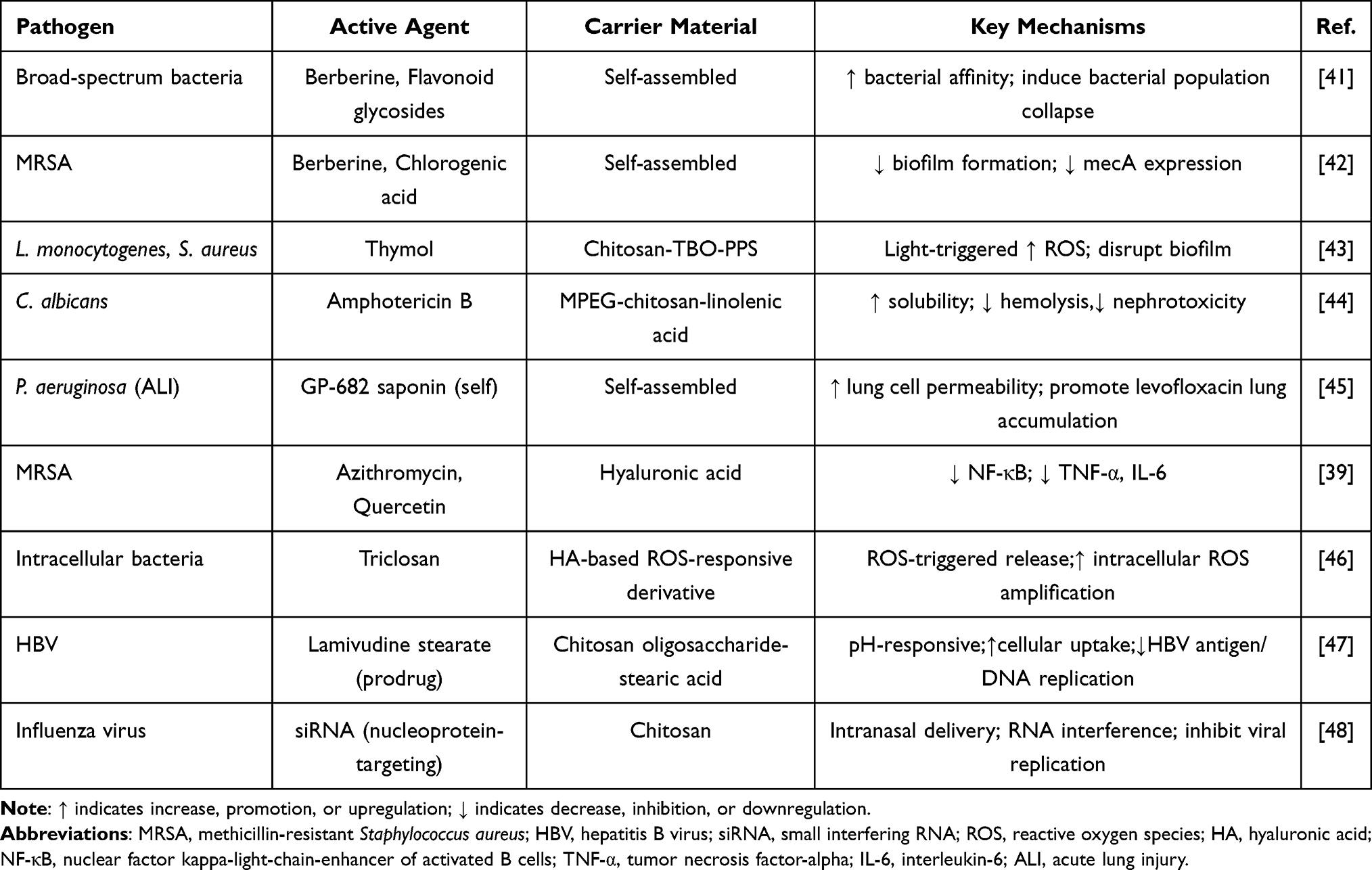 |
Table 2 Mechanisms of NSNs in Anti-Infective Therapy |
 |
Figure 3 Mechanisms of natural product-based nanomicelles against infections. (A) Antibacterial mechanisms: disruption of bacterial membranes, biofilm eradication, and downregulation of resistance gene expression. (B) Antiviral mechanisms: inhibition of viral entry, interference with intracellular replication, and RNA interference (siRNA delivery). (C) Immune modulation: activation of host immune responses to clear pathogens. (D) Microenvironmental regulation: scavenging of reactive oxygen species (ROS) and alleviation of infection-induced inflammation. Note: downward arrow (↓) indicates decrease, inhibition, or downregulation. |
Antibacterial Effects
Pathogenic microorganism infections are a major factor restricting human quality of life, with bacterial infections being predominant. These factors often lead to antibiotic misuse, consequently causing antimicrobial resistance, forming a vicious cycle.49 NSNs can combat bacterial and fungal infections through multiple synergistic mechanisms, with strategies extending beyond direct pathogen killing to include reversing drug resistance, disrupting biofilms, and modulating host immune responses.
Disruption of microbial structure and biofilms is the most direct antibacterial mechanism. Many plants, herbs, and spices serve as important sources of natural antibacterial and analgesic drugs.50 For example, the self-assembly of berberine, a natural isoquinoline alkaloid with broad-spectrum antibacterial activity, into nanomicelles with flavonoid glycosides more effectively induces bacterial population collapse and reduces biofilm formation.41 Nanomicelles coassembled from berberine and chlorogenic acid can significantly inhibit the formation of methicillin-resistant Staphylococcus aureus biofilms and downregulate their resistance gene expression.42 Furthermore, light-controllable chitosan micelles loaded with thymol can generate reactive oxygen species upon light irradiation and release thymol, synergistically eradicating mature biofilms of Listeria monocytogenes and Staphylococcus aureus through dual physicochemical mechanisms.43 In terms of antifungal effects, MPEG-CS-LNA micelles (a natural-synthetic hybrid system) loaded with amphotericin B achieve passive targeted fungal therapy by leveraging the inherent antifungal activity of the carrier and enhancing drug uptake.44
NSNs can enhance targeted delivery and modulate the infection microenvironment. NSNs can increase antibiotic accumulation at the infection site and regulate the local environment to be unfavorable for pathogens. A pulmonary drug delivery system based on self-assembled platycodin saponins can increase lung cell membrane permeability, promote the transmembrane uptake of the antibiotic levofloxacin, and produce synergistic therapeutic effects with the drug.45 Hyaluronic acid-modified azithromycin-quercetin micelles can target inflammatory sites via HA, downregulate key proinflammatory factors, and inhibit the NF-κB pathway while delivering antibiotics, combating MRSA infection through the dual pathways of bacteriostasis and anti-inflammation.39
NSNs can activate and amplify intracellular killing signals within bacteria. The novel micelles composed of hyaluronic acid derivatives and the ROS-responsive antibacterial agent triclosan can release the drug precisely in the highly oxidative stress environment at the infection site. Concurrently, the released cinnamaldehyde can maintain and amplify intracellular ROS levels, resulting in a self-amplifying killing effect that greatly enhances the elimination of intracellular drug-resistant bacteria.46
In summary, NSNs contribute to antibacterial therapy not only by delivering antibiotics but also by disrupting biofilms, downregulating resistance genes, and even achieving clearance of intracellular bacteria through light-controlled ROS or amplification of intracellular oxidative signals. This multimechanism parallel strategy offers a new approach to combating bacterial drug resistance, which is distinct from traditional antibiotics.
Antiviral Effects
Viruses are structurally simple, noncellular microorganisms that can only replicate and proliferate within living cells. After host infection, the pathogenic mechanisms are complex, and high mutation rates facilitate immune escape and drug resistance, posing ongoing challenges for clinical prevention and treatment. The mechanisms of action of antiviral nanomedicines can be summarized into multiple pathways, including direct inactivation, inhibition of invasion, inhibition of proliferation, and immune assistance.51
Inhibition of viral invasion and intracellular replication. NSNs, as efficient carriers, can significantly increase the intracellular delivery efficiency of traditional antiviral drugs. For example, stearic acid-g-chitosan oligosaccharide micelles loaded with the prodrug lamivudine stearate form a CSO-SA/LAS system. Leveraging pH-responsive drug release characteristics and enhanced cellular uptake, this system can more effectively deliver the drug to target cells, thereby significantly inhibiting the reverse transcription process of the hepatitis B virus, resulting in superior in vitro antiviral efficacy compared with that of the free drug.47 NSNs are ideal carriers for delivering nucleic acid drugs such as siRNAs, enabling highly specific viral gene silencing.52 For highly variable influenza viruses, chitosan/siRNA nanomicelles delivered intranasally can efficiently deliver siRNAs targeting the viral nucleoprotein to respiratory tract cells. This triggers RNA interference intracellularly, precisely degrading viral mRNA, thereby effectively inhibiting viral replication and providing protection in animal models.48 These findings suggest that combining the targeting, intelligent drug release, and efficient delivery capabilities of NSNs with modern antiviral drugs is a promising research direction for overcoming viral drug resistance and improving treatment efficiency.
In anti-infective applications, NSNs offer clear advantages by disrupting biofilms, downregulating resistance genes, and enabling intracellular bacterial killing through ROS amplification or light-controlled release. However, most studies remain in early preclinical stages, and the in vivo fate of these micelles in infected tissues—particularly their penetration into biofilm matrices—is poorly understood. A major unresolved challenge is whether the carrier itself may inadvertently suppress host immune responses that are essential for clearing opportunistic pathogens. Future work must distinguish between direct antimicrobial effects and host immunomodulation, and establish pharmacokinetic‒pharmacodynamic relationships that guide dosing regimens.
Anticancer Effects
Cancer is characterized by complexity and heterogeneity. In 2020, there were an estimated 19.3 million new cancer cases and nearly 10 million cancer deaths globally, presenting a severe situation for prevention and treatment.53 Currently, surgery, radiotherapy, and chemotherapy are the three main clinical treatment modalities for cancer, but they are associated with limitations such as numerous adverse effects, induction of drug resistance, and poor specificity,54 as detailed in Table 3 and Figure 4.
 |
Table 3 Mechanisms of NSNs in Antitumor Therapy |
 |
Figure 4 Antitumor mechanisms of natural product‑based nanomicelles.(A) Drug delivery and intracellular anti‑tumor mechanisms: (i) Passive targeting (EPR effect) and active targeting (ligand‑receptor recognition).(ii) Chemotherapy drug action on cancer cells: downregulating P‑glycoprotein (P‑gp), targeting organelles, and improving the tumor microenvironment.(B) Three synergistic therapeutic modalities: (i) Chemotherapy synergy, (ii) Chemotherapy and phototherapy combined (iii)Immune synergy. (C) Tumor microenvironment‑responsive drug release: (i) Triggered by low pH. (ii)Elevated ROS. (iii)Specific enzymes (eg, hyaluronidase). (D) Immunomodulation: including macrophage polarization, regulation of cell proliferation, promotion of anti‑tumor cytokine secretion, and induction of apoptosis. Note: Upward arrow (↑) indicates increase, promotion, or upregulation; downward arrow (↓) indicates decrease, inhibition, or downregulation. |
Delivery of Chemotherapeutic and Immunotherapeutic Agents
As efficient drug delivery vehicles in tumor therapy, the core function of NSNs lies in improving drug solubility, targeting, and stability, thereby enhancing efficacy and reducing toxicity.
The oral bioavailability of anticancer drugs is often limited by first-pass metabolism and inter-individual variability.68 To address this, Zhang et al developed disodium glycyrrhizinate (Na2GA)-based natural micelles for camptothecin (CPT) delivery. The optimized formulation exhibited a mean diameter of 80 nm, a critical micelle concentration of 0.303 mg/mL, a zeta potential of −33 mV, a polydispersity index of 0.25, and a drug loading of 6.22%. These micelles demonstrated excellent stability and permeability in simulated intestinal environments, and significantly enhanced the oral bioavailability of CPT, leading to superior in vitro and in vivo antitumor activity.55 Similarly, Wang et al constructed carboxymethyl chitosan-rhein (CR) conjugate micelles for oral paclitaxel (PTX) delivery. The PTX-loaded CR micelles achieved an encapsulation efficiency of 86.99 ± 12.26% and a loading capacity of 35.24 ± 1.58%. Pharmacokinetic studies confirmed a marked increase in oral bioavailability, and confocal imaging revealed that intact CR micelles were absorbed across the intestinal membrane. Furthermore, synergistic antitumor effects between the rhein carrier and PTX were observed in both Caco-2 cell assays and in vivo tumor models.56
Furthermore, NSNs can achieve precise drug delivery through active or passive targeting. Passive targeting relies primarily on the size effect of the nanoparticles. For example, Bletilla striata polysaccharide-vitamin E succinate micelles delivering andrographolide for colon cancer treatment can selectively accumulate in colon cancer tissues via the EPR effect.57 Building upon this, more precise active targeting can be achieved by surface modification with specific ligands. Folate-modified chitosan micelles can improve targeting to tumor cells and sites through folate receptor-mediated internalization. Codelivery of doxorubicin and all-trans retinoic acid synergistically induces apoptosis and inhibits cancer stem cell properties, achieving a tumor inhibition rate of 85.5% in a breast cancer mouse model.58
Multidrug resistance is a major cause of chemotherapy failure in cancer.69 NSNs can overcome MDR through various mechanisms, including inhibiting drug efflux pumps,70 targeting and disrupting organelles,71 and altering the tumor microenvironment.72 A multifunctional self-assembled micelle system, galactosamine-hyaluronic acid-vitamin E succinate, was developed to deliver the antihepatocellular carcinoma drug norcantharidin. The vitamin E TPGS component within the carrier acts as a P-gp inhibitor, blocking drug efflux and successfully reversing multidrug resistance.59 A natural polysaccharide-based nanoplatform with dual cellular- and mitochondrial-targeting capabilities was designed for the codelivery of ursolic acid and doxorubicin. After HA-mediated cellular internalization, these micelles anchor to the mitochondria. Triggered by high levels of intracellular ROS, UA and DOX are specifically released within mitochondria, synergistically inducing mitochondrial dysfunction. This fundamentally undermines the survival of malignant tumor cells, effectively reversing drug resistance.60
Synergistic Therapeutic Effects
In cancer treatment, single drugs or therapies have limited efficacy and are prone to inducing resistance. Therefore, integrating therapies with different mechanisms into a single nanoplatform can achieve synergistic efficacy enhancement.
In terms of chemical synergy, Gaber et al constructed natural polymer‑based micelles (zein‑ChS PMs) based on zein and chondroitin sulfate (ChS) for the co‑delivery of etoposide (ETP) and all‑trans retinoic acid (ATRA). In this system, ChS binds to the CD44 receptor overexpressed on the surface of tumor cells, thereby achieving active targeting. ETP induces tumor cell apoptosis by inhibiting topoisomerase II, while ATRA exerts synergistic antitumor effects by inducing differentiation and regulating the cell cycle. The combination of the two agents exhibited a significant synergistic effect (combination index CI < 1) and effectively reduced the dose requirement of each single drug. This natural‑derived micelle system demonstrates favorable biosafety and tumor‑targeting ability, providing a green and efficient nanoplatform for combination chemotherapy of solid tumors such as breast cancer.73 Further advancements involve combining different treatment modalities. Ding et al designed a dextran-based nanomicelle coloaded with a paclitaxel prodrug, the photosensitizer Ce6, and the chemiluminescent agent luminol. When H2O2 is overexpressed in the tumor microenvironment, chemiluminescence resonance energy transfer activates Ce6 to produce singlet oxygen. Simultaneously, the generated 1O2 activates the PTX prodrug, achieving a synergistic cascade effect of chemotherapy and photodynamic therapy, enhancing the efficacy against invasive carcinoma.61
Furthermore, immuno-synergistic therapy, as one of the most cutting-edge current strategies, focuses primarily on activating immunity74 and relieving immunosuppression.75 One study utilized mannose-modified stearic acid-chitosan micelles to actively capture endogenous tumor antigens and target them to dendritic cells in tumor-draining lymph nodes. This efficiently activates antigen-specific CD4+ and CD8+ T-cell responses, essentially functioning as an efficient personalized vaccine.62 Another study employed functionalized carboxymethyl chitosan micelles to codeliver doxorubicin and PD-L1 siRNA. While DOX induces immunogenic cell death, the siRNA blocks the PD-1/PD-L1 immune checkpoint, effectively reversing the tumor immunosuppressive microenvironment and achieving durable antitumor immune activation.63
Tumor Microenvironment-Responsive Drug Release
Cancer involves continuous and dynamic interactions between cancer cells and the tumor microenvironment. The establishment and maintenance of cancer depend, to varying degrees, on contributions from the TME. Compared with direct killing of cancer cells, targeting the TME offers significant therapeutic advantages.76 Microenvironment-responsive drug delivery systems can exploit specific signals within the TME, such as pH, ROS, and specific enzymes, to achieve targeted drug delivery and reduce side effects.77
pH responsiveness is one of the most widely employed designs because of its ability to target the uniquely acidic tumor microenvironment.78 Based on this strategy, dHAD‑PTX and dHAT‑PTX micelles were designed with a pH‑dependent surface charge‑switching capability. Under physiological conditions (pH 7.4), the micelles carry a negative surface charge, which favours prolonged blood circulation and reduces non‑specific uptake. Upon reaching the acidic tumor microenvironment (pH 5.6), their surface charge rapidly switches to positive, thereby greatly enhancing electrostatic interactions with tumor cells, triggering rapid release of paclitaxel (PTX) and promoting cellular uptake. Experimental results demonstrated that this charge‑switching mechanism led to a cellular uptake efficiency exceeding 95% in MCF‑7 cells, with corresponding IC50 values of 1.94 μg/mL for dHAD‑PTX and 1.87 μg/mL for dHAT‑PTX. More importantly, in an in vivo breast cancer model, these two micelles achieved tumour inhibition rates of 92.96% and 78.65%, respectively, without observable systemic toxicity. This design fully illustrates the great potential of exploiting pH‑regulated charge reversal in the tumour microenvironment to achieve highly efficient and low‑toxicity targeted chemotherapy.64
Reactive oxygen species-responsive drug delivery exploits the significantly higher intracellular ROS levels in tumor cells than in normal cells, offering potent intracellular killing capability. ROS-sensitive chemical bonds, such as disulfide bonds,79 are integrated into the structure of NSNs. Research has shown that in treating hepatocellular carcinoma, glycyrrhetinic acid-modified carboxymethyl chitosan-thioketal-rhein micelles rapidly disassemble upon entering cells under high intracellular ROS conditions, releasing celastrol. This significantly improves its bioavailability and achieves targeted therapeutic effects against liver cancer.65
Enzymes, which can accelerate chemical reactions, serve as ideal targets. NSNs can utilize enzymes specifically overexpressed at tumor sites to achieve precise drug release.80 Owing to its favorable biological properties and high affinity for the CD44 receptor, HA stands out as one of the most representative enzyme-responsive materials.81 For example, in protamine/hyaluronic acid multilayered micelles that deliver gambogic acid, upon reaching tumor regions enriched with hyaluronidase, the outer HA layer is rapidly degraded. This exposes the inner protamine, which then activates the “proton sponge” effect, greatly enhancing the endosomal escape efficiency of the micelles and increasing the cytoplasmic drug concentration.66 After CD44 receptor-mediated endocytosis, micelles based on hyaluronic acid-tetraphenylethylene have HA shells that are degraded by lysosomal hyaluronidase. This accelerates the release of the loaded doxorubicin around the nucleus, demonstrating excellent tumor inhibition efficacy in CD44-overexpressing tumor models.67
In addition to single stimuli, multiresponsive nanosystems have become a research frontier to further improve therapeutic effectiveness and safety, better leveraging the advantages of environment-responsive carriers.82 For example, micelles that integrate both pH-sensitive bonds and disulfide bonds can achieve sequential drug release in acidic and high-ROS microenvironments.
In summary, the core challenges in tumor therapy include the off-target effects of drugs, tumor heterogeneity, and the development of multidrug resistance. The unique value of NSNs lies in the ability of natural carriers such as hyaluronic acid and chitosan to achieve active targeting; carrier components such as rhein and ursolic acid can synergistically inhibit P-gp efflux or induce mitochondrial dysfunction; and platforms that codeliver chemotherapeutics and siRNA can simultaneously activate immune responses and relieve immunosuppression. Furthermore, the antioxidant activity of some carriers may increase chemotherapy sensitivity by modulating the tumor redox state.
Anti-Inflammatory and Immunomodulatory Effects
Inflammation underpins numerous physiological and pathological processes, and its dysregulation is a key driver of many inflammatory and autoimmune diseases.83 As key signaling molecules, ROS regulate various functions, including inflammatory responses.84 This section will use inflammatory bowel disease, rheumatoid arthritis, and skin inflammatory diseases as examples to systematically elaborate how NSNs provide new strategies for treating these complex diseases through mechanisms such as microenvironment-responsive drug release, immune regulation, and tissue repair,85 as detailed in Table 4 and Figure 5.
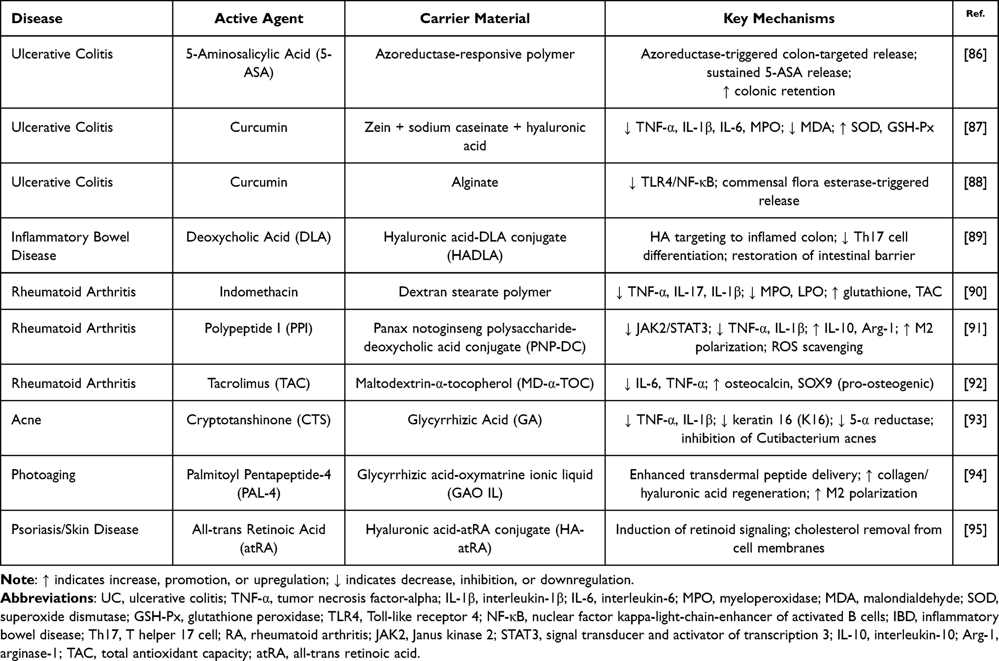 |
Table 4 Mechanisms of NSNs in Anti-Inflammatory Therapy |
 |
Figure 5 Therapeutic mechanisms of natural product-based nanomicelles in inflammatory and autoimmune diseases. (A) Inflammatory bowel disease: targeting inflamed colonic mucosa via CD44, repairing intestinal barrier, and modulating gut microbiota. (B) Rheumatoid arthritis: synovial targeting, M1-to-M2 macrophage polarization via the JAK2/STAT3 pathway, and protection of cartilage and bone. (C) Skin inflammatory diseases (eg, acne, psoriasis, photoaging): antimicrobial activity, sebum regulation, anti-inflammation, and collagen regeneration. Note: Upward arrow (↑) indicates increase, promotion, or upregulation; downward arrow (↓) indicates decrease, inhibition, or downregulation. |
Inflammatory Bowel Disease: Barrier Repair and Immune Modulation
Inflammatory bowel disease is a complex autoimmune condition resulting from the interplay of multiple factors, including genetic susceptibility, a dysregulated intestinal immune system, defective intestinal barrier function, and a disturbed gut microbiota. This disease mainly includes Crohn’s disease and ulcerative colitis.96 Current medications for IBD typically focus on suppressing the overactive immune response, which can only slow disease progression.
To achieve site-specific drug release at the lesion site, researchers have developed amphiphilic azo polymer micelles that utilize the unique azoreductase environment of the colon for localized release of 5-aminosalicylic acid. This system achieved a 60% dose reduction compared to conventional therapy, 77–97% inhibition of inflammatory markers (TNF-α, IL-6, etc)., and prolonged colonic retention up to 24 hours, significantly increasing drug accumulation in the lesion area.86 In modulating the intestinal immune and inflammatory microenvironments, various natural micelle systems have demonstrated clear regulatory efficacy. Curcumin-loaded biopolymer nanocomposites effectively alleviate intestinal inflammatory damage and promote tissue repair by regulating oxidative stress indicators and inflammatory mediators.87 Stable micelles formed from esterified alginate-curcumin conjugates can effectively inhibit TLR4 expression in intestinal epithelial cells, reducing downstream NF-κB pathway activation and the subsequent production of proinflammatory cytokines such as TNF-α and IL-1β. They also achieve colon-targeted therapy through a drug release mechanism triggered by the commensal microbiota.88
More importantly, NSNs can achieve superior therapeutic outcomes through synergistic treatment involving multiple mechanisms. For example, an oral micelle system constructed from hyaluronic acid and dehydrocholic acid not only accumulates at inflammatory sites via HA-CD44-mediated targeting but also repairs the intestinal physical barrier, protects epithelial cell integrity, modulates the adaptive immune system by precisely inhibiting Th17-mediated inflammation, and synergistically regulates microbial ecology. This multidimensional approach collectively reverses the pathological progression of IBD.89
In summary, the core challenge in IBD treatment lies in the vicious cycle of persistent intestinal barrier damage, immune homeostasis imbalance, and gut microbiota dysbiosis. The irreplaceable role of NSNs is that carriers such as hyaluronic acid can target inflammatory sites and repair the physical barrier; active ingredients such as curcumin can directly inhibit the TLR4/NF-κB pathway and regulate the Th17/Treg balance; and some systems, such as alginate-curcumin, can even respond to the microbiota for drug release. Behind these effects, scavenging excess ROS and restoring redox balance are crucial for inhibiting the amplification of inflammatory cascades.
Rheumatoid Arthritis: Synovial Targeting and Immune Remodeling
Rheumatoid arthritis is a chronic inflammatory disease resulting from the interplay of genetic, environmental, and immune dysregulation, characterized by persistent synovitis, systemic inflammation, and the production of autoantibodies.97 Current treatments primarily use disease-modifying antirheumatic drugs, which may exacerbate side effects and increase the risk of liver and kidney damage.98
In RA treatment, NSNs exert significant effects through multitargeted interventions. Indomethacin-loaded dextran stearate polymer micelles can significantly reduce key inflammatory cytokine levels while increasing glutathione and total antioxidant capacity, effectively alleviating joint inflammatory responses and oxidative stress damage.90 To address the specific immune imbalance in RA, folic acid-modified Panax notoginseng polysaccharide-deoxycholic acid micelles target the highly expressed folate receptor in the synovium. By inhibiting the JAK2/STAT3 pathway, they promote the transformation of proinflammatory M1 macrophages to anti-inflammatory M2 macrophages, fundamentally reshaping the immune microenvironment.91 To prevent joint structural damage, tacrolimus-loaded maltodextrin-α-tocopherol nanomicelles, while exerting immunosuppressive effects, have carrier components that promote osteogenic differentiation, counteracting the characteristic bone erosion in RA.92
RA treatment has long faced the dilemma of balancing immunosuppression with bone protection. The advantages of NSNs include the following: Panax notoginseng polysaccharides can regulate macrophage M1/M2 polarization via the JAK2/STAT3 pathway, remodeling the immune microenvironment; maltodextrin-α-tocopherol micelles, while tacrolimus is delivered, have carrier components that synergistically promote osteogenic differentiation. In this process, the carrier’s ability to scavenge ROS in the joint cavity and inhibit NF-κB activation create favorable conditions for M2 polarization.
Skin Inflammatory Diseases: Multitarget Local Intervention
Although the pathological features of skin inflammatory diseases vary, their treatment commonly faces challenges such as poor drug penetration and insufficient targeting. NSNs stand out because of their excellent skin permeability and multitarget regulatory capabilities, demonstrating unique advantages in treating conditions such as acne, photoaging, and psoriasis.
Acne primarily involves four core pathological processes: follicular hyperkeratinization, excessive sebum production, Cutibacterium acnes proliferation, and the inflammation/immune response.99 In 25 nm glycyrrhizic acid-cryptotanshinone self-assembled micelles, glycyrrhizic acid directly inhibits C. acnes and exerts anti-inflammatory effects, whereas cryptotanshinone inhibits sebum production. These micelles downregulate the expression of proinflammatory factors and reduce keratin 16 gene expression to improve follicular hyperkeratosis. Furthermore, downregulating 5-α reductase mRNA may influence androgen signaling pathways, indirectly regulating sebaceous gland function, demonstrating a more comprehensive therapeutic approach than monotherapy.93
Photoaging refers to the premature, nonnatural aging of skin caused by chronic, repeated ultraviolet exposure,100 with the primary tissue characteristic being destruction of the collagen fiber network.101 When glycyrrhizic acid and oxymatrine self-assemble into ionic liquid micelles for delivering palmitoyl pentapeptide-4, they can promote collagen and hyaluronic acid regeneration and inhibit the NF-κB signaling pathway to reduce inflammation and apoptosis,94 suggesting new ideas for clinical intervention in photoaging.
Psoriasis is an immune-mediated chronic inflammatory skin disease with characteristic changes, including abnormal differentiation of keratinocytes and activation of a complex immune‒inflammatory network.102 Hyaluronic acid-trans retinoic acid conjugate micelles can, on the one hand, induce retinoid signaling to help regulate the differentiation and proliferation of keratinocytes, normalizing them. On the other hand, they can actively remove cholesterol from cells, indirectly influencing the activation state of inflammatory signaling pathways. This cleverly combines nanotechnology with traditional drugs and newly discovered functions to address complex pathologies.95
In summary, NSNs achieve multilevel precision treatment, ranging from pathogen control and inflammation regulation to tissue repair, by targeting the specific pathological links of different skin inflammatory diseases, offering new technological strategies for the clinical management of these conditions.
Effects of Metabolic Regulation
Metabolic diseases encompass a wide range of conditions, including diabetes, obesity, and nonalcoholic fatty liver disease. Among these, the application of NSNs has been studied in depth in the field of diabetes and its complications, as shown in Table 5 and Figure 6.
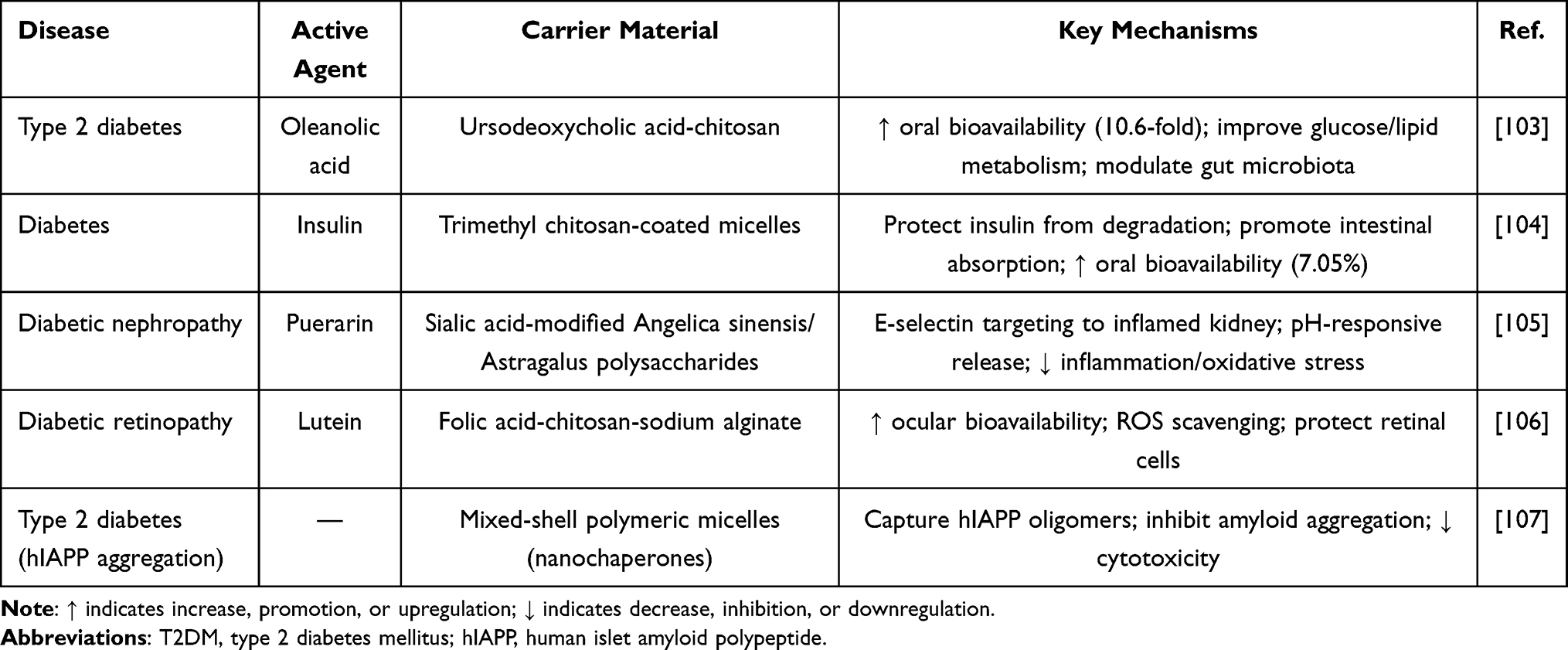 |
Table 5 Mechanisms of NSNs in Metabolic Disease Therapy |
 |
Figure 6 Therapeutic mechanisms of natural product-based nanomicelles in diabetes mellitus and its complications. (A) Enhanced oral delivery: improving the bioavailability of poorly soluble antidiabetic drugs and enabling oral insulin delivery. (B) Smart delivery to complication sites: targeting the renal/retinal microenvironment with pH/ROS responsiveness. (C) Systemic metabolic regulation: correcting glucose and lipid metabolism disorders and modulating gut microbiota. (D) Pathological intervention: inhibiting hIAPP aggregation, protecting β-cells, and alleviating oxidative stress injury. Note: downward arrow (↓) indicates decrease, inhibition, or downregulation. |
Diabetes and Its Complications: Targeted Intervention and Oxidative Stress Regulation
Diabetes is a common, incurable metabolic syndrome primarily characterized by chronic hyperglycemia resulting from defective insulin secretion or action, often accompanied by disturbances in glucose, protein, fat, water, and electrolyte metabolism.108 Currently, subcutaneous insulin injection remains the first-line therapy for blood glucose control, but it has limitations such as poor patient compliance, low bioavailability, risk of hypoglycemia, and inability to fundamentally correct metabolic disturbances.109 NSNs offer new, more convenient, safer, and noninvasive approaches for treating diabetes and its complications.110
Large-molecule drugs such as insulin are susceptible to degradation upon oral administration,111 and many natural hypoglycemic components suffer from poor water solubility,112 resulting in extremely low oral bioavailability. The hydrophobic cores of nanomicelles can efficiently encapsulate these drugs, solubilizing them. Research has shown that encapsulating oleanolic acid in ursodeoxycholic acid-modified chitosan micelles increases its oral bioavailability 10.6-fold and has significant hypoglycemic and hepatoprotective effects in a type 2 diabetes mouse model.103 For insulin, the construction of chitosan-based polyelectrolyte complex nanomicelles can protect it from degradation by gastrointestinal enzymes and promote intestinal absorption, achieving effective oral insulin delivery with a relative bioavailability of 7.05%.104
Lesion sites associated with diabetic complications, such as nephropathy113 and retinopathy,114 often exhibit specific microenvironments or overexpress particular receptors, providing targets for the intelligent design of nanomicelles. A pH-responsive hybrid micelle system based on Angelica sinensis polysaccharide and Astragalus polysaccharide was developed. This system utilizes sialic acid-modified APS to target E-selectin, which is highly expressed on inflamed endothelial cells, and releases the drug puerarin upon hydrazone bond cleavage in the acidic environment of the kidney, achieving precise drug delivery to renal inflammatory sites.105 Folate-modified chitosan-alginate nanocarriers have been shown to significantly increase the bioavailability of lutein in the eye tissues of diabetic rats and, by effectively scavenging ROS, protect retinal pigment epithelial cells from oxidative stress-induced damage, suggesting a new approach for preventing and treating diabetic retinopathy.106
Correcting glucose and lipid metabolism disorders is fundamental to diabetes treatment. In type 2 diabetes, the abnormal folding and aggregation of human islet amyloid polypeptide not only directly leads to β-apoptosis but also exacerbates local insulin resistance, acting as an upstream driver of metabolic dysregulation.115 Researchers have developed biomimetic nanochaperones that capture hIAPP oligomers through their surface-tunable hydrophobic microdomains, effectively inhibiting the formation of toxic amyloid aggregates and significantly reducing hIAPP-related cytotoxicity, providing an innovative strategy for protecting β-cell function at the source.107
Recent research has revealed a close association between the gut microbiota and the development of diabetes116,117. Some natural nanocarrier materials, such as chitosan118 and pectin,119 themselves possess prebiotic properties. The aforementioned study on UCOS-OA micelles not only confirmed their hypoglycemic effect but also revealed that they ameliorate gut microbiota dysbiosis and fecal metabolite profiles in diabetic mice, suggesting that they may influence host systemic metabolism through modulation of the gut microbiota.104
In summary, the complexity of diabetes lies in the low bioavailability of oral hypoglycemic agents, the heterogeneity of complication lesions, and the frequent neglect of upstream drivers such as hIAPP aggregation. NSNs enable multilevel intervention: chitosan carriers achieve efficient oral insulin delivery; sialic acid-modified polysaccharides target the renal inflammatory endothelium; and biomimetic nanochaperones capture hIAPP oligomers, protecting β-cells at the source. The ability of lutein micelles to scavenge ROS in retinal cells highlights the value of antioxidants in complex interventions. Furthermore, chronic diseases such as diabetes require long-term administration, yet the repeated-dose safety of most NSN materials has never been evaluated beyond a few weeks. The gut microbiota-modulating effects reported for some polysaccharide-based micelles are intriguing but highly variable across individuals, making predictive design difficult.
Anti-Fibrotic Effects
Fibrosis is a common terminal pathological outcome of dysregulated tissue repair in various chronic diseases, and is characterized by excessive extracellular matrix deposition and disruption of organ structure.120 The liver and kidney are organs with high susceptibility to fibrotic diseases, and their pathological progression is closely linked to injury from ischemia, inflammation, and other factors. NSNs, with their precise targeting capabilities and versatile functional design, offer innovative strategies for intervening in and even reversing the fibrotic process, as detailed in Table 6 and Figure 7.
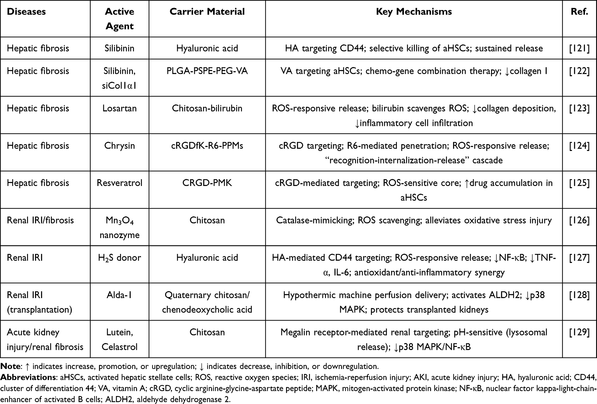 |
Table 6 Mechanisms of NSNs in Antifibrotic Therapy |
 |
Figure 7 Mechanisms of natural product-based nanomicelles in counteracting fibrosis. (A) Hepatic fibrosis, presented in three temporal stages: (i) Acute stage: scavenging of reactive oxygen species (ROS) to alleviate the initial “ROS storm” and reduce inflammatory cell infiltration. (ii) Chronic progressive stage: targeting activated hepatic stellate cells (aHSCs) via CD44 or vitamin A (VA) receptors, with ROS‑responsive drug release to inhibit collagen gene expression and suppress fibrogenesis. (iii) Fibrotic stage: chemo‑gene combination therapy to reverse established fibrosis and promote tissue repair.(B) Renal fibrosis and ischemia‑reperfusion injury, also shown in three stages: (i) Acute ischemic injury: ROS scavenging and anti‑inflammatory effects to protect against initial damage. (ii) Chronic progressive stage: megalin receptor‑mediated targeting enabling pH‑responsive delivery to injured renal tubules, along with inhibition of the TGF‑β1/MAPK/NF‑κB signaling axis to reduce inflammation and extracellular matrix (ECM) accumulation.(iii) Fibrotic stage: direct reduction of excessive ECM deposition to alleviate established renal fibrosis. Note: downward arrow (↓) indicates decrease, inhibition, or downregulation. |
Liver Fibrosis: Targeting Activated HSCs and Release of ROS-Responsive Drugs
Liver fibrosis is a common pathological feature of various chronic liver diseases, with the activation of hepatic stellate cells being a central event.130 Owing to the lack of specific targeting of activated HSCs in vivo and difficulties in achieving controlled release at the lesion site, the clinical efficacy of existing drugs is limited, and systemic side effects are significant.131
Achieving specific enrichment of drugs in the fibrotic liver and within aHSCs is key to improving efficacy and reducing side effects. Research indicates that the use of specific receptors that are overexpressed on the surface of aHSCs enables the precise targeting of nanomicelles. Silibinin-loaded hyaluronic acid micelles, which leverage the specific binding of HA to CD44 receptors, achieve targeted killing of aHSCs and sustained release therapy.121 Similarly, vitamin A, a classic targeting ligand for HSCs, was used to synthesize PLGA-PSPE-PEG-VA polymer micelles. These micelles efficiently accumulate in the fibrotic liver and specifically deliver drugs to aHSCs.122
The fibrotic liver microenvironment presents various pathophysiological features that are distinct from those of normal tissue, such as significantly elevated ROS levels. Stimuli-responsive nanomicelles designed on the basis of these features enable intelligent, controlled drug release at the lesion site. Chitosan-bilirubin micelles utilize the ROS-scavenging and -responsive properties of bilirubin. After being loaded with losartan, these cells respond to ROS levels within aHSCs, precisely releasing the drug and significantly reducing collagen deposition and inflammatory cell infiltration in liver tissue.123 In addition to the use of intact natural polymers, current research frontiers have drawn inspiration from natural mechanisms, such as the construction of superior intelligent delivery systems through molecular engineering. For example, cRGDfK-R6-PPM micelles integrate targeting, cell-penetrating, and ROS-responsive functions, achieving a cascade delivery of “recognition-internalization-release”, greatly increasing the intracellular bioavailability of drugs within aHSCs.124 Similarly, CRGD-PMK micelles, through cRGD-mediated active targeting and an ROS-sensitive PMK core, successfully achieved specific release and efficient accumulation of resveratrol in aHSCs.125
To address the complex mechanisms of liver fibrosis, codelivery systems have emerged. The aforementioned PLGA-PSPE-PEG-VA micelle platform not only achieved precise targeting of aHSCs but also successfully enabled the codelivery of the chemical drug silibinin and the gene drug siCol1α1. Studies have confirmed that, compared with monotherapies, these chemogenes coloaded with micelles, through the dual mechanisms of drugs inhibiting HSC activation and gene silencing blocking collagen synthesis, have significantly superior synergistic effects on the inhibition of collagen I production and amelioration of liver fibrosis.123
The key to reversing liver fibrosis lies in precisely targeting activated HSCs and breaching the fibrotic scar barrier. NSNs offer solutions: natural ligands such as hyaluronic acid and vitamin A target aHSCs; bilirubin and ROS-sensitive bonds respond to the high oxidative stress microenvironment for drug release; and the codelivery of silibinin and siCol1α1 simultaneously inhibits HSC activation and collagen synthesis. The inherent ROS-scavenging ability of the carrier bilirubin plays a fundamental role in this process.
Renal Fibrosis and Ischemia‒reperfusion Therapy: Antioxidant and Anti-Inflammatory Effects
As a highly perfused organ, the kidney is highly susceptible to damage from ischemia, toxic substances, and immune inflammation. This damage typically begins with acute kidney injury, with ischemia‒reperfusion injury being a major cause, and effective treatments are currently lacking. If not effectively effective, AKI can progress to chronic kidney disease characterized by renal fibrosis.132 The pathological mechanisms of AKI, especially IRI, generally involve oxidative stress, inflammatory responses, and apoptosis.133,134 Targeting and neutralizing excess ROS is the most direct approach. Chitosan-nanozyme micelles encapsulating Mn3O4 nanozymes can be specifically enriched at renal inflammatory sites and mimic catalase to efficiently scavenge excess ROS, mitigating direct cellular damage caused by oxidative stress from the source.126 In the context of ROS scavenging, further modulation of endogenous protective systems is possible. Hyaluronic acid-H2S donor micelles utilize HA’s targeting ability to deliver hydrogen sulfide. H2S, as a gaseous signaling molecule, not only directly scavenges ROS but also exerts potent anti-inflammatory effects by inhibiting the NF-κB pathway, resulting in synergistic antioxidant and anti-inflammatory effects. Notably, these micelles achieved intracellular H2S concentrations three times higher than those in the control group.127 For mitochondrial dysfunction and cellular metabolic disturbances caused by ROS, precise intervention in specific pathways is crucial. Quaternary chitosan/chenodeoxycholic acid micelles, which are used during hypothermic machine perfusion in kidney transplantation, deliver Alda-1. These micelles exhibited a mean particle size of 132.4 ± 0.3 nm, a zeta potential of 45.0 ± 1.0 mV, and a high drug loading capacity of 41.9%, with 80% of the drug released within 3.5 hours under hypothermic conditions. By activating ALDH2 and inhibiting the p38 MAPK pathway, they directly protect transplanted kidneys from IRI.128 For complex injury networks, multitarget synergistic therapy offers more comprehensive protection. Chitosan-based micelles codelivering lutein and celastrol achieve renal targeting via megalin receptor-mediated endocytosis and release drugs in the acidic lysosomal environment. This system synergistically provides antioxidant effects while simultaneously inhibiting the p38 MAPK/NF-κB pathways, two key inflammatory and stress signaling pathways, to achieve potent combined therapy.129
When damage persists, it will inevitably progress to chronic kidney disease characterized by renal fibrosis. At this stage, the core task of nanomicelles is to efficiently deliver antifibrotic drugs. Immune modulation is crucial in CKD progression.135 Research has shown that glycol chitosan-tacrolimus micelles utilize the renal targeting property of glycol chitosan to significantly increase the accumulation of the immunosuppressant tacrolimus at the lesion site. By inhibiting the TGF-β1/MAPK/NF-κB signaling axis, this system more potently alleviates glomerular fibrosis and inflammation, with efficacy superior to that of the free drug.136
In summary, the irreversibility of renal fibrosis dictates that the timing of intervention is critical. In the AKI stage, NSNs such as chitosan-nanozymes and HA-H2S donors scavenge excess ROS and inhibit NF-κB, blocking the oxidative stress‒inflammation cascade. In the CKD stage, glycol chitosan-tacrolimus micelles slow the fibrotic process. From direct ROS scavenging to activating the Nrf2/HO-1 pathway, antioxidant effects remain a core mechanism throughout AKI-to-CKD intervention. However, the reversibility of advanced fibrosis remains unproven with these systems. Most studies employ prophylactic or early‑intervention regimens, whereas clinical fibrosis is typically diagnosed at later stages when dense extracellular matrix barriers physically exclude nanoparticles. An underappreciated challenge is that the same ROS‑responsive mechanisms may be exhausted in the highly oxidative fibrotic milieu, leading to premature drug leakage.
Neuroprotective Effects
The treatment of central nervous system diseases has long been hampered by the low efficiency of drug delivery imposed by the blood‒brain barrier. NSNs, with their excellent biocompatibility, functional modifiability, and inherent biological activities, offer innovative strategies to overcome this bottleneck and achieve precise treatment for neurodegenerative and acute injury diseases, as detailed in Table 7 and Figure 8.
 |
Table 7 Mechanisms of NSNs in Neurological Disease Therapy |
 |
Figure 8 Mechanisms of natural product‑based nanomicelles for neurological disease therapy. (A) Blood–brain barrier crossing strategies. (B) Alzheimer’s disease: inhibition of Aβ fibrillation and tau aggregation, clearance of pre‑formed aggregates by artificial chaperone micelles, and early diagnosis via curcumin‑loaded micelles for retinal Aβ imaging. (C) Parkinson’s disease: antioxidant effects, reduction of α‑synuclein aggregation, and restoration of dopamine/serotonin balance. (D) Cerebral ischemia‑reperfusion injury: anti‑inflammatory effects (NF‑κB inhibition), reactive oxygen species (ROS) scavenging, and neurovascular repair. Note: Downward arrow (↓) indicates decrease, inhibition, or downregulation. |
Alzheimer’s Disease: Aβ Clearance, Tau Inhibition, and Early Diagnosis
Alzheimer’s disease is a neurodegenerative disorder characterized by Aβ deposition, tau protein hyperphosphorylation, neuroinflammation, and metabolic dysregulation.144
The active targeting capability of natural ligands is key to increasing the drug concentration in the brain. For example, lactoferrin-conjugated linoleic acid micelles efficiently cross the BBB via lactoferrin receptor-mediated transcytosis, significantly enhancing the distribution of loaded drugs in brain tissue, thereby effectively clearing Aβ, alleviating oxidative stress, and improving cognitive function.137 NSNs with biomimetic designs have unique advantages in addressing Aβ aggregation and deposition. Mixed-shell polymer micelles, inspired by natural molecular chaperones, possess tunable surface properties and can act as “artificial chaperones”, selectively inhibiting Aβ fibrillation and promoting the clearance of preformed aggregates, effectively remodeling Aβ homeostasis.138 As challenges with Aβ-targeted strategies increase clinically, intervening in tau pathology has become a new focus in AD treatment.145 Studies have shown that micelles modified with tau-targeting peptides can efficiently inhibit the abnormal aggregation of tau protein through multivalent binding peptides on their surface and can recognize and bind preformed tau aggregates, blocking their pathological “seeding” and propagation between neurons.139 Furthermore, NSNs demonstrate unique value in early disease diagnosis. Curcumin micelles, which leverage the high affinity of curcumin for Aβ, serve as efficient optical probes for noninvasive early imaging of retinal Aβ plaques, offering a revolutionary tool for early AD diagnosis.146
Parkinson’s Disease: Antioxidant Effects and Inhibition of α-Syn Aggregation
Parkinson’s disease is characterized by the progressive loss of dopaminergic neurons in the substantia nigra pars compacta, with a core pathology involving the abnormal aggregation of α-synuclein, oxidative stress, and neuroinflammation.147
Leveraging the synergistic effects of natural components is an effective strategy against the multifaceted pathology of PD. Studies have shown that nanomicelle complexes self-assembled from the natural dipeptide carnosine and the antioxidant α-lipoic acid, through synergistically increasing total antioxidant activity, successfully reverse dopamine and serotonin metabolism disturbances in an MPTP-induced early PD model, effectively restoring neurotransmitter balance in the striatum.140 In addition to the design of single antioxidants, the design of multitarget nanoprodrugs can achieve more comprehensive neuroprotection. Idebenone nanoprodrug micelles were designed as a multitarget therapeutic system. In PD models, they simultaneously reduce oxidative stress, inhibit neuroinflammation, and decrease α-syn aggregation, demonstrating comprehensive efficacy at the cellular and animal levels and significantly extending lifespan.141
Notably, a growing body of epidemiological and mechanistic research reveals a bidirectional connection between inflammatory bowel disease and neurodegenerative diseases (such as PD) via the “gut‒brain axis”, suggesting that chronic intestinal inflammation may be a risk factor for distant neurological pathology.148 This association inspires future research on NSNs. NSNs can both target and modulate the intestinal immune microenvironment, repair the barrier, and possess the potential to cross the BBB and intervene in neuroinflammation and protein misfolding. Future designs might integrate multifunctional platforms to synergistically intervene in gastrointestinal and nervous system-related diseases, starting from their common pathological origins.
Cerebral Ischemic Stroke and Reperfusion Injury: Anti-Inflammatory Effects and Neurovascular Repair
Cerebral ischemia‒reperfusion injury is a key challenge in stroke treatment, as its pathological process involves a vicious cycle of oxidative stress, neuroinflammation, and apoptosis.149
Effectively curbing neuroinflammation is crucial for blocking secondary damage. Triblock copolymer nanomicelles loaded with curcumin can efficiently deliver this natural anti-inflammatory component. By inhibiting the NF-κB signaling pathway, they significantly downregulate the expression of key proinflammatory factors, thereby effectively reducing brain tissue inflammatory damage in animal models.142 Furthermore, NSNs that integrate multiple natural components can achieve synergistic and comprehensive neuroprotection. For example, lactoferrin-baicalein nanomicelles utilize the ability of lactoferrin to target the brain to codeliver two naturally active molecules. This system not only exerts antioxidant and antiapoptotic effects but also effectively regulates the inflammatory microenvironment, resulting in significant improvements in neuronal apoptosis and cognitive dysfunction in both in vitro and in vivo experiments, highlighting the unique advantages of multitarget intervention.143
In summary, drug delivery for the nervous system faces the dual challenges of BBB obstruction and the multifactorial complexity of lesions. The strategies used by NSNs include lactoferrin-mediated BBB crossing, the use of biomimetic chaperones that selectively inhibit Aβ/Tau/α-syn aggregation, and the use of ROS-responsive systems to release drugs in ischemic lesions and activate the Nrf2/HO-1 pathway. The synergistic antioxidant effects of carnosine-α-lipoic acid and the multitarget protection of idebenone both point toward the common goal of restoring neuronal redox balance. The epidemiological association between IBD and PD also suggests the possibility of designing “gut‒brain axis” dual-targeting platforms in the future, which may intervene in central lesions from peripheral origins.
Discussion and Outlook
Based on the systematic analysis above, this section summarizes the core strategies by which NSNs overcome the delivery barriers of natural products, proposes a framework for formulation selection, identifies oxidative stress regulation as a common mechanism, and discusses the major challenges and future directions for clinical translation.
The poor solubility, rapid metabolism, and low bioavailability of natural products can be addressed by NSNs through three general mechanisms. First, physical encapsulation. The hydrophobic core of NSNs directly increases the aqueous solubility of poorly soluble natural compounds and protects them from premature enzymatic degradation or metabolism. Second, chemical conjugation and ligand‑mediated targeting. By covalently linking natural polymers (eg, hyaluronic acid, chitosan) to the drug or decorating the micelle surface with targeting ligands, NSNs achieve active accumulation in diseased tissues. Third, the intrinsic bioactivity of the carrier material creates a synergistic effect with the encapsulated drug. Many natural materials (polyphenols, saponins, polysaccharides) possess intrinsic anti‑inflammatory, antioxidant, or immunomodulatory activities.150 When these molecules serve as carrier materials to construct micelles, their function extends beyond traditional drug delivery, allowing them to directly participate in disease intervention.151,152
Regarding formulation selection, three factors should be considered: disease microenvironment, route of administration, and manufacturing consistency. Similar to synthetic polymer micelles, NSNs also face the challenge of premature drug release.153 Therefore, for solid tumors characterized by acidic pH and elevated ROS levels, stimuli‑responsive micelles are preferred. For inflammatory diseases such as rheumatoid arthritis or inflammatory bowel disease, hyaluronic acid‑based micelles (CD44‑targeting) are advantageous because of their inherent tropism toward activated macrophages and inflamed endothelium. For the route of administration, oral delivery requires formulations that resist gastric degradation and penetrate the mucus barrier; chitosan‑ or alginate‑based mucoadhesive micelles have proven effective. Intravenous administration demands stealth properties to avoid reticuloendothelial system uptake, with PEGylation or hyaluronic acid coating being common strategies. For manufacturing consistency, batch‑to‑batch reproducibility is a fundamental requirement for clinical translation; well‑defined derivatives or natural–synthetic hybrid systems are superior to crude extracts with variable compositions.
Notably, across the six disease categories reviewed, oxidative stress repeatedly emerges as a pathophysiological driver. NSNs often exert therapeutic effects by scavenging ROS through antioxidant moieties embedded in the carrier (eg, bilirubin, curcumin, resveratrol, polyphenols) or by activating the Nrf2/HO‑1 pathway. This cross‑disease consistency suggests that redox regulation is a fundamental common mechanism of NSNs, intrinsically provided by the natural building blocks. This insight provides a rational basis for designing NSNs against diseases driven by oxidative stress, without needing to re‑engineer the carrier for each new indication.
Although preclinical data are impressive, the translation of NSNs into clinical products faces several well‑recognized obstacles. Drawing on experience from the broader nanomedicine field, we analyze these obstacles and discuss potential solutions. (i) Batch‑to‑batch variability. Differences in molecular weight and degree of substitution of natural polymers between batches directly affect self‑assembly, size distribution, drug loading, and release kinetics, thereby complicating compliance with good manufacturing practice (GMP). Solutions include using well‑defined derivatives or constructing hybrid systems in which natural targeting ligands are conjugated to synthetic backbones (eg, PEG‑polysaccharide) to improve consistency while retaining bioactivity. (ii) Discrepancies between in vitro and in vivo results. Many NSNs exhibit potent ROS‑scavenging or anti‑inflammatory activity in cell assays but show limited or inconsistent effects in animal models. Potential reasons include protein corona formation, pH‑ or enzyme‑induced degradation, rapid clearance, and poor penetration into dense tissues. Overcoming this issue requires more physiologically relevant models (including orthotopic, chronic, and large‑animal models) and standardized efficacy endpoints.154 (iii) Lack of standardized evaluation protocols. There is currently no consensus on how to benchmark NSN performance against synthetic controls or approved formulations. Parameters such as catalytic activity (for ROS‑scavenging NSNs), drug loading efficiency, release kinetics, and toxicity are measured using different methods, making cross‑study comparisons difficult. Establishing unified protocols—mandating reporting of particle size, polydispersity, zeta potential, drug loading percentage, encapsulation efficiency, and cumulative release profiles—would significantly improve reproducibility and translational relevance. (iv) Regulatory uncertainty. NSNs occupy a gray zone between nanomedicines, natural health products, and drug‑device combinations. Their natural origin does not automatically guarantee safety; long‑term toxicity, immunogenicity, and potential metal ion accumulation require systematic evaluation.155 Moreover, the drug‑excipient synergy effect complicates attribution of efficacy, and regulatory agencies may require comparative studies of the carrier alone, the drug alone, and the intact NSN formulation.
Looking forward, three complementary approaches may accelerate the clinical translation of NSNs. First, mechanism‑driven rational design should replace empirical trial‑and‑error. Quantitative structure–activity relationship models and computational simulations can help predict self‑assembly, drug release, and in vivo behavior. Second, theranostics that integrate therapy and diagnosis enable real‑time monitoring of drug accumulation and therapeutic response. Third, scalability should be considered from an early stage, evaluating batch consistency, storage stability, sterilization compatibility, and cost‑effectiveness alongside efficacy. Technologies such as microfluidics and spray drying offer promising manufacturing routes. Collectively, these strategies will help bridge the gap between laboratory discoveries and clinical products.
Declaration of Generative AI and AI-Assisted Technologies in Manuscript Preparation
The authors confirm that during the preparation of this manuscript, the generative AI tool DeepSeek-R1-0528 only was used solely for language polishing, grammar checking, and academic expression optimization. All content, core ideas, literature analysis, logical structure, and conclusions were independently conceived, reviewed, revised, and finalized by the authors. The authors take full responsibility for the accuracy, integrity, and originality of the published manuscript.
Abbreviations
AD, Alzheimer’s disease; HSCs, Hepatic Stellate Cells; IBD, Inflammatory Bowel Disease; IRI, Ischemia-Reperfusion Injury; MDR, Multidrug Resistance; MRSA, Methicillin-Resistant Staphylococcus aureus; NF-κB, Nuclear Factor kappa-B; NSNs, Natural product-based Nanomicelles; OA, Oleanolic Acid; PD, Parkinson’s disease; PTX, Paclitaxel; RA, Rheumatoid Arthritis; RES, Reticuloendothelial System; ROS, Reactive Oxygen Species; siRNA, small interfering RNA; T2DM, Type 2 Diabetes Mellitus; TAC, Tacrolimus; TDLN, Tumor-Draining Lymph Nodes; TME, Tumor Microenvironment; UA, Ursolic Acid; UC, Ulcerative Colitis.
Data Sharing Statement
Not applicable. This is a review article and no new datasets were generated or analyzed during the current study. All data discussed in this manuscript are derived from published articles that are publicly available and cited in the reference list.
Ethics Approval and Consent to Participate
Not applicable. This review article does not involve any studies with human participants, human data, or human tissue conducted by any of the authors. It also does not involve any studies with animals.
Author Contributions
All authors made a significant contribution to the work reported, whether that is in the conception, study design, execution, acquisition of data, analysis and interpretation, or in all these areas; took part in drafting, revising or critically reviewing the article; gave final approval of the version to be published; have agreed on the journal to which the article has been submitted; and agree to be accountable for all aspects of the work.
Funding
This work was supported by the National Training Program of Innovation and Entrepreneurship for Undergraduates [grant number:202510541029], the Natural Science Foundation of Hunan Province for Basic Research Projects for Young Students [grant numbers: 2026JJ90291, 2026JJ90290].
Disclosure
The authors declare that they have no competing interests—financial or non-financial—that could influence the work reported in this manuscript.
References
1. Bose A, Roy Burman D, Sikdar B, Patra P. Nanomicelles: types, properties and applications in drug delivery. IET Nanobiotechnol. 2021;15(1):19–32. PMID: 34694727; PMCID: PMC8675821. doi:10.1049/nbt2.12018
2. Öztürk K, Kaplan M, Çalış S. Effects of nanoparticle size, shape, and zeta potential on drug delivery. Int J Pharm. 2024;666:124799. PMID: 39369767. doi:10.1016/j.ijpharm.2024.124799
3. Zhou Q, Zhang L, Yang T, Wu H. Stimuli-responsive polymeric micelles for drug delivery and cancer therapy. Int J Nanomedicine. 2018;13:2921–2942. PMID: 29849457; PMCID: PMC5965378. doi:10.2147/IJN.S158696
4. Tawfik SM, Azizov S, Elmasry MR, Sharipov M, Lee YI. Recent advances in nanomicelles delivery systems. Nanomaterials. 2020;11(1):70. PMID: 33396938; PMCID: PMC7823398. doi:10.3390/nano11010070
5. Wu Y, Wang XJ, Shang HZ, et al. Research progress of natural saccharide-based nanomedicine delivery systems. Chemistry Bulletin. 2022;85(04):441–445. doi:10.14159/j.cnki.0441-3776.2022.04.001
6. Burlatsky SF, Atrazhev VV, Dmitriev DV, et al. Surface tension model for surfactant solutions at the critical micelle concentration. J Colloid Interface Sci. 2013;393:151–160. PMID: 23153677. doi:10.1016/j.jcis.2012.10.020
7. Hamill RJ. Amphotericin B formulations: a comparative review of efficacy and toxicity. Drugs. 2013;73(9):919–934. PMID: 23729001. doi:10.1007/s40265-013-0069-4
8. Barenholz Y. Doxil®--the first FDA-approved nano-drug: lessons learned. J Control Release. 2012;160(2):117–134. PMID: 22484195. doi:10.1016/j.jconrel.2012.03.020
9. Bernabeu E, Cagel M, Lagomarsino E, Moretton M, Chiappetta DA. Paclitaxel: what has been done and the challenges remain ahead. Int J Pharm. 2017;526(1–2):474–495. PMID: 28501439. doi:10.1016/j.ijpharm.2017.05.016
10. Zhao Y, Cao WQ, Liu Y. Research progress of polymeric nanomedicines. Chem J Chin Univ. 2020;41(05):909–923.
11. Zhang L, Song J, Kong L, et al. The strategies and techniques of drug discovery from natural products. Pharmacol Ther. 2020;216:107686. PMID: 32961262. doi:10.1016/j.pharmthera.2020.107686
12. Kashyap D, Tuli HS, Yerer MB, et al. Natural product-based nanoformulations for cancer therapy: opportunities and challenges. Semin Cancer Biol. 2021;69:5–23. PMID: 31421264. doi:10.1016/j.semcancer.2019.08.014
13. Beigoli S, Boskabady MH. The molecular basis of the immunomodulatory effects of natural products: a comprehensive review. Phytomedicine. 2024;135:156028. PMID: 39276685. doi:10.1016/j.phymed.2024.156028
14. Gouda NA, Alshammari SO, Abourehab MAS, Alshammari QA, Elkamhawy A. Therapeutic potential of natural products in inflammation: underlying molecular mechanisms, clinical outcomes, technological advances, and future perspectives. Inflammopharmacology. 2023;31(6):2857–2883. PMID: 37950803. doi:10.1007/s10787-023-01366-y
15. Mohanty SK, Swamy MK, Sinniah UR, Anuradha M. Leptadenia reticulata (Retz.) wight & arn. (Jivanti): botanical, agronomical, phytochemical, pharmacological, and biotechnological aspects. Molecules. 2017;22(6):1019. PMID: 28629185; PMCID: PMC6152761. doi:10.3390/molecules22061019
16. Bilia AR, Piazzini V, Risaliti L, et al. Nanocarriers: a successful tool to increase solubility, stability and optimise bioefficacy of natural constituents. Curr Med Chem. 2019;26(24):4631–4656. PMID: 30381065. doi:10.2174/0929867325666181101110050
17. Lin X, Huang X, Tian X, et al. Natural small-molecule-based carrier-free self-assembly library originated from traditional Chinese herbal medicine. ACS Omega. 2022;7(48):43510–43521. PMID: 36506183; PMCID: PMC9730315. doi:10.1021/acsomega.2c04098
18. Jafernik K, Ładniak A, Blicharska E, et al. Chitosan-based nanoparticles as effective drug delivery systems-a review. Molecules. 2023;28(4):1963. PMID: 36838951; PMCID: PMC9959713. doi:10.3390/molecules28041963
19. Song Z, Sun J, Williams GR, et al. Glucan-based biomaterials and their applications in the biomedical field. Int J Biol Macromol. 2025;318(Pt 1):144940. PMID: 40473181. doi:10.1016/j.ijbiomac.2025.144940
20. Choi KY, Han HS, Lee ES, et al. Hyaluronic acid-based activatable nanomaterials for stimuli-responsive imaging and therapeutics: beyond CD44-mediated drug delivery. Adv Mater. 2019;31(34):e1803549. PMID: 30773699. doi:10.1002/adma.201803549
21. Karim A, Rehman A, Feng J, et al. Alginate-based nanocarriers for the delivery and controlled-release of bioactive compounds. Adv Colloid Interface Sci. 2022;307:102744. PMID: 35878506. doi:10.1016/j.cis.2022.102744
22. Moghaddam FD, Zare EN, Hassanpour M, et al. Chitosan-based nanosystems for cancer diagnosis and therapy: stimuli-responsive, immune response, and clinical studies. Carbohydr Polym. 2024;330:121839. PMID: 38368115. doi:10.1016/j.carbpol.2024.121839
23. Mu L, Wu L, Wu S, Ye Q, Zhong Z. Progress in chitin/chitosan and their derivatives for biomedical applications: where we stand. Carbohydr Polym. 2024;343:122233. PMID: 39174074. doi:10.1016/j.carbpol.2024.122233
24. Jin MZ, Jin WL. The updated landscape of tumor microenvironment and drug repurposing. Signal Transduct Target Ther. 2020;5(1):166. PMID: 32843638; PMCID: PMC7447642. doi:10.1038/s41392-020-00280-x
25. Shi Y, van der Meel R, Chen X, Lammers T. The EPR effect and beyond: strategies to improve tumor targeting and cancer nanomedicine treatment efficacy. Theranostics. 2020;10(17):7921–7924. PMID: 32685029; PMCID: PMC7359085. doi:10.7150/thno.49577
26. Li M, Sun J, Zhang W, Zhao Y, Zhang S, Zhang S. Drug delivery systems based on CD44-targeted glycosaminoglycans for cancer therapy. Carbohydr Polym. 2021;251:117103. PMID: 33142641. doi:10.1016/j.carbpol.2020.117103
27. Hall CL, Yang B, Yang X, et al. Overexpression of the hyaluronan receptor RHAMM is transforming and is also required for H-ras transformation. Cell. 1995;82(1):19–26. PMID: 7541721. doi:10.1016/0092-8674(95)90048-9
28. Herrlich P, Sleeman J, Wainwright D, et al. How tumor cells make use of CD44. Cell Adhes Commun. 1998;6(2–3):141–147. PMID: 9823465. doi:10.3109/15419069809004470
29. Rajitha P, Gopinath D, Biswas R, Sabitha M, Jayakumar R. Chitosan nanoparticles in drug therapy of infectious and inflammatory diseases. Expert Opin Drug Deliv. 2016;13(8):1177–1194. PMID: 27087148. doi:10.1080/17425247.2016.1178232
30. Nagarsheth N, Wicha MS, Zou W. Chemokines in the cancer microenvironment and their relevance in cancer immunotherapy. Nat Rev Immunol. 2017;17(9):559–572. PMID: 28555670; PMCID: PMC5731833. doi:10.1038/nri.2017.49
31. Zhang G, Ma L, Bai L, et al. Inflammatory microenvironment-targeted nanotherapies. J Control Release. 2021;334:114–126. PMID: 33887284. doi:10.1016/j.jconrel.2021.04.018
32. Hu HQ, Cheng GW, Wu YF, et al. Application of “unification of medicine and excipient” concept in design of traditional Chinese medicine preparation delivery systems. China J Chin Materia Medica. 2023;48(07):1800–1807. doi:10.19540/j.cnki.cjcmm.20221129.302
33. Qiu A, Wang Y, Zhang G, Wang H. Natural polysaccharide-based nanodrug delivery systems for treatment of diabetes. Polymers. 2022;14(15):3217. PMID: 35956731; PMCID: PMC9370904. doi:10.3390/polym14153217
34. Mu D, Li J, Qi Y, et al. Hyaluronic acid-coated polymeric micelles with hydrogen peroxide scavenging to encapsulate statins for alleviating atherosclerosis. J Nanobiotechnology. 2020;18(1):179. PMID: 33287831; PMCID: PMC7720571. doi:10.1186/s12951-020-00744-w
35. Min Y, Akbulut M, Kristiansen K, Golan Y, Israelachvili J. The role of interparticle and external forces in nanoparticle assembly. Nat Mater. 2008;7(7):527–538. PMID: 18574482. doi:10.1038/nmat2206
36. Lepeltier E, Bourgaux C, Couvreur P. Nanoprecipitation and the “Ouzo effect”: application to drug delivery devices. Adv Drug Deliv Rev. 2014;71:86–97. PMID: 24384372. doi:10.1016/j.addr.2013.12.009
37. Atanase LI. Micellar drug delivery systems based on natural biopolymers. Polymers. 2021;13(3):477. PMID: 33540922; PMCID: PMC7867356. doi:10.3390/polym13030477
38. Feng WW, Chen ZW. Research progress in preparation methods of solid lipid nanoparticles. Chin J Med Biotechnol. 2011;6(03):218–221.
39. Zhang Z, Chen M, Wang J, et al. Hyaluronic acid-modified micelles of azithromycin and quercetin against infections caused by methicillin-resistant Staphylococcus aureus. Int J Nanomedicine. 2024;19:9637–9658. PMID: 39309186; PMCID: PMC11414643. doi:10.2147/IJN.S476471
40. Kakizawa Y, Kataoka K. Block copolymer micelles for delivery of gene and related compounds. Adv Drug Deliv Rev. 2002;54(2):203–222. PMID: 11897146. doi:10.1016/s0169-409x(02)00017-0
41. Li T, Wang P, Guo W, et al. Natural berberine-based chinese herb medicine assembled nanostructures with modified antibacterial application. ACS Nano. 2019;13(6):6770–6781. PMID: 31135129. doi:10.1021/acsnano.9b01346
42. Fu S, Yi X, Li Y, et al. Berberine and chlorogenic acid-assembled nanoparticles for highly efficient inhibition of multidrug-resistant Staphylococcus aureus. J Hazard Mater. 2024;473:134680. PMID: 38795486. doi:10.1016/j.jhazmat.2024.134680
43. Wang Z, Bai H, Lu C, et al. Light controllable chitosan micelles with ROS generation and essential oil release for the treatment of bacterial biofilm. Carbohydr Polym. 2019;205:533–539. PMID: 30446137. doi:10.1016/j.carbpol.2018.10.095
44. Song Z, Wen Y, Deng P, et al. Linolenic acid-modified methoxy poly (ethylene glycol)-oligochitosan conjugate micelles for encapsulation of amphotericin B. Carbohydr Polym. 2019;205:571–580. PMID: 30446143. doi:10.1016/j.carbpol.2018.10.086
45. Zhang M, Ye L, Huang H, et al. Micelles self-assembled by 3-O-β-D-glucopyranosyl latycodigenin enhance cell membrane permeability, promote antibiotic pulmonary targeting and improve anti-infective efficacy. J Nanobiotechnology. 2020;18(1):140. PMID: 33008413; PMCID: PMC7532624. doi:10.1186/s12951-020-00699-y
46. Qiu Y, Shang K, Xu N, et al. Clearance of intracellular bacterial infections by hyaluronic acid-based ROS responsive drug delivery micelles. Int J Biol Macromol. 2023;245:125506. PMID: 37356691. doi:10.1016/j.ijbiomac.2023.125506
47. Li Q, Du YZ, Yuan H, et al. Synthesis of lamivudine stearate and antiviral activity of stearic acid-g-chitosan oligosaccharide polymeric micelles delivery system. Eur J Pharm Sci. 2010;41(3–4):498–507. PMID: 20728535. doi:10.1016/j.ejps.2010.08.004
48. Jamali A, Mottaghitalab F, Abdoli A, et al. Inhibiting influenza virus replication and inducing protection against lethal influenza virus challenge through chitosan nanoparticles loaded by siRNA. Drug Deliv Transl Res. 2018;8(1):12–20. PMID: 29063498. doi:10.1007/s13346-017-0426-z
49. Fisher RA, Gollan B, Helaine S. Persistent bacterial infections and persister cells. Nat Rev Microbiol. 2017;15(8):453–464. PMID: 28529326. doi:10.1038/nrmicro.2017.42
50. Tian X, Wang P, Li T, et al. Self-assembled natural phytochemicals for synergistically antibacterial application from the enlightenment of traditional Chinese medicine combination. Acta Pharm Sin B. 2020;10(9):1784–1795. PMID: 33088696; PMCID: PMC7564035. doi:10.1016/j.apsb.2019.12.014
51. Pu Y, Zhu C, Liao J, et al. Antiviral nanomedicine: advantages, mechanisms and advanced therapies. Bioact Mater. 2025;52:92–122. PMID: 40530413; PMCID: PMC12173073. doi:10.1016/j.bioactmat.2025.05.030
52. Paunovska K, Loughrey D, Dahlman JE. Drug delivery systems for RNA therapeutics. Nat Rev Genet. 2022;23(5):265–280. PMID: 34983972; PMCID: PMC8724758. doi:10.1038/s41576-021-00439-4
53. Sung H, Ferlay J, Siegel RL, et al. Global cancer statistics 2020: GLOBOCAN estimates of incidence and mortality worldwide for 36 cancers in 185 countries. CA Cancer J Clin. 2021;71(3):209–249. PMID: 33538338. doi:10.3322/caac.21660
54. Peng Q, Qian Z, Gao H, Zhang K. Recent advances in transition-metal based nanomaterials for noninvasive oncology thermal ablation and imaging diagnosis. Front Chem. 2022;10:899321. PMID: 35494651; PMCID: PMC9047733. doi:10.3389/fchem.2022.899321
55. Zhang Q, Feng Z, Wang H, et al. Preparation of camptothecin micelles self-assembled from disodium glycyrrhizin and tannic acid with enhanced antitumor activity. Eur J Pharm Biopharm. 2021;164:75–85. PMID: 33878433. doi:10.1016/j.ejpb.2021.04.012
56. Wang X, Guo Y, Qiu L, et al. Preparation and evaluation of carboxymethyl chitosan-rhein polymeric micelles with synergistic antitumor effect for oral delivery of paclitaxel. Carbohydr Polym. 2019;206:121–131. PMID: 30553305. doi:10.1016/j.carbpol.2018.10.096
57. Yue Z, Zhu Y, Chen T, et al. Bletilla striata polysaccharide-coated andrographolide nanomicelles for targeted drug delivery to enhance anti-colon cancer efficacy. Front Immunol. 2024;15:1380229. PMID: 38911867; PMCID: PMC11190162. doi:10.3389/fimmu.2024.1380229
58. Liu Y, Yu F, Dai S, et al. All-trans retinoic acid and doxorubicin delivery by folic acid modified polymeric micelles for the modulation of pin1-mediated DOX-induced breast cancer stemness and metastasis. Mol Pharm. 2021;18(11):3966–3978. PMID: 34579532. doi:10.1021/acs.molpharmaceut.1c00220
59. Jiang S, Li M, Hu Y, Zhang Z, Lv H. Multifunctional self-assembled micelles of galactosamine-hyaluronic acid-vitamin E succinate for targeting delivery of norcantharidin to hepatic carcinoma. Carbohydr Polym. 2018;197:194–203. PMID: 30007605. doi:10.1016/j.carbpol.2018.05.090
60. Guo Y, Yang X, Zhang Y, et al. Hyaluronic acid/dextran-based polymeric micelles co-delivering ursolic acid and doxorubicin to mitochondria for potentiating chemotherapy in MDR cancer. Carbohydr Polym. 2024;332:121897. PMID: 38431408. doi:10.1016/j.carbpol.2024.121897
61. Ding C, Shi Z, Ou M, et al. Dextran-based micelles for combinational chemo-photodynamic therapy of tumors via in vivo chemiluminescence. Carbohydr Polym. 2023;319:121192. PMID: 37567697. doi:10.1016/j.carbpol.2023.121192
62. Yang X, Yu T, Zeng Y, et al. Tumor-draining lymph node targeting chitosan micelles as antigen-capturing adjuvants for personalized immunotherapy. Carbohydr Polym. 2020;240:116270. PMID: 32475559. doi:10.1016/j.carbpol.2020.116270
63. Song P, Wang B, Pan Q, et al. GE11-modified carboxymethyl chitosan micelles to deliver DOX·PD-L1 siRNA complex for combination of ICD and immune escape inhibition against tumor. Carbohydr Polym. 2023;312:120837. PMID: 37059562. doi:10.1016/j.carbpol.2023.120837
64. Liu Z, Chen X, Jin Q, et al. Dual functionalized hyaluronic acid micelles loading paclitaxel for the therapy of breast cancer. Front Bioeng Biotechnol. 2023;11:1230585. PMID: 37600308; PMCID: PMC10436080. doi:10.3389/fbioe.2023.1230585
65. Zhang X, Xu X, Wang X, et al. Hepatoma-targeting and reactive oxygen species-responsive chitosan-based polymeric micelles for delivery of celastrol. Carbohydr Polym. 2023;303:120439. PMID: 36657834. doi:10.1016/j.carbpol.2022.120439
66. Ke Z, Yang L, Wu H, Li Z, Jia X, Zhang Z. Evaluation of in vitro and in vivo antitumor effects of gambogic acid-loaded layer-by-layer self-assembled micelles. Int J Pharm. 2018;545(1–2):306–317. PMID: 29654896. doi:10.1016/j.ijpharm.2018.04.016
67. Yan K, Feng Y, Gao K, Shi X, Zhao X. Fabrication of hyaluronic acid-based micelles with glutathione-responsiveness for targeted anticancer drug delivery. J Colloid Interface Sci. 2022;606(Pt 2):1586–1596. PMID: 34500160. doi:10.1016/j.jcis.2021.08.129
68. Parodi A, Buzaeva P, Nigovora D, et al. Nanomedicine for increasing the oral bioavailability of cancer treatments. J Nanobiotechnology. 2021;19(1):354. PMID: 34717658; PMCID: PMC8557561. doi:10.1186/s12951-021-01100-2
69. Assaraf YG, Brozovic A, Gonçalves AC, et al. The multi-factorial nature of clinical multidrug resistance in cancer. Drug Resist Updat. 2019;46:100645. PMID: 31585396. doi:10.1016/j.drup.2019.100645
70. Zhang H, Xu H, Ashby CR Jr, Assaraf YG, Chen ZS, Liu HM. Chemical molecular-based approach to overcome multidrug resistance in cancer by targeting P-glycoprotein (P-gp). Med Res Rev. 2021;41(1):525–555. PMID: 33047304. doi:10.1002/med.21739
71. Wang X, Xu Z, Wang J, et al. A mitochondria-targeted biomimetic nanomedicine capable of reversing drug resistance in colorectal cancer through mitochondrial dysfunction. Adv Sci. 2025;12(13):e2410630. PMID: 39943694; PMCID: PMC11967798. doi:10.1002/advs.202410630
72. Wang Y, Chai Y, Liu Y, et al. Inhibition of tumor cell macropinocytosis driver DHODH reverses immunosuppression and overcomes anti-PD1 resistance. Immunity. 2025;58(10):2456–2471.e6. PMID: 40816268. doi:10.1016/j.immuni.2025.07.013
73. Gaber M, Elhasany KA, Sabra S, et al. Co-administration of tretinoin enhances the anti-cancer efficacy of etoposide via tumor-targeted green nano-micelles. Colloids Surf B Biointerfaces. 2020;192:110997. PMID: 32361378. doi:10.1016/j.colsurfb.2020.110997
74. Galluzzi L, Humeau J, Buqué A, Zitvogel L, Kroemer G. Immunostimulation with chemotherapy in the era of immune checkpoint inhibitors. Nat Rev Clin Oncol. 2020;17(12):725–741. PMID: 32760014. doi:10.1038/s41571-020-0413-z
75. Xiao Z, Todd L, Huang L, et al. Desmoplastic stroma restricts T cell extravasation and mediates immune exclusion and immunosuppression in solid tumors. Nat Commun. 2023;14(1):5110. PMID: 37607999; PMCID: PMC10444764. doi:10.1038/s41467-023-40850-5
76. Xiao Y, Yu D. Tumor microenvironment as a therapeutic target in cancer. Pharmacol Ther. 2021;221:107753. PMID: 33259885; PMCID: PMC8084948. doi:10.1016/j.pharmthera.2020.107753
77. He Q, Chen J, Yan J, et al. Tumor microenvironment responsive drug delivery systems. Asian J Pharm Sci. 2020;15(4):416–448. PMID: 32952667; PMCID: PMC7486519. doi:10.1016/j.ajps.2019.08.003
78. Ding H, Tan P, Fu S, et al. Preparation and application of pH-responsive drug delivery systems. J Control Release. 2022;348:206–238. PMID: 35660634. doi:10.1016/j.jconrel.2022.05.056
79. Meng X, Shen Y, Zhao H, Lu X, Wang Z, Zhao Y. Redox-manipulating nanocarriers for anticancer drug delivery: a systematic review. J Nanobiotechnology. 2024;22(1):587. PMID: 39342211; PMCID: PMC11438196. doi:10.1186/s12951-024-02859-w
80. Shahriari M, Zahiri M, Abnous K, Taghdisi SM, Ramezani M, Alibolandi M. Enzyme responsive drug delivery systems in cancer treatment. J Control Release. 2019;308:172–189. PMID: 31295542. doi:10.1016/j.jconrel.2019.07.004
81. Dosio F, Arpicco S, Stella B, Fattal E. Hyaluronic acid for anticancer drug and nucleic acid delivery. Adv Drug Deliv Rev. 2016;97:204–236. PMID: 26592477. doi:10.1016/j.addr.2015.11.011
82. Zhang CY, Zhang CX, Wang HY, et al. Research progress of multi-stimuli responsive nano drug delivery systems. Chin J New Drugs. 2013;22(20):2383–2387.
83. Medzhitov R. Origin and physiological roles of inflammation. Nature. 2008;454(7203):428–435. PMID: 18650913. doi:10.1038/nature07201
84. Liu J, Han X, Zhang T, Tian K, Li Z, Luo F. Reactive oxygen species (ROS) scavenging biomaterials for anti-inflammatory diseases: from mechanism to therapy. J Hematol Oncol. 2023;16(1):116. PMID: 38037103; PMCID: PMC10687997. doi:10.1186/s13045-023-01512-7
85. Dou Y, Li C, Li L, Guo J, Zhang J. Bioresponsive drug delivery systems for the treatment of inflammatory diseases. J Control Release. 2020;327:641–666. PMID: 32911014; PMCID: PMC7476894. doi:10.1016/j.jconrel.2020.09.008
86. Tang S, Wang W, Wang Y, et al. Sustained release of 5-aminosalicylic acid from azoreductase-responsive polymeric prodrugs for prolonged colon-targeted colitis therapy. J Nanobiotechnology. 2024;22(1):468. PMID: 39103846; PMCID: PMC11302195. doi:10.1186/s12951-024-02724-w
87. Zhang S, Li C. A curcumin-loaded biopolymeric nanocomposite alleviates dextran sulfate sodium induced ulcerative colitis via suppression of inflammation and oxidative stress. Int J Biol Macromol. 2024;275(Pt 2):133665. PMID: 38971294. doi:10.1016/j.ijbiomac.2024.133665
88. Wang Y, Li Y, He L, et al. Commensal flora triggered target anti-inflammation of alginate-curcumin micelle for ulcerative colitis treatment. Colloids Surf B Biointerfaces. 2021;203:111756. PMID: 33865087. doi:10.1016/j.colsurfb.2021.111756
89. Cheng W, Zhou X, Jin C, et al. Acid-base transformative HADLA micelles alleviate colitis by restoring adaptive immunity and gut microbiome. J Control Release. 2023;364:283–296. PMID: 37898344. doi:10.1016/j.jconrel.2023.10.039
90. Abdollahi AR, Firouzian F, Haddadi R, Nourian A. Indomethacin loaded dextran stearate polymeric micelles improve adjuvant-induced arthritis in rats: design and in vivo evaluation. Inflammopharmacology. 2021;29(1):107–121. PMID: 33179175. doi:10.1007/s10787-020-00776-6
91. Yu Y, Kong L, Guo RB, et al. Engineered Panax notoginseng polysaccharide micelles inhibit macrophage polarization and delay the progression of rheumatoid arthritis via JAK2-STAT3 signaling pathway. J Nanobiotechnology. 2025;23(1):509. PMID: 40660236; PMCID: PMC12261558. doi:10.1186/s12951-025-03576-8
92. Helal HM, Samy WM, Kamoun EA, et al. Potential privilege of maltodextrin-α-tocopherol nano-micelles in seizing tacrolimus renal toxicity, managing rheumatoid arthritis and accelerating bone regeneration. Int J Nanomedicine. 2021;16:4781–4803. PMID: 34290503; PMCID: PMC8286967. doi:10.2147/IJN.S317409
93. Liang T, Wu Y, Zeng Q, et al. Development of a self-assembled micelles based on cryptotanshinone and glycyrrhizic acid: an efficient strategy for acne treatment. Int J Pharm. 2025;674:125411. PMID: 40020947. doi:10.1016/j.ijpharm.2025.125411
94. Wang Z, Liu J, Chen Q, et al. Bioactive Glycyrrhizic acid ionic liquid self-assembled nanomicelles for enhanced transdermal delivery of anti-photoaging signal peptides. Adv Sci. 2025;12(8):e2412581. PMID: 39783908; PMCID: PMC11848569. doi:10.1002/advs.202412581
95. Pavlík V, Machalová V, Čepa M, et al. Retinoic acid grafted to hyaluronic acid activates retinoid gene expression and removes cholesterol from cellular membranes. Biomolecules. 2022;12(2):200. PMID: 35204701; PMCID: PMC8961547. doi:10.3390/biom12020200
96. Hodson R. Inflammatory bowel disease. Nature. 2016;540(7634):S97. PMID: 28002398. doi:10.1038/540S97a
97. Scott DL, Wolfe F, Huizinga TW. Rheumatoid arthritis. Lancet. 2010;376(9746):1094–1108. PMID: 20870100. doi:10.1016/S0140-6736(10)60826-4
98. Zhao F, Hu Z, Li G, et al. Angiogenesis in rheumatoid Arthritis: pathological characterization, pathogenic mechanisms, and nano-targeted therapeutic strategies. Bioact Mater. 2025;50:603–639. PMID: 40453697; PMCID: PMC12124647. doi:10.1016/j.bioactmat.2025.04.026
99. Williams HC, Dellavalle RP, Garner S. Acne vulgaris. Lancet. 2012;379(9813):361–372. Erratum in: Lancet. 2012 Jan 28;379(9813):314. PMID: 21880356. doi:10.1016/S0140-6736(11)60321-8
100. Shen J, Tower J. Effects of light on aging and longevity. Ageing Res Rev. 2019;53:100913. PMID: 31154014; PMCID: PMC6663583. doi:10.1016/j.arr.2019.100913
101. Rijken F, Bruijnzeel-Koomen CA. Photoaged skin: the role of neutrophils, preventive measures, and potential pharmacological targets. Clin Pharmacol Ther. 2011;89(1):120–124. PMID: 21107312. doi:10.1038/clpt.2010.221
102. Guo J, Zhang H, Lin W, Lu L, Su J, Chen X. Signaling pathways and targeted therapies for psoriasis. Signal Transduct Target Ther. 2023;8(1):437. Erratum in: Signal Transduct Target Ther. 2024 Jan 22;9(1):25. doi: 10.1038/s41392-023-01733-9. PMID: 38008779; PMCID: PMC10679229. doi:10.1038/s41392-023-01655-6
103. Yuan M, Wan Y, Wang Y, et al. Ursodeoxycholic acid grafted chitosan oligosaccharide self-assembled micelles with enhanced oral absorption and antidiabetic effect of oleanolic acid. Food Chem. 2025;470:142708. PMID: 39752745. doi:10.1016/j.foodchem.2024.142708
104. Zhang P, Xu Y, Zhu X, Huang Y. Goblet cell targeting nanoparticle containing drug-loaded micelle cores for oral delivery of insulin. Int J Pharm. 2015;496(2):993–1005. PMID: 26541299. doi:10.1016/j.ijpharm.2015.10.078
105. Guo C, Cao M, Diao N, et al. Novel pH-responsive E-selectin targeting natural polysaccharides hybrid micelles for diabetic nephropathy. Nanomedicine. 2023;52:102696. Erratum in: Nanomedicine. 2025 Jul;67:102832. doi: 10.1016/j.nano.2025.102832. PMID: 37394108. doi:10.1016/j.nano.2023.102696
106. Toragall V, Baskaran V. Chitosan-sodium alginate-fatty acid nanocarrier system: lutein bioavailability, absorption pharmacokinetics in diabetic rat and protection of retinal cells against H2O2 induced oxidative stress in vitro. Carbohydr Polym. 2021;254:117409. PMID: 33357895. doi:10.1016/j.carbpol.2020.117409
107. Niu H, Hou X, Zhang Y, et al. Self-assembled nanochaperones inhibit the aggregation of human islet amyloid polypeptide associated with type 2 diabetes. ACS Macro Lett. 2021;10(6):662–670. PMID: 35549098. doi:10.1021/acsmacrolett.1c00200
108. Ceriello A, Prattichizzo F. Variability of risk factors and diabetes complications. Cardiovasc Diabetol. 2021;20(1):101. PMID: 33962641; PMCID: PMC8106175. doi:10.1186/s12933-021-01289-4
109. Khafagy ES, Morishita M, Onuki Y, Takayama K. Current challenges in non-invasive insulin delivery systems: a comparative review. Adv Drug Deliv Rev. 2007;59(15):1521–1546. PMID: 17881081. doi:10.1016/j.addr.2007.08.019
110. Souto EB, Souto SB, Campos JR, et al. Nanoparticle delivery systems in the treatment of diabetes complications. Molecules. 2019;24(23):4209. PMID: 31756981; PMCID: PMC6930606. doi:10.3390/molecules24234209
111. Iyer H, Khedkar A, Verma M. Oral insulin - a review of current status. Diabetes Obes Metab. 2010;12(3):179–185. Erratum in: Diabetes Obes Metab. 2010 Mar;12(3).doi: 10.1111/j.1463-1326.2010.01249.x. Erratum in: Diabetes Obes Metab. 2010 12(10):928. PMID: 20151994. doi:10.1111/j.1463-1326.2009.01150.x
112. Pan-On S, Dilokthornsakul P, Tiyaboonchai W. Trends in advanced oral drug delivery system for curcumin: a systematic review. J Control Release. 2022;348:335–345. PMID: 35654170. doi:10.1016/j.jconrel.2022.05.048
113. Zhao Y, Fan S, Zhu H, et al. Podocyte OTUD5 alleviates diabetic kidney disease through deubiquitinating TAK1 and reducing podocyte inflammation and injury. Nat Commun. 2024;15(1):5441. PMID: 38937512; PMCID: PMC11211476. doi:10.1038/s41467-024-49854-1
114. Uemura A, Fruttiger M, D’Amore PA, et al. VEGFR1 signaling in retinal angiogenesis and microinflammation. Prog Retin Eye Res. 2021;84:100954. PMID: 33640465; PMCID: PMC8385046. doi:10.1016/j.preteyeres.2021.100954
115. Bortoletto AS, Graham WV, Trout G, et al. Human islet amyloid polypeptide (hIAPP) protofibril-specific antibodies for detection and treatment of type 2 diabetes. Adv Sci. 2022;9(34):e2202342. PMID: 36257905; PMCID: PMC9731688. doi:10.1002/advs.202202342
116. Hu R, Liu Z, Geng Y, et al. Gut microbiota and critical metabolites: potential target in preventing gestational diabetes mellitus? Microorganisms. 2023;11(7):1725. PMID: 37512897; PMCID: PMC10385493. doi:10.3390/microorganisms11071725
117. Wang X, Tian R, Liang C, et al. Biomimetic nanoplatform with microbiome modulation and antioxidant functions ameliorating insulin resistance and pancreatic β-cell dysfunction for T2DM management. Biomaterials. 2025;313:122804. PMID: 39236631. doi:10.1016/j.biomaterials.2024.122804
118. Moine L, Canali MM, Porporatto C, Correa SG. Reviewing the biological activity of chitosan in the mucosa: focus on intestinal immunity. Int J Biol Macromol. 2021;189:324–334. PMID: 34419549. doi:10.1016/j.ijbiomac.2021.08.098
119. Sanders ME, Merenstein DJ, Reid G, Gibson GR, Rastall RA. Probiotics and prebiotics in intestinal health and disease: from biology to the clinic. Nat Rev Gastroenterol Hepatol. 2019;16(10):605–616. Erratum in: Nat Rev Gastroenterol Hepatol. 2019 Oct;16(10):642. doi: 10.1038/s41575-019-0199-6. PMID: 31296969. doi:10.1038/s41575-019-0173-3
120. Distler JHW, Györfi AH, Ramanujam M, Whitfield ML, Königshoff M, Lafyatis R. Shared and distinct mechanisms of fibrosis. Nat Rev Rheumatol. 2019;15(12):705–730. PMID: 31712723. doi:10.1038/s41584-019-0322-7
121. Li W, Zhou C, Fu Y, et al. Targeted delivery of hyaluronic acid nanomicelles to hepatic stellate cells in hepatic fibrosis rats. Acta Pharm Sin B. 2020;10(4):693–710. PMID: 32322471; PMCID: PMC7161713. doi:10.1016/j.apsb.2019.07.003
122. Qiao JB, Fan QQ, Xing L, et al. Vitamin A-decorated biocompatible micelles for chemogene therapy of liver fibrosis. J Control Release. 2018;283:113–125. PMID: 29857004. doi:10.1016/j.jconrel.2018.05.032
123. Surendran SP, Thomas RG, Moon MJ, Park R, Lee JH, Jeong YY. A bilirubin-conjugated chitosan nanotheranostics system as a platform for reactive oxygen species stimuli-responsive hepatic fibrosis therapy. Acta Biomater. 2020;116:356–367. PMID: 32927089. doi:10.1016/j.actbio.2020.09.014
124. Liu XY, Mao HY, Hu JS, et al. Reactive oxygen species-responsive micelles targeting activated hepatic stellate cells for treating liver fibrosis. J Control Release. 2025;385:113997. PMID: 40617516. doi:10.1016/j.jconrel.2025.113997
125. Hao Y, Song K, Tan X, et al. Reactive oxygen species-responsive polypeptide drug delivery system targeted activated hepatic stellate cells to ameliorate liver fibrosis. ACS Nano. 2022;16(12):20739–20757. PMID: 36454190. doi:10.1021/acsnano.2c07796
126. Choi HS, Mathew AP, Uthaman S, et al. Inflammation-sensing catalase-mimicking nanozymes alleviate acute kidney injury via reversing local oxidative stress. J Nanobiotechnology. 2022;20(1):205. PMID: 35477452; PMCID: PMC9044883. doi:10.1186/s12951-022-01410-z
127. Zhou X, Chen Q, Guo C, et al. CD44 receptor-targeted and reactive oxygen species-responsive hs donor micelles based on hyaluronic acid for the therapy of renal ischemia/reperfusion injury. ACS Omega. 2022;7(46):42339–42346. PMID: 36440107; PMCID: PMC9686187. doi:10.1021/acsomega.2c05407
128. Hu Q, Lu Z, Yasen K, et al. Nano-self-assembled particles loaded with aldehyde dehydrogenase 2 agonist inhibit ischemia-reperfusion injury of transplanted kidneys. ACS Nano. 2025;19(40):35853–35870. PMID: 41044054. doi:10.1021/acsnano.5c12681
129. Pang M, Duan S, Zhao M, et al. Co-delivery of celastrol and lutein with pH sensitive nano micelles for treating acute kidney injury. Toxicol Appl Pharmacol. 2022;1;450:116155. Erratum in: Toxicol Appl Pharmacol. 2025 Sep;502:117451. doi: 10.1016/j.taap.2025.117451. PMID: 35803437. doi:10.1016/j.taap.2022.116155
130. Hernandez-Gea V, Friedman SL. Pathogenesis of liver fibrosis. Annu Rev Pathol. 2011;6:425–456. PMID: 21073339. doi:10.1146/annurev-pathol-011110-130246
131. Friedman SL. Evolving challenges in hepatic fibrosis. Nat Rev Gastroenterol Hepatol. 2010;7(8):425–436. PMID: 20585339. doi:10.1038/nrgastro.2010.97
132. Guo R, Duan J, Pan S, et al. The road from AKI to CKD: molecular mechanisms and therapeutic targets of ferroptosis. Cell Death Dis. 2023;14(7):426. PMID: 37443140; PMCID: PMC10344918. doi:10.1038/s41419-023-05969-9
133. Lee SA, Cozzi M, Bush EL, Rabb H. Distant organ dysfunction in acute kidney injury: a review. Am J Kidney Dis. 2018;72(6):846–856. PMID: 29866457; PMCID: PMC6252108. doi:10.1053/j.ajkd.2018.03.028
134. Chen Z, Li Y, Yuan Y, et al. Single-cell sequencing reveals homogeneity and heterogeneity of the cytopathological mechanisms in different etiology-induced AKI. Cell Death Dis. 2023;14(5):318. PMID: 37169762; PMCID: PMC10175265. doi:10.1038/s41419-023-05830-z
135. Foresto-Neto O, Menezes-Silva L, Leite JA, Andrade-Silva M, Câmara NOS. Immunology of kidney disease. Annu Rev Immunol. 2024;42(1):207–233. PMID: 38211945. doi:10.1146/annurev-immunol-090122-045843
136. Kim CS, Mathew AP, Vasukutty A, et al. Glycol chitosan-based tacrolimus-loaded nanomicelle therapy ameliorates lupus nephritis. J Nanobiotechnology. 2021;19(1):109. PMID: 33865397; PMCID: PMC8052756. doi:10.1186/s12951-021-00857-w
137. Agwa MM, Abdelmonsif DA, Khattab SN, Sabra S. Self- assembled lactoferrin-conjugated linoleic acid micelles as an orally active targeted nanoplatform for Alzheimer’s disease. Int J Biol Macromol. 2020;162:246–261. PMID: 32531361. doi:10.1016/j.ijbiomac.2020.06.058
138. Huang F, Wang J, Qu A, et al. Maintenance of amyloid β peptide homeostasis by artificial chaperones based on mixed-shell polymeric micelles. Angew Chem Int Ed Engl. 2014;53(34):8985–8990. PMID: 24985739. doi:10.1002/anie.201400735
139. Zhu L, Xu L, Wu X, et al. Tau-targeted multifunctional nanoinhibitor for alzheimer’s disease. ACS Appl Mater Interfaces. 2021;13(20):23328–23338. PMID: 33999598. doi:10.1021/acsami.1c00257
140. Kulikova OI, Berezhnoy DS, Stvolinsky SL, Lopachev AV, Orlova VS, Fedorova TN. Neuroprotective effect of the carnosine - α-lipoic acid nanomicellar complex in a model of early-stage Parkinson’s disease. Regul Toxicol Pharmacol. 2018;95:254–259. PMID: 29601911. doi:10.1016/j.yrtph.2018.03.025
141. Yan X, Mao M, Feng S, et al. Nanotechnology-enhanced neuroprotection: a novel idebenone nanoprodrug conjugate strategy for Parkinson’s disease. Bioorg Chem. 2025;163:108777. PMID: 40700923. doi:10.1016/j.bioorg.2025.108777
142. Li F, Xu Y, Li X, et al. Triblock copolymer nanomicelles loaded with curcumin attenuates inflammation via inhibiting the NF-κB pathway in the rat model of cerebral ischemia. Int J Nanomedicine. 2021;16:3173–3183. PMID: 34007172; PMCID: PMC8121676. doi:10.2147/IJN.S300379
143. Zhang LK, Liu L, Li Z, et al. Polyphenylalanine-baicalein nanomicelles reduce nerve cell apoptosis and inflammation to enhance neuroprotection and poststroke rehabilitation. Biomacromolecules. 2025;26(2):1149–1160. PMID: 39874462. doi:10.1021/acs.biomac.4c01473
144. Ballard C, Gauthier S, Corbett A, Brayne C, Aarsland D, Jones E. Alzheimer’s disease. Lancet. 2011;377(9770):1019–1031. PMID: 21371747. doi:10.1016/S0140-6736(10)61349-9
145. Bakota L, Brandt R. Tau biology and tau-directed therapies for alzheimer’s disease. Drugs. 2016;76(3):301–313. PMID: 26729186; PMCID: PMC4757605. doi:10.1007/s40265-015-0529-0
146. Chibhabha F, Yang Y, Ying K, et al. Non-invasive optical imaging of retinal Aβ plaques using curcumin loaded polymeric micelles in APPswe/PS1ΔE9 transgenic mice for the diagnosis of Alzheimer’s disease. J Mater Chem B. 2020;8(33):7438–7452. PMID: 32662804. doi:10.1039/d0tb01101k
147. Chu Y, Hirst WD, Federoff HJ, Harms AS, Stoessl AJ, Kordower JH. Nigrostriatal tau pathology in parkinsonism and Parkinson’s disease. Brain. 2024;147(2):444–457. Erratum in: Brain. 2025 May 13;148(5):e50-e51. doi: 10.1093/brain/awaf146. PMID: 38006313; PMCID: PMC10834249. doi:10.1093/brain/awad388
148. Monaghan TM, Ugun-Klusek A, Finelli M, et al. Connecting inflammatory bowel and neurodegenerative diseases: microRNAs as a shared therapeutic intervention. Gut. 2024;73(6):1034–1036. PMID: 37137683. doi:10.1136/gutjnl-2022-327301
149. Salem SN. Recurrent Cerebral Ischaemia. Lancet. 1964;2(7363):799–800. PMID: 14187475.
150. Huang YX, Li HF, Huang ZB. Research progress of antioxidant active substances from natural medicines. J Guangdong Pharm Univ. 2016;32(04):532–536. doi:10.16809/j.cnki.1006-8783.2016041803
151. Wang H, Yang Z, He Z, et al. Self-assembled amphiphilic chitosan nanomicelles to enhance the solubility of quercetin for efficient delivery. Colloids Surf B Biointerfaces. 2019;179:519–526. PMID: 31075678. doi:10.1016/j.colsurfb.2019.04.007
152. Chen S, Wu J, Tang Q, et al. Nano-micelles based on hydroxyethyl starch-curcumin conjugates for improved stability, antioxidant and anticancer activity of curcumin. Carbohydr Polym. 2020;228:115398. PMID: 31635734. doi:10.1016/j.carbpol.2019.115398
153. Ghezzi M, Pescina S, Padula C, et al. Polymeric micelles in drug delivery: an insight of the techniques for their characterization and assessment in biorelevant conditions. J Control Release. 2021;332:312–336. PMID: 33652113. doi:10.1016/j.jconrel.2021.02.031
154. Yu M, Yang W, Yue W, Chen Y. Targeted cancer immunotherapy: nanoformulation engineering and clinical translation. Adv Sci. 2022;9(35):e2204335. PMID: 36257824; PMCID: PMC9762307. doi:10.1002/advs.202204335
155. Mohammapdour R, Ghandehari H. Mechanisms of immune response to inorganic nanoparticles and their degradation products. Adv Drug Deliv Rev. 2022;180:114022. PMID: 34740764; PMCID: PMC8898339. doi:10.1016/j.addr.2021.114022
 © 2026 The Author(s). This work is published and licensed by Dove Medical Press Limited. The
full terms of this license are available at https://www.dovepress.com/terms
and incorporate the Creative Commons Attribution
- Non Commercial (unported, 4.0) License.
By accessing the work you hereby accept the Terms. Non-commercial uses of the work are permitted
without any further permission from Dove Medical Press Limited, provided the work is properly
attributed. For permission for commercial use of this work, please see paragraphs 4.2 and 5 of our Terms.
© 2026 The Author(s). This work is published and licensed by Dove Medical Press Limited. The
full terms of this license are available at https://www.dovepress.com/terms
and incorporate the Creative Commons Attribution
- Non Commercial (unported, 4.0) License.
By accessing the work you hereby accept the Terms. Non-commercial uses of the work are permitted
without any further permission from Dove Medical Press Limited, provided the work is properly
attributed. For permission for commercial use of this work, please see paragraphs 4.2 and 5 of our Terms.
Recommended articles
Extracellular Vesicles for Disease Treatment

Wang F, Feng J, Jin A, Shao Y, Shen M, Ma J, Lei L, Liu L
International Journal of Nanomedicine 2025, 20:3303-3337
Published Date: 17 March 2025

Stem Cell-Derived Exosomes: A Comprehensive Review of Biomedical Applications, Challenges, and Future Directions

Zhang J, Tian X, Li Y, Fang C, Yang F, Dong L, Shen Y, Pu S, Li J, Chang D, Lei L, Yu X
International Journal of Nanomedicine 2025, 20:10857-10905
Published Date: 4 September 2025
Biopolymer Hydrogel-Based Nanocomposites Functionalized with Natural Products for Wound Dressings: Translational Advances in Drug Design, Development, and Therapeutic Wound Care
Ranatunga B, Sekar M, Hashmi AR, Zahra F, Ravi RN, Prashantha Kumar BR, Al Hamod M, Al Hamood N, Begum MY, Wong LS, Kumarasamy V, Molugulu N
Drug Design, Development and Therapy 2026, 20:578261
Published Date: 20 March 2026