Back to Journals » International Journal of Nanomedicine » Volume 21
Hydrogels in Neurological Disorders: Emerging Diagnostic and Therapeutic Applications
Received 18 April 2026
Accepted for publication 29 May 2026
Published 8 June 2026 Volume 2026:21 618081
DOI https://doi.org/10.2147/IJN.S618081
Checked for plagiarism Yes
Review by Single anonymous peer review
Peer reviewer comments 2
Editor who approved publication: Professor Dong Wang
Nan-Nan Wang,* Fei Cao,* Da Xu
Department of Neurology, Union Hospital, Tongji Medical College, Huazhong University of Science and Technology, Wuhan, Hubei, People’s Republic of China
*These authors contributed equally to this work
Correspondence: Da Xu, Email [email protected]
Abstract: The clinical management of neurological disorders remains a major challenge worldwide, constrained by fundamental limitations in both diagnosis and therapy. Electroencephalography (EEG), the cornerstone of neurological assessment, is limited by low spatial resolution and inconsistent signal quality. Therapeutically, the blood-brain barrier (BBB) restricts drug delivery to the brain, resulting in subtherapeutic intracerebral concentrations. These convergent diagnostic and delivery bottlenecks underscore an urgent imperative for innovative materials and technologies. Hydrogels, characterized by biomimetic three-dimensional (3D) architectures, have emerged as a versatile material platform to bridge this gap. From a diagnostic perspective, hydrogels-based electrodes exhibit exceptional biocompatibility and low interfacial impedance, enabling high-fidelity EEG acquisition while minimizing insult to sensitive neural and skin tissues. From a therapeutic perspective, their 3D architecture provides versatile scaffolds for therapeutic agents, supporting high loading efficiency and programmable release profiles for neurological interventions. In this review, we first outline the physicochemical properties and fabrication techniques of hydrogels. We then discuss their applications, with particular emphasis on neural bio-electrodes, brain-computer interfaces (BCIs), drug delivery, and neuro-bioengineering. Finally, we examine the challenges impeding the clinical translation of hydrogels and outline prospective mitigation strategies. The integration of these functionalities is anticipated to advance closed-loop therapeutic systems for the precise management of complex neurological disorders.
Keywords: hydrogel, neurological disorders, flexible electrode, brain computer interface, drug delivery
Introduction
Neurological disorders represent a paramount challenge to human health,1 progressively impairing fundamental neural dimensions including motor coordination, sensory perception, cognition, and emotional regulation.2 Currently, neurological disorders are regarded as the second leading cause of death worldwide, with absolute mortality increasing by 39% and disability-adjusted life-years (DALYs) rising by 15% over the past three decades.3 Moreover, the continued aging of population and increased life expectancy have contributed to a concerning rise in the age-standardized incidence rates of neurological disorders, outpacing rates of other diseases globally.4 Nevertheless, the clinical management of neurological disorders remains constrained by critical bottlenecks in both diagnosis and therapy, necessitating transformative technological breakthroughs.
Hydrogels have emerged as a versatile material platform, defined by three-dimensional (3D) crosslinked polymer networks with exceptional water-retention capacity.5 This distinctive structural characteristic, combined with the versatility to tailor their physicochemical properties for specific applications under diverse conditions6 (Figure 1), enable them to uniquely bridge the gap between materials and biological applications.7 Structurally, hydrogels are broadly divided into natural polymers and synthetic matrices. Natural hydrogels generally exhibit superior biocompatibility, with degradation products comprising endogenous metabolites such as amino acids and monosaccharides,8 whereas synthetic hydrogels offer robust chemical stability and precise tunability of physicochemical properties.9 Functionally, these systems are categorized into conductive and non-conductive networks. By integrating crosslinked networks with functional conductive nanocomposites,10 conductive hydrogels achieve hypersensitive acquisition of electrochemical signals.11
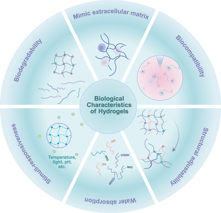 |
Figure 1 The biological properties of hydrogels. |
In neuro-diagnostics, electroencephalography (EEG) is vital for capturing electrophysiological biomarkers of most neurological disorders.12–16 However, conventional non-invasive electrodes are frequently compromised by high contact impedance and motion artifacts,17,18 compromising their spatial resolution. Conversely, invasive electrodes achieve superior resolution yet are typically constructed from rigid metals or silicon, exhibiting a profound mechanical mismatch with soft brain tissue.19 Their elevated Young’s modulus and acute geometries elicit chronic neuroinflammation and physical trauma.20 Hydrogel-based electrodes offer a compelling solution to these challenges. On the one hand, characterized by a low Young’s modulus and favorable biocompatibility, they inflict minimal trauma on neural tissue. On the other hand, abundant electrolytes entrapped within hydrogels matrix establish ionic-ionic interfaces with both skin and brain tissues, markedly lowering interfacial impedance. Furthermore, the inherent viscoelasticity and fluidity of hydrogels permit spontaneous adaptation to microscopic surface under applied pressure, generating intimate conformal contact that substantially improves signal quality.
With regard to therapeutic intervention, standard systemic administration of bioactive agents21 is severely compromised by the blood-brain barrier (BBB),22 necessitating supratherapeutic dosages that inevitably lead to side effects and systemic toxicity.23 Although alternative strategies such as deep brain stimulation (DBS), vagus nerve stimulation (VNS), and surgical interventions provide symptomatic relief, they primarily attenuate pathological signal propagation without targeting underlying disease mechanisms. The distinct 3D architecture of hydrogels establishes a biomimetic reservoir that enables high-capacity loading of therapeutic agents, protects payloads from enzymatic degradation, and permits spatiotemporally controlled release at the pathological site.24 Furthermore, brain tissue engineering is a transformative strategy for functional restoration following severe central nervous system (CNS) injuries, providing biomimetic scaffolds to mitigate long-term sequelae.25 By recapitulating the physicochemical properties of the native extracellular matrix (ECM),26 hydrogel-based scaffolds can be functionalized with incorporated stem cells, adhesion motifs, growth factors, and biomolecules,27 promoting cell proliferation, axonal guidance, and de novo tissue formation. Unlike circulating carriers such as nanoparticles and liposomes, hydrogel-based delivery systems can be retained at the therapeutic site to form a localized drug depot, achieving sustained release over weeks to months. By contrast, circulating carriers are susceptible to rapid clearance and exhibit limited stability.
Whereas existing reviews have largely catalogued hydrogel synthesis or addressed isolated facets of neuro-interventions, this review uniquely bridges flexible diagnostic interfaces, BBB-penetrating drug delivery, and regenerative neural engineering. Moreover, we identify critical gaps and novel opportunities in the field, aiming to provide a roadmap for their clinical translation and to inspire further research in neuroscience.
Basic Characteristic of Hydrogels
Source and Development
The development of hydrogels has a glorious history dating back over a century. In 1894, van Bemmelen innovated the term “hydrogel”.28 Subsequently, in 1960, Wichterle and Lim developed the synthetic poly2-hydroxyethyl methacrylate (PHEMA) hydrogel, marking the first generation of hydrogel, which was applied in the production of hydrogel contact lenses.29 Since their inception, hydrogels have experienced alarming growth across various fields and exhibit a wide array of applications. Currently, the diversity and quantity of hydrogels are considerable, and they can be categorized based on varying criteria.
According to sources, hydrogels can be classified into two primary categories: natural and synthetic types. Natural hydrogels, including collagen, gelatin, cellulose, hyaluronic acid (HA), chitosan, and others, are recognized for their inherent biocompatibility, bioactivity, and biodegradability.30 For instance, the extracted collagen can be converted into hydrogels through pH and temperature stimuli via a self-fibrogenesis process under physiological conditions, which is recognized as a biomimetic substance mimicking the cellular microenvironment.31 It is important to note that natural types exhibit relatively weak stability, mechanical strength, and pose challenges in achieving precise formulations and drug loading. Synthetic hydrogels are synthesized through the polymerization of monomers that incorporate various polymers including polyethylene glycol (PEG),32 polyvinyl alcohol (PVA),33 polyethylene oxide (PEO),34 and poly-2-hydroxyethyl methacrylate (PHEMA).35 These hydrogels can be tailored under different conditions, including chemical structure, arrangement, biological functions, and physicochemical properties.
As technology has evolved, stimuli-responsive hydrogels have emerged as a significant area of interest. These hydrogels are synthesized through the integration of stimuli-responsive molecular complexes and biomolecular units, enabling triggered and reversible transitions in structure and physical properties. Such transitions can be elicited by various physical or chemical stimuli, such as pH changes, chemical agents, light, temperature, magnetic fields, and electrical fields. Moreover, the incorporation of biomaterials consistently enhances their unique recognition properties and facilitates chemical modification.36 This enhancement not only allows for responses to physical and chemical signals but also results in increased sensitivity to biochemical stimuli, such as enzymatic reactions37 and levels of reactive oxygen species (ROS).38 For example, tumor cells frequently deplete their blood supply to satisfy their energetic requirements, which cause permanent or transient deprivation of oxygen and nutrients. This deprivation leads to increased levels of ROS and the upregulation of antioxidant enzymes.39 In response to this condition, cancer cells exhibit high rates of glycolysis, leading to the production of excessive lactic acid and a lower extracellular pH compared to normal tissues.40 Given the altered metabolic profiles of cancer cells, Yi-Jun Jo et al developed a multi-stimuli responsive hydrogel to precisely control the release of doxorubicin (DOX). The hydrogel exhibited a rapid release of DOX under various conditions, including acidic environments (pH 5), reducing conditions (10 mmol DTT), oxidizing media (0.5% H2O2), and exposure to NIR irradiation.41 In vitro experiments indicated that multi-stimuli responsive hydrogel enhances the antitumor efficacy of DOX.
Despite extensive exploration of traditional hydrogels as carriers for molecules and cells, these materials often fail to effectively integrate with the defect site and lack the ability to adapt their behavior properties in response to the physiological and pathological conditions associated with disease. Intelligent, stimuli-responsive hydrogels facilitate precision interventions in neurological diseases by responding to changes in the brain’s microenvironment, as summarized in Table 1.
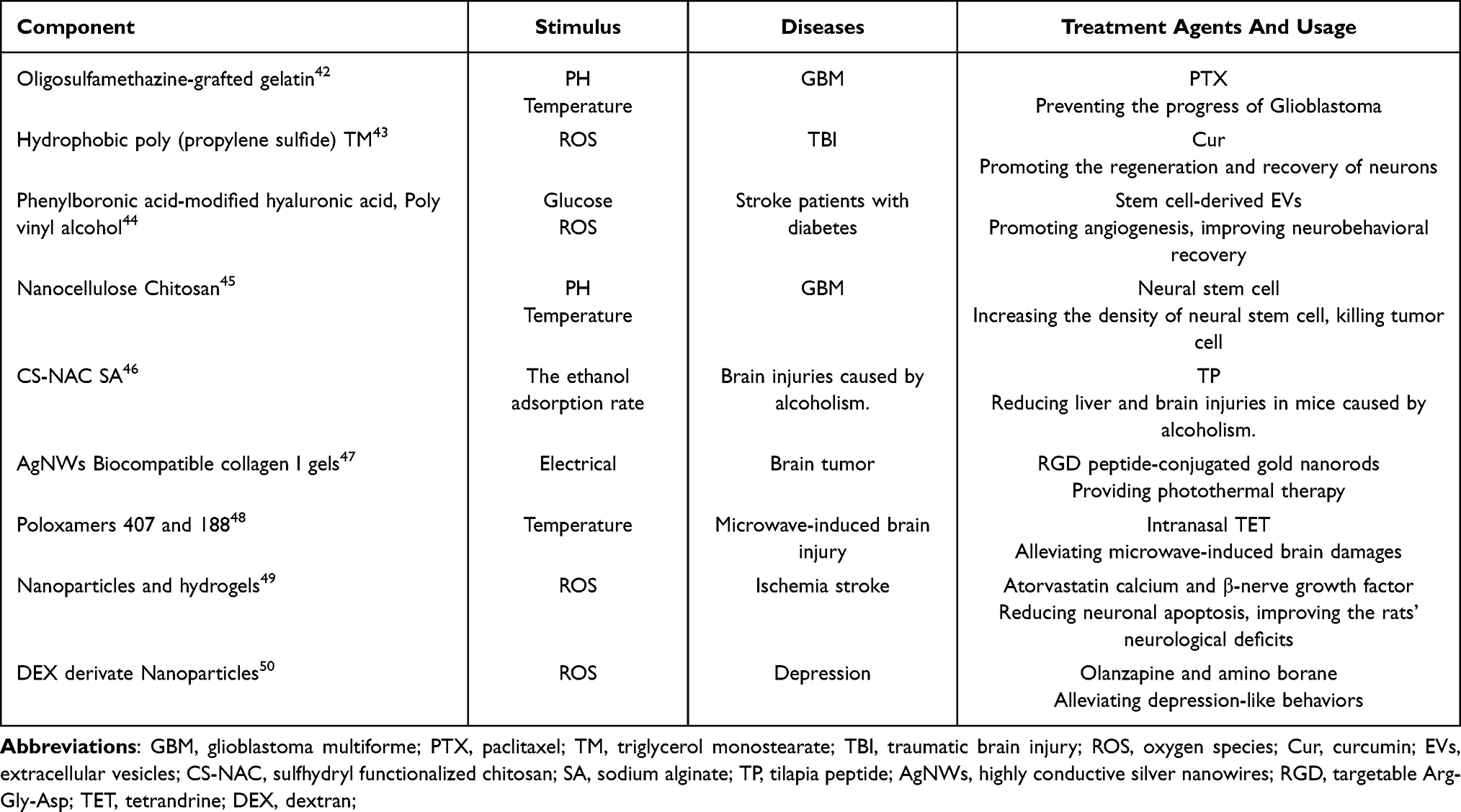 |
Table 1 Summary of the Hydrogel Component, Stimulus, Targeted Diseases and Related Treatment Agents of Some Typical Intelligent Stimuli-Responsive Polymers |
Synthesis of Hydrogels
The distinctive three-dimensional architectures of hydrogels arise from chemically or physically cross-linked hydrophilic polymer networks (Figure 2). The selection of cross-linking method critically determines their structural parameters, including mesh size, swelling ratio, and permeability, which will be introduced subsequently.
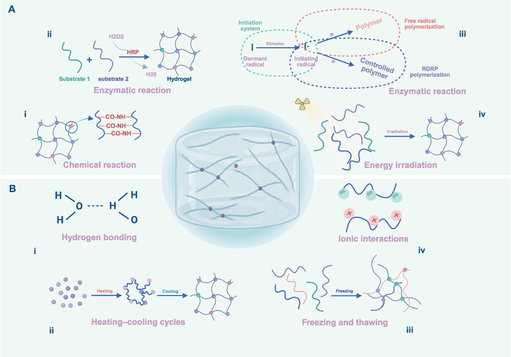 |
Figure 2 Hydrogel networks are typically categorized as chemically (A) and physically (B) crosslinked systems. Chemical crosslinking includes (Ai) chemical reaction, (Aii) enzymatic reaction, (Aiii) radical polymerization, and (Aiv) energy irradiation; physical crosslinking includes (Bi) hydrogen bonding, (Bii) heating–cooling cycles, (Biii) freezing and thawing, and (Biv) ionic interactions. |
Physical Crosslink
Physical crosslinking methods do not necessitate the use of crosslinking agents, providing advantages in convenience and non-toxicity. Advancements in science and technology have facilitated the development of diverse synthesis methods, including hydrogen bonding, anion-cation interactions, heating-cooling cycles, and freezing and thawing. Numerous natural hydrogels are constituted through self-assembly processes that involve physical crosslinking, despite their inherent instability and low mechanical strength resulting from the weak interactions between polymer chains.51
Hydrogen Bonding
Polymer chains typically contain numerous hydrogen atoms, which enable electrostatic interactions and the formation of hydrogen bonds among them, promoting the immediate assembly of hydrogels.52 For instance, Yan Wang et al developed a novel hydrogen-bonded polysaccharide hydrogel through the formation of strong intermolecular hydrogen bonds between ipsilateral hydroxyl groups.53 It exhibited favorable mechanical strength, antibacterial ability, and excellent biocompatibility, all attributed to the dynamic and reversible linkages. Notably, the formation of hydrogen bonds in hydrogels is intrinsically governed by pH.54 This pH-dependent dynamic enables hydrogels with dense hydrogen-bonding networks to exhibit programmable pH-responsive behaviors, a cornerstone for engineering smart hydrogels capable of autonomous adaptation to physiological pH fluctuations.
Ionic Interactions
It has been established that ions can participate in ion-dipole interactions with both polymers and water,55 facilitating the formation of hydrogels. Moreover, ionic hydrogels generally display exceptional conductivity, rendering them suitable for applications in electricity collection equipment, such as electrodes. Wen Li et al utilized tannic acid (TA) to reinforce polyacrylamide (PAM) and sodium carboxymethyl cellulose (CMC) polymers to create a strain sensor for human-machine interaction (HMI) devices.56 This hydrogel-based sensor exhibits optimal electromechanical performance and achieves a high motion recognition accuracy of 97.85%, characterized by high sensitivity (GF = 4.03), favorable linearity (0.997), and a low detection limit (0.5%).
Heating–Cooling Cycles
Heating-cooling cycles are essential for the dissociation of oligomers into monomers and the subsequent refolding process.57 Initially, these cycles induce a coil conformation in the polymer chains, which, after additional cycles, result in the formation of double helices, stabilizing the hydrogel structure through the involvement of salt. Tingrui Lin et al combining heating-cooling cycles with γ-ray radiation methods to develop a novel agarose/Ti3C2Tx-crosslinked-polyacrylamide hydrogel.58 In the experiment, the researchers mixed a solution of Ti3C2Tx nanosheets with agarose (AG), acrylamide (AM), and water in a glass vial. The mixture was heated to 90°C until it achieved homogeneity, followed by cooling to room temperature. Following several cycles, a strain sensor hydrogel was produced, which exhibited superior mechanical properties with 4250% stretchability, and enhanced adhesion to various substrates, demonstrated by an adhesive strength of 1148 kPa to copper at 30 °C. These advancements make it an effective strain sensor in large-scale strain detection scenarios that require high sensitivity and adhesion.
Freezing and Thawing
During the freezing step, water solidifies while solutes and bound water remain in a liquid state. This freeze-concentration effect drives extreme polymer chain compaction. This condition results in high local concentrations and the occurrence of “proximity effects”, which facilitate physical entanglement and the formation of multiple hydrogen bonds.59 Similar conditions for cross-linking are maintained during the thawing step, leading to the establishment of a three-dimensional polymer network characterized by unique wall and pore structures. The number of freezing−thawing cycles, freezing temperature and time significantly influence the structure and mechanical properties of hydrogels.60 Zhengyu Gong et al prepared a hydrogel composed of poly (acrylamide-co-acrylic acid) (PAM-co-PAA) and polyvinyl alcohol (PVA) through copolymerization and freezing/thawing. They demonstrated that hydrogels created in this manner exhibit exceptional rapid recovery, superior fatigue resistance, and self-healing capabilities. These attributes arise from the integrated fatigue resistance and self-healing properties, which are measured at 1230 ± 90 kPa and 1250 ± 50 kJ/m3, respectively.61
Chemical Crosslink
Chemical methods leverage site-specific reactions between functional groups on polymer chains and multifunctional crosslinkers This process induces irreversible covalent cross-links and excellent mechanical strength. Conventional chemical crosslinking strategies often rely on exogenous agents like epichlorohydrin (ECH) or glutaraldehyde, which pose dual challenges regarding the toxicity of cross-linking agents and uneven cross-linking distribution.62
Radical Polymerization
The free radical polymerization (FRP) mechanism governs hydrogel network formation through four fundamental steps: initiation, propagation, chain transformation, and termination,63 which enables precise spatiotemporal control over covalent network architecture. Recent breakthroughs in reversible deactivation radical polymerization (RDRP) techniques, such as atom transfer radical polymerization (ATRP), have revolutionized hydrogel synthesis by enabling precise control over network topology. Citing a case, Yerneni et al prepared a type of hydrogel using ATRP, a powerful and versatile RDRP process. They subsequently employed cholesterol-modified DNA tethers to functionalize the lipid membrane of exosomes for polymer grafting,64 enabling the localized delivery of therapeutic exosomes for a duration of one month, which is longer than that of conventional exosomes.
Chemical Reaction
Chemical reactions occur between complementary functional groups or pendant hydrophilic moieties and polymers mediated by multifunctional crosslinking agents. Crosslinking agents feature multiple reactive termini that covalently conjugate with complementary functional moieties on hydrophilic polymers to orchestrate the assembly of three-dimensional polymeric networks. Junjie Qin et al designed a glucose-driven self-regulating hydrogel with resilient structure, using beta-cyclodextrin (β-CD) and ferrocene (Fc) as partial crosslinkers, and a chemical reaction network (CRN).65 The designed supramolecular photonic hydrogel (SPH) has been successfully employed to detect glucose in human plasma and H2O2in liver tumor tissue, yielding results comparable to those of commercial assay kits. However, it is crucial to highlight that a majority of crosslinkers often exhibit inherent cytotoxicity and environmental persistence due to non-degradable byproducts and ecotoxic leakage. Scientists are challenged to develop safer alternatives.
Enzymatic Reaction
Enzyme-mediated hydrogel synthesis emerges as a biocompatible paradigm shift, offering spatiotemporal control over gelation kinetics and network precision unattainable by conventional physicochemical methods.66 This method is uniquely suited for polymers functionalized with enzyme-sensitive molecules. In recent decades, several technologies have been developed, including enzymatic polymerization of monomers, enzymatic cross-linking of existing polymers, and enzyme-manipulated self-assembly of small molecules.67 For instance, tyrosinase-mediated reactions were utilized to cross-link monophenol-modified glycol chitosan with hyaluronic acid, generating a bioactive hydrogel film.68 This film has been demonstrated to regulate blood glucose levels in a type 1 diabetes mouse model following transplantation. Furthermore, enzyme-catalyzed crosslinking approaches, especially the horseradish peroxidase (HRP)-catalysed crosslinking approaches, are promising approaches for materialization, due to their high specificity and mild reaction conditions.69 A notable advancement by Sara Baptista-Silva et al introduced a new horseradish peroxidase-mediated cross-linked hydrogel, composed of silk sericin.70 This study validated the methodology and demonstrated potent anti-inflammatory efficiency, which can promote cell adhesion and massive cell colonization after 7 days of culture in an in vivo diabetic wound model.
Energy Irradiation
Energy irradiation method employs high-energy radiation, including X-rays, gamma rays, and electron beams to synthesis hydrogels via radiation-induced polymerization or crosslinking. For example, Maria Demeter et al reported an elaborate hydrogel based on collagen-poly (vinyl pyrrolidone) (PVP)-poly (ethylene oxide) (PEO) cross-linked by e-beam irradiation;71 Moises Bustamante-Torres et al presented a pH-sensitive hydrogels copolymerized by gamma rays to localized release of ciprofloxacin and silver nanoparticles (AgNPs).72 Compared to traditional methods, energy irradiation eliminates the need for cytotoxic chemical initiators, while enable controlled network architectures. Moreover, the materials can be sterilized using energetic radiation, integrating synthesis and sterilization simultaneously, reducing both costs and time.
Fabrication Techniques
3D Printing
In recent years, the proliferation of 3D printing technologies (Figure 3), particularly utilizing inks containing multiple macromolecules and biological components, have revolutionized the fabrication of customizable hydrogels. 3D hydrogel-forming extrusion (HFE) employs the computer-controlled, layer-by-layer dispensing of semi-solid polymeric solutions, dispersions, or pastes through programmable nozzles traversing (x, y, z) axes.73 The chemical, mechanical, and stimulus-responsive properties of hydrogels can be tailored through the printing process. For instance, articular cartilage tissues connect articular surfaces to subchondral bones. However, due to its avascular environment and low cellular density, the capacity for regenerative restoration is limited. When damaged, articular cartilage extends into the underlying subchondral bone.74 To promote tissue regeneration, Qi Li et al produced a flash bioinspired double-network hydrogel scaffold through 3D printing, encapsulating tissue-specific decellularized extracellular matrix (dECM) and exosomes deriving from human adipose mesenchymal stem cell (MSC).75 The bionic hydrogel scaffolds promoted the attachment, spread, migration, proliferation, and differentiation of MSC, accelerating the simultaneous regeneration of cartilage and subchondral bone tissues in rat models.
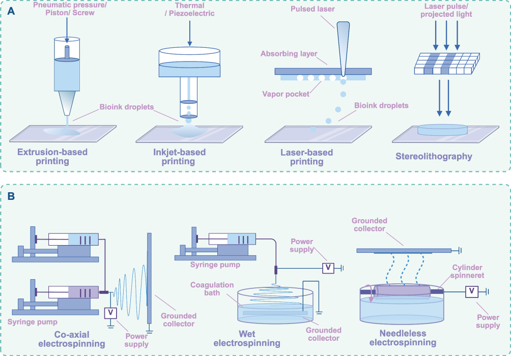 |
Figure 3 The synthetic methodologies for hydrogels, including three-dimensional (3D) printing (A) and electrospinning (B). |
However, ensuring printing quality poses significant challenges. The high-water content of hydrogels hinders achieving adequate printing accuracy and shape fidelity. Most hydrogel inks are influenced by gravitational forces and surface tension, which predisposes them to collapse or spread after printing.76 More importantly, it is an insurmountable barrier to assure biocompatibility, non-toxicity, and appropriate mechanical properties in 3D-printed hydrogels 3D printing hydrogels adopted in biomedical engineering.77
Electrospinning
Electrospinning is a sophisticated manufacturing technique that employs a high-voltage electrostatic field to convert polymer solutions or melts into ultrafine fibers, generally characterized by nanoscale diameters78 (Figure 3). Typically, electrospinning setup includes a high-voltage power supply, a spinneret (a metallic needle), and a grounded collector. When a sufficient voltage is applied to the spinneret, the electrostatic forces surpass the liquid’s surface tension, initiating a charged polymer jet that undergoes whipping and stretching in the electric field.79 This process leads to solvent evaporation or melt solidification, ultimately producing a nanofibrous network with exceptional properties, including high surface-to-volume ratio, controlled porosity, and tailored mechanical strength.
Similarly, the ability to precisely control fiber morphology and alignment through electrospinning provides a powerful tool for the fabrication of intricate hydrogel network. Moreover, the controlled nanofibrous architecture exhibits a rapid swelling response and faster shape recovery compared to bulk hydrogels. For instance, Shenglian Yao and coworkers incorporated carbon nanotubes (CNTs) into methacrylate acylated gelatin (GelMA) hydrogel to develop aligned conductive hydrogel fibers using rotating liquid bath electrospinning technique.80 Compared to bulk hydrogels with irregular structures, the aligned f hydrogel fibers supported the proliferation and aligned adhesion of PC12 cells, promoting the regeneration of neural fibers.
The integration of 3D printing and electrospinning technology can address some limitations inherent to each individual method, such as restricted cell migration resulting from the dense intertwining of electrospun fibers and the limited resolution associated with certain 3D printing techniques. For example, Sitian Liu et al presented an anisotropic multiscale cardiac scaffold achieved by combining the two methods. The 3D-printed micrometer-scale scaffold frames effectively mimic the interwoven anatomical structure of the myocardium, while the branched-aligned electrospun nanofiber network directionally guides cellular arrangements.81
Recording Neural Electrophysiological Signals
Neurons continuously generate and transmit electrophysiological signals. The EEG serves as the gold standard noninvasive modality for capturing the aggregate postsynaptic potentials from cerebral pyramidal cells. Currently, various devices for recording EEG signals have been developed, classified as invasive, semi-invasive, and noninvasive installations. These devices authorize the interaction between biological system and external electronic devices, functioning as operational instructions for activating brain computer interface (BCI) systems (Figure 4). In recent years, flexible hydrogels have attracted considerable interest as viable alternative materials for neuro-electrodes and interface components, promoting the development of electrochemical biosensors.
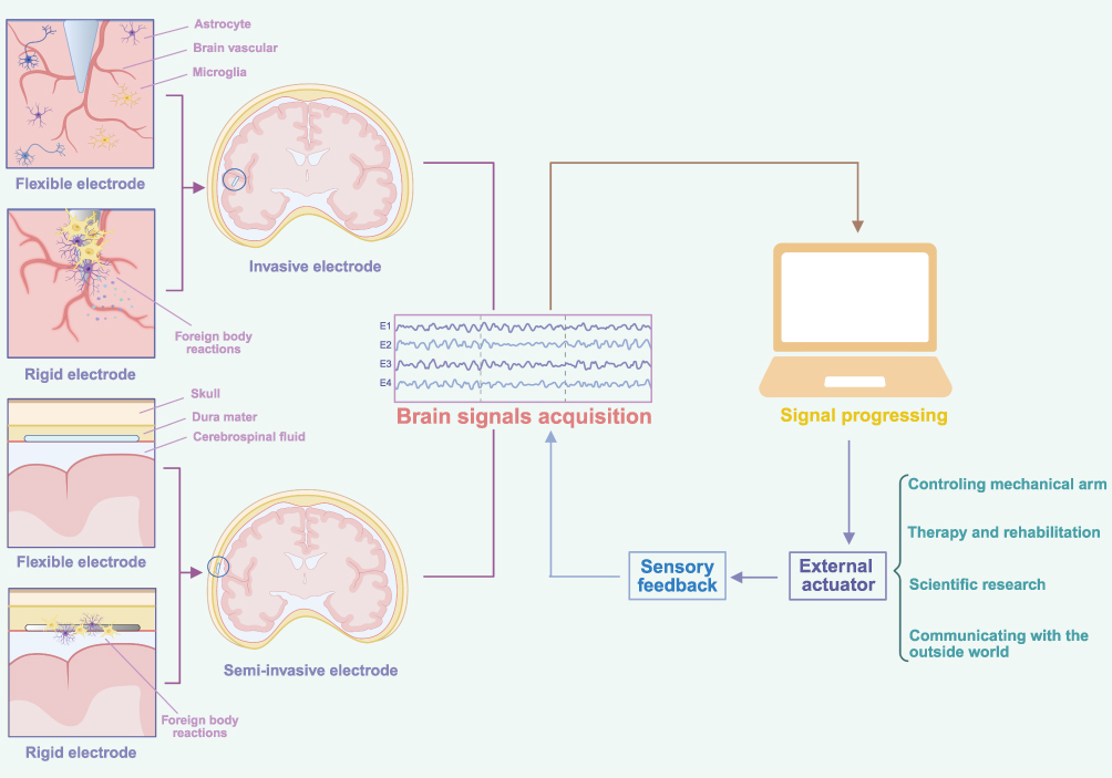 |
Figure 4 Schematic illustration of the closed-loop brain-computer interface (BCI) workflow. |
Flexible Electrode Materials
Conducive materials are essential components of electrodes, forming the electrical connections between the neuron and external device. Metals are the most widely utilized electrode materials, primarily due to their superior electrical properties and chemical stability. Research has demonstrated that liquid metal conductors fabricated onto polydimethylsiloxane substrates can create highly stretchable neural electrode arrays with a resolution of 50 µm, which are particularly effective for neural recording.82 However, these electrodes face challenges related to biocompatibility with both brain tissue and our scalp, which can lead to inflammation and irreversible brain damage. The adaptation of bio-materials, particularly hydrogels, represents a visionary alternative to address this issue, as summarized in Table 2
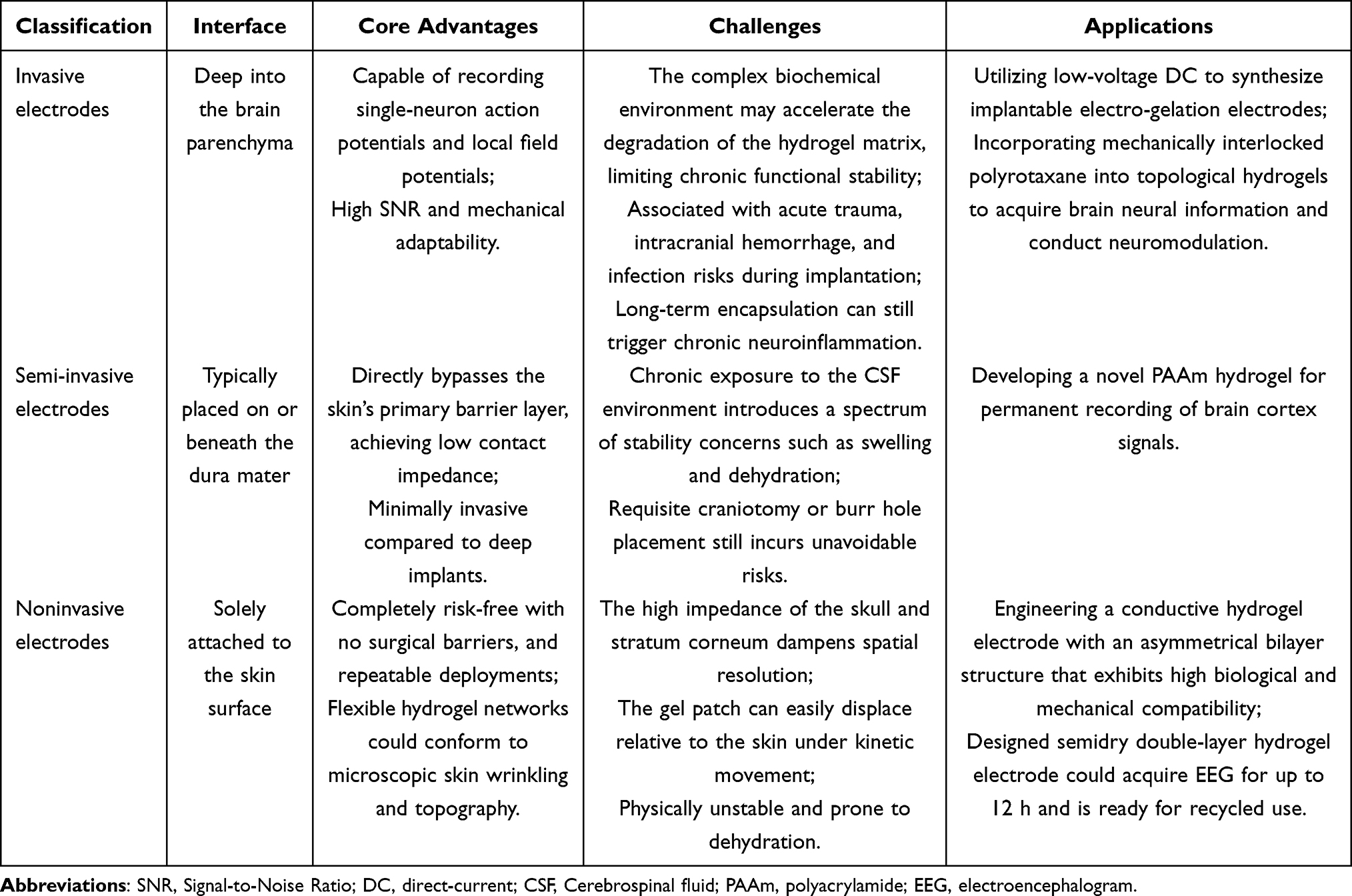 |
Table 2 Classification and Comparison of Hydrogel-Based Electrodes |
Invasive Electrodes Materials
Invasive neural signal acquisition entails a deep penetration into brain tissues to record voltage potentials from individual neurons near electrodes with high precision and specificity. However, conventional metallic electrodes trigger a cascade of foreign body reactions (FBR)83 when they contact with brain tissue. Furthermore, metals and semiconductors exhibit high rigidity, with Young’s moduli in the tens of gigapascals, whereas brain tissues are significantly softer, characterized by Young’s moduli in the kilopascal range. This disparity in mechanical properties can lead to irreversible brain damage and aggravate the inflammatory response. In the context of long-term recording, these rigid electrodes may be encapsulated by microglia,84 which can increase electrical impedance and compromise their biocompatibility and functionality. Furthermore, studies have demonstrated that, despite stringent aseptic implantation protocols, certain gut bacterial constituents might translocate to the brain following electrode insertion, compromising microelectrode performance and exacerbating chronic inflammatory responses.85 Moreover, when inflammation escalates and implanted electrodes fail, they must be surgically removed, causing escalates damage. This scenario highlights the critical need for developing appropriate materials to overcome the limitations of invasive electrode systems.
Among flexible biomaterials, hydrogels are distinguished as a notably active and widely utilized class. For instance, Zhen-Kai Lin et al utilized low-voltage direct-current (DC) to synthesize implantable electro-gelation electrodes, by inducing sol-to-gel transitions in an aqueous reconstituted silk fibroin solution.86 The electrode exhibits a high Young’s modulus (11–150 MPa) under ex vivo conditions to facilitate penetration through brain tissue, while transitioning to a brain-tissue-matching lower modulus (50–3200 kPa) post-insertion to minimize mechanical mismatch-induced damage. Simultaneously, the controlled release of dexamethasone (DEX) interrupt astrocyte growth to reduce the inflammatory response. Additionally, microscale needle-like electrode technologies provide neural extracellular recording with a high spatiotemporal resolution while minimize tissue injuries by reducing the surface area of electrodes and increasing their number.87 Zhenzhen Shen et al incorporated mechanically interlocked polyrotaxane into topological hydrogels to acquire brain neural information and conduct neuromodulation, enabling the continuous recording of neural local field potentials for eight weeks.88
However, invasive hydrogel-based electrodes are subject to dual destabilization mechanisms, dehydration can degrade electrical conductivity and promote mechanical stiffening, whereas excessive swelling drives volumetric expansion and potential structural disintegration. Moreover, the simultaneous optimization of electrical conductivity, modulus matching, biocompatibility, and mechanical stability remains inherently challenging, necessitating judicious trade-offs among these competing requirements.
Semi-Invasive Electrodes Materials
Despite being considered the gold standard for diagnosing certain neurological conditions, invasive methods are not universally accepted for their unavoidable tissue trauma. Semi-invasive electrodes, commonly referred to as Electrocorticography (ECoG) electrodes, are small devices strategically placed on or beneath the dura mater. This procedure is typically performed before and during neurosurgical operations for epilepsy or brain tumors. Remarkably, due to the mild immune response, these electrodes can reliably record signals for many years after implantation.89 Furthermore, ECoG electrodes are in direct contact with the brain and record the local field potential (LFP) from the cerebral cortex with high signal to noise ratio, fewer artifacts, and greater spatial specificity.90 However, conventional semi-invasive electrodes do not conform to the intricate mechanical properties of the brain. Upon implantation, these electrodes can disrupt brain tissues and compromise the BBB, which may accelerate the formation of a glial sheath around the electrodes and thicken the dura mater.91 Moreover, while semi-invasive electrodes must be sufficiently flexible to maintain contact with the brain’s convoluted surface, most electrodes lack the conformability. The loose connection between electrodes and the cortex can cause electrode displacement, potentially leading to inaccurate signal recordings and even misdiagnosis. Micromotion between the implanted electrodes and neural tissues may occur over time, primarily induced by vascular pulsations.92
Hydrogel-based electrodes offer adequate conformability to the brain’s curved and grooved surface, ensuring reliable neural signal acquisition. More importantly, hydrogels can absorb heat, avoiding local burns, lesions, and pain.93 For instance, through the integration of MRI-based anatomical mapping, finite element analysis (FEA)-optimized mechanical design, and direct ink writing (DIW) 3D printing, an ultrasoft hydrogel electrode featuring a bioinspired honeycomb architecture was developed. Its bending stiffness matches that of brain tissue, ensuring excellent cortical integration and conformability.94
Notably, the chronic exposure of semi-invasive electrodes to the cerebrospinal fluid (CSF) environment introduces a spectrum of stability concerns, ranging from uncontrolled swelling and dehydration-driven stiffening to inadequate conductivity and mechanical fatigue. Compounding these material challenges, the requisite craniotomy or burr hole placement still incurs unavoidable risks of hemorrhage, infection, and CSF leakage. Consequently, future investigations must judiciously address swelling restriction and interfacial adhesion optimization.
Noninvasive EEG Electrodes
EEG is the most widely used neuroimaging technique in clinical practice, due to its non-invasive nature, portability, ease of use, and lack of associated trauma. Typically, non-invasive electrodes can be categorized into three types: dry electrodes, semi-dry electrodes, and wet electrodes. Hydrogels can significantly enhance technological advancements by optimizing various types of electrodes for improved performance.
Wet electrodes, particularly those commonly used in EEG recording, are frequently preferred. Conventional gel electrodes incorporating Silver/Silver-Chloride (Ag/AgCl) materials and a conductive electrolyte gel are considered the gold standard for EEG signal recording,95 due to their low intrinsic noise levels and skin-contact impedance. However, the use of conductive gels often results in adherence to patients’ hair and skin, which are cumbersome and time-consuming for patients to clean. Moreover, while conductive paste promotes the formation of ion channels and guarantees stable signal acquisition progress, unintended leakage of paste can cause short circuits between adjacent electrodes.96 Additionally, the water content in the gel inevitably evaporates over time, necessitating regular maintenance to preserve optimal signal quality for extended periods,97 which does not allow for continuous long-term signal recording. Hydrogels are characterized by low impedance and a high signal-to-noise ratio (SNR), providing a stable and seamless interface between skin and wet electrodes. Moreover, hydrogel-based wet electrodes exhibit favorable electrical conductivity and are anticipated to enable long-term neuroelectric signal recording. Researchers have engineered a conductive hydrogel electrode with an asymmetrical bilayer structure that exhibits high biological and mechanical compatibility while remaining compatible with magnetic resonance imaging (MRI) and computed tomography (CT), obtaining high spatiotemporal resolution multi-dimensional brain information.98
Unlike wet electrodes, dry electrodes eliminate the need for conductive gel and maintain stable contact with the scalp through external pressure. This offers advantages such as convenience, comfort, ease of operation, and stability.99 Nevertheless, the primary disadvantage is their reduced conductivity and higher skin-electrode contact impedance, which makes them more susceptible to motion artifacts. The multichannel microneedle array (MMA) can penetrate the non-conductive stratum corneum to reduce electrode-skin contact impedance,100 however, the potential breakage of microneedles within the skin may increase the risk of infection and inflammatory reactions. To address these issues, researchers have designed semi-dry electrodes, which combine the advantages of both wet and dry electrodes while mitigating their respective drawbacks. Semi-dry electrodes, which contain a minimal amount of electrolyte, can reduce electrode-scalp impedance to a level comparable to wet electrodes while avoiding hair contamination and the risk of short circuits.101 However, semi-dry electrodes also suffer from liquid evaporation, which can be mitigated by applying hydrogels. For example, Hailing Xue et al propose a semi-dry double-layer hydrogel electrode.102 This electrode comprises two distinct layers. The first layer is constructed from conductive, robust hydrogels, which provide low skin-electrode impedance and high durability, facilitating prolonged use. The second layer consists of adhesive hydrogels formulated to bond with various substrate materials through chemical anchors. The designed semi-dry electrode demonstrated comparable signal capture capabilities to wet electrodes in both N170 and P300 event-related potential (ERP) tests, while causing less harm.
Nevertheless, the inevitable evaporation of moisture from wet electrodes induces polymeric network shrinkage, aberrant electrolyte concentration, and elevated contact impedance. Moreover, the high-water content compromises mechanical robustness and tissue adhesion. Semi-dry electrodes partially reconcile signal quality with wearing convenience, yet struggle to maintain a constant and controllable electrolyte exudation rate under variable environmental conditions and interindividual skin differences. Future research must transition from single-parameter optimization to multi-physics design, developing environmentally adaptive intelligent hydrogel materials and optimizing dynamic impedance-matching algorithms to ensure long-term clinical utility.
Interface Materials
For implantable electrodes, the practical and economic approach involves decorating the basic electrode with functional coating materials, rather than developing new designs and configurations.103 The electrode interface, where electrodes directly contact brain tissue, is crucial for the transmission of electrical signals. Functional coatings applied to the electrode surface enhance mechanical properties, biocompatibility, and durability.
Researchers have produced various coating materials, with hydrogels being a particularly favorable choice. For instance, Manuele Gori et al fabricated a sulfobetaine-based zwitterionic hydrogel as coating material for Polyimide (PI)-based intraneural electrodes. The PI surfaces were coated with a thin film of the hydrogel through covalent bonding to prevent the delamination of the hydrogel coating from the surface of neural electrodes.104 The Young’s modulus was 2.7 ± 0.24 kPa similar to brain tissue, minimizing the mechanical mismatch between electrodes and brain. Besides, the hydrogel coating was inspected and verified to successfully reduced adhesion and activation of fibrogenic and pro-inflammatory cells, ensuring the possibility of long-term implantation. Researchers created an organic subdural electrode composed of a hydrogel substrate and a patterned stretchable carbon fabric (CF).105 The hydrogel substrate acted as a buffer layer between the rigid electrodes and soft brain tissues, mitigating the immune responses induced by foreign matter.
However, hydrogel-based coatings, while demonstrating excellent biological performance, inherently possess low electrical conductivity. The incorporation of electrically conductive materials, such as conductive polymers (CPs) and metal oxides, could addresses this issue by retaining the desired biological and electrical properties.106 Sungjun Lee et al utilized poly (3,4-ethylenedioxythiophene): polystyrene sulfonate (PEDOT: PSS), alginate (Alg), polyacrylamide (PAAm), and CMC to synthesize a soft, tough ionic conductive hydrogel, which was subsequently covered with stretchable metal electrodes.107 The coated electrodes demonstrated a reduction in impedance from 60 kΩ to 10 kΩ at 1 kHz and acute in vivo experiments successfully validated their success, demonstrating sensitivity and accuracy.
Working as Part of a Brain-Computer Interfaces
Brain-computer interfaces (BCIs), which enable interaction between the human brain and machines, have garnered global attention and are undergoing rapid technological advancement. BCIs generally utilize electrodes to continuously encode and decode brain signals, translating these into the desired output following analysis and processing. This technology has diverse applications across multiple domains, such as the restoration of communication and motor function, the treatment of neurological and psychiatric disorders, daily life applications, and the gaming industry.108
The Development of BCIs
Since Berger first discovered the human electroencephalogram in 1924,17 researchers have embarked on a longstanding effort to investigate electrical neuronal activity, establishing the foundation for the development of BCI systems. Real BCIs devices did not begin to emerge until the 1970s. During this period, Jacques J. Vidal pioneered the evaluation of the feasibility and practicality of using brain signals for human-computer interaction, coining the phrase “Brain-Computer Interface”109 To date, BCI technology has evolved into a multidisciplinary field encompassing neuroscience, cognitive psychology, linguistics, artificial intelligence, philosophy, anthropology, robotics, and information technology.110 For example, chemotherapy‐induced peripheral neuropathy (CIPN)is a detrimental side effect of neurotoxic cancer treatment, affecting quality of life and potentially disabling cancer survivors.111 Sarah Prinsloo PhD et al proposed a closed-loop BCIs in which breast cancer survivors, who had undergone chemotherapy, were required to play a video game while their brain activity was monitored by electroencephalography. Both auditory and visual reinforcement were provided as the signal input command changed. The study demonstrated that the BCIs group reported significant symptom reduction and continuous improvement, exhibiting larger effect size differences compared to the placebo and waitlist control groups.112 A typical BCI system operates in three stages. The first stage involves the acquisition of neuroelectric signals from the brain. The second stage focuses on analyzing and integrating these signals, followed by converting the input signals into predefined output commands through translation algorithms. The final stage involves the operation of external devices according to the command.110
BCIs can be categorized into invasive, semi-invasive, and noninvasive systems, according to the type of electrodes employed. Conventional rigid electrodes may cause or worsen brain damage, diminishing the efficiency and long-term viability of BCIs. Therefore, biocompatibility, electrical conductivity, and mechanical properties resembling those of soft brain tissue are crucial for the fabrication of high-performance devices.113 Multifunctional hydrogel bioelectronics have authentically established reliable and compatible Human-Machine Interface (HMI) platforms for high-quality bioelectronic recording. For example, Chiara Rinoldi et al designed a novel soft neural bio-interface made of polyacrylamide hydrogels loaded with plasmonic silver nano-cubes.114 This type hydrogel-based electrodes, characterized by their low electrical impedance (<3 kΩ at 10 Hz) and Young’s modulus (<10 kPa), conform well to the brain surface and maintain stability amidst the brain’s subtle movements. These properties render them particularly suitable for applications in neuroscience. Ju-Chun Hsieh et al115 focused on EEG-based long-term and wearable BCIs. They developed an enhanced PEDOT: PSS/PAMPS hydrogel EEG electrode exhibiting improved ionic and electrical conductivity, along with long-term stability and low electrode-skin interfacial impedance. The authors demonstrated that the designed hydrogel electrode could capture oscillatory rhythms in motor imagery protocols, a feature applicable to BCI-based functional electrical stimulation (FES) for motor rehabilitation.
What Can BCIs Do
BCIs capture neuronal electric activity and convert it into commands for a computer or other device, enabling the user to control mechanical arm solely through thoughts. Recent advances in BCI technology hold significant potential for applications ranging from entertainment116 to medical rehabilitation,117 particularly in treating neurological disorders.
The majority of neuropsychiatric diseases can cause irreversible brain damage, presumably leading to various complications. For example, stroke is the leading cause of paralysis, often resulting in upper limb impairments and speech disorders;118 Alzheimer’s disease can cause cognitive and memory impairment;119 spinal cord and brain injuries are significant risk factor for motor dysfunction.120 BCIs are anticipated to restore or enhance brain functions impaired by debilitating disorders, as shown in Table 3.
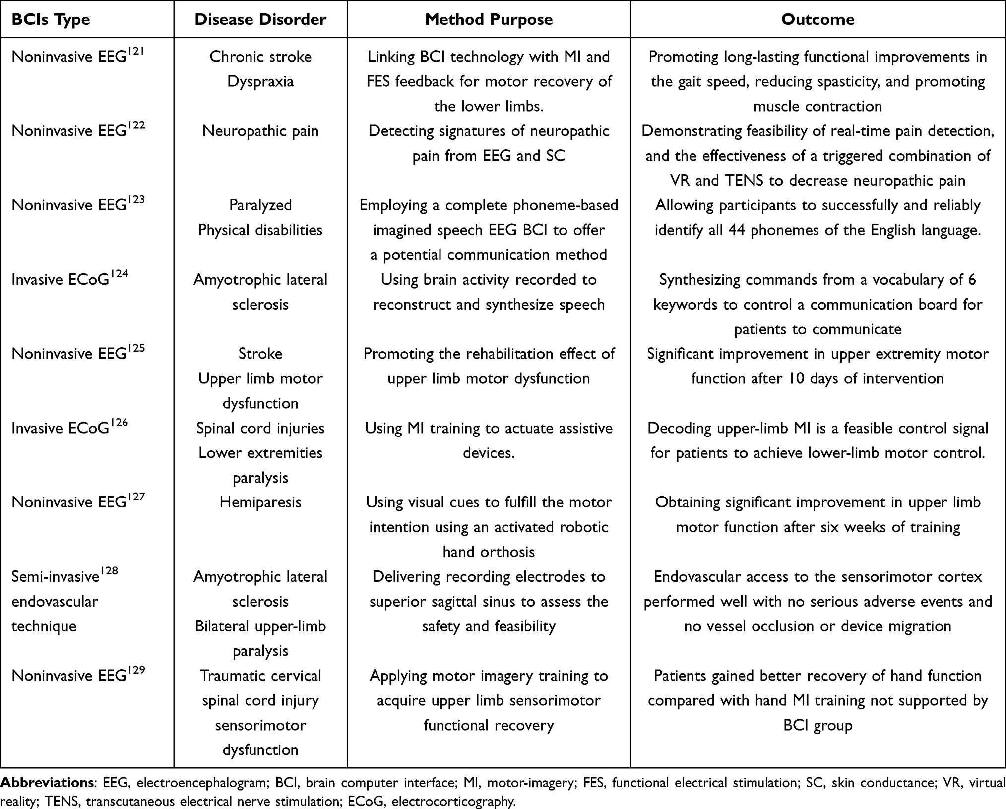 |
Table 3 The Role of Brain Computer Interfaces in Neurological Disorders |
Recently, researchers have successfully developed bidirectional BCIs that enable comprehensive communication between the brain and an actuator. Bidirectional BCIs enable not only the control of external mechanisms but also the delivery of somatosensory feedback by converting external sensor information into electric stimuli directed to the cortex.130 The development of the reverse pathway requires diverse feedback methods to establish a closed-loop system. For example, since vision provides limited feedback information, Sharlene N Fleshe et al applied tactile sensations generated by intracortical micro-stimulation of somatosensory cortex to enhance the completeness of feedback pathways.131 The authors reported that a bidirectional BCI system capable of eliciting tactile perceptions significantly improved the performance of an individual with tetraplegia using a robotic limb. This enhancement reduced the median time required to grasp objects from 20.9 seconds to 10.2 seconds. Bidirectional BCIs are expected to significantly restore or enhance human capabilities and are bound to become a mainstream technology in the future.
Functions as Biosensors
Beyond neural electrical signals, recent studies have demonstrated that hydrogels can also be engineered as biosensors to detect neurochemical signals or monitor motor function, enabling integrated diagnosis and management of neurological disorders. For instance, Metastasis-associated lung adenocarcinoma transcript 1 (MALAT1) in interstitial fluid (ISF) is aberrantly downregulated in patients with Alzheimer’s disease (AD),132 representing a potential neural biomarker. By integrating GelMA hydrogel microneedles, effective transdermal penetration of the skin epidermis can be achieved, enabling quantitative assessment of ISF MALAT1 levels and providing a practical tool for early non-invasive diagnosis of AD.133 A highly elastic and self-healing hydrogels conductor comprising catechol, alginate, and diatomite was fabricated as a stretchable triboelectric nanogenerator (TENG). This device harvests energy from human motion and functions as a self-powered tremor sensor when applied to the skin, enabling detection and tracking of low-frequency vibrational movements in patients with Parkinson’s disease (PD).134
However, the high-water content and inherent softness of hydrogel-based biosensors are intrinsically incompatible with long-term stable performance. Additionally, conventional hydrogels suffer from insufficient mechanical strength and poor toughness, rendering them susceptible to fatigue cracking, permanent deformation, or conductive network disruption during motion detection. Future improvements necessitate interdisciplinary strategies, synergistically integrating materials design, structural engineering, and algorithmic optimization to progressively overcome these existing limitations.
Carrying Therapeutic Agents
The treatment of major neurological disorders heavily depends on therapeutic agents. However, these agents frequently fail to achieving effective concentrations in the brain. Administering excessive doses of drugs tends to cause irreversible side effects. The utilization of advanced carriers presents a promising opportunity. Various carriers have been developed using a range of materials, among these hydrogels have been rapidly evolved due to their unique properties (Figure 5).
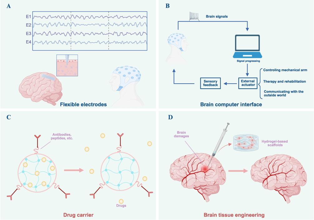 |
Figure 5 Biomedical applications of hydrogels, encompassing electrophysiological (EEG) signal monitoring (A), brain-computer interface (BCI) construction (B), drug delivery (C), and engagement in neural tissue engineering and repair (D). |
The Treatment of Neurological Disorders
Hydrogels are capable of encapsulating hydrophilic drugs, enabling sustainable and controllable drug release. They can access brain tissues through invasive and non-invasive routes. For example, injectable hydrogels can deliver ASM to the epileptic focus. Specific conditions, such as electrical stimulation and pH changes, can modify the physical properties of these hydrogels, facilitating the controlled release of drugs over an extended period.135 Here, focus on the intranasal administration pathway, heralded as a promising alternative for drug delivery. This method not only circumvents the BBB, ensuring effective intracranial concentrations, but also remains harmless.136 The combination of intranasal administration with a hydrogel carrier is expected to usher in a new era in the treatment of neurological disorders. Yujing Liu et al have conducted a study on the treatment of AD using the combination of hydrogel technology and intranasal administration.137 They developed a thermosensitive hydrogel by cross-linking carboxymethyl chitosan (CMCS) with aldehyde Pluronic F127 micelles, which were loaded with black phosphorus (BP) and methylene blue (MB). Researchers have demonstrated that the fabricated hydrogel alleviates neuroinflammation and improves cognitive function in mouse models of AD.
Conventional hydrogel delivery systems, characterized by micrometer-scale structures, are notably larger in size, which restricts their ability to cross BBB. Scientists have proposed a novel technique for manufacturing nanogels, by transforming hydrogel from microgels to nanogels. This method shows promise for allowing hydrogels to reach the smallest capillary vessels and cross the BBB due to reduced size. Furthermore, hydrogel carriers have limitations in encapsulating specific drug types, such as hydrophobic drugs, antibodies, and nucleic acids,138 due to their hydrophilic nature and relative instability. Additionally, their mechanical properties are often weaker compared to other materials. This challenge can be addressed by synthesizing hydrogel nanocomposites, which integrate nanotechnology with hydrogel technology. Specifically, incorporating nanomaterials into hydrogels creates an interpenetrating network structure,139 improves mechanical properties, and enhances drug encapsulation within a single delivery system. For example, the deep infiltration of glioblastoma multiforme (GBM) into the brain parenchyma limits efficacy of postsurgical treatment such as radiotherapy and systemic chemotherapy. Taegyu Kang et al developed an injectable thermos-responsive hydrogel nanocomposite.140 At room temperature, the composite consists of a liquid solution comprising drug-loaded micelles and water-dispersible ferrimagnetic iron oxide nano-cubes, which is injected into the resected tumor site following surgery. At body temperature, the composite undergoes sol-gel transitions, serving as a soft, deep intracortical drug reservoir. In orthotopic mouse GBM models, the drug delivery system remarkably suppressed tumor growth and improved survival rates. It is important to distinguish between nanogels and hydrogel nanocomposites, as they are fundamentally different. A nanogel is essentially a pure hydrogel, while a hydrogel nanocomposite consists of a combination of hydrogels and nanomaterials.
Compared with other drug delivery vehicles, hydrogels offer distinctive advantages as intracranial drug delivery devices. They can be minimally invasively injected as a liquid at room temperature, then undergo rapid gelation upon reaching the intracranial target site to achieve precise cavity filling. Their 3D polymeric networks enable sustained and controllable drug release over weeks to months, circumventing the peak-trough fluctuations and systemic toxicities associated with systemic administration.141 When engineered as intelligent systems responsive to stimuli such as pH, temperature, or enzymes, they further permit spatiotemporally precise on-demand drug release. However, current research has yet to achieve ideal outcomes, as significant challenges persist, including insufficient stability, heterogeneous drug distribution, and imprecise controlled release. Consequently, interdisciplinary convergence remains imperative to ensure the safety of clinical translation.
Rehabilitation of Neurological Dysfunction After Injury
Both BCIs and brain tissue engineering aim to reconstruct neural function. BCIs primarily achieve this by controlling external devices, whereas brain tissue engineering focuses on the activation or inhibition of molecular pathways within the brain. In both approaches, hydrogels play a crucial role. As previously summarized, their functions in brain-computer interfaces have been outlined, followed by a discussion of their effects in neuro-prosthetic engineering.
Restoration of Neurological Functions
The CNS consists of complex neural networks that regulate a spectrum of physiological activities, ranging from basic autonomic processes to advanced cognitive functions. CNS insults or diseases can trigger a cascade of reactions, including rapid cell death and severe neuroinflammatory responses. These events cause substantial cerebral damage and irreversible functional loss, further contributing to the disintegration and collapse of intricate neural circuits and connections within the affected regions.142 Although surviving neurons can sprout new axons and form synapses, reconstructing lost functions presents significant challenges, resulting in various complications. For example, during a stroke, neurological functions associated with the affected area are lost. After a few months, a partial recovery of nerve function typically occurs. However, approximately half of young stroke patients experience cognitive impairment, with around a quarter also suffering from aphasia.143 The global pooled prevalence of post-stroke fatigue among survivors was as high as 46.79%.144 In terms of specific deficits, the incidence rate of upper limb weakness (FAST score ≤8) is 35%, upper limb coordination impairment 46%, delayed recall impairment 41%, and upper limb sensation impairment 26%.145
Brain tissue engineering seeks to recreate living, functional tissue, playing a crucial role in restoring brain function and cognitive health. This goal is achieved by employing biological scaffolds that provide a supportive environment for cellular growth and storage spaces for molecules. Hydrogels can mimic the extracellular matrix, providing a supportive environment for transplanted and host cells to adhere, proliferate, and differentiate, making them an ideal choice for scaffold fabrication. Additionally, hydrogels extend the half-lives of injected molecules and alleviate their adverse effects by confining them within the hydrogel matrix, minimizing burst release, rapid proteolysis, and unintended diffusion.146 Over time, implanted hydrogel-based scaffolds gradually degrade and are replaced by the natural extracellular matrix synthesized by the neurons,147 a process that is non-toxic and safe. To date, multiple hydrogel-based scaffolds have been fabricated and applied in the treatment of neurological disorders, as list in Table 4.
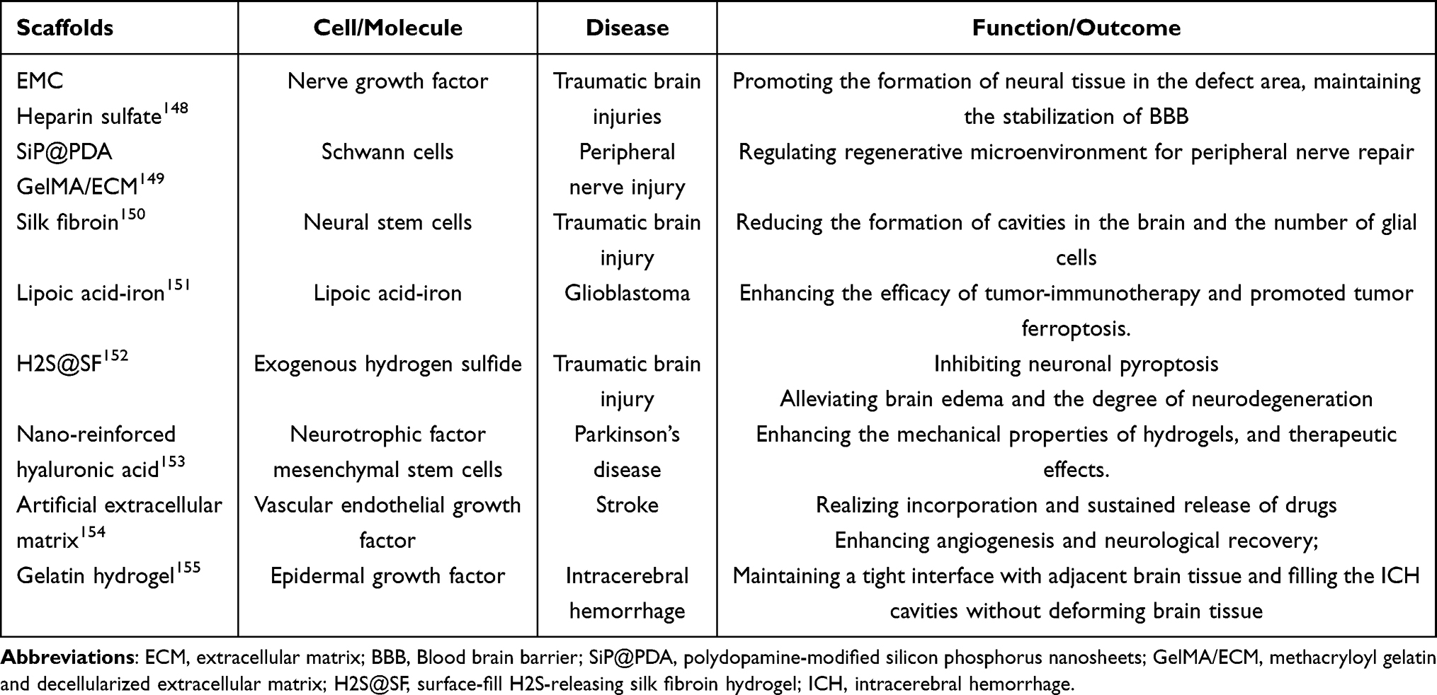 |
Table 4 Summary of Hydrogel-Based Scaffolds Loaded with Cells or Molecules for Nerve Regeneration in Brain Tissue Engineering |
Establishing Brain-Like Tissue
Generally, scientists employed animal models and two-dimensional (2D) cell culture systems to study the mechanisms of the nervous system. However, there exist significant differences in the characteristics of neurodevelopment and neurological diseases between humans and animals, contributing to the poor translation of therapeutic agents derived from animal models to the clinic.156 Additionally, traditional 2D cell culture models, like hippocampal slices, commonly reflect disease-related changes in a singular dimension, failing to preserve the intricate hierarchical structure of the brain and the dynamic interaction between cells and their environment.157 Therefore, it is necessary to design 3D brain-like models that accurately replicate mimic the structure and composition of human brains. Hydrogel possess properties similar to the extracellular matrix of brain tissue, enabling the specific distribution and growth of cells when properly designed. This capability makes hydrogels an appealing support material for 3D brain organoids. For instance, Raimondi et al employed hydrogel-based semi-interpenetrating polymer networks to develop a millimeter-thick brain-like tissue model,158 containing neurons and glial cells. The model effectively reproduced the functional relationships with peripheral body districts and the key biochemical parameters of brain tissue.
Future Perspective
The true benchmark for advances in biomedical science lies in their successful integration into clinical practice. Despite the recent surge in hydrogel research and the proliferation of promising preclinical findings, challenges in clinical translation persist.
Hydrogels provide a highly biomimetic interface that minimizes disruption to brain tissue. However, the crosslinked networks of these materials are mechanically fragile, particularly natural hydrogels, limiting long-term implant durability. Conversely, synthetic hydrogels exhibit superior physicochemical stability and enable facile shape modulation alongside scalable manufacturing, positioning them as a dominant platform for future investigations. Furthermore, the potential for bioaccumulation of degradation byproducts such as metallic elements like copper and toxic cross-linking agents, poses a significant risk of chronic neurotoxicity. Future research should prioritize the long-term biostability and functional reliability of these devices. The degradation rate, mechanical properties, and potential for chronic inflammation of hydrogels must be rigorously assessed.159 Additionally, hydrogels inherently exhibit limited intrinsic electrical conductivity. While the integration of nanomaterials, such as graphene oxide and carbon nanotubes, could enhance signal acquisition,160 it often introduces heterogeneous phases within the matrix. The non-uniform distribution of these fillers can inadvertently elevate the Young’s modulus and induce uneven electroosmotic flow, compromising the interfacial stability and long-term bio-compliance of the electrodes. Consequently, the central challenge lies in how to balance conductivity, transparency, and mechanical compliance while preserving biocompatibility.
Hydrogels are effective drug delivery systems enabling sustained drug release. However, the complex anatomy of brain poses challenges for precise targeting and delivery to pathological foci. Furthermore, the therapeutic efficacy of many current systems is tethered to passive stimuli-responsive kinetics, relying on localized fluctuations in pH or temperature.161 In the absence of such physiological triggers, the drug release profile may become unpredictable or suboptimal. Future efforts should concurrently enhance drug transport efficiency while modulating swelling rates to match brain tissue density, integrating molecular recognition moieties to enable precise controlled release, and ensuring long-term safety. At the fundamental research level, 3D brain-like models provide biomimetic platforms that recapitulate the multicellular complexity and anatomical architecture of the CNS. Due to the intricate structure of brain tissue and its highly nonlinear characteristics,162 there is an urgent need for advanced electrophysiological mapping of these hydrogel-integrated systems.
Despite challenges persist, the evolution of smart hydrogel systems offers a transformative vision for neuro-medicine. The synergy between material innovation and clinical necessity ensures that hydrogels will remain at the forefront of efforts to diagnose and treat neurological disorders.
Conclusions
Our review summarizes the fundamental physicochemical properties of hydrogels, with particular emphasis on their applications in the diagnosis and treatment of neurological disorders, including EEG signal acquisition and targeted therapeutic delivery. Furthermore, we critically analyze current research bottlenecks and discuss feasible solutions. Bioelectrode applications primarily rely on the ionic conductivity of hydrogels, enabling the transduction of ionic currents into measurable electronic signals. Conversely, drug delivery vehicles exploit the 3D network architecture to regulate mass transport, wherein mesh size dictates drug diffusion rates and swelling ratios modulate release kinetics. Recently, efforts have also been directed toward integrating these two modalities, yielding theranostic intelligent hydrogel systems.
However, translating these laboratory breakthroughs into clinical practice remains challenging. From a material perspective, hydrogel matrices inherently possess lower intrinsic electrical conductivity compared to conventional metallic or semiconductor elements, necessitating the integration of conductive additives to achieve desired electroactive properties. Biologically, the macromolecular size and complex hydrodynamics of many advanced hydrogel carriers frequently impede their efficient transit across BBB, undermining targeted central delivery. Furthermore, current therapeutic validations are predominantly confined to preclinical animal models, which fail to replicate the pathophysiological complexity and microenvironmental dynamics of the human brain. Rigorous, long-term biosafety profiling, precise degradation kinetics, and large-cohort clinical trials are imperative.
Consequently, the clinical translation of hydrogel devices requires robust interdisciplinary collaboration across materials science, neuroscience, and engineering. Future work should advance biomaterial design, improve fabrication techniques, and optimize device interfaces to enable early diagnosis and precision therapy for neurological disorders.
Abbreviations
EEG, Electroencephalography; BBB, Blood-brain Barrier; 3D, Three-dimensional; BCIs, Brain-computer Interfaces; DALYs, Disability-adjusted Life-years; DBS, Deep Brain Stimulation; VNS, Vagus Nerve Stimulation; CNS, Central Nervous System; ECM, Extracellular Matrix; PHEMA, Poly2-hydroxyethyl Methacrylate; HA, Hyaluronic Acid; PEG, Polyethylene Glycol; PVA, Polyvinyl Alcohol; PEO, Polyethylene Oxide; ROS, Reactive Oxygen Species; DOX, Doxorubicin; YA, Tannic Acid; PAM, Polyacrylamide; CMC, Carboxymethyl Cellulose; HMI, Human-machine Interaction; AG, Agarose; AM, Acrylamide; PAM-co-PAA, Poly (acrylamide-co-acrylic acid); ECH, Epichlorohydrin; FRP, Free Radical Polymerization; RDRP, Reversible Deactivation Radical Polymerization; ATRP, Atom Transfer Radical Polymerization; β-CD, Beta-cyclodextrin; Fc, Ferrocene; CRN, Chemical Reaction Network; SPH, Supramolecular Photonic Hydrogel; HRP, Horseradish Peroxidase; PVP, Poly (vinyl pyrrolidone); AgNPs, Silver Nanoparticles; HFE, Hydrogel-forming Extrusion; dECM, Decellularized Extracellular Matrix; MSC, Mesenchymal Stem Cell; CNTs, Carbon Nanotubes; GelMA, Methacrylate Acylated Gelatin; FBR, Foreign Body Reactions; DC, Direct-current; DEX, Dexamethasone; ECoG, Electrocorticography; LFP, Local Field Potential; FEA, Finite Element Analysis; DIW, Direct Ink Writing; CSF, Cerebrospinal Fluid; Ag/AgCl, Silver/Silver-Chloride; SNR, Signal-to-noise Ratio; MRI, Magnetic Resonance Imaging; CT, Computed Tomography; MMA, Multichannel Microneedle Array; ERP, Event-related Potential; PI, Polyimide; CF, Carbon Fabric; CPs, Conductive Polymers; PEDOT: PSS, Poly (3,4-ethylenedioxythiophene): Polystyrene Sulfonate; Alg, Alginate; PAAm, Polyacrylamide; CIPN, Chemotherapy‐induced Peripheral Neuropathy; FES, Functional Electrical Stimulation; MALAT1, Metastasis-associated Lung Adenocarcinoma Transcript 1; ISF, Interstitial Fluid; AD, Alzheimer’s Disease; TENG, Triboelectric Nanogenerator; PD, Parkinson’s disease; CMCS, Carboxymethyl Chitosan; BP, Black Phosphorus; MB, Methylene Blue; GBM, Glioblastoma Multiforme; 2D, Two-dimensional; PTX, Paclitaxel; TM, Triglycerol Monostearate; TBI, Traumatic Brain Injury; Cur, Curcumin; EVs, Extracellular Vesicles; SA, Sodium Alginate; TP, Tilapia Peptide; AgNWs, Highly Conductive Silver Nanowires; TET, Tetrandrine; MI, Motor-imagery; SC, Skin Conductance; VR, Virtual Reality; TENS, Transcutaneous Electrical Nerve Stimulation; SiP@PDA, Polydopamine-modified Silicon Phosphorus Nanosheets; GelMA/ECM, Methacryloyl Gelatin and Decellularized Extracellular Matrix; H2S@SF, Surface-fill H2S-releasing Silk Fibroin Hydrogel; ICH, Intracerebral Hemorrhage.
Acknowledgments
We thanked the authors whose literatures were included in our review. Figures 1–5 were created with BioRender (BioRender.com).
Author Contributions
All authors made a significant contribution to the work reported, whether that is in the conception, study design, execution, acquisition of data, analysis and interpretation, or in all these areas; took part in drafting, revising or critically reviewing the article; gave final approval of the version to be published; have agreed on the journal to which the article has been submitted; and agree to be accountable for all aspects of the work.
Funding
This study was supported by Beijing Medical Award Foundation (yxjl-2022-0351-0432).
Disclosure
The authors declare that they have no competing interests in this work.
References
1. Zhang C, Yang X, Wan D, et al. Burden of neurological disorders in China and its provinces, 1990–2021: findings from the global burden of disease study 2021. Med. 2025;6(8):100692. doi:10.1016/j.medj.2025.100692
2. Chen S, Gao J, Zhou Y, et al. Implications of neuromuscular electrical stimulation on gait ability, balance and kinematic parameters after stroke: a systematic review and meta-analysis. J Neuroeng Rehabil. 2024;21(1):164. doi:10.1186/s12984-024-01462-2
3. Feigin VL, Vos T, Nichols E, et al. The global burden of neurological disorders: translating evidence into policy. Lancet Neurol. 2020;19(3):255–25. doi:10.1016/S1474-4422(19)30411-9
4. Huang Y, Li Y, Pan H, Han L. Global, regional, and national burden of neurological disorders in 204 countries and territories worldwide. J Glob Health. 2023;13:04160. doi:10.7189/jogh.13.04160
5. Wu P, Xu C, Zou X, et al. Capacitive-coupling-responsive hydrogel scaffolds offering wireless in situ electrical stimulation promotes nerve regeneration. Adv Mater. 2024;36(14):e2310483. doi:10.1002/adma.202310483
6. Khan ZM, Munson JM, Long TE, Vlaisavljevich E, Verbridge SS. Development of a synthetic, injectable hydrogel to capture residual glioblastoma and glioblastoma stem-like cells with CXCL12-mediated chemotaxis. Adv Healthc Mater. 2023;12(14):e2300671. doi:10.1002/adhm.202300671
7. Chen Z, Liu X, Ding J, et al. Tissue-like electrophysiological electrode interface construction by multiple crosslinked polysaccharide-based hydrogel. Carbohydr Polym. 2022;296:119923. doi:10.1016/j.carbpol.2022.119923
8. Gharakhloo M, Jagleniec D, Romanski J, Karbarz M. A novel self-healing hydrogel based on derivatives of natural α-amino acids with potential applications as a strain sensor. J Mater Chem B. 2022;10(23):4463–4472. doi:10.1039/D2TB00534D
9. Xue B, Bashir Z, Guo Y, et al. Strong, tough, rapid-recovery, and fatigue-resistant hydrogels made of picot peptide fibres. Nat Commun. 2023;14(1):2583. doi:10.1038/s41467-023-38280-4
10. Moghaddasi M, Oktay B, Bingol AB, et al. Conductive nanocomposite hydrogels for neural tissue engineering: a systematic scoping review of recent trends. Adv Sci. 2025;12(38):e16085. doi:10.1002/advs.202416085
11. Su H, Mao L, Chen X, et al. A complementary dual-mode ion-electron conductive hydrogel enables sustained conductivity for prolonged electroencephalogram recording. Adv Sci. 2024;11(38):e2405273. doi:10.1002/advs.202405273
12. Dong C, Zhang Z, Sun D. Multi-channel EEG-based neurological disorder classification using cross-dependency spatiotemporal interactive network. Comput Methods Programs Biomed. 2025;271:108982. doi:10.1016/j.cmpb.2025.108982
13. Sun C, Jing J, Turley N, et al. Harvard electroencephalography database: a comprehensive clinical electroencephalographic resource from four Boston hospitals. Epilepsia. 2025;66(9):3411–3425. doi:10.1111/epi.18487
14. Schubert KM, Dasari V, Oliveira AL, et al. The role of electroencephalography in predicting post-stroke seizures and an updated prognostic model (SeLECT-EEG). Ann Neurol. 2025;98(4):814–825. doi:10.1002/ana.27301
15. Ebadi A, Allouch S, Mheich A, et al. Beyond homogeneity: charting the landscape of heterogeneity in neurodevelopmental and psychiatric electroencephalography. Transl Psychiatry. 2025;15(1):223. doi:10.1038/s41398-025-03441-0
16. Zhu L, Cai M, Pei Z, et al. Concurrent TMS-EEG to characterize cortical responses in the motor and prefrontal cortices in Parkinson’s disease. Neurotherapeutics. 2025;22(4):e00577. doi:10.1016/j.neurot.2025.e00577
17. Violante IR, Alania K, Cassarà AM, et al. Non-invasive temporal interference electrical stimulation of the human hippocampus. Nat Neurosci. 2023;26(11):1994–2004. doi:10.1038/s41593-023-01456-8
18. Lomoio U, Lió P, Guzzi PH, Veltri P. Bidirectional Mamba-2 boosts EEG super-resolution via regression and diffusion. Bioinformatics. 2026;42(5):btag169. doi:10.1093/bioinformatics/btag169
19. Zhou Y, Yang H, Wang X, et al. A mosquito mouthpart-like bionic neural probe. Microsyst Nanoeng. 2023;9:88. doi:10.1038/s41378-023-00565-5
20. Stacey WC, Kellis S, Greger B, et al. Potential for unreliable interpretation of EEG recorded with microelectrodes. Epilepsia. 2013;54(8):1391–1401. doi:10.1111/epi.12202
21. Li C, Yao S, Li Z, Gao Y. Application of novel drug-delivery strategies in neurological disorders. Adv Mater. 2025;37(34):e2503646. doi:10.1002/adma.202503646
22. Gao X, Liu X, Wang N, et al. Nanoparticles hijack calvarial immune cells for CNS drug delivery and stroke therapy. Cell. 2026;189(5):1341–1355.e17. doi:10.1016/j.cell.2025.12.008
23. He Z, Liu C, Lin L, Feng G, Wu G. Real-world safety of levetiracetam: mining and analysis of its adverse drug reactions based on FAERS database. Seizure. 2024;117:253–260. doi:10.1016/j.seizure.2024.03.009
24. Luo W, Yang Z, Zheng J, et al. Small molecule hydrogels loading small molecule drugs from Chinese medicine for the enhanced treatment of traumatic brain injury. ACS Nano. 2024;18(42):28894–28909. doi:10.1021/acsnano.4c09097
25. Reinehr P, Diel LF, Diz FM, et al. Three-dimensional bioactive collagen scaffolds incorporated with titanate nanotubes for tissue regeneration. Colloids Surf B Biointerfaces. 2025;252:114638. doi:10.1016/j.colsurfb.2025.114638
26. Chen Z, Zhang J, Lee FY, Kyriakides TR. Bone-derived extracellular matrix hydrogel from thrombospondin-2 knock-out mice for bone repair. Acta Biomater. 2024;186:85–94. doi:10.1016/j.actbio.2024.08.011
27. Fan L, Liu C, Chen X, et al. Exosomes-loaded electroconductive hydrogel synergistically promotes tissue repair after spinal cord injury via immunoregulation and enhancement of myelinated axon growth. Adv Sci. 2022;9(13):e2105586. doi:10.1002/advs.202105586
28. van Bemmelen FJ, Schouten MJ, Fekkes D, Bruinvels J. Succinic semialdehyde as a substrate for the formation of gamma-aminobutyric acid. J Neurochem. 1985;45(5):1471–1474. doi:10.1111/j.1471-4159.1985.tb07214.x
29. Hanyková L, Šťastná J, Krakovský I. Responsive acrylamide-based hydrogels: advances in interpenetrating polymer structures. Gels. 2024;10(7):414. doi:10.3390/gels10070414
30. Janas-Naze A, Zhang W. Perioperative anaphylaxis to fibrin sealants in children with Noonan syndrome: a retrospective study. Ann Allergy Asthma Immunol. 2022;129(1):95–100. doi:10.1016/j.anai.2022.03.014
31. Huang D, Li Y, Ma Z, et al. Collagen hydrogel viscoelasticity regulates MSC chondrogenesis in a ROCK-dependent manner. Sci Adv. 2023;9(6):eade9497. doi:10.1126/sciadv.ade9497
32. Fan Y, Lüchow M, Badria A, Hutchinson DJ, Malkoch M. Placenta powder-infused thiol-ene PEG hydrogels as potential tissue engineering scaffolds. Biomacromolecules. 2023;24(4):1617–1626. doi:10.1021/acs.biomac.2c01355
33. Sun M, Li H, Hou Y, et al. Multifunctional tendon-mimetic hydrogels. Sci Adv. 2023;9(7):eade6973. doi:10.1126/sciadv.ade6973
34. Hong Y, Kim JM, Jung H, et al. Facile synthesis of poly(ethylene oxide)-based self-healable dynamic triblock copolymer hydrogels. Biomacromolecules. 2020;21(12):4913–4922. doi:10.1021/acs.biomac.0c01140
35. Schumacher L, Siemsen K, Appiah C, et al. A co-polymerizable linker for the covalent attachment of fibronectin makes pHEMA hydrogels cell-adhesive. Gels. 2022;8(5):258. doi:10.3390/gels8050258
36. Rosa E, Pizzella M, Cimmino L, et al. Stimuli-responsive hydrogels from liquid-liquid phase separations of FUS-derived peptides. ACS Appl Mater Interfaces. 2025;17(40):55981–55993. doi:10.1021/acsami.5c15249
37. Zhou L, Jiao X, Liu S, et al. Functional DNA-based hydrogel intelligent materials for biomedical applications. J Mater Chem B. 2020;8(10):1991–2009. doi:10.1039/C9TB02716E
38. Wu Y, Wang Y, Long L, Hu C, Kong Q, Wang Y. A spatiotemporal release platform based on pH/ROS stimuli-responsive hydrogel in wound repairing. J Control Release. 2022;341:147–165. doi:10.1016/j.jconrel.2021.11.027
39. Huang L, Zhu J, Xiong W, et al. Tumor-generated reactive oxygen species storm for high-performance ferroptosis therapy. ACS Nano. 2023;17(12):11492–11506. doi:10.1021/acsnano.3c01369
40. Li T, Hu C, Huang T, et al. Cancer-associated fibroblasts foster a high-lactate microenvironment to drive perineural invasion in pancreatic cancer. Cancer Res. 2025;85(12):2199–2217. doi:10.1158/0008-5472.CAN-24-3173
41. Jo YJ, Gulfam M, Jo SH, et al. Multi-stimuli responsive hydrogels derived from hyaluronic acid for cancer therapy application. Carbohydr Polym. 2022;286:119303. doi:10.1016/j.carbpol.2022.119303
42. Kang JH, Turabee MH, Lee DS, Kwon YJ, Ko YT. Temperature and pH-responsive in situ hydrogels of gelatin derivatives to prevent the reoccurrence of brain tumor. Biomed Pharmacother. 2021;143:112144. doi:10.1016/j.biopha.2021.112144
43. Qian F, Han Y, Han Z, et al. In situ implantable, post-trauma microenvironment-responsive, ROS depletion hydrogels for the treatment of traumatic brain injury. Biomaterials. 2021;270:120675. doi:10.1016/j.biomaterials.2021.120675
44. Jiang Y, Wang R, Wang C, et al. Brain microenvironment responsive and pro-angiogenic extracellular vesicle-hydrogel for promoting neurobehavioral recovery in type 2 diabetic mice after stroke. Adv Healthc Mater. 2022;11(22):e2201150. doi:10.1002/adhm.202201150
45. King JL, Maturavongsadit P, Hingtgen SD, Benhabbour SR. Injectable pH thermo-responsive hydrogel scaffold for tumoricidal neural stem cell therapy for glioblastoma multiforme. Pharmaceutics. 2022;14(10):2243. doi:10.3390/pharmaceutics14102243
46. Lu S, Zhang L, Hu Z, Kong S, Zhang Z, Li G. Optimized preparation of gastric acid-response sulfhydryl functionalized chitosan/alginate/tilapia peptide hydrogel and its protective effects on alcohol-induced liver and brain injury. RSC Adv. 2021;11(55):34544–34557. doi:10.1039/D1RA06361H
47. Ha JH, Shin HH, Choi HW, et al. Electro-responsive hydrogel-based microfluidic actuator platform for photothermal therapy. Lab Chip. 2020;20(18):3354–3364. doi:10.1039/D0LC00458H
48. Zhang L, Pang L, Zhu S, et al. Intranasal tetrandrine temperature-sensitive in situ hydrogels for the treatment of microwave-induced brain injury. Int J Pharm. 2020;583:119384. doi:10.1016/j.ijpharm.2020.119384
49. Zhang W, Liu Y, Wang Z, et al. Remodeling brain pathological microenvironment to lessen cerebral ischemia injury by multifunctional injectable hydrogels. J Control Release. 2024;369:591–603. doi:10.1016/j.jconrel.2024.03.050
50. Liu L, Liu M, Xiu J, et al. Stimuli-responsive nanoparticles delivered by a nasal-brain pathway alleviate depression-like behavior through extensively scavenging ROS. Acta Biomater. 2023;171:451–465. doi:10.1016/j.actbio.2023.09.038
51. Khan F, Atif M, Haseen M, et al. Synthesis, classification and properties of hydrogels: their applications in drug delivery and agriculture. J Mater Chem B. 2022;10(2):170–203. doi:10.1039/D1TB01345A
52. Lyu F, Zeng S, Jia Z, et al. Two-dimensional mineral hydrogel-derived single atoms-anchored heterostructures for ultrastable hydrogen evolution. Nat Commun. 2022;13(1):6249. doi:10.1038/s41467-022-33725-8
53. Wang Y, Yang M, Zhao Z. Facile fabrication of self-healing, injectable and antimicrobial cationic guar gum hydrogel dressings driven by hydrogen bonds. Carbohydr Polym. 2023;310:120723. doi:10.1016/j.carbpol.2023.120723
54. Nam HG, Nam MG, Yoo PJ, Kim JH. Hydrogen bonding-based strongly adhesive coacervate hydrogels synthesized using poly(N-vinylpyrrolidone) and tannic acid. Soft Matter. 2019;15(4):785–791. doi:10.1039/C8SM02144A
55. Cai Y, Xin L, Li H, Sun P, Liu C, Fang L. Mussel-inspired controllable drug release hydrogel for transdermal drug delivery: hydrogen bond and ion-dipole interactions. J Control Release. 2024;365:161–175. doi:10.1016/j.jconrel.2023.11.016
56. Li W, Li SM, Kang MC, Xiong X, Wang P, Tao LQ. Multi-characteristic tannic acid-reinforced polyacrylamide/sodium carboxymethyl cellulose ionic hydrogel strain sensor for human-machine interaction. Int J Biol Macromol. 2024;254(Pt 2):127434. doi:10.1016/j.ijbiomac.2023.127434
57. Diaz-Baca JA, Fatehi P. Temperature responsive crosslinked starch-kraft lignin macromolecule. Carbohydr Polym. 2023;313:120846. doi:10.1016/j.carbpol.2023.120846
58. Lin T, Li S, Hu Y, et al. Ultrastretchable and adhesive agarose/Ti3C2Tx-crosslinked-polyacrylamide double-network hydrogel for strain sensor. Carbohydr Polym. 2022;290:119506. doi:10.1016/j.carbpol.2022.119506
59. Sekine Y, Nankawa T, Hiroi K, et al. Nanocellulose hydrogels formed via crystalline transformation from cellulose I to II and subsequent freeze cross-linking reaction. Carbohydr Polym. 2024;327:121538. doi:10.1016/j.carbpol.2023.121538
60. Ren H, Guo A, Luo C. Sandwich hydrogel to realize cartilage-mimetic structures and performances from polyvinyl alcohol, chitosan and sodium hyaluronate. Carbohydr Polym. 2024;328:121738. doi:10.1016/j.carbpol.2023.121738
61. Gong Z, Zhang G, Zeng X, et al. High-strength, tough, fatigue resistant, and self-healing hydrogel based on dual physically cross-linked network. ACS Appl Mater Interfaces. 2016;8(36):24030–24037. doi:10.1021/acsami.6b05627
62. Salleh KM, Zakaria S, Zainul Armir NA, Khairunnisa-Atiqah MK, Wang B. Electrovalent effects of sodium carboxymethyl cellulose and hydroxyethyl cellulose on regeneration of empty fruit bunch cellulose to a superabsorbent hydrogel. Int J Biol Macromol. 2024;278(Pt 3):134816. doi:10.1016/j.ijbiomac.2024.134816
63. Rigby ADM, Alipio AR, Chiaradia V, Arno MC. Self-healing hydrogel scaffolds through PET-RAFT polymerization in cellular environment. Biomacromolecules. 2023;24(7):3370–3379. doi:10.1021/acs.biomac.3c00431
64. Yerneni SS, Lathwal S, Cuthbert J, et al. Controlled release of exosomes using atom transfer radical polymerization-based hydrogels. Biomacromolecules. 2022;23(4):1713–1722. doi:10.1021/acs.biomac.1c01636
65. Qin J, Dong B, Wang W, Cao L. Self-regulating bioinspired supramolecular photonic hydrogels based on chemical reaction networks for monitoring activities of enzymes and biofuels. J Colloid Interface Sci. 2023;649:344–354. doi:10.1016/j.jcis.2023.06.094
66. Zhang Q, Zheng Z, Zhao Y, et al. D-peptide engineered hydrogel with dual-enzyme-ALA cascades enables multimodal oxygen modulation for self-sustaining EDT-PDT synergy. Bioact Mater. 2025;59:161–173. doi:10.1016/j.bioactmat.2025.12.037
67. Cheng C, Sun Q, Wang X, He B, Jiang T. Enzyme-manipulated hydrogelation of small molecules for biomedical applications. Acta Biomater. 2022;151:88–105. doi:10.1016/j.actbio.2022.08.016
68. Kim M, Kim H, Lee YS, et al. Novel enzymatic cross-linking-based hydrogel nanofilm caging system on pancreatic β cell spheroid for long-term blood glucose regulation. Sci Adv. 2021;7(26):eabf7832. doi:10.1126/sciadv.abf7832
69. Wang L, Chai P, Zhang W, et al. Enzymatic preparation of xanthan gum/polyvinyl alcohol-based nanocomposite hydrogel with antibacterial and immunoregulatory properties for accelerated skin wound healing. Int J Biol Macromol. 2025;331(Pt 2):148415. doi:10.1016/j.ijbiomac.2025.148415
70. Baptista-Silva S, Borges S, Costa-Pinto AR, et al. In situ forming silk sericin-based hydrogel: a novel wound healing biomaterial. ACS Biomater Sci Eng. 2021;7(4):1573–1586. doi:10.1021/acsbiomaterials.0c01745
71. Demeter M, Călina I, Scărișoreanu A, Micutz M. E-beam cross-linking of complex hydrogels formulation: the influence of poly(ethylene oxide) concentration on the hydrogel properties. Gels. 2021;8(1):27. doi:10.3390/gels8010027
72. Bustamante-Torres M, Pino-Ramos VH, Romero-Fierro D, Hidalgo-Bonilla SP, Magaña H, Bucio E. Synthesis and antimicrobial properties of highly cross-linked pH-sensitive hydrogels through gamma radiation. Polymers. 2021;13(14):2223. doi:10.3390/polym13142223
73. Bom S, Ribeiro R, Ribeiro HM, Santos C, Marto J. On the progress of hydrogel-based 3D printing: correlating rheological properties with printing behaviour. Int J Pharm. 2022;629:122334. doi:10.1016/j.ijpharm.2022.122334
74. Liu N, Ma Y, Gong W, et al. Osteocyte-derived extracellular vesicles mediate the bone-to-cartilage crosstalk and promote osteoarthritis progression. Nat Commun. 2025;16(1):4746. doi:10.1038/s41467-025-59861-5
75. Li Q, Yu H, Zhao F, et al. 3D printing of microenvironment-specific bioinspired and exosome-reinforced hydrogel scaffolds for efficient cartilage and subchondral bone regeneration. Adv Sci. 2023;10(26):e2303650. doi:10.1002/advs.202303650
76. Zhang Y, Pang T, Sun Z, et al. Up-and-down transformable embedded ink writing strategy for soft 3D architectures. Nat Commun. 2025;16(1):11348. doi:10.1038/s41467-025-66418-z
77. Guo A, Zhang S, Yang R, Sui C. Enhancing the mechanical strength of 3D printed GelMA for soft tissue engineering applications. Mater Today Bio. 2023;24:100939. doi:10.1016/j.mtbio.2023.100939
78. Tang D, Han B, He C, et al. Electrospun poly-l-lactic acid membranes promote M2 macrophage polarization by regulating the PCK2/AMPK/mTOR signaling pathway. Adv Healthc Mater. 2024;13(22):e2400481. doi:10.1002/adhm.202400481
79. Lee JW, Song KH. Fibrous hydrogels by electrospinning: novel platforms for biomedical applications. J Tissue Eng. 2023;14:20417314231191881. doi:10.1177/20417314231191881
80. Yao S, Yang Y, Li C, et al. Axon-like aligned conductive CNT/GelMA hydrogel fibers combined with electrical stimulation for spinal cord injury recovery. Bioact Mater. 2024;35:534–548. doi:10.1016/j.bioactmat.2024.01.021
81. Liu S, Wang Z, Chen X, et al. Multiscale anisotropic scaffold integrating 3D printing and electrospinning techniques as a heart-on-a-chip platform for evaluating drug-induced cardiotoxicity. Adv Healthc Mater. 2023;12(24):e2300719. doi:10.1002/adhm.202300719
82. Wu Y, Mei R, Zhou Y, Qi J, Hang C, Jiang X. Elastic thermoplastic polyurethane/graphene microneedle-mesh interfaces via microfluidic patterning for electrophysiology in neural organoids. ACS Nano. 2025;19(42):36958–36968. doi:10.1021/acsnano.5c08827
83. Ribeiro JF, Perna A, Orban G, et al. ChroMOS: a “microwire-like” CMOS neural probe for chronic neural recordings in mice. Biosens Bioelectron. 2025;290:117942. doi:10.1016/j.bios.2025.117942
84. Wang X, Xu M, Yang H, et al. Ultraflexible neural electrodes enabled synchronized long-term dopamine detection and wideband chronic recording deep in brain. ACS Nano. 2024;18(50):34272–34287. doi:10.1021/acsnano.4c12429
85. Hoeferlin GF, Grabinski SE, Druschel LN, et al. Bacteria invade the brain following intracortical microelectrode implantation, inducing gut-brain axis disruption and contributing to reduced microelectrode performance. Nat Commun. 2025;16(1):1829. doi:10.1038/s41467-025-56979-4
86. Lin ZK, Lin JS, Chen ZH, Cheng HW, Huang WC, Chen SY. Electrogelated drug-embedded silk/gelatin/rGO degradable electrode for anti-inflammatory applications in brain-implant systems. J Mater Chem B. 2024;12(5):1361–1371. doi:10.1039/D3TB02715E
87. Banno T, Tsuruhara S, Seikoba Y, et al. Nanoneedle-electrode devices for in vivo recording of extracellular action potentials. ACS Nano. 2022;16(7):10692–10700. doi:10.1021/acsnano.2c02399
88. Shen Z, Liang Q, Chang Q, Liu Y, Zhang Q. Topological hydrogels for long-term brain signal monitoring, neuromodulation, and stroke treatment. Adv Mater. 2024;36(7):e2310365. doi:10.1002/adma.202310365
89. Chen L, Zhong H, Wang L, et al. Long-term stable subdural recordings enabled by fibrosis-resistant hydrogel-integrated µECoG arrays. Adv Sci. 2025;12(47):e15453. doi:10.1002/advs.202515453
90. Merk T, Peterson V, Lipski WJ, et al. Electrocorticography is superior to subthalamic local field potentials for movement decoding in Parkinson’s disease. eLife. 2022;11:e75126. doi:10.7554/eLife.75126
91. Wu S, Issa NP, Rose SL, et al. Depth versus surface: a critical review of subdural and depth electrodes in intracranial electroencephalographic studies. Epilepsia. 2024;65(7):1868–1878. doi:10.1111/epi.18002
92. Alahi MEE, Liu Y, Khademi S, et al. Slippery epidural ECoG electrode for high-performance neural recording and interface. Biosensors. 2022;12(11):1044. doi:10.3390/bios12111044
93. Ye Q, Huang Y, Yao B, et al. Radiative coupled evaporation cooling hydrogel for above-ambient heat dissipation and flame retardancy. Nanomicro Lett. 2025;18(1):50. doi:10.1007/s40820-025-01903-0
94. Momin M, Feng L, Chen X, et al. 3D-printable, honeycomb-inspired tissue-like bioelectrodes for patient-specific neural interface. Adv Mater. 2026;38:e16291. doi:10.1002/adma.202516291
95. Zhang A, Shyam AB, Cunningham AM, et al. Adhesive wearable sensors for electroencephalography from hairy scalp. Adv Healthc Mater. 2023;12(22):e2300142. doi:10.1002/adhm.202300142
96. Zeng F, Wang G, Sun C, Gao J, Ji S, Zhang Q. Flexible and washable poly(3,4-ethylenedioxythiophene): polystyrene sulfonate/polyvinyl alcohol fabric dry electrode for long-term electroencephalography signals measurement. Polymers. 2025;17(5):683. doi:10.3390/polym17050683
97. Lin S, Jiang J, Huang K, et al. Advanced electrode technologies for noninvasive brain-computer interfaces. ACS Nano. 2023;17(24):24487–24513. doi:10.1021/acsnano.3c06781
98. Ren G, Zhang M, Zhuang L, et al. MRI and CT compatible asymmetric bilayer hydrogel electrodes for EEG-based brain activity monitoring. Microsyst Nanoeng. 2024;10(1):156. doi:10.1038/s41378-024-00805-2
99. Liu J, Yang X, Li M, et al. Application of graphene dry electrode in 512-lead EEG cap and real-time monitoring EEG system. ACS Appl Mater Interfaces. 2026;18(18):25806–25820. doi:10.1021/acsami.5c25282
100. Liu Z, Xu X, Huang S, et al. Multichannel microneedle dry electrode patches for minimally invasive transdermal recording of electrophysiological signals. Microsyst Nanoeng. 2024;10:72. doi:10.1038/s41378-024-00702-8
101. Wang D, Xue H, Xia L, et al. A tough semi-dry hydrogel electrode with anti-bacterial properties for long-term repeatable non-invasive EEG acquisition. Microsyst Nanoeng. 2025;11(1):105. doi:10.1038/s41378-025-00908-4
102. Xue H, Wang D, Jin M, et al. Hydrogel electrodes with conductive and substrate-adhesive layers for noninvasive long-term EEG acquisition. Microsyst Nanoeng. 2023;9:79. doi:10.1038/s41378-023-00524-0
103. Ma C, Li W, Gao C, et al. Multifunctional hydrogel materials for advanced neural interfaces. Small Methods. 2025;9(9):e01134. doi:10.1002/smtd.202501134
104. Gori M, Giannitelli SM, Vadalà G, et al. A soft zwitterionic hydrogel as potential coating on a polyimide surface to reduce foreign body reaction to intraneural electrodes. Molecules. 2022;27(10):3126. doi:10.3390/molecules27103126
105. Oribe S, Yoshida S, Kusama S, et al. Hydrogel-based organic subdural electrode with high conformability to brain surface. Sci Rep. 2019;9(1):13379. doi:10.1038/s41598-019-49772-z
106. Cheah E, Gao X, Jaw WQ, et al. Customizable fabrication of 2D and conformal multielectrode arrays for 3D printed organotypic bioelectronic interfaces. Adv Healthc Mater. 2026;15(8):e02757. doi:10.1002/adhm.202502757
107. Lee S, Park K, Kum J, et al. Stretchable surface electrode arrays using an alginate/PEDOT: PSS-based conductive hydrogel for conformal brain interfacing. Polymers. 2022;15(1):84. doi:10.3390/polym15010084
108. Wang J, Wang T, Liu H, et al. Flexible electrodes for brain-computer interface system. Adv Mater. 2023;35(47):e2211012. doi:10.1002/adma.202211012
109. Vidal JJ. Toward direct brain-computer communication. Annu Rev Biophys Bioeng. 1973;2:157–180. doi:10.1146/annurev.bb.02.060173.001105
110. Robinson JT, Norman SL, Angle MR, et al. An application-based taxonomy for brain-computer interfaces. Nat Biomed Eng. 2025;9(6):789–791. doi:10.1038/s41551-024-01326-z
111. Molinares D, Kurtevski S, Zhu Y. Chemotherapy-induced peripheral neuropathy: diagnosis, agents, general clinical presentation, and treatments. Curr Oncol Rep. 2023;25(11):1227–1235. doi:10.1007/s11912-023-01449-7
112. Prinsloo S, Kaptchuk TJ, De Ridder D, et al. Brain-computer interface relieves chronic chemotherapy-induced peripheral neuropathy: a randomized, double-blind, placebo-controlled trial. Cancer. 2024;130(2):300–311. doi:10.1002/cncr.35027
113. Ziai Y, Zargarian SS, Rinoldi C, et al. Conducting polymer-based nanostructured materials for brain-machine interfaces. Wiley Interdiscip Rev Nanomed Nanobiotechnol. 2023;15(5):e1895. doi:10.1002/wnan.1895
114. Rinoldi C, Ziai Y, Zargarian SS, et al. In vivo chronic brain cortex signal recording based on a soft conductive hydrogel biointerface. ACS Appl Mater Interfaces. 2023;15(5):6283–6296. doi:10.1021/acsami.2c17025
115. Hsieh JC, Alawieh H, Li Y, et al. A highly stable electrode with low electrode-skin impedance for wearable brain-computer interface. Biosens Bioelectron. 2022;218:114756. doi:10.1016/j.bios.2022.114756
116. Landau O, Nissim N. Mining multi-electrode and multi-wave electroencephalogram based time-interval temporal patterns for improved classification capabilities and explainability. Artif Intell Med. 2025;170:103269. doi:10.1016/j.artmed.2025.103269
117. Wairagkar M, Card NS, Singer-Clark T, et al. An instantaneous voice-synthesis neuroprosthesis. Nature. 2025;644(8075):145–152. doi:10.1038/s41586-025-09127-3
118. Brannigan JFM, Fry A, Opie NL, Campbell BCV, Mitchell PJ, Oxley TJ. Endovascular brain-computer interfaces in poststroke paralysis. Stroke. 2024;55(2):474–483. doi:10.1161/STROKEAHA.123.037719
119. Wang Q, Wang Y, Li S, Shi J. PACAP-Sirtuin3 alleviates cognitive impairment through autophagy in Alzheimer’s disease. Alzheimers Res Ther. 2023;15(1):184. doi:10.1186/s13195-023-01334-2
120. Wu Z, Feng K, Huang J, et al. Brain region changes following a spinal cord injury. Neurochem Int. 2024;174:105696. doi:10.1016/j.neuint.2024.105696
121. Sebastián-Romagosa M, Cho W, Ortner R, et al. Brain-computer interface treatment for gait rehabilitation in stroke patients. Front Neurosci. 2023;17:1256077. doi:10.3389/fnins.2023.1256077
122. Aurucci GV, Preatoni G, Damiani A, Raspopovic S. Brain-computer interface to deliver individualized multisensory intervention for neuropathic pain. Neurotherapeutics. 2023;20(5):1316–1329. doi:10.1007/s13311-023-01396-y
123. LaRocco J, Tahmina Q, Lecian S, Moore J, Helbig C, Gupta S. Evaluation of an English language phoneme-based imagined speech brain computer interface with low-cost electroencephalography. Front Neuroinf. 2023;17:1306277. doi:10.3389/fninf.2023.1306277
124. Angrick M, Luo S, Rabbani Q, et al. Online speech synthesis using a chronically implanted brain-computer interface in an individual with ALS. Sci Rep. 2024;14(1):9617. doi:10.1038/s41598-024-60277-2
125. Jiang Y, Yin J, Zhao B, et al. Motor imagery brain-computer interface in rehabilitation of upper limb motor dysfunction after stroke. J Vis Exp. 2023. doi:10.3791/65405
126. Cajigas I, Davis KC, Prins NW, et al. Brain-computer interface control of stepping from invasive electrocorticography upper-limb motor imagery in a patient with quadriplegia. Front Hum Neurosci. 2023;16:1077416. doi:10.3389/fnhum.2022.1077416
127. Carino-Escobar RI, Rodriguez-Barragan MA, Carrillo-Mora P, Cantillo-Negrete J. Brain-computer interface as complementary therapy for hemiparesis in an astrocytoma patient. Neurol Sci. 2022;43(4):2879–2881. doi:10.1007/s10072-022-05924-0
128. Mitchell P, Lee SCM, Yoo PE, et al. Assessment of safety of a fully implanted endovascular brain-computer interface for severe paralysis in 4 patients: the Stentrode with thought-controlled digital switch (SWITCH) study. JAMA Neurol. 2023;80(3):270–278. doi:10.1001/jamaneurol.2022.4847
129. Colamarino E, Lorusso M, Pichiorri F, et al. DiSCIoser: unlocking recovery potential of arm sensorimotor functions after spinal cord injury by promoting activity-dependent brain plasticity by means of brain-computer interface technology: a randomized controlled trial to test efficacy. BMC Neurol. 2023;23(1):414. doi:10.1186/s12883-023-03442-w
130. Lim J, Wang PT, Sohn WJ, et al. Real-time brain-computer interface control of walking exoskeleton with bilateral sensory feedback. Brain Stimul. 2026;19(3):103065. doi:10.1016/j.brs.2026.103065
131. Flesher SN, Downey JE, Weiss JM, et al. A brain-computer interface that evokes tactile sensations improves robotic arm control. Science. 2021;372(6544):831–836. doi:10.1126/science.abd0380
132. Chanda K, Jana NR, Mukhopadhyay D. Long non-coding RNA MALAT1 protects against Aβ induced toxicity by regulating the expression of receptor tyrosine kinase EPHA2 via quenching miR-200a/26a/26b in Alzheimer’s disease. Life Sci. 2022;302:120652. doi:10.1016/j.lfs.2022.120652
133. Zuo Y, He S, Zhang W, et al. Hydrogel microneedle based transdermal fluorescent patch for the on-site and non-invasive diagnosis of Alzheimer’s disease. Biosens Bioelectron. 2025;288:117823. doi:10.1016/j.bios.2025.117823
134. Kim JN, Lee J, Lee H, Oh IK. Stretchable and self-healable catechol-chitosan-diatom hydrogel for triboelectric generator and self-powered tremor sensor targeting at Parkinson disease. Nano Energy. 2021;82:105705. doi:10.1016/j.nanoen.2020.105705
135. Han Z, Zhao Z, Yu H, et al. Microenvironment-responsive hydrogel reduces seizures after traumatic brain injury in juvenile rats by reducing oxidative stress and hippocampal inflammation. Macromol Biosci. 2024;24(8):e2400050. doi:10.1002/mabi.202400050
136. Zhou X, Deng X, Liu M, et al. Intranasal delivery of BDNF-loaded small extracellular vesicles for cerebral ischemia therapy. J Control Release. 2023;357:1–19. doi:10.1016/j.jconrel.2023.03.033
137. Liu Y, Tan Y, Cheng G, et al. Customized intranasal hydrogel delivering methylene blue ameliorates cognitive dysfunction against Alzheimer’s disease. Adv Mater. 2024;36(19):e2307081. doi:10.1002/adma.202307081
138. Kass LE, Nguyen J. Nanocarrier-hydrogel composite delivery systems for precision drug release. Wiley Interdiscip Rev Nanomed Nanobiotechnol. 2022;14(2):e1756. doi:10.1002/wnan.1756
139. Jiao B, Shang Y, Zhou D, Wang X, Wang Q. In situ assembly of bilayer hydrogels by templated enzymatic polymerization for ultrahigh UV absorption and photothermal transdermal therapy. Small. 2026;22(5):e10083. doi:10.1002/smll.202510083
140. Kang T, Cha GD, Park OK, et al. Penetrative and sustained drug delivery using injectable hydrogel nanocomposites for postsurgical brain tumor treatment. ACS Nano. 2023;17(6):5435–5447. doi:10.1021/acsnano.2c10094
141. Yu Y, Zou Y, Yang M, et al. Quercetin-loaded zeolitic imidazolate framework-8 nanoparticles through borneol-embedded hyaluronic acid hydrogel for ischemic stroke treatment. Int J Biol Macromol. 2026;340(Pt 1):149821. doi:10.1016/j.ijbiomac.2025.149821
142. Tao J, Li J, Fan X, et al. Unraveling the protein post-translational modification landscape: neuroinflammation and neuronal death after stroke. Ageing Res Rev. 2024;101:102489. doi:10.1016/j.arr.2024.102489
143. Weterings RP, Kessels RP, De leeuw FE, Piai V. Cognitive impairment after a stroke in young adults: a systematic review and meta-analysis. Int J Stroke. 2023;18(8):888–897. doi:10.1177/17474930231159267
144. Zhan J, Zhang P, Wen H, et al. Global prevalence estimates of poststroke fatigue: a systematic review and meta-analysis. Int J Stroke. 2023;18(9):1040–1050. doi:10.1177/17474930221138701
145. Dalton EJ, Jamwal R, Augoustakis L, et al. Prevalence of arm weakness, pre-stroke outcomes and other post-stroke impairments using routinely collected clinical data on an acute stroke unit. Neurorehabil Neural Repair. 2024;38(2):148–160. doi:10.1177/15459683241229676
146. Shan BH, Wu FG. Hydrogel-based growth factor delivery platforms: strategies and recent advances. Adv Mater. 2024;36(5):e2210707. doi:10.1002/adma.202210707
147. Han S, Kim J, Kim SH, et al. In vitro induction of in vivo-relevant stellate astrocytes in 3D brain-derived, decellularized extracellular matrices. Acta Biomater. 2023;172:218–233. doi:10.1016/j.actbio.2023.09.046
148. Kim BS, Kim JU, Lee J, Ryu KM, Kim SH, Hwang NS. Decellularized brain extracellular matrix based NGF-releasing cryogel for brain tissue engineering in traumatic brain injury. J Control Release. 2024;368:140–156. doi:10.1016/j.jconrel.2024.02.017
149. Xu C, Wu P, Yang K, et al. Multifunctional biodegradable conductive hydrogel regulating microenvironment for stem cell therapy enhances the nerve tissue repair. Small. 2024;20(23):e2309793. doi:10.1002/smll.202309793
150. Yang Z, You Y, Liu X, et al. Injectable Bombyx mori (B. mori) silk fibroin/MXene conductive hydrogel for electrically stimulating neural stem cells into neurons for treating brain damage. J Nanobiotechnology. 2024;22(1):111. doi:10.1186/s12951-024-02359-x
151. Jia M, Zhou X, Li P, Zhang S. An injectable biomimetic hydrogel adapting brain tissue mechanical strength for postoperative treatment of glioblastoma without anti-tumor drugs participation. J Control Release. 2024;373:699–712. doi:10.1016/j.jconrel.2024.07.068
152. Chen X, Huang X, Liu C, et al. Surface-fill H2S-releasing silk fibroin hydrogel for brain repair through the repression of neuronal pyroptosis. Acta Biomater. 2022;154:259–274. doi:10.1016/j.actbio.2022.11.021
153. Torres-Ortega PV, Del Campo-Montoya R, Plano D, et al. Encapsulation of MSCs and GDNF in an injectable nanoreinforced supramolecular hydrogel for brain tissue engineering. Biomacromolecules. 2022;23(11):4629–4644. doi:10.1021/acs.biomac.2c00853
154. Yaguchi A, Oshikawa M, Watanabe G, et al. Efficient protein incorporation and release by a jigsaw-shaped self-assembling peptide hydrogel for injured brain regeneration. Nat Commun. 2021;12(1):6623. doi:10.1038/s41467-021-26896-3
155. Lim TC, Mandeville E, Weng D, et al. Hydrogel-based therapy for brain repair after intracerebral hemorrhage. Transl Stroke Res. 2020;11(3):412–417. doi:10.1007/s12975-019-00721-y
156. Milior G, Morin-Brureau M, Pallud J, Miles R, Huberfeld G. Animal models and human tissue compared to better understand and treat the epilepsies. Epilepsia. 2023;64(5):1175–1189. doi:10.1111/epi.17552
157. Lokai T, Albin B, Qubbaj K, Tiwari AP, Adhikari P, Yang IH. A review on current brain organoid technologies from a biomedical engineering perspective. Exp Neurol. 2023;367:114461. doi:10.1016/j.expneurol.2023.114461
158. Raimondi I, Tunesi M, Forloni G, Albani D, Giordano C. 3D brain tissue physiological model with co-cultured primary neurons and glial cells in hydrogels. J Tissue Eng. 2020;11:2041731420963981. doi:10.1177/2041731420963981
159. Khan WU, Shen Z, Mugo SM, Wang H, Zhang Q. Implantable hydrogels as pioneering materials for next-generation brain-computer interfaces. Chem Soc Rev. 2025;54(6):2832–2880. doi:10.1039/D4CS01074D
160. Han K, Bai Q, Wu W, Sun N, Cui N, Lu T. Gelatin-based adhesive hydrogel with self-healing, hemostasis, and electrical conductivity. Int J Biol Macromol. 2021;183:2142–2151. doi:10.1016/j.ijbiomac.2021.05.147
161. Garg A, Agrawal R, Singh chauhan C, Deshmukh R. In-situ gel: a smart carrier for drug delivery. Int J Pharm. 2024;652:123819. doi:10.1016/j.ijpharm.2024.123819
162. Wang L, Bai L, Wang S, et al. Biomimetic design and integrated biofabrication of an in-vitro three-dimensional multi-scale multilayer cortical model. Mater Today Bio. 2024;28:101176. doi:10.1016/j.mtbio.2024.101176
 © 2026 The Author(s). This work is published and licensed by Dove Medical Press Limited. The
full terms of this license are available at https://www.dovepress.com/terms
and incorporate the Creative Commons Attribution
- Non Commercial (unported, 4.0) License.
By accessing the work you hereby accept the Terms. Non-commercial uses of the work are permitted
without any further permission from Dove Medical Press Limited, provided the work is properly
attributed. For permission for commercial use of this work, please see paragraphs 4.2 and 5 of our Terms.
© 2026 The Author(s). This work is published and licensed by Dove Medical Press Limited. The
full terms of this license are available at https://www.dovepress.com/terms
and incorporate the Creative Commons Attribution
- Non Commercial (unported, 4.0) License.
By accessing the work you hereby accept the Terms. Non-commercial uses of the work are permitted
without any further permission from Dove Medical Press Limited, provided the work is properly
attributed. For permission for commercial use of this work, please see paragraphs 4.2 and 5 of our Terms.
Recommended articles
Plant Exosome-Loaded Intelligent Hydrogels for Osteoporotic Bone Regeneration: Mechanisms and Applications
Li L, Ye C, Wu ZQ, Wu R
International Journal of Nanomedicine 2025, 20:15863-15881
Published Date: 29 December 2025
Biopolymer Hydrogel-Based Nanocomposites Functionalized with Natural Products for Wound Dressings: Translational Advances in Drug Design, Development, and Therapeutic Wound Care
Ranatunga B, Sekar M, Hashmi AR, Zahra F, Ravi RN, Prashantha Kumar BR, Al Hamod M, Al Hamood N, Begum MY, Wong LS, Kumarasamy V, Molugulu N
Drug Design, Development and Therapy 2026, 20:578261
Published Date: 20 March 2026