
Back to Journals » International Journal of Nanomedicine » Volume 21
An E3 Aptamer-Modified T Cell-Derived Exosomal Nanoplatform for Codelivery of Astragaloside IV and PESV: Enhancing Prostate Cancer Therapy Through Immune Modulation with Implications for Clinical Translation
Authors Zhang Q, Liu C, Wu S, Zhong N, Qiu J, Fu W  , You X
Received 17 April 2026
Accepted for publication 30 June 2026
Published 9 July 2026 Volume 2026:21 614400
DOI https://doi.org/10.2147/IJN.S614400
Checked for plagiarism Yes
Review by Single anonymous peer review
Peer reviewer comments 2
Editor who approved publication: Prof. Dr. Anderson Oliveira Lobo
Qing Zhang,1 Chenguang Liu,2 Sirui Wu,2 Nan Zhong,2 Junfeng Qiu,3 Wei Fu,4,* Xujun You4,*
1Department of General Medicine Center, Shenzhen Bao’an Traditional Chinese Medicine Hospital, Guangzhou University of Chinese Medicine, Shenzhen, 518133, People’s Republic of China; 2The Seventh Clinical Medical College of Guangzhou University of Traditional Chinese Medicine, Shenzhen, 518133, People’s Republic of China; 3Department of Andrology, Shenzhen Traditional Chinese Medicine Hospital, Guangzhou University of Chinese Medicine, Shenzhen, 518033, People’s Republic of China; 4Department of Andrology, Shenzhen Bao’an Traditional Chinese Medicine Hospital, Guangzhou University of Chinese Medicine, Shenzhen, 518133, People’s Republic of China
*These authors contributed equally to this work
Correspondence: Xujun You, Department of Andrology, Shenzhen Bao’an Traditional Chinese Medicine Hospital, Guangzhou University of Chinese Medicine, Shenzhen, People’s Republic of China, Email [email protected] Wei Fu, Department of Andrology, Shenzhen Bao’an Traditional Chinese Medicine Hospital, Guangzhou University of Chinese Medicine, Shenzhen, People’s Republic of China, Email [email protected]
Purpose: Prostate cancer (PCa) remains a leading cause of cancer-related mortality in men, with current treatments often limited by drug resistance and systemic toxicity. Although traditional Chinese medicine components such as Astragaloside IV and polypeptide extract from scorpion venom (PESV) have demonstrated promising antitumor activity, their clinical translation is hampered by poor bioavailability and lack of tumor specificity. To address these limitations, we engineered an E3 aptamer-modified T cell-derived exosomal nanoplatform (EAPE) for the targeted co-delivery of Astragaloside IV and PESV in prostate cancer therapy.
Methods: EAPE was constructed and characterized, and its targeting capability, biosafety, and therapeutic performance were evaluated in vitro and in vivo. In vitro, the antitumor efficacy was assessed by proliferation, migration and apoptosis assays, while the immunomodulatory effects were investigated using a co-culture system of LNCaP cells and T lymphocytes. In vivo, the antitumor efficacy and immune activation were examined in prostate cancer xenograft mouse model, with tumor growth inhibition, apoptosis and immune responses measured.
Results: EAPE demonstrated efficient tumor-targeting capability and favorable biosafety profiles both in vitro and in vivo. EAPE demonstrated superior therapeutic efficacy against PCa by inhibiting proliferation and migration of prostate cancer cells and inducing apoptosis, while suppressing immunosuppression and activating antitumor immune response.
Conclusion: This study presents a biologically derived, targeted nanodelivery system that improves the delivery efficiency and therapeutic efficacy of Astragaloside IV and PESV. These findings support the potential of exosome-based nanoplatforms as promising strategies for enhancing the translational application of traditional Chinese medicine-derived therapeutics in prostate cancer.
Keywords: prostate cancer, Astragaloside IV-PESV, exosome delivery system, antitumor therapy, tumor microenvironment
Introduction
Prostate cancer (PCa), the most common malignant tumor of the male urogenital system, is a leading cause of cancer-related deaths among men globally.1 With approximately 1.5 million new cases diagnosed annually and a rising trend in both incidence and mortality rates in recent years, particularly among younger populations, PCa has emerged as a critical global public health challenge, garnering widespread attention.2,3 Current therapeutic options for PCa, including surgery, chemotherapy, androgen deprivation therapy (ADT), and radiation therapy, have significantly improved outcomes for patients with localized disease.4 However, clinical utility of these therapies remains compromised by severe systemic toxicity, the development of drug resistance, and a lack of tumor-specific targeting, especially in advanced stages such as castration-resistant prostate cancer (CRPC).5 Hence, there is an urgent need to develop highly efficacious and tumor-specific therapeutic strategies with minimal systemic toxicity for PCa treatment.
Traditional Chinese medicine (TCM) has recently demonstrated therapeutic potential for PCa, supported by accumulating evidence.6–8 Among various TCM formulations, the Astragalus–Scorpion drug pair has attracted particular attention, with Astragaloside IV and polypeptide extract from scorpion venom (PESV) identified as the primary bioactive constituents.9 Additionally, our previous studies have confirmed the therapeutic potential of Astragaloside IV-PESV against PCa, as evidenced by its inhibition of PCa cell proliferation and migration, while promoting apoptosis and autophagy.10–12 Despite the promising pharmacological activities of these TCM-derived agents, the clinical translation remains limited by poor bioavailability, limited delivery efficiency and insufficient tumor-specific accumulation.13 Therefore, improving drug delivery efficiency has become a key strategy for facilitating the clinical application of TCM-derived therapeutics.
In recent years, various nanomaterial-based drug delivery systems have been extensively investigated for cancer therapy to enhance tumor-targeted drug delivery while reducing systemic toxicity.8,14–16 Exosomes, nano-sized membrane vesicles secreted by cells, have attracted particular interest due to their superior biocompatibility, low immunogenicity, and high delivery efficiency, compared to conventional drug carriers (such as liposomes and inorganic mesoporous materials),17 which support their feasibility for clinical translation. Moreover, exosomes possess high engineerability, which can be modified with specific targeting molecules, such as aptamers, targeting peptides or antibodies, allowing for more precise delivery of therapeutic cargo.18 Notably, an RNA aptamer designated E3 has been identified to selectively bind and be internalized by PCa cells, but not normal prostate cells.19 Subsequent studies have further validated the targeting potential of the E3 aptamer, demonstrating that E3-aptamer-modified exosomes loaded with SIRT6-targeting siRNA exhibit significant tumor‑targeting capability and antitumor efficacy in PCa models.20 Consequently, E3 aptamer-functionalized exosomes represent a promising platform for targeted drug delivery in PCa, providing a rationale for the application of Astragaloside IV and PESV as a potential therapeutic strategy.
Given the need for targeted therapy in PCa and the therapeutic potential of Astragaloside IV and PESV, we developed a precision drug delivery system that integrates the tumor-targeting capability of the E3 aptamer with inherent biocompatibility of exosomes. Specifically, T lymphocyte-derived exosomes loaded with Astragaloside IV-PESV and modified with E3 aptamer (Exo-Astragaloside IV-PESV-E3, EAPE) were constructed with active targeting capability. Then the tumor-targeting specificity and antitumor efficacy of EAPE were systematically evaluated in prostate cancer models in vitro and in vivo. Taken together, this study provides a targeted delivery strategy for TCM-derived therapeutics in PCa, and establish a transferable framework that may be extended to the delivery of other poorly bioavailable compounds or nucleic acid drugs, supporting its potential for translational application in cancer therapy.
Materials and Methods
Exosome Isolation and Identification
CD8 + T cells were isolated from human peripheral blood by flow cytometry, and cultured in RPMI-1640 medium (BL303A, Biosharp, China) supplemented with exosome-depleted serum at 37°C for 48h. Exosomes were obtained and enriched from supernatant collected from CD8 + T cells by a series of ultracentrifugation as follows: supernatant was centrifuged first at 300 × g for 10 min, then at 2000 × g for 10 min, and at 10000× g for 30 min, finally at 100000× g for 70 min, the pellet was resuspended in phosphate-buffered saline (PBS) (P1022, Solarbio, China) for further experiments. All procedures were conducted under 4°C.
For exosome characterization, morphology was visualized using transmission electron microscopy (TEM) (HT7800, HITACHI, Japan). Exosome suspension (15 μL) was placed into copper grids for 1 min, and subsequently stained with 2% uranyl acetate solution for 1 min, washed with pure water, and then observed and photographed under a TEM at an accelerating voltage of 80 kV. Exosome size was assessed by nanoparticle tracking analyzer (NTA) (NanoBrook Omni, Brookhaven, USA). The specific surface markers of exosomes were validated by Western blot analysis. The following antibodies were used: anti-CD9 (1:2000, 20,597-1-AP, Proteintech), anti-CD63 (1:3000, 25,682-1-AP, Proteintech), anti-CD81 (1:2000, 66,866-1-Ig, Proteintech) and anti-Calnexin (1:5000, 10,427-2-AP, Proteintech).
Engineered Exosomes Construction and Characterization
Construction of Exo-Astragaloside IV-PESV (EAP): Astragaloside IV-PESV (5 mg/mL) (A274907, Aladdin, China) and exosomes (5 mg/mL) were mixed and electroporated (400 mV, 125 μF, 10–15 ms) in electroporation buffer Then electroporated mixture was incubated in an ice bath at 4°C for 24 h, followed by purification using a 10 kDa ultrafiltration device. The loaded exosomes were collected and stored at −80°C.
Construction of Exo-Astragaloside IV-PESV-E3 (EAPE): The E3 aptamer was designed and then synthesized in Sangon Biotech, followed by thiol modification. The sequence of the E3 aptamer is GGCUUUCGGGCUUUCGGCAACAUCAGCCCCUCAGCC. Next, to obtain the Mal-PEG-Chol-modified exosomes, pure exosomes (1 mg) were incubated with Mal-PEG-Chol (5 μM) at 37°C for 2 h, followed by purification using ultrafiltration centrifuge (14,000 g, 4°C, 10 min). The E3 aptamer was pre-treated with 500 mM Tris (2-carboxyethyl) phosphine hydrochloride (TCEP) to activate the thiol. The thiol-activated E3 aptamer was conjugated to the Mal-PEG-Chol-modified exosomes overnight at 4°C, followed by quenching with L-cysteine, and Exosome-E3 (Exo-E3) was obtained after further purification. Finally, Astragaloside IV-PESV was loaded into the Exo-E3 via electroporation.
As described above, the morphology and size of EAP and EAPE were examined using TEM and analyzed by NTA, respectively. The specific surface markers of exosome were verified by Western blot. The successful conjugation of the E3 aptamer and exosomes was confirmed by agarose gel electrophoresis. In brief, 2% agarose gel was prepared with nucleic acid dye, samples mixed with loading buffer, and then loaded into the wells, running at 120 V for 10 min, and finally imaging the gel.
Drug Loading and Release
Astragaloside IV-PESV was loaded into both blank exosome (Exo) and Exo-E3 via electroporation as previously described. The mixtures were then incubated at 4°C for 1, 3, 5, and 7 days, respectively, followed by purification using 10 kDa ultrafiltration to remove free drugs. The encapsulation efficiency (EE%) and loading rate (LR%) were calculated by measuring the absorbance of the collected free Astragaloside IV at 203 nm using a microplate reader (SpectraMax M2, Molecular Devices, USA).


In vitro release profiles were determined using a dialysis method. Briefly, 1 mL EAP or EAPE was placed into a dialysis bag (1 kDa cutoff) and then immersed in PBS (pH=7.4), respectively. The system was maintained at 37°C with stirring at 80 rpm. Samples (1 mL) of the release medium were collected at predetermined time points (0.5, 1, 2, 4, 6, 8, 12, and 24 h). The concentration of released Astragaloside IV at each time points was quantified by measuring the absorbance at 203 nm using a microplate reader.
Cell Culture and Treatment
The LNCaP cell lines were purchased from Procell system. The cells were cultivated in DMEM (CM-0143, Procell system, China) containing fetal bovine serum (AB-FBS-0500, ABW, China) and 1% penicillin-streptomycin (15140, Biosharp, China) at 37°C in a 5% CO2 incubator.
CCK8 Assay
The LNCaP cells in the logarithmic growth phase were seeded into 96-well plates at a density of 10,000 cells per well and cultured for 24 h. Then, the LNCaP cells were treated with Exo, Astragaloside IV-PESV (AP), EAP, or EAPE formulations containing varying concentrations of Astragaloside IV (2.5, 5, 10 μM) and PESV (10, 20, 40 mg/mL) for another 24 h. CCK-8 working solution (CA1210, Solarbio, China) was added to each well at a 1:100 dilution. After incubation for 4 h at 37 °C, the absorbance values were recorded with a microplate reader (SpectraMax M2, Molecular Devices, USA) at 450 nm.
The CD8 + T cells (1×10⁵/well) and LNCaP cells (4×10⁵/well) were co-cultured in a Transwell system for 24 h, with T cells in the upper chamber being treated with the respective exosome formulations (10 μM Astragaloside IV + 40 mg/mL PESV). Cell viability was then assessed using the CCK‑8 reagent as described above.
EdU Staining
Cell proliferation ability was assessed using EdU staining. Cells in the control group were treated with PBS, whereas the experimental groups received Exo, AP, EAP or EAPE. Subsequently, the cells were incubated with 0.5 mL of 2× EdU working solution (BL915A, Biosharp, China) in a CO2 incubator for 2 h. After washing with PBS, the cells were fixed at room temperature for 15 min and permeabilized for another 15 min. According to the manufacturer’s protocol, a click-reaction mixture was prepared and applied to the cells for a 30 min incubation at room temperature in the dark. Finally, the nuclei were counterstained with DAPI (S2110, Solarbio, China) for 10 min. The samples were mounted and imaged using confocal microscopy (Sp8, LEICA, Germany).
Flow Cytometry
For apoptosis analysis, LNCaP cells were seeded in 6-well plates at 50% confluency and cultured for 24 h. The cells were then treated with PBS, Exo, AP, EAP or EAPE for 24 h. Subsequently, the cells were harvested, trypsinized, and centrifuged at 1,500 rpm for 5 min. 5 μL of Annexin V-FITC and 5 μL of propidium iodide (PI) (C0001, GPSource, China) were added into the cells, mixed gently, and then incubated for 15 min at room temperature in the dark. Finally, cells were filtered to obtain a single-cell suspension and analyzed by flow cytometry (CytoFLEX, Backman, USA).
To determine the frequency of CD3+CD8+ T lymphocytes: T cells (1×10⁵/well) and LNCaP cells (4×10⁵/well) were co-cultured in a Transwell system for 24 h, with T cells in the upper chamber was supplemented with the respective exosomes. Then cells were collected, and incubated with anti-CD3 (60181-1-Ig, Proteintech, China) and anti-CD8 antibodies (66868-1-Ig, Proteintech, China) at 4°C for 30 min, subjected to flow cytometric analysis.
To quantify the proportion of CD3+CD8+ T cells in tumor-bearing mouse: freshly collected spleen tissues were homogenized using the rubber plunger of a syringe, and homogenate was collected and transferred to a centrifuge tube, followed by centrifugation at 1,500 rpm for 5 min. Subsequently, the isolated splenocytes were incubated with 0.5 µg each of anti-CD3 and anti-CD8 antibodies at 4°C for 30 min, then filtered and analyzed via flow cytometry.
Nile Red Labeling
Nile Red powder (N121291, Aladdin, China) was first dissolved in dimethyl sulfoxide (DMSO) to generate 1 mM stock solution. Engineered exosomes (10 µg) were labeled by incubation with 100 µM Nile Red (1 µL) for 20 min in the dark. To remove unincorporated dye, the mixture was purified using a 100 kDa ultrafiltration centrifuge tube and centrifuged at 14,000 × g and 4°C for 10 min. After centrifugation, the labeled exosomes were resuspended in an appropriate volume of PBS. Then, LNCaP cells in the control group were treated with PBS, whereas the experimental groups were incubated with Nile Red-labeled Exo, EAP or EAPE for 24 h. After washing with PBS, the cells were fixed with 4% paraformaldehyde (PFA) (P1110, Solarbio, China) for 5min. Subsequently, cell nuclei were stained with DAPI for 15 min at room temperature, and visualized using confocal microscopy.
Wound Healing and Invasion Assays
The scratch wound healing assay was performed as follows: LNCaP cells were seeded into 12-well plates at 100% confluency and cultured for 24 h. A scratch was created using a 200 μL pipette tip, an image was taken after PBS washed. Cells in the control group were treated with PBS, whereas the experimental groups were treated with Exo, AP, EAP or EAPE for 24 h. Subsequently, images of the wounds were captured again to evaluate cell migration. The relative migration area was calculated using the following formula, where A0 is the wound area at 0 h and At is the wound area at the indicated time point (24h).

For the invasion assay, matrigel (827045, Mogengel, China) was added to the upper chamber on ice and then incubated at 37°C in a 5% CO2 incubator for 4 h. LNCaP cells (1×10⁵/well) were seeded into the upper chamber, the PBS was added in the upper chamber of control group, while Exo, EAP and EAPE groups received the corresponding treatments. After a 24 h incubation period, cells were fixed with methanol (XK13-011, SINOPHARM, China) and stained with 1% crystal violet (BS941, Biosharp, China) for 30 min at room temperature. Finally, washed with PBS and mounted for microscopic observation.
Establishment of Tumor-Bearing Mouse Model
All animal procedures were approved by the Institutional Animal Care and Use Committee of Shenzhen Lingfu Top Biotechnology Co., Ltd (Approval No. TOP-1PZ-GM250708) and were conducted in accordance with the Guide for the Care and Use of Animals for research purposes. A total of 40 male BALB/c (6 weeks old) nude mice were purchased from Wu’s Animal Center, housed in cages under a 12 h light/dark cycle at 24 °C with free access to water and food. After one-week adaptive feeding, the nude mouse subcutaneously injected with 200 µL LNCaP (1×10⁷/mL) cells to establish a tumor-bearing mouse model. After 7 days, the mouses were randomly divided into Control, Exo, EAP and EAPE groups (n = 10 per group). Exosomes were labeled with DIR (D131031, Aladdin, China) by incubation at 37°C for 20 min in the dark, followed by resuspension in PBS. DIR or DIR-labeled exosomes were injected into nude mice via tail vein, once a day for 21 consecutive days. The control group injected with free DIR, Exo group injected with DIR-labeled blank exosomes, EAP group injected with DIR-labeled EAP, and EAPE group injected with DIR-labeled EAPE. Mouse body weight and tumor size were monitored every 4 days. Following the final measurement, the mouses were anesthetized with isoflurane for in vivo fluorescence imaging (Series III900/1700, Yingrui, China). Subsequently, the mouses were maintained under deep anesthesia and euthanized by cervical dislocation, in accordance with the guidelines of the American Veterinary Medical Association (AVMA). Death was confirmed by cessation of respiration and absence of reflexes. Then, major organs (heart, liver, spleen, lungs and kidneys) were collected, weighed, and imaged to quantify the distribution of fluorescence signals using the same imaging system.
ELISA Assay
Supernatants from T lymphocyte cultures or serum from tumor-bearing mouse were collected, the levels of IFN-γ (E-EL-H0108, Elabscience, China), TNF-α (CN-E-EL-M3063, Elabscience, China) and IL-2 (CN-E-MSEL-M0036, Elabscience, China) were determined by ELISA according to the manufacturer’s instructions.
Western Blotting Assay
Cells or tissues were collected and lysed in RIPA buffer (R0010, Solarbio, China) supplemented with protease (04693132001, Roche, Switzerland) and phosphatase inhibitors (04906837001, Roche, Switzerland). The total protein concentration was determined using a BCA kit (QB214754, Thermo Scientific, China) and then adjusted to the same protein concentration. After being separated by 10% SDS-PAGE gel electrophoresis, the measured proteins were transferred to the PVDF membrane (K5NA8023B, Amersham, USA) and sealed in 5% skimmed milk powder for 90 min. Then the membranes were incubated with diluted primary antibody overnight at 4°C. Following incubation, the membranes were incubated with a diluted goat anti-rabbit antibody (SA00001-2, Proteintech, China) or goat anti-mouse antibody (SA00001-1, Proteintech, China) for 60 min at room temperature. Finally, freshly prepared ECL emitting solution (K-12045-D50, Advansta, USA) was added on the membrane for 2 min, the PVDF membrane was imaged and analyzed.
The following antibodies were used: anti-Cleaved caspase3 (1:5000, 68,773-1-Ig, Proteintech), anti-Cleaved PARP (1:1000, AF7023, Affinity), anti-E-cadherin (1:800, BF0219, Affinity), anti-Vimentin (1:1000, BF8006, Affinity) and anti-GAPDH (1:10000, A0103-am, GPSource).
RNA Isolation and RT-PCR
Total RNA was extracted using TRIzol reagent (BS258A, Biosharp, China) according to the manufacturer’s instructions, and RNA concentration was quantified (SpectraMax M2, Molecular Devices, USA). The RNA was reversely transcribed into cDNA and then amplified. The products were separated by electrophoresis on a 1% agarose gel for 10 min and visualized using a gel imaging system. The primer sequences used for RT-PCR are listed Supplementary Table S1.
Hematoxylin and Eosin (H&E) Staining
Tumor and organ samples (heart, liver, spleen, lungs and kidneys) were soaked in paraformaldehyde overnight, then dehydrated in ethanol (10009218, SINOPHARM, China), cleared in xylene (X112051, Adaddin, China) and embedded in paraffin blocks. Sections of 5 μm thickness were cut from the paraffin blocks, then deparaffinized with xylene and rehydrated through a graded ethanol series. Following staining with hematoxylin and eosin solution (BL700B, Biosharp, China), the sections were sealed using neutral tree resin, and finally examined and captured under a microscope.
Immunohistochemistry (IHC)
Paraffin-embedded sections were deparaffinized in xylene, rehydrated through a graded ethanol series, then immersed in citrate buffer (pH 6.0) (C1010, Solarbio, China) for 1–2 min under high pressure and cooled with water. After incubated with 3% H2O2 for 30 min and sealed with 10% goat serum for 60 min, sections were incubated sequentially with primary antibody at 37°C for 2 h, followed by incubation with HRP-conjugated secondary antibody for 30 min. DAB (BL732A, Biosharp, China) was used for chromogenic detection, the nuclei were counterstained with hematoxylin solution. Finally, the sections were routinely dehydrated, cleared, dried, sealed, and examined under a microscope.
Immunofluorescence (IF)
IF assay was performed to examine the expression levels and localization of CD3 and CD8 in T lymphocytes or spleen tissues of tumor-bearing mouse. Cells were collected and fixed with 4% paraformaldehyde. Following fixation, samples were permeabilized with 0.1% Triton X-100 (V900502, VETEC, Germany) for 5 min and blocked with 5% BSA for 30 min. Then, the samples were incubated with primary antibodies overnight at 4°C, followed by incubation with the secondary antibodies for 1 h at room temperature in the dark. Finally, the samples were mounted with anti-fluorescence mounting medium containing DAPI (S2110, Solarbio, China) and imaged under a fluorescence microscope.
For IF staining of tumors and tissues, paraffin-embedded sections underwent the same initial processing steps as described for IHC, including deparaffinization, antigen retrieval and seal. Subsequently, the sections were incubated with a primary antibody for the entire night at 4°C, followed by incubation with a fluorophore-conjugated secondary antibody at room temperature for 1 h in the dark. Finally, the slices were sealed using an anti-fluorescence quenching sealer contained DAPI and imaged using a fluorescence microscope.
TUNEL Assay
Paraffin-embedded sections of tumors were deparaffinized in xylene and rehydrated through a graded ethanol series. The sections were then incubated with 20 µg/mL DNase-free proteinase K at 37°C for 30 min. The TUNEL reaction mixture (C1088, Beyotime, China) was prepared according to the manufacturer’s instructions, applied to the sections, and incubated at 37°C for 60 min. Finally, sections were mounted and imaged using a fluorescence microscope.
Statistical Analysis
For Western blot quantification, band intensities were analyzed using ImageJ software to obtain gray values, and each target protein was normalized to corresponding internal control (eg., β-actin or GAPDH). All graphs were generated using GraphPad Prism software (version 5). Statistical significance among multiple groups was determined by one-way analysis of variance (ANOVA), with a P < 0.05 considered statistically significant.
Results
Preparation and Characterization
Exosomes derived from different cell types exhibit distinct targeting specificities and biological functions.21 While tumor-derived exosomes possess intrinsic homing ability, their potential residual oncogenic molecules (such as oncogenic miRNAs) raise safety concerns for therapeutic delivery.22,23 By contrast, immune cell-derived exosomes represent promising drug carriers owing to their biocompatibility and potential immunomodulatory properties.24 In this study, exosomes were derived from human CD8+ T cells and isolated by ultracentrifugation. The purified exosomes (Exo) exhibited saucer-shaped vesicles with a bilayer membrane as visualized by TEM with negative staining, and the presence of the exosomal markers (including CD63, CD81 and CD9) was also verified by Western blotting (Figure 1A and D).
 |
Figure 1 Synthesis and characterization of Exo, EAP and EAPE. (A) TEM micrographs of Exo, EAP and EAPE (scale bar: 50 nm). (B) Size distribution of Exo, EAP and EAPE. (C) Gel electrophoresis analysis of the E3 aptamer in Exo, EAP and EAPE. (D) Immunoblotting analysis of CD63, CD81, CD9 and Calnexin expression in exosome membranes. (E and F) Encapsulation efficiency (EE%) and drug loading capacity (DL%) of EAP and EAPE. (G) Cumulative drug release profiles of EAP and EAPE. |
To construct Astragaloside-IV-PESV-loaded exosomes (EAP) and the E3 aptamer-modified version (EAPE), Astragaloside-IV-PESV was passively loaded into CD8+ T cell-derived exosomes via ultrasonication, followed by surface modification with the E3 aptamer to confer active targeting properties. Successful aptamer conjugation was confirmed by gel electrophoresis, which showed a new band in EAPE compared to Exo and EAP (Figure 1C). The EAP and EAPE were also observed by TEM, as shown in Figure 1A, EAP and EAPE have the same completely bilayer membrane structure as Exo. The results of NTA in Figure 1B further confirmed that drug loading and E3 adapter modification did not alter the particle size. In Figure 1D, Western blotting results identified that the exosome characteristic marker proteins CD63, CD81 and CD9 were expressed in EAP and EAPE. Moreover, the endoplasmic reticulum-specific protein Calnexin was not detected in any of the samples. These results indicated successful engineering of exosomes with preserved biophysical properties and high exosome purity. Drug loading efficiency and cumulative release rate of EAP and EAPE were further quantified. As shown in Figure 1E and F, the EE% and DL% of both EAP and EAPE increased with incubation time, with no significant differences at any time point, indicating that E3 aptamer modification did not alter the drug loading efficiency. A subsequent in vitro drug release test was performed, and the results are shown in Figure 1G. The results exhibited that both EAP and EAPE are capable of showing a rapid release trend within 12 h, followed by a sustained slow release thereafter. The maximum cumulative drug release for EAP and EAPE were achieved at 48 h, reaching 77.75 ± 2.30% for EAP and 84.19 ± 0.45% for EAPE. These results demonstrated that E3 aptamer modification preserves the core drug-loading and release properties of the exosomes.
Cellular Uptake and Cytotoxicity of EAPE in vitro
The effect of engineered exosomes loaded with varying concentrations of Astragaloside-IV and PESV on cell viability was evaluated using CCK8 assay. The results revealed that compared with Exo group, treatment with Astragaloside-IV and PESV (AP), EAP and EAPE resulted in dose-dependent reduces in viability of LNCaP cells (Figure 2A). Based on the above results, 10 μM Astragaloside IV combined with 40 mg/mL PESV exhibited the most pronounced inhibitory effect. Consistent with our previous study, this concentration was therefore selected for all subsequent experiments.25,26 Subsequently, to evaluate the tumor-targeting ability of EAPE in vitro, flow cytometry and Nile red staining were performed. Flow cytometry analysis demonstrated that compared with Control group, the cellular uptake capacity was significantly increased in the Exo, EAP and EAPE group, moreover, the EAPE displayed higher cellular uptake than Exo and EAP at the same incubation time (Figure 2B and C). The results of Nile red staining exhibited that exosomes were successfully phagocytized into the recipient cells and predominantly localized in the cytoplasm, with the strongest fluorescence signal observed in the EAPE group (Figure 2D and E). Furthermore, additional uptake and cytotoxicity studies were performed in RWPE-1, PC-3 and DU145 cells (Supplementary Figures S1–S3). The results demonstrated preferential uptake and stronger growth-inhibitory effects of EAPE in prostate cancer cells compared with normal prostate epithelial cells, further supporting the targeting specificity of the E3 aptamer-modified exosomal platform. These results collectively indicate that T cell-derived exosomes possess a certain targeting ability towards LNCaP cells, which can be further enhances by E3 aptamer modification.
 |
Figure 2 Cellular uptake and cytotoxicity of EAPE in vitro. (A) Cell viability was determined by CCK-8 assay after treatment with different concentrations of Astragaloside IV and PESV. (B and C) Flow cytometry images and quantitative analysis of cellular uptake in LNCaP cells. (D and E) Fluorescence microscopy images and quantitative analysis of cellular uptake in LNCaP cells (scale bar: 50 μm). Data are presented as mean ± SD (n = 3), statistical significance levels are represented as: *p < 0.05, **p < 0.01, ***p < 0.001 vs. Control group. |
EAPE Suppresses Proliferation, Migration and Induces Apoptosis in LNCaP Cell
To evaluate the antitumor efficacy of engineered exosomes in vitro, cytotoxic effects were first assessed using CCK8 assay. As shown in Figure 3A, treatment with EAP or EAPE significantly reduced the viability of LNCaP cells compared with the Control group. Notably, EAPE exhibited a significantly stronger inhibitory effect on LNCaP cell viability than AP. To further validate the inhibitory effect of engineered exosomes on the proliferation of LNCaP cells, EdU staining was used to label cells in the DNA synthesis phase, followed by confocal imaging. The staining results (Figure 3B and C) showed that numerous EdU-positive signals (green) were observed in the Control group, indicating active proliferation, Exo group showed a trend of reduced EdU signal compared to the control, suggesting a mild intrinsic inhibitory effect of lymphocyte‑derived exosomes. Furthermore, EdU-positive signals were significantly weakened in cells treated with AP, EAP or EAPE. Notably, the EAPE-treated group exhibited the most pronounced reduction in EdU signal, indicating the strongest inhibition of cell proliferation.
 |
Figure 3 Effects of EAPE on proliferation and metastasis in LNCaP cells. (A) Cell viability was determined by CCK-8 assay after treatment with Exo, AP, EAP or EAPE in LNCaP cells. (B and C) Fluorescence images and quantitative analysis of LNCaP cells treated with EdU-labeled exosomes (scale bar: 50 μm). (D and E) Representative images and relative migration area analysis of wound healing assays in LNCaP cells. (F and G) Representative images and cell quantitative analysis of Transwell invasion assays in LNCaP cells. (H–J) Immunoblotting analysis and relative expression quantification of E-cadherin and Vimentin levels in LNCaP cells. Data are presented as mean ± SD (n = 3), statistical significance levels are represented as: *p < 0.05, **p < 0.01, ***p < 0.001 vs. Control group; #p < 0.05, ##p < 0.01 vs. AP group. |
Cell migration and invasion abilities were validated by wound-healing and invasion assay. The wound-healing assay revealed that the LNCaP cells treated with engineered exosomes (EAP and EAPE) showed a significant reduction in motility. Furthermore, EAPE treatment resulted in a significantly greater inhibition of cell migration compared with AP. (Figure 3D and E). Consistent with the migration results, EAPE exhibited the strongest inhibitory effect on cell invasion (Figure 3F and G). In addition, the expression of epithelial-mesenchymal transition (EMT) markers (E-cadherin and Vimentin) were examined by Western blotting. As shown in Figure 3H–J, EAP or EAPE treatment significantly upregulated E-cadherin expression and downregulated Vimentin expression compared with the Control group. Furthermore, EAPE treatment resulted in a more pronounced increase in E-cadherin and decrease in Vimentin expression than AP.
Apoptosis test was performed by Annexin V-FITC staining and flow cytometry analysis to further verify the antitumor effects of engineered exosomes. The flow cytometry results as shown in Figure 4A and B demonstrated that the percentage of cells undergoing apoptosis significantly increased after the treated with AP or engineered exosomes (EAP and EAPE) compared with the Control group of LNCaP cells, and EAPE induced a significantly higher apoptotic rate than AP. Moreover, compared with the Con group, the protein expression levels of the apoptotic markers Cleaved Caspase-3 and Cleaved PARP in the EAPE group were significantly elevated, and EAPE treatment induced significantly higher expression levels of both proteins than AP treatment (Figure 4C–E).
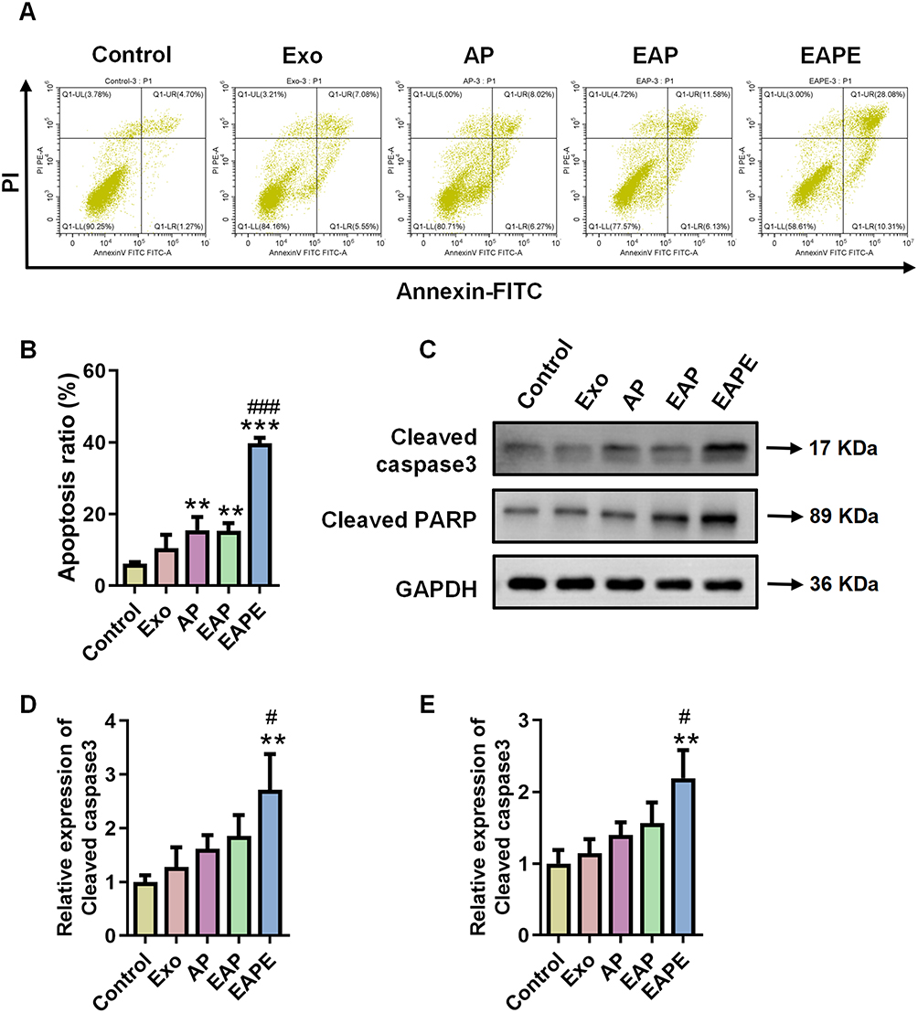 |
Figure 4 Effects of EAPE on apoptosis in LNCaP cells. (A and B) Flow cytometry plots and quantitative analysis of apoptosis in LNCaP cells. (C–E) Immunoblotting analysis and relative expression quantification of Cleaved caspase3 and Cleaved PARP levels in LNCaP cells. Data are presented as mean ± SD (n = 3), statistical significance levels are represented as: **p < 0.01, ***p < 0.001 vs. Control group; #p < 0.05, ###p < 0.001 vs. AP group. |
Collectively, these findings indicate that the engineered nanodelivery system, particularly EAPE, significantly enhances the antitumor activity of Astragaloside IV and PESV, as evidenced by the inhibition of LNCaP cell proliferation and migration, as well as the induction of apoptosis, providing a basis for subsequent antitumor evaluation in vivo.
EAPE Reverses Immunosuppression and Activates T Cells in vitro
Given the excellent antitumor activity of EAPE, its impact on the tumor immune microenvironment was further investigated. The LNCaP cells were co-cultured with T cells in a transwell to simulate the cellular interactions within the tumor immune microenvironment, and treated with engineered exosomes (Exo, EAP and EAPE). The CCK8 assay demonstrated that compared with the control group, treated with blank exosomes resulted in a significant decrease in cell viability, moreover, the inhibitory effect was further enhanced in the EAP group and was most pronounced in the EAPE group (Figure 5A). Subsequently, the content of IFN-γ in the cell supernatant was measured. IFN-γ is a cytokine that possesses immunoregulatory, antiviral and antiproliferative properties, and exerting inhibitory effects on tumors.20 The results of ELISA assay showed a significant upregulation of IFN-γ in the EAP and EAPE groups, and IFN-γ contents were also elevated in the Exo group, but the increase was not statistically significant (Figure 5B). Similarly, the levels of TNF-α and IL-2 were significantly increased in both the EAP and EAPE groups, with the highest expression observed in the EAPE group (Figure 5C and D). Although a slight increase was also observed in the EXO group, the difference did not reach statistical significance. Then, the protein and mRNA expression levels of PD-1 in T cells were assessed. The results showed that PD-1 expression was significantly increased in the EAP and EAPE groups, compared to the control, Exo treated led to a non-significant increase (Figure 5E–I). In contrast, the results of PD-L1 expression levels in LNCaP cells showed that blank exosomes induced a non-significant decrease, while EAP and EAPE treatments significantly downregulated PD-L1 expression (Figure 5E–J). Subsequently, flow cytometry analysis was carried out to detect the proportion of CD3+CD8+ cells in co-incubation environment. The positive proportion of CD3+CD8+ cells was elevated in Exo group, and significantly increased in both EAP and EAPE treatment groups, with a higher proportion observed in the EAPE group (Figure 5K and L). Similarly, the results of fluorescence co-localization showed that, the percentage of CD8/CD3 was significantly increased in the EAP and EAPE treatment groups (Figure 5M and N). These results suggest that engineered exosomes, especially EAPE, can effectively suppress the expression of the immune checkpoint PD-L1 on prostate cancer cells, indirectly activate T cells and enhancing the recognition and clearance of tumor cells by CD8+ T cells.
 |
Figure 5 Effects of EAPE on tumor immune microenvironment in vitro. (A) Cell viability was determined by CCK-8 assay after treatment with Exo, EAP and EAPE in a co-culture system of T cells and LNCaP cells. (B–D) The levels of IFN-γ, TNF-α and IL-2 in co-culture supernatants were determined by ELISA assay. (E–G) Immunoblotting analysis and relative expression quantification of PD-1 and PD-L1 protein levels. (H–J) RT-PCR analysis and relative expression quantification of PD-1 and PD-L1 mRNA levels. (K and L) Flow cytometry plots and quantitative analysis of CD3+CD8+ T cells. (M and N) Fluorescence microscopy images and quantitative analysis of CD8/CD3 co-localization (scale bar: 20 μm). Data are presented as mean ± SD (n = 3), statistical significance levels are represented as: *p < 0.05, **p < 0.01, ***p < 0.001 vs. Control group. |
Distribution and Biosafety of EAPE in vivo
Next, a subcutaneous tumor model of LNCaP in nude mouse was established to assess the safety and targeting ability of engineered exosomes in vivo. The results showed that the body weight of mouse in the EAPE group exhibited an overall upward trend, while the body weight of mouse in the other groups exhibited declining trends after different time points (Figure 6A). The fluorescence imaging was conducted to evaluate the in vivo targeting capability of EAPE. As illustrated in Figure 6B and C, the strong fluorescence signals were observed in the blank-exosomes and engineered exosomes compared to the control group. Moreover, to further assess the biodistribution of the engineered exosomes in vivo, the mouse major organs were harvested and subjected to fluorescence imaging. As shown in Figure 6D and E, no significant difference in organ fluorescence intensity was observed between the Exo and Control groups. In the EAP and EAPE group, fluorescence signals were elevated in tumors but not in organs. Notably, the EAPE group showed the strongest fluorescence signal in tumors. Histological examination of HE-stained organ sections revealed no apparent pathological changes in the different exosome-treated groups compared to the control group (Figure 6F), further demonstrating the safety of the EAPE in vivo. All these results demonstrate that E3 aptamer-modified exosomes also exhibit favorable biosafety and potent targeting capability in vivo.
 |
Figure 6 Distribution and biosafety of EAPE in vivo. (A) Body weight changes of mice following treatment with Exo, EAP or EAPE over 28 days. (B and C) In vivo fluorescence images and fluorescence intensity analysis of mice treated with Nile Red-labeled Exo, EAP and EAPE. (D and E) Ex vivo fluorescence distribution images and quantitative analysis of tumor and major organs in mice. (F) Representative HE stained sections of major organs from mice (scale bar: 50 μm). Data are presented as mean ± SD (n = 5), statistical significance levels are represented as: ***p < 0.001 vs. Control group. |
EAPE Suppresses Tumor Progression and Induces Apoptosis in vivo
The antitumor effects of EAPE were further evaluated in a subcutaneous tumor model. As depicted in Figure 7A–D, tumor growth remained largely unrestrained in both untreated and Exo-treated mice. However, mice treated with EAP and EAPE exhibited substantial tumor volume reduction, particularly in the EAPE group. To further evaluate the antitumor efficacy of engineered exosomes, histological examination was conducted on tumor samples collected from various treatment groups after 28 days. HE staining revealed that cells in the EAPE group demonstrated a reduced squamous-like morphology compared to those in the Control, Exo, and EAP groups (Figure 7E). Furthermore, the interfering efficiency of engineered exosome in subcutaneous tumor model was confirmed by the KI67 staining of tumor tissues, the results revealed a marked decrease in KI67-positive cells in the EAP and EAPE groups (Figure 7F and G). Additionally, TUNEL assay was conducted and showed a significant increase in the apoptosis rate of tumor tissues in both the EAP and EAPE groups (Figure 8A and B). Then the activation of apoptosis was confirmed to be associated with the cleavage activation of the Caspase-3 pathway, a marked increase in both the positive immunohistochemical staining of cleaved Caspase‑3 and the protein expression level of cleaved Caspase‑3 and cleaved PARP within tumor tissue, detected by Western blotting and IHC (Figure 8C–G). The expression of EMT markers (E-cadherin and Vimentin) in vivo were examined by Western blotting. As shown in Figure 8E and H, the expression of E-cadherin in tumor tissue was significantly increased in the group treated with E3 aptamer-modified exosomes. The results of WB and IHC showed that Vimentin expressed at low levels in mouse tumors of the Exo, EAP and EAPE groups (Figure 8E–K). Together, these data strongly indicate that EAPE effectively enhances antitumor efficacy of Astragaloside IV and PESV by suppressing tumor progression and inducing apoptosis in vivo.
 |
Figure 7 Effects of EAPE on tumor growth and proliferation in vivo. (A) Representative photographs of tumor-bearing mice. (B) Representative images of excised tumors from each group. (C) Quantitative analysis of tumor weights. (D) Quantitative analysis of tumor volumes. (E) Representative HE stained sections of tumor tissues (scale bar: 100 μm). (F and G) Immunohistochemical staining images and quantitative analysis of KI67 in tumor tissues (magnification × 200, scale bar = 50 μm; magnification × 400, scale bar = 25 μm). Data are presented as mean ± SD (n=5), statistical significance levels are represented as: *p < 0.05, **p < 0.01, ***p < 0.001 vs. Control group. |
 |
Figure 8 Effects of EAPE on apoptosis in vivo. (A and B) Quantitative analysis of TUNEL-positive cells and TUNEL fluorescence images in tumor tissues (scale bar: 50 μm). (C and D) Immunohistochemical staining images and quantitative analysis of Cleaved caspase3 in tumor tissues (magnification × 200, scale bar = 50 μm; magnification × 400, scale bar = 25 μm). (E–I) Immunoblotting analysis and relative expression quantification of Cleaved caspase3, Cleaved PARP, E-cadherin and Vimentin levels in tumor tissues. (J and K) Immunohistochemical staining images and quantitative analysis of Vimentin in tumor tissues (magnification × 200, scale bar = 50 μm; magnification × 400, scale bar = 25 μm). Data are presented as mean ± SD (n = 5), statistical significance levels are represented as: *p < 0.05, **p < 0.01, ***p < 0.001 vs. Control group. |
EAPE Remodels the Tumor Immune Microenvironment in vivo
Subsequently, the impact of EAPE on the tumor immune microenvironment was further validated in a tumor-bearing mouse model. The serum levels of IFN-γ, TNF-α and IL-2 in mice were measured by ELISA. As shown in Figure 9A–C, the Exo-treated group showed a slight increase in the levels of IFN-γ, TNF-α and IL-2 compared with the Control group, but no statistically significant differences were observed. Significant elevations of all three cytokines were found in both the EAP and EAPE groups, with the latter showing the highest levels. The analysis of PD‑1 expression in spleen tissue revealed that (Figure 9D–H), compared to the Control, the Exo group had a significant rise in protein but a non-significant increase in mRNA. Both the EAP and EAPE groups exhibited significant upregulation in both PD-1 protein and mRNA expression, which were further enhanced in the EAPE group. The results of PD-L1 protein and mRNA levels in tumor tissue showed a decreasing trend in the Exo group, though the changes were not statistically significant. The EAP and EAPE treatments led to a significant reduction in PD-L1 expression (Figure 9D–I). In addition, both confocal microscopy and flow cytometry analyses demonstrated a progressive increase in the CD8+CD3+ cell ratio across treatment groups. While the rise in the Exo group was not significant, and the EAP and EAPE groups showed significant elevations, with the EAPE group exhibiting the most pronounced increase (Figure 9J–M). Taken together, these findings indicate that EAPE improves antitumor effect through targeted drug delivery that disrupts local immune evasion, which in turn initiates a potent systemic antitumor immune response mediated by antigen-specific cytotoxic T lymphocytes, highlighting its potential as a clinically relevant immunotherapeutic strategy.
 |
Figure 9 Effects of EAPE on tumor immune microenvironment in vivo. (A–C) The levels of IFN-γ, TNF-α and IL-2 in serum were measured by ELISA. (D–F) RT-PCR analysis and relative expression quantification of PD-1 and PD-L1 mRNA levels. (G–I) Immunoblotting analysis and relative expression quantification of PD-1 and PD-L1 protein levels. (J and K) Flow cytometry plots and quantitative analysis of CD3+CD8+ T cells. (L and M) Fluorescence microscopy images and quantitative analysis of CD8/CD3 co-localization (scale bar: 50 μm). Data are presented as mean ± SD (n = 5), statistical significance levels are represented as: *p < 0.05, **p < 0.01, ***p < 0.001 vs. Control group. |
Discussion
Traditional Chinese medicine (TCM) has recently garnered increasing attention in cancer therapy due to its multi‑component nature, multi-target effects, and synergistic therapeutic advantages.27 The therapeutic potential of bioactive TCM constituents, such as Astragaloside IV and PESV, in prostate cancer treatment has been previously demonstrated.12,25,28 However, the clinical translation of bioactive TCM components is often limited by low bioavailability and lack of tumor specificity. These limitations significantly hinder the clinical application of these agents, thus necessitating effective delivery strategies to enhance their therapeutic efficacy in PCa treatment. Herein, we developed an E3 aptamer-modified T cell-derived exosomal nanodrug delivery system for the co-delivery of Astragaloside IV and PESV, representing an innovative and versatile strategy to enhance tumor-targeted accumulation and therapeutic efficacy while minimizing systemic toxicity. Moreover, we further explored the immunomodulatory effects of EAPE, demonstrating that it effectively suppresses immune evasion and promotes T cell activation, thereby providing mechanistic insights that further support its potential for clinical translation.
Exosomes have attracted increasing attention as promising nanocarriers for drug delivery due to their unique biological advantages, including inherent biocompatibility, low immunogenicity, and natural targeting capability. In recent years, a variety of nanomaterial-based delivery systems, including liposomes, polymeric nanoparticles and engineered extracellular vesicles, have been developed for PCa therapy to improve tumor-specific drug accumulation and reduce systemic toxicity.29–31 Among these platforms, exosome-based therapeutic strategies have gained growing recognition owing to their favorable biocompatibility, low immunogenicity, targeting capability and promising translational potential, attracting increasing attention from both academia and major global regulatory agencies.30,32,33 Notably, optimizing the cellular origin of exosomes is critical for enhancing therapeutic efficacy and translational potential. Serving as essential mediators of intercellular communication, exosomes carry distinct bioactive macromolecules depending on their cellular origin, which enables to perform diverse biological functions.18,24 Hence, T lymphocyte-derived exosomes were employed in this study due to their unique immunomodulatory properties. Accumulating evidence has demonstrated that T cell-derived exosomes can promote the proliferation of resting T cells, expand cytotoxic T cell populations and directly deliver killer molecules such as granzymes, thereby exhibiting antitumor potential.24,34, Thus, in this study, CD8+ T cell-derived exosomes were selected as carriers to retain the immunomodulatory functions and tumor cell cytotoxicity characteristic of their parental cells.
While T cell-derived exosomes exhibit a degree of natural tropism toward tumor cells, active targeting strategies can significantly improve their tumor accumulation and therapeutic precision. To this end, E3 aptamer was conjugated to the exosomal surface to achieve enhanced tumor-specific delivery via chemical coupling. Successful conjugation and structural integrity of the engineered exosomes (EAP and EAPE) were confirmed, ensuring that the modifications did not compromise their fundamental properties such as membrane integrity, size distribution, or drug loading and release capacity. These findings demonstrate that E3 aptamer-modified and drug-loaded exosomes retain the key characteristics required for effective drug delivery, laying a solid foundation for subsequent in vitro and in vivo functional evaluations.
Following successful construction and characterization, the active targeting capability and delivery advantages of E3 aptamer modified exosomes were systematically validated. In vitro cellular uptake experiments revealed that E3 modification significantly enhanced the internalization efficiency of drug-loaded exosomes by LNCaP cells, ensuring the precise and efficient delivery of Astragaloside IV-PESV to the intracellular targets, which underpins the enhanced in vitro efficacy. This enhanced uptake accounts for the superior antitumor efficacy observed in subsequent functional assays. Notably, blank exosomes also exhibited a certain degree of targeting capability toward LNCaP cells, confirming the intrinsic tumor-homing capacity of CD8+ T cell-derived exosomes.35 Collectively, the intrinsic tropism of CD8+ T cell-derived exosomes, combined with the active targeting conferred by E3 aptamers, further validates the targeting advantage of this engineered platform, which can reduce off-target effects and improve therapeutic precision in clinical settings.
Given the demonstrated efficient active targeting capability of the E3-modified exosomes, we further evaluated the antitumor efficacy of EAPE in vitro. Experimental results revealed that compared to blank exosomes, both EAPE and EAP exhibited significantly enhanced activity against prostate cancer cells, effectively reducing LNCaP cell viability, suppressing proliferative potential, and impairing cell motility and invasive capacity. Notably, EAPE exhibited the most pronounced inhibitory effects among all treatment group. Consistent with our expectations, the drug-loaded exosomes, including EAPE and EAP, exhibited potent antitumor efficacy, which can be attributed to the synergistic effects of Astragaloside IV and PESV. Both Astragaloside IV and PESV have been individually reported to exert anti‑tumor effects across a range of cancer types through promoting apoptosis and inhibiting proliferation of tumor cells.36–41 Furthermore, quantitative flow cytometry revealed that EAPE treatment induced the highest proportion of apoptotic cells among all groups, and this pro-apoptotic effect was further corroborated by Western blot analysis. Together, these findings suggest that E3 aptamer-mediated efficient internalization significantly enhances the intracellular accumulation of Astragaloside IV-PESV, thereby potentiating its pro-apoptotic effect on prostate cancer cells.
Beyond the direct cytotoxic effects on tumor cells, Astragaloside IV and PESV also exhibit immunomodulatory properties that contribute to remodeling the tumor microenvironment.36,42 In particular, Astragaloside IV has been shown to block IDO‑induced immune escape by enhancing the activity of cytotoxic T lymphocytes and promoting the infiltration of pro‑inflammatory factors such as IFN‑γ into tumor tissues.42,43 Furthermore, scorpion venoms exert antitumor activity by promoting the expression of IFN-γ within the tumor microenvironment, thereby activating the immune system.44 In this study, LNCaP cells were co-cultured with T lymphocytes to simulate tumor–immune interactions. The co-culture experiments demonstrated that EAPE treatment significantly increased IFN-γ secretion and promoted CD8⁺ T cell activation, indicating enhanced antitumor immune responses. IFN-γ is a crucial factor in antitumor immunity, primarily produced by activated immune cells, such as cytotoxic CD8⁺ T cell and NK cells.45–47 Consistent with this, flow cytometry and colocalization analysis revealed a marked activation of CD8+ T cells, providing cellular evidence for the elevated IFN-γ production. IFN-γ secreted by activated T cells triggers a series of downstream signaling events that induce chemokine production to promote the recruitment of additional CD8+ T cells, while simultaneously upregulating multiple checkpoint inhibitors (such as PD-L1, TIGIT and LAG3) on tumor cells as a feedback mechanism to restrain the antitumor immune response.48,49 A recent study demonstrated that Astragaloside IV alleviates immunosuppression and potentiates immune cytotoxic activity via suppressing IFN-γ-induced PD-L1 expression on HCC cells;50 similarly, the TCM compound CFF-1 reduces PD-L1 levels in PCa cells to potentiate T cell-mediated antitumor immunity by counteracting PD-1/PD-L1 upregulation.51 Consistent with these reports, PD-L1 expression on LNCaP cells was markedly downregulated following treatment with Astragaloside IV and PESV-loaded exosomes in our study, suggesting this formulation similarly weakens immune suppression in PCa. Although PD-1 expression on T cells was elevated, the concurrent increase in IFN-γ production indicates that this likely reflects an activation-associated feedback response rather than functional exhaustion.52 Taken together, these findings indicated that EAPE enhances antitumor immunity by delivering drugs specifically to cancer cells, which is highly relevant for improving the efficacy of immunotherapy in clinical settings.
Building upon the promising antitumor activity and immunomodulatory potential of EAPE in vitro, a nude mouse xenograft models were established to evaluate therapeutic efficacy within a complex in vivo system. In vivo imaging showed that EAPE specifically accumulated at the tumor site after injection, providing direct in vivo evidence for “active targeting” delivery. Histopathological analysis of major organs revealed a favorable safety profile, while tumor tissue assessment demonstrated extensive necrosis and marked suppression of the proliferation marker KI67 in EAPE-treated mice. Consistent with the in vitro results, TUNEL staining and the significant upregulation of Cleaved Caspase-3 and Cleaved PARP demonstrated that EAPE targeted to the tumor site and induced Caspase-3-dependent apoptosis in vivo. Additionally, EAPE treatment elicited robust systemic immune responses as evidenced by significantly elevated serum IFN-γ levels, upregulated PD-1 expression in spleen, and markedly reduced PD-L1 expression in tumor tissue. Fluorescence confocal microscopy and flow cytometry further validated the activation and expansion of T cells. These results collectively demonstrate the favorable biosafety profile and effective antitumor efficacy of EAPE in vivo, further supporting its translational potential as a novel drug delivery platform.
Taken together, EAPE demonstrates favorable tumor targeting, antitumor efficacy and immunomodulatory activity. These findings indicate that EAPE may serve as a clinically relevant delivery system capable of improving therapeutic outcomes while minimizing systemic toxicity. Moreover, the modular design of this platform enables its adaptation for the delivery of other therapeutic agents, supporting its broader applicability in prostate cancer treatment. While the present findings support the therapeutic potential of EAPE, several limitations should be acknowledged. Although the nude mouse xenograft model provided valuable evidence supporting the antitumor efficacy and immune-related activity of EAPE, future studies using immunocompetent or humanized models will further strengthen the mechanistic understanding of its immunomodulatory effects. Moreover, although comprehensive in vitro experiments demonstrated superior antitumor efficacy of EAPE compared with AP alone, the contribution of E3 aptamer-mediated delivery to the enhanced therapeutic efficacy observed in vivo requires further validation. In addition, the large-scale production, long-term safety and clinical efficacy also require further evaluation in future studies.
Conclusions
In summary, we have successfully constructed an E3 aptamer-modified T lymphocyte-derived exosome system as an actively targeted delivery platform for Astragaloside IV and PESV in PCa. This system exhibits excellent tumor-targeting capability, favorable biosafety, and potent antitumor efficacy both in vitro and in vivo. The work provides a precise delivery strategy that enhances therapeutic efficacy and reduces toxicity for supporting the clinical translation of Astragaloside IV-PESV or other TCM-derived components in PCa treatment, while also offering innovative experimental evidence and a strategic framework for integrated targeted immunotherapy of PCa.
Abbreviations
PCa, prostate cancer; PESV, polypeptide extract from scorpion venom; Exo, exosome; EAPE, E3 aptamer-modified T cell-derived exosome loaded with Astragaloside IV and PESV; EAP, T cell-derived exosome loaded with Astragaloside IV and PESV; AP, Astragaloside IV-PESV; ADT, androgen deprivation therapy; CRPC, castration-resistant prostate cancer; TCM, traditional Chinese medicine; PBS, phosphate-buffered saline; TEM, transmission electron microscopy; NTA, nanoparticle tracking analyzer; TCEP, tris (2-carboxyethyl) phosphine hydrochloride; EE, encapsulation efficiency; DL, drug loading; DMEM, dulbecco’s modified eagle medium; H&E, Hematoxylin and eosin; IF, immunofluorescence; IHC, immunohistochemistry; WB, Western blot.
Data Sharing Statement
All data that support the conclusions in the paper are presented in the paper and/or the Supplementary Materials. Additional data related to this paper may be requested from the corresponding author (Xujun You) upon request.
Ethics Approval
All animal procedures were carried out ethically and humanely, and approved by the Institutional Animal Care and Use Committee of Shenzhen Lingfu Top Biotechnology Co., Ltd (Approval No. TOP-1PZ-GM250708) and were conducted in accordance with institutional guidelines for the care and use of laboratory animals. All efforts were made to minimize animal suffering and to reduce the number of animals used.
Author Contributions
All authors made a significant contribution to the work reported, whether that is in the conception, study design, execution, acquisition of data, analysis and interpretation, or in all these areas; took part in drafting, revising or critically reviewing the article; gave final approval of the version to be published; have agreed on the journal to which the article has been submitted; and agree to be accountable for all aspects of the work.
Funding
This work is financially supported by the General funding project of China Postdoctoral Science Foundation (Program no. 2024M750265), Project of Guangdong Provincial Department of Science and Technology (Program no. 2024A1515012209), Project of Guangdong Provincial Administration of Traditional Chinese Medicine (Program No.20261340) and Shenzhen Bao’an Chinese Medicine Hospital Research Program (Program No. BAZYY20220703).
Disclosure
The authors declare no conflicts of interest in this study.
References
1. Sekhoacha M, Riet K, Motloung P, Gumenku L, Adegoke A, Mashele S. Prostate cancer review: genetics, diagnosis, treatment options, and alternative approaches. Molecules. 2022;27(17):5730. doi:10.3390/molecules27175730
2. Guruvayurappan GK, Frankenbach-Désor T, Laubach M, et al. Clinical challenges in prostate cancer management: metastatic bone-tropism and the role of circulating tumor cells. Cancer Lett. 2024;606:217310. doi:10.1016/j.canlet.2024.217310
3. Raychaudhuri R, Lin DW, Montgomery RB. Prostate cancer: a review. JAMA. 2025;333(16):1433–23. doi:10.1001/jama.2025.0228
4. Luo ZW, Xia K, Liu YW, et al. Extracellular vesicles from akkermansia muciniphila elicit antitumor immunity against prostate cancer via modulation of cd8(+) t cells and macrophages. Int J Nanomedicine. 2021;16:2949–2963. doi:10.2147/IJN.S304515
5. Tilki D, van den Bergh RCN, Briers E, et al. EAU-EANM-ESTRO-ESUR-ISUP-SIOG Guidelines on Prostate Cancer. Part II-2024 Update: treatment of Relapsing and Metastatic Prostate Cancer. Eur Urol. 2024;86(2):164–182. doi:10.1016/j.eururo.2024.04.010
6. Li S, Jing S, Zhou J, et al. Chinese medicines as therapeutic options for treating prostate cancer: therapeutic effects and underlying mechanisms. J Ethnopharmacol. 2026;354:120471. doi:10.1016/j.jep.2025.120471
7. Wang J, Han J, Feng J, et al. Effect of Bushen Huoxue Decoction combined with moxibustion on inflammation and urinary symptoms in patients with prostate cancer. Am J Transl Res. 2022;14(12):8991–9000.
8. Zou B, Long Y, Gao R, et al. Nanodelivery system of traditional Chinese medicine bioactive compounds: application in the treatment of prostate cancer. Phytomedicine. 2024;135:155554. doi:10.1016/j.phymed.2024.155554
9. Gao QT, Choi RCY, Cheung AWH, et al. Danggui buxue tang--a Chinese herbal decoction activates the phosphorylations of extracellular signal-regulated kinase and estrogen receptor alpha in cultured MCF-7 cells. FEBS Lett. 2007;581(2):233–240. doi:10.1016/j.febslet.2006.12.018
10. Wu L, Chen Y, Chen M, et al. Application of network pharmacology and molecular docking to elucidate the potential mechanism of Astragalus-Scorpion against prostate cancer. Andrologia. 2021;53(9):e14165. doi:10.1111/and.14165
11. You XJ, Wen Z, Zheng QX, et al. Explore the mechanism of astragaloside IV-PESV on proliferation, migration, and autophagy of prostate cancer cells based on the PI3K/AKT signaling pathway. Zhonghua Nan Ke Xue = National Journal of Andrology. 2023;29(12):963–972.
12. You X, Wu Y, Li Q, Sheng W, Zhou Q, Fu W. Astragalus-scorpion drug pair inhibits the development of prostate cancer by regulating GDPD4-2/PI3K/AKT/mTOR pathway and autophagy. Front Pharmacol. 2022;13:895696. doi:10.3389/fphar.2022.895696
13. Fu Z, Wang S, Zhou X, Ouyang L, Chen Z, Deng G. Harnessing the power of traditional chinese medicine in cancer treatment: the role of nanocarriers. Int J Nanomedicine. 2025;20:3147–3174. doi:10.2147/IJN.S502104
14. Sam Lee J, Kim M, Jin H, et al. DNA aptamer-conjugated lipid nanoparticle for targeted PTEN mRNA delivery to prostate cancer cells. Int J Pharm. 2024;662:124519. doi:10.1016/j.ijpharm.2024.124519
15. Tiburcius S, Krishnan K, Yang JH, et al. Silica-based nanoparticles as drug delivery vehicles for prostate cancer treatment. Chemical Record. 2021;21(6):1535–1568. doi:10.1002/tcr.202000104
16. Jiang Z, Zhang W, Zhang J, et al. Nanomaterial-based drug delivery systems: a new weapon for cancer immunotherapy. Int J Nanomedicine. 2022;17:4677–4696. doi:10.2147/IJN.S376216
17. Tian J, Han Z, Song D, et al. Engineered exosome for drug delivery: recent development and clinical applications. Int J Nanomedicine. 2023;18:7923–7940. doi:10.2147/IJN.S444582
18. Zhang M, Hu S, Liu L, et al. Engineered exosomes from different sources for cancer-targeted therapy. Signal Transduct Target Ther. 2023;8(1):124. doi:10.1038/s41392-023-01382-y
19. Powell Gray B, Kelly L, Ahrens DP, et al. Tunable cytotoxic aptamer-drug conjugates for the treatment of prostate cancer. Proc Natl Acad Sci U S A. 2018;115(18):4761–4766. doi:10.1073/pnas.1717705115
20. Han Q, Xie QR, Li F, et al. Targeted inhibition of SIRT6 via engineered exosomes impairs tumorigenesis and metastasis in prostate cancer. Theranostics. 2021;11(13):6526–6541. doi:10.7150/thno.53886
21. Luan X, Sansanaphongpricha K, Myers I, Chen H, Yuan H, Sun D. Engineering exosomes as refined biological nanoplatforms for drug delivery. Acta Pharmacol Sin. 2017;38(6):754–763. doi:10.1038/aps.2017.12
22. Becker A, Thakur BK, Weiss JM, Kim HS, Peinado H, Lyden D. Extracellular vesicles in cancer: cell-to-cell mediators of metastasis. Cancer Cell. 2016;30(6):836–848. doi:10.1016/j.ccell.2016.10.009
23. Mahaweni NM, Kaijen-Lambers MEH, Dekkers J, Aerts JGJV, Hegmans JPJJ. Tumour-derived exosomes as antigen delivery carriers in dendritic cell-based immunotherapy for malignant mesothelioma. J Extracell Vesicles. 2013;2:10–3402. doi:10.3402/jev.v2i0.22492
24. Choi SJ, Cho H, Yea K, Baek MC. Immune cell-derived small extracellular vesicles in cancer treatment. BMB Rep. 2022;55(1):48–56. doi:10.5483/BMBRep.2022.55.1.133
25. You X, Li H, Li Q, et al. Astragaloside IV-PESV facilitates pyroptosis by enhancing palmitoylation of GSDMD protein mediated by ZDHHC1. Naunyn-Schmiedeberg’s Archives of Pharmacology. 2025;398(10):13925–13939. doi:10.1007/s00210-025-04122-x
26. You X, Qiu J, Li Q, et al. Astragaloside IV-PESV repressed t cell immunosuppression by inhibiting pd-l1 expression in prostate cancer through stat3 pathway. J Food Biochem. 2023;2023:1–15. doi:10.1155/2023/8884430
27. Liu Y, Yang S, Wang K, et al. Cellular senescence and cancer: focusing on traditional Chinese medicine and natural products. Cell Prolif. 2020;53(10):e12894. doi:10.1111/cpr.12894
28. You X, Qiu J, Li Q, et al. Astragaloside IV-PESV inhibits prostate cancer tumor growth by restoring gut microbiota and microbial metabolic homeostasis via the AGE-RAGE pathway. BMC Cancer. 2024;24(1):472. doi:10.1186/s12885-024-12167-z
29. Kumar N, Bose P, Kumar S, et al. Development and evaluation of choline tagged mesoporous silica nanoparticles for targeted delivery and imaging in prostate cancer. Nanomedicine: Nanotechnology, Biology, and Medicine. 2026;74:102948. doi:10.1016/j.nano.2026.102948
30. An Y, Xu JZ, Ye GC, et al. Therapeutic translation of traditional Chinese medicine Huangqi derived exosome like nanoparticles: targeting prostate cancer through ferroptosis activation, immune reprogramming, and microbiome modulation. J Nanobiotechnology. 2026;24(1):447. doi:10.1186/s12951-026-04160-4
31. Martinez De Castilla P E, Verdi V, de Voogt W, et al. Nanobody-decorated lipid nanoparticles for enhanced mrna delivery to tumors in vivo. Adv Healthc Mater. 2025;14(24):e2500605. doi:10.1002/adhm.202500605
32. Mehr FK, Emtiazi N, Zolfi E, Moradi Y. Comparison of the therapeutic potential of exosomes derived from different cells for the treatment of prostate cancer. Critical Reviews in Oncology/Hematology. 2026;217:105055. doi:10.1016/j.critrevonc.2025.105055
33. Liang B, Zou S. Exosome-mediated delivery of miRNA-1290 inhibitor enhances JNK-dependent tissue-resident memory T cell immunity in prostate cancer. Sci Rep. 2026.
34. Fu W, Lei C, Liu S, et al. CAR exosomes derived from effector CAR-T cells have potent antitumour effects and low toxicity. Nat Commun. 2019;10(1):4355. doi:10.1038/s41467-019-12321-3
35. Del Vecchio F, Martinez-Rodriguez V, Schukking M, Cocks A, Broseghini E, Fabbri M. Professional killers: the role of extracellular vesicles in the reciprocal interactions between natural killer, CD8+ cytotoxic T-cells and tumour cells. J Extracell Vesicles. 2021;10(6):e12075. doi:10.1002/jev2.12075
36. Arcos SSS, Aguiar MRDC, Oliveira JLAD, et al. Scorpion venom as a source of cancer drugs: a comprehensive proteomic analysis and therapeutic potential. International Journal of Molecular Sciences. 2025;26(20):9907. doi:10.3390/ijms26209907
37. Bai B, Zhang L, Zhang Y, et al. Astragaloside IV potentiates cisplatin sensitivity in triple-negative breast cancer via STING signaling pathway activation. Phytomedicine: International Journal of Phytotherapy and Phytopharmacology. 2025;148:157330. doi:10.1016/j.phymed.2025.157330
38. Chen T, Yang P, Jia Y. Molecular mechanisms of astragaloside‑IV in cancer therapy (Review). Int J Mol Med. 2021;47(3):13. doi:10.3892/ijmm.2021.4846
39. Dueñas-Cuellar RA, Santana CJC, Magalhães ACM, Pires ORJ, Fontes W, Castro MS. Scorpion toxins and ion channels: potential applications in cancer therapy. Toxins. 2020;12(5):326. doi:10.3390/toxins12050326
40. Li L, Wu XN, Pu YZ, et al. Astragaloside IV alleviates colorectal cancer metastases by regulating RALY/PLD2 axis and inhibiting tumoral exosome biogenesis. Phytotherapy Research. 2026;40(2):681–700. doi:10.1002/ptr.70154
41. Zhou L, Li M, Chai Z, et al. Anticancer effects and mechanisms of Astragaloside‑IV (Review). Oncol Rep. 2023;49(1):5. doi:10.3892/or.2022.8442
42. Zhang A, Zheng Y, Que Z, et al. Astragaloside IV inhibits progression of lung cancer by mediating immune function of Tregs and CTLs by interfering with IDO. J Cancer Res Clin Oncol. 2014;140(11):1883–1890. doi:10.1007/s00432-014-1744-x
43. Liu F, Ran F, He H, Chen L. Astragaloside IV exerts anti-tumor effect on murine colorectal cancer by re-educating tumor-associated macrophage. Arch Immunol Ther Exp (Warsz). 2020;68(6):33. doi:10.1007/s00005-020-00598-y
44. Amirgholami N, Karampour NS, Ghadiri A, Tagavi Moghadam A, Ghasemi Dehcheshmeh M, Crassicauda PMHA. M. eupeus and H. lepturus scorpion venoms initiate a strong in vivo anticancer immune response in CT26-tumor mice model. Toxicon. 2020;180:31–38. doi:10.1016/j.toxicon.2020.04.001
45. Fang C, Weng T, Hu S, et al. IFN-γ-induced ER stress impairs autophagy and triggers apoptosis in lung cancer cells. Oncoimmunology. 2021;10(1):1962591. doi:10.1080/2162402X.2021.1962591
46. Gocher AM, Workman CJ, Vignali DAA. Interferon-γ: teammate or opponent in the tumour microenvironment? Nature Reviews. Immunology. 2022;22(3):158–172. doi:10.1038/s41577-021-00566-3
47. Han J, Wu M, Liu Z. Dysregulation in IFN-γ signaling and response: the barricade to tumor immunotherapy. Front Immunol. 2023;14:1190333. doi:10.3389/fimmu.2023.1190333
48. Ayers M, Lunceford J, Nebozhyn M, et al. IFN-γ-related mRNA profile predicts clinical response to PD-1 blockade. The Journal of Clinical Investigation. 2017;127(8):2930–2940. doi:10.1172/JCI91190
49. Li Q, Han J, Yang Y, Chen Y. PD-1/PD-L1 checkpoint inhibitors in advanced hepatocellular carcinoma immunotherapy. Front Immunol. 2022;13:1070961. doi:10.3389/fimmu.2022.1070961
50. Ma Y, Li Y, Wu T, Li Y, Wang Q. Astragaloside IV attenuates programmed death-ligand 1-mediated immunosuppression during liver cancer Development via the miR-135b-5p/CNDP1 Axis. Cancers. 2023;15(20):5048. doi:10.3390/cancers15205048
51. Zhang Y, Wei Y, Jiang S, et al. Traditional Chinese medicine CFF-1 exerts a potent anti-tumor immunity to hinder tumor growth and metastasis in prostate cancer through EGFR/JAK1/STAT3 pathway to inhibit PD-1/PD-L1 checkpoint signaling. Phytomedicine. 2022;99:153939. doi:10.1016/j.phymed.2022.153939
52. Liu J, Chen Z, Li Y, Zhao W, Wu J, Zhang Z. PD-1/PD-L1 checkpoint inhibitors in tumor immunotherapy. Front Pharmacol. 2021;12:731798. doi:10.3389/fphar.2021.731798
 © 2026 The Author(s). This work is published and licensed by Dove Medical Press Limited. The
full terms of this license are available at https://www.dovepress.com/terms
and incorporate the Creative Commons Attribution
- Non Commercial (unported, 4.0) License.
By accessing the work you hereby accept the Terms. Non-commercial uses of the work are permitted
without any further permission from Dove Medical Press Limited, provided the work is properly
attributed. For permission for commercial use of this work, please see paragraphs 4.2 and 5 of our Terms.
© 2026 The Author(s). This work is published and licensed by Dove Medical Press Limited. The
full terms of this license are available at https://www.dovepress.com/terms
and incorporate the Creative Commons Attribution
- Non Commercial (unported, 4.0) License.
By accessing the work you hereby accept the Terms. Non-commercial uses of the work are permitted
without any further permission from Dove Medical Press Limited, provided the work is properly
attributed. For permission for commercial use of this work, please see paragraphs 4.2 and 5 of our Terms.
Recommended articles

M1 Macrophage-Derived Sonoresponsive Nanoparticles for Sonodynamic Anticancer Therapy
Chen S, Wang J, Liao H, Tang K, Xu Y, Wang L, Niu C
International Journal of Nanomedicine 2022, 17:4725-4741
Published Date: 10 October 2022
Prostate Cancer Bone Metastasis: Molecular Mechanisms of Tumor and Bone Microenvironment
Jiang H
Cancer Management and Research 2025, 17:219-237
Published Date: 1 February 2025
Advances of Drug-Loaded Microsphere Technology for Targeted Immunotherapy Against Prostate Cancer
Feng W
International Journal of Nanomedicine 2025, 20:11479-11489
Published Date: 20 September 2025
Nanomedicine Targeting Cancer-Associated Fibroblasts in Prostate Cancer: From Biological Mechanisms to Integrated Theranostic Strategies
Qian F, Zhou J, Tang Y, Chen J, Zuo J, Gou K, Tan C, Fang J, Zhao Z, Zhao J, Wen L, Fu S, Yang J, Wang C, Zhang Z
International Journal of Nanomedicine 2026, 21:588669
Published Date: 17 March 2026