
Back to Journals » International Journal of General Medicine » Volume 18
Admission Serum Receptor-Interacting Protein Kinase-1 as a Biomarker of Severity and a Prognostic Factor in Aneurysmal Subarachnoid Hemorrhage
Authors Wang Y, Ye J, Huang J, Ye H, Zhang Z, Cai Y, Su C
Received 25 July 2025
Accepted for publication 11 October 2025
Published 17 October 2025 Volume 2025:18 Pages 6279—6299
DOI https://doi.org/10.2147/IJGM.S556157
Checked for plagiarism Yes
Review by Single anonymous peer review
Peer reviewer comments 3
Editor who approved publication: Dr Redoy Ranjan
Yanfeng Wang,1,* Jianping Ye,2,* Jing Huang,3 Huifeng Ye,4 Zhixing Zhang,5 Yong Cai,6 Chang Su1
1Department of Neurosurgery, Lishui Hospital of Wenzhou Medical University, Lishui People’s Hospital, Lishui, Zhejiang, People’s Republic of China; 2Department of Intensive Care Unit, Lishui Hospital of Wenzhou Medical University, Lishui People’s Hospital, Lishui, Zhejiang, People’s Republic of China; 3Department of Neurosurgery, Longquan People’s Hospital, Lishui, Zhejiang, People’s Republic of China; 4Department of Nursing, Songyang County Hospital of Traditional Chinese Medicine, Lishui, Zhejiang, People’s Republic of China; 5Department of Neurosurgery, Jinyun County Hospital of Traditional Chinese Medicine, Lishui, Zhejiang, People’s Republic of China; 6Department of Neurosurgery, First People’s Hospital of Linping District, Hangzhou, Zhejiang, People’s Republic of China
*These authors contributed equally to this work
Correspondence: Chang Su, Department of Neurosurgery, Lishui Hospital of Wenzhou Medical University, Lishui People’s Hospital, Lishui, Zhejiang, People’s Republic of China, Email [email protected]
Objective: Receptor-interacting protein kinase-1 (RIPK1), a regulator of necrotic apoptosis, is involved in acute brain injury. This study was designed to investigate whether serum RIPK1 levels are related to severity and poor clinical outcomes after aneurysmal subarachnoid hemorrhage (aSAH).
Methods: In this multicenter prospective cohort study of 224 patients with aSAH and 100 controls, severity appraisal was completed by applying the Hunt-Hess and modified Fisher (mFisher) gradings, and neurological function was evaluated by using the modified Rankin Scale (mRS) at post-aSAH three months. A single blood-drawing was performed at admission of patients, and the enzyme-linked immunosorbent assay was used to measure serum RIPK1 levels. Multivariate analyses were adopted to determine relation of serum RIPK1 levels with severity, delayed cerebral ischemia (DCI) and 3-month poor prognosis following aSAH (mRS 3– 6).
Results: Median interval time between symptom ictus and blood drawings was 6.9 h (lower-upper quartiles, 4.8– 11.3 h). Serum RIPK1 levels were significantly elevated in aSAH patients compared to controls and were independently associated with Hunt-Hess and mFisher scores. Higher RIPK1 levels correlated with increased risk of DCI and poor 3-month outcomes. The two respective combined predictive models incorporating RIPK1, Hunt-Hess, and mFisher scores demonstrated superior prognostic accuracy compared to each variable alone.
Conclusion: Elevated serum RIPK1 levels after aSAH are closely associated with disease severity, and can effectively predict the occurrence of DCI and poor prognosis at 3 months post-aSAH. Thus, serum RIPK1 may be a potential prognostic biomarker of aSAH.
Keywords: aneurysmal subarachnoid hemorrhage, receptor-interacting protein kinase-1, disease severity, delayed cerebral ischemia, poor prognosis
Introduction
Aneurysmal subarachnoid hemorrhage (aSAH), the most common type of subarachnoid hemorrhage (SAH), accounts for approximately 85% of all cases. It is characterized by high disability and mortality rates, and gravely imposes the social burden.1–3 Delayed cerebral ischemia (DCI) appears as one of the most common complications of aSAH and its happenings significantly worsens the clinical prognosis of patients.4 DCI has a multifactorial pathophysiological bases, involving large vessel spasm, microthrombosis, microcirculatory dysfunction, inflammation, and cortical diffusion inhibition.5 Additionally, oxidative stress, inflammatory cascades, cytotoxic effects, and apoptosis following aSAH further facilitate DCI development and impact patient prognosis.6–8 The modified Fisher (mFisher) grading and Hunt-Hess scaling, which are regularly applied to assess hemorrhage severity and forecast prognosis, strongly predict DCI.9,10 Circulating biomarkers, such as C-reactive protein, apelin-13, NADPH oxidase 2 and so on, participate in oxidative stress and inflammatory response after aSAH; and in consideration of their relative objectivity and acceptable roles in prediction of DCI and prognosis in aSAH patients, these biomarkers have attracted enormous attentions in neurological field during recent decades.11–13
Receptor-interacting protein kinase-1 (RIPK1), a serine/threonine protein kinase, serves as a key mediator in cell death and inflammation.14 Its complex structure determines the diversity of RIPK1’s functions, with the crucial roles in the pathophysiological signal transduction processes of various diseases.15–17 Also, RIPK1 is highly expressed in the central nervous system, particularly in glial cells and neurons.18,19 In acute brain injury diseases, such as traumatic brain injury, cerebral infarction, and intracerebral hemorrhage, significantly elevated RIPK1 expressions were closely associated with disease severity.20–22 Following aSAH, red blood cells lyse, and then release substances, mainly including hemoglobin and iron ions, subsequently activating death and pattern recognition receptors, triggering the cell’s RIPK1 related death signaling pathway, altogether exaggerating brain oxidative stress and inflammatory response and eventually leading to neuronal death.23,24 Therefore, blood RIPK1 may be related to acute brain injury. Up to date, there is a paucity of data available on circulating RIPK1 levels post-aSAH in humans. This multicenter prospective cohort study was done to measure serum RIPK1 levels and further to elucidate the relationship between serum RIPK1 levels, and disease severity, DCI and poor prognosis so as to guide clinical treatments in patients with aSAH.
Materials and Methods
Participant Enrollments and Ethical Consents
This multicenter prospective cohort study was performed at the Lishui Hospital of Wenzhou Medical University (Lishui, China) and First People’s Hospital of Linping District (Hangzhou, China), and patients with aSAH were consecutively recruited between October 2020 and October 2023. All patients met the following criteria: (1) age > 18 years; (2) no history of trauma; (3) SAH diagnosed by head computerized tomography; (4) intracranial aneurysm confirmed by computed tomography angiography or digital subtraction angiograph; (5) hospital admission within 24 hours of symptom onset; (6) endovascular intervention to secure the aneurysm within 48 hours of hospitalization. Exclusion criteria included: (1) aneurysmal rerupture, multiple aneurysm ruptures, or pseudoaneurysm rupture; (2) history of brain injury, such as cerebral infarction, aneurysmal subarachnoid hemorrhage, intracerebral hemorrhage, and moderate-severe craniocerebral trauma; (3) traumatic subarachnoid hemorrhage; (4) history of neurological diseases, such as Alzheimer’s disease, neurodegenerative diseases, and intracranial tumors; (5) severe underlying diseases, such as heart failure, myocardial infarction, cirrhosis, malignant tumors, etc.; (6) pregnancies; (7) surgery or severe infection within past a month; (8) refusal to participate in the study, incomplete clinical data, ineligible blood samples or loss to follow-up. Healthy individuals at the Health Examination Center of the Lihua Hospital of Wenzhou Medical University (Lizhi, China) from August 2022 to October 2022 for routine physical examinations were enrolled as controls. Controls should be aged at 18 years or greater; be free of some chronic diseases, such as hyperlipidemia, hypertension, and diabetes; and have normal results in routine laboratory tests, such as blood glucose levels, leucocyte counts, and potassium levels; and not experience surgery or infection within recent a month. This study complies with the principles outlined in the Declaration of Helsinki, and the study protocol was approved by the Ethics Committee at Lishui Hospital of Wenzhou Medical University (Approval Numbers: 2020–001) and First People’s Hospital of Linping District (Approval Numbers: 2021–045). Written informed consent was garnered from the legal representatives of all patients and controls themselves.
Data Gathering, Clinical Evaluation and Outcome Metrics
We collected demographic data (age and gender), lifestyle habits (cigarette smoking and alcohol consumption), and comorbidities (hypertension, diabetes mellitus and hyperlipidemia). Two time-parameters referred to interval time from symptom onset to hospital admission and interval time between symptom onset and blood drawing. Baseline radiological findings included acute hydrocephalus and intraventricular accumulation of hemorrhage. The baseline severity was assessed by using the mFisher and Hunt-Hess scoring systems. DCI was identified based on the following criteria: (1) clinical deterioration (ie, new focal deficits, decreased level of consciousness, or both), and/or (2) new infarction on head CT scan at admission or immediately postoperatively that could not be attributed to other causes by clinical assessment, brain imaging, or appropriate laboratory studies. Neurological functional status was evaluated by applying the modified Rankin Scale (mRS).11–13 In a blinded mode, the mRS scores were acquired via telephone visits in form of structured interviews at 3 months after aSAH. The mRS scores of 0–2 was defined as good prognosis; while the scores of 3–6, poor prognosis.11–13
Blood Obtainments, Sample Processing and Immune Analysis
Blood samples were garnered within 24 hours of symptom onset, and then were put in serum-separation tubes, allowing the samples to clot at room temperature for 30 minutes, followed by centrifugation at 1000×g for 15 minutes to obtain serum samples for storage at −80°C for later analysis. Using the commercially available enzyme-linked immunosorbent assay kit (Yuanju Biotechnology Center, Shanghai, China; catalogue number: YJ35903), serum RIPK1 levels were measured by the technician blinded to clinical data. The minimum detectable concentration of this assay kit is 0.08 ng/mL, with the detection range of 0.157 to 10 ng/mL. The coefficients of variation for both inter-assay and intra-assay precision are less than 10%. All samples were tested in duplicate, and two measurements were converted to average value for statistical analysis.
Statistical Analysis
Data presentation, statistical analysis and graphing were finished by using the SPSS 23.0 (SPSS Inc., Chicago, IL, USA), R 3.5.1 (https://www.r-project.org), GraphPad Prism 7.01 (GraphPad Software, Inc., San Diego, California, USA) and MedCalc 20 (MedCalc Software, Ltd., Ostend, Belgium). Sample size estimation was performed using the G-Power 3.1.9.4 (Heinrich-Heine-Universität Düsseldorf, Universitätsstraße 1, Düsseldorf, Germany). Continuous variables were examined for their normality using the Shapiro–Wilk test, next were expressed as mean ± standard deviation or median (upper and lower quartiles), and were compared between two groups by applying the t-tests or Mann–Whitney U-tests as deemed appropriate. Categorical variables were expressed as numbers (percentages) and underwent two-group comparisons via the chi-square test or Fisher’s exact test as applicable. As per the Spearman correlation test, correlations were ascertained between serum RIPK1 levels or mRS scores and continuous variables. Multivariate linear regression models were created so as to identify variables, which were independently associated with serum RIPK1 levels and mRS scores. Multivariate logistic regression analysis was done to determine factors which had independent association with the occurrence of DCI and poor prognosis after aSAH, and the associations were reported as odds ratios (OR) and related 95% confidence intervals (CI). All variables, which were significantly different on univariate analyses, were given entry into the multivariate models, and goodness of fit in models got appraisal via the Hosmer-Lemeshow test and by computing brier scores. The receiver operating characteristic (ROC) curve was plotted to determine the predictive value of RIPK1 for DCI and poor prognosis following aSAH. The nomogram was configured to describe the predictive ability of combination of serum RIPK1 levels, mFisher scores, and Hunt-Hess scores for DCI risk and poor prognosis. The calibration curve was constructed so as for assessing the stability of the predictive model, and the decision curve was drawn to ascertain the clinical application value of the predictive model. A two-tailed P < 0.05 was considered statistically significant.
Results
Participant Selections and Characteristics
A total of 263 patients with aSAH got an initial enrollment. Subsequently, 39 patients were excluded because of aneurysmal rerupture (n=5), pseudoaneurysm rupture (n=2), history of brain injury (n=4), history of neurological diseases (n=4), severe underlying diseases (n=8), surgery or severe infection within past a month (n=2), refusal to participate in the study (n=3), incomplete clinical data (n=5), ineligible blood samples (n=4) and loss to follow-up (n=2). Ultimately, 224 patients participated in the study. The baseline characteristics of all patients are shown in Table 1. Totally, 100 controls, 48 being males and 52 being females, were aged from 34 to 77 years (median, 57 years; lower-upper quartiles, 52–67 years), and encompassed 27 tobacco smokers and 31 alcohol drinkers. There were no statistically significant differences between all patients and all controls in terms of age, gender, smoking, and alcohol consumption (all P > 0.05).
 |
Table 1 Baseline Features of Patients with Aneurysmal Subarachnoid Hemorrhage |
Serum RIPK1 Levels and aSAH Severity
As shown in Figure 1, serum RIPK1 levels were substantially higher in patients than in controls (P<0.001). Serum RIPK1 levels were significantly lowest in patients with Hunt-Hess score of 1, followed by the scores from 2 to 4, and the levels were substantially highest in those with the score of 5 (P<0.001; Figure 2A). Similarly, biomarker levels were markedly lowest in patients with mFisher score of 1, followed by the scores of 2 and 3, and the levels were notably highest in those with the score of 4 (P<0.001; Figure 2B). Also, serum RIPK1 levels were intimately correlated with Hunt-Hess scores (P<0.001; Figure 2C) and mFisher scores (P<0.001; Figure 2D). In addition to the two severity indicators, serum RIPK1 levels were closely correlated with blood leucocyte count, blood C-reactive protein levels, blood glucose levels, intraventricular hemorrhage, acute hydrocephalus, and external ventricular drainage (all P<0.05, Table 2). Similar results were revealed by using univariate linear regression analysis (all P<0.05; Supplemental Table 1). When the aforementioned variables were input into the multivariate linear regression model, serum RIPK1 levels were independently correlated with Hunt Hess scores and mFisher scores (both P<0.05; Table 3).
 |
Table 2 Factors in Relation to Serum Receptor-Interacting Protein Kinase-1 Levels Following Aneurysmal Subarachnoid Hemorrhage |
 |
Table 3 Variables in Relevance to Serum Receptor-Interacting Protein Kinase-1 Levels Following Aneurysmal Subarachnoid Hemorrhage by Applying Multivariate Linear Regression Analysis |
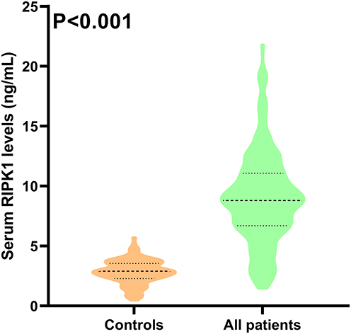 |
Figure 1 Serum receptor-interacting protein kinase-1 levels between patients with aneurysmal subarachnoid hemorrhage and controls. Serum receptor-interacting protein kinase-1 levels were apparently higher in patients than in controls (P<0.001). Abbreviation: RIPK1, receptor-interacting protein kinase-1. |
 |
Figure 2 Relationships between serum receptor-interacting protein kinase-1 levels and disease severity of aneurysmal subarachnoid hemorrhage. Serum receptor-interacting protein kinase-1 levels were firmly correlated with categorial Hunt-Hess scores (P<0.001; (A)), categorial modified Fisher scores (P<0.001; (B)), continuous Hunt-Hess scores (P<0.001; (C)) and continuous modified Fisher scores (P<0.001; (D)) after aneurysmal subarachnoid hemorrhage. Abbreviations: RIPK1, receptor-interacting protein kinase-1; mFisher, modified Fisher. |
Serum RIPK1 Levels and Post-aSAH mRS Scores
As shown in Figure 3, the mRS scores at post-stroke 3 months in aSAH patients were significantly positively correlated with serum RIPK1 levels (P<0.001). Also, the levels were apparently related to mFisher scores, Hunt-Hess scores, blood leucocyte count, blood C-reactive protein levels, blood glucose levels, intraventricular hemorrhage, acute hydrocephalus, and external ventricular drainage (all P<0.05; Table 4). Analogous findings were demonstrated by aidance of univariate linear regression analysis (all P<0.05; Supplemental Table 2). As delineated in Table 5, Hunt-Hess scores, mFisher scores and serum RIPK1 levels were independently related to mRS scores at 3 months post-aSAH (all P<0.05).
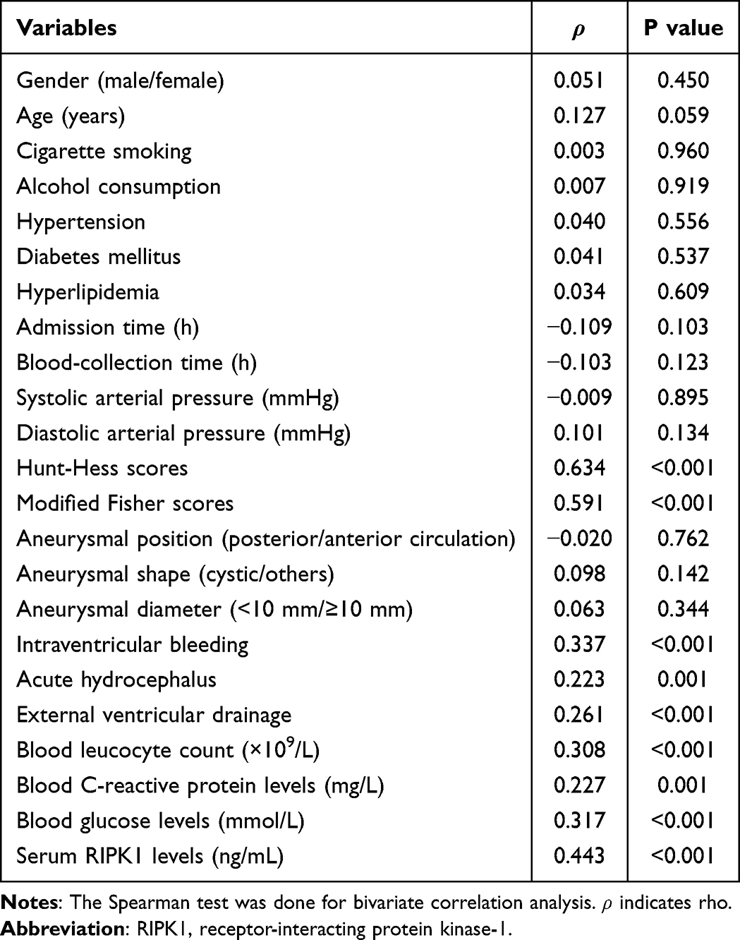 |
Table 4 Factors in Correlation with Modified Rankin Scale Scores Three Months After Aneurysmal Subarachnoid Hemorrhage |
 |
Table 5 Factors Correlated with Modified Rankin Scale Scores Three Months After Aneurysmal Subarachnoid Hemorrhage by Adopting Multivariate Linear Regression Analysis |
 |
Figure 3 Relation of modified Rankin Scale scores to serum receptor-interacting protein kinase-1 levels post- aneurysmal subarachnoid hemorrhage. Serum receptor-interacting protein kinase-1 levels were substantially positively related to modified Rankin Scale scores subsequent to aneurysmal subarachnoid hemorrhage (P<0.001). Abbreviations: RIPK1, receptor-interacting protein kinase-1; mRS, modified Rankin Scale. |
Serum RIPK1 Levels and DCI Post-aSAH
DCI occurred in 54 patients. As show in Figure 4, serum RIPK1 levels were significantly higher in patients with DCI than in those without DCI (P < 0.001). Alternatively, the risk of DCI was linearly correlated with serum RIPK1 levels (P for nonlinear > 0.05; Figure 5). Additionally, compared with patients without DCI, those with DCI exhibited higher Hunt-Hess scores, mFisher scores, blood leucocyte counts, blood C-reactive protein levels and blood glucose levels, as well as displayed higher rates of intraventricular hemorrhage, acute hydrocephalus and external ventricular drainage (all P < 0.05; Table 6). Consistently, those associations were verified in help of univariate logistic regression analysis (all P<0.05; Supplemental Table 3). Inclusion of the above variables into the binary logistic regression model gave rise to the results that Hunt-Hess scores, mFisher scores and serum RIPK1 levels were independent predictors of DCI following aSAH (all P < 0.05; Table 7). The model had satisfactory goodness of fit (P=0.328 via the Hosmer-Lemeshow test; brier score=0.241).
 |
Table 6 Factors Associated with Delayed Cerebral Ischemia Post-Aneurysmal Subarachnoid Hemorrhage |
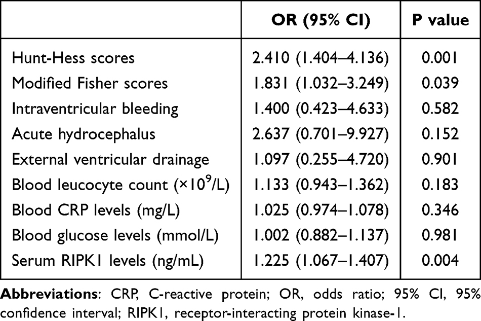 |
Table 7 Factors Related to Delayed Cerebral Ischemia Post-Aneurysmal Subarachnoid Hemorrhage by Multivariate Binary Logistic Regression Analysis |
 |
Figure 4 Serum receptor-interacting protein kinase-1 levels and delayed cerebral ischemia following aneurysmal subarachnoid hemorrhage. Serum receptor-interacting protein kinase-1 levels were markedly higher in patients with delayed cerebral ischemia than in those without in this cohort of patients diseased of aneurysmal subarachnoid hemorrhage (P<0.001). Abbreviations: RIPK1, receptor-interacting protein kinase-1; DCI, delayed cerebral ischemia. |
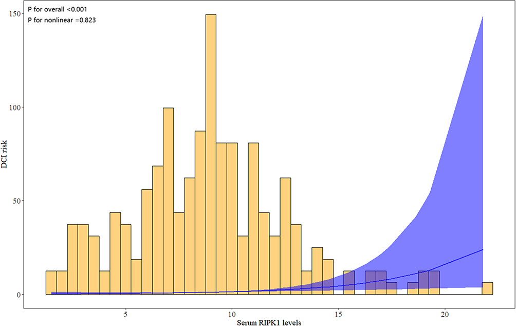 |
Figure 5 Restricted cubic spline depicting linear correlation of serum receptor-interacting protein kinase-1 levels with likelihood of development of delayed cerebral ischemia after aneurysmal subarachnoid hemorrhage. There was a linear correlation between serum receptor-interacting protein kinase-1 levels and the probability of delayed cerebral ischemia following aneurysmal subarachnoid hemorrhage (P for nonlinear >0.05). Abbreviations: RIPK1, receptor-interacting protein kinase-1; DCI, delayed cerebral ischemia. |
By applying ROC curve analysis, serum RIPK1 levels effectively predicted DCI in aSAH patients (Figure 6A). As shown in Figure 6B, area under ROC curve (AUC) for predicting DCI using serum RIPK1 levels was 0.745 (95% CI, 0.682–0.801), and its predictive ability was comparable to that of Hunt-Hess scores (AUC, 0.804; 95% CI, 0.746–0.854; P = 0.156) and mFisher scores (AUC, 0.795; 95% CI, 0.737–0.846; P = 0.181). To predict DCI occurrence, Hunt-Hess scores, mFisher scores and serum RIPK1 levels were integrated to construct a predictive model. In Figure 6B, the combined model had AUC at 0.841 (95% CI, 0.787–0.886) for DCI prediction and also displayed significantly higher predictive ability, as compared to the Hunt-Hess scores (P = 0.038), mFisher scores (P = 0.009), and serum RIPK1 levels (P = 0.002) alone. The model was visually presented via a nomogram (Figure 7), and had stable performance (Figure 8) and clinical efficacy (Figure 9).
 |
Figure 6 Predictive ability of serum receptor-interacting protein kinase-1 levels for delayed cerebral ischemia in patients diagnosed of aneurysmal subarachnoid hemorrhage. In the paradigm of receiver operating characteristic curve, serum receptor-interacting protein kinase-1 levels were in possession of effective discrimination ability for risk of delayed cerebral ischemia following aneurysmal subarachnoid hemorrhage (A). The model was made up of the Hunt-Hess scores, mFisher scores and serum receptor-interacting protein kinase-1 levels. The preceding three metrics possessed similar predictive capability in the scenario of receiver operating characteristic curve (all P>0.05; (B) and combined model were in possession of significantly highest predictive effect for delayed cerebral ischemia (all *P<0.05; (B)). Arrow indicates cutoff value. Abbreviations: AUC, area under curve; 95% CI, 95% confidence interval; RIPK1, receptor-interacting protein kinase-1; mFisher, modified Fisher; ns, non-significant. |
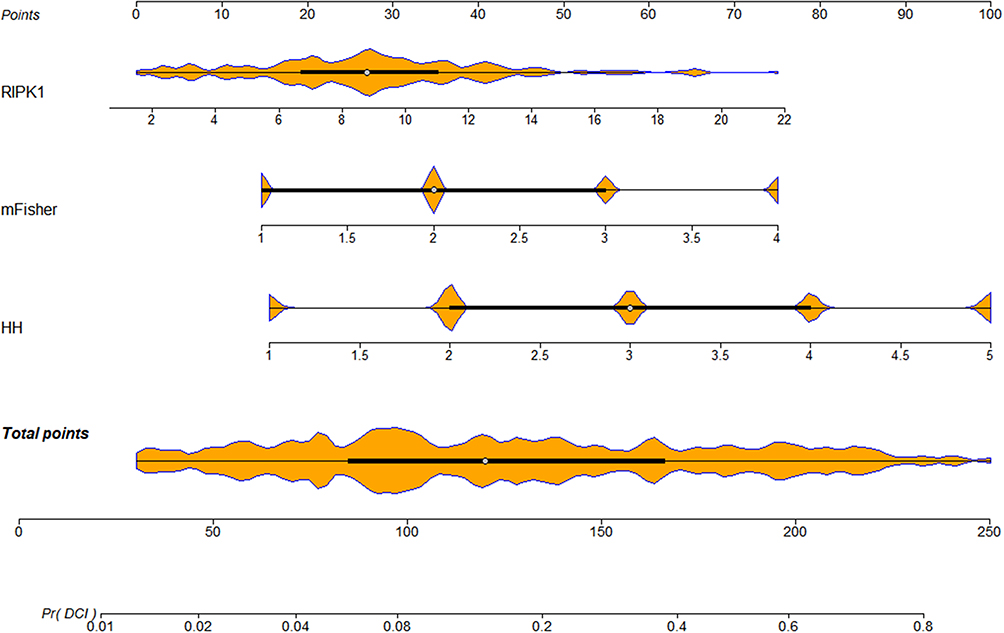 |
Figure 7 Nomogram outlining combination model of delayed cerebral ischemia following aneurysmal subarachnoid hemorrhage. The nomogram providing a visual description for predicting delayed cerebral ischemia using combined model. Abbreviations: RIPK1, receptor-interacting protein kinase-1; DCI, delayed cerebral ischemia; mFisher, modified Fisher; HH, Hunt-Hess. |
 |
Figure 8 Calibration curve assessing the dependability of the model for predicting delayed cerebral ischemia following aneurysmal subarachnoid hemorrhage. The model, in which Hunt-Hess scores, modified Fisher scores and serum receptor-interacting protein kinase-1 levels were integrated, was comparatively stable for prediction of delayed cerebral ischemia after aneurysmal subarachnoid hemorrhage. Abbreviation: DCI, delayed cerebral ischemia. |
 |
Figure 9 Decision curve displaying clinical fit of the model for predicting delayed cerebral ischemia following aneurysmal subarachnoid hemorrhage. The model was rather effective in predicting delayed cerebral ischemia post-aneurysmal subarachnoid hemorrhage. Abbreviations: RIPK1, receptor-interacting protein kinase-1; mFisher, modified Fisher. |
Serum RIPK1 Levels and 3-Month Poor Prognosis Post-aSAH
As depicted in Figure 10, serum RIPK1 levels were markedly lowest in patients with mRS score 0, had a tendency of gradual enhancement from patients with the score 1 to those with the score 6, and were notably highest in those with the score 6 (P<0.001). A total of 63 patients suffered from poor prognosis at 3 months post-stroke. As portrayed in Figure 11, serum RIPK1 levels were significantly higher in patients with poor prognosis compared to those with good prognosis (P < 0.001). Moreover, likelihood of poor prognosis was linearly correlated with serum RIPK1 levels (P for nonlinear >0.05; Figure 12). As exhibited in Table 8, compared with patients with good prognosis, those with poor prognosis had significantly higher percentages of acute hydrocephalus, intraventricular hemorrhage, and extracerebral drainage, as well as held higher Hunt-Hess scores, mFisher scores, serum RIPK1 levels, blood C-reactive protein levels, blood leucocyte counts and blood glucose levels (all P<0.05). Also, these variables were verified to be substantially different by employing univariate logistic regression analysis (all P<0.05; Supplemental Table 4). When all the aforementioned variables were entered into a binary logistic regression model, Hunt-Hess scores, mFisher scores and serum RIPK1 levels were independently associated with poor prognosis (all P<0.05; Table 9). The model harbored acceptable goodness of fit (P=0.452 via the Hosmer-Lemeshow test; brier score=0.218).
 |
Table 8 Factors in Association with 3-month Poor Prognosis Following Aneurysmal Subarachnoid Hemorrhage |
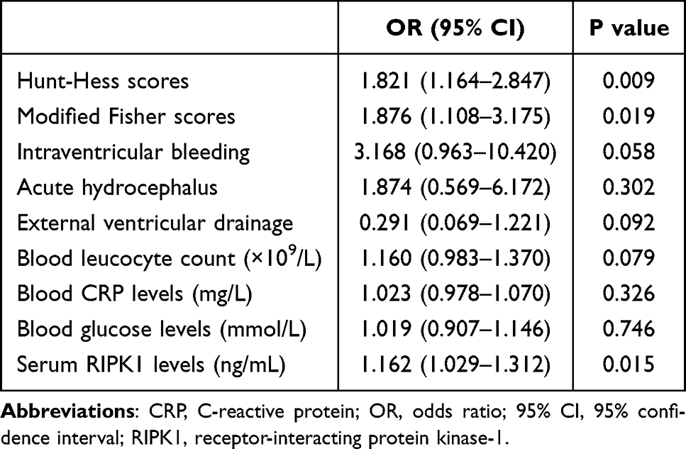 |
Table 9 Factors in Connection with Three-month Poor Prognosis Following Aneurysmal Subarachnoid Hemorrhage via Multivariate Binary Logistic Regression Analysis |
 |
Figure 10 Serum receptor-interacting protein kinase-1 levels and modified Rankin Scale scores at 3 months after aneurysmal subarachnoid hemorrhage. Serum receptor-interacting protein kinase-1 levels were substantially elevated in order of modified Rankin Scale scores from 0 to 6 at 3 months after aneurysmal subarachnoid hemorrhage (P<0.001). Abbreviations: RIPK1, receptor-interacting protein kinase-1; mRS, modified Rankin Scale. |
 |
Figure 11 Serum receptor-interacting protein kinase-1 levels and 3-month poor prognosis following aneurysmal subarachnoid hemorrhage. Serum receptor-interacting protein kinase-1 levels were markedly higher in patients with poor prognosis than in those without the event in this cohort of patients diseased of aneurysmal subarachnoid hemorrhage (P<0.001). Abbreviation: RIPK1, receptor-interacting protein kinase-1. |
 |
Figure 12 Restricted cubic spline depicting linear correlation of serum receptor-interacting protein kinase-1 levels with likelihood of 3-month poor prognosis after aneurysmal subarachnoid hemorrhage. There was a linear correlation between serum receptor-interacting protein kinase-1 levels and the probability of 3-month poor prognosis following aneurysmal subarachnoid hemorrhage (P for nonlinear >0.05). Abbreviation: RIPK1, receptor-interacting protein kinase-1. |
As delineated in Figure 13A, the AUC of serum RIPK1 levels for predicting poor prognosis at 3 months post-stroke was 0.739 (95% CI, 0.677–0.796). And, Figure 13B shows that its predictive ability resembled those of the Hunt-Hess scores (AUC, 0.815; 95% CI, 0.758–0.863; P = 0.066) and mFisher scores (AUC, 0.800; 95% CI, 0.741–0.850; P = 0.144). To distinguish the risk of poor prognosis, the three independent predictors of poor prognosis: Hunt-Hess scores, mFisher scores, and serum RIPK1 levels, were integrated to build a predictive model. In Figure 13B, the combined model had AUC at 0.847 (95% CI, 0.794–0.892), and displayed significantly higher predictive ability for poor prognosis compared with the Hunt-Hess scores (P = 0.042), mFisher scores (P = 0.016), and serum RIPK1 levels (P <0.001). Additionally, the combined model was visualized by the nomogram for predicting poor prognosis (Figure 14). The model was at a stable state (Figure 15) and had good clinical applicability (Figure 16).
 |
Figure 13 Predictive ability of serum receptor-interacting protein kinase-1 levels for 3-month poor prognosis in patients diagnosed of aneurysmal subarachnoid hemorrhage. In the paradigm of receiver operating characteristic curve, serum receptor-interacting protein kinase-1 levels were in possession of effective discrimination ability for risk of 3-month poor prognosis following aneurysmal subarachnoid hemorrhage (A). The model was made up of the Hunt-Hess scores, mFisher scores and serum receptor-interacting protein kinase-1 levels. The preceding three metrics possessed similar predictive capability in the scenario of receiver operating characteristic curve (all P>0.05; (B)) and combined model were in possession of significantly highest predictive effect for 3-month poor prognosis (all *P<0.05; (B)). Arrow indicates cutoff value. Abbreviations: AUC, area under curve; 95% CI, 95% confidence interval; RIPK1, receptor-interacting protein kinase-1; mFisher, modified Fisher; ns, non-significant. |
 |
Figure 14 Nomogram outlining combination model of 3-month poor prognosis following aneurysmal subarachnoid hemorrhage. The nomogram providing a visual description for predicting 3-month poor prognosis using combined model. Abbreviations: RIPK1, receptor-interacting protein kinase-1; mFisher, modified Fisher; HH, Hunt-Hess. |
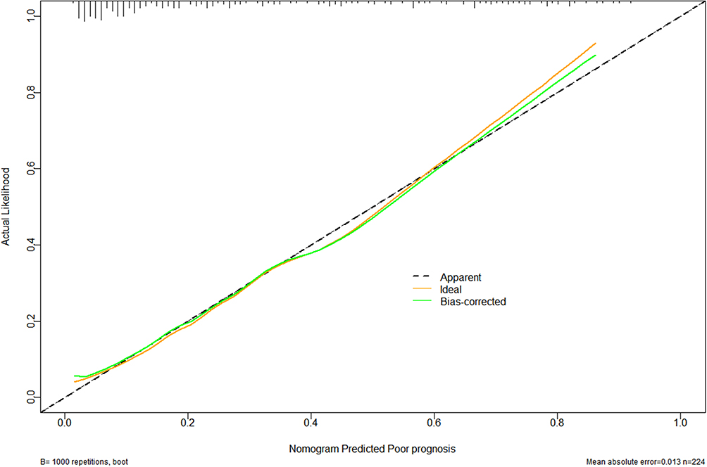 |
Figure 15 Calibration curve assessing the dependability of the model for predicting 3-month poor prognosis following aneurysmal subarachnoid hemorrhage. The model, in which Hunt-Hess scores, modified Fisher scores and serum receptor-interacting protein kinase-1 levels were integrated, was comparatively stable for prediction of 3-month poor prognosis after aneurysmal subarachnoid hemorrhage. |
 |
Figure 16 Decision curve displaying clinical fit of the model for predicting 3-month poor prognosis following aneurysmal subarachnoid hemorrhage. The model was rather effective in predicting 3-month poor prognosis post-aneurysmal subarachnoid hemorrhage. Abbreviations: RIPK1, receptor-interacting protein kinase-1; mFisher, modified Fisher. |
Discussion
To the best of our knowledge, it is unclear regarding blood RIPK1 levels after aSAH. The main findings of this study are: (1) admission serum RIPK1 levels after aSAH were significantly increased; (2) serum RIPK1 levels were independently correlated with two conventional severity indicators, that is Hunt-Hess scores and mFisher scores; (3) serum RIPK1 had strong predictive ability for occurrence of DCI and poor prognosis following aSAH; (4) the combined models containing serum RIPK1 levels possessed satisfactory clinical effectiveness in predicting DCI and poor prognosis after aSAH. In summary, serum RIPK1 may be valuable in assessing disease severity and predicting clinical outcomes in the entity of aSAH.
aSAH, a severe neurosurgical urgency, is frequently complicated with DCI.25,26 Roughly 30% of patients with aSAH exhibit DCI, and similarly, 24.1% (54/224) among this cohort of patients were inflicted by DCI.27 In clinical practice, the Hunt-Hess and mFisher grading systems, the two conventional severity indicators, are preferred to predict DCI and neurological outcomes in aSAH patients.9,10 In our study, the two indicators were demonstrated to be independently associated with DCI and poor prognosis as well, alluding to the notion that Hunt-Hess and mFisher should be worthy of clinical application in aSAH.
Apoptosis and programmed necrosis are the two forms of cellular death.28 RIPK1, a key regulator of cellular decisions between pro-survival necrosis factor-kappa B signaling and death in human diseases, is highly expressed by neurons and glial cells in response to a spectrum of inflammatory and pro-apoptotic stimuli.20,29 Under specific death signal induction, RIPK1 kinase activity is activated, and subsequently leads to programmed necrosis (necroptosis), which is marked by the cell membrane ruptures, resultant release of a large amount of cellular contents, activation of an explosive inflammatory response and further exacerbation of tissue damage.30,31 Accumulating experimental data of cerebral ischemia, traumatic brain injury and SAH showed that RIPK1 expression was significantly elevated in brain tissue surrounding the lesion, RIPK1 gene-deficiency or RIPK1-specific inhibitor resulted in reduced brain injury and improved neurological outcomes.24,32–34 Taken together, RIPK1 may be a detrimental factor and can be recognized as a therapeutic target for acute brain injury.
Serum RIPK1 levels were significantly higher in amyotrophic lateral sclerosis patients than in healthy controls and were positively correlated with the severity of bulbar symptoms.35 Additionally, among 159 patients with severe traumatic brain injury undergoing craniectomy for decompression due to brain herniation, elevated serum RIPK1 levels were closely associated with injury severity and poor prognosis, and demonstrated high discriminatory efficiency for 180-day mortality, overall survival, and poor prognosis.36 In our clinical study, serum RIPK1 levels were elevated in the early stage of aSAH and showed a significantly positive correlation with Hunt-Hess scores and mFisher scores. In a multivariable logistic regression model, serum RIPK1 levels, Hunt-Hess scores, and mFisher scores were independently predictive factors of DCI and poor prognosis. Moreover, serum RIPK1 levels effectively distinguished the risk of DCI and poor prognosis. Additionally, the predictive abilities of serum RIPK1 levels were comparable to those of Hunt-Hess scores and mFisher scores. Here, the combined model was configured by containing the three independent predictors of poor prognosis and DCI. The combined model yielded higher predictive capability than any one of the three independent predictors. In the current study, both severity correlation and outcome association were verified by applying multivariate analyses; and the nomogram, altogether with calibration curve, ROC curve and decision curve, were employed. All these statistical methods, in conjunction with multi-center analysis, have been strongly supportive of the notion that serum RIPK1 may be an appealing biomarker of severity appraisal and outcome anticipation in human aSAH.
This study has several limitations to be noted. (1) serum RIPK1 levels were measured only in the early stage of aSAH onset, and continuous measurements could better define the evolutional trajectory and clinical value of serum RIPK1. (2) considering that RIPK1 is one of the markers of inflammatory response, further investigating its specific mechanisms in secondary brain injury following aSAH could help to identify new therapeutic avenues. (3) as for how to handle missing data, it should be comprehensively considered. The prospective cohort study basically features fewer missing data. Currently, five patients were removed from this study, because they had incomplete clinical data. Altogether, patients with missing data only accounted for 2.23% (5/224), subsequently causing a small impact on entire data. However, to keep integrity of the whole data is an effortfully-pursued goal in order to make study conclusions more scientific and reliable. (4) According to the exclusion criteria, severe infection within past a month had been excluded from this group of patients. Nevertheless, mild-moderate infections may be existent in this cohort, possibly to some extent affecting study results. So, comorbid inflammation as a potential confounder should be taken into consideration in future study. (5) DCI is different from cerebral vasospasm, because cerebral vasospasm is only a cause of DCI, and other causes include microthrombosis, endothelial injury, inflammatory activation and so forth.37,38 Moreover, mFisher is the most related to DCI, as compared to World Federation of Neurological Societies Scale, Glasgow coma scale and Hunt-Hess scoring systems; because mFisher is a radiological scoring system for reflecting hemorrhagic severity.37,38 Naturally, mFisher has been used in this study. Hopefully, both WFNS and GCS are proposed to be added as the comparative metrics in future. (6) although our multicenter study included an adequate sample size based on statistical principle, conducting large-scale cohort studies in the future to validate the conclusions and enhance the generalizability of our results holds significant importance.
Conclusion
Elevated serum RIPK1 levels after aSAH, in tight correlation with Hunt-Hess scores and modified Fisher scores, can independently predict DCI and 3-month poor prognosis. The combined models incorporating serum RIPK1 perform well. Therefore, serum RIPK1 may be a potential prognostic predictor of aSAH.
Data Sharing Statement
The data that support the findings of this study are available on request from the corresponding author. The data are not publicly available due to privacy or ethical restrictions.
Acknowledgments
We would express gratitude to everyone who helped in this study.
Funding
The work has been financially supported by the Medical and Health Research Project of Zhejiang province (2025KY1969, 2025KY1970), the City-level Public Welfare Technology Application Research Project of Lishui (2023SJZC078, 2023SJZC101), and the Public Welfare Technology Research Program of Lishui (2024GYX69).
Disclosure
All authors have no disclosures to report for this work.
References
1. Neifert SN, Chapman EK, Martini ML, et al. Aneurysmal subarachnoid hemorrhage: the last decade. Transl Stroke Res. 2021;12(3):428–446. doi:10.1007/s12975-020-00867-0
2. Grimm JW. Aneurysmal subarachnoid hemorrhage: a potentially lethal neurological disease. J Emerg Nurs. 2015;41(4):281–284. doi:10.1016/j.jen.2014.12.018
3. de Rooij NK, Linn FH, van der Plas JA, Algra A, Rinkel GJ. Incidence of subarachnoid haemorrhage: a systematic review with emphasis on region, age, gender and time trends. J Neurol Neurosurg Psychiatry. 2007;78(12):1365–1372. doi:10.1136/jnnp.2007.117655
4. Zhang C, Tang W, Cheng L, et al. Early and delayed blood-brain barrier permeability predicts delayed cerebral ischemia and outcomes following aneurysmal subarachnoid hemorrhage. Eur Radiol. 2024;34(8):5287–5296. doi:10.1007/s00330-023-10571-w
5. Abdulazim A, Heilig M, Rinkel G, Etminan N. Diagnosis of delayed cerebral ischemia in patients with aneurysmal subarachnoid hemorrhage and triggers for intervention. Neurocrit Care. 2023;39(2):311–319. doi:10.1007/s12028-023-01812-3
6. Suzuki H, Kanamaru H, Kawakita F, Asada R, Fujimoto M, Shiba M. Cerebrovascular pathophysiology of delayed cerebral ischemia after aneurysmal subarachnoid hemorrhage. Histol Histopathol. 2021;36(2):143–158. doi:10.14670/HH-18-253
7. Balança B, Bouchier B, Ritzenthaler T. The management of delayed cerebral ischemia after aneurysmal subarachnoid hemorrhage. Rev Neurol. 2022;178(1–2):64–73. doi:10.1016/j.neurol.2021.11.006
8. Mehra A, Gomez F, Bischof H, Diedrich D, Laudanski K. Cortical spreading depolarization and delayed cerebral ischemia; rethinking secondary neurological injury in subarachnoid hemorrhage. Int J Mol Sci. 2023;24(12):9883. doi:10.3390/ijms24129883
9. Wang HR, Ma J, Guo YZ, et al. Combination of albumin/fibrinogen ratio and admission Hunt-Hess scale score as an independent predictor of clinical outcome in aneurysmal subarachnoid hemorrhage. World Neurosurg. 2024;181:e322–e329. doi:10.1016/j.wneu.2023.10.047
10. Zhang Q, Zhang G, Wang L, et al. Clinical value and prognosis of C reactive protein to lymphocyte ratio in severe aneurysmal subarachnoid hemorrhage. Front Neurol. 2022;13:868764. doi:10.3389/fneur.2022.868764
11. Yao H, Lv C, Luo F, He C. Plasma cellular prion protein concentrations correlate with severity and prognosis of aneurysmal subarachnoid hemorrhage. Clin Chim Acta. 2021;523:114–119. doi:10.1016/j.cca.2021.09.010
12. Lin Q, Ye L, Dai J, et al. A prospective cohort study on decreased serum apelin-13 levels after human aneurysmal subarachnoid hemorrhage: associations with severity and prognosis. Neurosurg Rev. 2023;46(1):235. doi:10.1007/s10143-023-02142-w
13. Wu X, Ji D, Wang Z, et al. Elevated serum NOX2 levels contribute to delayed cerebral ischemia and a poor prognosis after aneurysmal subarachnoid hemorrhage: a prospective cohort study. Neuropsychiatr Dis Treat. 2023;19:1027–1042. doi:10.2147/NDT.S407907
14. Newton K. RIPK1 and RIPK3: critical regulators of inflammation and cell death. Trends Cell Biol. 2015;25(6):347–353. doi:10.1016/j.tcb.2015.01.001
15. DeRoo E, Zhou T, Liu B. The role of RIPK1 and RIPK3 in cardiovascular disease. Int J Mol Sci. 2020;21(21):8174. doi:10.3390/ijms21218174
16. Malireddi RKS, Gurung P, Kesavardhana S, et al. Innate immune priming in the absence of TAK1 drives RIPK1 kinase activity-independent pyroptosis, apoptosis, necroptosis, and inflammatory disease. J Exp Med. 2020;217(3):
17. Speir M, Lawlor KE. RIP-roaring inflammation: RIPK1 and RIPK3 driven NLRP3 inflammasome activation and autoinflammatory disease. Semin Cell Dev Biol. 2021;109:114–124. doi:10.1016/j.semcdb.2020.07.011
18. Ofengeim D, Mazzitelli S, Ito Y, et al. RIPK1 mediates a disease-associated microglial response in Alzheimer’s disease. Proc Natl Acad Sci U S A. 2017;114(41):E8788–E8797. doi:10.1073/pnas.1714175114
19. Yuan J, Amin P, Ofengeim D. Necroptosis and RIPK1-mediated neuroinflammation in CNS diseases. Nat Rev Neurosci. 2019;20(1):19–33. doi:10.1038/s41583-018-0093-1
20. Liu J, Zhu Z, Wang L, et al. Functional suppression of Ripk1 blocks the NF-κB signaling pathway and induces neuron autophagy after traumatic brain injury. Mol Cell Biochem. 2020;472(1–2):105–114. doi:10.1007/s11010-020-03789-5
21. Song X, Lan Y, Lv S, et al. Downregulation of Ripk1 and Nsf mediated by CRISPR-CasRx ameliorates stroke volume and neurological deficits after ischemia stroke in mice. Front Aging Neurosci. 2024;16:1401038. doi:10.3389/fnagi.2024.1401038
22. Lule S, Wu L, McAllister LM, et al. Genetic inhibition of receptor interacting protein kinase-1 reduces cell death and improves functional outcome after intracerebral hemorrhage in mice. Stroke. 2017;48(9):2549–2556. doi:10.1161/STROKEAHA.117.017702
23. Yan F, Qiao Y, Pan S, Kang A, Chen H, Bai Y. RIPK1: a promising target for intervention neuroinflammation. J Neuroimmune Pharmacol. 2025;20(1):59. doi:10.1007/s11481-025-10208-3
24. Wu Y, Xu Y, Sun J, Dai K, Wang Z, Zhang J. Inhibiting RIPK1-driven neuroinflammation and neuronal apoptosis mitigates brain injury following experimental subarachnoid hemorrhage. Exp Neurol. 2024;374:114705. doi:10.1016/j.expneurol.2024.114705
25. Daou BJ, Koduri S, Thompson BG, Chaudhary N, Pandey AS. Clinical and experimental aspects of aneurysmal subarachnoid hemorrhage. CNS Neurosci Ther. 2019;25(10):1096–1112. doi:10.1111/cns.13222
26. Rouanet C, Silva GS. Aneurysmal subarachnoid hemorrhage: current concepts and updates. Arq Neuropsiquiatr. 2019;77(11):806–814. doi:10.1590/0004-282X20190112
27. Ikram A, Javaid MA, Ortega-Gutierrez S, et al. Delayed cerebral ischemia after subarachnoid hemorrhage. J Stroke Cerebrovasc Dis. 2021;30(11):106064. doi:10.1016/j.jstrokecerebrovasdis.2021.106064
28. Bertheloot D, Latz E, Franklin BS. Necroptosis, pyroptosis and apoptosis: an intricate game of cell death. Cell Mol Immunol. 2021;18(5):1106–1121. doi:10.1038/s41423-020-00630-3
29. Liu Y, Meng X, Tang C, Zheng L, Tao K, Guo W. Aerobic exercise modulates RIPK1-mediated MAP3K5/JNK and NF-κB pathways to suppress microglia activation and neuroinflammation in the hippocampus of D-gal-induced accelerated aging mice. Physiol Behav. 2024;286:114676. doi:10.1016/j.physbeh.2024.114676
30. Shi FL, Yuan LS, Wong TS, et al. Dimethyl fumarate inhibits necroptosis and alleviates systemic inflammatory response syndrome by blocking the RIPK1-RIPK3-MLKL axis. Pharmacol Res. 2023;189:106697. doi:10.1016/j.phrs.2023.106697
31. Wang J, Zhong W, Su H, et al. Histone methyltransferase dot1L contributes to RIPK1 kinase-dependent apoptosis in cerebral ischemia/reperfusion. J Am Heart Assoc. 2021;10(23):e022791. doi:10.1161/JAHA.121.022791
32. Zhang Y, Li M, Li X, et al. Catalytically inactive RIP1 and RIP3 deficiency protect against acute ischemic stroke by inhibiting necroptosis and neuroinflammation. Cell Death Dis. 2020;11(7):565. doi:10.1038/s41419-020-02770-w
33. Mitroshina EV, Loginova MM, Yarkov RS, et al. Inhibition of neuronal necroptosis mediated by RIPK1 provides neuroprotective effects on hypoxia and ischemia in vitro and in vivo. Int J Mol Sci. 2022;23(2):735. doi:10.3390/ijms23020735
34. Wehn AC, Khalin I, Duering M, et al. RIPK1 or RIPK3 deletion prevents progressive neuronal cell death and improves memory function after traumatic brain injury. Acta Neuropathol Commun. 2021;9(1):138. doi:10.1186/s40478-021-01236-0
35. Wei J, Li M, Ye Z, et al. Elevated peripheral levels of receptor-interacting protein kinase 1 (RIPK1) and IL-8 as biomarkers of human amyotrophic lateral sclerosis. Signal Transduct Target Ther. 2023;8(1):451. doi:10.1038/s41392-023-01713-z
36. Cai L, Dou X, Dong W, et al. Serum RIPK1, acute lung injury, and outcomes in severe traumatic brain injury: a multicenter prospective study. Ther Clin Risk Manag. 2025;21:385–405. doi:10.2147/TCRM.S502775
37. Suzuki H, Kawakita F, Asada R. Neuroelectric mechanisms of delayed cerebral ischemia after aneurysmal subarachnoid hemorrhage. Int J Mol Sci. 2022;23(6):3102. doi:10.3390/ijms23063102
38. Marques IP, Albuquerque CRC, Souza NVO, Andrade JBC, Silva GS, Kurtz P. Delayed cerebral ischemia after aneurysmal subarachnoid hemorrhage: a narrative review. Arq Neuropsiquiatr. 2025;83(6):1–14. doi:10.1055/s-0045-1809885
 © 2025 The Author(s). This work is published and licensed by Dove Medical Press Limited. The
full terms of this license are available at https://www.dovepress.com/terms
and incorporate the Creative Commons Attribution
- Non Commercial (unported, 4.0) License.
By accessing the work you hereby accept the Terms. Non-commercial uses of the work are permitted
without any further permission from Dove Medical Press Limited, provided the work is properly
attributed. For permission for commercial use of this work, please see paragraphs 4.2 and 5 of our Terms.
© 2025 The Author(s). This work is published and licensed by Dove Medical Press Limited. The
full terms of this license are available at https://www.dovepress.com/terms
and incorporate the Creative Commons Attribution
- Non Commercial (unported, 4.0) License.
By accessing the work you hereby accept the Terms. Non-commercial uses of the work are permitted
without any further permission from Dove Medical Press Limited, provided the work is properly
attributed. For permission for commercial use of this work, please see paragraphs 4.2 and 5 of our Terms.
Recommended articles
Elevated Serum NOX2 Levels Contribute to Delayed Cerebral Ischemia and a Poor Prognosis After Aneurysmal Subarachnoid Hemorrhage: A Prospective Cohort Study
Wu X, Ji D, Wang Z, Yu W, Du Q, Hu W, Zheng Y, Dong X, Chen F
Neuropsychiatric Disease and Treatment 2023, 19:1027-1042
Published Date: 29 April 2023