Back to Journals » Diabetes, Metabolic Syndrome and Obesity » Volume 18
Abdominal Massage Decreases Food Intake and Body Weight in High-Fat Diet-Induced Obese Rats Through Upregulating GPR41/GPR43-PYY/GLP-1 Axis
Authors Ma F, Li H, Huang C, Shuai C, An C, Zhang W
Received 19 August 2024
Accepted for publication 1 May 2025
Published 21 May 2025 Volume 2025:18 Pages 1673—1682
DOI https://doi.org/10.2147/DMSO.S492185
Checked for plagiarism Yes
Review by Single anonymous peer review
Peer reviewer comments 2
Editor who approved publication: Dr Rebecca Baqiyyah Conway
Fei Ma,1,* Huanan Li,2,* Caiyin Huang,2 Chenghao Shuai,2 Chengfei An,2 Wei Zhang2
1Tianjin Academy of Traditional Chinese Medicine Affiliated Hospital, Tianjin, 300120, People’s Republic of China; 2Department of Tuina, First Teaching Hospital of Tianjin University of Traditional Chinese Medicine, National Clinical Research Center for Chinese Medicine Acupuncture and Moxibustion, Tianjin, 300000, People’s Republic of China
*These authors contributed equally to this work
Correspondence: Fei Ma, Tianjin Academy of Traditional Chinese Medicine Affiliated Hospital, Hongqiao District, Tianjin, 300120, People’s Republic of China, Tel +86 022 27345050, Email [email protected]
Background: Abdominal massage has been found to exert an important role in helping people in overcoming obesity. However, the mechanism by which abdominal massage induces weight loss remains largely unclear.
Methods: Healthy male Sprague-Dawley (SD) rats were randomly grouped into standard diet control (15% fat content) group and high-fat diet (HFD, 40% fat content) group. After 6 weeks of high-fat feeding, rats in the HFD group were successfully modeled, and then separated into the HFD group and HFD plus abdominal massage group. Rats in the HFD plus abdominal massage group were then subjected to abdominal massage for 12 continuous days.
Results: Compared to the HFD group, abdominal massage could decrease body weight, food intake and abdominal fat index (AFI) of HFD-fed rats. Meanwhile, compared to the HFD group, abdominal massage obviously attenuated mucosal epithelial damage and reduced inflammatory cell infiltration in colon mucosal tissues of HFD-fed rats. Furthermore, compared to the HFD group, abdominal massage significantly increased GPR42 and GPR43 levels in the colon tissues of HFD-fed rats, and upregulated the production of glucagon-like peptide-1 (GLP-1) and peptide YY (PYY) in colon mucosal tissues of HFD-fed rats.
Conclusion: Collectively, abdominal massage could decrease food intake and body weight in HFD-induced obese rats through upregulating GPR41/GPR43-PYY/GLP-1 axis.
Keywords: abdominal nudging, obesity, SCFAs, GLP-1, PYY, food intake, body weight
Introduction
Obesity is a type of metabolic diseases, which has become a major public health issue worldwide.1,2 Obesity is characterized by excessive fat accumulation, overweight, energy imbalance, metabolic disorders and chronic low-grade inflammation.3,4 Obesity can increase the risk of multiple diseases, such as hypertension, hyperlipidemia, hyperglycemia, fatty liver disease, coronary heart diseases, osteoarthritis and several cancers.5 The pathogenesis of obesity is complex, genetic, economic and environmental risk factors are all responsible for the obesity development.6 Approximately, simple obesity denotes an unusual accumulation of body fat that leads to a rise in body weight in the absence of any major illness, encompassing 95% of all cases of obesity.7,8 Overeating is the most common cause of simple obesity. Evidence has shown that excessive fat intake could contribute to an imbalance of energy consumption and intake, and induce excessive body fat accumulation and weight gain, eventually leading to obesity.9,10 Thus, diet control and regulating the energy metabolism are the fundamental principles for the treatment of obesity.
The brain-gut-microbiota axis is a complex bidirectional communication system,11,12 which plays a key role in food intake and energy consumption.13,14 Some potent satiety hormones [eg glucagon-like peptide 1 (GLP-1) and peptide YY (PYY)] involved in feeding can be released from the small intestine to the colon, and these intestinal satiety signals can be transmit to the brain through vagal afferent nerves.15–17 The brain can integrate multiple signals such as gastrointestinal signals, hormonal signals and signals from other brain regions, and then send output signals via vagal afferent nerves to the digestive system for controlling the food intake.18 Activation of PYY and GLP-1 has been found to reduce food intake.19
Furthermore, it has been shown that the gut microbiota could regulate dietary intake through influencing microbial metabolite production such as short-chain fatty acids (SCFAs).20,21 SCFAs (such as acetate, propionate, butyrate) can activate its receptors G-protein-coupled receptor 41 (GPR41) and G-protein-coupled receptor 43 (GPR43) to regulate glucose and lipid metabolism.22,23 Thereafter, the activation of GPR41 and GPR43 could elevate PYY and GLP-1 levels in intestinal L cells to suppress food intake.23
Abdominal massage is an ancient external therapy in traditional Chinese medicine, which has been widely used in clinical treatment.24,25 Abdominal massage has proven beneficial in alleviating abdominal distention, relieving constipation, and reducing gastric residual volume in critically ill patients.26,27 Furthermore, abdominal massage can also effectively enhance the function of digestion and absorption of the gastrointestinal tract and stimulate the brain-gut axis, playing an important role in helping people overcome obesity.28,29 Zhang et al found that abdominal massage could reduce the body weight of high-fat diet (HFD)-induced obese mice.30 However, the mechanism by which abdominal massage could reduce the body weight and food intake remains largely unclear. Thus, in the current research, we aimed to explore whether abdominal massage could prevent obesity development through targeting SCFAs-GPR41/43 or PYY and GLP-1 signalings.
Materials and Methods
Animals
A total of 36 Sprague-Dawley rats (SPF level, male, 6–8 weeks old, weighting 200 ± 20g) were obtained from SiPeiFu (Beijing, laboratory animal production license number: SCXK (Jing) 2019–0010). All animals were fed and watered freely in the Experimental Animal Center of Nankai Hospital, Tianjin, China and maintained under constant environmental conditions (temperature, 25 ± 2°C; 30–40% relative humidity; a 12 h dark/light cycle). All animal studies were approved by the Animal Ethical Committee of Tianjin University of Chinese Medicine and performed according to national guidelines.
Establishment of a Simple Obesity Rat Model
After 7 days of adaptive feeding, all animals were randomized into two groups: standard diet control group (n = 8) and high-fat diet (HFD) group (n = 20). Rats in the standard diet control group were fed with standard feed (15% fat content). Rats in the HFD groups were fed with high-fat feed (including 60% ordinary feed, 15% lard, 15% sucrose, 2% cholesterol, 4% egg yolk powder, 2% maltodextrin, 2% fish meal; 40% fat content).31 Rats were fed with standard diet or HFD for 6 weeks. Compared to the standard diet control group, rats with at least a 20% increase in body weight in the HFD group were selected as experimental fat rats (n = 16) (four rats were excluded because their body weight did not meet the requirements). Then, these experimental fat-fed rats were divided into HFD (n = 8) and HFD plus (+) abdominal massage (n = 8) groups.
Experimental Design
Rats in the standard diet control group were fed with ordinary feed and received no treatment. Rats in the HFD and HFD + abdominal massage groups were fed with high-fat feed. Rats in the HFD group were put into a binding service for 20 minutes once per day for 12 consecutive days, without any abdominal massage treatment. Rats in the HFD + abdominal massage group were received abdominal massage intervention for 20 minutes once per day for 12 consecutive days.
Abdominal massage was performed according to the methods described in two books.32,33 Briefly, the researchers rubbed the abdomen in a clockwise manner for 12 min at a frequency of 20–30 cycles per minute. Next, the thumb of the operator was also put on the abdomen of rats. Next, the thumb of the operator depressed the subxiphoid skin and moved from the subxiphoid to the tail region of rats. In this process, the operator needed to apply a force slowly and steadily, and the massage direction was always from top (subxiphoid skin) to bottom (tail region). The frequency was 15–20 times per min, and the massage time for this part was 8 min. To ensure the consistency of the massage manipulation, all operators received unified training learn the correct massage technique including strength and the frequency of massage.
Samples Collection
After 12 days of massage intervention, all rats were anesthetized with isoflurane. The liver tissue and the fat that is inside the retroperitoneal cavity and around both kidneys were collected and stored in liquid nitrogen. Meanwhile, colon mucosal tissues were collected by scraping from part of the colon and then lysed using the RIPA buffer for further ELISA analysis.
Measurement of Physiological Parameters
As shown in Figure 1A, food intake of each rat was detected every day. The body weight of each rat was weighted at day 0, day 7 and day 12. The body length of each rat was measured at day 12. The Lee’s index was calculated as Lee’s = [body weight (g)×1000]1/3÷[body length (cm)]. The abdominal fat index (AFI) was calculated as AFI (%) = fat weight/body weight (%). The liver-to-body weight ratio (LBW) was calculated as LBW = liver weight/body weight.
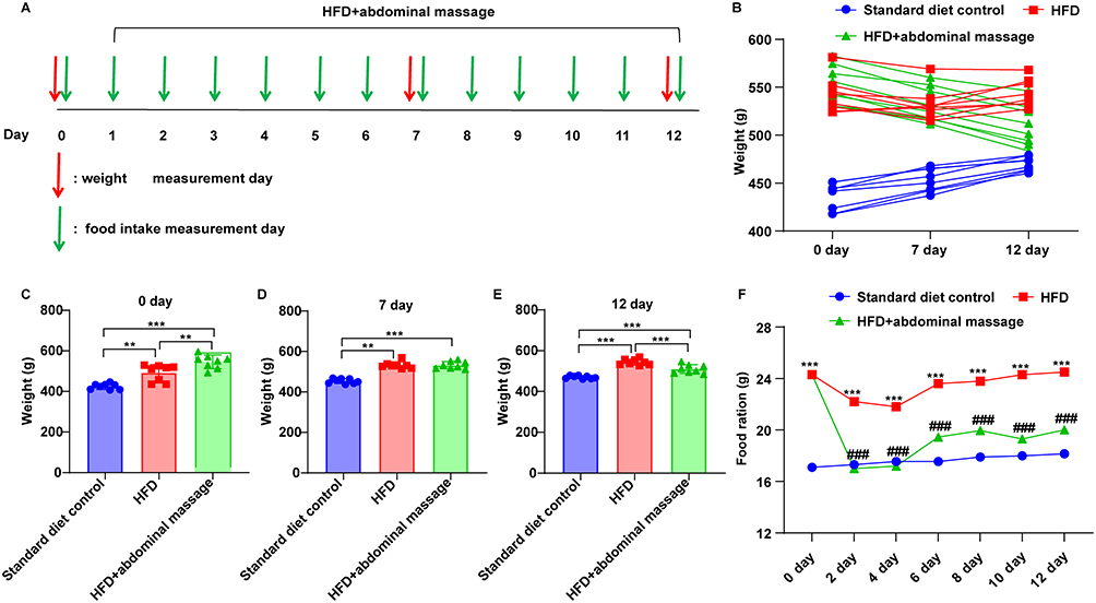 |
Figure 1 The effects of abdominal massage on the body weight and food intake of HFD-fed rats. (A) Schedule of the animal experiment. (B) The body weight of each rat in standard diet control, HFD and HFD + abdominal massage groups (n = 8 per group) on the tested day 0, 7 and 12. (C–E) The average body weight of rats in three groups (n = 8 per group) on the tested day (C) 0, (D) 7 and (E) 12. **P < 0.01, ***P < 0.001 (F) The food ration of rats (n = 8 per group) on the tested day 0, 2, 4, 6, 8, 10 and 12. ***P < 0.001 vs standard diet control group; ###P < 0.001 vs HFD group. |
Quantification of SCFAs in Fat Samples
The fat tissues were added in 300 µL of 50% (v:v) pre-cooled acetonitrile-water solution. Samples were then grinded for 3 min, and extracted by ultrasound instrument in ice-water bath for 10 min. After centrifugation at 12000 rpm for 10 min at 4°C, the supernatant (80 µL) was transferred into an autosampler vial. Next, 40 μL of 200 mm 3-NPH and 40 μL of 120 mm EDC-6% pyridine were then added into this autosampler vial and maintained at 40°C for 30 min. The mixture was allowed to cool on ice for 1 min, the supernatant (160 µL) was collected and then filtered through a 0.22 µm filter and then transferred into a clean autosampler vial. Samples were stored at −80°C. Liquid chromatography-mass spectrometry (LC-MS) analysis was then conducted to determine the contents of acetic acid, propionic acid and butyric acid in fat tissues using the Nexera UHPLC LC-30A Chromatograph/AB Sciex Qtrap 5500 mass spectrometer. Detailed materials and methods are provided in Supplementary material 1.
Immunohistochemical Staining Assay
Paraffin-embedded colon tissues were cut into 4-μm thick slices. Next, the slices were blocked in the normal goal serum for 20 min and then treated with anti-GPR41 (No. PA5-99629, 1:200, ThermoFisher), anti-GPR43 (No. PA5-33718, 1:150, ThermoFisher) antibodies overnight at 4°C, followed by incubation with the HRP-labeled secondary antibody. Photographs were taken with a light microscope after staining with DAB solution, and the results were analyzed using the ImageJ software.
Hematoxylin and Eosin (H&E) Staining Assay
Paraffin-embedded colon sections (4-μm thick) were subjected to routine H&E staining, as described previously.34 Finally, photographs were taken with a light microscope.
Enzyme-Linked Immunosorbent Assay (ELISA)
The rat Peptide YY (PYY) ELISA kit and rat Glucagon-like Peptide-1 (GLP-1) ELISA kit were obtained from Gelatins. The concentration of PYY and GLP-1 in colon mucosal tissues were measured by the commercial kits according to the manufacturers’ protocols. A microplate reader (DNM-9602, PERLONG) was applied for reading the results.
Statistical Analysis
All values are expressed as means ± standard deviation (SD). One-way analysis of variance (ANOVA) followed by Tukey’s test was used to analyze differences among groups. GraphPad Prism 9.5.0 was used for statistical analysis. A value of p < 0.05 was considered statistically significant.
Results
The Effects of Abdominal Massage on the Body Weight and Food Intake of HFD-Fed Rats
During the treatment period, the body weight of rats in the standard diet control group gradually increased, while, there was no significant change in body weight in the HFD group (Figure 1B). However, rats in the HFD + abdominal massage group experienced a gradual reduction in body weight following the massage intervention (Figure 1B). Additionally, prior to the intervention (at day 0), the body weight of rats in the HFD and HFD + abdominal massage groups were significantly higher than that of the standard diet control group (Figure 1C). Significantly, after the intervention (at day 12), the body weight of mice in the HFD + abdominal massage group was reduced compared to the HFD group (Figure 1D and E). These results showed that abdominal massage could reduce the body weight of HFD-fed rats.
Furthermore, compared to the rats fed standard feed in the standard diet control group, the average daily food intake of HFD-fed rats in the HFD group was remarkably elevated (Figure 1F). However, compared to the HFD group, abdominal massage notably reduced the food intake of HFD-fed rats (Figure 1F). To sum up, abdominal massage demonstrated the potential to reduce body weight and decrease food intake in HFD-fed rats.
The Effects of Abdominal Massage on Physiological Indicators of HFD-Fed Rats
Next, we then explored the effects of abdominal massage on body length, Lee’s index, AFI and LBW in HFD-fed rats. As shown in Figure 2A and B, the body length and Lee’s index of rats were not significantly different among the three groups. Additionally, AFI was notably elevated in HFD-fed rats in the HFD group compared to those on a regular diet in the standard diet control group; however, abdominal massage intervention significantly reduced AFI of HFD-fed rats when compared to the HFD group (Figure 2C). Moreover, the LBW of rats did not exhibit a significant difference among the three groups (Figure 2D).
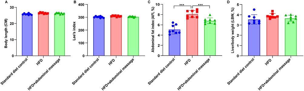 |
Figure 2 The effects of abdominal massage on physiological indicators of HFD-fed rats. (A) Body length, (B) Lee’s index, (C) AFI and (D) LBW of rats were calculated in standard diet control, HFD and HFD + abdominal massage groups (n = 8 per group). ***P < 0.001. |
The Effects of Abdominal Massage on the Production of SCFAs in the Intra-Abdominal Fat of HFD-Fed Rats
Next, the impact of abdominal massage on the production of SCFAs (acetic acid, propionic acid and butyric acid) in the intra-abdominal fat of HFD-fed rats was investigated. As shown in Figure 3A, acetic acid production in the intra-abdominal fat of rats did not show significant difference among standard diet control, HFD and HFD + abdominal massage groups. Additionally, the production of propionic acid was slightly increased in HFD-fed rats in the HFD group compared to the standard diet control rats, whereas abdominal massage led to a slight reduction in propionic acid production in the intra-abdominal fat of HFD-fed rats compared to the HFD group, although the differences were not statistically significant among the three groups (Figure 3B). Furthermore, compared to the standard diet control group, HFD notably declined the production of butyric acid in the intra-abdominal fat of rats (Figure 3C). Meanwhile, abdominal massage further reduced the production of butyric acid compared to the HFD group (Figure 3C).
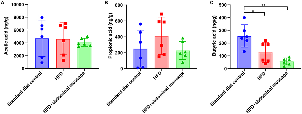 |
Figure 3 The effects of abdominal massage on the production of SCFAs in the intra-abdominal fat of HFD-fed rats. The production of (A) acetic acid, (B) propionic acid and (C) butyric acid in the intra-abdominal fat of rats was quantified in standard diet control, HFD and HFD + abdominal massage groups (n = 6 per group). *P < 0.05, **P < 0.01. |
The Effects of Abdominal Massage on the Colon Tissues of HFD-Fed Rats
Next, H&E staining was performed to examine the pathological changes of colon tissues of HFD-fed rats. As shown in Figure 4A, the colon of rats in the standard diet control group showed intact mucosal epithelium, ordered arrangement of glands within lamina propria and normal number of goblet cells. However, mucosal epithelial damage, branched and irregular glands, disarray of the crypts and crypt atrophy, a reduced number of goblet cells, and inflammatory cell infiltration were observed in colon tissues of HFD-fed rats in the HFD group, whereas abdominal massage obviously improved these changes (Figure 4A).
 |
Figure 4 The effects of abdominal massage on the colon tissues of HFD-fed rats. (A) H&E staining analysis of pathological changes of colon tissues of rats in standard diet control, HFD and HFD + abdominal massage groups (n = 6 per group). (B–D) IHC staining analysis of GPR41 and GPR43 expressions in the colon tissues of rats in three groups (n = 6 per group). (E and F) PYY and GLP-1 levels in intestinal mucosal tissues of rats in three groups were evaluated by ELISA (n = 6 per group). ***P < 0.001. |
Furthermore, the results of IHC staining assay showed that compared to the standard diet control group, GPR41 and GPR43 levels were significantly reduced in the HFD group (Figure 4B–D). Conversely, abdominal massage notably elevated GPR41 and GPR43 levels in the colon tissues of HFD-fed rats, compared to the HFD group (Figure 4B–D). Additionally, the concentrations of colonic PYY and GLP-1 in HFD-fed rats in the HFD group were notably reduced, compared to the standard diet control group; however, abdominal massage intervention notably elevated PYY and GLP-1 levels in colon mucosal tissues of HFD-fed rats (Figure 4E and F).
Discussion
With the continuous development of society, people’s lifestyles and dietary patterns have undergone tremendous changes. This has led to a remarkable increase in the number of obese people.35 An analysis of 195 countries reported that the prevalence of obesity has doubled since 1980 over 70 countries; in 2015, over 600 million adults were obese.36 Modern medicine believes that the main causes of obesity are imbalanced energy metabolism and lack of physical activity.37,38 Meanwhile, traditional Chinese medicine believes that intemperate diet is the main reason of obesity.39 A theory stems from a medical book of Huangdi Neijing (The Yellow Emperor’s Inner Canon) indicated that eating fatty and sweet foods and excessive consumption of alcohol can lead to weakness of the spleen and stomach and dysfunction of spleen transportation.39 Because of the dysfunction of spleen and stomach, excess energy was then converted into fat that accumulates in the body, eventually leading to obesity.
In the current research, we focused on the acupoints located on the abdomen, and massage therapy was performed on the abdomen of HFD-fed rats. The abdominal massage method used in this research not only conforms to the understanding of traditional Chinese medicine in the theory of meridians and acupoints but also echoes modern anatomy thoughts. This massage method included two parts: circular kneading (“Mo” method) and pushing (“Tui” method).40 Circular kneading on the middle part of gastric cavity (Zhongwan) acupoint can exert “strengthen the spleen and stomach” and “dispel dampness and resolve turbidity” roles; pushing the Ren meridian can regulate the Qi of the whole body.40 Zhongwan acupoint belongs to the Ren meridian, which is the intersection acupoint of the small intestine meridian, Sanjiao meridian, stomach meridian and Ren meridian. This acupoint plays an essential role in regulating the spleen and stomach and controlling diet. Meanwhile, it also plays a role in expelling turbid and purging fu (Tong fu xie zhuo) and regulating the Qi circulation (Tiao li qi ji). Zhongwan acupoint-centered massage can stimulate Qi in Ren meridian, spleen meridian, stomach meridian and kidney-meridian, and then promote circulation of Qi in Sanjiao meridian. Meanwhile, rubbing the abdomen in a clockwise manner is also consistent with the direction of large intestinal peristalsis, which can enhance the conduction function of the large intestine.40 Because the intestine is sensitive to the mechanical force, thus abdominal massage (“Mo” method) can exhibit a good role in expelling turbid and purging fu (Tong fu xie zuo).40 Meanwhile, Ren meridian is located in front of the human at the midline of the body. Multiple acupoints located on the Ren meridian have a role in regulating Qi. Thus, pushing the Ren meridian can exert crucial roles in regulating the Qi circulation, strengthening the spleen and stomach, blood-tonifying and qi-tonifying, and promoting digestion and removing stagnated food.
Many studies showed that SCFAs are key players in the pathophysiology of obesity-related diseases, which exert crucial roles in energy metabolism regulation.41–45 Some SCFAs (acetate, propionate and butyrate) could stimulate insulin secretion and increase insulin sensitivity through activating GPR41 and GPR43, thereby affecting the host metabolism and energy balance status.46,47 It has been shown that overproduction of SCFAs in the bowel may be toxic to intestinal mucosa.48 Additionally, some reports indicated that the abundance of SCFAs (eg propionate) was significantly higher in obese participants than that in lean participants.49,50 These findings suggested that an overproduction of SCFAs may contribute to obesity. Conversely, Barczyńska et al found that the total amount of SCFAs were notably declined in the stool of obese children compared to normal weight children.51 These findings above showed that SCFAs may exert both obesity-inhibiting and obesity-promoting properties. SCFAs play important roles in modulating energy homeostasis, glucose and lipid metabolism.52 Consequently, its dysregulation may lead to obesity. In the current research, HFD slightly elevated propionic acid production and significantly reduced butyric acid production, but did not affect acetic acid production in intra-abdominal fat of rats compared to the standard diet control group, suggesting that HFD may lead to the dysfunction of energy metabolism. In general, targeting SCFAs may be a strategy for improving obesity. Our results showed that abdominal massage could obviously reduce body weight and food intake of HFD-fed rats. Additionally, abdominal massage significantly upregulated the expressions of GPR41 and GPR43 in the colon tissues of HFD-fed rats. Kimura et al found that mice lacking GPR43 were prone to obese even on a normal diet; however, mice with enhanced GPR43 expression in adipose tissue maintain a lean body mass even when fed with HFD.53 These findings suggested that the weight loss effect of abdominal massage may be exerted through activation of GPR41/43 signaling. However, abdominal massage has limited effects on the abundance of SCFAs in HFD-fed rats. Thus, further studies with large sample size are needed to explore the relation among abdominal massage, the amount of SCFAs and host energy metabolism in the future.
Furthermore, evidence has shown that obesity often related to chronic inflammation.54 In the current research, the results of H&E analysis showed that obvious inflammatory infiltration was observed in the colon of HFD-fed rats, which was consistent with the previous study. Meanwhile, abdominal massage intervention obviously attenuated mucosal epithelial damage, crypt atrophy and reduced inflammatory cell infiltration in colon mucosal tissues of HFD-fed rats. These data suggested that abdominal massage could suppress local inflammation, regulate intracellular environment and repair damaged tissue.
PYY and GLP-1, two kinds of satiety hormones, play an important role in maintaining intestinal energy balance.55 SCFAs have been found to affect food intake and satiety by increasing the release of the PYY and GLP-1 in L cells via GPR41 and GPR43, which is a pivotal link in the gut-brain axis.56–58 Mechanically, PYY and GLP1 can inhibit food intake via inhibition of the neuropeptide Y and activation of pro-opiomelanocortin neurons in the arcuate nucleus of the hypothalamus.59–62 Additionally, Tolhurst et al found that GPR43 could trigger Ca2+ elevation in L cells, thereby promoting the secretion of GLP-1.63 These findings showed that SCFAs could exert a obesity-inhibiting role through activating GPR43-PYY/GLP-1 axis. In the current research, we found that compared to the HFD group, abdominal massage could significantly elevated GLP-1 and PYY levels in colon mucosal tissues of HFD-fed rats, suggesting that abdominal massage could inhibit food intake and body weight of HFD-fed rats through upregulating GPR41/GPR43-PYY/GLP-1 axis.
Conclusion
In the current research, we found that abdominal massage could inhibit the progression of obesity. Through the gut-brain axis, abdominal massage could upregulate GPR41 and GPR43 protein expression, stimulate the release of GLP-1 and PYY, elevate the satiety and then reduce food intake of HFD-fed rats. Abdominal massage could break the balance between energy intake and consumption (eg energy expenditure exceeds energy intake), leading to weight loss. Collectively, abdominal massage could decrease food intake and body weight in HFD-induced obese rats through upregulating GPR41/GPR43-PYY/GLP-1 axis.
Data Sharing Statement
The original contributions presented in the study are included in the article and supplementary material.
Ethical Approval and Consent to Participate
Animal Ethical Committee of Tianjin University of Traditional Chinese Medicine (TCM-LAEC2023176).
Funding
Scientific Research Project on Combination of Traditional Chinese Medicine and Western Medicine, Tianjin Municipal Administration of Traditional Chinese Medicine, Project No. 2021005, High-level Key Discipline of Tuina Science, State Administration of Traditional Chinese Medicine, National Regional TCM (Tuina) Diagnostic and Treatment Center Project (20200424), National Natural Science Foundation of China (NSFC), Grant No. 81503675, 81873393.
Disclosure
All authors report no conflicts of interest in this work.
References
1. Lu X, Kong X, Wu H, et al. UBE2M-mediated neddylation of TRIM21 regulates obesity-induced inflammation and metabolic disorders. Cell Metab. 2023;35(8):1390–405.e8. doi:10.1016/j.cmet.2023.05.011
2. Sáenz de Urturi D, Buqué X, Porteiro B, et al. Methionine adenosyltransferase 1a antisense oligonucleotides activate the liver-brown adipose tissue axis preventing obesity and associated hepatosteatosis. Nat Commun. 2022;13(1):1096. doi:10.1038/s41467-022-28749-z
3. Wong HS, Tsai SY, Chu HW, et al. Genome-wide association study identifies genetic risk loci for adiposity in a Taiwanese population. PLoS Genetics. 2022;18(1):e1009952. doi:10.1371/journal.pgen.1009952
4. Ke X, Walker A, Haange SB, et al. Synbiotic-driven improvement of metabolic disturbances is associated with changes in the gut microbiome in diet-induced obese mice. Mol Metabol. 2019;22:96–109. doi:10.1016/j.molmet.2019.01.012
5. Blüher M. Obesity: global epidemiology and pathogenesis. Nat Rev Endocrinol. 2019;15(5):288–298. doi:10.1038/s41574-019-0176-8
6. Lin X, Li H. Obesity: epidemiology, pathophysiology, and therapeutics. Front Endocrinol. 2021;12:706978. doi:10.3389/fendo.2021.706978
7. Pang Y, Wang K, Chen S, et al. Massage for simple obesity: a protocol for systematic review. Medicine. 2021;100(6):e24336. doi:10.1097/MD.0000000000024336
8. Chen L, Cong D, Wang G, et al. Tuina combined with diet and exercise for simple obesity: a protocol for systematic review. Medicine. 2022;101(6):e28833. doi:10.1097/MD.0000000000028833
9. Guo C, Han L, Li M, Yu L. Seabuckthorn (Hippophaë rhamnoides) freeze-dried powder protects against high-fat diet-induced obesity, lipid metabolism disorders by modulating the gut microbiota of mice. Nutrients. 2020;12(1):265. doi:10.3390/nu12010265
10. Venkatakrishnan K, Chiu HF, Wang CK. Extensive review of popular functional foods and nutraceuticals against obesity and its related complications with a special focus on randomized clinical trials. Food Funct. 2019;10(5):2313–2329. doi:10.1039/C9FO00293F
11. Berentsen B, Nagaraja BH, Teige EP, et al. Study protocol of the Bergen brain-gut-microbiota-axis study: a prospective case-report characterization and dietary intervention study to evaluate the effects of microbiota alterations on cognition and anatomical and functional brain connectivity in patients with irritable bowel syndrome. Medicine. 2020;99(37):e21950. doi:10.1097/MD.0000000000021950
12. Chen QY, Li N, Jiang J. [Relationship between chronic constipation and brain-gut microflora axis]. Zhonghua Wei Chang Wai Ke Za Zhi = Chin J Gastrointestinal Surg. 2021;24(12):1048–1053. doi:10.3760/cma.j.cn441530-20210921-00377
13. Jethwa PH, Small CJ, Smith KL, et al. Neuromedin U has a physiological role in the regulation of food intake and partially mediates the effects of leptin. Am J Physiol Endocrinol Metab. 2005;289(2):E301–5. doi:10.1152/ajpendo.00404.2004
14. Delbès AS, Castel J, Denis RGP, et al. Prebiotics supplementation impact on the reinforcing and motivational aspect of feeding. Front Endocrinol. 2018;9:273. doi:10.3389/fendo.2018.00273
15. Sun EWL, Martin AM, Young RL, Keating DJ. The regulation of peripheral metabolism by gut-derived hormones. Front Endocrinol. 2018;9:754. doi:10.3389/fendo.2018.00754
16. Koda S, Date Y, Murakami N, et al. The role of the vagal nerve in peripheral PYY3-36-induced feeding reduction in rats. Endocrinology. 2005;146(5):2369–2375. doi:10.1210/en.2004-1266
17. Grau-Bové C, González-Quilen C, Cantini G, et al. GLP1 exerts paracrine activity in the intestinal lumen of human colon. Int J Mol Sci. 2022;23(7):3523. doi:10.3390/ijms23073523
18. Baumard L, Weerts Z, Masclee AAM, Keszthelyi D, Michael-Titus AT, Peiris M. Effect of obesity on the expression of nutrient receptors and satiety hormones in the human colon. Nutrients. 2021;13(4):1271. doi:10.3390/nu13041271
19. Østergaard S, Paulsson JF, Kjaergaard Gerstenberg M, Wulff BS. The Design of a GLP-1/PYY Dual Acting Agonist. Angew Chem. 2021;60(15):8268–8275. doi:10.1002/anie.202016464
20. Hughes RL, Holscher HD. Fueling gut microbes: a review of the interaction between diet, exercise, and the gut microbiota in athletes. Adv Nutr. 2021;12(6):2190–2215. doi:10.1093/advances/nmab077
21. Xiong RG, Zhou DD, Wu SX, et al. Health benefits and side effects of short-chain fatty acids. Foods. 2022;11(18):2863. doi:10.3390/foods11182863
22. Yang X, Liu X, Song F, et al. Seasonal expressions of GPR41 and GPR43 in the colon of the wild ground squirrels (Spermophilus dauricus). Eur J Histochem. 2022;66(1). doi:10.4081/ejh.2022.3351.
23. Yan J, Sheng L, Li H. Akkermansia muciniphila: is it the Holy Grail for ameliorating metabolic diseases? Gut Microbes. 2021;13(1):1984104. doi:10.1080/19490976.2021.1984104
24. Qi S, Lou S, Tan T. The “Tongmai Tiaoshen” abdominal massage therapy of traditional Chinese medicine improves sleep quality of chronic insomnia patients: a case report. Comp Ther Clin Practice. 2021;42:101292. doi:10.1016/j.ctcp.2020.101292
25. Kim JS, Jo YJ, Hwang SK. [The effects of abdominal Meridian massage on menstrual cramps and dysmenorrhea in full-time employed women]. Taehan Kanho Hakhoe chi. 2005;35(7):1325–1332. doi:10.4040/jkan.2005.35.7.1325
26. Paluchamy T, Monisha M. Effect of Swedish abdominal massage on gastrointestinal outcomes of critical ill patients. J Pharm Bioallied Sci. 2024;16(Suppl 3):S3019. doi:10.4103/jpbs.jpbs_602_24
27. Turan M, Cengiz Z. The effect of abdominal massage and in-bed ROM exercise on gastrointestinal complications and comfort in intensive care unit patients receiving enteral nutrition: a randomized controlled trial. Jpn J Nurs Sci. 2024;21(3):e12602. doi:10.1111/jjns.12602
28. Zeng X, He J, Li X, et al. Clinical efficacy of one-finger meditation massage on IBS-C based on the “gut-brain axis” theory: study protocol for a randomized controlled trial. BMC Complement Med Therap. 2023;23(1):185. doi:10.1186/s12906-023-04019-3
29. Sheng J, Yang G, Jin X, et al. Electroacupuncture combined with diet treatment has a therapeutic effect on perimenopausal patients with abdominal obesity by improving the community structure of intestinal flora. Front Physiol. 2021;12:708588. doi:10.3389/fphys.2021.708588
30. Zhang J, Wang T, Shi Y, Liu Y, Lu T. abdominal massage ameliorates inguinal fat accumulation via augmentation of pparγ signaling in high-fat diet-induced obese mice. Diabetes Metabolic Syndrome Obesity. 2023;16:2409–2418. doi:10.2147/DMSO.S412218
31. Chandler PC, Viana JB, Oswald KD, Wauford PK, Boggiano MM. Feeding response to melanocortin agonist predicts preference for and obesity from a high-fat diet. Physiol Behav. 2005;85(2):221–230. doi:10.1016/j.physbeh.2005.04.011
32. Wang JG. Experimental Tuina Science. Beijing: China Traditional Chinese Medicine Press; 2017.
33. Shuan Y. Traditional Chinese Veterinary Acupuncture and Moxibustion. Beijing: Agricultural Press; 2017:
34. Wang P, Zhang X, Zheng X, et al. Folic acid protects against hyperuricemia in C57BL/6J mice via ameliorating gut-kidney axis dysfunction. J Agricultural Food Chem. 2022;70(50):15787–15803. doi:10.1021/acs.jafc.2c06297
35. Zhang Y, Chen F, Zhang F, Huang X. Characterization of DNA methylation as well as mico-RNA expression and screening of epigenetic markers in adipogenesis. J Transl Med. 2022;20(1):93. doi:10.1186/s12967-022-03295-w
36. Afshin A, Forouzanfar MH, Reitsma MB, et al. Health effects of overweight and obesity in 195 Countries over 25 Years. New Engl J Med. 2017;377(1):13–27.
37. Tomassoni D, Martinelli I, Moruzzi M, et al. Obesity and age-related changes in the brain of the Zucker lepr (fa/fa) Rats. Nutrients. 2020;12(5):1356. doi:10.3390/nu12051356
38. Chen S, Mei X, Yin A, Yin H, Cui XB, Chen SY. Response gene to complement 32 suppresses adipose tissue thermogenic genes through inhibiting beta3-adrenergic receptor/mTORC1 signaling. FASEB J. 2018;32(9):4836–4847. doi:10.1096/fj.201701508R
39. Liu M. Yellow Emperor’s Internal Classic. Central South University Press; 2007.
40. Wang J. Wang Jingui Jin Gu Zang-Fu Tuina Xin Fa. Beijing: China Press of Traditional Chinese Medicine; 2017.
41. De Vadder F, Kovatcheva-Datchary P, Goncalves D, et al. Microbiota-generated metabolites promote metabolic benefits via gut-brain neural circuits. Cell. 2014;156(1–2):84–96. doi:10.1016/j.cell.2013.12.016
42. den Besten G, Bleeker A, Gerding A, et al. Short-chain fatty acids protect against high-fat diet-induced obesity via a PPARγ-Dependent switch from lipogenesis to fat oxidation. Diabetes. 2015;64(7):2398–2408. doi:10.2337/db14-1213
43. Frost G, Sleeth ML, Sahuri-Arisoylu M, et al. The short-chain fatty acid acetate reduces appetite via a central homeostatic mechanism. Nat Commun. 2014;5:3611. doi:10.1038/ncomms4611
44. Rowland I, Gibson G, Heinken A, et al. Gut microbiota functions: metabolism of nutrients and other food components. Eur J Nutr. 2018;57(1):1–24. doi:10.1007/s00394-017-1445-8
45. Sanna S, van Zuydam NR, Mahajan A, et al. Causal relationships among the gut microbiome, short-chain fatty acids and metabolic diseases. Nature Genet. 2019;51(4):600–605. doi:10.1038/s41588-019-0350-x
46. Canfora EE, Jocken JW, Blaak EE. Short-chain fatty acids in control of body weight and insulin sensitivity. Nat Rev Endocrinol. 2015;11(10):577–591. doi:10.1038/nrendo.2015.128
47. Priyadarshini M, Wicksteed B, Schiltz GE, Gilchrist A, Layden BT. SCFA receptors in pancreatic β cells: novel diabetes targets? Trends in endocrinology and metabolism. TEM. 2016;27(9):653–664. doi:10.1016/j.tem.2016.03.011
48. Peng L, He Z, Chen W, Holzman IR, Lin J. Effects of butyrate on intestinal barrier function in a Caco-2 cell monolayer model of intestinal barrier. Pediatr Res. 2007;61(1):37–41. doi:10.1203/01.pdr.0000250014.92242.f3
49. de la Cuesta-Zuluaga J, Mueller NT, Álvarez-Quintero R, et al. higher fecal short-chain fatty acid levels are associated with gut microbiome dysbiosis, obesity, hypertension and cardiometabolic disease risk factors. Nutrients. 2018;11(1):51. doi:10.3390/nu11010051
50. Schwiertz A, Taras D, Schäfer K, et al. Microbiota and SCFA in lean and overweight healthy subjects. Obesity. 2010;18(1):190–195. doi:10.1038/oby.2009.167
51. Barczyńska R, Litwin M, Sliżewska K, et al. Bacterial microbiota and fatty acids in the faeces of overweight and obese children. Polish Jmicrobiology. 2018;67(3):339–345. doi:10.21307/pjm-2018-041
52. Morales-Ferré C, Azagra-Boronat I, Massot-Cladera M, et al. Effects of a postbiotic and prebiotic mixture on suckling rats’ microbiota and immunity. Nutrients. 2021;13(9):2975. doi:10.3390/nu13092975
53. Kimura I, Ozawa K, Inoue D, et al. The gut microbiota suppresses insulin-mediated fat accumulation via the short-chain fatty acid receptor GPR43. Nat Commun. 2013;4:1829. doi:10.1038/ncomms2852
54. Ravaut G, Légiot A, Bergeron KF, Mounier C. Monounsaturated fatty acids in obesity-related inflammation. Int J Mol Sci. 2020;22(1):330. doi:10.3390/ijms22010330
55. Jones LA, Sun EW, Lumsden AL, et al. Alterations in GLP-1 and PYY release with aging and body mass in the human gut. Mol Cellular Endocrinol. 2023;578:112072. doi:10.1016/j.mce.2023.112072
56. Nakamura A, Yokoyama Y, Tanaka K, et al. Asperuloside improves obesity and type 2 diabetes through modulation of gut microbiota and metabolic signaling. iScience. 2020;23(9):101522. doi:10.1016/j.isci.2020.101522
57. Wang SZ, Yu YJ, Adeli K. Role of gut microbiota in neuroendocrine regulation of carbohydrate and lipid metabolism via the microbiota-gut-brain-liver axis. Microorganisms. 2020;8(4):527. doi:10.3390/microorganisms8040527
58. Mazhar M, Zhu Y, Qin L. The interplay of dietary fibers and intestinal microbiota affects type 2 diabetes by generating short-chain fatty acids. Foods. 2023;12(5):1023. doi:10.3390/foods12051023
59. Ruska Y, Szilvásy-Szabó A, Kővári D, et al. Expression of glucagon-like peptide 1 receptor in neuropeptide Y neurons of the arcuate nucleus in mice. Brain Struct Funct. 2022;227(1):77–87. doi:10.1007/s00429-021-02380-y
60. Salehi M, Purnell JQ. The role of glucagon-like peptide-1 in energy homeostasis. Metabol Syndrome Related Dis. 2019;17(4):183–191. doi:10.1089/met.2018.0088
61. Challis BG, Pinnock SB, Coll AP, Carter RN, Dickson SL, O’Rahilly S. O’Rahilly S. Acute effects of PYY3-36 on food intake and hypothalamic neuropeptide expression in the mouse. Biochem Biophys Res Commun. 2003;311(4):915–919. doi:10.1016/j.bbrc.2003.10.089
62. Shen L, Keenan MJ, Martin RJ, et al. Dietary resistant starch increases hypothalamic POMC expression in rats. Obesity. 2009;17(1):40–45. doi:10.1038/oby.2008.483
63. Tolhurst G, Heffron H, Lam YS, et al. Short-chain fatty acids stimulate glucagon-like peptide-1 secretion via the G-protein-coupled receptor FFAR2. Diabetes. 2012;61(2):364–371. doi:10.2337/db11-1019
 © 2025 The Author(s). This work is published and licensed by Dove Medical Press Limited. The
full terms of this license are available at https://www.dovepress.com/terms
and incorporate the Creative Commons Attribution
- Non Commercial (unported, 4.0) License.
By accessing the work you hereby accept the Terms. Non-commercial uses of the work are permitted
without any further permission from Dove Medical Press Limited, provided the work is properly
attributed. For permission for commercial use of this work, please see paragraphs 4.2 and 5 of our Terms.
© 2025 The Author(s). This work is published and licensed by Dove Medical Press Limited. The
full terms of this license are available at https://www.dovepress.com/terms
and incorporate the Creative Commons Attribution
- Non Commercial (unported, 4.0) License.
By accessing the work you hereby accept the Terms. Non-commercial uses of the work are permitted
without any further permission from Dove Medical Press Limited, provided the work is properly
attributed. For permission for commercial use of this work, please see paragraphs 4.2 and 5 of our Terms.
Recommended articles
Is Nigella sativa an Effective Bodyweight Lowering Agent and a Mitigator of Obesity Risk? A Literature Review
Al Asoom LI
Vascular Health and Risk Management 2022, 18:495-505
Published Date: 12 July 2022
Clinical Impact of Semaglutide, a Glucagon-Like Peptide-1 Receptor Agonist, on Obesity Management: A Review
Alorfi NM, Algarni AS
Clinical Pharmacology: Advances and Applications 2022, 14:61-67
Published Date: 3 August 2022
Clinical Insight on Semaglutide for Chronic Weight Management in Adults: Patient Selection and Special Considerations
Chao AM, Tronieri JS, Amaro A, Wadden TA
Drug Design, Development and Therapy 2022, 16:4449-4461
Published Date: 29 December 2022