
Back to Journals » Clinical, Cosmetic and Investigational Dermatology » Volume 19
A Single-Center Profile of Pemphigus in China: Significant Diagnostic Delay and Evolving Treatment Patterns
Authors Zhang H  , Li D, Zhang Y, Lu X, Tang K, Zuo YG , Jin H
Received 8 November 2025
Accepted for publication 10 January 2026
Published 15 January 2026 Volume 2026:19 578636
DOI https://doi.org/10.2147/CCID.S578636
Checked for plagiarism Yes
Review by Single anonymous peer review
Peer reviewer comments 2
Editor who approved publication: Dr Monica K. Li
Hanlin Zhang,* Dianmo Li,* Yuqiao Zhang,* Xinyi Lu, Keyun Tang, Ya-Gang Zuo, Hongzhong Jin
Department of Dermatology, State Key Laboratory of Complex Severe and Rare Diseases, Peking Union Medical College Hospital, Chinese Academy of Medical Sciences and Peking Union Medical College, Beijing, People’s Republic of China
*These authors contributed equally to this work
Correspondence: Hongzhong Jin, Email [email protected]
Purpose: Pemphigus encompasses a group of rare and potentially life-threatening autoimmune bullous diseases. Epidemiological research on pemphigus in China remains limited. This study aimed to analyze the clinical characteristics, diagnostic delay, and initial treatment patterns of patients with newly diagnosed pemphigus at a tertiary center in China.
Patients and Methods: This retrospective study included patients newly diagnosed with pemphigus between January 2020 and December 2024 at the outpatient department of Peking Union Medical College Hospital. Demographic and clinical data were collected and analyzed.
Results: A total of 138 patients were included. Pemphigus vulgaris was the most prevalent subtype (68 cases, 49.3%), followed by pemphigus erythematosus (34 cases, 24.6%), pemphigus foliaceus (13 cases, 9.4%), and pemphigus herpetiformis (11 cases, 8.0%). Notably, the diagnostic delay was substantial, with a median of 5.0 (2.0– 12.0) months, and 106 patients (76.8%) had been misdiagnosed before a definitive diagnosis was made. Regarding initial treatment, the most frequently used drugs were corticosteroids (104/132, 78.8%), followed by mycophenolate mofetil (33/132, 25.0%), Tripterygium wilfordii Hook F (29/132, 22.0%), minocycline (28/132, 21.2%), and rituximab (24/132, 18.2%). There was no significant difference among the subtypes in the proportion of patients receiving non-steroidal therapies.
Conclusion: These findings highlight a significant diagnostic delay and outline evolving treatment patterns for pemphigus in a contemporary Chinese cohort. This information may inform future research directions and health policy decisions for managing this rare disease.
Keywords: pemphigus, epidemiology, diagnostic delay, treatment patterns, China
Introduction
Pemphigus is a group of rare and severe autoimmune bullous diseases characterized by blisters, bullae, and erosions on the skin and/or mucous membranes.1,2 The diagnosis is based on clinical presentation, histopathology, direct immunofluorescence (DIF), indirect immunofluorescence (IIF), and enzyme-linked immunosorbent assay (ELISA).3 Systemic corticosteroids are the traditional first-line treatment, while rituximab has been increasingly used in recent years for moderate to severe cases.4–6
Epidemiological research on pemphigus in China remains limited. Wang et al analyzed the distribution of autoimmune bullous diseases in the dermatology department of Peking Union Medical College Hospital from 1983 to 2013.7 Zhu et al summarized the characteristics of 221 hospitalized pemphigus vulgaris patients in Northeast China from 2001 to 2010, revealing a male-to-female ratio of 1:1.4, a peak incidence between ages 30–50, and more than half presenting with mucocutaneous involvement.8 Chen et al conducted an epidemiological analysis of autoimmune bullous diseases in Shanghai, identifying workers as commonly affected occupational group, with patients generally having lower education levels and socioeconomic status.9
Since 2020, the management of pemphigus has evolved in response to two major developments. First, the COVID-19 pandemic introduced distinct challenges. Increased infection risks, treatment challenges for infected patients, and management strategies during the pandemic have become major concerns in the field.10,11 Second, this period witnessed updates in international consensus guidelines and recommendations on pemphigus diagnosis and treatment.12–14 These evolving circumstances highlight the importance of characterizing pemphigus in current clinical practice. This study aims to retrospectively analyze the clinical profiles, diagnostic challenges, and initial treatment patterns of patients newly diagnosed with pemphigus at a tertiary center between 2020 and 2024, to provide insights for the management of pemphigus patients in contemporary China.
Material and Methods
This study included patients diagnosed with pemphigus for the first time in the outpatient department of Peking Union Medical College Hospital between January 2020 and December 2024. Inpatient population at our center was a subset of this outpatient population, as patients were typically admitted following an initial outpatient assessment. Patients who had been previously diagnosed with pemphigus at our center before this period, or initially diagnosed with pemphigus at other institutions during this period were excluded. The diagnosis of pemphigus was based on clinical manifestations, histopathology, and immunological tests (DIF, IIF on monkey esophagus substrate, and ELISA).3,15,16 This study was conducted in accordance with the Declaration of Helsinki and approved by the Ethics Committee of Peking Union Medical College Hospital (Approval No. K5313). The requirement for informed consent was waived by the Ethics Committee because of the retrospective nature of the study. All patient data were handled with strict confidentiality, and all personally identifiable information was removed prior to analysis to protect patient privacy.
Demographic and clinical data of the enrolled patients were retrospectively collected from the outpatient medical record system, including sex, age at onset, pemphigus subtype, initial visiting department, diagnostic delay, misdiagnosed conditions, skin and mucosal involvement, histopathological findings, immunological test results, and initial systemic treatment regimens. The classification of pemphigus subtypes was based on previous literature.17 The initial visiting department referred to the outpatient department where the patient first sought medical attention at our center for pemphigus-related clinical manifestations. The diagnostic delay was defined as the duration from symptom onset to the definitive diagnosis of pemphigus. A positive histopathological result was defined as the presence of acantholysis and/or intraepidermal blisters. A positive DIF result was defined as a net-like deposition of IgG and/or C3 (rarely IgA or IgM) in the intercellular space of the epidermis (IgG subclasses were not routinely assessed). A positive IIF result was defined as the presence of serum antibodies against intercellular substances on monkey esophagus substrate. ELISA was used to detect serum anti-desmoglein 1 (Dsg1) antibody (reference value <20 U/mL), anti-desmoglein 3 (Dsg3) antibody (reference value <20 U/mL), and anti-bullous pemphigoid antigen 2 (BP180) antibody (reference value <9 U/mL). Non-steroidal therapies were defined as systemic therapies without using systemic corticosteroids.
Continuous data were assessed for normality using the Shapiro–Wilk test. Continuous data conforming to a normal distribution were expressed as mean ± standard deviation  , while non-normally distributed continuous data were expressed as median (Q1, Q3). Categorical data were presented as frequencies (percentages). The chi-square test was used to compare sex differences among pemphigus subtypes, and one-way analysis of variance was employed to compare age differences. The Kruskal–Wallis test was used to compare diagnostic delay among different pemphigus subtypes. Fisher’s exact test with Bonferroni correction was applied to compare differences in the positive rates of histopathology, DIF, and IIF among pemphigus subtypes. Fisher’s exact test was also used to compare the proportions of non-steroidal therapy usage across subtypes. One-way analysis of variance with Tukey’s post hoc test was performed to compare systemic corticosteroid doses among pemphigus subtypes. Cases with missing data for a specific variable were excluded from that particular analysis, and no imputation was performed. Data analysis was conducted using RStudio 2024, and data visualization was performed using GraphPad Prism 10. A p-value <0.05 was considered statistically significant.
Results
A total of 138 pemphigus patients were enrolled, including 63 males (45.7%) and 75 females (54.3%), with a mean age of onset of 53.2 ± 13.8 years. The most common subtype was pemphigus vulgaris (68 cases, 49.3%), followed by pemphigus erythematosus (34 cases, 24.6%), pemphigus foliaceus (13 cases, 9.4%), and pemphigus herpetiformis (11 cases, 8.0%). Less frequent subtypes included IgA pemphigus (four cases, 2.9%), paraneoplastic pemphigus (four cases, 2.9%), drug-induced pemphigus (three cases, 2.2%), and pemphigus vegetans (one case, 0.7%). The sex and age distributions of the four most common subtypes are presented in Table 1. Regarding sex distribution, no significant differences were observed among the four subtypes. Except for pemphigus herpetiformis, female patients outnumbered males in the other three subtypes. In terms of age distribution, the mean age ranged from 51.4 to 54.7 years, with no significant differences among the four subtypes.
 |
Table 1 Sex and Age Distribution of Patients with Four Common Pemphigus Subtypes |
The majority of patients (122 cases, 88.4%) first visited Dermatology, followed by Stomatology (eight cases, 5.8%), Rheumatology & Immunology (six cases, 4.3%), and Otolaryngology (two cases, 1.4%). Among the 69 patients with pemphigus vulgaris or pemphigus vegetans, the initial visiting departments were as follows: Dermatology (56 cases, 81.2%), Stomatology (eight cases,11.6%), Rheumatology & Immunology (four cases,5.8%), and Otolaryngology (one case,1.4%). All 47 patients with pemphigus foliaceus or pemphigus erythematosus first sought medical care in Dermatology.
Notably, the diagnostic delay for the 138 patients was substantial, with a median of 5.0 (2.0–12.0) months. No significant differences were observed in diagnostic delay across the four most common subtypes.
Among the 138 patients, 106 (76.8%) were misdiagnosed prior to the definitive diagnosis of pemphigus. 14 patients had undocumented misdiagnoses, and 92 patients were misdiagnosed with 153 disease entities (some patients were misdiagnosed with multiple conditions), as detailed in Table 2. The top five most frequent misdiagnoses included unspecified dermatitis and eczema (30 cases, 19.6%), oral ulcers (19 cases, 12.4%), pemphigoid (15 cases, 9.8%), drug-induced dermatitis (10 cases, 6.5%), and unspecified skin infections (nine cases, 5.9%).
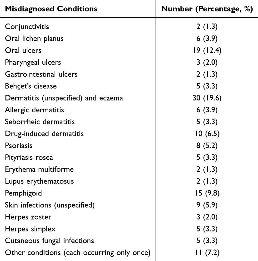 |
Table 2 Misdiagnosis Spectrum and Distribution in Pemphigus Patients |
Regarding skin and mucosal involvement, all patients with non-vulgaris subtypes exhibited skin involvement. Among 68 patients with pemphigus vulgaris, 59 (86.8%) had oral mucosal involvement, 11 (16.2%) had ocular involvement, 30 (44.1%) had nasopharyngeal involvement, and 30 (44.1%) had anogenital involvement. For the 34 patients with pemphigus erythematosus, 4 (11.8%) showed oral mucosal involvement, while ocular, nasopharyngeal, and anogenital involvement were each observed in one case (2.9%).
The histopathological, DIF, IIF, and ELISA results are presented in Table 3. The highest testing rates were observed for IIF and ELISA detection of anti-Dsg1 and anti-Dsg3 antibodies (both 94.2%), while DIF showed the lowest testing rate (69.6%). Among the 138 patients, 10 (7.9%) patients were positive for anti-BP180 antibodies, all with low titers (≤20 U/mL).
 |
Table 3 Auxiliary Examination Results of 138 Pemphigus Patients |
As shown in Table 4, no significant differences were observed in the positive rates of histopathology and DIF among the four major pemphigus subtypes. Pemphigus vulgaris patients demonstrated significantly higher IIF positive rates compared to pemphigus erythematosus patients (p < 0.01), while no significant differences in IIF positivity were found between other subtypes.
 |
Table 4 Positive Rates of Auxiliary Examinations in Four Common Pemphigus Subtypes |
Among the 138 pemphigus patients, four cases (one IgA pemphigus and three pemphigus vulgaris) only received definitive diagnosis at our center and sought further treatment at other institutions. One pemphigus vulgaris patient and one drug-induced pemphigus patient only received topical treatment. Disease control was defined as the cessation of new lesion formation and the beginning of healing of existing lesions.18 All 132 patients who received systemic treatment achieved disease control with the initial regimen. The drug distribution for these 132 patients is shown in Figure 1 (azathioprine [three cases], cyclophosphamide [one case], dapsone [one case] were excluded from presentation due to low usage, and plasmapheresis [three cases] was excluded because it is not a kind of drug). The most commonly used drug was corticosteroids (104/132, 78.8%), followed by mycophenolate mofetil (33/132, 25.0%), Tripterygium wilfordii Hook F (29/132, 22.0%), minocycline (28/132, 21.2%), rituximab (24/132, 18.2%), nicotinamide (18/132, 13.6%), thalidomide (14/132, 10.6%), methotrexate (9/132, 6.8%), intravenous immunoglobulin (IVIG, 5/132, 3.8%), and compound glycyrrhizin (4/132, 3.0%).
 |
Figure 1 Drug distribution for achieving disease control in 132 pemphigus patients. |
Among the 64 patients with pemphigus vulgaris who received systemic therapy, the treatment regimens were as follows: corticosteroids alone (15 cases), corticosteroids combined with immunosuppressants (mycophenolate mofetil, methotrexate, azathioprine, or cyclophosphamide) (18 cases), corticosteroids combined with rituximab (five cases), corticosteroids combined with Tripterygium wilfordii Hook F (one case), corticosteroids combined with thalidomide (six cases), corticosteroids combined with minocycline (one case), corticosteroids combined with two or more treatments (such as immunosuppressants, rituximab, Tripterygium wilfordii Hook F, thalidomide, minocycline, IVIG, or plasmapheresis) (10 cases), and non-steroidal therapies (combinations of Tripterygium wilfordii Hook F, minocycline, nicotinamide, or thalidomide) (eight cases). The most intensive regimen was prednisone-equivalent 100 mg/d combined with mycophenolate mofetil 2 g/d, rituximab, IVIG, and plasmapheresis.
Among the 34 patients with pemphigus erythematosus who received systemic therapy, the treatment regimens were as follows: corticosteroids alone (nine cases), corticosteroids combined with immunosuppressant (mycophenolate mofetil) (six cases), corticosteroids combined with rituximab (five cases), corticosteroids combined with Tripterygium wilfordii Hook F (one case), corticosteroids combined with two or more treatments (two cases, of which one received corticosteroids combined with Tripterygium wilfordii Hook F, minocycline, and nicotinamide, while the other received corticosteroids combined with Tripterygium wilfordii Hook F and minocycline), and non-steroidal therapies (11 cases).
There was no significant difference in the proportion of non-steroidal therapy usage among the four major pemphigus subtypes. Among the 104 patients who received systemic corticosteroids, the dose distribution (prednisone-equivalent) is shown in Figure 2. The mean corticosteroid doses for the four subtypes were as follows: 45.1 ± 20.3 mg/d for pemphigus vulgaris, 36.3 ± 13.0 mg/d for pemphigus erythematosus, 60.6 ± 19.0 mg/d for pemphigus foliaceus, and 41.7 ± 7.5 mg/d for pemphigus herpetiformis. Patients with pemphigus erythematosus received significantly lower corticosteroid doses than those with pemphigus foliaceus (p = 0.01), while no significant differences were observed among the other subtypes.
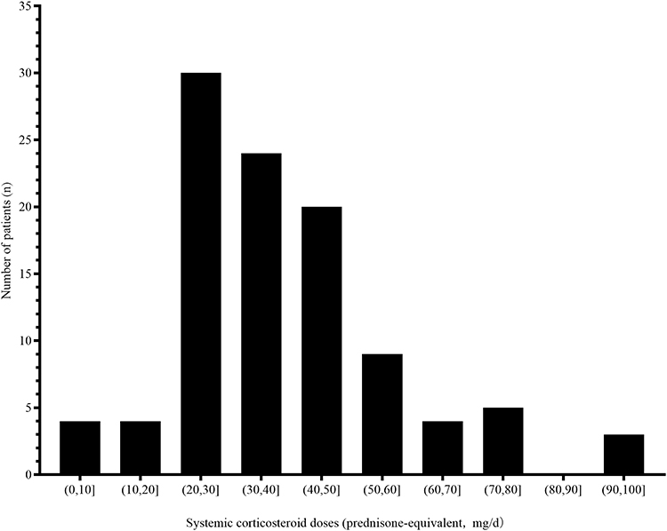 |
Figure 2 Distribution of systemic corticosteroid doses in 104 patients with pemphigus. |
Discussion
This study conducted a retrospective analysis of pemphigus patients first diagnosed at Peking Union Medical College Hospital from 2020 to 2024. The male-to-female ratio among the included patients was approximately 1:1.2. For pemphigus vulgaris, pemphigus erythematosus, and pemphigus foliaceus, female patients were more common, whereas pemphigus herpetiformis showed a male predominance. This finding aligns with a previous retrospective analysis of 26 pemphigus herpetiformis cases.19 Pemphigus primarily affected individuals aged 45–60, suggesting that pemphigus should be considered in the differential diagnosis of middle-aged patients presenting with relevant mucocutaneous manifestations. The youngest patient in this study was 17 years old, and the oldest was 83, highlighting the need for paying attention to both juvenile and elderly pemphigus patients.20,21 Traditionally, pemphigus vulgaris and pemphigus foliaceus are considered the two most common subtypes, with pemphigus vegetans regarded as a variant of pemphigus vulgaris and pemphigus erythematosus as a variant of pemphigus foliaceus.4 This study included patients first diagnosed with pemphigus at our center, providing insights into the subtype distribution of pemphigus in contemporary China. Pemphigus erythematosus accounted for 24.6% of cases, second only to pemphigus vulgaris (49.3%) and exceeding the proportion of pemphigus foliaceus (9.4%). More attention should be paid to the clinical characteristics of pemphigus erythematosus, along with further exploration of its relationship with pemphigus foliaceus and lupus erythematosus.
This study revealed that 88.4% of pemphigus patients first sought medical care in Dermatology, highlighting the crucial role of dermatologists in the diagnosis, treatment, and management of pemphigus. 5.8% of patients initially visited Stomatology, all of whom had pemphigus vulgaris. Stomatologists play a significant role in identifying characteristic mucosal lesions,22 selecting optimal biopsy sites,23 and assessing mucosal disease severity.24 A small proportion of patients first consulted Rheumatology & Immunology, partly because pemphigus is broadly classified as an immune-mediated disease and it should be differentiated from Behçet’s disease. Additionally, otolaryngologists should consider pemphigus in the differential diagnosis of pharyngeal ulcers.
A key finding of this study was the substantial diagnostic delay (median 5.0 months). Delayed treatment may lead to worsening mucocutaneous lesions and secondary infections, underscoring the importance of early recognition of skin and mucosal lesions as well as timely diagnosis and treatment.25 Approximately three-quarters of patients were misdiagnosed before receiving a definitive pemphigus diagnosis, with common misdiagnoses including dermatitis and eczema, oral ulcers, pemphigoid, drug-induced dermatitis, and skin infections. These data may serve as a reference for policymaking regarding pemphigus as a rare disease.
This study found that 88.2% of pemphigus vulgaris patients showed skin involvement while all patients with other subtypes exhibited skin lesions. This study also documented mucosal involvement rates for each subtype. The observed higher skin involvement rate may reflect bias, as oral mucosal cases may present to specialized hospitals.26 Regarding auxiliary examinations, IIF and ELISA for anti-Dsg1/Dsg3/BP180 antibodies were all performed in over 90% of cases, histopathology was obtained in approximately 80% of patients, and DIF was used in approximately 70% of cases. For patients who did not undergo DIF or histopathology, this was primarily because the physician considered the diagnosis to be sufficiently supported by clinical manifestations and other auxiliary examinations (eg, IIF and ELISA), to minimize invasive procedures and reduce patient burden. Serological tests predominated likely due to their convenience and variations in clinicians’ understanding of disease severity assessment. There is a critical need to standardize pemphigus evaluation protocols to better guide systemic treatment decisions. Notably, 10 patients demonstrated low-titer anti-BP180 positivity. Low-titer anti-BP180 antibody positivity has been reported in several dermatological conditions including severe drug eruptions and generalized eczema, warranting further investigation into its pathogenic role.27 This study also showed significantly higher IIF positivity in pemphigus vulgaris (92.1%) compared to pemphigus erythematosus (58.1%). More attention should be paid to the comprehensive evaluation of suspected pemphigus erythematosus cases to prevent missed diagnoses.
In this study, all 132 pemphigus patients receiving systemic therapy achieved disease control. Corticosteroids were the most frequently prescribed drug, used in approximately 80% of patients. Mycophenolate mofetil emerged as the most common immunosuppressants, employed in one-quarter of cases. Despite its established efficacy, azathioprine was used in only three patients, likely due to limited availability of required genetic testing and enzyme activity assays, along with concerns about adverse effects such as severe bone marrow suppression.28 Tripterygium wilfordii Hook F, known for its anti-inflammatory and immunomodulatory properties, were utilized in over one-fifth of patients. However, the frequency of use reported here does not constitute evidence of efficacy or safety for Tripterygium wilfordii Hook F in pemphigus. While it has shown effectiveness in rheumatoid arthritis29 and bullous pemphigoid,30 its role in pemphigus warrants further investigation. Minocycline was administered to more than 20% of patients. Minocycline combined with nicotinamide in treating pemphigus herpetiformis31 and pemphigus vegetans,32 as well as minocycline alone or in conjunction with low-dose corticosteroids to treat pemphigus foliaceus were reported.33 Further research is needed to clarify the therapeutic potential of minocycline in pemphigus. Rituximab was employed in 18.2% of patients, with its efficacy shown in many studies.5,34 Future studies should explore optimal rituximab-based combination strategies, as well as the utility of low-dose and ultralow-dose regimens in pemphigus management. The choice of systemic therapy is influenced by multiple factors, such as disease severity, comorbidities, physician experience, patient preference, and cost. In line with Chinese guidelines, our practice emphasizes corticosteroids combined with steroid-sparing agents (eg, rituximab or immunosuppressants like mycophenolate mofetil) for moderate-to-severe cases.
This study analyzed treatment regimens for four major pemphigus subtypes, revealing no significant differences in the utilization rates of non-steroidal therapies across these subtypes. Analysis of corticosteroid doses in 104 steroid-treated patients showed the most common prednisone-equivalent doses were 20–50 mg/d, though severe cases required up to 100 mg/d combined with adjuvant therapies (mycophenolate mofetil, rituximab, IVIG, and plasmapheresis). Notably, pemphigus erythematosus patients received significantly lower corticosteroid doses compared to pemphigus foliaceus patients. Contrary to conventional understanding that pemphigus vulgaris represents the most severe form,35 its corticosteroid requirements did not significantly differ from other subtypes. This suggests contemporary therapeutic approaches increasingly favor combination regimens incorporating adjunctive therapies rather than dose-escalation of corticosteroids for disease control.
This study has several limitations. First, its retrospective, single-center design may introduce bias. Second, the sample size is limited. While pemphigus is a rare disease, our center, as a diagnostic and treatment center for autoimmune blistering diseases, also manages many patients who were either diagnosed before the study period or initially diagnosed at other institutions during the study period. Future research should investigate the clinical characteristics of these patient groups. Third, this study only analyzed treatment patterns used to achieve disease control. The study period coincided with the COVID-19 pandemic, which significantly complicated patient follow-up and management. This study did not assess longitudinal treatment outcomes such as time to remission, cumulative steroid dosage, relapse rates, or adverse effects. Future studies should assess long-term treatment responses, pemphigus comorbidities, pemphigus relapses, and treatment-related adverse effects.
Conclusion
This study summarized the demographic and clinical data of 138 pemphigus patients first diagnosed at Peking Union Medical College Hospital between 2020 and 2024. Approximately half of the patients were pemphigus vulgaris, while about one-quarter were pemphigus erythematosus. The diagnostic delay was substantial, with a median of 5.0 (2.0–12.0) months, and 106 patients (76.8%) had been misdiagnosed before a definitive diagnosis was made. All 132 patients who received systemic treatment achieved disease control with the initial regimen. Corticosteroids were the most frequently used systemic treatment, with mycophenolate mofetil being the most common immunosuppressants. Other frequently used drugs included Tripterygium wilfordii Hook F, minocycline, and rituximab. This study quantifies a significant diagnostic delay and outlines evolving treatment patterns among patients with pemphigus, providing epidemiological data on Chinese pemphigus patients in the new era and offering references for policy-making and future research on this rare disease.
Funding
This study was supported by National Key R&D Program of China (2022YFC3601800), Beijing Natural Science Foundation (7242109), National Key Clinical Specialty Project of China, and Beijing Key Clinical Specialty Construction Project.
Disclosure
The authors report no conflicts of interest in this work.
References
1. Vafaeian A, Mahmoudi H, Daneshpazhooh M. What is novel in the clinical management of pemphigus vulgaris? Expert Rev Clin Pharmacol. 2024;17(5–6):489–9. doi:10.1080/17512433.2024.2350943
2. Lan Y, Zhang H, Jin H. Pregnancy in pemphigus vulgaris: a systematic review. Am J Reprod Immunol. 2024;91(1):e13813. doi:10.1111/aji.13813
3. Zuo Y-G, Li L, Chen J-B, et al. Chinese Expert Proposal on the Diagnosis and Management of Pemphigus Vulgaris (2020). Int J Dermatol Venereol. 2020;3(3):148–155. doi:10.1097/jd9.0000000000000088
4. Schmidt E, Kasperkiewicz M, Joly P. Pemphigus. Lancet. 2019;394(10201):882–894. doi:10.1016/s0140-6736(19)31778-7
5. Cao S, Yang B, Wang Z, et al. Efficacy, safety, and B-cell depletion capacity of 3 rituximab dosing regimens in the treatment of moderate-to-severe pemphigus vulgaris and pemphigus foliaceus: a 52-week clinical trial. J Am Acad Dermatol. 2025. doi:10.1016/j.jaad.2025.05.1374
6. Durães SMB, Santos NR, Batzner CN, Cerqueira FGM. Use of low-dose rituximab to treat pemphigus. Anais Brasileiros Dermatol. 2024;99(5):791–792. doi:10.1016/j.abd.2023.07.019
7. Wang YL, Tie D, Yan TM, et al. Survey of the epidemiology of autoimmune bullous disorders in Peking Union Medical College Hospital: a 30-year review. Eur J Dermatol. 2017;27(3):331–332. doi:10.1684/ejd.2017.3010
8. Zhu X, Pan J, Yu Z, Wang Y, Cai L, Zheng S. Epidemiology of pemphigus vulgaris in the Northeast China: a 10-year retrospective study. J Dermatol. 2014;41(1):70–75. doi:10.1111/1346-8138.12286
9. Chen Z, Wang L, Ma L, et al. Epidemiological Insights into Autoimmune Bullous Diseases in China: a Comprehensive Analysis. J Epidemiol Global Health. 2024;14(3):513–523. doi:10.1007/s44197-024-00277-7
10. Kasperkiewicz M, Woodley DT. COVID-19 and autoimmune bullous diseases: lessons learned. Autoimmunity Rev. 2023;22(4):103286. doi:10.1016/j.autrev.2023.103286
11. Zhang JL, Wang SH, Cui SN, et al. Severe acute respiratory syndrome coronavirus 2 infection in patients with autoimmune bullous diseases in China. J Dermatol. 2023;50(11):1433–1441. doi:10.1111/1346-8138.16900
12. Zhao W, Wang J, Zhu H, Pan M. Comparison of Guidelines for Management of Pemphigus: a Review of Systemic Corticosteroids, Rituximab, and Other Immunosuppressive Therapies. Clin Rev Allergy Immunol. 2021;61(3):351–362. doi:10.1007/s12016-021-08882-1
13. De D, Mehta H, Shah S, et al. Consensus Based Indian Guidelines for the Management of Pemphigus Vulgaris and Pemphigus Foliaceous. Indian Dermatol Online J. 2025;16(1):3–24. doi:10.4103/idoj.idoj_1059_24
14. Chu CY, Ch L, He L, et al. Taiwanese dermatological association (TDA) consensus for the management of pemphigus. J Formosan Med Assoc. 2023;122(7):540–548. doi:10.1016/j.jfma.2022.12.005
15. Murrell DF, Peña S, Joly P, et al. Diagnosis and management of pemphigus: recommendations of an international panel of experts. J Am Acad Dermatol. 2020;82(3):575–585.e1. doi:10.1016/j.jaad.2018.02.021
16. Amagai M, Tanikawa A, Shimizu T, et al. Japanese guidelines for the management of pemphigus. J Dermatol. 2014;41(6):471–486. doi:10.1111/1346-8138.12486
17. Timóteo RP, Pessoa-Gonçalves YM, Do Carmo Neto JR, Rodrigues WF, da Silva MV, Oliveira CJF. A Global View of Pemphigus: geographical Variations. Clin Rev Allergy Immunol. 2024;66(1):14–29. doi:10.1007/s12016-024-08980-w
18. Murrell DF, Dick S, Ahmed AR, et al. Consensus statement on definitions of disease, end points, and therapeutic response for pemphigus. J Am Acad Dermatol. 2008;58(6):1043–1046. doi:10.1016/j.jaad.2008.01.012
19. Wang YM, Mao XM, Zhao WL, et al. The clinical, immunological and pathological features for relapse of pemphigus herpetiformis: a univariate analysis of 26 cases. Br J Dermatol. 2020;182(3):802–804. doi:10.1111/bjd.18518
20. Filippi F, Loi C, Chessa MA, et al. Clinical course and therapeutic management of juvenile pemphigus vulgaris: a retrospective analysis from an Italian referral centre. JEADV. 2024;38(6):e487–e489. doi:10.1111/jdv.19671
21. Ingen-Housz-Oro S, Alexandre M, Le Roux-Villet C, et al. Pemphigus in elderly adults: clinical presentation, treatment, and prognosis. J Am Geriatr Soc. 2012;60(6):1185–1187. doi:10.1111/j.1532-5415.2012.03987.x
22. Endo H, Rees TD, Matsue M, Kuyama K, Nakadai M, Yamamoto H. Early detection and successful management of oral pemphigus vulgaris: a case report. J Periodontol. 2005;76(1):154–160. doi:10.1902/jop.2005.76.1.154
23. Liu J, Hu C, He J, et al. Diagnostic utility of gingival biopsy for pemphigus vulgaris and mucosa membrane pemphigoid: a 10-year cohort study. J Am Acad Dermatol. 2024;91(1):135–138. doi:10.1016/j.jaad.2024.03.005
24. Wang F, Zhang X, Yao Y, et al. Pemphigus oral lesions area index (POLAI): a new scoring scale for assessing oral pemphigus vulgaris. Oral Dis. 2025;31(1):160–167. doi:10.1111/odi.15054
25. Lu X, Zhang H, Zhang Y, Tang K, Jin H. Characteristics and risk factors for infection in hospitalized patients with moderate to severe pemphigus: a retrospective study of 102 patients. Archiv Dermatol Res. 2025;317(1):824. doi:10.1007/s00403-025-04326-4
26. Xu Y, He X, Jie W, Liu S, Hu JA, Li Y. The application of direct immunofluorescence in the pathological diagnosis of oral mucosal diseases: series of 116 cases. Pathol Res Pract. 2025;270:155989. doi:10.1016/j.prp.2025.155989
27. Liu Z, Chen L, Zhang C, Xiang LF. Circulating bullous pemphigoid 180 autoantibody can be detected in a wide spectrum of patients with other dermatologic conditions: a cross-sectional study. J Am Acad Dermatol. 2019;80(3):774–775. doi:10.1016/j.jaad.2018.06.006
28. Chams-Davatchi C, Mortazavizadeh A, Daneshpazhooh M, et al. Randomized double blind trial of prednisolone and azathioprine, vs. prednisolone and placebo, in the treatment of pemphigus vulgaris. JEADV. 2013;27(10):1285–1292. doi:10.1111/j.1468-3083.2012.04717.x
29. Lv QW, Zhang W, Shi Q, et al. Comparison of Tripterygium wilfordii Hook F with methotrexate in the treatment of active rheumatoid arthritis (TRIFRA): a randomised, controlled clinical trial. Ann Rheumatic Dis. 2015;74(6):1078–1086. doi:10.1136/annrheumdis-2013-204807
30. Ge XL, Li SZ, Jin X, Jin HZ, Zuo YG. Treatment of bullous pemphigoid in Chinese patients with Tripterygium wilfordii Hook F. Dermatol Ther. 2020;33(4):e13721. doi:10.1111/dth.13721
31. Wu C, Zuo Y, Jin H, Chen J, Li L. Treatment of pemphigus herpetiformis with minocycline and nicotinamide. Chinese Med J. 2014;127(19):3514.
32. von Köckritz A, Ständer S, Zeidler C, Metze D, Luger T, Bonsmann G. Successful monotherapy of pemphigus vegetans with minocycline and nicotinamide. JEADV. 2017;31(1):85–88. doi:10.1111/jdv.13779
33. Fan B, Chen X, Zhu X, Wang M. Pemphigus foliaceus treated with minocycline monotherapy or low-dose prednisolone combination therapy. J Dermatol Treatment. 2023;34(1):2113756. doi:10.1080/09546634.2022.2113756
34. Zhou X, Zhan T, Xu X, et al. The efficacy and safety of low-dose rituximab in the treatment of pemphigus vulgaris: a cohort study. J Dermatol Treatment. 2024;35(1):2302071. doi:10.1080/09546634.2024.2302071
35. Porro AM, Seque CA, Ferreira MCC, Enokihara M. Pemphigus vulgaris. Anais Brasileiros Dermatol. 2019;94(3):264–278. doi:10.1590/abd1806-4841.20199011
 © 2026 The Author(s). This work is published and licensed by Dove Medical Press Limited. The
full terms of this license are available at https://www.dovepress.com/terms
and incorporate the Creative Commons Attribution
- Non Commercial (unported, 4.0) License.
By accessing the work you hereby accept the Terms. Non-commercial uses of the work are permitted
without any further permission from Dove Medical Press Limited, provided the work is properly
attributed. For permission for commercial use of this work, please see paragraphs 4.2 and 5 of our Terms.
© 2026 The Author(s). This work is published and licensed by Dove Medical Press Limited. The
full terms of this license are available at https://www.dovepress.com/terms
and incorporate the Creative Commons Attribution
- Non Commercial (unported, 4.0) License.
By accessing the work you hereby accept the Terms. Non-commercial uses of the work are permitted
without any further permission from Dove Medical Press Limited, provided the work is properly
attributed. For permission for commercial use of this work, please see paragraphs 4.2 and 5 of our Terms.
Recommended articles

Epidemiology of Disorders Associated with Short Stature in Childhood: A 20-Year Birth Cohort Study in Finland
Harju S, Saari A, Sund R, Sankilampi U
Clinical Epidemiology 2022, 14:1205-1214
Published Date: 26 October 2022
Global Burden of Asthma, and Its Impact on Specific Subgroups: Nasal Polyps, Allergic Rhinitis, Severe Asthma, Eosinophilic Asthma
Rabe APJ, Loke WJ, Gurjar K, Brackley A, Lucero-Prisno III DE
Journal of Asthma and Allergy 2023, 16:1097-1113
Published Date: 6 October 2023
Comprehensive Analysis Reveals the Potential Roles of CDKN3 in Pancancer and Verification in Endometrial Cancer
Gao C, Fan X, Liu Y, Han Y, Liu S, Li H, Zhang Q, Wang Y, Xue F
International Journal of General Medicine 2023, 16:5817-5839
Published Date: 11 December 2023
Prevalence and Incidence of Treatment-Range Adolescent Idiopathic Scoliosis Diagnosed in Ontario Between 2012 and 2021: Validating a Population-Based Health Administrative Algorithm for Case Identification in Youth Aged 10–17 Years
Dermott JA, To T, Jaakkimainen L, Bouchard M, Howard A, Lebel DE
Clinical Epidemiology 2026, 18:578144
Published Date: 19 March 2026