Back to Journals » Orthopedic Research and Reviews » Volume 14
A New Test for the Advanced Diagnosis of Lateral Elbow Tendinopathy with Concomitant Intrasubstance Tear: Failure to Resist Extension Effort (the Free Test)
Authors Greene C, Droppelmann G , García N, Jorquera C, Verdugo A
Received 27 February 2022
Accepted for publication 26 July 2022
Published 30 December 2022 Volume 2022:14 Pages 495—503
DOI https://doi.org/10.2147/ORR.S364050
Checked for plagiarism Yes
Review by Single anonymous peer review
Peer reviewer comments 2
Editor who approved publication: Professor Clark Hung
Cristóbal Greene,1,2 Guillermo Droppelmann,1,3,4 Nicolás García,1 Carlos Jorquera,5 Arturo Verdugo1
1Centro de Medicina Ejercicio Deporte y Salud, Clínica MEDS, Santiago, Chile; 2Facultad de Medicina, Universidad Diego Portales, Santiago, Chile; 3Health Sciences Ph.D. Program, Universidad Católica de Murcia, Murcia, Spain; 4Principles and Practice of Clinical Research, Harvard T.H. Chan School of Public Health, Boston, MA, USA; 5Facultad de Ciencias, Escuela de Nutrición y Dietética, Universidad Mayor, Santiago, Chile
Correspondence: Guillermo Droppelmann, Centro de Medicina Ejercicio Deporte y Salud, Clínica MEDS, Santiago, Chile, Email [email protected]
Background: Lateral elbow tendinopathy (LET) is one of the most common causes of musculoskeletal pain. The diagnosis is based on the clinical history and different physical maneuvers. Ultrasound (US) is a complementary diagnostic method to detect degenerative tendon changes and intrasubstance tears (IST). To date, there is no available physical maneuver to identify an IST in patients with LET.
Aim: To evaluate the diagnostic accuracy of an index test to detect an IST confirmed by ultrasound in patients with LET.
Methods: A diagnostic retrospective study was performed. Patients who presented medical records with LET were recruited. Two orthopaedic surgeons developed the physical maneuver. The index test was considered positive when the position failed to resist the wrist extension maximum effort. Clinical findings were associated with confirmation of IST by US. Data were calculated using diagnostic accuracy, sensitivity, and specificity with 95% confidence intervals.
Results: Thirty-nine patients (39 elbows) were analyzed, 25 (64%) women and 14 (36%) men, with an average age of 47.7 years. The index test’s sensitivity was 0.86 (95% CI, 0.67– 0.96). Accuracy was 0.79 (95% CI, 0.64– 0.91), and the specificity was 0.64 (95% CI, 0.31– 0.89).
Conclusion: The index test presented very good sensitivity and good accuracy in patients with LET with US diagnostic confirmation of IST.
Level of Evidence: Diagnostic study, Level III.
Keywords: accuracy, diagnosis, sensitivity, specificity, tennis elbow
Introduction
Lateral elbow tendinopathy (LET), also commonly referred to as “tennis elbow”,1 is one of the most common causes of pain in the elbow.2 This condition affects the tendinous insertion of the wrist extensor muscles at the lateral epicondyle.3 Despite advances in the treatment, over 80% of cases recover within one year.4
LET diagnosis is based on an insidious history of pain in the lateral aspect of the elbow with irradiation down the extensor aspect of the forearm.5 The clinical examination can be complemented with provocation tests. The Cozen’s, Mill’s, or Maudsley’s tests are the most used6 in clinical practice and research for their diagnostic validity.7 All of these physical maneuvers are positive in the presence of lateral epicondylar pain.8 Nevertheless, it cannot detect degenerative tendon changes, such as bone irregularities, calcific deposits, neovascularization, thickening, thinning, and tears.
For this reason, imaging diagnostic is used as a complementary method for examining the structural changes on the elbow’s common extensor tendon (CET). Studies have proven that magnetic resonance imaging (MRI) is a reliable tool in determining the radiological severity of LET.9,10 However, ultrasonography (US) is often used to assess this disorder11 because it is more accurate and cost-effective. US findings have been well documented12 and can be helpful to detect degenerative tendon signs,13 and even better to detect intrasubstance tear (IST) over than MRI.14
There is no consensus in the current literature on the most important abnormal US findings that confirm LET.15 The IST is one of the most important because the repair process is slow and characterized by a scar with mechanically inferior tissue, at least initially.16 Impacting on peoples’ quality of life represents an enormous economic burden on the worldwide healthcare system17 due to lost workdays and can even disable some patients from working for weeks,18 Figure 1.
Traditionally, a long subjective history of pain, physical examination, and diagnostic imaging are used interchangeably or in conjunction for the clinical diagnosis of LET in the absence of a “gold diagnostic standard.” Most clinical tests are based on the provocation of symptoms, while imaging aims to identify degenerative tissue changes or abnormalities.7 Although numerous clinical tests for LET are described in the literature, provocative maneuvers are typically used to pinpoint or isolate specific tissues with pathology. To date, there is no available provocative maneuver to identify a specific IST of the CET in patients with LET.
Therefore, this study aimed to evaluate the diagnostic accuracy of an index test to detect an IST confirmed by ultrasound in patients with LET. We hypothesized that the index test would have high sensitivity and accuracy to detect IST in patients with LET. Secondly, we propose that nocturnal pain also presents similar values to the index test.
Methods
Ethics Statement
This study has been performed following the latest version of the Declaration of Helsinki and the Chilean scientific legislation. The study was approved by the “Comité de Ética Científico Adulto del Servicio Metropolitano Oriente de la ciudad de Santiago de Chile (SSMO)”. The ethical committee required no informed consent given the retrospective design of the study. The project was approved on November 23, 2021. No approval number was recorded. The participants’ privacy, such as confirmation that the data was anonymized and maintained with confidentiality.
Study Design
This research was designed as a diagnostic retrospective and multicentric study. It was written following the Reporting Diagnostic Accuracy Studies (STARD) guideline.19 All patients’ records with an elbow ultrasound exam at MEDS Clinic in Santiago, Región Metropolitana, Chile, were selected from January 1st, 2020, to June 30th, 2020. This study started on December 15th, 2021.
Participants
The eligibility criteria were all the electronic records of patients of both genders, over 18 years old, with LET suspicion with any unilateral or bilateral signs or symptoms in the selected period. Inclusion criteria considered first episode with pain from one week to six weeks, tenderness to palpation at the lateral epicondyle, and positive presence of any classical maneuvers such as Cozen’s, Mill’s, or Maudsley’s tests. We have also included pain at night, and tendon tear was confirmed with an ultrasound exam. This technique is not the gold standard as an MRI. However, ultrasonography is a valuable imaging modality that can be used as a screening tool in LET. Intrasubstance tear was defined as a linear hypoechoic focus associated with discontinuity of tendon fibers.20 Exclusion criteria were any previous treatment such as platelet-rich plasma (PRP), corticoid injection, or surgical intervention. Other diagnoses for lateral elbow pain, fracture around the elbow, shoulder, or neck condition were also excluded.
Sample Size
The sample size was estimated at least 28 subjects’ medical and ultrasound records, using information from two studies published in 2009 and 2014, respectively.21,22
Instrumentation
Our medical imaging software (Carestream RIS, v.11) allows a word search of the whole electronic ultrasound exam record database. Only GD and other external professional SM had access to the information and generated an online database structure with the inclusion criteria.
The exams were diagnosed by five musculoskeletal radiologists with more than ten years of experience. Only NG was the author of this article. Although ultrasound is a very operator-dependent diagnostic modality, the concordance between the five musculoskeletal radiologists was not measured. The professionals used an Aplio 500 US system (Toshiba America Medical Systems, Inc., Tustin, CA, ca.medical.canon) equipped with a multifrequency linear transducer with a frequency of 18 MHz to inform the IST in the LET exams.
Physical Maneuver
All patients were tested with the index test by two orthopaedic surgeon’s specialists in elbow and hand pathologies (CG, AV) with more than 15 years of practice each of them. The concordance between orthopaedic surgeon’s elbow specialists was not measured either.
The patients were positioned in a sitting position with the shoulder slightly abducted, elbow flexed to 90°, forearm pronated, and wrist flexed so that the palm would face downwards. Then, the orthopedic surgeon stood beside the patient’s affected side and resisted the maximum wrist extension effort (Figure 2). While maintaining this position, failure to resist the maximum wrist extension effort indicated a positive sign of this new physical maneuver (Figure 3). Clinical findings established to be associated with extensor tendon tear when patients were failed to resist the maximum dorsal wrist extension effort against resistance and presented nocturnal pain.
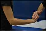 |
Figure 2 Index test initial position. Maximum wrist extension effort against resistance. |
 |
Figure 3 Index test final position. The index test is positive when patients fail to resist. |
Another author (GD) identified the US exams and confirmed the presence or absence of IST.
Statistical Analysis
Data were calculated using diagnostic accuracy (Ac), sensitivity (Sn), and specificity (Sp). The first step was to build a 2 × 2 table with patients divided by IST into rows and categories according to the index test, nocturnal pain, and Cozen’s test results in columns.
True-positive (TP) was defined as the number of cases correctly identified with IST. False-positive (FP) was the number of cases incorrectly identified with IST. True-negative (TN) was the number of cases correctly identified with IST, and False-negative (FN) was the number of cases incorrectly identified with IST.23 Ac was the correctly classified. In other words, it is correctly differentiating the patient with and without IST. The Ac has identified the proportion of true positive and true negative in all evaluated cases (TP+TN)/(TP+TN+FP+FN). Sn was expressed in percentage and defined the proportion of TP patients with the IST in a total group of patients with the tendon condition (TP/TP+FN). Sp was a measure of diagnostic test accuracy, complementary to sensitivity. It was defined as a proportion of patients without the IST with a negative test result in a total of subjects without IST (TN/TN+FP).24 Ninety-five percent confidence intervals (95% CI) also were calculated for all diagnostic metrics.
Positive and negative predictive values were not calculated because they largely depend on disease prevalence in the examined population. We assumed that all patients had symptoms when they were examined. The ROC curve was not calculated because we presented binary outcomes, with few thresholds significantly underestimating the real area under the curve.25,26
Neither final exposure variables nor outcome data presented missing values. Missing data analysis showed that missingness was at random. All graphs and analyses were performed using R (The R Foundation for Statistical Computing, v. 3.6.2) and RStudio, v.4.1.0.
Results
Participants
Forty-one patients were analyzed in this study. However, thirty-nine patients (39 elbows) presented complete information for the statistical analysis, Figure 4.
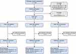 |
Figure 4 STARD flow diagram of the study. Abbreviation: n, number of patient’s medical records. |
The sample was constituted of 25 (64%) women and 14 (36%) men. The average age was 47.7 years (range 34 to 69 years). Of the total elbows, 37 (95%) were affected by the dominant side, and 32 (82%) were on the right side. Twenty-three patients (59%) reported nocturnal pain. Thirty-three patients (85%) presented a positive sign in the Cozen’s Test. Twenty-eight (72%) patients presented an intrasubstance tear, and other twenty-eight patients also had a positive sign in the index test. Table 1 shows the baseline demographic and clinical characteristics of participants.
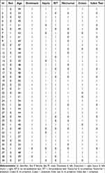 |
Table 1 Demographics Characteristics and Findings Examination |
Test Results
Overall, the index test’s accuracy (0.79; 95% CI, 0.64–0.91) was superior to the presence of nocturnal pain (0.72; 95% CI, 0.55–0.85) and positive sign in the Cozen’s test (0.61; 95% CI, 0.45–0.77). The index test’s sensitivity or true positive rate (0.86; 95% CI, 0.67–0.96) was close to the nocturnal pain (0.87; 95% CI, 0.66–0.97) but higher than the Cozen’s test (0.70; 95% CI, 0.51–0.84). Finally, the index test’s specificity or true negative rate (0.64; 95% CI, 0.31–0.89) was significantly higher than the nocturnal pain report (0.5; 95% CI, 0.25–0.75) and the Cozen-s test (0.17; 95% CI, 0.00–0.64). Table 2 shows diagnostic accuracy and precision (with 95% confidence intervals). Any adverse events were not recorded from performing the index test or the reference standard.
 |
Table 2 Diagnostic Measures of the Index Test, Nocturnal Presence and Cozen’s Test |
Discussion
The primary aim of this study was to evaluate the diagnostic accuracy of an index test to detect an IST confirmed by US in patients with LET. Our secondary aim was to propose that the nocturnal pain also presents similar values to the index test. Two orthopaedic surgeons developed the evaluation. A novel physical maneuver was applied to the diagnostic clinical experience of the professionals, electronic medical records, and ultrasound exams to confirm a LET with the presence of an IST.
The findings from the current study evidenced that nearly ninety percent of patients were correctly diagnosed when the index test was positive, and the presence of IST by ultrasound was confirmed. This result could be potentially explained by the high clinical acuity and the expertise of specialist physicians. Furthermore, data showed strong evidence in the proportion of correctly classified subjects among all subjects, also called diagnostic accuracy (effectiveness). This research’s value was nearly 80%, classified as very good. Also, there is some evidence that the index test had a particular ability in detecting patients who did not have the disease when the test was negative, also called specificity. This value was lower than the two others, and this could be explained because the specificity is the probability that the test will correctly classify healthy individuals or the probability that healthy individuals will have a negative result. In this case, all the patients were previously diagnosed with LET.
As we mentioned, LET diagnosis is confirmed using different clinical and imagenological strategies. One of the most used in the clinical practice is the reproduction of provocative physical maneuvers for LET such as Cozen’s test, Mill’s test, Maudsley’s test, and Polk’s test.27,28 While these tests have been extensively described in the literature, numerous clinical tests for the LET not accompany the condition on diagnostic accuracy.29 Indeed, the diagnostic capacity of these clinical provocation tests to confirm LET is under-investigated.30 Despite this, the Cozen test is the only one recommended by the United Kingdom Health Safety Executive Workshop to diagnose LET.31 For this reason, the authors explored the Cozen test performance in the same sample of patients. Surprisingly, we found the lowest sensitivity, specificity, and accuracy values. This could suggest that the Cozen’s test is not the best physical maneuver in patients with IST suspected.
Other authors have studied the reproduction of pain during daily activities for LET diagnosis. Since 1970, lifting a chair with a pronated hand and a modified chair pick-up test is one of the most used tests to diagnose LET.32 Researchers recently determined that the upper limb position affects pain-free grip strength in LET.33 However, most of these diagnostic tests have in common that they do not discriminate according to the type of ultrasonographic finding such an IST. They only make the diagnosis by confirming the presence or absence of the LET condition. For this reason, detecting the presence or absence of the condition is not enough. To the authors’ knowledge and experience, detecting IST is crucial in determining the best therapeutic outcomes in patients with LET. Tendon tear is the final phase of the degenerative tissue process, and the number of US findings could determine the severity of the tendinopathy.20
On the other hand, severe pain may cause sleep disturbance.34 According to the authors’ experience, nocturnal pain is typical of patients with tendon tears. The results section demonstrated a higher sensitivity with this clinical sign but a moderate specificity. To date, this is the only study in the recent literature that attempts to identify a physical maneuver and nocturnal sign independently with the presence of an intrasubstance tendon tear diagnosed by musculoskeletal ultrasound in patients with LET.
This study has some limitations. First, it was a retrospective design, which is often considered inferior to prospective, randomized, and controlled clinical trials.35 Inter- and intra-rater reliability between radiologists and orthopaedic surgeons were not considered, losing external validity. Further limitations include only ultrasound diagnosis confirmation of IST, not included other imagenological findings, hindering results generalization. An additional limitation was that this study focused solely on the presence of IST and the positive sign of the index test and the nocturnal pain, but not pain scores, evolution times, physical activity levels, comorbidities, and other clinical manifestations were included. Finally, binary outcomes considered did not allow for more complex statistical analyses.
Future studies require reproducibility and the implementation of the new test. A cohort design with a bigger sample size, inter- and intra-rater reliability between professionals, and continuous variables for analysis should be considered.
Conclusion
In summary, this study provides a new physical maneuver and clinical sign for the advanced diagnosis of LET with concomitant intrasubstance tear. The index test and nocturnal pain presented very good sensitivity and good accuracy in patients with IST. These findings are not a definitive conclusion. Further research is necessary to provide more robust evidence to establish the effectiveness of the index test and nocturnal pain presence to diagnose IST in patients with LET. Perhaps more importantly, these findings provide more information to clinicians for more accurate LET diagnoses.
Disclosure
The authors report no conflicts of interest in this work.
References
1. Flatt AE. Tennis elbow. Proc Bayl Univ Med Cent. 2008;21(4):400. doi:10.1080/08998280.2008.11928437
2. Keijsers R, de Vos RJ, Kuijer PPFM, van den Bekerom MPJ, van der Woude HJ, Eygendaal D. Tennis elbow. Shoulder Elb. 2019;11(5):392. doi:10.1177/1758573218797973
3. Pitzer ME, Seidenberg PH, Bader DA. Elbow tendinopathy. Med Clin North Am. 2014;98(4):833–849. doi:10.1016/J.MCNA.2014.04.002
4. Bisset L, Beller E, Jull G, Brooks P, Darnell R, Vicenzino B. Mobilisation with movement and exercise, corticosteroid injection, or wait and see for tennis elbow: randomised trial. BMJ. 2006;333(7575):939. doi:10.1136/bmj.38961.584653.AE
5. Speers CJB, Bhogal GS, Collins R. Lateral elbow tendinosis: a review of diagnosis and management in general practice. Br J Gen Pract. 2018;68(676):549. doi:10.3399/BJGP18X699725
6. Zwerus EL, Somford MP, Maissan F, Heisen J, Eygendaal D, van den Bekerom MP. Physical examination of the elbow, what is the evidence? A systematic literature review. Br J Sports Med. 2018;52(19):1253–1260. doi:10.1136/BJSPORTS-2016-096712
7. Karanasios S, Korakakis V, Moutzouri M, et al. Diagnostic accuracy of examination tests for lateral elbow tendinopathy (LET) - A systematic review. J Hand Ther. 2021;S0894–1130(21):39–49. doi:10.1016/J.JHT.2021.02.002
8. Laratta J, Caldwell JM, Lombardi J, Levine W, Ahmad C. Evaluation of common elbow pathologies: a focus on physical examination. Phys Sportsmed. 2017;45(2):184–190. doi:10.1080/00913847.2017.1292831
9. Qi L, Zhang YD, Bin YR, Bin SH. Magnetic resonance imaging of patients with chronic lateral epicondylitis: is there a relationship between magnetic resonance imaging abnormalities of the common extensor tendon and the patient’s clinical symptom? Medicine. 2016;95(5):e2681. doi:10.1097/MD.0000000000002681
10. Cha YK, Kim SJ, Park NH, Kim JY, Kim JH, Park JY. Magnetic resonance imaging of patients with lateral epicondylitis: relationship between pain and severity of imaging features in elbow joints. Acta Orthop Traumatol Turc. 2019;53(5):366–371. doi:10.1016/J.AOTT.2019.04.006
11. Krogh TP, Fredberg U, Ammitzbøll C, Ellingsen T. Clinical value of ultrasonographic assessment in lateral epicondylitis versus asymptomatic healthy controls. Am J Sports Med. 2020;48(8):1873–1883. doi:10.1177/0363546520921949
12. Levin D, Nazarian LN, Miller TT, et al. Lateral epicondylitis of the elbow: US findings. Radiology. 2005;237(1):230–234. doi:10.1148/RADIOL.2371040784
13. Poltawski L, Ali S, Jayaram V, Watson T. Reliability of sonographic assessment of tendinopathy in tennis elbow. Skeletal Radiol. 2012;41(1):83–89. doi:10.1007/S00256-011-1132-4
14. Bachta A, Rowicki K, Kisiel B, et al. Ultrasonography versus magnetic resonance imaging in detecting and grading common extensor tendon tear in chronic lateral epicondylitis. PLoS One. 2017;12(7):e0181828. doi:10.1371/JOURNAL.PONE.0181828
15. Dones VC, Grimmer K, Thoirs K, Suarez CG, Luker J. The diagnostic validity of musculoskeletal ultrasound in lateral epicondylalgia: a systematic review. BMC Med Imaging. 2014;14:10. doi:10.1186/1471-2342-14-10
16. James R, Kesturu G, Balian G, Chhabra AB. Tendon: biology, biomechanics, repair, growth factors, and evolving treatment options. J Hand Surg Am. 2008;33(1):102–112. doi:10.1016/J.JHSA.2007.09.007
17. Docheva D, Müller SA, Majewski M, Evans CH. Biologics for tendon repair. Adv Drug Deliv Rev. 2015;84:239. doi:10.1016/J.ADDR.2014.11.015
18. State W, Silverstein B, Welp E, Nelson N, Kalat J. Claims incidence of work-related disorders of the upper extremities: Washington state, 1987 through 1995. Am J Public Health. 1998;88(12):1827–1833. doi:10.2105/AJPH.88.12.1827
19. Bossuyt PM, Reitsma JB, Bruns DE, et al. STARD 2015: an updated list of essential items for reporting diagnostic accuracy studies. BMJ. 2015;351:h5527. doi:10.1136/BMJ.H5527
20. Droppelmann G, Feijoo F, Greene C, et al. Ultrasound findings in lateral elbow tendinopathy: a retrospective analysis of radiological tendon features [version 1; peer review: awaiting peer review]. F1000Reaearch. 2022:1–12. doi:10.12688/f1000research.73441.1
21. Tarhan S, Ünlü Z, Ovalı GY, Pabuşçu Y. Value of ultrasonography on diagnosis and assessment of pain and grip strength in patients with lateral epicondylitis. Arch Rheumatol. 2009;24(3):123–130.
22. Saroja G, Leo Aseer A, Sai V. Diagnostic accuracy of provocative tests in lateral epicondylitis. Int J Physiother Res. 2014;2(6):815–838. doi:10.16965/ijpr.2014.699
23. Baratloo A, Hosseini M, Negida A, El AG. Part 1: simple definition and calculation of accuracy, sensitivity and specificity; 2015. Available from: www.jemerg.com.
24. Šimundić A-M. Measures of diagnostic accuracy: basic definitions. EJIFCC. 2009;19(4):203–211.
25. DeLong ER, DeLong DM, Clarke-Pearson DL. Comparing the areas under two or more correlated receiver operating characteristic curves: a nonparametric approach. Biometrics. 1988;44(3):837. doi:10.2307/2531595
26. Iii JM. ROC and AUC with a binary predictor: a potentially misleading metric. J Classif. 2020;37:696–708. doi:10.1007/s00357-019-09345-1
27. Polkinghorn BS. A novel method for assessing elbow pain resulting from epicondylitis. J Chiropr Med. 2002;1(3):117–121. doi:10.1016/S0899-3467(07)60015-9
28. MacDermid JC, Michlovitz SL. Examination of the elbow: linking diagnosis, prognosis, and outcomes as a framework for maximizing therapy interventions. J Hand Ther. 2006;19(2):82–97. doi:10.1197/J.JHT.2006.02.018
29. Zwerus EL, Somford MP, Maissan F, Heisen J, Eygendaal D, Van Den Bekerom MP. Physical examination of the elbow, what is the evidence? A systematic literature review. Br J Sports Med. 2018;52(19):1253–1260. doi:10.1136/BJSPORTS-2016-096712
30. Wright JG. Evidence-based orthopaedics E-book: the best answers to clinical questions; 2008. Available from: https://books.google.cl/books?hl=es&id=EuHFdiN5Vx8C&oi=fnd&pg=PP1&ots=cnx25aS-e1&sig=vSDtKZyn9mtXH8NX_IfN6VfL-po&redir_esc=y#v=onepage&q&f=false.
31. Palmer K, Walker-Bone K, Linaker C, et al. The Southampton examination schedule for the diagnosis of musculoskeletal disorders of the upper limb. Ann Rheum Dis. 2000;59:5–11. doi:10.1136/ard.59.1.5
32. Paoloni JA, Appleyard RC, Murrell GAC. The orthopaedic research institute-tennis elbow testing system: a modified chair pick-up test-interrater and intrarater reliability testing and validity for monitoring lateral epicondylosis. J Shoulder Elb Surg. 2004;13(1):72–77. doi:10.1016/J.JSE.2003.09.017
33. Cooke N, Obst S, Vicenzino B, Hodges PW, Heales LJ. Upper limb position affects pain-free grip strength in individuals with lateral elbow tendinopathy. Physiother Res Int. 2021;26(3):e1906. doi:10.1002/PRI.1906
34. Vaquero-Picado A, Barco R, Antuña SA. Lateral epicondylitis of the elbow. EFORT Open Rev. 2016;1(11):391. doi:10.1302/2058-5241.1.000049
35. Abbott KV, Barton FB, Terhorst L, Shembel A. Retrospective studies: a fresh look. Am J Speech Language Pathol. 2016;25(2):157–163. doi:10.1044/2016_AJSLP-16-0025
 © 2022 The Author(s). This work is published and licensed by Dove Medical Press Limited. The full terms of this license are available at https://www.dovepress.com/terms.php and incorporate the Creative Commons Attribution - Non Commercial (unported, v3.0) License.
By accessing the work you hereby accept the Terms. Non-commercial uses of the work are permitted without any further permission from Dove Medical Press Limited, provided the work is properly attributed. For permission for commercial use of this work, please see paragraphs 4.2 and 5 of our Terms.

